HAMSTRING FLEXIBILITY - University of Pretoria
224
HAMSTRING FLEXIBILITY: MEASUREMENT, STRETCHING AND INJURY SUSCEPTIBILITY by Sally Waterworth Student number: 13218591 submitted in partial fulfillment of the requirements for the degree DOCTOR PHILOSOPHIAE in the FACULTY OF HUMANITIES (Department of Biokinetics, Sport and Leisure Science) University of Pretoria Promoter: Prof. P.E. Krüger September 2013 © University of Pretoria
-
Upload
khangminh22 -
Category
Documents
-
view
0 -
download
0
Transcript of HAMSTRING FLEXIBILITY - University of Pretoria

HAMSTRING FLEXIBILITY: MEASUREMENT, STRETCHING AND
INJURY SUSCEPTIBILITY
by
Sally Waterworth
Student number: 13218591
submitted in partial fulfillment of the requirements for the degree
DOCTOR PHILOSOPHIAE
in the
FACULTY OF HUMANITIES (Department of Biokinetics, Sport and Leisure Science)
University of Pretoria
Promoter: Prof. P.E. Krüger September 2013
©© UUnniivveerrssiittyy ooff PPrreettoorriiaa
i
CONTENTS
Page No.
ACKNOWLEDGEMENTS vi
SYNOPSIS vii
ABBREVIATIONS xi
LIST OF TABLES xii
LIST OF FIGURES xiii
LIST OF EQUATIONS xiv
CHAPTER 1
INTRODUCTION 1
1. INTRODUCTION 2
1.1 FLEXIBILITY 3
1.2 INJURY RISK IN SPORT 5
1.3 HAMSTRING STRAIN MECHANISMS 8
1.4 STRETCHING 10
1.5 RATIONALE 11
1.6 STATEMENT OF THE PROBLEM 13
1.7 ORGANISATION OF THE THESIS 13
1.8 AIMS OF THE STUDY 14
CHAPTER 2
LITERATURE REVIEW 15
2. LITERATURE REVIEW 16
2.1 MEASUREMENT OF STATIC FLEXIBILITY 16
2.2 STATIC FLEXIBILITY MEASURES 17
2.2.1 Reliability of static hamstring flexibility tests 17
©© UUnniivveerrssiittyy ooff PPrreettoorriiaa
ii
2.2.2 Validity of static hamstring flexibility tests 19
2.3 MUSCLE AND JOINT STRUCTURE AND FLEXIBILTY 19
2.4 DYNAMIC HAMSTRING FLEXIBILITY 21
2.5 STRETCHING TECHNIQUES 23
2.6 STRETCHING AND FLEXIBILITY 25
2.7 STRETCHING AND PERFORMANCE 28
2.8 STRETCHING AND JOINT POSITION SENSE 29
2.9 STRETCHING AND INJURY PREVENTION 31
2.10 FATIGUE AND INJURY SUSCEPTIBILITY 33
2.11 SUMMARY 36
CHAPTER 3
INTRA AND INTERDAY RELIABILITY OF HAMSTRING FLEXIBILITY MEASURES 38
3.1 INTRODUCTION 39
3.1.1 Methods for determining reliability 40
3.1.2 Reliability of flexibility measurements 43
3.1.3 Aims and hypotheses 44
3.2 PILOT STUDIES 45
3.2.1 Previous research procedures 45
3.2.2 Procedures 46
3.2.2.1 Set-up 46
3.2.2.2 Passive straight leg raise approach 46
3.2.2.3 Passive knee extension approach 47
3.2.2.4 Warm-up 48
3.2.2.5 Data analysis 49
3.2.2.6 Sample size estimation 50
3.3 METHODS 50
3.3.1 Participants 50
3.3.2 Study design 51
3.3.3 Procedures 51
3.3.3.1 Passive properties 51
©© UUnniivveerrssiittyy ooff PPrreettoorriiaa

iii
3.3.3.2 Static flexibility 52
3.3.3.3 Statistical analysis 53
3.4 RESULTS 54
3.5 DISCUSSION 58
3.5.1 Static flexibility reliability 58
3.5.2 Dynamic flexibility reliability 60
3.5.3 Dynamometer comparison 61
3.5.4 Practical implications 63
3.5.5 Limitations 64
3.5.6 Reflections 63
3.6 CONCLUSIONS 65
CHAPTER 4
COMPARISON BETWEEN DIFFERENT METHODS OF MEASURING STATIC AND
DYNAMIC HAMSTRING FLEXIBILITY 66
4.1 INTRODUCTION 67
4.1.1 Aims and hypotheses 71
4.2 METHODS 72
4.2.1 Participants 72
4.2.2 Procedures 72
4.2.3 Statistical Analysis 73
4.3 RESULTS 73
4.3.1 Relationship between static flexibility measurements 73
4.3.2 Relationship between static and dynamic flexibility measurements 74
4.4 DISCUSSION 76
4.4.1 Static flexibility measurements 76
4.4.2 Static and dynamic flexibility measurements 79
©© UUnniivveerrssiittyy ooff PPrreettoorriiaa

iv
4.4.3 Limitations 81
4.4.4 Reflections 82
4.5 CONCLUSIONS 83
CHAPTER 5
IMPACT OF FATIGUE AND POST-EXERCISE STATIC HAMSTRING STRETCHING ON
MEASURES OF STATIC AND DYNAMIC HAMSTRING FLEXIBILITY AND PERFORMANCE
84
5.1 INTRODUCTION 85
5.1.1 Injury prevention 86
5.1.2 Fatigue and injury risk 88
5.1.3 Stretching as part of recovery 90
5.1.4 Aims and hypotheses 92
5.2 METHODS 92
5.2.1 Participants 92
5.2.2 Study design 93
5.2.3 Sprint protocol 94
5.2.4 Post-exercise stretching intervention 95
5.2.5 Measurement procedures 97
5.2.5.1 Eccentric torque 97
5.2.5.2 Perceived stiffness 98
5.2.5.3 Joint position sense 98
5.2.6 Statistical analysis 98
5.3 RESULTS 99
5.4 DISCUSSION 109
5.4.1 Passive dynamic flexibility measurements to 0.99 rad 110
5.4.2 Final passive dynamic flexibility measurements 111
5.4.3 Active dynamic flexibility measurements 113
5.4.4 Static flexibility measurements 115
5.4.5 Perception of stiffness 116
©© UUnniivveerrssiittyy ooff PPrreettoorriiaa
v
5.4.6 Joint position sense 118
5.4.7 Practical implications 119
5.4.8 Limitations 120
5.4.9 General limitations 122
5.4.10 Reflections 124
5.4.11 Future directions of research 127
5.5 CONCLUSIONS 129
CHAPTER 6
REFERENCES 130
CHAPTER 7
APPENDIXES 189
7.1 APPENDIX 1 190
7.2 APPENDIX 2 194
7.3 APPENDIX 3 195
7.4 APPENDIX 4 196
7.5 APPENDIX 5 200
7.6 APPENDIX 6 202
7.7 APPENDIX 7 203
7.8 APPENDIX 8 204
7.9 APPENDIX 9 205
7.10 APPENDIX 10 206
©© UUnniivveerrssiittyy ooff PPrreettoorriiaa

vi
ACKNOWLEDGEMENTS
To the Lord Almighty, without whom nothing is possible.
I wish to acknowledge the following persons and institutions for their assistance and support:
My parents, for the opportunities they awarded me.
Prof. P.E. Krüger - Promoter (Department of Biokinetics, Sport and Leisure Sciences, University
of Pretoria), for his guidance and valuable time afforded to me.
Dr. M. Jones – (Head of Sports Science, Edge Hill University), for initial guidance, mentoring
and time.
Mr Mark Leather – Head Physiotherapist Wigan Warriors RLFC, for his help in coordinating
players and testing sessions.
Mr Andy Jones – Head Physiotherapist Widnes Vikings RLFC, for his help in coordinating
players and testing sessions.
All the participants in the studies, for affording me their time and for their enthusiasm.
©© UUnniivveerrssiittyy ooff PPrreettoorriiaa

vii
Dit het al tradisie geword om fleksiteit te beskou as ’n belangrike komponent van die mens se
fisieke fiksheid, maar daar bestaan nie stawende empiriese bewyse vir hierdie veronderstelling
nie. Hoewel uitgebreide gepubliseerde navorsing onderneem is oor die relatiewe belangrikheid
van fleksiteit en die impak van verskillende strekmetodes op vlakke van fleksiteit, prestasie en
beseringsrisiko, het die kwaliteit van die studies aansienlik gewissel; betroubaarheid en
geldigheid van die metodologie is nie altyd bewys nie; en die rasionaal kon by tye bevraagteken
word. Verder het baie literatuur gefokus op statiese lenigheid, wat nie noodwendig verband hou
met eienskappe van die muskulotendon-eenheid en dus dinamiese lenigheid nie. Hierdie
proefskrif is ontwerp om gapings in die bestaande literatuur te oorbrug met behulp van
aanvaarbare metodes om die relatiewe en absolute betroubaarheid van fleksiteitstoetse vir die
hampese vas te stel; die vergelykbaarheid van statiese en dinamiese komponente van die
globale konsep van fleksiteit te oorweeg; en uit te vind hoe dinamiese fleksiteit beïnvloed word
deur uitputtende oefening en daaropvolgende statiese strekking. Die eerste doelwit is bereik
deur gebruik te maak van ’n herhaalde metingstudie, ontwerp om die geldigheid van die
intradaaglikse en interdaaglikse intrakoersbepaler vas te stel, en die metingsfout van statiese en
dinamiese metings van hampees fleksiteit vas te stel. Die beduidende relatiewe geldigheid van
metings van statiese en dinamiese hampees fleksiteit is deur middel van interklas korrelasie
gedemonstreer, maar limiete van ooreenkomsanalise het aangedui dat daar ’n graad van
absolute metingsfout was wat in verhouding tot analitiese doelwitte geïnterpreteer moes word.
Die tweede doelwit het evaluasie vereis van verhoudings wat gedeel word deur statiese en
dinamiese metings van hampeesfleksiteit. Beduidende verhoudings tussen die verskillende
Titel: HAMPEES FLEKSITEIT: METINGE, STREKKING EN VATBAARHEID VIR
BESERINGS
Kandidaat: Sally Waterworth
Promotor: Prof. P.E. Krüger
Graad: DPhil (MBK)
©© UUnniivveerrssiittyy ooff PPrreettoorriiaa
viii
statiese fleksiteitstoetse is vasgestel, maar die omvang van onverklaarde variansie het
aangedui dat slegs metings van dieselfde toets direk met mekaar vergelyk behoort te word.
Verhoudings tussen verskillende metings van dinamiese fleksiteit en statiese fleksiteit het
gewissel van nie-beduidend tot redelik sterk, wat kon beteken dat metings van statiese en
dinamiese fleksiteit nie identies is nie en dat resultate tussen die twee tipes toetse nie
uitgewissel kon word nie. Weens ’n gebrek aan verklarende empiriese bewyse is die finale
hoofstuk, wat ten doel gehad het om deur middel van ’n prospektiewe ewekansige herhaalde
metingstudie om metings van dinamiese fleksiteit en prestasie te bepaal, nie voltooi nie.
Uitputting het geen beduidende veranderings aan passiewe of aktiewe dinamiese
fleksiteitsmetings meegebring nie, maar wel ’n beduidende verswakking van statiese
fleksiteitsvlakke en waargenome styfheid getoon. Strekking na oefening het gelei tot beduidend
verhoogde vlakke van aktiewe en passiewe energie-absorpsie onmiddellik na oefening en 18
uur daarna, en in beduidend verlaagde gewrigsposisie gewaarwording onmiddellik na oefening.
Effekgroottes was klein, dus kan die kliniese beduidendheid van die uitvoering van statiese
strekking na oefening bevraagteken word, veral as dit in plaas van ander, potensieel voordeliger
praktyke uitgevoer word.
Lys van sleutel woorde: Muskulo-tendineus eenheid, Fleksiteit, Strekking, Passiewe
fleksiteit, Aktiewe fleksiteit, Energie absorpsie, Uitputting, Beserings
risikofaktore, Prestasie, Top sportlui
©© UUnniivveerrssiittyy ooff PPrreettoorriiaa
ix
Flexibility has traditionally been considered an important component of human physical fitness
but this conjecture lacks supporting empirical evidence. While there is extensive published
research examining the relative importance of flexibility and the impact of various methods of
stretching on levels of flexibility, performance and injury risk, the quality of studies has varied
considerably, reliability and validity of methodology has not always been proven, and rationale
has at times been questionable. Additionally, much literature has focused on static flexibility
which is not necessarily related to properties of the musculotendinous unit and thus dynamic
flexibility. This thesis was designed to fill gaps in the existing literature by using accepted
methods to establish relative and absolute reliability of hamstring flexibility tests, consider the
comparability of static and dynamic components of the global concept of flexibility and explore
how dynamic flexibility and performance are influenced by fatiguing exercise and subsequent
static stretching. The first aim was realised by a repeated measures study designed to establish
the intraday and interday, intrarater reliability and measurement error of static and dynamic
measures of hamstring flexibility. Significant relative reliability for measures of static and
dynamic hamstring flexibility was demonstrated via intraclass correlation coefficient (3,1) but
limits of agreement analysis indicated there was a degree of absolute measurement error that
must be interpreted in relation to analytical goals. The second aim required evaluation of
relationships shared by static and dynamic measures of hamstring flexibility. Significant
relationships between the different static flexibility tests were established but the extent of
unexplained variance indicated that only measurements from the same tests should be directly
compared to each other. Relationships between different measures of dynamic flexibility and
Title: HAMSTRING FLEXIBILITY: MEASUREMENT, STRETCHING
AND INJURY SUSCEPTIBILITY
Candidate: Sally Waterworth
Promoter: Prof. P.E. Krüger
Degree: DPhil (HMS)
©© UUnniivveerrssiittyy ooff PPrreettoorriiaa

x
static flexibility varied from non-significant to moderately strong, suggesting that measures of
static and dynamic flexibility are not identical and results should not be interchanged between
the two types of tests. Due to a lack of explanatory empirical evidence, the final chapter aimed
via a prospective randomised repeated measures study to investigate the impact of fatigue and
post-exercise static stretching on measures of dynamic flexibility and performance. Fatigue
resulted in no significant changes to passive or active dynamic flexibility measures but a
significant worsening of static flexibility levels and perceived stiffness. Post-exercise stretch
resulted in significantly increased passive and active energy absorption immediately and 18
hours post-exercise and in significantly reduced joint position sense immediately post-exercise.
Effect sizes were small so the clinical meaningfulness of performing post-exercise static
stretching is questionable, particularly if performed in place of other, potentially more beneficial
practices.
List of key words: Musculotendinous unit, Flexibility, Stretching, Passive stiffness, Active
stiffness, Energy absorption, Fatigue, Injury risk factors, Performance,
Elite athletes
©© UUnniivveerrssiittyy ooff PPrreettoorriiaa

xi
LIST OF ABBREVIATIONS
AKE - Active knee extension
ANOVA - Analysis of variance
ASLR - Active straight leg raise
DOMS - Delayed onset muscle soreness
EDL - Extensor digitorum longus
EMG - Electromyography
ICC - Intraclass correlation
JPS - Joint position sense
KE - Knee extension
LOA - Limits of agreement
PKE - Passive knee extension
PNF - Proprioceptive neuromuscular facilitation
PSLR - Passive straight leg raise
PPM - Pearson product moment
ROM - Range of motion
SD - Standard deviation
SLR - Straight leg raise
< - Smaller than / Less than
> - Larger than / More than
≥ - Larger than and equal to / More than and equal to
- Smaller than and equal to / Less than and equal to
©© UUnniivveerrssiittyy ooff PPrreettoorriiaa

xii
LIST OF TABLES
Table Page No.
3.1 One hour test-retest reliability values for measures of static and 55
dynamic hamstring flexibility
3 2 Interday test-retest reliability values for measures of static and 57
dynamic hamstring flexibility
3.3 Values of passive dynamic hamstring flexibility recorded on different 58
models of Isokinetic dynamometer
4.1 Relationship between static and dynamic flexibility measures 75
4.2 Differences between dynamic flexibility of ‘tight’ and ‘normal’ participants 76
5.1 Impact of repeated sprint exercise and a 30 s post-exercise static stretch 100
on passive dynamic hamstring flexibility measured at the highest common point
5.2 Impact of repeated sprint exercise and a 30 s post-exercise static stretch on 100
peak passive dynamic hamstring flexibility
5.3 Impact of repeated sprint exercise and a 30 s post-exercise static stretch on 102
active properties during eccentric hamstring contraction at 2.09 rad.s-1
5.4 Impact of a 30 s post-exercise static stretch on static hamstring flexibility levels 104
5.5 Impact of a 30 s post-exercise static stretch on perception of stiffness and JPS 106
©© UUnniivveerrssiittyy ooff PPrreettoorriiaa
xiii
LIST OF FIGURES
Figure Page No.
1.1 Conceptual model of injury predisposition 7 2.1 A three-element model of a sarcomere 20 2.2 Example of a load-deformation (torque–angle) curve of a material with 21 viscoelastic properties 3.1 Test position used for stretch manoeuvre by Magnusson et al. (1995) 45 3.2 Example of limits of agreement for PKE 56 4.1 Scatter plot of AKE against PKE 74 4.2 Scatter plot of total passive energy absorbed against PKE 75 5.1 Conceptual model of injury predisposition 85 5.2 Factors constraining flexibility 87 5.3 Flow diagram of testing procedure for Chapter 5 96 5.4 Mean sprint times for 20 maximal sprints performed during the repeated 99 sprint protocol 5.5 Impact of a 30 s post-exercise static hamstring stretch on hamstring passive 101 peak torque 5.6 Impact of a 30 s post-exercise static hamstring stretch on total passive energy 102
absorbed by the hamstring muscle 5.7 Impact of a 30 s post-exercise static hamstring stretch on peak torque 103 generated during maximal eccentric hamstring contraction 5.8 Impact of a 30 s post-exercise static hamstring stretch on total energy 104
absorbed (work done) by the hamstring during a maximal eccentric contraction 5.9 Impact of a 30 s post-exercise static hamstring stretch on AKE 105 5.10 Impact of a 30 s post-exercise static hamstring stretch on perception of 107 stiffness as measured on a visual analogue scale 5.11 Impact of a 30 s post-exercise static hamstring stretch on JPS 108 5.12 Scatter plot of passive peak torque against peak eccentric hamstring torque 108 5.13 Scatter plot of total passive against total active hamstring energy absorption 109
©© UUnniivveerrssiittyy ooff PPrreettoorriiaa

xiv
LIST OF EQUATIONS
Equation Page No. 3.1 Theoretical formula for reliability 40 3.2 Generic reliability formula 40 3.3 Formula for ICCs 42
©© UUnniivveerrssiittyy ooff PPrreettoorriiaa

1
CHAPTER 1
INTRODUCTION
©© UUnniivveerrssiittyy ooff PPrreettoorriiaa

2
1 INTRODUCTION
Flexibility is considered to be a component of human physical fitness, ranking alongside others
such as strength, stamina and speed (ACSM, 1998). Flexibility testing became of interest at the
beginning of the 20th
century when there was a need to assess disability, especially losses in
range of motion (ROM), in response to two conditions; widespread poliomyelitis and injuries
suffered by participants in World War 1 (Alquier, 1916; Albee & Gilliland, 1920). Cureton
(1941) first presented flexibility as a component of physical fitness and suggested that flexibility
had never been studied intensively because some of the necessary aspects are not measurable in
human subjects and because of the joint specific nature of flexibility (Cureton, 1941). This
suggestion still holds a degree of truth today. Corbin and Noble (1980) again highlighted
flexibility testing as part of fitness in 1980 and in 1985, Corbin and Fox described flexibility as
“The forgotten part of fitness” (Corbin & Fox, 1985: 191).
Anthropometric research has demonstrated differences in flexibility between athletes from
various sports but the retrospective nature of most studies and varying measurement protocols
limits understanding of these differences, and consequentially, the importance of flexibility for
sports performance (Maud, 1983; Corbin & Fox, 1985; Gleim et al., 1990; Roetert et al., 1992;
Roetert et al., 1996; Wiesler et al., 1996; Twellaar et al., 1997; Tyler et al., 2001; Watson, 2001;
Beaudoin & Whatley Blum, 2005; Brooks et al., 2006; Espana-Romero et al., 2009; Platzer et
al., 2009; Singh et al., 2011; Anloague et al., 2012; Silva et al., 2013). The early rationale for the
inclusion of flexibility as a component of physical fitness was largely based on reason and belief
that appropriate levels of flexibility are required for safe and effective movement (Clarke, 1975).
While it is logical that flexibility below an acceptable threshold will more likely result in an
overstretched muscle, the presumption that higher levels of flexibility will decrease injury risk is
less plausible and lacks empirical support. Consensus that higher flexibility will lead to improved
performance has also been common within coaching and teaching practice but experimental
evidence to support this is lacking (Corbin & Noble, 1980; Gleim & McHugh, 1997; McHugh &
Cosgrave, 2010; Kay & Blazevich, 2012; Stathokostas et al., 2012).
The belief that flexibility is an important component of fitness has led to extensive research
evaluating methods by which flexibility can be improved, usually through the use of a stretching
regimes (Bandy & Irion, 1994; Bandy et al., 1997; Bandy et al., 1998; Roberts & Wilson, 1999;
©© UUnniivveerrssiittyy ooff PPrreettoorriiaa

3
Funk et al., 2001; Zakas et al., 2003; Nelson & Bandy, 2004; Young et al., 2004; Davis et al.,
2005; Zakas et al., 2006; O'Sullivan et al., 2009; Marshall et al., 2011; Maddigan et al., 2012;
Nakamura et al., 2012; Ghanbari et al., 2013; Matsuo et al., 2013). The acute effects of
stretching on flexibility and performance and the efficacy of longer-term stretching regimes have
been widely researched but study designs have been of variable quality and often there is
conflicting evidence (Shrier, 2004; McNeal & Sands, 2006; Behm & Chaouachi, 2011; Kay &
Blazevich, 2012; Simic et al., 2013). One of the key problems with literature surrounding
flexibility and stretching is the inconsistent use of terminology and thus it is important to define
key terms.
1.1 FLEXIBILITY
Flexibility comes from the Latin term flexibilis, which means to bend (Ingraham, 2003), and has
technically been defined as the ability of matter to deform without breaking (Shewchuk &
Moodie, 1998). A commonly accepted and widely utilised definition of human flexibility is the
ROM at a joint or series of joints (Shellock & Prentice, 1985). This definition over simplifies a
complex phenomenon because assessing only the ROM available at a joint does not give
information about the properties of the musculotendinous unit, and joint ROM can be affected by
neural, mechanical and anatomical constraints (Guissard & Duchateau, 2006). A more acceptable
definition of flexibility is “…the intrinsic property of body tissues, which determines the range
of motion achievable without injury at a joint or group of joints” (Holt et al., 1996: 172). This
definition better embraces the role of the musculotendinous units and also fits conceptually with
early research that identified static and dynamic expressions of flexibility (Fleishman, 1963),
giving rise to the commonly applied terms of static and dynamic flexibility (Anderson & Burke,
1991).
Static flexibility is a linear or angular measurement of the limits of motion in a joint or series of
joints and thus defines the available range (Holt et al., 1996). Static flexibility is generally
assessed using ROM tests as a consequence them of being cheap, accessible and easy to
administer (Armstrong et al., 1998; Holm et al., 2000; Gajdosik, 2001b; Sporis et al., 2011;
Ayala et al., 2012a). There are inherent problems with static flexibility testing including the
subjective nature of the end-point of the test and factors besides the musculotendinous unit
influencing the measure, such as ligamentous constraints, neural tension and bony congruencies
©© UUnniivveerrssiittyy ooff PPrreettoorriiaa
4
(Armstrong et al., 1998; Bierma-Zeinstra et al., 1998). Furthermore, it has been suggested that
some individuals have longer or shorter muscles as a result of the number of sarcomeres in series
and therefore measures of static flexibility are not related to the actual stiffness of muscles
(Gajdosik et al., 1999). Despite the widespread application of static flexibility testing within
fitness and rehabilitation settings, reports of reliability for different movements and at different
joints are limited (Armstrong et al., 1998; Awan et al., 2002; Peeler & Anderson, 2008; Pua et
al., 2008; Bozic et al., 2010; Shimon et al., 2010; Sporis et al., 2011; Ayala et al., 2012b). The
majority of existing research examining the reliability of static flexibility measures is typically
confounded by poor statistical analysis of data with a focus on the relative relationship between
repeated measures as opposed to consideration of the amount of error and its clinical
meaningfulness (Atkinson & Nevill, 1998).
Because static flexibility tests do not give information on the resistance of the musculotendinous
unit to motion, focus has begun to switch to consideration of dynamic measures of flexibility
(Granata et al., 2002; Blackburn et al., 2004; Reid & McNair, 2004; Aquino et al., 2006; Nordez
et al., 2006; Eiling et al., 2007; Ryan et al., 2008; Watsford et al., 2010; Marshall et al., 2011;
Nakamura et al., 2012; Pruyn et al., 2012; Ditroilo et al., 2013; Palmer et al., 2013). Dynamic
flexibility refers to the increase in resistance with muscle elongation and can be quantified in
terms of stiffness (Gleim & McHugh, 1997). Studies have attempted to measure active dynamic
flexibility using variations of a damped oscillation technique, which requires participants to
maintain constant muscular activity while a perturbation is applied to the loaded
musculotendinous unit and the damping response of the system is recorded (Wilson et al., 1992;
Wilson et al., 1994; Walshe et al., 1996; Hunter & Spriggs, 2000; Fukashiro et al., 2001;
Ditroilo et al., 2011; Jarocka et al., 2012; Faria et al., 2013). This procedure has a degree of
subjectivity in requiring participants to maintain constant muscle activity and consequently large
variation in results has been found, even when the same muscle groups have been assessed
(Aruin et al., 1979; Greene & McMahon, 1979; Luhtanen & Komi, 1980). An alternative
approach has made use of isokinetic dynamometers to measure passive responses to stretch,
enabling evaluation of stiffness alongside appraisal of energy absorbing capabilities of
musculotendinous units throughout their available range (Klinge et al., 1997; Reid & McNair,
2004; Ryan et al., 2008; McHugh et al., 2012; Mizuno et al., 2013). As it is difficult to separate
the viscous and elastic properties of musculotendinous units as they are stretched from initial to
©© UUnniivveerrssiittyy ooff PPrreettoorriiaa
5
final length, these have generally been measured together and the terms passive viscoelastic
stiffness, passive elastic stiffness and passive stiffness have been used interchangeably.
Measurements of passive biomechanical responses to stretch on isokinetic dynamometers have
been deemed reliable through the use of Pearson product moment (PPM) correlation coefficients
(Magnusson et al., 1995), but as these are interclass rather than intraclass coefficients, they are
inappropriate for use in reliability studies (Nevill & Atkinson, 1997; Atkinson & Nevill, 1998;
Weir, 2005).
1.2 INJURY RISK IN SPORT
Rugby League is a full contact team sport played internationally (Gabbett & Seibold, 2013). It is
considered one of the toughest and most physically demanding team sports in the world and as
such, participants have a relatively high risk of injury (Hodgson et al., 2006; Gabbett et al.,
2008). Many studies have reported the injury incidence of rugby league participants in matches
and training (Gibbs, 1993; Seward et al., 1993; Estell et al., 1995; Stephenson et al., 1996;
Gissane et al., 2002; Orchard, 2002; Orchard & Seward, 2002; Orchard, 2004; Hodgson et al.,
2006; Gissane et al., 2012) but comparison of these injury surveillance studies is difficult due to
inconsistencies in injury definitions used (Ekstrand & Karlsson, 2003; Orchard et al., 2005) and
methodologies undertaken (van Mechelen et al., 1992; Finch, 1997; Ekstrand & Karlsson, 2003;
Brooks & Fuller, 2006). Conclusions have varied widely with reported injury rates ranging from
139 per 1000 playing hours to 346 per 1000 playing hours in professional rugby league players
(Seward et al., 1993; Hodgson et al., 2006; Orchard & Hoskins, 2007; Gissane et al., 2012; King
et al., 2012). Sixteen to 30% of all rugby league injuries have been reported as severe and
resulted in players missing five or more matches (Gibbs, 1993; Gabbett, 2001; Gabbett, 2003;
Gabbett, 2005; Gabbett & Domrow, 2005; Hodgson et al., 2006; Gissane et al., 2012; King et
al., 2012).
From a biomechanical perspective, injury is the failure of a structure due to the transfer of energy
to that structure (Stauber, 2004). Muscle strains are among the most common injures in sports
involving high-speed movement and physical contact. They have been reported to account for 10
to 20% of injures in American football (Meeuwisse et al., 2000), 25% of injuries in Australian
Rules football (Seward et al., 1993), 13 to 41% of injuries in soccer (Hawkins & Fuller, 1999;
Arnason et al., 2004; Woods et al., 2004; Cross et al., 2013; Hägglund et al., 2013) and 30% of
©© UUnniivveerrssiittyy ooff PPrreettoorriiaa
6
injuries in professional rugby league (Stephenson et al., 1996), with 137 muscular strains being
sustained per 1000 training hours (Gissane et al., 1993; Gabbett, 2002). Most match injuries are
sustained during tackles whilst during training sessions, injury incidence is more related to
players rapidly accelerating, decelerating and changing direction (Meir, 1994; Gabbett, 2004;
Gabbett et al., 2008; Gabbett, 2008; King et al., 2012). At elite levels, the hamstring is the most
commonly strained muscle group and hamstring strains account for 5 to 16% of total injury
occurrence in professional rugby and football, with 5 to 6 strains per club per season being
reported and an average of 18 days and 3 to 3.5 matches missed per injury (Seward et al., 1993;
Woods et al., 2004; Watsford et al., 2010; Elliott et al., 2011; Fousekis et al., 2011). The re-
injury rate is up to 31% (Hawkins et al., 2001; Orchard & Best, 2002), possibly because the
hamstring is a biarticular muscle group, subject to high strains and is disproportionately weak
compared to the quadriceps (Costa et al., 2009; Liu et al., 2012). Inadequate flexibility is often
cited as a risk factor for hamstring muscle strain injury (Gabbe et al., 2005; Brooks & Fuller,
2006; Bradley & Portas, 2007; Fuller, 2007; Dallinga et al., 2012).
Risk factors for injury are factors that do not seem to be a direct cause of the injury but seem to
be associated in some way (Bahr & Holme, 2003; Hopkins et al., 2007). Having a risk factor
makes the chances of sustaining an injury higher but does not guarantee that the injury will occur
(Fuller, 2007; Van Tiggelen et al., 2008). The absence of any risk factors or having a protective
factor does not necessarily guard against sustaining an injury (Hopkins et al., 2007; Meeuwisse
et al., 2007). Risk factors can be extrinsic (from outside the body) or intrinsic (from within the
body) and are multifactorial in nature (Arnason et al., 2004; Gabbe et al., 2004; Gabbett &
Domrow, 2005; Liu et al., 2012; Hägglund et al., 2013). Extrinsic risk factors include skill level,
use of protective equipment or braces, playing surface, level of competition and fatigue. Intrinsic
risk factors include age, gender, previous injury, aerobic fitness, muscle strength, reaction time,
muscle imbalances and flexibility (Hagglund et al., 2006; Gabbett & Domrow, 2007; Arnason et
al., 2008; Alentorn-Geli et al., 2009; Liu et al., 2012). In order to examine the relative
contribution of these, intrinsic predisposing factors as well as any interaction from extrinsic risk
factors that might make an athlete susceptible to injury need to be examined before injury
actually occurs (Brooks et al., 2005; Willems et al., 2005; Ekstrand et al., 2006; Hagglund et al.,
2006).
©© UUnniivveerrssiittyy ooff PPrreettoorriiaa
7
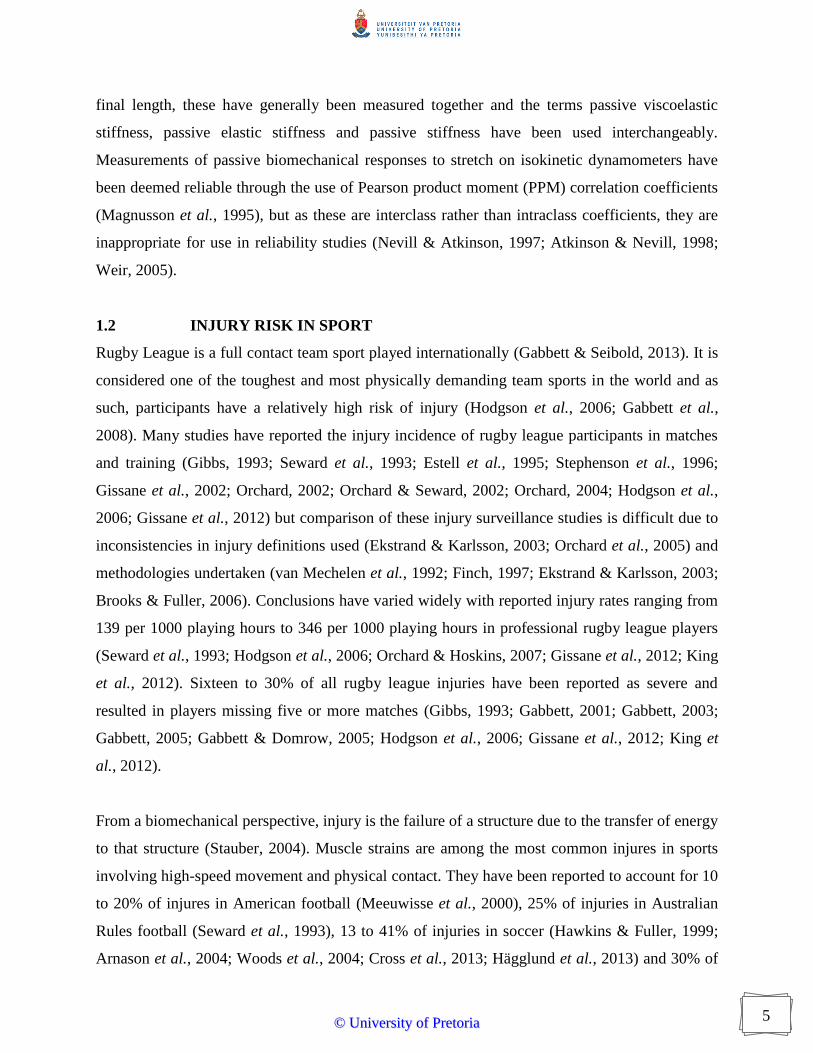
Figure 1.1 - Conceptual model of injury predisposition (Meeuwisse, 1994)
Flexibility is included as an intrinsic risk factor in most conceptual models of injury such as the
one shown in Figure 1.1 (Meeuwisse, 1994) and is considered a primary aetiological factor
associated with musculotendinous strain injuries (Agre, 1985; Worrell et al., 1991; Jonhagen et
al., 1994; Kroll & Raya, 1997; Kujala et al., 1997; Bennell et al., 1999; Devlin, 2000;
Hrysomallis, 2009; Engebretsen et al., 2010; Beijsterveldt et al., 2012). Many studies examining
the relationship between flexibility and injury have been epidemiological and retrospective in
nature (Worrell et al., 1994; Krivickas, 1997; Orchard, 2001; Murphy et al., 2003). Retrospective
research designs fail to address whether theorised risk factors predisposed to, or were the result
of injury (Bennell et al., 1999). Results and conclusions from injury surveillance studies have
varied widely as researchers have considered match hours, training hours, or combined match
and training hours (King et al., 2010), have used substantial differences in the definitions of
injury (Ekstrand & Karlsson, 2003; Orchard et al., 2005) and have generally focused only on
static flexibility. Several studies have identified a relationship between flexibility levels and
injury risk (Krivickas & Feinberg, 1996; Kaufman et al., 1999; Beynnon et al., 2001; Knapik et
al., 2001; McKay et al., 2001; Soderman et al., 2001; Bradley & Portas, 2007; Lowther et al.,
2012) while others have reported no association (Milgrom et al., 1991; Arnason et al., 1996;
Wiesler et al., 1996; Twellaar et al., 1997; Engebretsen et al., 2010). A more preferable study
design is a prospective cohort study as these can provide more direct and accurate estimates of
incidence and relative risk. The main disadvantage of such studies is that sample size is critical
Internal Risk Factors;
- Age
- Gender
- Body Composition
- Health (including
previous injury history)
- Physical Fitness
(including Flexibility)
- Anatomical Factors
- Skill Level
External Risk Factors;
- Other players
- Protective Equipment
- Sports Equipment
- Environmental Factors
- Fatigue
Inciting Event;
- Joint Motion
- Playing Situation
- Training Programme
- Match Schedule
PREDISPOSED
ATHLETE
SUSCEPTIBLE
ATHLETE INJURY
©© UUnniivveerrssiittyy ooff PPrreettoorriiaa

8
and it may be necessary to include and monitor large numbers of athletes for long study periods
(Bahr & Holme, 2003).
Eleven prospective cohort studies examining the relationship between flexibility and hamstring
injury have been conducted. These studies used amateur (Bennell et al., 1998; Gabbe et al.,
2005; Yeung et al., 2009; Engebretsen et al., 2010) and professional participants (Orchard et al.,
1997; Witvrouw et al., 2003; Arnason et al., 2004; Gabbe et al., 2006; Bradley & Portas, 2007;
Arnason et al., 2008; Henderson et al., 2010) and measurements were either ROM (Witvrouw et
al., 2003; Arnason et al., 2004; Gabbe et al., 2005; Gabbe et al., 2006; Bradley & Portas, 2007;
Engebretsen et al., 2010) or sit and reach test scores (Orchard et al., 1997). Eight of these studies
demonstrated no significant association between flexibility and hamstring injury incidence via
logistic regression analysis (Orchard et al., 1997; Bennell et al., 1998; Arnason et al., 2004;
Gabbe et al., 2005; Gabbe et al., 2006; Arnason et al., 2008; Yeung et al., 2009; Engebretsen et
al., 2010). Three studies involving professional footballers reported lower ROM of hip and knee
flexors in players who subsequently sustained muscle strain injury (Witvrouw et al., 2003;
Bradley & Portas, 2007; Henderson et al., 2010). The disparity in findings may be associated
with the type of analysis applied and the inclusion or exclusion of participants with previous
injury. Consistently it must be remembered that risk factors only lead to injury susceptibility and
to provide a greater understanding of patterns leading up to an injury situation, the inciting event
should also be described (Bahr & Holme, 2003).
1.3 HAMSTRING STRAIN MECHANISMS
During an eccentric contraction, when force exerted on a muscle exceeds the force developed by
a muscle, work is done on the stretching muscle and in the process the muscle absorbs
mechanical energy. The degree of energy that a muscle can absorb is thought to be related to
injury risk (Noonan et al., 1994; Garrett, 1996; Mair et al., 1996; Magnusson et al., 2000).
Hamstring strain injuries usually occur near the musculotendinous junction (Proske et al., 2004)
and normally whilst an individual is accelerating or sprinting (Verrall et al., 2003; Woods et al.,
2004), particularly if there is a degree of forward flexion (Verrall et al., 2003). Conjecture exists
regarding the precise point when hamstring strains occur; some researchers argue that initial
stance is the critical point while others maintain that hamstrings are most biomechanically
susceptible to injury during terminal swing (Stanton & Purdham, 1989; Thelen et al., 2005;
©© UUnniivveerrssiittyy ooff PPrreettoorriiaa
9
Chumanov et al., 2007; Chumanov et al., 2012). Such conclusions have generally been based on
theoretical rationale (Stanton & Purdham, 1989) or analysis of asymptomatic subjects (Mann &
Sprague, 1980; Wood, 1987; Orchard & Seward, 2002); these approaches are unable to definitely
establish when in the sprinting cycle the hamstrings fail.
While there are models to estimate the moments affecting individual joints during the human gait
cycle (Mann & Hagy, 1980; Novacheck, 1995) as well as muscle length during sprinting
(Hawkins & Hull, 1990; Gerritsen et al., 1998), there is no model to determine the external force
or stress that individual muscle or muscle groups are subject to at any given time of the gait cycle
making it difficult to assess when and why strain injuries actually occur. The only two studies
that have managed to capture data of a running athlete at the time of an actual hamstring strain
occurring both identified terminal swing phase as the most likely time of injury (Heiderscheit et
al., 2005; Schache et al., 2009). At this time the hamstrings demonstrate peak electromyographic
activity as most of the inertial force acting about the knee joint is potentially imparted onto the
hamstrings as they attempt to decelerate the swinging lower limb (Kryolainen et al., 1999;
Kuitunen et al., 2002) with the gastrocnemius being the only other significant muscle capable of
providing some assistance (Li et al., 2002). In contrast, a number of large muscles are thought to
contribute to the generation of hip extensor torque and control of trunk flexion during the initial
stance (Waters et al., 1974; Pandy & Zajac, 1991; Arnold et al., 2005). Although the forces
required to control upper body deflections and propel hip extension at initial stance would be
overall greater than to control the relatively light weight of the lower limb at terminal swing,
high tensile stress when the muscle is shortening is not thought a primary injury inducing
mechanism (Moens et al., 1993; Petrof et al., 1993; Gao et al., 2008). Conversely it is widely
agreed that muscle strain injury is associated with exercise that involves eccentric contractions
(Warren et al., 1993; Morgan & Allen, 1999; Allen, 2001; Friden & Lieber, 2001), when
extracellular matrix and lateral linkage is under large shear stress from the myofibrils (Petrof et
al., 1993). If this stress is sufficiently large, it can break the extracellular matrix linking adjacent
fibres and peel off the basal lamina from the myofibrils (Stauber, 1989; Kääriäinen et al., 2000)
as well as causing sarcolemma damage (Friden & Lieber, 2001). The magnitude of this stress
and resulting strain has been shown to relate to muscle damage (Talbot & Morgan, 1998).
©© UUnniivveerrssiittyy ooff PPrreettoorriiaa

10
1.4 STRETCHING
Practitioners have long advocated stretching before exercise as a method of achieving improved
flexibility, enhanced performance and consequentially reduced injury risk. Epidemiological
research examining the relationship between injury occurrence and stretching has yielded
equivocal findings. The studies are frequently retrospective in nature, often fail to account for the
array of other factors related to injury incidence and have included a range of athletic and non-
athletic participants (Kerner & D'Amico, 1983; Jacobs & Berson, 1986; Blair et al., 1987;
Macera et al., 1989; Brunet et al., 1990; Wilber et al., 1995). Experimental approaches have
typically looked at the effect of pre-exercise stretching on static flexibility and various
performance measures as opposed to injury risk, and this empirical evidence is equivocal in
nature (Moller et al., 1985; Wessling et al., 1987; Godges et al., 1989; Cornelius et al., 1992;
Clark et al., 1999; Cornwell et al., 2002; Gill et al., 2002; Funk et al., 2003; Zakas et al., 2003;
Bonnar et al., 2004; Egan et al., 2006). Published articles of an experimental or quasi-
experimental nature have frequently lacked randomisation or have failed to include control
measures and as such do not qualify for inclusion in meta-analyses or systematic reviews (Moller
et al., 1985; Wessling et al., 1987; Godges et al., 1989; Cornelius et al., 1992; Clark et al., 1999;
Cornwell et al., 2002; Gill et al., 2002; Funk et al., 2003; Zakas et al., 2003; Bonnar et al., 2004;
Egan et al., 2006). Furthermore, many studies which have suggested that static stretching prior to
exercise might prevent injury have included co-interventions such as a warm-up and the use of
braces in addition to stretching, thus making it impossible to identify the unique effect of the
stretching (Ekstrand & Gillquist, 1983; Bixler & Jones, 1992; Amako et al., 2003).
Systematic reviews considering the impact of stretching on injury risk are equivocal and
collectively suggest that, based on the limited and low quality evidence available, stretching may
or may not provide a meaningful decrease in the risk of injury but because appropriate evidence
is lacking, that no definite recommendations should be made (Weldon & Hill, 2003; Thacker et
al., 2004; Small et al., 2008). The most recent of these reviews identified 364 published peer-
reviewed articles related to stretching and injury risk but only seven studies met inclusion and
exclusion criteria (Small et al., 2008). Of these, pre-exercise static stretching was considered
ineffective in six studies (Bixler & Jones, 1992; van Mechelen et al., 1993; Pope et al., 1998;
Amako et al., 1999; Cross & Worrell, 1999; Pope et al., 2000) while only one controlled clinical
trial concluded that pre-exercise stretching had a positive impact on injury risk (Hartig &
©© UUnniivveerrssiittyy ooff PPrreettoorriiaa
11
Henderson, 1999). Other quasi-randomised (Amako et al., 2003) or non-randomised (Hadala &
Barrios, 2009) interventions have shown some beneficial effect of pre-exercise stretching. Yeung
and Yeung (2001) in an investigation of experimental and quasi-experimental studies pertaining
to the prevention of lower limb running injuries analysed the collective results of five studies
with 1944 participants in stretch groups and 3159 participants in control groups and reported that
there was no clear evidence to support the notion that pre-exercise static stretching exercises are
effective in reducing lower limb injuries (Andrish et al., 1974; van Mechelen et al., 1993; Hartig
& Henderson, 1999; Pope et al., 2000). This conclusion needs to be treated with a degree of
caution as it is not based on meta-analysis findings and the quality of the studies included was
not evaluated. Reviews conducted into the relationship between stretching and injury risk have
not always attempted to distinguish between pre-exercise, post-exercise or general stretching and
furthermore stretching has been typically related to overall injury incidence. More recently Small
et al. (2008) considered injuries to the musculotendinous unit independently of overall injury rate
and suggested that there was some evidence to indicate that pre-exercise stretching might be
beneficial in the prevention of musculotendinous strain injuries. Intuitively it is plausible that
stretching is more likely to be linked to injuries of the musculotendinous unit rather than to joint,
bone and vascular-related injuries but empirical evidence to substantiate this belief is lacking.
1.5 RATIONALE
Given the widespread application of flexibility testing there is a need to re-evaluate the reliability
of static and dynamic flexibility measures using more acceptable statistical analysis procedures
(Lamb, 1998). Reliability is the extent to which the measurements of a test remain constant over
repeated tests of the same participants under identical conditions, and knowledge of the type and
magnitude of error is important to determine whether a small but potentially meaningful effect
would be detected (Nevill & Atkinson, 1997). Such analyses need to consider the population for
which the measurement tool is intended, as reliability in one population does not necessarily
infer reliability in a different population. Many previous studies considering the reliability of
flexibility measures have used non-athletic convenience samples taken from University or
general populations and used results to make assumptions about athletic individuals.
Furthermore, applicability to the proposed testing schedule must be considered as conducting
reliability studies on the same day is inappropriate if testing is to take place on different days, as
©© UUnniivveerrssiittyy ooff PPrreettoorriiaa

12
is common in flexibility and stretching studies, when between day (interday) reliability should be
reported (Hopkins, 2000).
Some attempts have been made to consider the relationships shared between measures of static
flexibility, passive dynamic flexibility and active dynamic flexibility (Cameron & Bohannon,
1993; Gajdosik et al., 1993; Gleim & McHugh, 1997; McHugh et al., 1998; Hunter & Spriggs,
2000; Gajdosik, 2002; Blackburn et al., 2004; Rolls & George, 2004; Davis et al., 2008; Ayala et
al., 2011; Ayala et al., 2012a), although results have not been conclusive. Furthermore there is
disparity among results of studies examining different aspects of the same type of flexibility
measurement (Wilson et al., 1991; Hunter & Spriggs, 2000; Bojsen-Moller et al., 2007). As well
as being highly specific to individual joints, it is possible that different tests which are attempting
to investigate flexibility of the same muscle group (e.g. straight leg raise and knee extension tests
for the hamstring) are examining, at least in part, different constraining factors (Gajdosik, 1987;
Cameron & Bohannon, 1993; Gajdosik et al., 1993; Rolls & George, 2004; Davis et al., 2008).
Examination of whether tests of static flexibility are related to each other and to the more
complex dynamic flexibility tests is thus warranted.
While there is extensive published research examining the impact of stretching on levels of
flexibility, performance and injury risk, the quality of studies has varied considerably. Reviewers
have often failed to consider this, leading to inappropriate and unsubstantiated conclusions being
drawn. Moreover, studies investigating stretching and injury risk have not always attempted to
provide rationale for their findings and few studies examining flexibility or performance
measures have been holistic in their approaches. An integrated review of available literature
considering the impact of stretching on flexibility levels and performance measures would assist
in better informing practitioners and sports personnel as to overall risks and benefits of this
practice.
Existing research has almost exclusively focused on static flexibility as a potential risk factor for
injury and there is clearly a need to examine whether dynamic flexibility, which provides a better
indicator of the mechanical properties of the muscle, is related to injury risk. Understanding the
mechanisms behind passive extensibility modifications that have been studied in animal models
(Taylor et al., 1993; Noonan et al., 1994; Mair et al., 1996) and considering how they apply to
©© UUnniivveerrssiittyy ooff PPrreettoorriiaa
13
interventions with human muscles is necessary to develop methods that promote favourable
passive extensibility adaptations, functional activities and athletic performance (Gajdosik,
2001b). Given the conceptual model for injury development (Figure 1.1) and the number of and
possible interactions between potential risk factors, it is unlikely that epidemiological approaches
will be able to correctly identify the potential role of static and dynamic flexibility in
predisposing injury. Experimental approaches might thus be better able to identify the potential
theoretical framework through which flexibility may be a risk factor for injury.
1.6 STATEMENT OF THE PROBLEM
Existing literature related to both flexibility and stretching in humans has almost exclusively
focused on static flexibility. Despite extensive published research, the evidence supporting static
flexibility as an injury risk factor is equivocal and often poor quality. Static flexibility is not
necessarily related to the mechanical properties of the musculotendinous unit and it is plausible
that dynamic flexibility is better associated with injury risk and performance measures. Some
research studies have explored dynamic flexibility in humans in terms of developing valid and
reliable assessment techniques, comparing static and dynamic measures of flexibility and
investigating factors influencing dynamic flexibility. Research exploring dynamic flexibility has
tended to be experimental in nature to attempt to understand the mechanisms through which
flexibility may be a risk factor for injury. Existing published research has tended to use
heterogeneous groups of active adults, frequently has poorly described methodologies and lacks
provision of reliability indices. Athletic populations consistently demonstrate a high incidence of
strains to the musculotendinous unit, frequently located in the hamstring muscle group, and thus
there is a need for research focusing on this subgroup of individuals. This thesis was designed to
fill gaps in the existing literature by evaluating methods of assessing dynamic flexibility,
considering the comparability of static and dynamic components of the global concept of
flexibility and exploring how dynamic flexibility is influenced by exercise and stretching.
1.7 ORGANISATION OF THESIS
The central theme of this thesis is hamstring flexibility and the influence of fatiguing exercise
and stretching on this. A review of the literature is provided in Chapter 2. The key topics
addressed are the measurement of flexibility, reliability and validity of flexibility tests and the
impact of exercise and post-exercise stretching on measures of flexibility and performance, with
©© UUnniivveerrssiittyy ooff PPrreettoorriiaa
14
consideration given to how these might influence injury risk. The review attempts to appraise
current knowledge and suggest future directions in a critical manner. The reliability of different
methods of measuring hamstring flexibility is evaluated in Chapter 3. To allow meaningful
interpretation for a specific population, Chapter 3 focuses on professional rugby league players.
The same population takes part in subsequent studies. A comparison of static and dynamic
hamstring flexibility measurements is reported in Chapter 4. Findings from previous Chapters
are used to inform study design of Chapter 5 which is a repeated measures randomised
controlled study examining the consequence of fatiguing exercise and subsequent static
stretching on measures of flexibility and performance and implications for injury risk. The thesis
finishes with a critical review of the research and suggestions for future investigations.
1.8 AIMS OF THE STUDY
The aims of this thesis were to;
1) Establish the intraday and interday, relative and absolute reliability of static and dynamic
measurements of hamstring flexibility;
2) Examine relationships shared by static and dynamic measurements of hamstring
flexibility;
3) Investigate the impact of fatigue and post-exercise static stretching on measurements of
static and dynamic flexibility and performance.
©© UUnniivveerrssiittyy ooff PPrreettoorriiaa

15
CHAPTER 2
LITERATURE REVIEW
©© UUnniivveerrssiittyy ooff PPrreettoorriiaa
16
2 LITERATURE REVIEW
Static flexibility testing has been commonplace since the beginning of the 1940s when flexibility
was first identified as an important component of human physical fitness (Cureton, 1941).
Substantial research using various approaches of evaluating the reliability and validity of static
flexibility tests has been conducted and as these are essential to any measurement tool, it is
important that their exploration and critical evaluation is always undertaken. Research pertaining
to measures of dynamic flexibility is more limited and there is a need to understand the
properties of the musculotendinous unit and investigate methods and applicability of dynamic
flexibility testing. Various stretching techniques are used to improve levels of flexibility and it is
necessary to explore reasons behind their possible effectiveness and consider the impact that
stretching might have on injury mechanisms and recovery from exercise.
2.1 MEASUREMENT OF STATIC FLEXIBILITY
Static flexibility tests are based on linear or angular measurements of the motion of a joint or
series of joints, and have been classified as single joint or compound (multiple joint) tests
(Corbin & Noble, 1980). Single joint static flexibility tests are common clinical measures in
sports medicine and typically involve angular (goniometers or inclinometers) measurements
(Gajdosik, 2001a) rather than the linear measurements common in field tests of flexibility
(Baltaci et al., 2003). Single joint tests are considered better measures of static flexibility than
compound tests because they are more specific but nonetheless are subject to testing limitations
such as accurately identifying the centre of rotation for goniometry measurement, the influence
of skin and fat movement and the difficulty in isolating the movement (Armstrong et al., 1998;
Peeler & Anderson, 2008; Pua et al., 2008). Alongside the distinction between single and
compound tests of static flexibility, there is a further division of common static flexibility tests
into passive and active tests (Harvey et al., 2002; Ylinen et al., 2010). Passive static flexibility
tests require the investigator or clinician to move the required joint to the end of its ROM
without assistance from the participant. Participants are asked to indicate when they feel at their
maximum range and the joint is held still and the angle measured (Maud & Cortez-Cooper,
1994). Active static flexibility tests require the participant to themselves move the required joint
to end of its ROM themselves, using muscles surrounding the joint alone and then hold the joint
still while the angle measured (Kim et al., 2005). Passive static flexibility measurements may be
influenced by factors such as the pain threshold of an individual, subject bias and application of
©© UUnniivveerrssiittyy ooff PPrreettoorriiaa
17
different levels of torque whilst differences in extent of muscle activation and stretch reflex
might affect active measurements (Marshall et al., 2011; Ayala et al., 2012b).
2.2 STATIC HAMSTRING FLEXIBILITY MEASURES
Static flexibility of the hamstring muscle group has traditionally been measured using the
straight leg raise (SLR) test (Gajdosik et al., 1993; Halbertsma et al., 2001), although movement
of the pelvis means that the test is not necessarily specific to the hamstrings (Hu et al., 2010).
The SLR test can also be affected by deep fascia of the lower limb and neurological tissue
(McHugh et al., 2012) and has been described as a ‘key tension test’ of the lower limb and trunk
(Boyd et al., 2009), making it potentially more useful as a neurological test rather than a static
flexibility test in the clinical setting. Other tests of static hamstring flexibility include active and
passive knee extension (KE) tests which involve movement at the knee joint but not the hip
making the joint movement easier to control and isolate (Gajdosik et al., 1993). The most widely
used compound and linear field based measurement of static hamstring flexibility is the sit and
reach test (Sporis et al., 2011; Ayala et al., 2012b). This test is however only moderately related
to hamstring flexibility, is influenced by anthropometric factors, e.g. leg length, adipose tissue,
and as a compound movement includes a contribution from flexibility of the lower back (Ayala
et al., 2011).
2.2.1 Reliability of static hamstring flexibility tests
Before any useful information can be gleaned from flexibility assessment it is important to
ensure that measurements are adequately valid and reliable. Validity is the ability of the
measurement tool to reflect what it is designed to measure, while reliability is defined as the
consistency of measurements or the absence of measurement error (Atkinson & Nevill, 1998).
Intratester (intrarater) reliability reflects the consistency of repeated measurements taken by one
tester over time while intertester reliability reflects the reproducibility of measurements taken by
different individuals (Hopkins et al., 2001). Some testers have found intratester reliability of the
Active Knee Extension (AKE) test to be high (r = 0.99, p < 0.05) using a metal rig to assist in
measurement and straps to limit pelvic and leg motion (Gajdosik & Lusin, 1983). Whilst this is
likely to lead to improved isolation and therefore smaller measurement error, the applicability of
this procedure has been questioned since these types of apparatus are rarely available clinically
and the procedure therefore lacks ecological validity (Worrell et al., 1991). The reliability of the
©© UUnniivveerrssiittyy ooff PPrreettoorriiaa
18
manual techniques and basic goniometry that are routinely used in clinical environments is
variable with ICCs of 0.18 to 0.99 being reported for intratester (Rothstein et al., 1983; Youdas
et al., 1993; Bierma-Zeinstra et al., 1998; Pua et al., 2008; Ayala et al., 2011; Nunes et al., 2012)
and 0.28 to 0.97 being reported for intertester reliability (Rothstein et al., 1983; Youdas et al.,
1993; Holm et al., 2000; Gabbe et al., 2004; Norris & Matthews, 2005; Bozic et al., 2010; Nunes
et al., 2012; Poulsen et al., 2012; Reurink et al., 2013). Researchers generally interpret repeated
measures of static flexibility exhibiting high correlation coefficients as being very reliable but
these measure the strength of association rather than the agreement and if data is widely
distributed, can exhibit poor intra-individual agreement but high correlations (Weir, 2005).
Additionally, some studies inappropriately use the Pearson product moment correlation to assess
reliability (Gajdosik & Lusin, 1983; Gajdosik & Bohannon, 1987). This is an interclass and
bivariate statistic and thus should only be used to establish extent of relationships between
different groups of variables and not to assess reliability, which involves repeated measurements
within the same group (Weir, 2005). Little attempt has been made to establish the absolute
reliability of static flexibility tests, for instance by reporting statistics such as SEM or Limits of
Agreement (Bland & Altman, 1986). Studies that have reported absolute reliability identify that
actual measurements can vary by as much as 6o to 20
o between repeated measures (Hsieh et al.,
1983; Gajdosik et al., 1993; Chow et al., 1994; Mullaney et al., 2010; Ayala et al., 2011). Many
reasons have been proposed for the presence of such measurement errors and include differing
tolerance of participants to sensations of stretch and pain (Magnusson et al., 1996), lack of
stabilisation (Gajdosik & Lusin, 1983), influence from structures other than those being tested
(Krabak et al., 2001) and inconsistent force application on passive tests (Reid & McNair, 2004).
It has been suggested that researchers should quantify force applied to passive movements in
attempts to ensure better repeatability although the recent move toward post-positivism in
orthopaedic and sports physical therapy research suggests that the tactic knowledge that forms
parts of the skills of an experienced clinician is sufficient (Greenfield et al., 2007). This
inductive approach argues that although something cannot be empirically quantified, it should
not be disregarded (Ferlie et al., 1999). As clinicians are experienced in measuring static
flexibility, they are therefore likely to apply similar levels of force and stabilisation thus giving
high intrarater reliability of the tests.
©© UUnniivveerrssiittyy ooff PPrreettoorriiaa
19
2.2.2 Validity of static hamstring flexibility tests
Few studies have compared results obtained using different methods of assessing static
hamstring flexibility (Hsieh et al., 1983; Cameron & Bohannon, 1993; Gajdosik et al., 1993; Hui
& Yuen, 2000; Rolls & George, 2004; Davis et al., 2008; Shimon et al., 2010; Ayala et al., 2011;
Ayala et al., 2012a). Many have considered common variance of tests as an indicator of validity
(Hsieh et al., 1983; Gajdosik et al., 1993; Hui & Yuen, 2000; Gajdosik, 2001b; Awan et al.,
2002). Common variance, also called coefficient of determination, is in simple regression the
proportion of the variance of the target or dependent variable that is accounted for by regression
upon the regressor or independent variable and is given by the square of the PPM correlation
(Kinnear & Gray, 2008). Gajdosik et al. (1993) reported Passive Knee Extension (PKE) and
Passive Straight Leg Raise (PSLR) common variance of 43.5%, Active Knee Extension (AKE)
and PKE common variance of 49 to 74% and ASLR and AKE common variance of only 4%.
These common variances indicate that generally more than half of the available range is affected
by different factors for each test. Linked to low shared common variances between active and
passive knee extension tests, significant differences (p < 0.05) between joint angles achieved
have also been identified (Gajdosik & Lusin, 1983; Cameron & Bohannon, 1993). Authors
suggested that this might be because the AKE tests represent measurement of initial length of the
hamstring and PKE tests represent a measurement of the final length. Initial length was described
as the point where first passive resistance to stretch is detected, which is not identical to the
resting length of the muscle (Gajdosik, 2001c). This suggests that active and passive static
flexibility tests are not necessarily measuring the same constraints of static flexibility and these
findings highlight the necessity to standardise between the use of active and passive tests.
2.3 MUSCLE AND JOINT STRUCTURE AND FLEXIBILITY
To understand the concept of dynamic flexibility it is important to consider the mechanical
properties of muscle and factors that may influence quantification of resistance. The proposed
mechanism through which changes in muscle length occur is through the sliding of thick and thin
filaments past one another within the functional units of the muscle (Maganaris, 2001). Animal
models have shown that muscles, like most biological materials, display viscoelastic properties
(Taylor et al., 1990) and thus demonstrate the characteristics of stress relaxation, creep and
hysteresis (Magnusson et al., 1997). These models have been extrapolated to explain the
behaviour of human muscle in vivo and imply that muscles have both viscous (rate dependent)
©© UUnniivveerrssiittyy ooff PPrreettoorriiaa
20
and elastic (force dependent) properties (Fukashiro et al., 2001). The mechanical characteristics
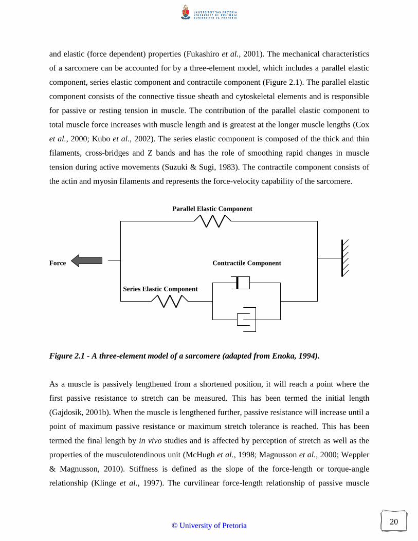
of a sarcomere can be accounted for by a three-element model, which includes a parallel elastic
component, series elastic component and contractile component (Figure 2.1). The parallel elastic
component consists of the connective tissue sheath and cytoskeletal elements and is responsible
for passive or resting tension in muscle. The contribution of the parallel elastic component to
total muscle force increases with muscle length and is greatest at the longer muscle lengths (Cox
et al., 2000; Kubo et al., 2002). The series elastic component is composed of the thick and thin
filaments, cross-bridges and Z bands and has the role of smoothing rapid changes in muscle
tension during active movements (Suzuki & Sugi, 1983). The contractile component consists of
the actin and myosin filaments and represents the force-velocity capability of the sarcomere.
Parallel Elastic Component
Force Contractile Component
Series Elastic Component
Figure 2.1 - A three-element model of a sarcomere (adapted from Enoka, 1994).
As a muscle is passively lengthened from a shortened position, it will reach a point where the
first passive resistance to stretch can be measured. This has been termed the initial length
(Gajdosik, 2001b). When the muscle is lengthened further, passive resistance will increase until a
point of maximum passive resistance or maximum stretch tolerance is reached. This has been
termed the final length by in vivo studies and is affected by perception of stretch as well as the
properties of the musculotendinous unit (McHugh et al., 1998; Magnusson et al., 2000; Weppler
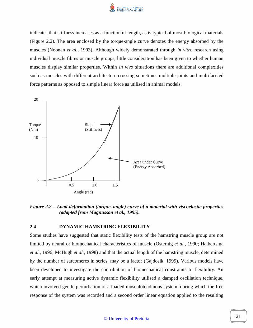
& Magnusson, 2010). Stiffness is defined as the slope of the force-length or torque-angle
relationship (Klinge et al., 1997). The curvilinear force-length relationship of passive muscle
©© UUnniivveerrssiittyy ooff PPrreettoorriiaa
21
indicates that stiffness increases as a function of length, as is typical of most biological materials
(Figure 2.2). The area enclosed by the torque-angle curve denotes the energy absorbed by the
muscles (Noonan et al., 1993). Although widely demonstrated through in vitro research using
individual muscle fibres or muscle groups, little consideration has been given to whether human
muscles display similar properties. Within in vivo situations there are additional complexities
such as muscles with different architecture crossing sometimes multiple joints and multifaceted
force patterns as opposed to simple linear force as utilised in animal models.
20
10
0.5 1.0 1.5
Figure 2.2 – Load-deformation (torque–angle) curve of a material with viscoelastic properties
(adapted from Magnusson et al., 1995).
2.4 DYNAMIC HAMSTRING FLEXIBILITY
Some studies have suggested that static flexibility tests of the hamstring muscle group are not
limited by neural or biomechanical characteristics of muscle (Osternig et al., 1990; Halbertsma
et al., 1996; McHugh et al., 1998) and that the actual length of the hamstring muscle, determined
by the number of sarcomeres in series, may be a factor (Gajdosik, 1995). Various models have
been developed to investigate the contribution of biomechanical constraints to flexibility. An
early attempt at measuring active dynamic flexibility utilised a damped oscillation technique,
which involved gentle perturbation of a loaded musculotendinous system, during which the free
response of the system was recorded and a second order linear equation applied to the resulting
Area under Curve
(Energy Absorbed)
Slope
(Stiffness)
Torque
(Nm)
0
Angle (rad)
©© UUnniivveerrssiittyy ooff PPrreettoorriiaa

22
damped oscillation to calculate the stiffness of the system (Bach et al., 1983; Shorten, 1987).
This technique has possible errors in that the downward force applied by the experimenter is
generally described as a ‘downward gentle push’ in the region of 100-200N magnitude (Ditroilo
et al., 2011) and often estimated rather than being objectively measured. Differences in
perturbation amplitude can influence stiffness assessment, particularly in terms of reflex
contribution (Kearney et al., 1999; Hunter & Spriggs, 2000). There is evidence that the damping
response is somewhat curvilinear as opposed to completely linear (Watsford et al., 2010). While
Wilson et al. (1994) described reliability values for performance measures determined through
the use of intraclass correlations (ICCs), they did not report any values for stiffness measures and
two earlier studies failed to make any attempts at confirming reliability of the system (Wilson et
al., 1991; Wilson et al., 1992). Other studies have reported ICCs of r = 0.87 to 0.94 (p < 0.01)
but did not state which particular ICC was used (Walshe et al., 1996; Murphy et al., 2003). The
choice of ICC test is not inherently obvious and different tests can yield different results, which
should be considered when interpreting this value. Additionally, convenience samples of
participants were used in all studies and as these can exhibit high inter-subject variability,
artificially high reliability values can be obtained (Atkinson & Nevill, 1998).
In an attempt to improve objectivity and to allow further understanding of dynamic flexibility,
Magnusson et al. (1995) developed a model which used an isokinetic dynamometer to examine
the stiffness and energy in a dynamic phase, and viscoelastic response in a static phase of a
standardised stretch manoeuvre of the human hamstring muscle group. Due to the insignificant
differences between trials as measured by paired t-tests and having Pearson product moment
correlation coefficients of r = 0.91 to 0.99 (p < 0.05), the method was considered highly reliable
(Magnusson et al., 1995). The use of PPM correlation coefficients in reliability studies is
however inappropriate (Chen & Barnhart, 2008) as they are bivariate statistics and reliability
measures are univariate in nature (Weir, 2005). Despite the challenges associated with the
objective measurement of flexibility and the equivocal nature of the research considering the
importance of flexibility for injury prevention, a variety of strategies aimed at improving
flexibility and performance are utilised.
©© UUnniivveerrssiittyy ooff PPrreettoorriiaa
23
2.5 STRETCHING TECHNIQUES
Static stretching is generally the most widely used and recommended stretching technique
because the exercises are relatively easy to perform and have little associated risk of injury
(Kolber & Zepeda, 2004). These involve joints being placed in the outer limits of their ROM and
then subjected to an elongation torque or force, which is maintained for a period ranging from a
few seconds to minutes (Thacker et al., 2004). The slow build-up of tension and the absence of
pain involved with static stretching are believed to minimise stretch reflex responses thus
inducing muscular relaxation and allowing further stretching (Guissard & Duchateau, 2006).
Ballistic stretching is a dynamic and fast movement in which a bouncing type of stretch torque is
applied into the extreme ROM limits of the joints concerned (Covert et al., 2010). This technique
was thought a useful method of developing dynamic flexibility even though research often
reports ballistic stretching to be less effective at improving flexibility than other types of
stretching (Sady et al., 1982; Wallin et al., 1985; Bandy et al., 1998; Bacurau et al., 2009;
Covert et al., 2010). The inhibitory effect of the stretch reflex is thought to be one reason for the
lower effectiveness of ballistic stretching for improving static flexibility (Guissard & Duchateau,
2006). Additionally, because higher forces are involved in ballistic stretching compared to other
stretching methods, it is associated with increased potential of injury to the musculotendinous
unit and thus has traditionally been avoided (Hartig & Henderson, 1999; Hedrick, 2000). While
it is possible that the musculotendinous units of untrained and sedentary individuals may not be
able to withstand this vigorous type of stretching without sustaining muscle damage, sports
people often put their joints through large ranges while exerting considerable forces. It is
therefore possible that ballistic-type stretching may be appropriate in these groups of people
(Shrier, 2005). More recently, ballistic stretching procedures have been adapted to become more
controlled, through range movements rather than end of range techniques, and these types of
stretches have been termed dynamic stretching (Behm & Chaouachi, 2011).
Proprioceptive Neuromuscular Facilitation (PNF) is a collection of techniques for facilitating
muscle contraction, strengthening and increasing flexibility. These techniques were originally
formulated and developed as a physical therapy procedure for the rehabilitation of stroke patients
(Knott & Voss, 1957; Kabat, 1958) and PNF stretching procedures were subsequently developed
on the basis of several important neurophysiological mechanisms (Chalmers, 2004). Contract-
©© UUnniivveerrssiittyy ooff PPrreettoorriiaa

24
relax stretching involves an initial maximal isometric contraction of the muscle to be stretched
(the antagonist) followed by the relaxation and passive stretch of the muscle to the limit of its
ROM (Sharman et al., 2006). The isometric contraction of the muscle to be stretched is followed
by relaxation, thought to stem from autogenic inhibition by the Golgi tendon organs (Ferber et
al., 2002; Chalmers, 2004; Guissard & Duchateau, 2006). This suppresses the excitability of
muscle spindles and thus allows the muscle to relax and stretch further (Bonnar et al., 2004).
Consequently, the intention of PNF stretching is to reduce reflex activity, thus diminishing
resistance and thereby improving joint ROM (Etnyre & Abraham, 1986). Paradoxically, it has
been found that PNF stretching techniques that were most effective in increasing static flexibility
also produced the highest levels of EMG activity in the stretched muscles, indicating that full
muscular relaxation is not necessary for elongation to take place (Sharman et al., 2006; Mitchell
et al., 2009).
It has been suggested that PNF alters stretch perception, providing a stretch-induced analgesic
effect and thus increasing the tolerance of an individual to stretch (Sharman et al., 2006;
Weppler & Magnusson, 2010). There is little evidence for proposed mechanisms but this might
be through activation of the descending pain suppression system. PNF techniques have been
described as painful and painful stimuli cause the release of endorphins and enkephalins, which
inhibit the release of Substance P, a neurotransmitter in the Lateral Spinothalamic Tract (the
pathway in which pain signals travel from the receptor to the brain) (Piercey et al., 1986; De
Felipe et al., 1998). Activation of the descending pain suppression system results in an individual
becoming less sensitive to painful stimuli (i.e. maximal stretch) and thus is able to tolerate higher
levels of this stimulus (i.e. greater degree of stretch resulting in higher achievable ROM) (Pert,
1982). Additionally it is accepted that pain perception can be affected by dynamic exercise
(Sforzo, 1989), thus altered pain perception as a result of the isometric contraction could
contribute to the mechanism of PNF stretching. Alternatively, there is a possibility that
mechanoreceptors contribute to a decreased pain perception via gate control. This occurs when
stimulation of these receptors by distortion, pressure or stretch causes presynaptic inhibition of
nociceptive signals (Terman et al., 1984).
©© UUnniivveerrssiittyy ooff PPrreettoorriiaa

25
2.6 STRETCHING AND FLEXIBILITY
Several studies have compared the effectiveness of different stretching techniques for improving
static (Cornelius et al., 1992; Bandy & Irion, 1994; Bandy et al., 1997; Klinge et al., 1997;
Wiemann & Hahn, 1997; Bandy et al., 1998; McHugh et al., 1998; Roberts & Wilson, 1999;
Depino et al., 2000; Chan et al., 2001; Funk et al., 2001; Funk et al., 2003; Zakas et al., 2003;
Nelson & Bandy, 2004; Young et al., 2004; Davis et al., 2005; Zakas et al., 2006; Bacurau et al.,
2009; Fasen et al., 2009; O'Sullivan et al., 2009; Ayala & Sainz de Baranda Andújar, 2010;
Covert et al., 2010; O'Hora et al., 2011; Maddigan et al., 2012; Mizuno et al., 2013; Place et al.,
2013) and dynamic flexibility (Mahieu et al., 2007; Herda et al., 2008; Jaggers et al., 2008;
Morse et al., 2008; Hough et al., 2009; Mahieu et al., 2009; Herda et al., 2010; Nakamura et al.,
2011; Matsuo et al., 2013) with equivocal results. Many studies are of poor to moderate
methodological quality (Moher et al., 2009) and often fail to differentiate between an acute
single stretch, series of stretches or a longer term stretching programme when setting up their
rationale (Behm & Chaouachi, 2011). Additionally, some studies utilise an aerobic warm up
before commencing the stretch intervention while others do not (Fradkin et al., 2006). As well as
different methods of stretching being used, stretches are held for differing amounts of time (Ryan
et al., 2008; Ayala & Sainz de Baranda Andújar, 2010; Matsuo et al., 2013). Prospective
randomised controlled trials or clinical controlled trials have shown that stretching increases
static flexibility by between 5 and 30% (Wessling et al., 1987; Halbertsma et al., 1996;
Wiemann & Hahn, 1997; Kokkonen et al., 1998; Clark et al., 1999; Depino et al., 2000;
Magnusson et al., 2000; Church et al., 2001; Draper et al., 2002; de Weijer et al., 2003; Power et
al., 2004; Bazett-Jones et al., 2005; Young et al., 2006; O'Hora et al., 2011). The practical
consequence of these findings is not always considered and differentiation should be made
between stretching to restore normal osteokinematic movements in individuals with identifiable
muscle tightness and stretching to produce hyperflexibility (beyond normal ROM) in athletes
with otherwise functional movements (McCormack et al., 2004).
Theoretical explanations of the effectiveness of stretching techniques in improving static
flexibility have included overcoming the stretch reflex, reciprocal inhibition and autogenic
inhibition (Hutton, 1992). Results of studies comparing PNF and static stretching techniques
have been equivocal. Many researchers have found PNF to be the superior technique for
improving static flexibility (Wallin et al., 1985; Etnyre & Lee, 1988; Cornelius et al., 1992;
©© UUnniivveerrssiittyy ooff PPrreettoorriiaa
26
Magnusson et al., 1996; Funk et al., 2003; Sharman et al., 2006; O'Hora et al., 2011) while
others have concluded that PNF is equivalent to static stretching (Worrell et al., 1994; Marek et
al., 2005; Maddigan et al., 2012). Part of the disparity in such findings may be associated with
the lack of distinction between short and long-term effects of stretching and a lack of uniformity
among measurement procedures (McHugh & Cosgrave, 2010). Some studies through
electromyography analysis have found excitability to be lower during PNF stretching than during
static stretching which provided evidence for the rationale behind PNF techniques (Etnyre &
Abraham, 1986). However, other studies contradict this and further question the mechanism
through which PNF stretching is effective (Moore & Hutton, 1980; Hutton, 1992; Chalmers,
2004; Sharman et al., 2006; Mitchell et al., 2009). While there is evidence both static and PNF
stretching result in improved flexibility, static stretching is generally performed before or after
periods of exercise while PNF stretching is more often used as part of treatment or rehabilitation
programmes (Westwater-Wood et al., 2010).
While there is largely consensus that static stretching results in an improvement in static
flexibility, no consistency is apparent with regard to how long stretches should be held to obtain
optimum benefits. Recommendations for duration of stretching in flexibility training
programmes range from 5 to 60 s yet justifications for these selections have largely been absent
and the duration of the stretching protocols used in some studies do not always coincide with
typical practice of athletes and fitness coaches (Behm & Chaouachi, 2011). A series of articles
conduced in professional sport reported average stretch durations of 12 to 18 s (Ebben et al.,
2004; Ebben et al., 2005; Simenz et al., 2005; Duehring et al., 2009). Madding et al. (1987)
compared the effects of 15, 45 and 120s of stretching on hip abduction and reported that
sustaining a stretch for 15s was as effective as 120s. Bandy and Irion (1994) found that a 30s
stretch was more beneficial than a 15s stretch but there was no advantage to holding the stretch
for 60s. Other authors have also concluded that 30s is the optimum duration to maintain a static
stretch (Grady & Saxena, 1991; Walter et al., 1994; Rubini et al., 2007; Young, 2007). In the
literature reviewed, no study investigating acute effects of stretching on flexibility indicated
attempts to control for total amount of time spent stretching while manipulating stretch duration.
Total time spent stretching may influence the improvements observed in static flexibility after a
flexibility training programme as holding stretches for 60s would result in a much longer total
time spent stretching compared to holding each stretch for 15s. The same duration of static
©© UUnniivveerrssiittyy ooff PPrreettoorriiaa

27
stretching completed either in multiple bouts of shorter durations or one single bout is thought to
elicit similar gains in static flexibility (Roberts & Wilson, 1999).
Because of questions of applicability of improvements in static flexibility to measures of
dynamic flexibility, research has considered the influence of stretching on viscoelastic properties
of skeletal muscle (Halbertsma et al., 1996; Magnusson et al., 1996; Magnusson et al., 1997;
McHugh et al., 1998; Magnusson et al., 2000; Kubo et al., 2001; Nordez et al., 2006; Mahieu et
al., 2007; Morse et al., 2008; Kay & Blazevich, 2009; Herda et al., 2010; Kato et al., 2010;
Nakamura et al., 2011). Results have indicated that while static stretching did increase static
flexibility, there was generally no significant change in measures of passive torque, stiffness and
energy absorbed during stretch to maximum pre-stretch angle achieved by each participant.
Static stretching consistently increased the maximum angle and peak torque that a participant
was able to achieve (McHugh et al., 1992; Magnusson et al., 1995; Magnusson et al., 1996;
Magnusson et al., 1996; Magnusson et al., 2000; Magnusson et al., 2000). The increase in ROM
and peak torque was attributed to stretch induced analgesia, as there were no apparent decreases
in force at the same angle or greater joint angles for the same load, and therefore no visible effect
on the tissue properties. Wiemann and Hahn (1997) agreed with this having found no change in
the resting tension or resistance of muscle to a stretching procedure as a result of stretching
exercises. In contrast, some authors reported a decreased passive tension in ankle dorsiflexion
after short-term stretching of the soleus muscle (Toft et al., 1989) and a reduced stretching
resistance after 15s passive stretch of the thigh adductor muscle (Madding et al., 1987; Ryan et
al., 2008). Prolonged static stretch (5 to 10 minutes) has been shown to decrease tendon and
aponeurosis stiffness and hysteresis as measured passively by ultrasonography (Kubo et al.,
2002). It was suggested that the decrease in stiffness was possibly due to acute changes in the
arrangement of collagen fibres in tendon but the stretch duration in these studies is considerably
longer than the 30 to 60s more routinely used. In experiments with isolated muscle fibres, results
showed that only stretches above 160% of the resting length reduced resting tension (Ramsey &
Street, 1940; Higuchi et al., 1988). Other studies utilising animal models have shown that
stretching resulted in altered viscoelastic responses of rabbit extensor digitorum longus and
tibialis anterior muscles (Taylor et al., 1993) and canine medial collateral ligaments (Woo et al.,
1981).
©© UUnniivveerrssiittyy ooff PPrreettoorriiaa
28
2.7 STRETCHING AND PERFORMANCE
Stretching before exercise has traditionally been believed to enhance subsequent performance
(McNeal & Sands, 2006). Given that pre-exercise routines usually consist of multiple
interventions including cardiovascular work, progressively intense muscular contractions as well
as stretching, it is impossible to ascertain the specific element of combination of elements
responsible for altering performance or injury risk (Kay & Blazevich, 2012; Simic et al., 2013).
Additionally, as it is difficult to test actual competition performance with sufficient scientific
rigour, various direct and indirect tests have been designed to provide reliable and valid objective
measures. Numerous researchers have reported that acute static stretching can induce significant
reductions in measures of performance indices including strength (Kokkonen et al., 1998;
Fowles et al., 2000; Behm et al., 2001; Nelson et al., 2001; Nelson et al., 2001; Knudson &
Noffal, 2005; Brandenburg, 2006; Cramer et al., 2006; Young et al., 2006; Ogura et al., 2007;
Kay & Blazevich, 2008; McHugh & Nesse, 2008; Matsuo et al., 2013), reaction or movement
time (Behm et al., 2004; McMillian et al., 2006; Maddigan et al., 2012), speed or efficiency
(Little & Williams, 2006; Winchester et al., 2008; Beckett et al., 2009; Esposito et al., 2012) and
jump height (McNeal & Sands, 2006; Behm & Kibele, 2007; Bradley et al., 2007; Holt &
Lambourne, 2008; Jaggers et al., 2008; Robbins & Scheuermann, 2008; Gonzalez-Rave et al.,
2009; Hough et al., 2009; Carvalho et al., 2012; Fortier et al., 2013). Conversely, others studies
have reported no reduction in strength, power or explosive muscular performance (Bazett-Jones
et al., 2005; Burkett et al., 2005; Unick et al., 2005; Cramer et al., 2007; Kinser et al., 2008;
Torres et al., 2008; Wallmann et al., 2008; Di Cagno et al., 2010; Haag et al., 2010; Handrakis et
al., 2010; Molacek et al., 2010; Murphy et al., 2010; Cannavan et al., 2012; Fortier et al., 2013;
Gonçalves et al., 2013; Morrin & Redding, 2013) or actual improvements in some measures of
performance (O'connor et al., 2006; Gonzalez-Rave et al., 2009; Haag et al., 2010).
These contradictory findings are likely to be due to a number of factors. Consideration is seldom
given to study quality and many studies lacked randomisation, did not blind participants or
investigators, did not have a control group and lacked homogeneity of participants at baseline.
Additionally, as well as some studies using co-interventions other than acute static stretching,
studies varied widely in the duration of stretch utilised. It has been suggested that the effect of
acute static stretching on performance is dose-related, that is, studies utilising shorter durations
of stretch are less likely to find pre-exercise stretching to be detrimental to performance. To date,
©© UUnniivveerrssiittyy ooff PPrreettoorriiaa

29
only ten studies have considered the effect of a < 30s acute static stretch on performance. Eight
of these reported no significant reduction in performance measures of speed (Beckett et al.,
2009), strength (Knudson & Noffal, 2005; Kay & Blazevich, 2008; Siatras et al., 2008), vertical
jump (Church et al., 2001; Holt & Lambourne, 2008; Murphy et al., 2010) and medicine ball
throw (McMillian et al., 2006) while two reported significant increases in jump distance
(McMillian et al., 2006) and peak cycling power (O'connor et al., 2006). Only one study utilising
a stretch duration of < 30s reported a significant but small reduction in speed (Fletcher & Jones,
2004). Conversely, when stretch durations were greater than 60s, studies generally report
significant reductions in measures of strength and performance which plateau when stretch
duration exceeds 2 minutes (Simic et al., 2012; Matsuo et al., 2013). This finding is congruent
with previous dose-response studies (Knudson & Noffal, 2005; Young et al., 2006; Ogura et al.,
2007; Kay & Blazevich, 2008; Siatras et al., 2008; Ayala & Sainz de Baranda Andújar, 2010;
Cannavan et al., 2012).
Contrary to the potentially detrimental effects of pre-exercise stretching on performance,
stretching after exercise or outside of activity periods might contribute to improved performance
as increased force production has been described after regular, long-term stretching programme
in the elderly (Kerrigan et al., 2003), and in athletes (Wilson et al., 1992; Worrell et al., 1994;
Handel et al., 1997; Kokkonen et al., 2007). There is negligible evidence to suggest that regular
stretching could decrease performance or be detrimental to performance when performed at
times other than immediately before exercise.
2.8 STRETCHING AND JOINT POSITION SENSE
Joint position sense (JPS) is generally defined as the ability to assess the position of a limb
without the assistance of vision (Ghaffarinejad et al., 2007). Although not extensively studied in
humans, JPS is considered important in musculotendinous function and performance (Herrington
et al., 2010), and is an accepted risk factor in injury occurrence and in higher re-injury rates,
particularly in knee and ankle joints (Miura et al., 2004; Givoni et al., 2007; Ergen & Ulkar,
2008) and has been suggested as a risk factor for muscle strain injuries (Larsen et al., 2005;
Givoni et al., 2007). JPS, also called proprioception, is the sense that indicates whether the body
is moving with required effort, as well as where the various parts of the body are located in
relation to each other. The integration of the information collected from various
©© UUnniivveerrssiittyy ooff PPrreettoorriiaa
30
mechanoreceptors, nociceptors and muscle afferents, allows feedback for motor control through
position sense and movement sense (kinaesthesia) and provides dynamic joint stability (Proske,
2005). Motor control is accomplished through properly monitored feedback mechanisms realised
during movement and the proper response to that feedback (Rahnama et al., 2006; Walsh et al.,
2009). If the mechanoreceptors are unable to accurately report position in the outer range
(stretch) position, then there is a potential for structures to become stressed before any
compensatory muscle contraction can take place (Givoni et al., 2007). Such alterations in JPS are
commonly linked to either previous injury (Docherty et al., 1998; Willems et al., 2005; Ergen &
Ulkar, 2008) or fatigue (Carpenter et al., 1998; Walsh et al., 2004; Givoni et al., 2007; Walsh et
al., 2009). Several authors have demonstrated statistically significant deficits in JPS after
localised fatigue protocols (Chappell et al., 2005; Givoni et al., 2007; Allen et al., 2010;
Mohammadi & Roozdar, 2010; Pinsault & Vuillerme, 2010; Gear, 2011). This is possibly due to
decreased joint stability as a result of the body's intrinsic protective mechanisms being slowed or
impaired. Different fatigue inducing exercise protocols have reduced electromechanical delay
(Gleeson et al., 1998; Cè et al., 2013; Conchola et al., 2013) and diminished coordination by
affecting energy transfer between limb segments and thus timing (Melnyk & Gollhofer, 2007). If
stress of sufficient magnitude is placed upon a structure it is possible that injury will occur and
epidemiological studies have shown that JPS is different in injured compared to non-injured
controls thus supporting this conjecture (Carter et al., 1997; Herrington et al., 2010; Yang et al.,
2010).
Little research has specifically considered the effect of static stretching on JPS. Ghaffarinejad et
al. (2007) showed a significant improvement in knee JPS at 45° of flexion as a result of static
stretching of the quadriceps, hamstring and adductors, suggesting that stretching may influence
mechanoreceptors in muscles around the knee joint. Studies evaluating the effect of static
stretching of quadriceps and hamstring muscles on knee JPS reported no significant difference
between the stretching and control groups (Larsen et al., 2005; Torres et al., 2012). Other results
indicated that after a 2-week stretching regime of the rectus femoris, stretch sensation was
decreased in an experimental group and suggested that receptors were responsible
for reduced
stretch sensation (Bjorklund et al., 2001). Muscle spindles have been shown to have a thixotropic
property therefore if stretching breaks some of the stable bonds, proprioceptive input of muscular
receptors may be improved and the positional sensitivity of the muscular receptors might be
©© UUnniivveerrssiittyy ooff PPrreettoorriiaa
31
adjusted (Proske et al., 2003). The adjustment may begin early after stretching and involve recoil
of the stretched elastic component of the tendon to a new equilibrium state and is a possible
reason for altered JPS as a result of static stretching. This suggests that because the muscular
receptors have an important role in the elaboration of limb positional sense, stretching
may
improve sensory and motor capabilities of perception of JPS. It has been hypothesised that the
accuracy of JPS would improve as the muscles stretched and that this increase in accuracy
might
be responsible for the increase in motor capabilities after stretching (Bjorklund et al., 2001).
This
theory is disputable because of the decrease in performance measures commonly seen after an
acute bout of static stretching.
2.9 STRETCHING AND INJURY PREVENTION
Muscle strain injuries frequently occur in sport and can result in significant periods during which
participants are unable to train or compete (Chan et al., 2012). Epidemiological studies have
estimated that hamstring strains in particular account for 5 to 16% of injures in professional
sport, which can have considerable financial effects on injured individuals and their clubs
(Seward et al., 1993; Orchard & Seward, 2002; Woods et al., 2004; Brooks et al., 2006;
Ekstrand et al., 2011; Elliott et al., 2011). It has been estimated that English professional football
players are absent through injury for an average of 39.6 days per year (Drawer & Fuller, 2002)
and in 2011, players in the English Premiership were earning an average salary of £1.16million
(Daily Mail, 2011), which equates to a financial loss of over £125,000 per player per season.
Compared against criteria from the occupational health setting, this level of risk is defined as
unacceptable (Drawer & Fuller, 2002). Studies and reviews have attempted to implicate
individual or groups of risk factors in injury causation but there is little agreement with respect to
the findings, which can in part be attributed to limitation in study design and the statistical
methods used to assess the results (Reurink et al., 2012). Poor flexibility is one of the more
commonly cited of these risk factors (Orchard et al., 1997; Witvrouw et al., 2003; Gabbe et al.,
2006; Engebretsen et al., 2010; Hägglund et al., 2013) which has led to the rationale that
stretching to improve flexibility is a necessary part of fitness regimes. This conjecture persists
despite little supporting empirical evidence.
The only prospective studies that have suggested that stretching prior to exercise might prevent
injury also included a warm-up and/or other co-interventions in addition to stretching (Ekstrand
©© UUnniivveerrssiittyy ooff PPrreettoorriiaa

32
et al., 1983; Bixler & Jones, 1992; Amako et al., 2003; Hadala & Barrios, 2009). Warming up
prior to activity has been reported to reduce the risk of injury in the subsequent activity session
(Wedderkopp et al., 1999; Olsen et al., 2005) and the incorporation of a warm-up with static
stretch has been shown to negate the detrimental effect of static stretching on ensuing exercise
performance (Taylor et al., 2009). The presence of such co-interventions therefore prevents any
useful conclusions being drawn from these studies as to the effectiveness of stretching before
exercise. Results of other studies have generally indicated that stretching before exercise is of no
benefit to injury prevention (Jacobs & Berson, 1986; Macera et al., 1989; Walter et al., 1989;
van Mechelen et al., 1993; Pope et al., 1998; Cross & Worrell, 1999; Pope et al., 2000). Some of
these studies have been epidemiological and retrospective rather than prospective in nature and
most have not controlled for previous injury (McHugh & Cosgrave, 2010). This can confound
cause and effect, i.e. an athlete may stretch more because of previous injury in an attempt to
prevent injury recurrence (Meeuwisse & Hagel, 2009). For example, a retrospective case control
study of sprinters found that those with hamstring injuries had weaker and less flexible hamstring
muscles than did sprinters who had never experienced hamstring injury (Jonhagen et al., 1994).
This finding can be interpreted as the hamstring weakness and inflexibility being the cause of the
injury when they might simply be the result of the injury. As muscle injuries heal by fibrosis and
the laying down of relatively inelastic scar tissue, those who have been previously injured often
exhibit reduced static flexibility levels (Toumi et al., 2006), and thus strong correlations found
between static flexibility measurements, stretching practice and injury occurrences may lead to
incorrect assumptions as to the relationship between stretching and injury incidence.
Reasons for the apparent ineffectiveness of acute pre-exercise stretching in reducing injury risk
have been suggested through the use of animal models. Because sarcomere length within a
muscle is heterogeneous, some sarcomeres lengthen during a contraction at the same time as
others are shortening (Horowits & Podolsky, 1987). When sarcomeres are stretched until the
actin and myosin filaments no longer overlap, the force is transmitted to the cytoskeleton of the
muscle fibre and muscle fibre damage occurs. At this point the joint is often within a normal
ROM (Shrier, 2008) suggesting that sarcomere length and not total muscle length might be more
related to musculotendinous strain injury, and thus that stretching to increase static flexibility or
decrease passive torque is irrelevant to likelihood of injury occurrence. It has been reported that
pre-injury stretch protocols do not reduce force deficit or work deficit resulting from contraction-
©© UUnniivveerrssiittyy ooff PPrreettoorriiaa

33
induced injury in mouse Extensor Digitorum Longus (EDL) muscle (Black & Stevens, 2001).
Additionally, stretching might produce small amounts of muscle damage and during subsequent
exercise when far greater forces are applied to the muscles, any small tears might increase in size
(Lieber & Friden, 2002). It is however plausible that stretching after exercise or outside of
strenuous exercise periods might help adaptively lengthen shorter sarcomeres thus helping to
reduce injury risk. Energy absorbed by a muscle is thought to be the most important variable
with respect to musculotendinous strain injury occurrence (Macpherson et al., 1996; Mair et al.,
1996). Active muscle has a lower compliance (higher stiffness) than resting muscle (Wilson et
al., 1991) but absorbs more energy (Garrett et al., 1987; Brooks et al., 1995). As most injuries
are believed to occur when a muscle is active, the assessment of static flexibility may not provide
sufficient information and that evaluation of the biomechanical properties of active muscle is
necessary. Only one study has prospectively investigated the relationship between lower body
stiffness and hamstring injury (Watsford et al., 2010). Stiffness was assessed using a free
oscillation technique. Results indicated that individuals who recorded higher lower leg stiffness
values might be at higher risk of sustaining a hamstring strain injury. Interestingly, it also
appeared that individuals with lowest recorded stiffness values were also at increased risk of
injury, supporting the assumption that there is an optimum range of flexibility, above and below
which predisposition to hamstring strain injury might be increased (Liu et al., 2012). Further
studies are needed to establish this, together with the effect of stretching on the properties of the
musculotendinous system in an aim to identify the impact that any stretch-induced changes
might have on injury risk.
2.10 FATIGUE AND INJURY SUSCEPTIBILITY
Fatigue is characterised by an inability to sustain the original work-rate (Robineau et al., 2012).
This can be through depletion of energy stores, thermal strain, failure of excitation-contraction at
muscular level and effects of altered metabolic activity locally (peripheral fatigue) and on the
central nervous system (central fatigue) (Rahnama et al., 2003). Muscular fatigue is associated
with altered JPS (Cote et al., 2008; Allen et al., 2010; Mohammadi & Roozdar, 2010; Gear,
2011) and impaired motor performance (Reilly et al., 2008), and is a commonly proposed risk
factor for injury (Gabbett & Domrow, 2005; Gabbe et al., 2006; Alentorn-Geli et al., 2009;
Hägglund et al., 2013). Epidemiological research that has reported that most hamstring strain
injuries occur late in training sessions or matches thus leading to the conjecture that fatigue is a
©© UUnniivveerrssiittyy ooff PPrreettoorriiaa

34
predisposing factor to injury (Brockett et al., 2004; Lattier et al., 2004; Woods et al., 2004;
Engebretsen et al., 2010; Small et al., 2010; Liu et al., 2012). In prospective cohort studies,
Hawkins et al. (2001) identified the numbers of injuries recorded in the second half were greater
than the first half in professional football, Pinto et al. (1999) reported 46% of injuries were
sustained in the third period and 47% of injuries were sustained in the last 5 minutes of a match-
play time in junior level ice hockey and Woods et al. (2004) stated that half of all hamstring
injuries sustained during matches occurred in the last 15 min of each half.
Accepted conjecture is that muscle strain injuries occur when a muscle is stretched passively or
activated during stretch (Garrett, 1996; Kirkendall & Garrett, 2002). This theoretical rationale
has been deduced from laboratory studies of isolated animal muscle which have shown that
strain (degree of lengthening expressed in SI units) has a higher degree of correlation with
muscle damage than with muscle force (Lieber & Friden, 2002), velocity (Brooks & Faulkner,
2001), strain rate (Best et al., 1995) and contraction status of the muscle (Garrett et al., 1987). If
extended to human running, this paradigm suggests that the hamstring muscles are prone to
strain injury in the late swing phase (eccentric phase) rather than early ground contact when the
hamstring contraction is concentric (Thelen et al., 2006; Chumanov et al., 2007; Yu et al., 2008;
Chumanov et al., 2012). In such a situation, stretch and negative work requirements may couple
together at high speeds to contribute to injury risk. For example, the cumulative negative work
done over repeated maximal stretch-shortening contractions may predispose a muscle to injury
(Butterfield & Herzog, 2005). Alternatively, injury can result from stride-to-stride variability in
the peak stretch imposed, with an excessive stretch in a single stride leading to the onset of injury
(Thelen et al., 2006).
As well as increasing injury risk, exercising to fatigue, particularly when unaccustomed eccentric
contractions are involved, can result in muscle damage that presents as soreness in the 24 to 48
hrs after exercise. This phenomenon is known as delayed onset muscle soreness (DOMS)
(Tufano et al., 2012). It is not known exactly what causes DOMS but theories include skeletal
muscle damage, inflammatory responses and oxidising free radical damage (Barnett, 2006;
Lewis et al., 2012). Damage to skeletal muscle is thought to result from excessive or
unaccustomed exercise causing disruption in some sarcomeres, resulting in the myosin and actin
filaments no longer interdigitating properly (Armstrong et al., 1991; Proske & Morgan, 2001)
©© UUnniivveerrssiittyy ooff PPrreettoorriiaa

35
thus potentially representing a weakness in the muscle. During repeated eccentric contractions,
this area of disruption grows until the membranes are torn and the muscle fibre contracts
uncontrollably, raising the passive tension of the whole muscle (Proske et al., 2004). This also
causes damage to the muscle cell membrane resulting in inflammatory response and leading to
the formation of metabolic waste products, which act as a chemical stimulus to the nerve endings
that directly cause a sensation of pain (MacIntyre et al., 2001). These metabolic waste products
also increase vascular permeability and attract neutrophils (a type of white blood cell) to the site
of injury. Once at the site of injury, neutrophils generate free radicals (molecules with unshared
electrons), which can further damage the cell membrane (Toumi & Best, 2003). Swelling is also
a common occurrence at the site of membrane injury, and can lead to additional sensations of
pain (Pyne, 1994).
Although empirical research has not established a sound and consistent treatment for DOMS,
suggested interventions have included pharmaceuticals, pre-exercise warm-up, nutritional
supplements, aerobic cool-down, massage and stretching (Connolly et al., 2003; Barnett, 2006;
Robey et al., 2009; Henschke & Lin, 2011; Herbert et al., 2011; Takizawa et al., 2012; Tufano et
al., 2012). Studies considering the effect of pre-exercise stretching have found no significant
effects on symptoms of DOMS (High et al., 1989; Wessel & Wan, 1994; Johansson et al., 1999).
Of these studies, one randomised participants to stretch and control conditions (High et al., 1989)
and the others stratified by participants and randomised legs to stretch and control conditions
(Wessel & Wan, 1994; Johansson et al., 1999). These studies were quasi-experimental in design,
consisted of 80 to 600s of stretch time per muscle group and used between 10 and 30 healthy
adult participants. None of the studies concealed allocation, blinded participants or assessors or
explicitly analysed by intention to treat and thus could not be considered of high methodological
quality (De Morton, 2009). Pooled estimates showed that pre-exercise stretching reduced
soreness one day after exercise by an average of 0.5 points on a 100 point scale (Herbert et al.,
2011) and thus the mean effect was too small to be of clinical relevance. Studies investigating
the effect of stretching on DOMS have differed in the stretching techniques they have used,
lengths of time stretches have been held for and when stretching has been performed in relation
to the exercise bout (High et al., 1989; Wessel & Wan, 1994; Johansson et al., 1999). It is
accepted that gentle exercise relieves the pain that is present in DOMS, even if only temporarily
(Connolly et al., 2003; Robey et al., 2009), possibly because of a break up of adhesions in the
©© UUnniivveerrssiittyy ooff PPrreettoorriiaa
36
affected muscles, increased removal of noxious waste products due to the increased blood flow
or endorphin release as a result of the activity (Cheung et al., 2003). It is therefore possible that
if static stretching is performed after exercise, lengthening the muscle through its full range,
formation of adhesions may be limited. Studies that have investigated the effect of post-exercise
stretching on other measures of exercise induced muscle damage have failed to find any
significant effects (Buroker & Schwane, 1989; Dawson et al., 2005; Weber, 2012; Torres et al.,
2013), possibly due to the heterogeneity of participants and low sample numbers. Johansson et
al. (1999) found that pre and post-exercise stretching did not attenuate force loss following
exercise in 10 female volunteers while Lund et al. (1998) showed that force loss was actually
emphasised following pre and post-exercise stretching in 7 untrained women. It was interesting
to note that the force loss in the stretching group continued for up to seven days post exercise
(Lund et al., 1998). Jamtvedt et al. (2010) conducted a 12 week program in which participants
were asked to stretch major muscle groups for 30s before and after physical activity. This did not
result in reduced injury risk but did reduce the risk of what was termed ‘bothersome soreness’
(Jamtvedt et al., 2010). Because of the use of both pre- and post-exercise stretching, it is
impossible to determine whether post-exercise stretching alone would have these effects. Some
of the participants in the study reported a spontaneous relief in pain following the post-exercise
stretching but the effect lasted only a few minutes. No attempts have yet been made to determine
whether these feelings correlate to alterations in measures of dynamic flexibility.
2.11 SUMMARY
This review of literature highlights some common misconceptions and misunderstandings in the
area of flexibility and stretching. The reliability of the techniques used to examine static and
dynamic flexibility is questionable due to insufficient investigation, use of inappropriate
statistical analysis and incorrect interpretation of results. Establishing reliability of static and
dynamic measures of flexibility is paramount before it can be investigated whether any expected
changes as a result of a stretching intervention would be detected or whether they would be
disguised by random error (type II error). Studies comparing different measures of static and
dynamic flexibility of the hamstring are necessary to consider the relationships shared by
different tests and thus applicability of findings to clinical and field practice.
©© UUnniivveerrssiittyy ooff PPrreettoorriiaa
37
Conventional clinical practice suggests that stretching can enhance performance and reduce
injury risk by increasing flexibility but current scientific research does not support this notion.
Extensive quasi-experimental and experimental studies have been conducted to examine the
effectiveness of different methods of stretching in improving static flexibility and whilst there
are some equivocal findings, the general consensus is that stretching does increase static
flexibility. There is less agreement on the impact of stretching on measures of dynamic
flexibility, strength, performance and JPS with conclusions ranging from stretching negatively to
positively affecting these measures. The disparity in findings is possibly in part caused by the use
of differing methodologies and measurement techniques and furthermore, it is uncertain whether
commonly used static flexibility tests give information about the mechanical properties of
musculotendinous units.
As a consequence of findings that pre-exercise stretching can cause reductions in subsequent
performance alongside having equivocal or no perceivable benefit to injury risk, suggestions
have been made that static stretching should instead be performed after exercise sessions as part
of the cool-down regime. Post-exercise stretching is believed to reduce the effect of DOMS
through limitation of adhesion formation, dispersal of oedema and restoration of resting length
and tension despite there being little supporting empirical evidence for this conjecture. Exercise
of sufficient volume and intensity to induce fatigue is thought to increase risk of musculoskeletal
injury occurrence. Proposed mechanisms for this include lowered energy absorbing capability of
fatigued muscle, reduced proprioception, altered electromechanical delay, exercise induced
microtrauma and increased resting tension and therefore stiffness of fatigued muscle. If
stretching after exercise can positively affect any of these parameters, it is likely to be of benefit.
These findings have generally been demonstrated though the use of isolated muscle in animal
models. It remains to be established whether extrapolation of these to whole muscle groups in
humans is appropriate and therefore further research into the effect of post-exercise stretching on
the biomechanical characteristics of human muscle is warranted.
©© UUnniivveerrssiittyy ooff PPrreettoorriiaa
38
CHAPTER 3
INTRA AND INTERDAY RELIABILITY
OF HAMSTRING FLEXIBILITY
MEASURES
©© UUnniivveerrssiittyy ooff PPrreettoorriiaa
39
3.1 INTRODUCTION
Despite flexibility assessment being commonplace within sport medicine and sciences since
Cureton’s early work in the 1940’s (Cureton, 1941), evidence of reliability is limited which may
partially account for equivocal findings in the flexibility literature (Hopkins, 2000; Gabbe et al.,
2004; Weir, 2005; Janssen & Le-Ngoc, 2009; Bozic et al., 2010; Ayala et al., 2011; Sporis et al.,
2011; Ayala et al., 2012; Frohm et al., 2012; Nagai et al., 2012; Nunes et al., 2012; Reurink et
al., 2013). In order to make judgements about the effectiveness of stretching interventions it is
essential that the extent of error in the assessment of stretching is known. Existing literature
reporting reliability of flexibility measurements is based on relatively few studies that have used
a range of different measurement tools and generally small and often heterogeneous samples.
Furthermore, reflecting changes in clinical and medical research (Bland & Altman, 1986; Nevill
& Atkinson, 1997; Atkinson & Nevill, 1998; Weir, 2005; Ayala et al., 2012b), a movement in
sport medicine and sciences has questioned the appropriateness of the statistical techniques (e.g.
correlation) traditionally used to reflect measurement reliability. Consequently, this threatens the
credibility of the findings of past studies that selected measures based on such statistics. This has
crucial implications for current and future empirical investigations (Lamb & Rogers, 2007) and
identifies the need for further research evaluating the reliability of flexibility measurements using
contemporary statistical approaches.
One basic assumption of reliability is the repeatability of measurements, i.e. the extent to which
the measurements of a test remain constant over repeated tests of the same participants under
identical conditions. A measurement is deemed reliable if it yields consistent results, or
unreliable if repeated measurements give different results (Reurink et al., 2013). Reliability can
thus be described as the absence of measurement error, although in reality some degree of
measurement error is always present with continuous data (Costa-Santos et al., 2011). For a
group of measurements, the total variance in the data can be thought of as being due to true score
variance and error variance and similarly, each observed score is composed of the true score and
error (Equation 3.1).
©© UUnniivveerrssiittyy ooff PPrreettoorriiaa
40
Reliability (R) = true score – error score
true score
Equation 3.1 – Theoretical formula for reliability (adapted from Weir, 2005)
As the true score for each participant is not known in reliability studies, an index of true score
variance based on between-participant variability is used (Equation 3.2).
R = between participants variability – measurement error or residual variance
between participants variability
Equation 3.2 – Generic reliability formula (adapted from Safrit & Wood, 1989)
Measurement error can be due to systematic bias (distorting the results in a specific direction,
e.g. later scores worse due to fatigue) or random error (adds variability but not in a particular
direction, e.g. biological variation or inconsistencies in measurement procedure). Systematic
error can be a natural phenomenon and not necessarily a defect of the procedure but should be
recognised and treated differently in different situations (Hopkins et al., 2009). Although
methodology should focus on equipment with small learning effects, or allow sufficient
familiarisation to reduce learning effects, this is not always possible in a clinical setting where
patient availability is limited and the extent of familiarisation that would be necessary is often
unknown. Quantification of type and magnitude of error is important to determine whether a
potentially small but meaningful effect would be detected (Dvir, 2003), although it is often
impossible to separate the effects of different sorts of error (Weir, 2005).
3.1.1 Methods for determining reliability
Intratester or intrarater reliability reflects the consistency of repeated measurements taken by one
tester over time (Rousson et al., 2002). It is assessed by having one tester take measurements on
one set of individuals on at least two separate occasions keeping all other testing conditions as
constant as possible. Intratester reliability is demonstrated by the tester making repeated
measurements and producing consistent scoring (Peeler & Anderson, 2008), and in relation to
clinical practice, is important to detect small but possibly meaningful changes as a result of time
or intervention. Intertester reliability is the reproducibility of measurements taken by different
testers (Hopkins et al., 2009). It is an assessment of a measurement situation rather than tool and
©© UUnniivveerrssiittyy ooff PPrreettoorriiaa

41
must be demonstrated if measures are to be taken by two or more individuals (Batterham &
George, 2003).
Pearson Product Moment (PPM) correlation coefficients (r) are often used in sport medicine and
sciences to quantify reliability (Weir, 2005) with the idea that if a high (> 0.80) and statistically
significant coefficient is obtained, the measurement is adequately reliable (Atkinson & Nevill,
1998). PPM is calculated using standard deviations about the mean and works on the assumption
that measures are bivariate in nature (Hopkins, 2000). As reliability studies involve repeats of the
same measurement and are thus univariate, the use of PPM correlation coefficients is
inappropriate and instead intraclass correlation coefficients (ICCs) should be reported (Chen &
Barnhart, 2008). These have become a popular choice for reporting reliability in sport sciences
and medicine, and have been utilised in some studies investigating the reliability of static
measures of flexibility. A high correlation coefficient infers the participants will mostly keep
their same places between tests, whereas a low correlation means they will be in a different
order, thus the coefficient is a dimensionless index that provides some measure of relative
reliability (Weir, 2005). A number of problems are evident with the use of correlation
coefficients as indexes of reliability including that the magnitude of the coefficient is influenced
by the heterogeneity (spread) of the values and is an indicator of relationship rather than
agreement between measures and thus do not detect systematic bias (Bland & Altman, 1986;
Nevill & Atkinson, 1997). Application of ICC is complex because there are six common
methods of calculating the ICC (Shrout & Fleiss, 1979). The choice of ICC model relates to the
number of testers and the extent of systematic error between measures and although there have
been attempts to simplify selection with a model nomenclature approach, this is not intuitively
obvious and thus there is confusion about both calculation and interpretation of ICCs.
ICCs are calculated from mean squares values derived from a within-subjects, single factor
ANOVA (i.e., a repeated measures ANOVA) and can theoretically vary between 0 and 1.0,
where a 0 indicates no reliability and 1.0 indicates perfect reliability, although with actual data
the extremes rarely exist (Equation 3.3).
©© UUnniivveerrssiittyy ooff PPrreettoorriiaa

42
ICC = Mean Square Variance between Participants – Mean Square Variance within Participants
Mean Square Variance between Participants
Equation 3.3 – Formula for ICCs (adapted from Fleiss, 1986)
ICC model selection depends on the decision to consider between trial differences as random or
systematic measurement error, whether or not error is considered a natural aspect of the variable
being measured and the population from which the raters are drawn. Static flexibility studies
have reported ICC models 1,1 [used when each subject is rated by multiple raters, raters assumed
to be randomly assigned to subjects, all subjects have the same number of raters and there is
some systematic difference between measurements (Pua et al., 2008)] and 2,2 [used when all
subjects are rated by the same raters who are assumed to be a random subset of all possible raters
and there is no systematic difference between measurements (Janssen & Le-Ngoc, 2009)].
Researchers have often misapplied the ICC (2,1) to data from a single item of equipment. This is
derived from the fully random model, where subjects and trials are considered as random effects
and should be used when retesting subjects on different equipment, methods or installations. This
makes the correct model for a simple test-retest study 3,1 (two way mixed model with absolute
agreement; all subjects are rated by the same raters who are assumed to be the entire population
of raters) providing one does not wish to generalise the ICCs beyond the confines of the study
(Shrout & Fleiss, 1979). Furthermore the ICC (3,1) is unbiased for any sample size (McGraw &
Wong, 1996) and can be calculated in such a way that it is sensitive to systematic bias within
data but combines this to provide an index of total error as opposed to distinguishing between
systematic and random error. The ICC nonetheless reflects relative reliability and whilst useful,
it should not be used as the sole index of reliability but reported with other measures of error that
can be more easily related to analytical goals (Weir, 2005).
Absolute reliability measures enable researchers to use their own judgement to assess the
reliability of measurements in terms of clinical significance or meaningfulness, rather than
accepting or rejecting a hypothesis on the basis of statistical significance (Ayala et al., 2011).
This involves knowing how much a dependent variable is expected to differ between defined
groups or change following some controlled intervention and if the magnitude of measurement
error is greater than the value of any expected change, considering that such a change might not
be detected and thus the chance of a type II error occurring. To give an index of expected trial-
©© UUnniivveerrssiittyy ooff PPrreettoorriiaa
43
to-trial noise, it has been suggested that Bland and Altman’s Limits of Agreement (LOA)
technique should be used to assess reliability of data that is parametric in nature (Bland &
Altman, 1986). LOA must only be performed if test-retest differences among subjects are
normally distributed, if test and retest means are not significantly different and if there is no
significant relationship between test-retest differences (expressed without sign) and test-retest
means (Atkinson & Nevill, 1998). As these limits contain 95% of differences if data are
normally distributed, researchers can use their own judgement to assess the suitability or
reliability of a measurement in terms of clinical significance or meaningfulness (Costa-Santos et
al., 2011).
As well as the amount of measurement error, consideration should be given to the manner in
which reliability studies are conducted. Reliability is frequently assessed by test and retest being
administered in the same day (intraday), referred to as internal consistence reliability (Rousson et
al., 2002). Assessing intraday reliability is appropriate if the measurements, when applied to
practice or research, are conducted in this manner, i.e. all testing of each participant completed
within a single day, although care must be taken if there is a long period between initial and final
testing sessions as results can be affected by circadian variation (Gribble et al., 2007). If applied
practice and/or research dictates participants are to be tested on different days, as is common in
sport science and medicine, between day (interday) reliability also needs to be reported. This is
known as stability reliability. Interday reliability values are generally lower than intraday
reliability values because values can be affected by biological variations as well as measurement
errors (Hopkins, 2000).
3.1.2 Reliability of flexibility measurements
Goniometric assessment of static flexibility is commonplace and allows the quantification of
movement using angular units, usually degrees (Peeler & Anderson, 2008) and to date, reliability
studies have tended to consider static flexibility measures as dependent variables (Watkins et al.,
1991; Holm et al., 2000; Gabbe et al., 2004; Norris & Matthews, 2005; Davis et al., 2008; Peeler
& Anderson, 2008; Pua et al., 2008; Ayala et al., 2012b). Different types of goniometer have
demonstrated different readings for the same measurement (Bierma-Zeinstra et al., 1998), which
identifies a need for reliability indices unique to each type of goniometer. Previous reliability
studies have reported ICCs ranging from 0.57 to 0.97 (p<0.05) for static hamstring flexibility
©© UUnniivveerrssiittyy ooff PPrreettoorriiaa
44
measures (Bierma-Zeinstra et al., 1998; Davis et al., 2008; Pua et al., 2008; Ayala et al., 2011;
Ayala et al., 2012b; Reurink et al., 2013). Despite coefficients of variation of between 5 and
28%, because of these moderate to high correlation coefficients and no significant differences
between test and retest scores, measures were deemed reliable. Studies have also reported that
static flexibility readings taken by experienced clinicians varied by 6 to 8o
in the same testing
session in healthy and previously injured individuals (ICC = 0.57 to 0.80, p<0.05) (Rothstein et
al., 1983; Armstrong et al., 1998; Reurink et al., 2013). Additionally, many studies examining
the reliability of static flexibility measures have attempted to control variables not ordinarily
controlled in a clinical setting in an attempt to maximise internal validity (Gajdosik et al., 1993;
Bierma-Zeinstra et al., 1998; Holm et al., 2000) therefore results do not necessarily reflect the
reliability of goniometry as it is used clinically and thus lack ecological validity (Riddle et al.,
1987).
There is very little published research reporting reliability data for measures of dynamic
flexibility. In terms of passive dynamic hamstring flexibility, intrarater reliability with test and
retest performed one hour apart produced PPM correlation coefficients in the range of r = 0.91 to
0.99 (p<0.05) and thus the tests were interpreted as being reliable. As discussed previously, the
use of PPM for reliability studies is inappropriate as they are bivariate statistics and additionally,
same day reliability studies do not take into account any variability that might occur as a result of
time (Lamb & Rogers, 2007) therefore there is no evidence that this methodology would be
reproducible if measures were taken on separate days. Furthermore, as such studies utilised a
convenience sample of healthy volunteers, results might not be applicable to a more specialised
population (Morrow & Jackson, 1993).
3.1.3 Aims and hypotheses
The aim of this investigation was to establish the intraday and interday, intratester reliability of
static flexibility measures (active and passive ROM) and passive dynamic flexibility measures
(energy absorbed, peak torque and stiffness) of the hamstring muscle group. The relative
reliability was examined via use of ICCs and it was hypothesised, on the basis of existing
literature, that the measures would provide high (> 0.80) and statistically significant ICCs. The
absolute reliability and thus degree of measurement error was examined to give a context for
acceptable intervention change in future studies and was established by 95% LoA (Bland &
©© UUnniivveerrssiittyy ooff PPrreettoorriiaa

45
Altman, 1986). The 95% LoA are not hypothesis driven since researchers and clinicians should
consider errors in relation to their analytical goals.
Summary aims of the chapter were to:
Develop criteria to help justify an appropriateness of the test;
Establish reliability between days;
Define and measure variability within the outcome measures; and
Acquire benchmark measurement error criteria to evaluate stretching in elite rugby
players.
3.2 PILOT STUDIES
3.2.1 Previous research procedures
There is limited published methodological information related to the procedures to measure
passive dynamic flexibility. The earliest method of using an isokinetic dynamometer to examine
passive torque of the human hamstring muscle during controlled passive stretch was developed
by Magnusson et al. (1995). Participants were seated in the chair of a KinCom dynamometer
with the trunk perpendicular to the seat. The thigh was rested on what was described as a
‘specially constructed thigh pad’, elevating it 0.52 rad above the horizontal. The lower leg was
moved at a speed of 0.087 rad.s-1
from a position of 0.85 rad below the horizontal to a
predetermined final position that provoked a sensation similar to a static stretching manoeuvre
but was below the level that would cause a painful response (Figure 3.1).
Figure 3.1 - Test position used for stretch manoeuvre by Magnusson et al. (1995)
©© UUnniivveerrssiittyy ooff PPrreettoorriiaa
46
Electromyographic (EMG) activity of the human hamstring group was measured and processed
but as it was not significantly different in the initial compared to final five seconds of passive
stretch manoeuvres, it was concluded that EMG was not a significant contributor during passive
static stretch (Magnusson et al., 1995).
3.2.2 Procedures
3.2.2.1 Set-up
Early pilot work for this study focused on attempting to replicate the method described by
Magnusson et al. (1995). When replicating this procedure the participants (n = 8) found the
positioning restrictive, uncomfortable and unfamiliar. It was therefore instead decided to utilise a
method based on the universally used PSLR test.
3.2.2.2 Passive straight leg raise approach
Isokinetic dynamometers do not have adaptors for a SLR movement so these had to be designed
and constructed. To ensure that any potential hypermobility at the knee did not influence testing,
these comprised of two parts. The knee part was 25cm long so that the knee could be held in full
extension without placing any stress on the posterior joint capsule, and thus preventing knee
hyperextension influencing results. The ankle part was 12cm long and positioned just proximal
to the medial malleolus so that the leg was comfortably supported but no pressure placed on the
gastrocnemius muscle, which might have affected flexion of the ankle and thus influenced
stretch of the sciatic nerve. Thick Velcro straps, positioned above and below the knee and above
the ankle, were securely fastened to ensure there was no movement of the limb away from the
adaptors. Participants lay supine in the dynamometer chair with their hip placed in line with the
axis of rotation of the lever arm. EMG activity was recorded by electrodes placed on the
posterior thigh midway between the gluteal fold and popliteal crease with a 3 cm inter-electrode
distance. The root mean square of muscle activity was normalised to a maximum voluntary
isometric contraction recorded at the completion of each test. EMG was expressed as a
percentage of the activity recorded during maximum isometric voluntary contraction. If recorded
EMG levels showed no visible change during the passive stretch manoeuvre, stretch reflex was
not considered to have occurred. Increased EMG activity was not apparent during trials thus
agreeing with previous studies (Magnusson et al., 1995; Magnusson et al., 1996; Klinge et al.,
1997; Magnusson et al., 1997).
©© UUnniivveerrssiittyy ooff PPrreettoorriiaa
47
Although results appeared to be reliable with test and retest scores showing little variation,
participants reported that the stretch was often limited by a burning sensation in their calf and
foot rather than tension in the hamstring. As the SLR test is also used for neural tension testing
(Guissard & Duchateau, 2006), it was considered possible that tests were placing stress on the
sciatic nerve rather than the hamstring muscle in some participants, despite the absence of
increased EMG activity. There was no way to alter the construction of the adaptors to minimise
this potential stress further therefore, because the model was being developed to assess
musculotendinous stiffness, the possible influence of neurological structures rendered it
unsuitable and focus therefore returned to initial methodology.
3.2.2.3 Passive knee extension approach
In the first attempts to replicate the Magnusson procedure it had been necessary to incline the
back of the dynamometer chair in front of the vertical to 80o to ensure that the more flexible
participants could not reach full knee extension using the 0.52 rad wedge described by
Magnusson et al. (1995). This led to participants reporting they felt ‘nervous’, ‘crunched up’ and
‘uncomfortable’ which was reflected in curves that were non-uniform and with large peaks and
troughs as participants shifted their positions. To reduce these negative sensations, a larger
wedge was designed to raise the thigh to 0.79 rad above the horizontal so that the back of the
dynamometer chair could remain vertical. The initial wedge was too short and placed painful
levels of pressure on the posterior thigh so a longer model was constructed. Special straps were
also developed to ensure that the thigh did not lift off this wedge during the terminal stages of
knee extension and thus give artificially high results.
EMG analysis was conducted for early trials to confirm the absence of increased EMG activity at
terminal ROM as described for the previous methodology. No increase in EMG activity at
terminal stretch was apparent. In addition to these findings, it has previously been shown that
EMG activity is unrelated to force decline or ROM (Moore & Hutton, 1980; Etnyre & Abraham,
1986; McHugh et al., 1992; Mitchell et al., 2009). Due to potential time constraints of using
professional athletes and the lack of useful information gleaned from measurement of EMG, it
was decided that this analysis would not be incorporated into the actual study.
©© UUnniivveerrssiittyy ooff PPrreettoorriiaa

48
Consideration was given to the use of the dominant compared to non-dominant limb. Early trials
did not demonstrate noticeable differences in flexibility of right and left, and dominant and non-
dominant legs, which is in agreement with previous reports of no significant differences between
flexibility of preferred and non-preferred kicking legs in soccer players (Rahnama et al., 2005).
Further assessing any differences between the two legs was not a consideration of this study.
Time restrictions also made it more favourable to keep the dynamometer chair in one position
throughout testing sessions and this also helped eliminate any error that might occur through
slight variations in chair positioning as a result of moving it.
Gravity correction was carried out with participants seated in the position to be used for the study
and with the leg at the starting position for the passive stretch manoeuvre (0.26 rad). The weight
of the limb in this position was recorded by the isokinetic dynamometer. It is necessary to
subtract the weight of the limb segment that produces a moment (rotational force) from the force
measurements obtained when the limb segment is moved against gravity (Nelson and Duncan,
1983; Winter et al., 1981). Manufacturers instructions state that there should be no muscular
tension when following gravity correction procedures as this can affect the gravity correction
algorithms of the dynamometers (Finucane et al., 1994). The position of 0.26 rad utilised for
gravity correction in this study was chosen as subsequent to this, the passive tension recorded
began to increase and it was thus deduced that passive tension of the hamstrings would affect the
value. Calibration was carried out in various positions and through range by using a 98N load.
This ensured that stress relaxation of the lead cell did not occur.
3.2.2.4 Warm-up
Prior to any flexibility testing, it is accepted that some warm-up is necessary to reduce
thixotropic associated muscle stiffness (Proske et al., 1993; Walsh et al., 2009) but varying tissue
temperature has been shown to affect stiffness (Strickler et al., 1990; Noonan et al., 1993).
Because it was difficult to ensure that any aerobic warm-up would raise tissue temperature
uniformly, it was decided to utilise cyclic warm-ups, similar to those performed in animal studies
(Garrett et al., 1987; Taylor et al., 1993; Mair et al., 1996) and to also use this as part of
familiarisation with the protocol. As the club routinely performed isokinetic training and testing,
participants were expected to be used to performing concentric, eccentric and isometric
contractions but many had never experienced passive movements. Five passive movements with
©© UUnniivveerrssiittyy ooff PPrreettoorriiaa
49
visual feedback from the isokinetic dynamometer computer screen and verbal feedback from the
tester were used to help ensure that participants were aware what was expected of them and they
were able to relax fully during these manoeuvres.
3.2.2.5 Data analysis
Early results showed that although passive torque increased as angle increased, the load
deformation plot was not always a uniform curve. Magnusson et al. (1995) considered the
torque-angle curve obtained from the dynamometer to have three approximately even portions,
an initial toe region, a transition period and a linear portion. Stiffness was calculated as the slope
of the linear portion of the curve. Energy absorbed (work done) figures for each third of and the
total curve were obtained from the dynamometer and used for subsequent analysis but authors
did not provide a biomechanical or physiological rationale for dividing the curve into thirds and
it may be that it was simply convenient as many isokinetic dynamometers provide data in this
way. Additionally, there was no information given pertaining to the smoothing or data fitting of
the described curves. Rather than following the methodology of these previous studies, gradient
was calculated at specific points of the curve by differentiating the equation of the curve and
substituting various values for x. After attempting to do this manually and using different
software, it was determined that the mathematical package ‘Derive’ would be most appropriate.
Initial tests found that results were more consistent if 3rd
order rather than 4th
order polynomial
curves were fitted to the data. This software was also used to integrate the curves giving values
for area under the curve (energy absorbed). This required identifying the most suitable limits and
extracting the appropriate data set from the values provided by the dynamometer for each trial.
As tests were performed to maximum achievable range, this was the upper limit and the lower
limit of 0.26 rad was chosen as it was deemed the point that the dynamometer begun to measure
resistance to the passive movement. Because some studies have examined biomechanical
characteristics of muscle to a common range, the ROM reached by the most inflexible participant
was used as a measurement point to provide information on this.
When more than one trial is given, the reliability is often reported as the mean of all trials, yet
practitioners typically administer a single trial. It has been suggested that an estimate of single
trial reliability should be reported if one trial is typically used when determining the measure
(Morrow and Jackson, 1993). Previous research into the reliability of instrumentation for
©© UUnniivveerrssiittyy ooff PPrreettoorriiaa
50
assessing flexibility has considered single (Watkins et al., 1991; Youdas et al., 1993; Norris &
Matthews, 2005) or the mean (Gajdosik & Lusin, 1983; Cornbleet & Woolsey, 1996) of
measurements. Although using the mean value of a raters repeated measurements to carry out
ICC calculations is a statistically valid approach, ICC based on the mean of several
measurements will be greater than the ICC of a single measurement (Eliasziw et al., 1994). It
was thus decided to use one measurement rather than mean of measurements and because
participants generally obtained the larger flexibility scores on the final test, this value was used
for subsequent analysis.
3.2.2.6 Sample size estimation
Sample size for reliability studies must be the same as would be used in an experiment to delimit
the smallest worthwhile effect of a treatment but the difficulty lies in not being able to estimate
the sample size without knowing the typical error (Hopkins, 2000). Estimation of sample size
must instead be based on consideration of precision for the typical error. Sample size
calculations indicated that 39 participants were necessary to detect greater than moderate
reliability ( = 0.05 and = 0.20) (Walter et al., 1998). This calculation is for samples from a
general population and as the population used in this study was elite rugby league players and
therefore more limited, it is likely that use of a smaller sample size would give an adequate
refection of the population (Morrow & Jackson, 1993). Additionally, Hopkins (2000) stated that
the likely range for typical error with 30 participants completing two trials is 0.77 to 1.30
whereas with 50 participants it is to 0.82 to 1.22, further supporting the use of 40 participants.
Previous studies investigating reliability of isokinetic dynamometers in measuring dynamic
flexibility have used only 8 to 15 participants from much larger populations composed of healthy
individuals, recreationally active individuals or university and school students (Magnusson,
1998). Sample size of this study, as well as meeting criteria established in the literature, was
therefore greater and more specialised than in previous studies.
3.3 METHODS
3.3.1 Participants
Forty rugby league players volunteered to participate in this study (age 19.72 years ± 2.49,
stature 1.78m ± 0.06, mass 88.58kg ± 9.84 [mean ± SD]). Sample size calculations indicated that
39 participants were necessary to detect greater than moderate reliability ( = 0.05 and = 0.20)
©© UUnniivveerrssiittyy ooff PPrreettoorriiaa

51
(Walter et al., 1998). All participants were part of a Great Britain Superleague Rugby League
Club and familiar with the use of an isokinetic dynamometer. Testing was conducted in the first
half of the season when participants were involved in training sessions four to five times a week
and matches once or twice a week. For inclusion in the study, participants were certified free of
lower limb injury by the club physiotherapist and considered to be in good physical condition.
Because participants were all completing the same training programme and were tested within
the season, they were considered to be of similar and close to peak fitness levels. Whilst fitness
differences were likely between players, in part related to their position, this was not considered
because a range of players from different positions (e.g. forwards and backs) were included
within the sample. Participants were excluded if they had previously sustained any form of
hamstring injury or were not currently participating fully in all training sessions scheduled by the
club. Written informed consent was obtained prior to the study from the participants themselves
if over the age of eighteen or via the club following letters being sent to parents or guardians if
participants were u/18. In such situations club personnel acted in loco parentis during testing
sessions. Ethical approval was obtained from the University of Pretoria’s Post-graduate
Committee and the Ethical Committee.
3.3.2 Study design
The study was of repeated measures design. One experienced experimenter took all
measurements. There was a period of one hour between testing sessions for the intraday test-
retest protocol and two weeks for the interday test-retest protocol, to fit in with the club training
schedule. Participants were asked to refrain from any strenuous activity for at least three hours
prior to testing to ensure that any influence from different levels of fatigue, tissue temperatures
and thixotropic state of the muscle were minimal (Cornelius et al., 1992; Noonan et al., 1993;
Proske & Morgan, 1999; Noakes, 2000).
3.3.3 Procedures
3.3.3.1 Passive properties
A Biodex system 2 isokinetic dynamometer and associated software (Biodex Medical Systems
Inc., USA) (intraday protocol) and a Con-Trex MJ isokinetic dynamometer and associated
software (Con-Trex Biomechanical Test and Training Systems, Switzerland) (interday protocol)
were used to assess the biomechanical properties of the hamstring muscle during passive knee
©© UUnniivveerrssiittyy ooff PPrreettoorriiaa

52
extension (Nm). Both dynamometers have been shown to exhibit similar reliability for knee
flexion and extension movements in healthy participants (Lund et al., 2005; Maffiuletti et al.,
2007). Prior to testing the dynamometers were calibrated with a load cell to ensure that stress
relaxation of the equipment was not occurring. All measurements were corrected for gravity.
Participants were the same for the intraday and interday protocols. Because there can be some
viscoelastic response to repeated stretch (Magnusson et al., 1995), tests were performed in the
same order. To minimise the effect of this, passive dynamic flexibility tests were conducted first
so that participants could be taken through five controlled passive knee extension movements.
Throughout testing, temperature of the lab remained constant.
Participants were seated in the chair of the dynamometer with the trunk perpendicular to the
thighs. The right thigh was placed on a 0.79 rad specially constructed thigh pad. The lateral
condyle of the femur was placed in line with the axis of rotation of the lever arm. The lower leg
was secured to the dynamometer attachment 2 cm proximal to the medial malleolus of the ankle.
The upper leg was fixed to the specially constructed thigh pad by a strap placed across the femur
5 cm proximal to the condyles. Additional straps were used to fix the upper body and
contralateral limb to the chair. The starting position for the test was set at an angle of 0.26 rad of
knee flexion. Prior to commencing testing, participants were taken through five passive knee
extensions at a rate of 0.09 rad.s-1
to a point where they experienced a feeling of stretch in their
posterior thigh. During the test, participants were asked to relax fully while their knee was
passively extended at 0.09 rad.s-1
to a point when they felt a strong but not painful stretching
sensation in their posterior thigh. At this point the test was immediately stopped and the leg
quickly returned to the starting position. This was repeated three times, partly for comparability
to other flexibility research (Gajdosik & Lusin, 1983; Magnusson et al., 1995; Cornbleet &
Woolsey, 1996) and partly to ensure participants were comfortable with the assessment
procedure before the final measurement was taken. Torque during passive knee extension was
recorded by the dynamometer. Data from the final test was exported for analysis.
3.3.3.2 Static flexibility
Static flexibility (ROM) measurements were taken using a clinical goniometer (MIE Medical
Research Ltd, UK). This was zeroed to the horizontal prior to testing being commenced. For all
measurements, participants lay supine on a treatment plinth with their legs fully supported and
©© UUnniivveerrssiittyy ooff PPrreettoorriiaa
53
were asked to try and keep their lower back and left limb flat on the table at all times. They were
instructed to move as far as possible for active tests and to a point where they felt a strong
stretching sensation similar to that experienced on the dynamometer for passive tests. Final
positions were held for 3 s and ROM recorded. This timing was chosen as it provided sufficient
time to ensure that the position was stable but would not have affected the viscoelastic properties
of the muscle and thus subsequent measurements (Ford et al., 2005; Brandenburg, 2006). As for
passive dynamic flexibility, measurements were repeated three times with the final value being
used for analysis. For AKE and PKE, participants were instructed to flex their right hip to 90o.
The experimenter placed a hand on the thigh to help ensure that it stayed in the same position
while the knee was extended. For ASLR and PSLR, participants were asked to keep their right
knee fully extended while their right hip was flexed.
3.3.3.3 Statistical analysis
Data obtained from the isokinetic dynamometer were exported into Microsoft Excel 2000. 3rd
Order Polynomial Curves were fitted and using Derive (version 7), equations of the fitted curves
(y = ax3 ± bx
2 ± c ± k) were differentiated to find the gradient at the highest common point
achieved by all participants (stiffness at 0.99 rad) in addition to the final position achieved by
individual participants (final stiffness) and integrated to find the area under the curve (energy
absorbed to 0.99 rad and total energy absorbed) during the passive knee extension manoeuvre.
Measures of passive torque at 0.99 rad, maximum range achieved and passive peak torque were
recorded.
The normality of the data sets was determined by a one-sample Kolmogorov-Smirnov test.
Descriptive statistics, intraclass correlation coefficients (3,1) and 95% limits of agreement
(Bland & Altman, 1986) were calculated using SPSS (Statistics Package for the Social Sciences,
version 16.0) and Microsoft Excel 2000. The 95% limits of agreement included plotting a graph
(Bland-Altman plot) of the mean for the participants test and retest results [(test + retest)/2] on
the x axis corresponding to the difference between each participants test and retest results (test -
retest) on the y axis. Heteroscedasticity occurs in test data when the amount of random error (test
- retest) increases as the measured value increases (mean of test and retest). Heteroscedasticity is
common in sports medicine-related variables as they are usually measured on a ratio scale
(Nevill & Atkinson, 1997). ICC analysis was performed to check for heteroscedastic errors and
©© UUnniivveerrssiittyy ooff PPrreettoorriiaa
54
as no significant relationships were identified and correlation coefficients were all considered
small (r < 0.20), absolute LoA were calculated (Bland & Altman, 1986). The 95% LoA were
expressed as ±1.96 multiplied by the mean of the differences between test and retest. Statistical
significance was set at p<0.05 for all hypothesis testing.
3.4 RESULTS
From Table 3.1 it can be seen that the mean values for test and retest were similar across all
measurements. Intraclass correlation coefficients ranged from 0.85 to 0.98 (p<0.05). These
scores represent the proportion of variance that is attributable to between subjects variability
(Weir, 2005) and indicate that 2 to 15% of the difference is attributable to measurement error
(Traub & Rowley, 2005).
The LoA indicate that there was some degree of measurement error present across all measures
in both test and retest. The first value of the limits of agreement is a measure of systematic bias
and the second value of the random error. It appears that this was a small amount of systematic
bias present in the measurements taken using the dynamometer and those with a goniometer. All
measurements exhibited some degree of random error. The measurement error for energy
absorbed indicated that, in 95% of the population, a typical measurement of 13 Nm for passive
torque at 0.99 rad could range from 8.2 Nm to 16.9 Nm while a measurement of 30 Nm for
passive peak torque could range from 21.5 Nm to 36.1 Nm. A maximum angle measurement of
1.4 rad could vary from 1.4 rad to 1.5 rad, for stiffness at 0.99 rad a gradient of 22 could range
from 12.9 to 30.9 and for final stiffness 53 could range from 32.3 to 79.3. A usual measurement
of 6 J for energy absorbed to 0.99 rad could range from 4.8 J to 6.8 J and for total energy
absorbed 15 J could range from 9.5 J to 18.2 J. For the ROM measurements, angles of 69o for
AKE could range from 62.6o to 74.9
o, 78
o for PKE could range from 72.7
o to 85.3
o (Figure 3.2),
71o for ASLR could range from 66.4
o to 75.3
o and 80
o for PSLR could range from 74.3
o to 86.8
o.
©© UUnniivveerrssiittyy ooff PPrreettoorriiaa
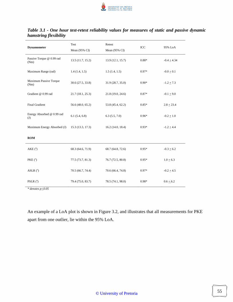
55
Table 3.1 - One hour test-retest reliability values for measures of static and passive dynamic
hamstring flexibility
Dynamometer Test
Mean (95% CI)
Retest
Mean (95% CI) ICC 95% LoA
Passive Torque @ 0.99 rad (Nm)
13.5 (11.7, 15.2) 13.9 (12.1, 15.7) 0.88* -0.4 + 4.34
Maximum Range (rad) 1.4 (1.4, 1.5) 1.5 (1.4, 1.5) 0.97* -0.0 + 0.1
Maximum Passive Torque
(Nm) 30.6 (27.5, 33.8) 31.9 (28.7, 35.0) 0.90* -1.2 + 7.3
Gradient @ 0.99 rad 21.7 (18.1, 25.3) 21.8 (19.0, 24.6) 0.87* -0.1 + 9.0
Final Gradient 56.6 (48.0, 65.2) 53.8 (45.4, 62.2) 0.85* 2.8 + 23.4
Energy Absorbed @ 0.99 rad
(J) 6.1 (5.4, 6.8) 6.3 (5.5, 7.0) 0.96* -0.2 + 1.0
Maximum Energy Absorbed (J) 15.3 (13.3, 17.3) 16.2 (14.0, 18.4) 0.93* -1.2 + 4.4
ROM
AKE (o) 68.3 (64.6, 71.9) 68.7 (64.8, 72.6) 0.95* -0.3 + 6.2
PKE (o) 77.5 (73.7, 81.3) 76.7 (72.5, 80.8) 0.95* 1.0 + 6.3
ASLR (o) 70.5 (66.7, 74.4) 70.6 (66.4, 74.8) 0.97* -0.2 + 4.5
PSLR (o) 79.4 (75.0, 83.7) 78.5 (74.1, 98.0) 0.98* 0.6 + 6.2
* denotes p<0.05
An example of a LoA plot is shown in Figure 3.2, and illustrates that all measurements for PKE
apart from one outlier, lie within the 95% LoA.
©© UUnniivveerrssiittyy ooff PPrreettoorriiaa
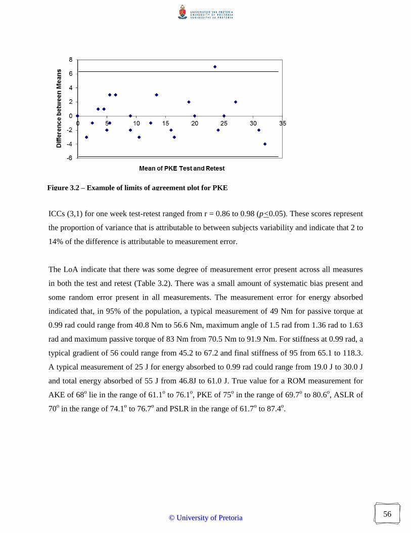
56
ICCs (3,1) for one week test-retest ranged from r = 0.86 to 0.98 (p<0.05). These scores represent
the proportion of variance that is attributable to between subjects variability and indicate that 2 to
14% of the difference is attributable to measurement error.
The LoA indicate that there was some degree of measurement error present across all measures
in both the test and retest (Table 3.2). There was a small amount of systematic bias present and
some random error present in all measurements. The measurement error for energy absorbed
indicated that, in 95% of the population, a typical measurement of 49 Nm for passive torque at
0.99 rad could range from 40.8 Nm to 56.6 Nm, maximum angle of 1.5 rad from 1.36 rad to 1.63
rad and maximum passive torque of 83 Nm from 70.5 Nm to 91.9 Nm. For stiffness at 0.99 rad, a
typical gradient of 56 could range from 45.2 to 67.2 and final stiffness of 95 from 65.1 to 118.3.
A typical measurement of 25 J for energy absorbed to 0.99 rad could range from 19.0 J to 30.0 J
and total energy absorbed of 55 J from 46.8J to 61.0 J. True value for a ROM measurement for
AKE of 68o lie in the range of 61.1
o to 76.1
o, PKE of 75
o in the range of 69.7
o to 80.6
o, ASLR of
70o in the range of 74.1
o to 76.7
o and PSLR in the range of 61.7
o to 87.4
o.
Figure 3.2 – Example of limits of agreement plot for PKE
©© UUnniivveerrssiittyy ooff PPrreettoorriiaa

57
Table 3.2 – Interday test-retest reliability values for measures of static and passive dynamic
hamstring flexibility
Dynamometer Test
Mean (95% CI)
Retest
Mean (95% CI) ICC 95% LoA
Passive Torque @ 0.99 rad (Nm) 48.9 (39.7, 58.1) 49.2 (39.6, 58.8) .97* -0.3 + 7.9
Maximum Range (rad) 1.5 (1.4, 1.6) 1.5 (1.4, 1.7) .97* -0.0 + 0.1
Maximum Passive Torque (Nm) 83.6 (74.3, 92.8) 85.4 (76.5, 94.3) .95* -1.8 + 10.7
Gradient @ 0.99 rad 56.3 (44.0, 68.6) 56.1 (43.8, 68.4) 0.97* 0.2 + 11.0
Final Gradient 95.2 (82.7, 107.7) 98.5 (82.7, 114.3) 0.89* -3.3 + 26.6
Energy Absorbed @ 0.99 rad (J) 24.6 (19.4, 29.8) 25.1 (19.9, 30.4) 0.96* -0.5 + 5.5
Total Energy Absorbed (J) 56.2 (47.4, 64.9) 57.3 (49.2, 65.4) 0.98* -1.1 + 7.1
ROM
AKE (o) 68.9 (63.9, 73.9) 68.3 (63.1, 73.5) .92* 0.6 + 7.5
PKE (o) 75.0 (70.2, 79.8) 74.8 (70.5, 79.1) .95* 0.2 + 5.4
ASLR (o) 70.0 (64.2, 75.8) 74.8 (70.5, 79.2) .87* -4.6 + 9.3
PSLR (o) 76.9 (69.9, 83.8) 80.3 (74.3, 86.3) .86* -3.5 + 10.8
* denotes P<0.05
Substantially different absolute measures of peak torque, total energy absorption and stiffness
were obtained depending on which isokinetic dynamometer was used (Table 3.3). Values of peak
torque were 277% greater on the Contrex compared to the Biodex in this study and 260% greater
on the KinCom in the study of Magnusson et al. (1996) compared to that of Reid and McNair
(2004). Values of total energy absorbed were 379% and final stiffness 171% as great on the
Contrex compared to the Biodex. Final stiffness values reported by Magnusson et al. (1996)
were 1158% greater than those reported by Reid and McNair (2004).
©© UUnniivveerrssiittyy ooff PPrreettoorriiaa
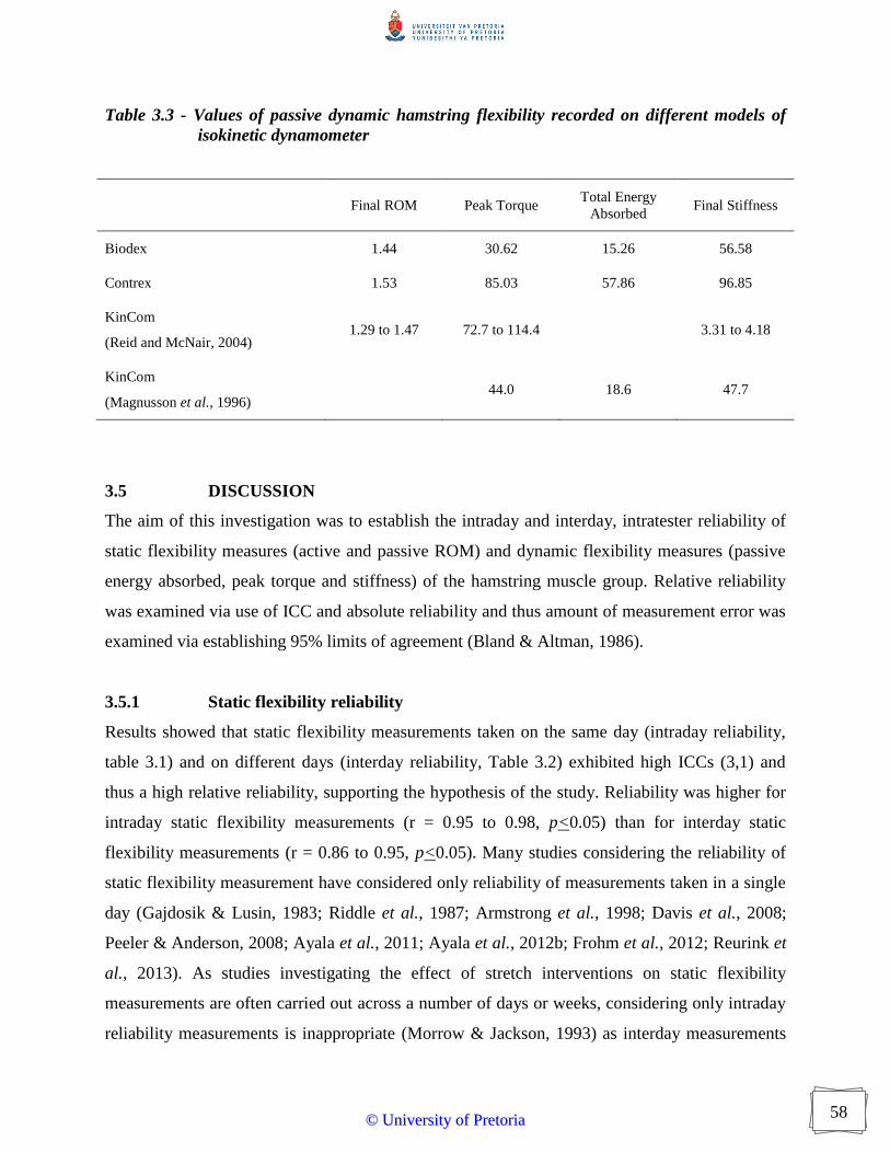
58
Table 3.3 - Values of passive dynamic hamstring flexibility recorded on different models of
isokinetic dynamometer
Final ROM Peak Torque Total Energy
Absorbed Final Stiffness
Biodex 1.44 30.62 15.26 56.58
Contrex 1.53 85.03 57.86 96.85
KinCom
(Reid and McNair, 2004) 1.29 to 1.47 72.7 to 114.4 3.31 to 4.18
KinCom
(Magnusson et al., 1996) 44.0 18.6 47.7
3.5 DISCUSSION
The aim of this investigation was to establish the intraday and interday, intratester reliability of
static flexibility measures (active and passive ROM) and dynamic flexibility measures (passive
energy absorbed, peak torque and stiffness) of the hamstring muscle group. Relative reliability
was examined via use of ICC and absolute reliability and thus amount of measurement error was
examined via establishing 95% limits of agreement (Bland & Altman, 1986).
3.5.1 Static flexibility reliability
Results showed that static flexibility measurements taken on the same day (intraday reliability,
table 3.1) and on different days (interday reliability, Table 3.2) exhibited high ICCs (3,1) and
thus a high relative reliability, supporting the hypothesis of the study. Reliability was higher for
intraday static flexibility measurements (r = 0.95 to 0.98, p<0.05) than for interday static
flexibility measurements (r = 0.86 to 0.95, p<0.05). Many studies considering the reliability of
static flexibility measurement have considered only reliability of measurements taken in a single
day (Gajdosik & Lusin, 1983; Riddle et al., 1987; Armstrong et al., 1998; Davis et al., 2008;
Peeler & Anderson, 2008; Ayala et al., 2011; Ayala et al., 2012b; Frohm et al., 2012; Reurink et
al., 2013). As studies investigating the effect of stretch interventions on static flexibility
measurements are often carried out across a number of days or weeks, considering only intraday
reliability measurements is inappropriate (Morrow & Jackson, 1993) as interday measurements
©© UUnniivveerrssiittyy ooff PPrreettoorriiaa

59
may introduce different biological and procedural variations to those encountered within a day
(Dixon & Keating, 2000). The results of this study are in agreement with previous studies in
finding lower relative reliability of interday compared to intraday studies (DeVita & Bates, 1988;
Hamill & McNiven, 1990) but as the values are greater than 0.8, the measurement can be
deemed to demonstrate high interday relative reliability. Reliability of static flexibility
measurements may be influenced by changes that result from repeated testing trials (Gajdosik &
Bohannon, 1987) and researchers have reported that increases in static flexibility occur
systematically over trials during a one to three day period (Cameron & Bohannon, 1993;
Ghoncheh & Smith, 2004). This suggests that recording the average of several measurements
might increase reliability of static flexibility tests in studies that have wide variations or
systematic increases, but will have little effect in studies with more stable measurements.
Additionally, it is known that an ICC based on the mean of several measurements will be greater
than the ICC of a single measurement (Eliasziw et al., 1994). The use of a single measurement in
this study reduced the possibility of an artificially high relative reliability value and further
confirms the high intra and interday reliability values obtained.
Although ICCs give accurate and easily interpreted measure of relative reliability or rank order
within a sample, they give no information on absolute reliability. LoA analysis allows the
reliability to be expressed in the units of measurement (Bland & Altman, 1986). Results showed
that little systematic bias was indicated in the static flexibility measurements but there was a
fairly large amount of random error. There are a number of possible reasons for this. All
participants in the study would have been familiar with static flexibility measurements as these
form part of many fitness assessments and thus there would be unlikely to be any learning effect
present (Lund et al., 2005). This is possibly the reason why there was little systematic bias
present. The random error could be due to the fact that measurements were taken in a manner
that would mimic a clinical setting, i.e. the pelvis and contralateral limb were not secured to the
table and for the knee extension tests, no apparatus was used to ensure that the knee remained at
90o. The degree of random error in maximum ROM achieved on the dynamometer was less
(0.10o) that that of PKE (5.4 to 6.3
o), possibly because unlike during PKE measurement,
participants were fully secured into the chair of the dynamometer which would have limited
unwanted movement of other body parts. The use of highly stabilised movements, whilst
seemingly reducing random error, has little application to applied practice and research since
©© UUnniivveerrssiittyy ooff PPrreettoorriiaa

60
clinically measures are not taken in this manner and thus the reliability findings cannot be
applied to these settings. Additionally, it is possible that different amounts of pressure were
applied to movements and that feedback from participants on what they felt was the end point
varied (Gajdosik, 2001b). According to the inductive approach, this was unlikely to be a
significant factor as the experimenter was experienced in taking measurements of passive static
flexibility and consciously attempted to apply consistent force to the limb (Ferlie et al., 1999;
Greenfield et al., 2007).
Magnitude of error is important when conducting longitudinal studies to determine the effects of
various interventions such as stretching on static or dynamic flexibility levels of the hamstring
muscle group (Jones et al., 2002). As the amount of error is fairly large for the static flexibility
measurements, it is possible that such tests, even though displaying high ICCs, are actually not
sensitive enough to detect small but potentially clinically relevant changes in these variables. For
example, as a result of a stretching programme, a participant might be expected to make gains of
8o in PKE (Harvey et al., 2002). According to the limits of agreement, the true value for the
measurement will lie in a range of approximately 13o
and these gains of 8o may therefore not be
detected. As static flexibility tests are commonly used, this is a potential reason for conflict in the
literature examining effects of stretching for clinical and performance reasons. Re-examination
of results with consideration given to absolute error of measurements is warranted to confirm or
deny previous findings and try to reach some consensus as to the effect of stretching
interventions on measures of static flexibility.
3.5.2 Dynamic flexibility reliability
Early studies investigating reliability of isokinetic dynamometers for measuring passive dynamic
hamstring flexibility inappropriately calculated PPM (Magnusson et al., 1995) and therefore
although isokinetic dynamometers have become accepted methods of measuring passive
dynamic flexibility, their reliability had not been correctly established. Shimon et al. (2010)
reported intraday, interrater ICC of r = 0.89 but did not describe which ICC they had used
(Shimon et al., 2010). Results of this study demonstrated intraday ICCs (3,1) of r = 0.85 to 0.98
(p<0.05) and interday ICCs (3,1) of r = 0.89 to 0.98 (p<0.05). From these, it can be deemed that
both dynamometers have good relative reliability in measuring the dynamic flexibility of the
©© UUnniivveerrssiittyy ooff PPrreettoorriiaa

61
hamstring muscle group during a controlled stretch manoeuvre thus supporting the hypothesis of
this study.
As an index of absolute reliability LoA analysis showed that there was some systematic bias in
the measurements taken on the dynamometer, with a small increase on the test compared to
retest, and a larger amount of random error present. Most of the participants were familiar with
the dynamometer but none had previously gone through passive movements to their maximum
ROM. Participants completed five passive movements before testing commenced, partly as a
warm-up to reduce thixotropic associated muscle stiffness (Proske et al., 1993) and partly as a
familiarisation session. It is possible that more familiarisation should be included prior to testing
but this was not possible due to the time constraints of professional sport. A warm-up involving
whole body physical activity was not undertaken to reduce any effects of altered muscle
temperature, as this has been shown to affect muscle stiffness (Noonan et al., 1993). Previous
studies have shown that acute stretch induced increases in static (Duong et al., 2001; Ryan et al.,
2008) and dynamic flexibility (Magnusson et al., 1995; Matsuo et al., 2013; Mizuno et al., 2013)
reset in under an hour. Based on these findings, the period between test and retest in intraday
measures would have allowed any changes in flexibility as a result of the measurement
procedures to have returned to baseline values. Although the passive manoeuvres on the
dynamometers were well stabilised, some random error could be due to movement or muscle
contraction during the test procedures, slight variations in participant positioning between tests
or insufficient sensitivity of test instrumentation.
3.5.3 Dynamometer comparison
Results showed substantial differences between the absolute values obtained on the Biodex
isokinetic dynamometer compared to those from the Contrex isokinetic dynamometer in this
study (Table 3.3). Comparison of results from this study with those obtained in previous studies
using KinCom isokinetic dynamometers also showed differences in absolute values achieved,
both between different dynamometers and between different protocols on the same
dynamometer. It has been suggested that there is a lack of comparability between separate makes
of dynamometers for measures of peak torque (Greenberger et al., 1994; Leggin et al., 1996) and
this difference is further increased if different subject positioning and protocols are used
(Croisier et al., 2002). Prior to this study, there has been no available data comparing values of
©© UUnniivveerrssiittyy ooff PPrreettoorriiaa
62
passive torque, stiffness and energy absorbed obtained from the same individuals on different
makes of dynamometer. It is possible that companies manufacture dynamometers according to
their own specifications, thus giving inconsistent results for the same measurement if different
models of dynamometer are used. One of the major factors affecting isokinetic measurements is
the moment of the gravitational forces of the involved segments and the dynamometer input arm
(Kellis & Baltzopoulos, 1996). Mechanical work has been shown to vary by 26 to 43% for knee
extensors and 55 to 510% for knee flexors in the absence of gravity correction (Winter et al.,
1981) and differences in participant positioning can affect gravitational moments. Some studies
have estimated gravitational moments in a seated position (Feiring et al., 1990; Griffin et al.,
1993; Tis et al., 1993) whereas others have used a supine position (Worrell et al., 1990;
Fitzgerald et al., 1991). Although some studies have found that measurements taken in a prone
and supine position were not significantly different (Nelson & Duncan, 1983), others reported
that gravitational moments were greater in a seated compared to supine position (Ford et al.,
1994). Furthermore, different gravity correction procedures are followed when using different
dynamometers with some manufacturers recommending protocols that measure gravitational
moments in a static position whereas others recommend a dynamic motion (Kellis &
Baltzopoulos, 1996). Results from this and previous studies suggest that absolute data values,
particularly those that are measured passively, should only be compared if collected from the
same type of dynamometer. Further investigation into the role of gravity correction on different
machines should be conducted using strain gauges or calculations based on anthropometric data.
A standard protocol for measuring gravity correction should also be established to examine the
extent that subject positioning and protocol affect the absolute measurements obtained from the
dynamometers. Because of the differences in the absolute values obtained, it was decided to use
only one dynamometer for further study. As the Contrex Isokinetic Dynamometer was more
readily available, further studies were conducted on this alone.
3.5.4 Practical Implications
Flexibility assessment methods with minimal or acceptable measurement error are essential to
monitor progression and effectiveness of injury prevention and recovery strategies (Atkinson &
Nevill, 1998). Existing published research examining reliability of flexibility measurements has
typically utilised highly stabilised static flexibility measures that are not consistent with practise,
participants primarily from convenience samples of University students or active individuals, and
©© UUnniivveerrssiittyy ooff PPrreettoorriiaa
63
have focused on intraday reliability and thus do not take into account any variability that might
occur as a result of time. Additionally, PPM correlations have been reported which is
inappropriate for reliability studies because PPM is a bivariate statistic that measures strength of
linear association, and reliability studies involve univariate measures (Maher, 1993). Chapter 3
utilised a homogenous sample from a specific athletic population in an endeavour to control for
Type II errors and provide meaningful results that could be applied to a professional sport setting
(Nevill & Atkinson, 1997). Significant interday static flexibility ICCs (3,1) suggested good
relative reliability (r = 0.86 to 0.98, P<0.05) but LoA detected fairly large amounts of random
error (5.4 to 10.8o). Few studies investigating the reliability of static flexibility measures have
considered magnitude of error and those that have reported measurement errors of up to 36o
despite correlations of 0.65 to 0.72 (Gabbe et al., 2004; Peeler & Anderson, 2008; Peeler &
Anderson, 2008). Stretching has been reported to increase static flexibility measures by a mean
of 8o
(Harvey et al., 2002) thus static flexibility tests might not be sensitive enough to detect
these potentially clinically relevant changes as a result of stretch interventions. The measurement
error associated with static flexibility tests may partly account for inconsistencies in literature
related to flexibility and injury risk.
3.5.5 Limitations
The intraday reliability part of the study utilised a one-hour period between test and retest.
Previous studies have shown that any changes in static flexibility or mechanical properties of a
muscle following a stretch reset to baseline in under an hour (Magnusson et al., 1996; Ryan et
al., 2008), although other studies have indicated that the effects of stretching last much longer
than this (de Weijer et al., 2003). It is possible that a period of one hour between tests in
insufficient for the hamstring to have returned to its baseline state. Studies have shown that time
of day can affect flexibility, strength and postural control (Reilly et al., 1984; Gifford, 1987;
Coldwells et al., 1994; Atkinson & Reilly, 1995; Michael et al., 2003). If the period between test
and retest had been too great, circadian variations in flexibility could potentially affect the results
but further studies are needed to investigate this.
The limits of agreement presented here can only be applied to males playing rugby league at a
semi-professional or professional level following the same procedures as the current study.
Further studies using different populations of participants are warranted to establish reliability of
©© UUnniivveerrssiittyy ooff PPrreettoorriiaa
64
measurement procedure and allow further investigation of biomechanical factors that might
influence static and dynamic flexibility.
3.5.6 Reflections
Previous studies using similar methodology have used small samples (n = 8 to 15) of school
children, university students or recreational athletes to calculate reliability (McHugh et al., 1992;
Magnusson et al., 1995; Magnusson et al., 1996; Magnusson et al., 1996; Shimon et al., 2010).
Convenience samples such as these can exhibit heteroscedascity as well as potentially large
variations in fitness levels, which might impact on results of planned interventions, therefore this
study utilised professional rugby league players. This provided some challenges in design and
implementation of protocols. Testing sessions were severely constrained by time and had to be
designed around participant availability. Additionally, there was no control over the activities
that were performed on the day prior to testing. Through liaison with the coach and club
physiotherapist, lists of players that were available to be tested on specific days were provided.
To ensure that maximum numbers possible were tested, it was necessary to adhere to a strict
testing timetable. Many pre-testing sessions on non-study participants were conducted to ensure
that procedures were fine-tuned and that the testing routine was well practiced and would run
smoothly. Later interventions also had to be designed around club requirements and player
availability. Although the use of such participants placed restrictions on the testing protocol and
procedures, it was felt that the opportunity of gaining access to such homogenous participants at
an elite level of sport outweighed the disadvantages of this.
Due to lack of clarity in methodology described by Magnusson et al. (1995), extensive work was
required to replicate the protocol. As isokinetic dynamometers are not generally used for passive
movements to maximum ROM, instructions were not available in operational manuals and had to
be determined from available functions. Furthermore, a number of wedges had to be designed
and constructed to provide sufficient thigh elevation to prevent full knee extension while
remaining comfortable for participants. This required a number of trials on non-athletic
volunteers and adaptation of first the wedge and then the straps used to fix the thigh and other
limb to the dynamometer chair. Challenges were also presented in determining the most
appropriate way of analysing the data from the isokinetic dynamometer. It was felt that using the
data directly as provided (split into thirds) as per previous methodology lacked theoretical
©© UUnniivveerrssiittyy ooff PPrreettoorriiaa
65
rationale and was thus not appropriate. Instead it was decided to use mathematical software to
facilitate the differentiation and integration of curves, therefore allowing measurements at or to
any points to be calculated. Learning to use the software proved challenging but was worthwhile
due to the information provided, although on reflection, rather than differentiating the curve at
specific points for measures of stiffness, it might have been more appropriate to consider a 1-2o
portion of the curve to minimise potential inconsistencies due to momentary deflections of the
curve as possibly helping to reduce the random error of stiffness measurements.
3.6 CONCLUSIONS
ICCs demonstrated high relative reliability for measures of static and dynamic hamstring
flexibility, thus accepting the hypothesis. There was a degree of absolute measurement error, the
magnitude of which must be interpreted in relation to analytical goals and the expected extent of
change. There was a small amount of systematic bias present in static and dynamic flexibility
measures and a larger amount of random error. The degree of random error in flexibility
measurements means that care must be taken before categorising any interventions as effective
or ineffective as the tests may not be sufficiently sensitive and thus type II errors might occur.
Dynamic flexibility measures were specific to the model of isokinetic dynamometer and this
should be taken into account when considering absolute reliability values.
©© UUnniivveerrssiittyy ooff PPrreettoorriiaa
66
CHAPTER 4
COMPARISON BETWEEN DIFFERENT
METHODS
OF MEASURING STATIC AND
DYNAMIC HAMSTRING FLEXIBILITY
©© UUnniivveerrssiittyy ooff PPrreettoorriiaa
67
4.1 INTRODUCTION
Flexibility measurements are commonly used by clinicians to evaluate the ability of the
musculotendinous unit to lengthen, with the assumption that a more flexible muscle can be
stretched further and to a higher ultimate strain and is therefore less susceptible to injury than a
stiffer muscle (Blackburn et al., 2004; Pruyn et al., 2012). This conjecture lacks substantive
supporting evidence and does not take into account that flexibility is constrained by a number of
factors including neural, mechanical, joint and other constraints, e.g. skin, temperature, etc.
(Bozic et al., 2010; Ditroilo et al., 2013). The passive stiffness of muscle has classically been
regarded as being determined by connective tissue elements surrounding individual muscle
fibres, bundles of fibres and whole muscles (Ramsey & Street, 1940). Later attempts to measure
the contribution of connective tissue to stiffness have suggested that it contributes only at long
sarcomere lengths (Casella, 1950; Rapoport, 1972; Tidball, 1986) and that structures within
muscle fibres contribute to resting stiffness (Magid & Law, 1985). These include portions of the
cytoskeletal network, especially connecting filaments composed of protein, titin (Horowits et al.,
1989) and low level cross bridge interactions (Hill, 1968). These findings have been elicited
using animal models and have often not included components of non-muscular origin.
Johns and Wright (1962) showed that in the cat wrist, the joint capsule actually contributed more
to stiffness (47%) than the muscles (41%), tendons (10%) and skin (2%). In vitro experimental
models have attempted to distinguish the mechanical consequences of reflex activity from those
due to the intrinsic properties of the joint and muscle by comparing the behaviour of a joint
under normal conditions with that observed after deafferenation, local contraction by artificial
stimulation, immobilisation in a shortened position or a combination of these methods
(Goldspink, 1976; Goldspink, 1977; Tardieu et al., 1982; Jarvinen et al., 1992; Herbert &
Balnave, 1993; Herbert & Crosbie, 1997; Yang et al., 1997; Joumaa et al., 2008). These results
have indicated that flexibility is primarily a result of myogenic rather than neurogenic
mechanisms but it cannot be assumed that conditions would be the same for in vivo models.
Flexibility is likely to be restricted to a greater extent at joints that have bi or multi-articular
muscles as nervous structures might be stressed at the limits of the range (McHugh et al., 2012).
In vivo studies have suggested that among other things, flexibility is dependent on the
viscoelasticity of the musculotendinous unit, ligaments and other connective tissues (Magnusson
et al., 2001; Sobolewski et al., 2013). This is somewhat simplistic and previous research has
©© UUnniivveerrssiittyy ooff PPrreettoorriiaa
68
often has not taken into account that flexibility has both static and dynamic components (Bojsen-
Moller et al., 2007; Ditroilo et al., 2011). Researchers and clinicians have measured static
flexibility and used results to make assumptions about movement or injury kinematics that would
be more related to dynamic flexibility (Bennell et al., 1999; Tyler et al., 2001; Witvrouw et al.,
2003; Rahnama et al., 2005; Bradley & Portas, 2007; Hrysomallis, 2009) which could be one of
the reasons for the disparity among researchers as to the effect of different levels of flexibility on
injury risk and performance levels (Thacker et al., 2004; Shrier, 2005; Foreman et al., 2006;
Small et al., 2008; Kay & Blazevich, 2012; Simic et al., 2013).
There is a lack of universally agreed definition as to what constitutes hamstring flexibility and
therefore no agreed measurement procedure (Weppler & Magnusson, 2010). Different methods
of measuring hamstring flexibility have been shown to yield different results. Early research
tended to consider only static hamstring flexibility measurements such as SLR and KE tests.
Researchers considering the association between different methods of assessing active and
passive static hamstring flexibility have shown relationships between tests of AKE and PKE (r =
0.64 to 0.87, p<0.01, common variance = 41% to 76%) but relationship between ASLR and
PSLR has not been specifically examined (Cameron & Bohannon, 1993; Gajdosik et al., 1993;
Rolls & George, 2004; Davis et al., 2008; Shimon et al., 2010; Ayala et al., 2011; Ayala et al.,
2012a). The unexplained variance of 24 to 59% between AKE and PKE tests suggests that there
are different factors constraining these tests. In order to allow the hamstring to lengthen there is
reciprocal inhibition from contraction of the quadriceps in the AKE as well as a force moment
around the knee joint. It is known that muscles with greater moment arms undergo greater
passive strain during joint motion (Magnusson et al., 2000) and that for a given static situation
the external moment about the knee joint is counterbalanced by the internal moment. As the
results from PKE are generally greater than AKE tests, it is likely that the force applied
externally by investigators is greater than that the body can generate through quadriceps
contraction alone.
Studies investigating the relationship between different passive static hamstring flexibility tests
have shown that PKE and PSLR (r = 0.16 to 0.66, p<0.01, common variance = 3% to 44%) share
significant positive relationships (Cameron & Bohannon, 1993; Gajdosik et al., 1993; Rolls &
George, 2004; Davis et al., 2008). Despite being significant, weak correlations in some instances
©© UUnniivveerrssiittyy ooff PPrreettoorriiaa
69
suggests that there are quite different factors influencing the result of each test. Similar to results
for passive static flexibility tests, results for active static flexibility tests indicate that AKE and
ASLR (r = -0.72, p<0.01, common variance = 51%) are significantly positively related but
nonetheless share little more than half their variance (Cameron & Bohannon, 1993; Gajdosik et
al., 1993; Rolls & George, 2004; Davis et al., 2008). Consequently researchers have questioned
the ability of SLR tests to assess static hamstring flexibility independently of other variables
(McHugh et al., 2012). It is possible that factors such as neural tension, movement of the pelvis
and varying degrees of stabilization affect measures of static flexibility although little attempt
has been made to quantify the extent of these potential influences (Davis et al., 2008).
Some attempts have been made to study dynamic flexibility in both humans (Bach et al., 1983;
Wilson et al., 1991; Wilson et al., 1994; Halbertsma et al., 1996; Walshe et al., 1996;
Magnusson, 1998; McHugh et al., 1998; Hunter & Spriggs, 2000; Fukashiro et al., 2001;
Blackburn et al., 2004; Reid & McNair, 2004; Owen et al., 2005; Ryan et al., 2008; Watsford et
al., 2010; Ditroilo et al., 2011; Palmer et al., 2013; Sobolewski et al., 2013) and animals (Garrett
et al., 1987; Nikolaou et al., 1987; Garrett et al., 1988; Taylor et al., 1993; Noonan et al., 1994;
Best et al., 1995; Mair et al., 1996; Taylor et al., 1997; Yang et al., 1997; Black & Stevens,
2001). Human models have used either damped oscillation techniques (Bach et al., 1983; Wilson
et al., 1991; Wilson et al., 1994; Walshe et al., 1996; Hunter & Spriggs, 2000; Fukashiro et al.,
2001; Blackburn et al., 2004; Owen et al., 2005; Watsford et al., 2010; Ditroilo et al., 2011) or
assessed load-deformation characteristics of muscle during passive movements (McNair &
Stanley, 1996; Klinge et al., 1997; Magnusson, 1998; Halbertsma et al., 1999; Hunter & Spriggs,
2000; Magnusson et al., 2000; Halbertsma et al., 2001; McNair et al., 2001; Gajdosik, 2002;
Reid & McNair, 2004; Ryan et al., 2008; Palmer et al., 2013; Sobolewski et al., 2013) while
animal models have assessed stress-strain relationships of individual muscle fibres or muscle
groups (Garrett et al., 1987; Nikolaou et al., 1987; Garrett et al., 1988; Taylor et al., 1990;
Noonan et al., 1994; Best et al., 1995; Mair et al., 1996; Taylor et al., 1997; Yang et al., 1997;
Black & Stevens, 2001).
Studies investigating the viscoelastic characteristics of the hamstring muscle group in stiff
(defined by not being able to extend hands past level of stool on toe touch test) compared to
normal participants suggested that at a given angle, stiffer muscle absorbs a greater amount of
©© UUnniivveerrssiittyy ooff PPrreettoorriiaa
70
energy than does more flexible muscle (Magnusson et al., 1997; Hobara et al., 2010). Animal
models have been used to examine factors that might contribute to muscle strain injury and have
repeatedly shown that the energy absorbing capability of muscle is an important variable
(Noonan et al., 1994; Sun et al., 1994; Garrett, 1996; Macpherson et al., 1996; Lieber & Friden,
2002). These findings have been based on failure characteristics of stimulated and non-
stimulated muscle and reported that muscle activation alone failed to result in strain injury, and
that to obtain an injury, stretch was necessary. Furthermore, although total strain at failure was
similar, force generated at failure was only 15% greater in the active muscles, but energy
absorption was increased by 100% compared to the non-active muscle (Garrett, 1996). Basic
science therefore suggests that in contrast to popular belief, it is possible that it is actually stiffer,
rather than more flexible individuals who are at reduced risk of injury due to the higher energy
absorbing capability of their muscles.
Few attempts have been made to compare values obtained from static hamstring flexibility tests
to dynamic hamstring flexibility tests and available results are equivocal. In a series of studies,
Magnusson et al. found that individuals with lower static flexibility exhibited lower passive peak
torque and energy absorption than more flexible individuals, but that more flexible individuals
demonstrated a higher stretch tolerance (Magnusson et al., 1995; Magnusson et al., 1996;
Magnusson et al., 1996; Magnusson et al., 1996; Magnusson et al., 1997; Magnusson et al.,
1998; Magnusson et al., 2001). McHugh et al. (1998) found a significant relationship between
energy absorbed by the hamstring, measured during an instrumented PSLR tests, and ROM on
PSLR test as measured clinically using a goniometer (r = 0.49, p<0.05; common variance =
25%). Common variance climbed to 79% when the increase in torque from 20 to 50o
was also
added into a stepwise multiple regression (r = 0.89, p<0.05). Researchers suggested that static
flexibility is predominantly limited by passive mechanical constraints and it was hypothesised
that the remaining variance may have been due to an active contractile component at terminal
ROM (McHugh et al., 1998). This study did not take into account the possibility that the
predictor variables were related to each other and thus the presence of multicollinearity (Lago et
al., 2010). Furthermore, McHugh et al. (1998) reported no significant relationship between
passive peak torque and ROM that participants were able to achieve (r = 0.09, p>0.05) which
conflicts with Magnusson et al. (1997) who found a difference in passive peak torque in
individuals exhibiting higher and lower levels of static flexibility.
©© UUnniivveerrssiittyy ooff PPrreettoorriiaa
71
Normally a high and statistically significant correlation coefficient is interpreted as a strong
relationship between two variables (Hopkins et al., 2009) but these coefficients can depend
greatly on the range of values present in the sample (Janssen & Le-Ngoc, 2009). Most studies
considering relationships between different measures of flexibility have used convenience
samples, predominantly University students or active adults. Such participant groups can exhibit
potentially a large range of measurements, which can result in artificially high relationships
being found (Weir, 2005; Hopkins et al., 2009). It remains to be established whether a
homogenous sample of participants, such as athletes competing at professional level in the same
sport, will share the same relationships between measures of flexibility as those found in more
general samples (Hopkins, 2000). Additionally, because of the high incidence of hamstring
injuries in sport, club physiotherapists routinely conduct testing of static hamstring flexibility
(Pruyn et al., 2012) and the applicability of these results to measures of dynamic flexibility
should be considered. Because of the dearth of evidence empirically examining the validity of
static flexibility tests for assessing dynamic properties of muscle, further research in this area is
warranted to determine the extent of the relationship between the end point of goniometric
measurements and the mechanical properties of the musculotendinous unit. Additionally, due to
conflicting findings, the relationship between measures of dynamic and static hamstring
flexibility needs further investigation.
4.1.1 Aims and hypotheses
The aim of this study was to evaluate the relationship shared by commonly used tests of static
hamstring flexibility with measures of dynamic hamstring flexibility in professional rugby
league players participating in the Great Britain Superleague.
Hypotheses were as follows;
1) There would be no significant positive relationship between KE and SLR tests;
2) AKE and PKE tests would share a significant positive relationship, as would ASLR and
PSLR tests;
3) There would be a significant positive relationship between KE tests and energy absorbed
by the hamstring during controlled KE on an isokinetic dynamometer;
©© UUnniivveerrssiittyy ooff PPrreettoorriiaa
72
4) Participants who were tight (< 80o on PSLR test) would have significantly greater
stiffness than normal participants (> 80o on PSLR test); and
5) There would be no significant positive relationship between KE tests and level of passive
peak torque and final stiffness.
4.2 METHODS
4.2.1 Participants
Twenty-five rugby league players volunteered to participate in this study (age 17.72 years ±
1.49, stature 1.78m ± 0.06, mass 88.58kg ± 9.84 [mean ± SD]). These were chosen from the
sample that participated in the study reported in Chapter 3. The whole sample was not available
due to National squad duties. SPSS sample power software indicated that this would give a
sufficient power ( = 0.05 and = 0.14) by one-tailed correlation comparing values in the
literature (r = 0.49, p<0.05) (Blackburn et al., 2004) against a theoretical value (constant) of
0.00. All participants were part of a Great Britain Super League rugby league club and testing
was conducted mid-season when participants were involved in training sessions four to five
times a week and matches once or twice a week. For inclusion in the study, participants were
certified free of lower limb injury by the club physiotherapist and considered to be in good
physical condition. Because participants were all participating in the same training programme
and were tested within the season, they were considered to be of similar fitness levels close to
their peak. Participants were excluded if they had previously sustained any form of hamstring
injury or were not currently participating fully in all training sessions scheduled by the club.
Written informed consent was obtained prior to the study from the participants themselves if
over the age of eighteen years or via the club following letters being sent to parents or guardians
if participants were under eighteen years, along with verbal assent from the participants
themselves. In such situations club personnel acted in loco parentis during testing sessions.
Ethical approval was obtained from the University of Pretoria’s Post-graduate Committee and
the Ethical Committee.
4.2.2 Procedures
Measurements were taken in a single testing session. Participants were asked to refrain from any
strenuous activity for at least three hours prior to testing to ensure that effects from fatigue,
different tissue temperatures and thixotropic state of the muscle were minimal (Cornelius et al.,
©© UUnniivveerrssiittyy ooff PPrreettoorriiaa
73
1992; Noonan et al., 1993; Proske & Morgan, 1999; Noakes, 2000). The study followed
assessment procedures described in Chapter 3.
4.2.3 Statistical analysis
Data obtained from the Isokinetic Dynamometer were exported into Microsoft Excel 2000. 3rd
Order Polynomial Curves were fitted and using Derive (version 7), equations of the fitted curves
(y = ax3 ± bx
2 ± c ± k) were differentiated to find the final stiffness (gradient) and integrated to
find the energy absorbed (area under the curve) during the passive knee extension manoeuvre.
To allow comparison with less stabilised static hamstring flexibility measurements, the
maximum angle each participant achieved during passive knee extension on the dynamometer
(MaxROM) was recorded.
Statistical analysis was performed using SPSS (Statistics Package for the Social Sciences,
Version 16.0). The normality of the data sets was determined by one-sample Kolmogorov-
Smirnov (K-S) test. Because data were not significantly different from normal distributions
curve, parametric tests were used for analysis. Relationships between different hamstring
flexibility measures were analysed by calculating PPM correlations and from the results,
coefficients of determination (R2) and common variances were calculated. Independent t-tests
were performed to test for differences in dynamic flexibility measurements of participants who
were tight (< 80o) and normal (> 80
o) based on the results of the PSLR test (Göeken & Hof,
1993; Halbertsma et al., 1999). Statistical significance was set at p<0.05.
4.3 RESULTS
4.3.1 Relationship between static flexibility measurements
MaxROM on the dynamometer was significantly positively related to all static flexibility tests (r
= 0.52 to 0.67, p<0.05, common variance = 27 to 45%). Significant positive relationships were
identified between AKE and ASLR (r = 0.91, p<0.05, common variance = 83%) and PKE and
PSLR (r = 0.86, p<0.05 common variance = 74%). The AKE and PKE (r = 0.83, p<0.05,
common variance = 69%) and ASLR and PSLR (r=0.79, p<0.05, common variance = 62%) also
showed strong significant relationships. Scatter plots illustrated the strong positive relationships,
©© UUnniivveerrssiittyy ooff PPrreettoorriiaa

74
for instance Figure 4.1 shows data falling close to the line of best fit across the range of
measures.
4.3.2 Relationship between static and dynamic flexibility measurements
Results were variable between measurements of dynamic flexibility and static flexibility. Torque
at 0.99 rad showed a moderate negative relationship with all static flexibility measurements (r = -
0.35 to -0.48, p<0.05, common variance = 12 to 23%). Energy absorbed to 0.99 rad showed a
significant moderate negative relationship with all static flexibility measurements (r = -0.39 to -
0.53, p<0.05, common variance = 15 to 28%) while total energy absorbed showed a moderate
positive relationship with PKE (r = 0.48, p<0.05, common variance = 23%) and PSLR (r = 0.43,
p<0.05, common variance = 18%). Scatter plots illustrated the moderate relationships, for
example Figure 4.2 shows that data lies in the region of the line of best fit but there is a degree of
spread.
Figure 4.1 – Scatter plot of AKE against PKE (n = 25).
R2 = 0.69
©© UUnniivveerrssiittyy ooff PPrreettoorriiaa

75
Table 4.1 - Relationship between static and dynamic flexibility measures
Passive Torque
at 0.99 rad Max Rad
Passive peak
Torque
Stiffness at
0.99 rad
Final
Stiffness
Energy to
0.99 rad Total Energy
AKE -.46* .52* .45 -.56* .08 -.49* .21
PKE -.48* .58* .08 -.55* .04 -.53* .48*
ASLR -.35* .62* .29 -.53* .28 -.39* .26
PSLR -.43* .67* .26 -.61* .26 -.47* .43*
* denotes p<0.05
Figure 4.2 – Scatter plot of total passive energy absorbed against PKE (n = 25).
Independent t-tests (Table 4.2) showed that there was no significant difference in the dynamic
flexibility results of stiffness or energy absorption at either a common measurement point or final
measurement point for participants classified as ‘tight’ or ‘normal’, based on whether they scored
below or above 80o on the PSLR test (p>0.05).
R2 = 0.23
©© UUnniivveerrssiittyy ooff PPrreettoorriiaa
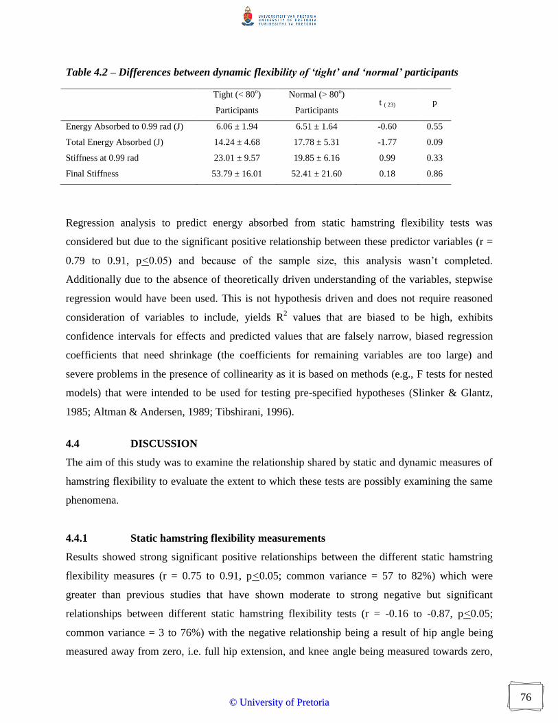
76
Table 4.2 – Differences between dynamic flexibility of ‘tight’ and ‘normal’ participants
Tight (< 80
o)
Participants
Normal (> 80o)
Participants t ( 23) p
Energy Absorbed to 0.99 rad (J) 6.06 ± 1.94 6.51 ± 1.64 -0.60 0.55
Total Energy Absorbed (J) 14.24 ± 4.68 17.78 ± 5.31 -1.77 0.09
Stiffness at 0.99 rad 23.01 ± 9.57 19.85 ± 6.16 0.99 0.33
Final Stiffness 53.79 ± 16.01 52.41 ± 21.60 0.18 0.86
Regression analysis to predict energy absorbed from static hamstring flexibility tests was
considered but due to the significant positive relationship between these predictor variables (r =
0.79 to 0.91, p<0.05) and because of the sample size, this analysis wasn’t completed.
Additionally due to the absence of theoretically driven understanding of the variables, stepwise
regression would have been used. This is not hypothesis driven and does not require reasoned
consideration of variables to include, yields R2 values that are biased to be high, exhibits
confidence intervals for effects and predicted values that are falsely narrow, biased regression
coefficients that need shrinkage (the coefficients for remaining variables are too large) and
severe problems in the presence of collinearity as it is based on methods (e.g., F tests for nested
models) that were intended to be used for testing pre-specified hypotheses (Slinker & Glantz,
1985; Altman & Andersen, 1989; Tibshirani, 1996).
4.4 DISCUSSION
The aim of this study was to examine the relationship shared by static and dynamic measures of
hamstring flexibility to evaluate the extent to which these tests are possibly examining the same
phenomena.
4.4.1 Static hamstring flexibility measurements
Results showed strong significant positive relationships between the different static hamstring
flexibility measures (r = 0.75 to 0.91, p<0.05; common variance = 57 to 82%) which were
greater than previous studies that have shown moderate to strong negative but significant
relationships between different static hamstring flexibility tests (r = -0.16 to -0.87, p<0.05;
common variance = 3 to 76%) with the negative relationship being a result of hip angle being
measured away from zero, i.e. full hip extension, and knee angle being measured towards zero,
©© UUnniivveerrssiittyy ooff PPrreettoorriiaa
77
i.e. full knee extension (Cameron & Bohannon, 1993; Gajdosik et al., 1993; Rolls & George,
2004; Davis et al., 2008). Participants in this study were screened for previous injury to avoid
any potential effects of scar tissue from previous injury influencing level of static flexibility and
impacting upon results (Toumi et al., 2006) and were all familiar with the tests as they form part
of screening and fitness testing programmes. Additionally, as participants were professional
sportsmen, it is likely that they would have greater muscle strength than non-athletic populations
and thus would be able to exert a greater force on the active tests, possibly resulting in the higher
relationships seen between active and passive tests in this study compared to previous studies.
The two active and two passive static hamstring flexibility tests shared significant strong
relationships with each other (AKE and ASLR r = 0.91, p<0.05, common variance = 83%; PKE
and PSLR r = 0.86, p<0.05 common variance = 74%) thus rejecting hypothesis 1. Despite the
strong relationships, the unexplained variance between the tests suggests that it is necessary to
standardise tests of hamstring flexibility and the use of different tests can in part explain the
disparity among researchers as to the implications of different levels of flexibility and stretching
regimes (Bojsen-Moller et al., 2007; Palmer et al., 2013). The unexplained variance could be due
to a number of factors including methodological issues and neural input. Both the ASLR and
PSLR tests might be influenced by neural factors and the PSLR in particular, is a commonly
used lower limb neural tension test (McHugh et al., 2012). If this, rather than other neural or
mechanical factors limited such tests, participants would be likely to score better on the knee
extension tests. Care should be taken over subject positioning for these tests to attempt to limit
effects from this (Shacklock, 2005). Congruency at the hip joint might affect the score on the
SLR tests compared to KE tests in some populations (Holm et al., 2000) but due to the utilisation
of relatively young participants with no previous history of injury, would have been unlikely to
have had significant effects in this study (Zakas et al., 2006). The length of the moment arm
differing between knee extension and straight leg raise tests might also have had an impact,
particularly if one group of muscles was disproportionate in strength compared to another, i.e.
participants with weaker hip flexor muscles might perform better on knee extension tests. This
concept has been promoted in previous literature (Devlin, 2000; Ylinen et al., 2010) but because
this study did not measure baseline muscle strength, muscle balance and quadriceps:hamstring
strength ratio it is not possibly to directly support these findings.
©© UUnniivveerrssiittyy ooff PPrreettoorriiaa

78
ASLR and PSLR shared a significant strong positive relationship (r = 0.79, p<0.05, common
variance = 62%), as did AKE and PKE (r = 0.83, p<0.05, common variance = 69%), thus
accepting hypothesis 2. Despite strong relationships, there was 31 to 38% unexplained variance
between active and passive SLR and KE tests. Methodological issues such as inability to apply
consistent force to the limb or different interpretation of the end point of passive tests has been
shown to affect results (Ylinen et al., 2010). Identification of this can be difficult because it
relies on psychophysiological phenomena, i.e. resistance of the muscle and a persons stretch
tolerance (Marshall et al., 2011; Sobolewski et al., 2013). It is possible that active static
hamstring flexibility tests might be influenced to a greater degree by the presence of a stretch
reflex than passive tests (Stein & Kearney, 1995). Although participants were clearly instructed
to move their limbs in a slow and controlled manner, due to the competitive nature of
professional athletes, it is possible that some were trying to use momentum to achieve a better
result thus triggering a stretch reflex. Conversely, had the tests been performed slowly, it is
possible that activation of the agonist might have caused a degree of autogenic inhibition to the
antagonist (Chalmers, 2004; Sharman et al., 2006). This causes muscular relaxation and
therefore could result in increased levels of static flexibility. EMG data from pilot and previously
published studies suggest that the altered neural activity does not significantly affect static
flexibility tests (Magnusson et al., 1997; Wallmann et al., 2008; Hough et al., 2009) but such
data would have been useful in eliminating this as a possible source of variance in this study.
The static flexibility measurements were taken in a manner that would mimic a clinical setting,
i.e. the pelvis and contralateral limb were not secured to the table and for the knee extension
tests, no apparatus was used to ensure that the knee remained at 90o. Previous studies have often
used devices such as metal rigs to assist in measurement and straps to limit pelvic and leg motion
(Rothstein et al., 1983; McCombe et al., 1989; Gajdosik et al., 1993; Hall et al., 1993; Li et al.,
1996; Armstrong et al., 1998; Davis et al., 2008). Whilst this is likely to lead to improved
isolation and therefore better reliability, the applicability of this procedure is questionable since
these types of apparatus are rarely available in the clinic and thus lack ecological validity.
Previous studies have suggested that in order to achieve valid results in both a clinical and
research setting the pelvis must be fully stabilised in order to prevent unwanted movement of the
hamstring origin and involvement of gluteal or lower back muscles (Gajdosik et al., 1993; Hall
et al., 1993; Li et al., 1996; Mens et al., 2010; Hu et al., 2012). The strong relationships obtained
©© UUnniivveerrssiittyy ooff PPrreettoorriiaa
79
between static hamstring flexibility as measured clinically using a goniometer and the highly
stabilised static flexibility measurements from the dynamometer dispute such findings and
suggest good concurrent validity, although further research in other populations is warranted to
confirm this.
Level of common variance between different static hamstring flexibility tests (57 to 82%)
suggests that the tests are measuring the same basic phenomenon. Although this implies that it is
not necessary to perform multiple static hamstring flexibility measurements clinically, the
unexplained variance between tests (18 to 43%) suggests only measurements from the same tests
should be directly compared to each other. Judgement of which test is most suitable should be
made on an individual basis and may include physiological or clinical factors (Shacklock, 2005;
Guissard & Duchateau, 2006). Consideration should also be given as to whether such
measurements of static flexibility are relevant if an individual’s sport is particularly dynamic or
involves movements that take place in the middle rather than towards the outer part of the
available range.
4.4.2 Static and dynamic hamstring flexibility measurements
Stiffness at 0.99 rad shared relationships of r = -0.53 to -0.61 (p<0.05; common variance = 28 to
37%) and energy absorbed to 0.99 rad relationships of r = -0.39 to -0.53 (p<0.05; common
variance = 15 to 28%) with static hamstring flexibility tests (Table 4.1). The low common
variance shared by energy absorbed to 0.99 rad and static flexibility levels is in disagreement
with the findings of McHugh et al. (1998) who found a strong relationship and moderate
common variance between PSLR ROM and energy absorbed from 20 to 50o in an instrumented
PSLR (r = -0.73, p<0.001, common variance 53%). Furthermore, results of this study
demonstrated no significant relationship between active static flexibility tests and total energy
absorbed and significant, but only moderately strong relationships, between passive static
flexibility tests measured clinically (r = 0.43 to 0.48, p<0.05) and those measured on an
isokinetic dynamometer (r =0.71, p<0.05) and total energy absorbed (Table 4.1) thus rejecting
hypothesis 3. The lack of significant relationship between active static flexibility and dynamic
flexibility tests and the unexplained variance between passive static flexibility and dynamic
flexibility tests suggests that these variables are not identical. This may be a reason for the
equivocal findings for the relationship between flexibility and injury risk in studies that have
©© UUnniivveerrssiittyy ooff PPrreettoorriiaa
80
utilised only static flexibility measures as their dependent variable and highlights the need to re-
examine the injury risk literature using dynamic flexibility measures.
Results showed no significant differences between stiffness or energy absorption at either a
common measurement point or final measurement point (p>0.05) in participants classified as
‘tight’ or ‘normal’ based on results of the PSLR test (Table 4.2), thus rejecting hypothesis 4. This
disputes findings of previous studies which reported participants with lower levels of static
hamstring flexibility and therefore classified as 'tight' exhibited greater stiffness and absorbed
more energy for a given angle than those classed as 'normal' (Magnusson et al., 1997). Previous
in vivo studies have assessed stiffness in terms of viscoelastic stress relaxation of a muscle
subjected to a stretch (McHugh et al., 1998) and found a significant relationship between energy
absorbed by the hamstring and maximum achievable ROM during passive SLR, as measured by
goniometry (r = 0.49, p<0.05). The current study calculated stiffness in terms of the gradient of a
load-deformation curve and results indicated that there is no significant relationship between
measurements of static flexibility and final stiffness of the muscle (r = 0.04 to 0.28, p>0.05),
thus rejecting hypothesis 5. These findings further illustrate that the use of purely static
flexibility measurements might be inappropriate and it is possible that global models of
flexibility need to be specifically adapted for static flexibility measurements compared to
dynamic flexibility measurements and should include factors related to the biomechanics and
neurodynamics of skeletal muscle. Care must therefore be taken in extrapolating findings from in
vitro research carried out on individual muscle fibres or groups of fibres, to the human in vivo
environment. The controversial results regarding the effects of stretching on flexibility that have
been reported may be linked to the unsubstantiated assumption of a relationship between
stiffness and static flexibility levels.
The low to moderate relationships between measures of static and dynamic flexibility obtained in
this and previous studies do not support the widely accepted view that static hamstring flexibility
is limited by predominantly neural or biomechanical characteristics of muscle (Osternig et al.,
1987; Halbertsma et al., 1996; McHugh et al., 1998). Factors such as the joint structure, joint
capsule, skin or neural tension might play a more major role in limiting static flexibility than
previously believed. Genetic factors such as the actual length of the hamstring muscle, rather
than its biomechanical characteristics may also be a limiting factor (Gajdosik, 2001b). Results of
©© UUnniivveerrssiittyy ooff PPrreettoorriiaa
81
this study suggest that extrapolation of findings from static flexibility tests to draw conclusions
about factors more related to dynamic flexibility might be inappropriate. Research that directly
assesses the dynamic flexibility of individual muscles or muscle groups rather than simply their
available range is warranted, along with re-examination of some of the conjecture associated
with flexibility, its assessment, relationships to factors such as injury risk and performance and
influence of commonly used interventions such as stretching.
4.4.3 Limitations
Sample size was governed by number of squad players made available by the club. Although
similar to previous studies (n = 10 to 30) that have attempted to compare different measures of
static and dynamic flexibility (Cameron & Bohannon, 1993; Gajdosik et al., 1993; Magnusson et
al., 1995; McHugh et al., 1998; Hunter & Spriggs, 2000; Blackburn et al., 2004), this was
insufficient to perform multiple regression as this requires at least 10 participants per predictor
variable. Most previous studies have used correlation to assess relationships between variables
while only one has used stepwise multiple regression with appropriate number of participants per
predictor variable (Blackburn et al., 2004).
While isokinetic dynamometers as used in this and previous studies (Magnusson et al., 1995;
McNair et al., 2001; Gajdosik, 2002; Reid & McNair, 2004; Ryan et al., 2008) give information
about the dynamic flexibility of passive muscles and therefore can be deemed more useful that
purely static flexibility measures, it is questionable whether these are related to the dynamic
flexibility of active muscles. Previous studies have reported significant weak to moderate
positive relationships between measures of active static flexibility and passive and active
stiffness of the knee flexors (Blackburn et al., 2004). It would thus be useful in future studies to
attempt to further examine the relationships shared by measures of passive and active dynamic
flexibility.
Previous research and pilot studies did not show alterations in EMG activity at terminal static
flexibility (Moore & Hutton, 1980; Etnyre & Abraham, 1986; McHugh et al., 1992). Because of
this and the extra time that would have been necessary to accurately record EMG, this was not
performed in this study. It was therefore not possible to determine whether stretch reflex did
contribute to limiting static flexibility, particularly in active tests. Participants were asked to
©© UUnniivveerrssiittyy ooff PPrreettoorriiaa

82
maintain their final position in active static flexibility tests for 3 s to try and reduce impact of this
but the extent of any neurological responses is unknown.
4.4.4 Reflections
The in vivo approach used in this study limits understanding of which factors relate to common
and which to unexplained variance of flexibility measurements. As no single research approach
is adequate in terms of validity, accuracy, and completeness of information provided, it would
necessary to combine different approaches such as human, cadaver, animal and computer
simulation studies to provide results that are both valid and accurate in furthering awareness of
constrains to dynamic flexibility. Combining evidence from studies using different approaches is
valuable, but care must be taken as differences in experimental set up or study design impact on
comparison of the findings from one study with those of another. Additionally, because of the
different absolute values obtained for passive dynamic hamstring flexibility when the same
protocols were performed on different types of isokinetic dynamometer, further research is
needed to establish whether it is possible, via mathematical manipulation, regression equations or
similar, to predict the absolute scores that individuals would achieve on different models of
isokinetic dynamometer from those attained on one type of machine.
Order of testing was constrained by participant availability and thus it was necessary to measure
static and dynamic flexibility in the same testing session. Although order of testing was kept the
same to attempt to reduce any potential impact of this, had there been more time available it
would possibly have been better to perform static flexibility testing on one day and dynamic
flexibility testing the subsequent day.
Attempts were made to make this study theoretically driven but there was little literature on
which to base the hypotheses. There is no conceptual model of factors constraining flexibility
and researchers have seldom considered static and dynamic components of flexibility
individually. To date, most studies have used a global model of flexibility, which needs to be
developed and adapted to fit static and dynamic components of flexibility. Additionally, focusing
on only passive dynamic flexibility meant that applicability of findings to dynamic flexibility of
active muscle was limited. Attempts should be made to address this in future research.
©© UUnniivveerrssiittyy ooff PPrreettoorriiaa
83
4.5 CONCLUSIONS
In conclusion, the aim of this study was to examine relationships shared by static and dynamic
measures of hamstring flexibility to evaluate the extent to which tests of these are possibly
examining the same phenomena. Results showed significant relationships between different
static flexibility tests but the strength of the relationships and extent of unexplained variance,
particularly between active and passive static hamstring flexibility tests indicates that only
measurements from the same tests should be directly compared to each other. Judgement of
which test is most suitable should be made on an individual basis and may include physiological
or clinical factors.
Results of passive dynamic flexibility tests at a common angle shared moderately strong
relationships with results of static flexibility tests. There were no significant relationships found
between measures of static flexibility and maximum peak torque and between active static
flexibility tests and total energy absorbed and significant but only moderately strong
relationships found between passive static flexibility tests and total energy absorbed. This
suggests that measures of static and dynamic flexibility are not identical and that results should
not be interchanged between the two types of test. This may be a reason for the equivocal
findings for the relationship between flexibility, performance and injury risk in studies that have
utilised only static flexibility measures as their dependent variable and highlights the need to re-
examine the literature using prospective studies considering dynamic rather than static flexibility.
©© UUnniivveerrssiittyy ooff PPrreettoorriiaa
84
CHAPTER 5
IMPACT OF FATIGUE AND POST-
EXERCISE
STATIC HAMSTRING STRETCHING ON
MEASURES OF STATIC AND DYNAMIC
HAMSTRING FLEXIBILITY AND
PERFORMANCE
©© UUnniivveerrssiittyy ooff PPrreettoorriiaa
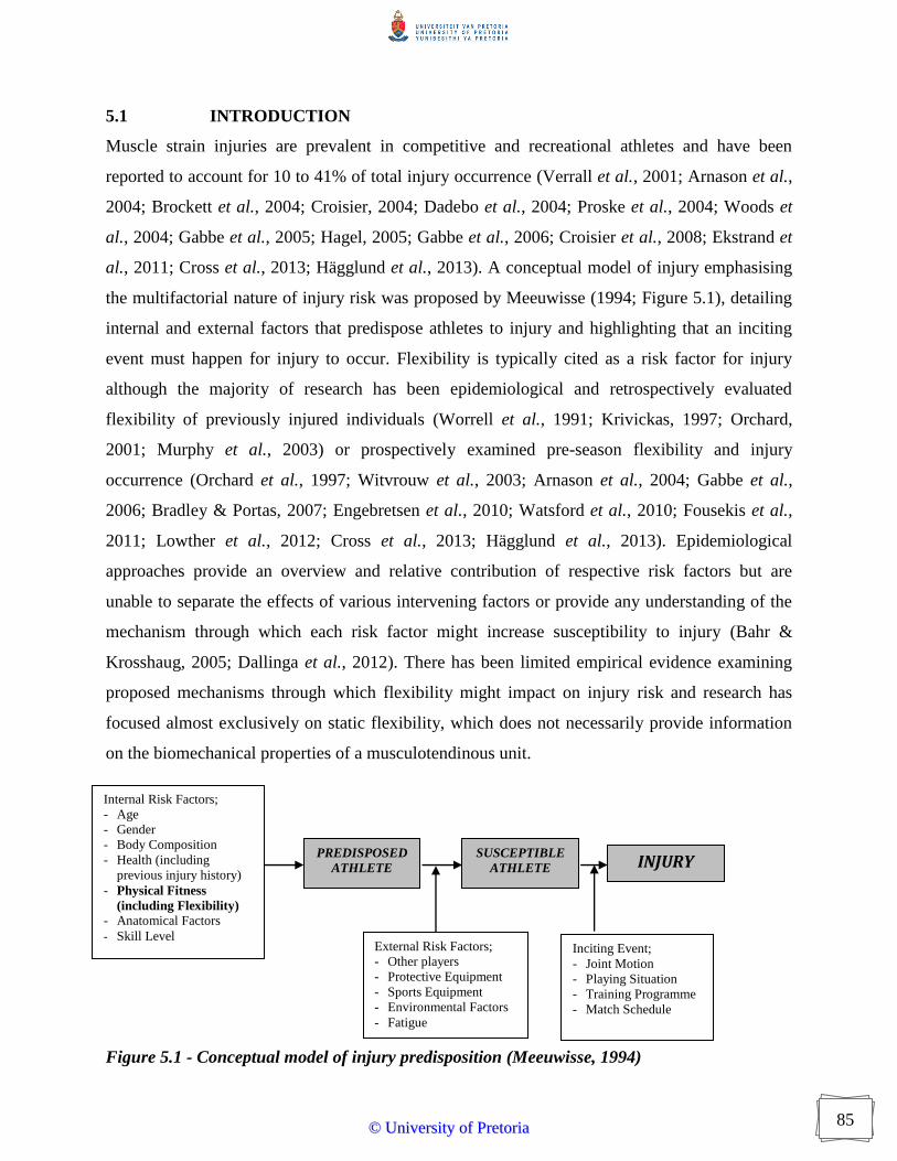
85
5.1 INTRODUCTION
Muscle strain injuries are prevalent in competitive and recreational athletes and have been
reported to account for 10 to 41% of total injury occurrence (Verrall et al., 2001; Arnason et al.,
2004; Brockett et al., 2004; Croisier, 2004; Dadebo et al., 2004; Proske et al., 2004; Woods et
al., 2004; Gabbe et al., 2005; Hagel, 2005; Gabbe et al., 2006; Croisier et al., 2008; Ekstrand et
al., 2011; Cross et al., 2013; Hägglund et al., 2013). A conceptual model of injury emphasising
the multifactorial nature of injury risk was proposed by Meeuwisse (1994; Figure 5.1), detailing
internal and external factors that predispose athletes to injury and highlighting that an inciting
event must happen for injury to occur. Flexibility is typically cited as a risk factor for injury
although the majority of research has been epidemiological and retrospectively evaluated
flexibility of previously injured individuals (Worrell et al., 1991; Krivickas, 1997; Orchard,
2001; Murphy et al., 2003) or prospectively examined pre-season flexibility and injury
occurrence (Orchard et al., 1997; Witvrouw et al., 2003; Arnason et al., 2004; Gabbe et al.,
2006; Bradley & Portas, 2007; Engebretsen et al., 2010; Watsford et al., 2010; Fousekis et al.,
2011; Lowther et al., 2012; Cross et al., 2013; Hägglund et al., 2013). Epidemiological
approaches provide an overview and relative contribution of respective risk factors but are
unable to separate the effects of various intervening factors or provide any understanding of the
mechanism through which each risk factor might increase susceptibility to injury (Bahr &
Krosshaug, 2005; Dallinga et al., 2012). There has been limited empirical evidence examining
proposed mechanisms through which flexibility might impact on injury risk and research has
focused almost exclusively on static flexibility, which does not necessarily provide information
on the biomechanical properties of a musculotendinous unit.
Figure 5.1 - Conceptual model of injury predisposition (Meeuwisse, 1994)
Internal Risk Factors;
- Age
- Gender
- Body Composition
- Health (including
previous injury history)
- Physical Fitness
(including Flexibility)
- Anatomical Factors
- Skill Level External Risk Factors;
- Other players
- Protective Equipment
- Sports Equipment
- Environmental Factors
- Fatigue
Inciting Event;
- Joint Motion
- Playing Situation
- Training Programme
- Match Schedule
PREDISPOSED
ATHLETE
SUSCEPTIBLE
ATHLETE INJURY
©© UUnniivveerrssiittyy ooff PPrreettoorriiaa

86
Flexibility is influenced by multiple factors and Figure 5.2 highlights the contribution from
neural constraints, mechanical constraints, joint related constraints and other constraints (Johns
& Wright, 1962; McHugh et al., 1998; Magnusson et al., 2001; Guissard & Duchateau, 2006;
Bojsen-Moller et al., 2007; Bozic et al., 2010; Weppler & Magnusson, 2010; McHugh et al.,
2012). Of these constraints, some are active (e.g. musculotendinous unit) and some are passive
(e.g. joint capsule). Empirical evidence detailing the relative roles played by these constraints is
limited and tends to be provided by animal or cadaver studies (Ramsey & Street, 1940; Casella,
1950; Hill, 1968; Rapoport, 1972; Magid & Law, 1985; Tidball, 1986; Horowits et al., 1989),
and finding are often extrapolated directly to humans without consideration of differences in
tissue structures and properties (MacNaughton & MacIntosh, 2007). Additionally it has been
suggested that there are static and dynamic expressions of flexibility (Gleim & McHugh, 1997)
but researchers generally consider flexibility as a global concept. It is plausible that the
constraints to flexibility differ in their relative contributions to static and dynamic flexibility,
which makes interpretation and comparison of previous research investigating flexibility difficult
because of differing dependent variables and methods of measurement used.
5.1.1 Injury prevention
Research on injury prevention is a step-by-step process in which information on causes of injury
is systematically collected and used to develop potentially useful intervention methods (Van
Tiggelen et al., 2008). One important goal for effective injury prevention is to identify the multi-
factorial extrinsic and intrinsic risk factors that contribute to the susceptibility of an athlete to
injury (Beijsterveldt et al., 2012). A number of retrospective and prospective epidemiological
studies have attempted to isolate risk factors related to hamstring strain injuries (Verrall et al.,
2001; Arnason et al., 2004; Brockett et al., 2004; Croisier, 2004; Dadebo et al., 2004; Proske et
al., 2004; Woods et al., 2004; Gabbe et al., 2005; Hagel, 2005; Gabbe et al., 2006; Croisier et
al., 2008; Engebretsen et al., 2010; Henderson et al., 2010; Watsford et al., 2010; Ekstrand et al.,
2011; Fousekis et al., 2011; Lowther et al., 2012; Cross et al., 2013; Hägglund et al., 2013).
Among other contributory factors, fatigue (Gabbett & Domrow, 2005; Gabbe et al., 2006;
Alentorn-Geli et al., 2009; Small et al., 2010) and inadequate flexibility (Agre, 1985; Jonhagen
et al., 1994; Worrell, 1994; Kroll & Raya, 1997; Kujala et al., 1997; Bennell et al., 1999; Devlin,
2000; Witvrouw et al., 2003; Bradley & Portas, 2007; Watsford et al., 2010) have been cited as
risk factors for hamstring strain injury. Despite widespread acceptance of the conceptual model
©© UUnniivveerrssiittyy ooff PPrreettoorriiaa

87
for injury (Figure 5.1), approaches to evaluating injury risk often fail to consider the inciting
event (Bahr & Krosshaug, 2005; Liu et al., 2012). Appreciation of this is important to help
understand the mechanism of a particular injury type in a given sport, accounting for the injury
situation (player and opponent behaviour and playing situation) and whole body and joint
biomechanics at time of injury.
Figure 5.2 – Factors constraining flexibility
Controlled prospective epidemiological studies are well suited to identifying the potential
contribution of a range of risk factors to injury but do not provide information about the possible
mechanism through which they act and do not account for the inciting event. To develop better
FLEXIBILITY
Mechanical
Factors
Muscle
Tone
Muscle
Length Viscoelastic
Properties
Excessive Fibrotic
Tissue Formation
Neural
Factors
Stretch
Reflex
Neural
Tension
Reciprocal
Inhibition
Joint
Related
Factors
Congruency
of Surfaces
Ligamentous/
Capsular
Tightness
Articular/
Fibrocartilage
Surfaces
Other
Factors
Skin
Elasticity
Ambient
Temperature
Circadian
Variation
Training
Status
Stretch
Tolerance
©© UUnniivveerrssiittyy ooff PPrreettoorriiaa

88
understanding of injury prevention, a range of research approaches are needed including
epidemiological studies exploring risk, athlete interviews, movement analysis of injury
occurrence, cadaver studies, mathematical modelling and experimental approaches (Krosshaug et
al., 2005; Finch, 2011). Each method to understanding injury prevention has inherent limitations
and no single research approach is adequate in terms of completeness of information provided. In
terms of hamstring strain injury, there is a rationale for experimental approaches to facilitate
examination and manipulation of recognised individual risk factors in a controlled manner thus
providing further insight into how susceptibility to injury might be affected.
5.1.2 Fatigue and injury risk
Injury prevalence studies suggest injuries often occur towards the end of matches and training
sessions and epidemiological approaches include fatigue as a risk factor for injury, but there is
limited understanding about how fatigue might influence injury susceptibility. Few experimental
studies in humans have considered the effect of fatigue on mechanical properties of human
muscle (Garrandes et al., 2007; Cote et al., 2008; Timmins et al., 2012; Semmler et al., 2013),
possibly because fatigue is difficult to quantify and not always synonymous with exposure time
(Verrall et al., 2001). Fatigue might be higher after shorter periods of time requiring repeated
high-intensity efforts than at the end of competition if player involvement has been low
(Robineau et al., 2012). Human studies have shown that muscular fatigue (Melnyk & Gollhofer,
2007; Gehring et al., 2009; Cè et al., 2013; Conchola et al., 2013) and muscle damage (Davies &
White, 1981; Kuipers, 1994; Hesselink et al., 1996) induce neuromuscular control deficiencies,
thereby influencing dynamic joint stability. Animal models have demonstrated that stimulated
fatigued muscle undergoes a greater length change and absorbs less energy than non-fatigued
muscle, and these models have been extrapolated to explain the increased predisposition to injury
seen in fatigued individuals (Mair et al., 1996; Gabbett & Domrow, 2005). Intense or sustained
exercise induces microtrauma in muscle which can have a cumulative effect as the exercise bout
progresses, causing micro-damage of the muscle fibres which could progress to more major
muscle tears of involved muscle groups (Proske & Morgan, 2001). Rodent EDL and soleus
muscle has demonstrated that fatigue increases resting tension because the muscles fail to relax
properly between contractions (Matar et al., 2000), and means that muscles will potentially
exhibit greater stiffness. This has been a suggested mechanism by which fatigued human muscle
©© UUnniivveerrssiittyy ooff PPrreettoorriiaa
89
is at increased risk of injury (Aagaard et al., 2000; Reilly et al., 2008; Alentorn-Geli et al., 2009)
but there is little empirical evidence, particularly of an in vivo nature, to support this conjecture.
Muscle fatigue has been quantified by a reduction in maximum force that a muscle can generate
compared to pre-exercise values (Cross et al., 2013) and research has shown that concentric and,
less frequently, eccentric strength of the knee flexors and extensors is impaired after exercise
protocols comprising isokinetic maximal contractions (Sangnier & Tourny-Chollet, 2008), high-
intensity cycling (Garrandes et al., 2007) and prolonged intermittent running (Rahnama et al.,
2006; Small et al., 2010). Different studies have found variations in magnitude of change with 8
to 15% of knee extensor and knee flexor peak torque being reported after a 90 minute fatiguing
protocol set to simulate the work rate during a soccer game (Rahnama et al., 2003), while
reductions in peak torque of nearly 25% have been described after prolonged intermittent high
intensity exercise (Mercer et al., 2003). Alterations have also been seen in the dynamic control
ratio of the leg (eccentric knee flexion:concentric knee extension) indicating a reduced ability to
stabilise the knee (Rahnama et al., 2003). Fatigue inducing exercise protocols have been shown
to increase electromechanical delay (Gleeson et al., 1998; Yavuz et al., 2010; Cè et al., 2013;
Conchola et al., 2013) and diminish coordination and timing by affecting energy transfer
between limb segments (Rozzi et al., 1999; Martin & Brown, 2009), thereby causing deficits in
proprioception and JPS (Kaufman et al., 2001; Givoni et al., 2007). Alterations in JPS and
neuromuscular function can cause an imbalance between afferent feedback and efferent
responses (Herrington et al., 2010) and such disruptions to the biological machinery for sensory-
motor performance may elicit compromised self-perception of the capability for neuromuscular
control. Imprecision of self-perception could contribute to a process by which injury becomes
more likely (Noakes et al., 2004); for instance, overestimation of performance capability may
leave the participant partially unprotected during episodes of externally imposed mechanical
stress. Skinner et al. (1986) studied knee proprioception performance in healthy young men and
found a significant decrease in the ability to reproduce knee joint angles after a series of interval
running sprints (3.75 miles in total), while in contrast, Marks and Quinney (1993) found that 20
maximal isokinetic quadriceps contractions did not significantly reduce knee proprioception
performance in young sedentary women. The conflicting finding may be attributable to the type
and duration of exercise protocol utilised and females being less likely to demonstrate significant
changes in JPS after a fatigue inducing protocol (Lattanizio et al., 1997; Yavuz et al., 2010).
©© UUnniivveerrssiittyy ooff PPrreettoorriiaa

90
Research investigating the effect of static stretching on JPS has been equivocal with studies
finding positive benefits (Ghaffarinejad et al., 2007) and no measureable effects (Larsen et al.,
2005; Bjorklund et al., 2006; Torres et al., 2012) but a lack of high quality empirical evidence
and differences in methodology and participant makes comparison of results difficult.
Through the use of animal models, it has been suggested that the most important factor in muscle
strain occurrence is the amount of energy that the muscle can absorb before failure (Brooks et
al., 1995; Garrett, 1996; Mair et al., 1996). Muscle strain injuries are thought to occur when
muscles are stretched passively or activated during stretch (Garrett, 1996; Kirkendall & Garrett,
2002; Schache et al., 2009; Chumanov et al., 2012; Lowther et al., 2012). Eccentric contractions
are an important factor because muscle forces can be higher during lengthening than when
shortening (Stauber, 2004), adding to the forces applied by passive connective tissue elements
(Elftman, 1966). Animal studies have shown that maximally activated muscle absorbs
significantly more energy before failure than less activated muscle, indicating that the passive
elements of the muscle have the ability to absorb energy but the ability is enhanced when the
muscle is active (Garrett et al., 1987). Fatigue has been reported to reduce the energy absorbing
capabilities of a muscle in vitro although failure was at the same length regardless of fatigue
(Mair et al., 1996). Fatigued muscle is less active than non-fatigued muscle, which might
contribute to a reduced ability to absorb energy and thus increased level of injury predisposition
(Garrandes et al., 2007; Enoka, 2012).
5.1.3 Stretching as part of recovery
Pre-exercise stretching has traditionally been thought to play a role in reducing injury risk and
subsequently, stretching has been an accepted and integral part of pre-exercise warm-up regimes
(ACSM, 1998). While immediate increases in static flexibility as a result of an acute bout of
stretching are well documented, little research has demonstrated benefits in terms of reduced
injury risk (Andrish et al., 1974; van Mechelen et al., 1993; Pope et al., 1998; Hartig &
Henderson, 1999; Pope et al., 2000; Amako et al., 2003; Hadala & Barrios, 2009). Furthermore
there is a growing body of evidence reporting concurrent decreases in measures of strength and
performance (Kokkonen et al., 1998; Avela et al., 1999; Fowles et al., 2000; Nelson et al., 2001;
Avela et al., 2004; Behm et al., 2004; Power et al., 2004; Marek et al., 2005; Nelson et al., 2005;
Cramer et al., 2006; Egan et al., 2006; Little & Williams, 2006; Yamaguchi et al., 2006; Bradley
©© UUnniivveerrssiittyy ooff PPrreettoorriiaa
91
et al., 2007; Cramer et al., 2007; Cramer et al., 2007; McBride et al., 2007; Stewart et al., 2007;
Herda et al., 2008; McHugh & Nesse, 2008; Ryan et al., 2008; Torres et al., 2008; Winchester et
al., 2008; Costa et al., 2009; Carvalho et al., 2012; Esposito et al., 2012; Maddigan et al., 2012;
Fortier et al., 2013; Matsuo et al., 2013). Stretch induced detriments in performance were
generally smaller than decrements in strength measures (approx. 3% vs. 8% for clinically
relevant stretch durations). In contrast to stretching before exercise, researchers have suggested
that stretching after exercise might have positive benefits in improving recovery (Reilly &
Ekblom, 2005; Shrier, 2008). Muscle recovery after physical activity is important because
periods between games and training sessions have become shorter, with athletes often partaking
in multiple training sessions per day on successive days of the week. It has been suggested that
post-exercise static stretching could help fatigued muscles return to their normal resting lengths
(Nelson & Hutton, 1985; Avela et al., 1999), thus lessening symptoms of DOMS as well as
decreasing subsequent risk of injury. This conjecture has not been supported empirically as no
significant reductions in subjective measures of soreness on a visual analogue scale or palpation
as a result of post-exercise static stretching have been demonstrated (McGlynn et al., 1979;
Buroker & Schwane, 1989; Dawson et al., 2005; Kinugasa & Kilding, 2009; Robey et al., 2009;
Torres et al., 2013). Studies examining the efficacy of post-exercise stretching have generally
used relatively untrained convenience samples of participants and exposed participants to
protocols designed to induce high levels of DOMS therefore it is not known whether the same
responses would present in trained athletes exposed to protocols simulating normal strenuous
training sessions. Furthermore, while most studies consider immediate effects of an intervention,
in sports people impact at commencement of the subsequent training session is perhaps more
important, as this would be when alterations in levels of performance and injury risk would be of
greater relevance.
The impact of post-exercise stretching on ensuing performance and consequence on factors that
might predispose injury remains to be firmly established and there is thus a need to examine
possible modifiable risk factors in an experimental setting to evaluate their individual responses.
It is difficult to directly measure the impact of post-exercise stretching on injury occurrence
because of the number of confounding variables that can occur in longitudinal studies; for
instance, alterations in training programmes, differences in fitness levels, use of braces or
supports, changing weather conditions and different levels of opposition (Fuller, 2007). As all of
©© UUnniivveerrssiittyy ooff PPrreettoorriiaa
92
these factors are thought to influence injury predisposition, the impact of the post-exercise
stretching alone cannot be isolated. To achieve sufficient scientific rigour to make any results
meaningful, factors which themselves influence injury risk can be measured and the impact of
post-exercise stretching on these established via experimental designs (Moher et al., 2009). From
results of such studies, inferences can be made as to the effect of post-exercise stretching on
subsequent injury predisposition.
5.1.4 Aims and hypotheses
The aim of this study was to investigate the impact of fatigue and post-exercise static stretching
on measures of static and dynamic flexibility and performance.
Hypotheses were as follows;
1) The fatigue inducing repeated spring protocol would lead to deterioration in measures of
static and dynamic flexibility and performance immediately and 18 hours post-exercise;
2) A post-exercise static stretch would significantly improve measures of static and
dynamic flexibility and performance in the stretch compared to the control condition
immediately and 18 hours post-exercise;
3) The fatigue inducing repeated spring protocol would lead to deterioration in JPS
immediately and 18 hours post-exercise; and
4) A post-exercise static stretch would significantly improve JPS in the stretch compared
to the control condition immediately and 18 hours post-exercise.
5.2 METHODS
5.2.1 Participants
Fifteen rugby league players volunteered to participate in this study (age 21.20 years ± 3.00,
stature 180cm ±5.47, mass 92.64kg ± 16.04 [mean ± SD]). Sample size calculations indicated
that 13 participants were needed for a repeated measures design to detect an expected change of
8o (Harvey et al., 2002) as a result of a stretching program ( = 0.05 and = 0.20). In case of
any participants withdrawing from the study and to further increase power, two extra participants
were recruited. All participants were part of a GB Super League rugby league club and so could
be considered a homogenous sample. Testing was conducted pre-season when participants were
involved in training sessions four to five times a week and training matches once a week. For
©© UUnniivveerrssiittyy ooff PPrreettoorriiaa
93
inclusion in the study, participants were certified free of lower limb injury by the club
physiotherapist and considered to be in good physical condition. Because participants were all
participating in the same training programme and were tested within the season, they were
considered to be of similar fitness levels close to their peak. Whilst fitness differences were
likely between players, in part related to their position, this was not considered because a range
of players from different positions (e.g. forwards and backs) were included within the sample.
Participants were excluded if they had previously sustained any form of hamstring injury or were
not currently participating fully in all training sessions scheduled by the club. Written informed
consent was obtained prior to the study from the participants themselves if over the age of
eighteen or via the club following letters being sent to parents or guardians if participants were
under eighteen. In such situations club personnel acted in loco parentis during testing sessions.
Ethical approval was obtained from the University of Pretoria’s Post-graduate Committee and
the Ethical Committee.
5.2.2 Study design
The study was of randomised cross-over repeated measures design as indicated in Figure 5.3.
Participants were asked to report to the lab on four occasions. The first and third occasions
involved measurement of static hamstring flexibility, passive dynamic hamstring flexibility,
eccentric peak torque, eccentric work done at 2.09 rad.s-1
, JPS and perceived stiffness before and
after a repeated sprint protocol. On one of these occasions the participant was randomly allocated
to a hamstring stretch intervention condition and on the other occasion a no hamstring stretch
(upper limb stretch) control condition. Immediately post-exercise measurements were taken one
minute after participants had performed 30s static stretch of their hamstring or upper limb. The
second and fourth occasions involved measurements of static and passive dynamic hamstring
flexibility, eccentric force and work done at 2.09rad.s-1
, JPS and perceived stiffness. The second
and fourth testing sessions took place 18 hours after the first and third sessions respectively. This
timing was chosen partly to allow physiological recovery to have taken place (Noakes, 2000;
Nédélec et al., 2013) and partly because, following discussion with team coaches and medical
team it was considered to mimic the normal club routine and training schedule and thus would
have good ecological validity. Having undertaken the first testing session in an afternoon,
participants were unlikely to have undertaken any further exercise by the following morning
when the subsequent testing session took place. Participants were asked to undergo only
©© UUnniivveerrssiittyy ooff PPrreettoorriiaa

94
activities of normal daily living during this 18 hour period. One week separated the first and
third testing sessions.
Allocation to control or stretch condition on the first occasion was randomised with seven and
eight participants being allocated to each condition respectively. This allocation was reversed
when participants presented for the third testing session so that all participants took part in both
the intervention and control conditions. All participants received treatment and control
conditions as allocated and none withdrew from the study. Allocation was concealed by the use
of sealed envelopes. The assessor was not made aware of which condition the participant was
allocated to until testing had been completed. A trained student who was not aware of the
purpose of the study supervised the stretching intervention. Attempts were made to blind the
participant by explaining that the purpose of the study was to examine the cross over effects of
upper limb stretching on lower limb flexibility. A repeated measures design was considered
optimal because differences between participants are removed as potential confounding variables
and fewer participants are required, since data for all conditions derive from the same condition
of participants (Hopkins et al., 2009), and given that the study was to be carried out on elite
rugby players, the available sample was smaller than if participants were from other, more
general, populations. The potential disadvantage of repeated measures design is from order
effects, which occur when people behave differently because of the order in which the conditions
are performed. For example, the participant’s performance may be enhanced because of a
practice effect, or performance may be reduced because of a boredom or fatigue effect. Any
order effects were counterbalanced and therefore limited in this study because participants were
randomised to condition. Because participants were considered close to peak fitness levels and
regularly carried out maximal sprint drills as part of their training, a wash out period of a week
was considered sufficient to prevent any carry over effects from the sprint protocol. Additionally,
the effects of an acute bout of static stretching are thought to return to baseline within one hour
(Duong et al., 2001; Ryan et al., 2008) and as such, this was not considered to have an influence
on the second set of testing.
5.2.3 Sprint protocol
Participants were asked to perform 30 x 20m sprints on an indoor track. This protocol was
chosen following discussion with coaches as it has been shown to induce fatigue and relates to
©© UUnniivveerrssiittyy ooff PPrreettoorriiaa
95
what a player would perform during a match. Gabbett (2005) identified the importance of
repeated sprinting in rugby league and suggested rugby league players are rarely required to
sprint over 40 m, commonly sprint 10-20 m, and have work:rest ratios in the region of 1:6 to
1:12. Pilot study data and data based on fitness testing of comparable GB super league teams,
led to the estimation the sprint time would be in the region of 3s, the recovery walk time in the
region of 12 to 15s, with 3s to commence the next sprint and therefore an initial work:rest ratio
in the region of 1:5/6. Previous research has tended to utilise single maximal efforts to induce
fatigue rather than repeated dynamic sprint efforts which are more typical of team sports
(Pinniger et al., 2000), whereas with similar sprint distances and work:rest ratios the protocol
was considered to have good ecological validity (Deutsch et al., 1998). Sprints were performed
from a standing start as and on completion of each sprint, the participant immediately turned and
walked back to the start. The work to rest ratio was approximately 1:3. As soon as the starting
position was reached, the next sprint was commenced. Electronic timing gates (Smart Speed,
Fusion Sport) were used to measure the start and finish of the sprints. The first five sprints were
conducted at 50% pace and the second five at 75% pace to ensure participants were sufficiently
warmed-up before being asked to produce maximum effort. Percentage of maximal effort was
determined by self-perception and these times did not count towards the study. The final twenty
sprints were conducted at maximal effort.
5.2.4 Post-exercise stretching intervention
The hamstring stretch was performed in a standing position and took place five minutes after the
repeated sprint protocol had been completed (Dorado et al., 2004). This was to mimic field
conditions where athletes usually perform an aerobic cool down prior to stretching. Few studies
examining effects of post-exercise static stretching have explicitly described the time period
between the completion of exercise and stretch intervention and those that have used times
ranging between 5 and 15 to 20 minutes (Dawson et al., 2005; Mika et al., 2007). A period of 5
minutes was chosen for this study following discussion with coaches as it was believed to
accurately reflect the time it would take for players to complete a training session and move to
the cool-down area.
©© UUnniivveerrssiittyy ooff PPrreettoorriiaa
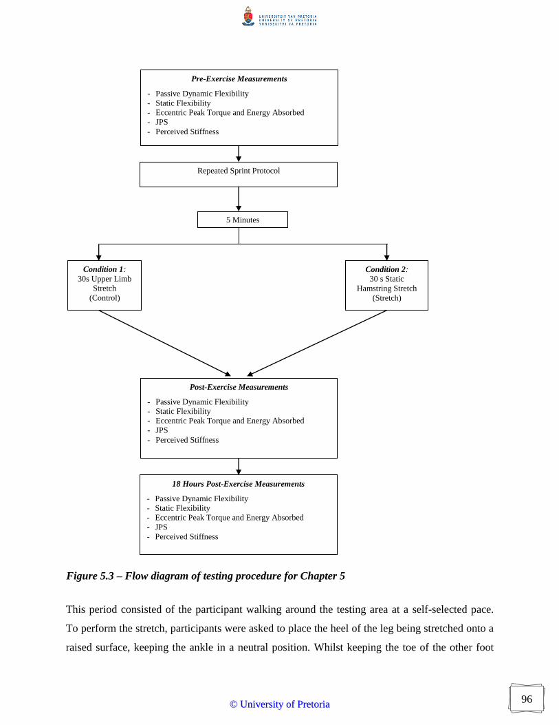
96
This period consisted of the participant walking around the testing area at a self-selected pace.
To perform the stretch, participants were asked to place the heel of the leg being stretched onto a
raised surface, keeping the ankle in a neutral position. Whilst keeping the toe of the other foot
Pre-Exercise Measurements
- Passive Dynamic Flexibility
- Static Flexibility
- Eccentric Peak Torque and Energy Absorbed
- JPS
- Perceived Stiffness
Condition 1:
30s Upper Limb
Stretch
(Control)
Condition 2:
30 s Static
Hamstring Stretch
(Stretch)
Repeated Sprint Protocol
Post-Exercise Measurements
- Passive Dynamic Flexibility
- Static Flexibility
- Eccentric Peak Torque and Energy Absorbed
- JPS
- Perceived Stiffness
Figure 5.3 – Flow diagram of testing procedure for Chapter 5
5 Minutes
18 Hours Post-Exercise Measurements
- Passive Dynamic Flexibility
- Static Flexibility
- Eccentric Peak Torque and Energy Absorbed
- JPS
- Perceived Stiffness
©© UUnniivveerrssiittyy ooff PPrreettoorriiaa
97
straight, the hips square and maintaining a lower lumbar lordosis, i.e. being instructed keep a
curve in the lower back rather than slumping forwards, participants were asked to place their
hands on the leg being stretched and move them forwards until a strong but not painful tension
was felt in the posterior thigh. During the no hamstring stretch (control) condition, participants
were asked to perform a posterior arm (tricep) stretch by raising one arm above their head,
flexing the elbow and applying a downward pressure with their other hand.
Studies with longer stretching durations are more likely to reach significance with their results
and many studies use durations of stretch far higher than is generally performed as part of a pre-
exercise warm-up (McHugh & Cosgrave, 2010). For increases in static flexibility, a series of
studies by Bandy and colleagues demonstrated significant improvements in the static flexibility
of the hamstrings with a single static stretch lasting 30s performed 5 days per week for 6 weeks
(Bandy & Irion, 1994; Bandy et al., 1997; Bandy et al., 1998). Other studies corroborated these
results, reporting that 30 s of static stretching was optimal when compared with other durations
for increasing static flexibility of the gastrocnemius (Grady & Saxena, 1991; Bandy & Irion,
1994) and hamstring muscles (Walter et al., 1994). To ensure good ecological validity, stretches
were maintained for 30s to mimic what is often practiced in a field setting.
5.2.5 Measurement procedures
Assessment of flexibility indices were taken according to the method described previously
(Chapter 3).
5.2.5.1 Eccentric torque
Participants were seated in the chair of the isokinetic dynamometer with the trunk perpendicular
to the thighs. The lateral condyle of the femur was placed in line with the axis of rotation of the
lever arm. The lower leg was secured to a pad placed 2 cm proximal to the medial malleolus of
the ankle. The upper leg was stabilised by a thigh strap placed across the femur 5 cm proximal to
the condyles. Additional straps were used to fix the upper body and contralateral limb to the
chair.
Speed for the test was set at 2.09 rad.s-1
. Although optimum velocities remain uncertain, a
relatively fast speed was required to attempt to mimic functional movements but if too great it
©© UUnniivveerrssiittyy ooff PPrreettoorriiaa
98
was anticipated that all participants would not be able to reach the speed of the dynamometer
(Yen, 2005). Participants performed three maximal eccentric hamstring contractions against the
dynamometer (Christou & Carlton, 2002). Data from all tests were exported. Final values
obtained for gravity corrected peak torque, energy absorbed (work done) and position of peak
torque were used for subsequent analysis.
5.2.5.2 Perceived stiffness
Participants were given a Visual Analogue Scale (10cm Line), with Absolutely No Stiffness
marked on the left side of the scale and Worst Stiffness Ever marked on the right side of the
scale. These scales have been shown to be reliable and valid (Mattacola et al., 1997; McCormack
et al., 2009). Participants were asked to mark on the line where they felt that the stiffness they
were currently experiencing fitted.
5.2.5.3 Joint position sense
JPS was measured using an accepted and deemed reliable method of testing the ability to
reproduce specific knee joint angles in an open-kinetic sitting position (Skinner et al., 1986;
Marks & Quinney, 1993). Participants were seated on a treatment plinth that was of sufficient
height that their feet did not touch the ground. With their eyes closed, the experimenter extended
the participants knee to different angles to prevent a learning effect. The chosen angle was
measured using a clinical goniometer (MIE Medical Research Ltd, UK) and the limb then
lowered. The participant was asked to return the limb to the previous position and the angle
measured. The difference between the first and second angles was used as a measure of JPS in
subsequent analyses.
5.2.6 Statistical analysis
Data obtained from the Isokinetic Dynamometer were exported into Microsoft Excel 2000. 3rd
Order Polynomial Curves were fitted, and using Derive (version 7), equations of the fitted curves
(y = ax3 ± bx
2 ± c ± k) were differentiated to find gradient at the highest common point achieved
by all participants (stiffness at 0.99 rad) as well the maximum position (final stiffness) and
integrated to find the area under the curve (energy absorbed to 0.99 rad and total energy
absorbed) during the passive knee extension manoeuvre. Measures of torque at 0.99 rad and
passive peak torque were also recorded.
©© UUnniivveerrssiittyy ooff PPrreettoorriiaa
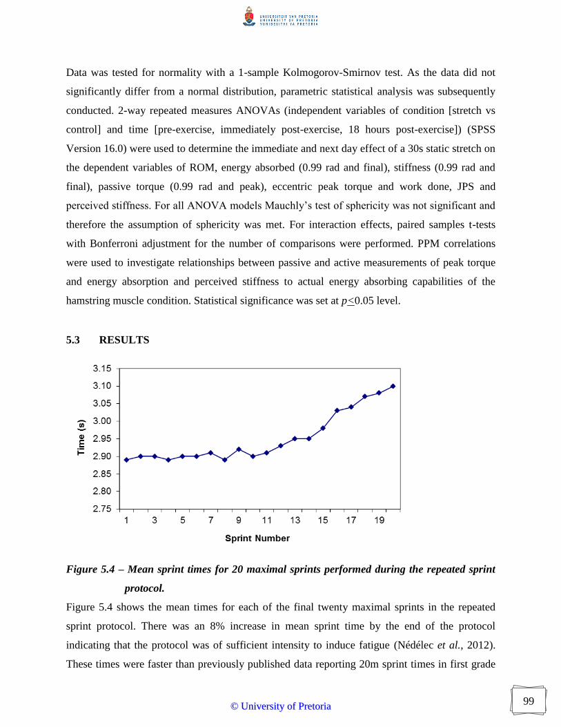
99
Data was tested for normality with a 1-sample Kolmogorov-Smirnov test. As the data did not
significantly differ from a normal distribution, parametric statistical analysis was subsequently
conducted. 2-way repeated measures ANOVAs (independent variables of condition [stretch vs
control] and time [pre-exercise, immediately post-exercise, 18 hours post-exercise]) (SPSS
Version 16.0) were used to determine the immediate and next day effect of a 30s static stretch on
the dependent variables of ROM, energy absorbed (0.99 rad and final), stiffness (0.99 rad and
final), passive torque (0.99 rad and peak), eccentric peak torque and work done, JPS and
perceived stiffness. For all ANOVA models Mauchly’s test of sphericity was not significant and
therefore the assumption of sphericity was met. For interaction effects, paired samples t-tests
with Bonferroni adjustment for the number of comparisons were performed. PPM correlations
were used to investigate relationships between passive and active measurements of peak torque
and energy absorption and perceived stiffness to actual energy absorbing capabilities of the
hamstring muscle condition. Statistical significance was set at p<0.05 level.
5.3 RESULTS
Figure 5.4 – Mean sprint times for 20 maximal sprints performed during the repeated sprint
protocol.
Figure 5.4 shows the mean times for each of the final twenty maximal sprints in the repeated
sprint protocol. There was an 8% increase in mean sprint time by the end of the protocol
indicating that the protocol was of sufficient intensity to induce fatigue (Nédélec et al., 2012).
These times were faster than previously published data reporting 20m sprint times in first grade
©© UUnniivveerrssiittyy ooff PPrreettoorriiaa
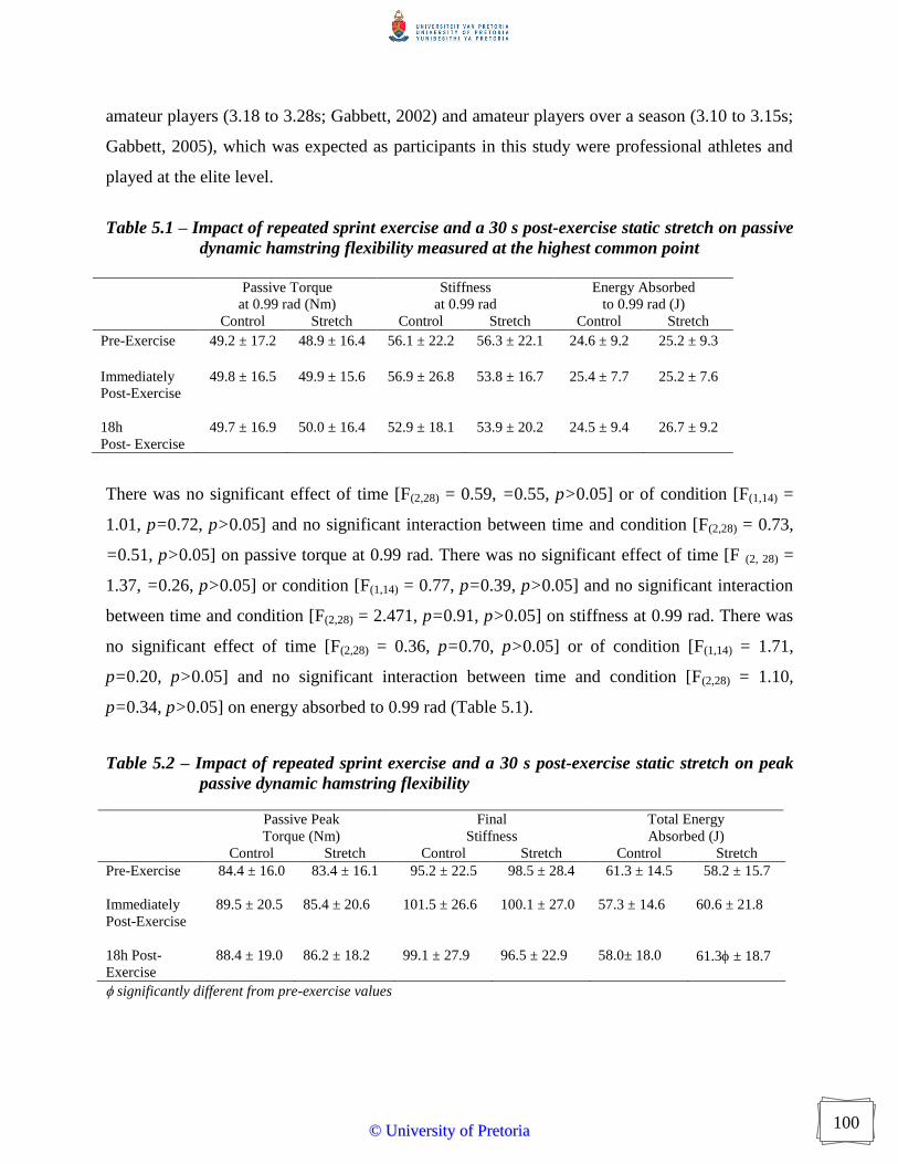
100
amateur players (3.18 to 3.28s; Gabbett, 2002) and amateur players over a season (3.10 to 3.15s;
Gabbett, 2005), which was expected as participants in this study were professional athletes and
played at the elite level.
Table 5.1 – Impact of repeated sprint exercise and a 30 s post-exercise static stretch on passive
dynamic hamstring flexibility measured at the highest common point
Passive Torque
at 0.99 rad (Nm)
Stiffness
at 0.99 rad
Energy Absorbed
to 0.99 rad (J)
Control Stretch Control Stretch Control Stretch
Pre-Exercise 49.2 ± 17.2 48.9 ± 16.4 56.1 ± 22.2 56.3 ± 22.1 24.6 ± 9.2 25.2 ± 9.3
Immediately
Post-Exercise
49.8 ± 16.5 49.9 ± 15.6 56.9 ± 26.8 53.8 ± 16.7 25.4 ± 7.7 25.2 ± 7.6
18h
Post- Exercise
49.7 ± 16.9 50.0 ± 16.4 52.9 ± 18.1 53.9 ± 20.2 24.5 ± 9.4 26.7 ± 9.2
There was no significant effect of time [F(2,28) = 0.59, =0.55, p>0.05] or of condition [F(1,14) =
1.01, p=0.72, p>0.05] and no significant interaction between time and condition [F(2,28) = 0.73,
=0.51, p>0.05] on passive torque at 0.99 rad. There was no significant effect of time [F (2, 28) =
1.37, =0.26, p>0.05] or condition [F(1,14) = 0.77, p=0.39, p>0.05] and no significant interaction
between time and condition [F(2,28) = 2.471, p=0.91, p>0.05] on stiffness at 0.99 rad. There was
no significant effect of time [F(2,28) = 0.36, p=0.70, p>0.05] or of condition [F(1,14) = 1.71,
p=0.20, p>0.05] and no significant interaction between time and condition [F(2,28) = 1.10,
p=0.34, p>0.05] on energy absorbed to 0.99 rad (Table 5.1).
Table 5.2 – Impact of repeated sprint exercise and a 30 s post-exercise static stretch on peak
passive dynamic hamstring flexibility
Passive Peak
Torque (Nm)
Final
Stiffness
Total Energy
Absorbed (J)
Control Stretch Control Stretch Control Stretch
Pre-Exercise 84.4 ± 16.0 83.4 ± 16.1 95.2 ± 22.5 98.5 ± 28.4 61.3 ± 14.5 58.2 ± 15.7
Immediately
Post-Exercise
89.5 ± 20.5 85.4 ± 20.6 101.5 ± 26.6 100.1 ± 27.0 57.3 ± 14.6 60.6 ± 21.8
18h Post-
Exercise
88.4 ± 19.0 86.2 ± 18.2 99.1 ± 27.9 96.5 ± 22.9 58.0± 18.0 61.3 ± 18.7
significantly different from pre-exercise values
©© UUnniivveerrssiittyy ooff PPrreettoorriiaa

101
There was no significant effect of time [F(2,28) = 2.68, p=0.08, p>0.05] on passive peak
hamstring torque and no significant interaction between time and condition [F(2,28) = 25.13,
p=0.59, p>0.05]. There was a significant effect of condition [F(1,14) = 8.38, p=0.01, p<0.05] on
passive peak torque. Figure 5.5 illustrates that the control condition (87.4Nm ± 18.5) appeared to
have a higher passive peak torque than the stretch condition (85.0Nm ± 18.5).
Figure 5.5 – Impact of a 30 s post-exercise static hamstring stretch on hamstring passive peak
torque (n = 15).
There was no significant effect of time [F(2,28) = 0.47, p=0.63, p>0.05] or condition [F(1,14) =
0.05, p=0.83, p>0.05] and no significant interaction between time and condition [F(2,28) = 1.54,
p=0.22, p>0.05] on final stiffness (Table 5.2).
There was no significant effect of time [F(2,28) = 0.49, p=0.61, p>0.05] or of condition [F(1,14) =
0.90, p=0.35, p>0.05] on total energy absorbed by the hamstring during the passive stretching
manoeuvre. There was a significant interaction between time and condition [F(2,28) = 6.84,
p=0.001, p<0.05]. Paired samples t-tests with Bonferroni adjustment for number of comparisons
indicated that there was no significant difference between stretch and control conditions pre-
exercise [t(14) = 1.52, p=0.137, p>0.01], post-exercise [t(14) = -2.10, p=0.04, p>0.01] or 18 hours
post-exercise [t(14) = -2.29, p=0.03, p>0.01]. Figure 5.6 illustrates that total energy absorbed
appears to decrease in the control condition post-exercise and remains lower than baseline
values. In comparison in the stretch condition total energy absorbed appears to increase post-
exercise although not significantly [t(14) = -1.47, p=0.15, p>0.01], but there was a significant
©© UUnniivveerrssiittyy ooff PPrreettoorriiaa

102
increase in total energy absorbed between pre and 18 hours post-exercise [t(14) = -3.51, p=0.001,
p<0.01].
Figure 5.6 – Impact of a 30 s post-exercise static hamstring stretch on total passive energy
absorbed by the hamstring muscle (n = 15).
Table 5.3 – Impact of repeated sprint exercise and a 30 s post-exercise static stretch on active
properties during eccentric hamstring contraction at 2.09 rad.s-1
Peak Torque (Nm) Energy Absorbed (J)
Position of Peak
Torque (rad)
Control Stretch Control Stretch Control Stretch
Pre-Exercise 146.9 ±
34.3
149.9 ±
34.5
161.9 ±
44.5
164.8 ±
44.7 1.2 ± 0.3 1.2 ± 0.2
Immediately
Post-Exercise
137.8 ±
26.7
148.9* ±
40.8
147.9 ±
39.9
162.4* ±
45.3 1.3 ± 0.2 1.3 ± 0.2
18h Post-
Exercise
139.4 ±
38.2
150.4* ±
41.2
153.1 ±
42.9
164.9* ±
43.0 1.2 ± 0.2 1.2 ± 0.2
* significantly different from control condition
There was no significant effect of time on hamstring eccentric peak torque [F(2,28) = 1.13,
p=0.33, p>0.05] and no significant interaction between time and condition [F(2,28) = 2.23,
p=0.11, p>0.05]. There was a significant effect of condition on hamstring eccentric peak torque
[F(1,14) = 12.39, p=0.00, p<0.05]. The control condition (141.0 Nm ± 33.1) appeared to produce a
lower peak torque during eccentric contraction than the stretch condition (149.7 Nm ± 38.8), as
illustrated in Figure 5.7.
©© UUnniivveerrssiittyy ooff PPrreettoorriiaa

103
Figure 5.7 – Impact of a 30 s post-exercise static hamstring stretch on peak torque generated
during maximal eccentric hamstring contraction (n = 15).
There was no significant effect of time on energy absorbed (work done) by the hamstring muscle
group during an eccentric contraction [F(2,28) = 3.02, p=0.06, p>0.05]. There was a significant
effect of condition on work done (energy absorbed) by the hamstring muscle group during an
eccentric contraction [F(1,14) = 8.62, p=0.01, p<0.05]. Overall the stretch condition (164.0 ± 44.3)
produced a higher total energy absorbed compared to the control condition (154.3 ± 42.4) (mean
values Table 5.3). There was a significant interaction between time and condition on energy
absorbed (work done) by the hamstring muscle group [F(2,28) = 7.03, p=0.00, p<0.05]. Paired
samples t-tests with Bonferroni adjustment for multiple comparisons showed no significant
difference between control and stretch conditions pre-exercise [t(14) = -0.84, p=0.40, p>0.01]
whereas there was a significant difference both post-exercise [t(14) = -3.38, p=0.00, p<0.01] and
18 hours post-exercise [t(14) = -3.08, p=0.00, p<0.01]. Figure 5.8 illustrates that the total energy
absorbed remains stable throughout the protocol in the stretch condition whereas it decreases
post-exercise in the control condition.
There was no significant effect of time [F(2,28) = 0.26, p=0.77, p>0.05] or condition [F(1,14) =
0.32, P=0.57, p>0.05] and there was no significant interaction between time and condition
[F(1,14) = 0.19, P=0.83, p>0.05] on position of the torque-angle curve that peak torque was
achieved during a maximal eccentric hamstring contraction at 2.09 rad.s-1
(Table 5.3).
©© UUnniivveerrssiittyy ooff PPrreettoorriiaa
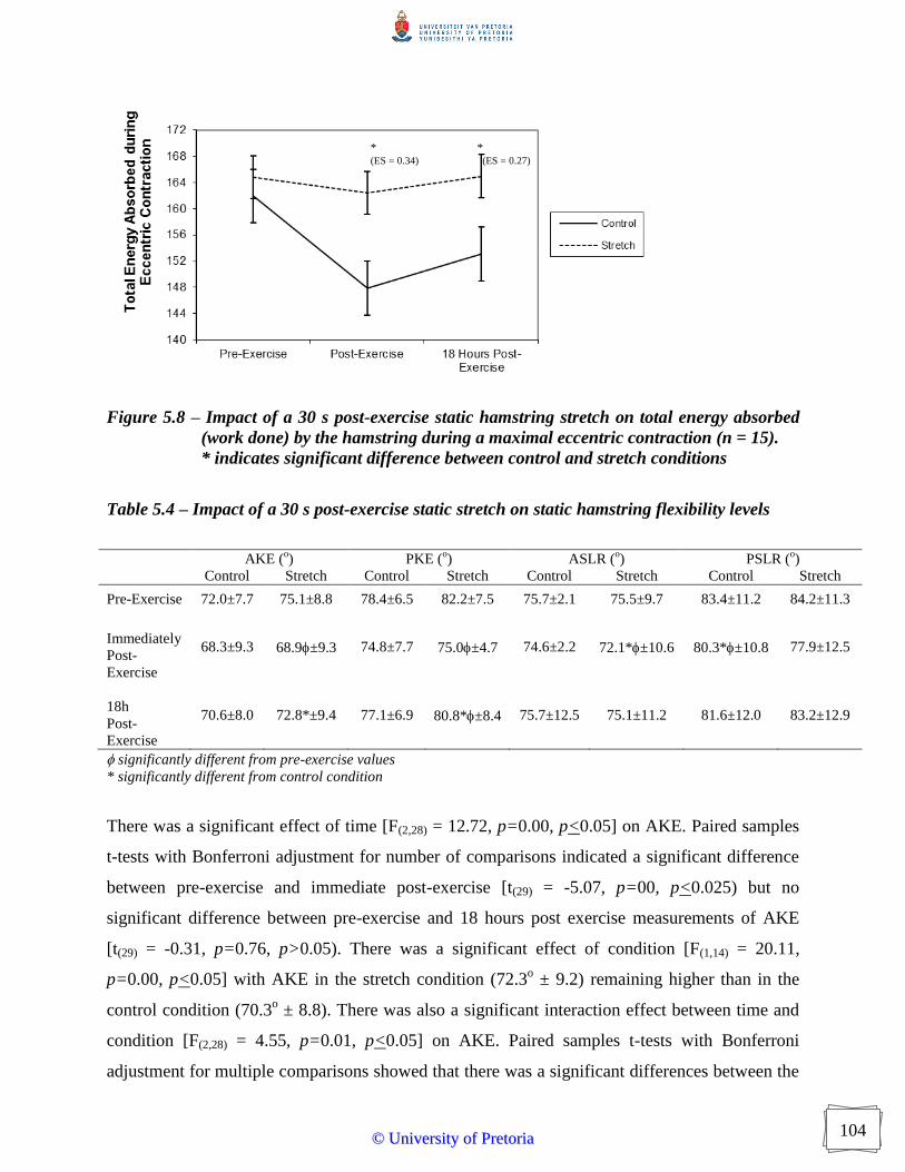
104
Figure 5.8 – Impact of a 30 s post-exercise static hamstring stretch on total energy absorbed
(work done) by the hamstring during a maximal eccentric contraction (n = 15).
* indicates significant difference between control and stretch conditions
Table 5.4 – Impact of a 30 s post-exercise static stretch on static hamstring flexibility levels
AKE (
o) PKE (
o) ASLR (
o) PSLR (
o)
Control Stretch Control Stretch Control Stretch Control Stretch
Pre-Exercise 72.0±7.7 75.1±8.8 78.4±6.5 82.2±7.5 75.7±2.1 75.5±9.7 83.4±11.2 84.2±11.3
Immediately
Post-
Exercise
68.3±9.3 68.9±9.3 74.8±7.7 75.0±4.7 74.6±2.2 72.1*±10.6 80.3*±10.8 77.9±12.5
18h
Post-
Exercise
70.6±8.0 72.8*±9.4 77.1±6.9 80.8*±8.4 75.7±12.5 75.1±11.2 81.6±12.0 83.2±12.9
significantly different from pre-exercise values
* significantly different from control condition
There was a significant effect of time [F(2,28) = 12.72, p=0.00, p<0.05] on AKE. Paired samples
t-tests with Bonferroni adjustment for number of comparisons indicated a significant difference
between pre-exercise and immediate post-exercise [t(29) = -5.07, p=00, p<0.025) but no
significant difference between pre-exercise and 18 hours post exercise measurements of AKE
[t(29) = -0.31, p=0.76, p>0.05). There was a significant effect of condition [F(1,14) = 20.11,
p=0.00, p<0.05] with AKE in the stretch condition (72.3o ± 9.2) remaining higher than in the
control condition (70.3o ± 8.8). There was also a significant interaction effect between time and
condition [F(2,28) = 4.55, p=0.01, p<0.05] on AKE. Paired samples t-tests with Bonferroni
adjustment for multiple comparisons showed that there was a significant differences between the
* (ES = 0.34)
* (ES = 0.27)
©© UUnniivveerrssiittyy ooff PPrreettoorriiaa

105
control and stretch conditions pre-exercise (t(14) = -4.43, p=0.00, p<0.01) and 18 hours post-
exercise [t(14) = -2.83, p=0.01, p<0.01] but no significant difference between conditions
immediately post-exercise [t(14) = -1.03, p=0.31, p>0.01]. Figure 5.9 illustrates that AKE in the
stretch condition appears to return closer to baseline values 18 hours post-exercise than the
control condition and there was no significant difference in AKE between pre and 18 hours post-
exercise in the stretch condition [t(14) = 1.49, p=0.14, p>0.01].
Figure 5.9 – Impact of a 30 s post-exercise static hamstring stretch on AKE (n=15).
* indicates significant difference between control and stretch conditions
There was a significant effect of time [F(2,28) = 11.13, p=0.00, p<0.05] on PKE. Paired samples t-
tests with Bonferroni adjustment for number of comparisons showed that there were significant
differences in PKE between pre-exercise and immediate post-exercise measurements [t(29) = -
6.61, p=0.00, p<0.025] but no significant difference between pre-exercise and 18 hours post-
exercise [t(29) = 0.52, p=0.62, p>0.025]. There was a significant effect of condition [F(1,14) =
17.81, p=0.00, p<0.05] with PKE in the stretch condition (79.3o ± 6.9) remaining higher than in
the control condition (76.8o ± 7.0) (mean values Table 5.4). There was also a significant
interaction effect of time and condition [F(2,28) = 3.69, p=0.03, p<0.05] on PKE. Paired samples
t-tests with Bonferroni adjustment for multiple comparisons indicated there was a significant
differences between the control and stretch conditions pre-exercise [t(14) = -7.34, p=0.00,
p<0.01] and 18 hours post-exercise [t(14) = -2.85, p=0.007, p<0.01] but no significant difference
between conditions immediately post-exercise [t(14) = -0.36, p=0.72, p>0.01]. PKE in the stretch
condition appears to return closer to baseline values 18 hours post-exercise than the control
*
* (ES = 0.25)
©© UUnniivveerrssiittyy ooff PPrreettoorriiaa

106
condition and there was no significant difference in PKE between pre and 18 hours post-exercise
in the stretch condition [t(14) = 2.74, p=0.009, p<0.01] (Table 5.4).
There was no significant effect of time [F(2,28) = 1.24, p=0.29, p>0.05] or condition [F(1,14) =
1.74, p=0.20, p>0.05] and no significant interaction between time and condition [F(2,28) = 5.70,
p=0.57, p>0.05] on ASLR (Table 5.4).
There was a significant effect of time [F(2,28) = 8.19, p=0.001, p<0.05] on PSLR. Paired samples
t-tests with Bonferroni adjustment for multiple comparisons indicated that there was a significant
difference in PSLR between pre-exercise and post-exercise measurements [t(29) = 4.40, p=0.00,
p<0.025] but no significant difference between pre and 18 hours post-exercise measurements
[t(29) = 0.64, p=0.51, p>0.025]. There was no significant effect of condition [F(1,14) = 1.70,
p=0.68, p>0.05] and no significant interaction between time and condition [F(2,28) = 2.24,
p=0.11, p>0.05] on PSLR (Table 5.4).
Table 5.5 – Impact of a 30 s post-exercise static stretch on perception of stiffness and JPS
Perceived Stiffness (mm) Joint Position Sense (
o)
Control Stretch Control Stretch
Pre-Exercise 39.5 ± 12.2 38.2 ± 15.4 4.7 ± 1.7 3.9 ± 1.7
Immediately
Post-Exercise
32.4 ± 21.1 29.9 ± 15.5 6.2 ± 1.1 7.2* ± 2.2
18h
Post-Exercise
53.8 ± 19.1 42.0* ± 16.8 4.5 ± 2.0 4.8 ± 2.3
significantly different from pre-exercise values
* significantly different from control condition
There was a significant effect of time [F(2,28) = 30.65, p=0.001, p<0.05] on perceived stiffness.
Paired sample t-tests with Bonferroni adjustment for multiple comparisons showed that there was
a significant difference in perceived stiffness between pre-exercise and immediately post-
exercise [t(29) = -2.74, p=0.00, p<0.025]. There was also a significant difference in perceived
stiffness between pre-exercise and 18 hours post-exercise [t(29) = -3.80, p=0.00, p<0.025],
illustrated in Figure 5.10 as an increase in perceived stiffness 18 hours post-exercise compared to
pre-exercise. There was also a significant effect of condition [F(1,14) = 5.76, p=0.02, p<0.05] on
perceived stiffness, with lower perceived stiffness in the stretch condition (34.7mm ± 15.9)
©© UUnniivveerrssiittyy ooff PPrreettoorriiaa
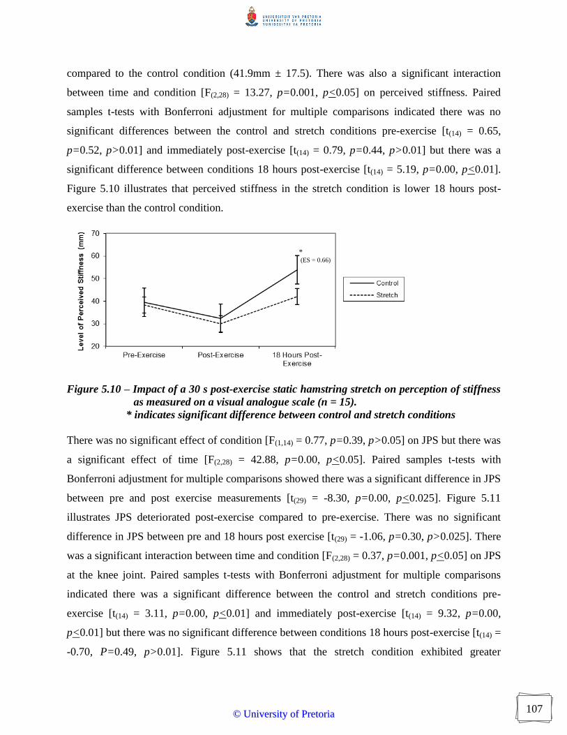
107
compared to the control condition (41.9mm ± 17.5). There was also a significant interaction
between time and condition [F(2,28) = 13.27, p=0.001, p<0.05] on perceived stiffness. Paired
samples t-tests with Bonferroni adjustment for multiple comparisons indicated there was no
significant differences between the control and stretch conditions pre-exercise [t(14) = 0.65,
p=0.52, p>0.01] and immediately post-exercise [t(14) = 0.79, p=0.44, p>0.01] but there was a
significant difference between conditions 18 hours post-exercise [t(14) = 5.19, p=0.00, p<0.01].
Figure 5.10 illustrates that perceived stiffness in the stretch condition is lower 18 hours post-
exercise than the control condition.
Figure 5.10 – Impact of a 30 s post-exercise static hamstring stretch on perception of stiffness
as measured on a visual analogue scale (n = 15).
* indicates significant difference between control and stretch conditions
There was no significant effect of condition [F(1,14) = 0.77, p=0.39, p>0.05] on JPS but there was
a significant effect of time [F(2,28) = 42.88, p=0.00, p<0.05]. Paired samples t-tests with
Bonferroni adjustment for multiple comparisons showed there was a significant difference in JPS
between pre and post exercise measurements [t(29) = -8.30, p=0.00, p<0.025]. Figure 5.11
illustrates JPS deteriorated post-exercise compared to pre-exercise. There was no significant
difference in JPS between pre and 18 hours post exercise [t(29) = -1.06, p=0.30, p>0.025]. There
was a significant interaction between time and condition [F(2,28) = 0.37, p=0.001, p<0.05] on JPS
at the knee joint. Paired samples t-tests with Bonferroni adjustment for multiple comparisons
indicated there was a significant difference between the control and stretch conditions pre-
exercise [t(14) = 3.11, p=0.00, p<0.01] and immediately post-exercise [t(14) = 9.32, p=0.00,
p<0.01] but there was no significant difference between conditions 18 hours post-exercise [t(14) =
-0.70, P=0.49, p>0.01]. Figure 5.11 shows that the stretch condition exhibited greater
* (ES = 0.66)
©© UUnniivveerrssiittyy ooff PPrreettoorriiaa

108
deterioration in JPS immediately post-exercise than did the control condition but these values
had returned to near baseline by 18 hours post-exercise.
Figure 5.11 – Impact of a 30 s post-exercise static hamstring stretch on JPS (n = 15).
* indicates significant difference between control and stretch conditions
There was a strong significant positive relationship between passive peak torque during a
controlled stretch manoeuvre of the hamstring muscle group and peak eccentric torque of the
hamstring muscle group generated during a maximal eccentric contraction at 2.09 rad.s-1
(r =
0.74, p<0.05; Figure 5.12).
Figure 5.12 – Scatter plot of passive peak against peak eccentric hamstring torque (n = 15).
R2 = 0.55
*
* (ES = 0.61)
©© UUnniivveerrssiittyy ooff PPrreettoorriiaa

109
There was a significant positive relationship between total energy absorbed during passive
stretch of the hamstring muscle group and total energy absorbed during a maximal eccentric
contraction of the hamstring muscle group at 2.09 rad.s-1
(r = 0.61, p<0.05; Figure 5.13).
Figure 5.13 – Scatter plot of total passive against total active hamstring energy absorption (n =
15).
A significant relationship was found between immediate post-exercise passive energy absorption
and perception of stiffness in the stretch condition (r = 0.53, p<0.05, R2 = 0.28). There was no
significant relationship between the two variables in the control condition (p >0.05). 18 hours
post-exercise there was a significant negative relationship between passive energy absorption
and perception of stiffness in the control condition (r = -0.73, p<0.05, R2 = 0.53) and a weaker
but still significant relationship between the two variables in the stretch condition (r = -0.32,
p<0.05, R2 = 0.10).
5.4 DISCUSSION
The aim of this study was to investigate the impact of fatigue and post-exercise static stretching
on measures of static and dynamic hamstring flexibility and performance, and it was
hypothesised that fatigue would have a negative influence and post-exercise static stretching a
positive influence on these measures. Previous research has generally induced fatigue in single
muscles or muscle groups rather than through repeated dynamic whole body efforts, which are
more typical of team sports (Pinniger et al., 2000; Enoka, 2012). The protocol used in this study
R2 = 0.37
©© UUnniivveerrssiittyy ooff PPrreettoorriiaa

110
specifically related to what a player would perform during a match with comparable sprint
distances and work:rest ratios (Deutsch et al., 1998). The mean sprint time increased by 8% from
the first to the final sprint and since fatigue has been characterised by an inability to sustain the
original work rate (Reilly et al., 2008) these results suggest the repeated sprint protocol
employed in this study was of adequate duration and intensity to evoke fatigue.
5.4.1 Passive dynamic flexibility measurements to 0.99 rad
There was no significant main effect for time, condition or interaction effect between time and
condition for passive torque, stiffness and energy absorbed to 0.99 rad. Fatigued muscles are
thought to show increased resting tension (Gong et al., 2000) and in animal models, fatigued
muscles could absorb less energy than non-fatigued muscles with the differences being much
more evident in the earlier part of the range (Mair et al., 1996). Findings of this study did not
support this in humans and therefore hypothesis 1 was rejected. The disagreement of results of
this and previous research conducted in vitro could be due to a number of factors. Fatigue has
multiple mechanical effects and it is possible that the contractile properties of muscle studied in
vitro are not predominantly important and local muscle factors induced by altered energy
supplies and metabolic changes are also critical (Rahnama et al., 2006). The utilisation of whole
body repeated dynamic efforts to induce fatigue rather than the cyclic stimulations of muscle
used by Mair et al. (1996) might have influenced results and it is also possible that muscles
would behave differently when purely longitudinal forces are applied in vitro than when the
forces resulting from the mechanical output of the agonist as well as all synergists and
antagonists crossing the joint are applied to multi-articular muscles (Maganaris, 2001).
Previous studies have attempted to identify the effect of stretching protocols on passive dynamic
flexibility of the hamstring muscle group (Magnusson et al., 1995; Magnusson et al., 1996;
Magnusson et al., 2000; Reid & McNair, 2004; Nordez et al., 2006; Whatman et al., 2006;
Marshall et al., 2011; Matsuo et al., 2013) and have shown that passive torque, stiffness and
energy absorbed remain unchanged during stretch to a common angle. Results of this study agree
with previous research in showing that stretching fatigued muscle appears to have no effect on
the viscoelastic properties in this early part of the range and supports research which has
considered the effect of stretch on non-fatigued human muscle (Magnusson et al., 1997). These
findings do not concur with those conducted on animal muscle fibres or individual muscles in
©© UUnniivveerrssiittyy ooff PPrreettoorriiaa
111
vitro (Brooks et al., 1995; Mair et al., 1996). As much in vitro research is conducted on single
muscle fibres or individual fusiform muscles it is possible that these have a different response to
multipennate muscle groups in vivo. Muscle fibres belonging to different motor unit types have
been shown to have different lengths (Maganaris, 2003; Epstein et al., 2006) and do not extend
from one tendon plate to another but are arranged in series along the length of the muscle. This
suggests that muscle deformation may be differentially distributed between muscle fibre types
and therefore that strain experienced by fiber type is not equivalent (Lieber & Friden, 2002). In
vitro models operate through the entire force-length relationship available while in vivo models
may only operate in a part of the curve, because of anatomical and neural constraints (Maganaris,
2001). Although the mechanisms for the difference are not clear and require further research, the
results of this study provide evidence that findings from in vitro investigations might not be
directly related to human hamstring muscles.
5.4.2 Final passive dynamic flexibility measurements
There was no significant effect of time or interaction effect between time and condition on
passive peak torque, suggesting that fatigue does not negatively influence this constraint of
passive dynamic flexibility. There was a significant effect of condition on passive peak torque
with the stretch condition having a lower mean throughout the protocol than the control
condition and therefore partly rejecting hypotheses 1 and 2. Previous research has examined the
effects of acute, cyclical or chronic stretch on passive dynamic flexibility and have repeatedly
shown that following stretch protocols increases in torque about the joint occur, and these have
been attributed to increases in muscle length (Gajdosik, 1991) or increased stretch tolerance
through induced natural analgesic responses of the body (Magnusson, 1998; Ben & Harvey,
2010; Weppler & Magnusson, 2010; Marshall et al., 2011). It is well accepted that exercise can
alter pain perception in humans and animals (Koltyn, 2000) due to activation of the endogenous
opioid system in which the release of endorphins and enkephalins blocks the productions of
‘Substance P’, a neurotransmitter in the pain circuit (Piercey et al., 1986), but it is possible that
as the participants used in this study were professional athletes and would thus be used to
exercising at maximal levels, that the fatiguing effect of the repeated sprint protocol was not
sufficient to activate this response.
©© UUnniivveerrssiittyy ooff PPrreettoorriiaa
112
There was no significant main effect of time or condition or interaction effect on final stiffness
thus partly rejecting hypothesis 1. It is commonly assumed in the clinical and laboratory setting
that stiffness and flexibility are synonymous concepts (Blackburn et al., 2004). Flexibility gives
an indication of the ability of the musculotendinous unit to elongate without consideration of the
associated force response, and hence stiffness. The final stiffness is the gradient of the load-
deformation curve at the maximal deformation (i.e. limit of flexibility). Because static stretching
can increase levels of static flexibility (Cornelius et al., 1992; Bandy & Irion, 1994; Bandy et al.,
1997; Klinge et al., 1997; Wiemann & Hahn, 1997; Bandy et al., 1998; McHugh et al., 1998;
Roberts & Wilson, 1999; Depino et al., 2000; Chan et al., 2001; Funk et al., 2001; Funk et al.,
2003; Zakas et al., 2003; Nelson & Bandy, 2004; Young et al., 2004; Davis et al., 2005; Zakas et
al., 2006; Bacurau et al., 2009; Fasen et al., 2009; O'Sullivan et al., 2009; Ayala & Sainz de
Baranda Andújar, 2010; Covert et al., 2010; O'Hora et al., 2011), it has been inferred to have
concurrent effects in reducing stiffness. Results of this and other studies do not support that this
conjecture is applicable to the hamstring muscle (Blackburn et al., 2004; Aquino et al., 2006).
Other studies have shown that acute passive stretching of the plantar flexor muscle group reduces
passive stiffness (Morse et al., 2008; Ryan et al., 2008; Herda et al., 2010; Kato et al., 2010;
Nakamura et al., 2011). LoA calculated in Chapter 3 showed a degree of random error in values
of stiffness from the torque-angle curve obtained from passive stretch on an isokinetic
dynamometer and it is possible this concealed a meaningful effect in this study (Bland &
Altman, 1986). Alternatively, studies examining the plantar flexor muscle group used stretch
durations of up to 20 minutes, unlike this study which used a clinically relevant stretch duration
of 30 seconds. Studies using greater stretch durations are more likely to find statistical
significance in their results, which may partly explain the conflicting findings (Behm &
Chaouachi, 2011; Matsuo et al., 2013).
There was no significant main effect of time or condition on total passive hamstring energy
absorption but there was a significant interaction effect. Post-hoc analysis identified no
significant difference between control and stretch conditions at any time interval but mean data
demonstrated decreased energy absorption post-exercise in the control condition compared to
increased energy absorption in the stretch condition, thus supporting hypothesis 2. Fatigue is an
identified risk factor for injury and accepted conjecture is that fatigued muscles absorb less
energy and are stiffer than non-fatigued muscles (Mair et al., 1996; Verrall et al., 2005; Gabbett
©© UUnniivveerrssiittyy ooff PPrreettoorriiaa

113
& Domrow, 2007; Alentorn-Geli et al., 2009; Small et al., 2010). Animal models have been used
to demonstrate significantly less energy absorption in fatigued muscle compared to non-fatigued
muscle, with the greatest loss occurring in the most fatigued muscle (Mair et al., 1996). Prior to
this study it remained to be established whether the energy absorbing capabilities of human
muscle are affected by fatigue in the same way, and the current study offers tentative support for
this. Performing a 30s static hamstring stretch post-exercise appears to negate the impact of
fatigue on total passive energy absorption. It has been suggested that fatigued muscle ‘bunches’
and because of it supposed shorter length, would be at greater injury risk (Westerblad et al.,
1993). This idea has been supported by findings of reduced static flexibility in fatigued muscle
(Cote et al., 2008). Researchers have suggested that post-exercise stretching might help muscle
return to its resting length and thus reduce likelihood of subsequent injury (Mika et al., 2007;
Robey et al., 2009). The impact of post-exercise stretching in increasing the total passive energy
absorption of the hamstring muscle 18 hours post-exercise provides a mechanism by which post-
exercise static stretching may reduce susceptibility to injury in ensuing training sessions or
games. The small effect size (0.18) between energy absorption of the hamstring muscle pre-
exercise and 18 hours post-exercise suggests, that although there is a significant difference, its
clinical meaningfulness is questionable.
5.4.3 Active dynamic flexibility measurements
There was no significant effect of time or interaction effect between time and condition on
eccentric peak torque, thus disputing hypothesis 1. There was a significant effect of condition
with the stretch condition overall resulting in higher eccentric peak torque than the control
condition and therefore supporting hypothesis 2. Previous research has shown that concentric
and less frequently, eccentric strength of the knee flexors and extensors is impaired after exercise
protocols comprising isokinetic maximal contractions (Sangnier & Tourny-Chollet, 2008), high-
intensity cycling (Garrandes et al., 2007), and prolonged intermittent running (Rahnama et al.,
2006). This has been attributed to biomechanical factors such as decreased neural stimulation as
measured by electromyographic activation of a muscle (Gleeson et al., 1998) or increased
muscle compliance (Wilson et al., 1994). The 8% reduction in peak eccentric hamstring torque
in the control condition in this study was similar to the 8 to 15% reduction of knee extensor and
knee flexor peak torque reported after a 90 min fatiguing protocol set to simulate the work rate
during a soccer game (Rahnama et al., 2003) but the difference was not significant. Other studies
©© UUnniivveerrssiittyy ooff PPrreettoorriiaa
114
have reported greater reductions in peak torque of nearly 25% (Mercer et al., 2003) and the
conflicting findings may be due to the nature and duration of fatiguing exercise protocols using
isokinetic dynamometers inducing more localised fatigue and maximal effort through full ROM
(Enoka & Stuart, 1992; Kellis, 1999). Despite common assumptions on the effectiveness of static
stretching as part of the recovery procedure, there is little research to indicate that this modality
reduces the degree or course of post-exercise muscle soreness or force generation (Buroker &
Schwane, 1989; Dawson et al., 2005; Herbert & de Noronha, 2007; Kinugasa & Kilding, 2009;
Robey et al., 2009; Torres et al., 2013). Prior to this study, no research has examined the effect
of post-exercise static stretching on eccentric force generation either immediately or at the
subsequent training session. Results provide some evidence that a post-exercise stretch might
prevent reduction in eccentric peak torque but the small effect size (0.26) means that the clinical
meaningfulness of this is questionable.
There was no significant effect of time but there was a significant effect of condition and a
significant interaction between time and condition on active energy absorbed by the hamstring
muscle group during eccentric contraction. The active energy absorbing capability of the control
condition was significantly lower than the stretch condition both immediately and 18 hours post-
exercise, thus supporting hypothesis 2. Effect sizes were small (0.34 immediately post-exercise
and 0.27 18 hours post-exercise) despite a reduction of 9% in active energy absorption. Fatigue
is an accepted risk factor for injury with most injuries occurring late in a training session or game
when individuals are exhibiting signs of fatigue such as reduced work rate (Dadebo et al., 2004;
Woods et al., 2004; Small et al., 2010; Mooney et al., 2013). Rabbit hind limb muscle has been
shown to absorb 100% more energy when active compared to when passive, suggesting that
passive elements of muscle have the ability to absorb energy but this ability is greatly enhanced
when a muscle is active (Nikolaou et al., 1987). Any condition that decreases the ability of a
muscle to contract therefore decreases its ability to store energy and increases injury
susceptibility (Kirkendall & Garrett, 2002). Results of this study provide evidence to support
application of basic science findings to human hamstring muscle that fatigued muscle might be at
increased predisposition to injury via the mechanism of its reduced active energy absorbing
capability.
©© UUnniivveerrssiittyy ooff PPrreettoorriiaa

115
There was no significant time, condition or interaction effect in position of the torque-angle
curve that peak torque was achieved, thus disputing the suggestion that fatigue alters the shape of
this curve. Previous research into the force-length relationship has shown
that the force
depression of fatigued muscle correlates with a rightward shift in force-length characteristics of
muscle (Whitehead et al., 2001; Biewener et al., 2004; MacIntosh & MacNaughton, 2005;
MacNaughton & MacIntosh, 2007). This implies that fatigued muscles favour force generation at
longer lengths, possibly due to stretch of series
elastic elements resulting in sarcomeres
contracting at a shorter length. Findings from this study do support those of a recent in vitro
study which reported that when the series elastic compliance of whole muscle (rat medial
gastrocnemius) and aponeurosis and/or tendon is taken into account, force depression resulting
from fatigue via repetitive in situ muscle stimulation did not result in a substantial
rightward shift
in the muscle's force-length relationship. Researchers demonstrated this using sonomicrometry to
measure directly muscle fascicle length, and reported no significant change in fascicle
length
before and after fatigue (MacNaughton & MacIntosh, 2006). Prior to this, the method of
calculating active force used the passive force at the actual fascicle length during contraction,
instead of the passive force at the length of the inactive muscle.
The authors did note that this
effect would be less important for muscles having less connective tissue
and, as a result, less
series elastic compliance. The gastrocnemius of rodents, like humans, is a muscle that has
considerable passive stiffness (MacNaughton & MacIntosh, 2007) and the difference between
results of this study and those that have previously demonstrated an in vivo shift in the force-
length relationship is probably due to differences in measurement techniques affecting these
factors.
5.4.4 Static flexibility measurements
There was a significant effect of time for all static hamstring flexibility measurements except
ASLR, thus supporting hypothesis 1. Increases in static flexibility are commonly seen after a
warm-up period (Murphy et al., 2010) and while general non-fatiguing exercise results in
increased static flexibility, probably due to the increased extensibility of warm tissues, (Noonan
et al., 1993; Magnusson et al., 2000), the significant reduction in static flexibility measurements
following the repeated sprint protocol performed in this study is likely to be due to a number of
factors. Fatigue is associated with failure of the contractile capacity of the exercised muscles and
as such, fatigued muscles are thought to exhibit greater stiffness and reduced tolerance to stretch
©© UUnniivveerrssiittyy ooff PPrreettoorriiaa

116
(Noakes, 2000; Ogai et al., 2008). Fatiguing protocols have induced significant reductions in
combined maximal hip flexion and knee extension angle during sprinting, which was attributed
to reduced hamstring length (Moller et al., 1985; Young et al., 2004) and resulting in reduced
stride length. Static flexibility measures in this study returned to values not significantly different
from pre-exercise by 18 hours post-exercise, thus these findings have limited consequence to
subsequent training sessions. There was a significant effect of condition and interaction between
time and condition for AKE and PKE, with stretch conditions maintaining higher levels of AKE
and PKE than control conditions, thus partly supporting hypothesis 2. The significant differences
between control and stretch conditions 18 hours post-exercise in AKE and PKE suggest that
performing a 30s static stretch post-exercise seems to negate the loss of static flexibility during
AKE and PKE tests. This might be related to the increased passive and active energy absorption
seen in the stretch condition 18 hours after exercise but further studies are needed to ascertain
possible mechanisms for this.
5.4.5 Perception of stiffness
There was a significant time effect on perceived stiffness with significantly lower perceived
stiffness immediately post-exercise and significantly higher perceived stiffness 18 hours post-
exercise compared to pre-exercise, and a significant interaction between time and stretch
condition. Mean data indicated that perceived stiffness was significantly greater in the control
condition compared to the stretch condition 18 hours post-exercise. It is possible that the
decrease in perception of stiffness following exercise can also be partly attributed to the
increased tissue temperature reducing the viscoelasticity of the muscles thus allowing easier
movements for a given force. Moreover, maximal sprinting will require relatively high levels of
hip and knee flexion and extension therefore thixotropic associated stiffness will be minimised.
In muscles, bonds between fibres become detached as a result of movement or stretch and
gradually reform during rest (Proske & Morgan, 1999; Walsh et al., 2009). Exercise is associated
with a ‘loosening up’ of muscles and joints and as the participants had been using their joints
through a relatively full part of their range, this would have ensured that most fibres had been
recruited throughout their range. Exercise has also been shown to induce analgesia via alterations
in the transmission of nociceptive (pain) fibres. This is caused by the endorphins and enkephalins
released as a result of the exercise blocking the signal at synapses (Pert, 1982; Sforzo, 1989).
This effect, in combination with the feelings of euphoria caused by the release of these
©© UUnniivveerrssiittyy ooff PPrreettoorriiaa
117
chemicals, might contribute to lessening in perception of stiffness. The first hint of fatigue in
humans is often the sense that it is necessary to increase the effort associated with the task, rather
than an inability to exert the necessary force (Gandevia et al., 1981; Jones & Hunter, 1983;
Ferrell et al., 1987; Twist et al., 2012). This suggests that individuals judgements are based on
the effort required to generate a force, rather than the absolute magnitude of the force generated,
and psychophysical experiments have demonstrated the independence of perceived effort and
force failure during sustained activity (Psek & Cafarelli, 1993; Noakes et al., 2004; Swart et al.,
2012). It has been proposed that these alterations in perception are due to the internal actions of
motor commands or corollary discharge (McCloskey et al., 1983; Cafarelli, 1988) and that the
association is influenced by humoral factors circulating in the cerebrospinal fluid (Gerald, 1978;
Heyes et al., 1985). Although related to force production, it is possible that similar mechanisms
influence perception of stiffness. Immediately post-exercise, there was decreased perceived
stiffness despite reduced static and dynamic flexibility measures. A greater perception of
capability relative to true capability may leave the individual partially unprotected during an
episode of externally imposed mechanical stressors (Doyle et al., 1998) and individuals may also
choose actions in competitive situations that may have inherent extra risk when in conjunction
with fatigue-related reduced neuromuscular performance capabilities (Gabbett, 2008; Cè et al.,
2013), for example stretching into a range that is no longer available and thus incurring a strain
injury.
PPM correlations showed that perception of stiffness shared a significant positive relationship
with passive energy absorption in the stretch condition immediately post-exercise (r = 0.53,
p<0.05, common variance 28%). Although this would be classed as a moderately strong
relationship according to the correlation coefficient value (Atkinson & Nevill, 1998), these
variables share a common variance of only 28% suggesting that there are other factors
influencing level of perceived stiffness. These might include psychological factors such as
motivation levels, anxiety, influence of peer group, and self-confidence (Smith, 2003; Tod et al.,
2003; McIntosh, 2005; Twist et al., 2012). 18 hours post-exercise both the stretch and the control
conditions shared significant negative relationships between passive energy absorption and
perception of stiffness (r = -0.32 and r = -0.71, p<0.05, common variance 10% and 50%).
Although these common variances are low, it is possible that at this time the participants who felt
looser had a reduced capacity for energy absorption and were thus at greater risk of sustaining a
©© UUnniivveerrssiittyy ooff PPrreettoorriiaa

118
muscle strain injury. Psychological factors play an important role in performance. The placebo
effect is a psychological phenomenon where the measurable, observable, or felt improvement in
health or behaviour is not attributable to a medication or invasive treatment that has been
administered (Beedie & Foad, 2009). If athletes feel that post-exercise stretching makes them
less stiff and better able to perform to their full capacity the following day, inclusion of these
techniques in a cool-down regime might be useful if there were no apparent negative effects.
5.4.6 Joint position sense
Results of this study supported those of previous studies in finding a significant effect of time on
JPS, with a significant deterioration in JPS immediately post-exercise in both control and stretch
conditions (Skinner et al., 1986; Walsh et al., 2004; Walsh et al., 2006; Givoni et al., 2007) and
supporting hypothesis 3. The magnitude of the change in the control condition in this study (1.5o)
was similar to the 2.1o to 4.5
o previously reported. While altered JPS has previously been
considered an injury risk factor (Larsen et al., 2005; Givoni et al., 2007; Bonsfills et al., 2008;
Ergen & Ulkar, 2008), the influence of fatigue on JPS has received only limited scrutiny (Miura
et al., 2004; Mohammadi & Roozdar, 2010; Pinsault & Vuillerme, 2010; Vafadar et al., 2012).
Muscles subjected to an eccentric fatiguing protocol exhibit greater deterioration in JPS that
those having undertaken a concentric fatiguing protocol, which might be as a result of the greater
muscle damage induced by eccentric exercise (Aarimaa et al., 2004).
There was a significant interaction between time and condition on JPS and post-hoc analysis
indicated a significant difference between stretch and control conditions at baseline and
immediately post-exercise, but not 18 hours post-exercise. Comparison of the means indicated
the stretch condition (3.3o) led to a greater deterioration in JPS than the control condition (1.5
o),
therefore hypothesis 4 was rejected. It is likely that the increased deterioration in JPS in the
stretch condition is due to altered muscle spindle activity as a result of the stretch. Muscle
spindles provide information about limb position and velocity and the loading of the knee joint
(Laskowski et al., 2000). This assumption is supported by the observation that the muscle
spindles are affected by a previous history of muscle stretching (Chalmers, 2004). It has also
been suggested that static stretching may adjust the positional sensitivity of the muscular
receptors by affecting the series elastic component of the muscles (Bjorklund et al., 2001). The
reduction in JPS was not evident 18 hours post-exercise but may be a consideration when
©© UUnniivveerrssiittyy ooff PPrreettoorriiaa
119
athletes are encouraged to stretch during breaks in play or training in attempts to minimise the
effects of fatigue, although any possible detriments must be balanced against potential benefits.
5.4.7 Practical implications
Results of this study suggest mechanisms via which fatigue might increase injury predisposition
and therefore can offer some tentative practical implications for sports science and medical
practitioners:
Although these data were gathered on an isokinetic dynamometer and therefore have limited
applicability to real life sprinting situations where speed of movement is much greater as well
as additional synergistic control being needed, it is possible that they provide more insightful
information that purely static flexibility measures alone;
Post-exercise static hamstring stretching reduced losses in active peak torque and increased
active and passive energy absorption both immediately and 18 hours post-exercise, which
might be beneficial to subsequent performance and level of injury predisposition;
Post-exercise static hamstring stretching had a significant effect, with moderate to high effect
size, on perception of stiffness with participants feeling that post-exercise stretching made
them looser 18 hours later, when a subsequent training session might be likely to take place.
Incorporation of post-exercise static stretching should not be at the exclusion of other factors
which have more supporting empirical evidence, such as an aerobic cool-down (Montgomery
et al., 2008; Robey et al., 2009);
Recommendations about post-exercise static hamstring stretching should be made on an
individual rather than general basis and take into account any prior injuries that the individual
has sustained. For example, previous injuries to the ankle can affect proprioception (Alentorn-
Geli et al., 2009; Han et al., 2009; Witchalls et al., 2012) and in individuals such as these,
altered JPS might be a more major factor than in those who have otherwise unaffected
proprioceptive ability; and
Because effect sizes were generally small, differences although significant have questionable
clinical meaningfulness. A clinically meaningful implies a change that is both appreciably
different, and of value to the participant (Atkinson & Nevill, 1998). Further research is needed
using larger samples and more reliable methodology to determine whether this will increase
the effect size of post-exercise static hamstring stretching.
©© UUnniivveerrssiittyy ooff PPrreettoorriiaa
120
5.4.8 Limitations
Fatigue was assumed in this study through the increase in sprint time seen between beginning
and end of the repeated sprint protocol. This does not provide any markers of fatigue so it was
not possible to determine whether fatigue was central or peripheral, and blood sampling and/or
muscle biopsies would have been a useful inclusion to provide information on this. Given that
much research has attributed improvements in static flexibility without concurrent changes in
passive dynamic flexibility to stretch induced analgesia, termed increased stretch tolerance,
measurement of pain or hormonal markers in participants undertaking stretching regimes would
be valuable to investigate the extent that this proposed mechanism might contribute to increases
in static flexibility seen following stretching interventions.
The repeated sprint protocol used in this study did not aim to specifically induce fatigue of the
hamstring muscle group, as do protocols using specific concentric, eccentric or isometric
movements, but rather a general fatigue similar to that experienced during matches and training
sessions. Co-activation patterns during static contractions are different to those evaluated during
a range of concentric and eccentric muscle contractions such as those performed repeatedly when
sprinting (Psek & Cafarelli, 1993; Weir et al., 1998) and significant differences have also been
reported in muscle activation between isometric, eccentric and concentric contractions (Kellis,
1999). It is possible that although the fatiguing protocol utilised actions that would be performed
regularly by participants, evaluation of the effect of fatigue through torque production on an
isokinetic dynamometer did not provide a complete picture. Further research investigating the
impact of fatigue on the biomechanical properties of the hamstring muscle group using
functional movements should be considered. The practical nature of the fatiguing protocol is also
likely to have induced fatigue in reciprocal muscle groups which may have affected static
flexibility measures, particularly those involving an active component, e.g. quadriceps fatigue
may have resulted in lesser achievements on AKE tests. Additionally, it is possible that
participants experienced an inability to relax fatigued muscles completely and this might have
further influenced some of the measurements. Replication of the experimental procedure with a
protocol to induce localised hamstring fatigue would determine the extent of possible effects
from these factors.
©© UUnniivveerrssiittyy ooff PPrreettoorriiaa
121
Attempts were made to control any effects of circadian variation for the studies in Chapters 3 and
4 by ensuring that testing sessions were held at similar times of the day. In order to mimic
training schedules of clubs and limit the impact of participants undertaking different types and
intensities of training sessions between the same day and next day post-exercise measurements
being taken, testing sessions for the study in Chapter 5 were held on successive afternoons and
mornings. As flexibility can be affected by circadian variation, this may have impacted on
results. Further research, which utilises a 24-hour post-exercise, could be considered to minimise
the potential impact of this. Additionally, the use of activity diaries in the periods between
testing should be considered to ensure that differences in activities of daily living among
participants did not influence results.
Because of the time constraints of the professional sportsmen used in this study and the necessity
of conducting testing during the same period of the season to minimise any effects of varying
levels of conditioning, it was necessary to employ an exercise protocol that related to a training
situation as opposed to simulating a match. This was deemed appropriate as many injuries do
occur in training situations and pre-season is a common injury point (Gabbett, 2004). Attempts
were made to relate the protocol to typical play and training as far as possible by utilising sprints
of a distance commonly seen in Rugby League and taking care that work:recovery intervals were
comparable to those seen in training situations. Despite this, it is possible that the repeated sprint
protocol used in this study induced fatigue via different mechanisms than would a longer
exercise sessions and replication of the study using an exercise protocol simulating a game
would be interesting to determine if the type of activity undertaken influences the effects of
fatigue on active and passive mechanical characteristics of the hamstring muscle.
The static stretch used in this study was chosen to mimic protocols described in texts (Maud &
Cortez-Cooper, 1994; Alter, 2004) and routinely used in coaching practice. Because time is often
constrained in professional sport, it was decided to use a stretch that a participant could perform
alone rather than an assisted stretch. As the participants assumed the stretch position, the
hamstring muscle would have been working eccentrically in reverse action to control the
lowering of the upper body and as such, the stretch was not purely a static one but had a contract
component. Getting the participants to perform seated stretches might have lessened the effect of
gravity on this but would still have had an impact. Additionally, although clear instructions were
©© UUnniivveerrssiittyy ooff PPrreettoorriiaa

122
given to keep the lower back curved, it was impossible to control for differences in lordosis. The
use of assisted stretching with the lower back kept flat would help control for this. Future studies
should consider using assisted stretches to determine if the effects are similar to those obtained
from these unassisted stretches.
5.4.9 General limitations
Conclusions made in relation to this thesis need to be examined in light of the respective
limitations, which can broadly be categorised in relation to threats to the internal and external
validity of studies. Internal validity refers to the extent to which it can be accurately stated that
the independent variable produced the observed effects, while external validity relates to the
generalisability of findings across different populations, tasks or environments (Godwin et al.,
2003).
Many of the threats to internal validity have been separately addressed in the respective studies
and are particular to the research approach undertaken. Specific aspects of the thesis that
provided some uniqueness but nonetheless some threats to internal validity were the
measurement of passive dynamic flexibility and the use of an elite athletic population. During
passive dynamic flexibility tests, participants were asked to maintain full muscular relaxation
while undergoing measurement procedures and because previous literature and pilot studies
indicated unchanged EMG levels, these were not monitored. During testing it appeared that not
all individuals were able to achieve the same levels of relaxation, as torque-angle curves of some
participants were less smooth than others. Observation of any EMG changes would have been
useful to ensure that results were not influenced by muscular resistance, but because of strict
time constraints, it was not possible to adapt methodology to include this after testing had
commenced. Although all passive measurements taken on isokinetic dynamometers were
corrected for gravity, gravity correction equations are derived for active movements. The passive
stretch protocol used in this study may have been influenced inappropriately by gravity
correction, resulting in errors in values of absolute torque produced. Although these are likely to
be constant and would therefore not affect relationship or comparison analyses, it is possible that
to compare actual passive torque values is inappropriate. In relation to the elite athletic
population, testing sessions had to be designed to fit around the club training schedule and it may
have been that some of the participants had previously partaken in more intense training sessions
©© UUnniivveerrssiittyy ooff PPrreettoorriiaa

123
than other. Fatigue after a strenuous training session has a neural manifestation with some
athletes unable to voluntarily fully activate muscle or experiencing stretch reflex inhibition after
heavy training (Kirsch & Rymer, 1992; Melnyk & Gollhofer, 2007). Had participants been
subjected to different intensities of training sessions in the days preceding testing sessions, this
might have had an influence on the results. Training diaries would have been useful in
quantifying any effect from this (Hartwig et al., 2008) and should be incorporated into future
studies of a similar nature.
Threats to external validity were related to the use of a specialised population, the specificity of
the musculoskeletal region and the procedures utilised. To limit occurrence of type II errors, a
homogenous sample of athletes (professional male rugby league players) participated in all
studies. In each study the sample size was considered sufficient to have enough power to detect
significant differences and/or relationships, as estimated by sample size calculations, but
nonetheless the elite nature of the population presented challenges in terms of developing
methodology with sufficient internal validity (see section 3.2.1) and recruiting sufficiently large
sample sizes. Furthermore, findings are only applicable to highly trained rugby players and
further research is needed before results can be extrapolated to other elite athletic populations
and more general populations. It is possible that results would be different if the same protocols
would be used on athletes with a greater than normal hamstring ROM (e.g. gymnasts) although
the lack of significant difference found in dynamic flexibility measures of participants classified
as ‘tight’ or ‘normal’ (Chapter 4) suggest that findings might be similar. Participants from a
clinical population who had irregularities in joint congruency and possible mechanical blocks
preventing them from reaching full passive knee extension warrant further investigation before
extrapolation of results. A further threat to external validity is that focus was purely on the
hamstring muscle group, the rationale for this relating to the high incidence and disability of
hamstring strain injuries. Results can therefore not be translated to other musculature in the body
as this will have different structural properties and will be subject to different magnitudes and
directions of force. Research is needed to examine the effect of fatigue on the biomechanical
properties of other muscle groups in vivo, particularly those that have a relatively high incidence
of strain injury. The procedure and application of isokinetic testing to measure passive and active
dynamic hamstring flexibility also threatened external validity as the protocols worked the
muscle groups in an isokinetic passive or eccentric manner in highly stabilised, isolated and open
©© UUnniivveerrssiittyy ooff PPrreettoorriiaa
124
chained movements. In real-life situations, musculature stabilises and fixes other joints and
movements are performed at much greater and variable velocities and involve multiple joints and
multiple muscle actions. Although findings from this study attempt to partly bridge the
difference between findings from animal models and human field-testing, a combination of
research approaches might provide a more complete picture. The development of mathematical
models to enable simulation of body kinematics and injury mechanisms in a computer
environment would be useful and isokinetic measures could then be tested for validity against
these.
Critical evaluation of the ethical approval procedures and governance associated with the
research conducted as part of the PhD process is warranted. Testing procedures throughout the
investigations conducted were considered to involve only activities that would be routinely
undertaken as part of club testing or training programmes. In line with University of Pretoria
ethics procedures, participants were required to provide written informed consent, and in the case
of minors (< 18 years), written parental or guardian consent together with verbal participant
assent. The informed consent adhered to the best practice principles established by the American
College of Sports Medicine by including disclosure of the risks and control measures for these
risks including specific risk assessment protocols (ACSM, 1998). Participants were informed
verbally and in writing of their right not to participate in the study and withdraw at any time
without needing to give a reason, nonetheless because coaches were involved in the testing and
approval of the club for the testing was sought there was a degree of implicit coercion. This is an
inherent challenge associated with any physical testing and research involving elite athletes
where club approval is necessary to practically conduct the research. A further consideration is
that of confidentiality in that research data collection was occasionally combined with fitness
testing data collection required by the club and was therefore disclosed to the club. Participants
did consent to this disclosure of information but nonetheless data is normally held confidentially
unless disclosure is required by law or to prevent harm (Shephard, 2002).
5.4.10 Reflections
This study was designed to examine the impact of fatigue and post-exercise static stretching on
measures of static and dynamic flexibility and performance. An experimental randomised cross-
over repeated measures design was utilised because of a relatively small population of available
©© UUnniivveerrssiittyy ooff PPrreettoorriiaa

125
participants and to enable as much control as possible over extraneous variables (Hopkins et al.,
1999; Godwin et al., 2003). The research study was designed with consideration to quality
assessment of methodology and was deemed to be of good methodological quality (PEDro score
9); it was not possible to blind participants completely but attempts were made by describing the
study as intending to assess the cross over effects of upper limb stretching (De Morton, 2009).
Fatigue is considered to be a modifiable risk factor for injury via suggested mechanisms
including reductions in energy absorption, force generation and proprioception (Moore et al.,
2002; Willems & Stauber, 2002; Miura et al., 2004; Melnyk & Gollhofer, 2007) but there is little
empirical in vivo evidence to substantiate these beliefs. Because of the intensity of sports training
schedules, identification of mechanisms by which fatigue might affect the properties of a muscle
to increase injury predisposition was considered important to aid in development of preventative
strategies. Extrapolations from animal literature have led to speculation that the increased injury
occurrence in fatigued muscle is due to the reduced ability of the muscle to absorb energy (Mair
et al., 1996; Lieber & Friden, 2002). Results from the control condition in Chapter 6 did not
support previous in vitro research as fatigue caused no significant changes in measures of passive
or active dynamic flexibility. Static flexibility generally significantly decreased immediately
post-exercise but had returned to baseline by 18 hours post-exercise. The reduction in static
flexibility might combine with the significantly lower perceived stiffness levels and decline in
JPS seen immediately post-exercise to provide a tentative means by which fatigue might cause
increased predisposition to hamstring injuries, although the mechanisms for this are not clear
from this study and warrant further investigation.
To explore the impact of post-exercise static stretching, measurements were taken after exercise
and 18 hours later to try and mimic normal club training schedules when individuals undertook
training sessions during afternoons and then again on subsequent mornings. Results
demonstrated significantly lower passive and active energy absorption post-exercise in the
control condition compared to the stretch condition both immediately and 18 hours post-exercise,
indicating that a 30 s post-exercise static hamstring stretch reduced fatigue-induced losses in the
energy absorbing capability of the hamstring muscle. It is possible that this might in part account
for the reduction in injury rate previously reported as a result of recovery interventions that
included stretching (Verrall et al., 2005), but further longitudinal research of experimental design
is required to confirm this. In addition to improved energy absorption, perceived stiffness was
©© UUnniivveerrssiittyy ooff PPrreettoorriiaa
126
significantly higher in the control condition compared to the stretch condition 18 hours post-
exercise. A detriment to post-exercise stretching was also revealed with the stretch condition
exhibiting greater deterioration in JPS than the control condition immediately post-exercise. This
is not likely to have major implications in subsequent exercise sessions but may be a
consideration when athletes are encouraged to stretch during breaks in play or training in
attempts to minimise the effects of fatigue (Verrall et al., 2005). This novel finding suggests that
care must be taken in prescribing stretching if individuals already have altered proprioceptive
abilities due to prior injury. These results provide tentative support for suggestions that post-
exercise static stretching could be beneficial to the recovery process but the small effect size
means that clinical meaningfulness might be questionable.
Previous research has attempted to measure dynamic flexibility in humans through damped
oscillation techniques (Wilson et al., 1991; Wilson et al., 1994; Walshe et al., 1996; Fukashiro et
al., 2001) or isokinetic resistance to controlled stretch (Klinge et al., 1997; Magnusson et al.,
1997; Reid & McNair, 2004; Ryan et al., 2008; Weppler & Magnusson, 2010). Controlled
passive stretch using isokinetic dynamometers was deemed to be the better of the available
methods because of the degree of subjectivity and lack of adequate reliability of damped
oscillation techniques, but nonetheless, there is some lack of ecological validity in that
manoeuvres are passive, slow, controlled and highly stabilised. Researchers have reported high
same-day intrarater PPM correlation coefficients (r = 0.91 to 0.99, P<0.05) for passive dynamic
hamstring flexibility measurements taken on a Biodex isokinetic dynamometer and interpreted
these as indicating high reliability. This evidence is limited by the inappropriate use of bivariate
correlations and same-day test and retest, and thus the reliability of these measures had not been
appropriately established prior to this study. Results of Chapter 3 demonstrated high relative
reliability with intraday ICCs (3,1) of r = 0.85 to 0.98 (P<0.05) and interday ICCs (3,1) of r =
0.89 to 0.98 (P<0.05). Absolute reliability values demonstrated random error of 7.3 to 10.7 Nm
for passive peak torque and 4.4 to 7.1 J for passive energy absorption. Previous research has
tended to consider decline in peak torque as a result of stretch rather than passive peak torque or
energy absorption when investigating impact of interventions on passive dynamic flexibility
(McHugh et al., 1992; Magnusson et al., 1996; LaRoche & Connolly, 2006; Marshall et al.,
2011). This study found non-significant changes in passive peak torque of 2.2 to 4.1 Nm and in
passive energy absorption of 3.3 J as a result of post-exercise stretch, therefore it is possible that
©© UUnniivveerrssiittyy ooff PPrreettoorriiaa
127
as the degree of random error is larger than the observed changes, the measurement technique
was not sufficiently sensitive and type II errors might have occurred (no impact of stretch was
detected on passive and active dynamic hamstring flexibility but in actuality there was one).
Procedures must be developed to reduce the extent of random error before future studies are
conducted
This was an opportunistic study designed around the availability of professional rugby league
players under the condition that testing would not adversely affect training or playing abilities.
Opportunities to conduct research on such populations are rare and provide a homogenous
sample but also present some difficulties. The testing battery took four hours on each of four
afternoons and two hours on each of the subsequent mornings. These were the times made
available by the coach and strict adherence to these was necessary. Because of this, there was no
way the testing protocol could be adapted after testing commenced. Additionally, the variables
selected, although providing more insight than has previously been gleaned into mechanisms by
which fatigue and post-exercise static stretching might influence measures of static and dynamic
flexibility, are insufficient to conclude on all aspects of movement science and further
investigation using a combination of research methods is warranted.
5.4.11 Future directions of research
This thesis attempted to use a fairly novel and infrequently reported measure of passive dynamic
flexibility to further understanding of flexibility, stretching and injury susceptibility. Despite
establishing via pilot studies a workable protocol and providing evidence the measurement
procedure had adequate relative reliability and acceptable measurement error, it nonetheless still
lacked some ecological validity. To further understanding, more active measurements that mimic
actions and speeds of real-life sport movements are required, and should also include
consideration of the complex interaction of factors that constrain flexibility. For instance,
fatiguing protocols have been reported to lead to significant reductions in combined maximal hip
flexion and knee extension angle during sprinting, resulting in decreased stride length and
attributed to reduced hamstring length (Moller et al., 1985; Young et al., 2004). Empirical
evidence supporting such assumptions is lacking and measurement of force-deformation
characteristics of muscle to establish active dynamic flexibility in a real life situation is
warranted to further understanding. This would be complicated and require the use of complex
©© UUnniivveerrssiittyy ooff PPrreettoorriiaa
128
mathematical modelling of data from force plates, accelerometers and motion analysis software
at the very least. A similar criticism can be made regarding the measure of JPS, which was
assessed using a commonly described and accepted method (Givoni et al., 2007; Walsh et al.,
2009; Gear, 2011). The movements involved were slow and controlled and performed in an open
chain manner, whereas during locomotion the adaptation of movements by proprioceptive
feedback is more dynamic and involves large forces which often need to be generated in split
seconds. Future research would be useful to determine fatigue induced alterations in JPS in more
real world situations using methodology that would mimic functional movements such as in
closed kinetic chain movements at higher velocities. Development of procedures to enable data
collection in more real-life scenarios and/or mathematical modelling of these situations is
necessary to add to data measured in clinical or field settings.
An underlying aspiration within the thesis was to better understand why flexibility is cited as an
injury risk factor, and how stretching might impact on flexibility, performance and susceptibility
to injury. To fully understand injury, research approaches need to be combined in order to better
understand relative risks of different internal and external factors, mechanisms of injury and
inciting events. It was beyond the scope and aims of this thesis to include a range of research
approaches, for instance athlete interviews to explore the inciting event or epidemiological
approaches to evaluate relative risks of different levels of passive and active dynamic flexibility.
The experimental approach taken in this study enabled investigation of the impact of post-
exercise stretching immediately and 18 hours post-exercise, but it was not possible to take
measurements between these two times and it would be interesting to observe the time course of
any changes. This could have implications for tournament situations where many games take
place in a relatively short period with little time for recovery in between, and would be of
particular relevance to JPS, which significantly deteriorated immediately post-stretch but had
returned to baseline 18 hours later. Prospective epidemiological investigations, ideally controlled
clinical trials where teams are followed for two or more seasons prior to implementation of the
intervention and two of more seasons after implementation of the intervention should be
conducted to develop further understanding of the potential benefits of post-exercise static
stretching. This research approach would limit influences of external variables such as weather
conditions and levels of fitness but would require large study population numbers and resources
to realise the research aims.
©© UUnniivveerrssiittyy ooff PPrreettoorriiaa

129
5.5 CONCLUSIONS
The purpose of this study was to investigate the impact of fatigue and post-exercise static
stretching on measures of static and dynamic flexibility and performance. Fatigue resulted in no
significant changes in passive dynamic flexibility to a common angle or final position, or to
active dynamic flexibility. Static flexibility generally significantly decreased immediately post-
exercise but had returned to baseline by 18 hours post-exercise. Perceived stiffness was
significantly lower immediately post-exercise and significantly higher 18 hours post-exercise
than pre-exercise. Post-exercise stretch resulted in significantly increased passive and active
energy absorption immediately and 18 hours post-exercise compared to the control condition and
in significantly reduced JPS in the stretch condition immediately post-exercise. Perceived
stiffness was significantly greater in the control condition compared to the stretch condition 18
hours post-exercise. Effect sizes were generally small so the clinical meaningfulness of
performing post-exercise static stretching is questionable, particularly as time constraints of
professional sport might mean it would at the exclusion of other, potentially more beneficial
practices.
©© UUnniivveerrssiittyy ooff PPrreettoorriiaa
130
CHAPTER 6
REFERENCES
©© UUnniivveerrssiittyy ooff PPrreettoorriiaa

131
AAGAARD, P. SIMONSEN, E.B. ANDERSEN, J.L. MAGNUSSON, S.P. HALKJAER-
KRISTENSEN, J. & DYHRE-POULSEN, P. (2000). "Neural inhibition during maximal
eccentric and concentric quadriceps contraction: effects of resistance training." Journal of
Applied Physiology, 89(6): 2249-2257.
AARIMAA, V. RANTANEN, J. BEST, T. SCHULTZ, E. CORR, D. & KALIMO, H. (2004).
"Mild eccentric stretch injury in skeletal muscle causes transient effects on tensile load and cell
proliferation." Scandinavian Journal of Medicine and Science in Sports, 14(6): 367-372.
ACSM (1998). "American College of Sports Medicine Position Stand. The recommended
quantity and quality of exercise for developing and maintaining cardiorespiratory and muscular
fitness, and flexibility in healthy adults." Medicine and Science in Sports and Exercise, 30(6):
975-991.
AGRE, J.C. (1985). "Hamstring injuries. Proposed aetiological factors, prevention, and
treatment." Sports Medicine (Auckland, N.Z.), 2(1): 21-33.
ALBEE, F.H. & GILLILAND, A.R. (1920). "Metrotherapy, or the measure of voluntary
movement." Journal of the American Medical Association, 75(15): 983-986.
ALENTORN-GELI, E. MYER, G.D. SILVERS, H.J. SAMITIER, G. ROMERO, D. LAZARO-
HARO, C. & CUGAT, R. (2009). "Prevention of non-contact anterior cruciate ligament injuries
in soccer players. Part 1: Mechanisms of injury and underlying risk factors." Knee Surgery,
Sports Traumatology Arthroscopy, 17(7): 705-729.
ALLEN, D.G. (2001). "Eccentric muscle damage: mechanisms of early reduction of force." Acta
Physiologica Scandinavica, 171(3): 311-319.
ALLEN, T.J. LEUNG, M. & PROSKE, U. (2010). "The effect of fatigue from exercise on
human limb position sense." The Journal of Physiology, 588(8): 1369-1377.
ALQUIER, L. (1916). "Un goniometre precis." Revue Neurologique, 23(2): 515-519.
ALTER, M.J. (2004). Science of Flexibility. Europe, Human Kinetics Ltd.
ALTMAN, D.G. & ANDERSEN, P.K. (1989). "Bootstrap investigation of the stability of a Cox
regression model." Statistics in Medicine, 8(7): 771-783.
AMAKO, M. ODA, T. MASUOKA, K. YOKOI, H. & CAMPISI, P. (2003). "Effect of static
stretching on prevention of injuries for military recruits." Military Medicine, 168(6): 442-446.
AMAKO, M. TSUDA, K. OYAMA, E. MASUOKA, K. YOKOI, H. & OSADA, M. (1999).
"Effect of Static Stretching on Muscle Flexibility of Military Recruits." Japanese Journal of
Orthopedic Sports Medicine, 19(4): 411-416.
©© UUnniivveerrssiittyy ooff PPrreettoorriiaa

132
ANDERSON, B. & BURKE, E.R. (1991). "Scientific, medical, and practical aspects of
stretching." Clinics in Sports Medicine, 10(1): 63-86.
ANDRISH, J.T. BERGFELD, J.A. & WALHEIM, J. (1974). "A prospective study on the
management of shin splints." The Journal of Bone and Joint Surgery, 56(8): 1697-1700.
ANLOAGUE, P.A. SPEES, V. SMITH, J. HERBENICK, M.A. & RUBINO, L.J. (2012).
"Glenohumeral range of motion and lower extremity flexibility in collegiate-level baseball
players." Sports Health: A Multidisciplinary Approach, 4(1): 25-30.
AQUINO, C.F. GONÇALVES, G.G.P. FONSECA, S.T. & MANCINI, M.C. (2006). "Analysis
of the relation between flexibility and passive stiffness of the hamstrings." Revista Brasileira de
Medicina do Esporte, 12: 195-200.
ARMSTRONG, A.D. MACDERMID, J.C. CHINCHALKAR, S. STEVENS, R.S. & KING, G.J.
(1998). "Reliability of range-of-motion measurement in the elbow and forearm." Journal of
Shoulder and Elbow Surgery, 7(6): 573-580.
ARMSTRONG, R.B. WARREN, G.L. & WARREN, J.A. (1991). "Mechanisms of exercise-
induced muscle fibre injury." Sports Medicine (Auckland, N.Z.,), 12(3): 184-207.
ARNASON, A. ANDERSEN, T.E. HOLME, I. ENGEBRETSEN, L. & BAHR, R. (2008).
"Prevention of hamstring strains in elite soccer: an intervention study." Scandinavian Journal of
Medicine and Science in Sports, 18(1): 40-48.
ARNASON, A. GUDMUNDSSON, A. DAHL, H.A. & JOHANNSSON, E. (1996). "Soccer
injuries in Iceland." Scandinavian Journal of Medicine and Science in Sports, 6(1): 40-45.
ARNASON, A. SIGURDSSON, S.B. GUDMUNDSSON, A. HOLME, I. ENGEBRETSEN, L.
& BAHR, R. (2004). "Risk factors for injuries in football." The American Journal of Sports
Medicine, 32(1 Suppl): 5S-16S.
ARNOLD, A.S. ANDERSON, F.C. PANDY, M.G. & DELP, S.L. (2005). "Muscular
contributions to hip and knee extension during the single limb stance phase of normal gait: a
framework for investigating the causes of crouch gait." Journal of Biomechanics, 38(11): 2181-
2189.
ARUIN, A.S. PRILUTSKII, B.I. RAITSIN, L.M. & SAVEL'EV, I.A. (1979). "Biomechanical
properties of muscles and efficiency of movement." Human Physiology, 5(4): 426-434.
ATKINSON, G. & NEVILL, A.M. (1998). "Statistical methods for assessing measurement error
(reliability) in variables relevant to sports medicine." Sports Medicine (Auckland, N.Z.), 26(4):
217-238.
ATKINSON, G. & REILLY, T. (1995). "Effects of age and time of day on preferred work rates
during prolonged exercise." Chronobiology International, 12(2): 121-134.
©© UUnniivveerrssiittyy ooff PPrreettoorriiaa

133
AVELA, J. FINNI, T. LIIKAVAINIO, T. NIEMELÄ, E. & KOMI, P.V. (2004). "Neural and
mechanical responses of the triceps surae muscle group after 1 h of repeated fast passive
stretches." Journal of Applied Physiology, 96(6): 2325-2332.
AVELA, J. KYROLAINEN, H. & KOMI, P.V. (1999). "Altered reflex sensitivity after repeated
and prolonged passive muscle stretching." Journal of Applied Physiology, 86(4): 1283-1291.
AWAN, R. SMITH, J. & BOON, A.J. (2002). "Measuring shoulder internal rotation range of
motion: a comparison of 3 techniques." Archives of Physical Medicine and Rehabilitation,
83(9): 1229-1234.
AYALA, F. DE BARANDA, P.S. CROIX, M.D.S. & SANTONJA, F. (2012a). "Reproducibility
and Concurrent Validity of Hip Joint Angle Test for Estimating Hamstring Flexibility in
Recreationally Active Young Men." The Journal of Strength and Conditioning Research,
26(9): 2372-2382.
AYALA, F. SAINZ, B.P. DE STE, C.M. & SANTONJA, F. (2012b). "Absolute reliability of
five clinical tests for assessing hamstring flexibility in professional futsal players." Journal of
Science and Medicine in Sport, 15(2): 142-147.
AYALA, F. & SAINZ DE BARANDA ANDÚJAR, P. (2010). "Effect of 3 different active
stretch durations on hip flexion range of motion." The Journal of Strength and Conditioning
Research, 24(2): 430-436.
AYALA, F. SAINZ DE BARANDA, P. DE STE CROIX, M. & SANTONJA, F. (2011).
"Absolute reliability of five clinical tests for assessing hamstring flexibility in professional futsal
players." Journal of Science and Medicine in Sport, 15(2): 142-147.
AYALA, F. SAINZ DE BARANDA, P. DE STE CROIX, M. & SANTONJA, F. (2011).
"Criterion-related validity of four clinical tests used to measure hamstring flexibility in
professional futsal players." Physical Therapy in Sport, 12(4): 175-181.
AYALA, F. SAINZ DE BARANDA, P. DE STE CROIX, M. & SANTONJA, F. (2012).
"Reproducibility and criterion-related validity of the sit and reach test and toe touch test for
estimating hamstring flexibility in recreationally active young adults." Physical Therapy in
Sport, 13(4): 219-226.
BACH, T.M. CHAPMAN, A.E. & CALVERT, T.W. (1983). "Mechanical resonance of the
human body during voluntary oscillations about the ankle joint." Journal of Biomechanics,
16(1): 85-90.
BACURAU, R.F.P. MONTEIRO, G.A. UGRINOWITSCH, C. TRICOLI, V. CABRAL, L.F. &
AOKI, M.S. (2009). "Acute effect of a ballistic and a static stretching exercise bout on flexibility
and maximal strength." The Journal of Strength and Conditioning Research, 23(1): 304-308.
BAHR, R. & HOLME, I. (2003). "Risk factors for sports injuries--a methodological approach."
British Journal of Sports Medicine, 37(5): 384-392.
©© UUnniivveerrssiittyy ooff PPrreettoorriiaa
134
BAHR, R. & KROSSHAUG, T. (2005). "Understanding injury mechanisms: a key component of
preventing injuries in sport." British Journal of Sports Medicine, 39(6): 324-329.
BALTACI, G. UN, N. TUNAY, V. BESLER, A. & GERCEKER, S. (2003). "Comparison of
three different sit and reach tests for measurement of hamstring flexibility in female university
students." British Journal of Sports Medicine, 37(1): 59-61.
BANDY, W.D. & IRION, J.M. (1994). "The effect of time on static stretch on the flexibility of
the hamstring muscles." Physical Therapy, 74(9): 845-850; discussion 850-842.
BANDY, W.D. IRION, J.M. & BRIGGLER, M. (1997). "The effect of time and frequency of
static stretching on flexibility of the hamstring muscles." Physical Therapy, 77(10): 1090-1096.
BANDY, W.D. IRION, J.M. & BRIGGLER, M. (1998). "The effect of static stretch and
dynamic range of motion training on the flexibility of the hamstring muscles." The Journal of
Orthopaedic and Sports Physical Therapy, 27(4): 295-300.
BARNETT, A. (2006). "Using recovery modalities between training sessions in elite athletes:
does it help?" Sports Medicine (Auckland, N.Z.), 36(9): 781-796.
BATTERHAM, A.M. & GEORGE, K.P. (2003). "Reliability in evidence-based clinical practice:
a primer for allied health professionals q." Physical Therapy in Sport, 4: 122-128.
BAZETT-JONES, D.M. WINCHESTER, J.B. & MCBRIDE, J.M. (2005). "Effect of
potentiation and stretching on maximal force, rate of force development, and range of motion."
Journal of Strength and Conditioning Research, 19(2): 421-426.
BEAUDOIN, C.M. & WHATLEY BLUM, J. (2005). "Flexibility and running economy in
female collegiate track athletes." The Journal of Sports Medicine and Physical Fitness, 45(3):
295-300.
BECKETT, J.R. SCHNEIKER, K.T. WALLMAN, K.E. DAWSON, B.T. & GUELFI, K.J.
(2009). "Effects of static stretching on repeated sprint and change of direction performance."
Medicine and Science in Sports and Exercise, 41(2): 444-450.
BEEDIE, C.J. & FOAD, A.J. (2009). "The placebo effect in sports performance: a brief review."
Sports Medicine (Auckland, N.Z.), 39(4): 313-329.
BEHM, D.G. BAMBURY, A. CAHILL, F. & POWER, K. (2004). "Effect of acute static
stretching on force, balance, reaction time, and movement time." Medicine and Science in
Sports and Exercise, 36(8): 1397-1402.
BEHM, D.G. BUTTON, D.C. & BUTT, J.C. (2001). "Factors affecting force loss with prolonged
stretching." Canadian Journal of Applied Physiology, 26(3): 261-272.
©© UUnniivveerrssiittyy ooff PPrreettoorriiaa
135
BEHM, D.G. & CHAOUACHI, A. (2011). "A review of the acute effects of static and dynamic
stretching on performance." European Journal of Applied Physiology, 111(11): 2633-2651.
BEHM, D.G. & KIBELE, A. (2007). "Effects of differing intensities of static stretching on jump
performance." European Journal of Applied Physiology, 101(5): 587-594.
BEIJSTERVELDT, A. PORT, I. VEREIJKEN, A. & BACKX, F. (2012). "Risk Factors for
Hamstring Injuries in Male Soccer Players: A Systematic Review of Prospective Studies."
Scandinavian Journal of Medicine & Science in Sports, 23(3): 253-262.
BEN, M. & HARVEY, L.A. (2010). "Regular stretch does not increase muscle extensibility: a
randomized controlled trial." Scandinavian Journal of Medicine and Science in Sports, 20(1):
136-144.
BENNELL, K. TULLY, E. & HARVEY, N. (1999). "Does the toe-touch test predict hamstring
injury in Australian Rules footballers?" The Australian Journal of Physiotherapy, 45(2): 103-
109.
BENNELL, K. WAJSWELNER, H. LEW, P. SCHALL-RIAUCOUR, A. LESLIE, S. PLANT,
D. & CIRONE, J. (1998). "Isokinetic strength testing does not predict hamstring injury in
Australian Rules footballers." British Journal of Sports Medicine, 32(4): 309-314.
BEST, T.M. MCELHANEY, J.H. GARRETT, W.E., JR. & MYERS, B.S. (1995). "Axial strain
measurements in skeletal muscle at various strain rates." Journal of Biomechanical
Engineering, 117(3): 262-265.
BEYNNON, B.D. RENSTROM, P.A. ALOSA, D.M. BAUMHAUER, J.F. & VACEK, P.M.
(2001). "Ankle ligament injury risk factors: a prospective study of college athletes." Journal of
Orthopaedic Research, 19(2): 213-220.
BIERMA-ZEINSTRA, S.M. BOHNEN, A.M. RAMLAL, R. RIDDERIKHOFF, J. VERHAAR,
J.A. & PRINS, A. (1998). "Comparison between two devices for measuring hip joint motions."
Clinical Rehabilitation, 12(6): 497-505.
BIEWENER, A.A. MCGOWAN, C. CARD, G.M. & BAUDINETTE, R.V. (2004). "Dynamics
of leg muscle function in tammar wallabies (M. eugenii) during level versus incline hopping."
The Journal of Experimental Biology, 207(Pt 2): 211-223.
BIXLER, B. & JONES, R.L. (1992). "High-school football injuries: effects of a post-halftime
warm-up and stretching routine." The Family Practice Research Journal, 12(2): 131-139.
BJORKLUND, M. DJUPSJOBACKA, M. & CRENSHAW, A.G. (2006). "Acute muscle
stretching and shoulder position sense." Journal of Athletic Training, 41(3): 270-274.
BJORKLUND, M. HAMBERG, J. & CRENSHAW, A.G. (2001). "Sensory adaptation after a 2-
week stretching regimen of the rectus femoris muscle." Archives of Physical Medicine and
Rehabilitation, 82(9): 1245-1250.
©© UUnniivveerrssiittyy ooff PPrreettoorriiaa

136
BLACK, J.D. & STEVENS, E.D. (2001). "Passive stretching does not protect against acute
contraction-induced injury in mouse EDL muscle." Journal of Muscle Research and Cell
Motility, 22(4): 301-310.
BLACKBURN, J.T. PADUA, D.A. RIEMANN, B.L. & GUSKIEWICZ, K.M. (2004). "The
relationships between active extensibility, and passive and active stiffness of the knee flexors."
Journal of Electromyography and Kinesiology, 14(6): 683-691.
BLAIR, S.N. KOHL, H.W. & GOODYEAR, N.N. (1987). "Relative risks for running and
exercise injuries: Studies in three populations." Research Quarterly, 58: 221-228.
BLAND, J.M. & ALTMAN, D.G. (1986). "Statistical methods for assessing agreement between
two methods of clinical measurement." Lancet, 1(8476): 307-310.
BOJSEN-MOLLER, J. BROGAARD, K. HAVE, M.J. STRYGER, H.P. KJAER, M.
AAGAARD, P. & MAGNUSSON, S.P. (2007). "Passive knee joint range of motion is unrelated
to the mechanical properties of the patellar tendon." Scandinavian Journal of Medicine and
Science in Sports, 17(4): 415-421.
BONNAR, B.P. DEIVERT, R.G. & GOULD, T.E. (2004). "The relationship between isometric
contraction durations during hold-relax stretching and improvement of hamstring flexibility."
The Journal of Sports Medicine and Physical Fitness, 44(3): 258-261.
BONSFILLS, N. GOMEZ-BARRENA, E. RAYGOZA, J.J. & NUNEZ, A. (2008). "Loss of
neuromuscular control related to motion in the acutely ACL-injured knee: an experimental
study." European Journal of Applied Physiology, 104(3): 567-577.
BOYD, B.S. WANEK, L. GRAY, A.T. & TOPP, K.S. (2009). "Mechanosensitivity of the lower
extremity nervous system during straight-leg raise neurodynamic testing in healthy individuals."
Journal of Orthopaedic and Sports Physical Therapy, 39(11): 780-790.
BOZIC, P.R. PAZIN, N.R. BERJAN, B.B. PLANIC, N.M. & CUK, I.D. (2010). "Evaluation of
the field tests of flexibility of the lower extremity: reliability and the concurrent and factorial
validity." The Journal of Strength and Conditioning Research, 24(9): 2523-2531.
BRADLEY, P.S. OLSEN, P.D. & PORTAS, M.D. (2007). "The effect of static, ballistic, and
proprioceptive neuromuscular facilitation stretching on vertical jump performance." Journal of
Strength and Conditioning Research, 21(1): 223-226.
BRADLEY, P.S. & PORTAS, M.D. (2007). "The relationship between preseason range of
motion and muscle strain injury in elite soccer players." Journal of Strength and Conditioning
Research, 21(4): 1155-1159.
BRANDENBURG, J.P. (2006). "Duration of stretch does not influence the degree of force loss
following static stretching." The Journal of Sports Medicine and Physical Fitness, 46(4): 526-
534.
©© UUnniivveerrssiittyy ooff PPrreettoorriiaa
137
BROCKETT, C.L. MORGAN, D.L. & PROSKE, U. (2004). "Predicting hamstring strain injury
in elite athletes." Medicine and Science in Sports and Exercise, 36(3): 379-387.
BROOKS, J.H. FULLER, C.W. KEMP, S.P. & REDDIN, D.B. (2005). "A prospective study of
injuries and training amongst the England 2003 Rugby World Cup squad." British Journal of
Sports Medicine, 39(5): 288-293.
BROOKS, J.H. FULLER, C.W. KEMP, S.P. & REDDIN, D.B. (2006). "Incidence, risk, and
prevention of hamstring muscle injuries in professional rugby union." The American Journal of
Sports Medicine, 34(8): 1297-1306.
BROOKS, J.H.M. & FULLER, C.W. (2006). "The influence of methodological issues on the
results and conclusions from epidemiological studies of sports injuries: illustrative examples."
Sports Medicine, 36(6): 459-472.
BROOKS, S.V. & FAULKNER, J.A. (2001). "Severity of contraction-induced injury is affected
by velocity only during stretches of large strain." Journal of Applied Physiology, 91(2): 661-
666.
BROOKS, S.V. ZERBA, E. & FAULKNER, J.A. (1995). "Injury to muscle fibres after single
stretches of passive and maximally stimulated muscles in mice." The Journal of Physiology,
488(2): 459-469.
BRUNET, M.E. COOK, S.D. BRINKER, M.R. & DICKINSON, J.A. (1990). "A survey of
running injuries in 1505 competitive and recreational runners." The Journal of Sports Medicine
and Physical Fitness, 30(3): 307-315.
BURKETT, L.N. PHILLIPS, W.T. & ZIURAITIS, J. (2005). "The best warm-up for the vertical
jump in college-age athletic men." Journal of Strength and Conditioning Research, 19(3): 673-
676.
BUROKER, K.C. & SCHWANE, J.A. (1989). "Does postexercise stretching alleviate delayed
muscle soreness?" The Physician and Sportsmedicine, 17(6): 65-83.
BUTTERFIELD, T.A. & HERZOG, W. (2005). "Quantification of muscle fiber strain during in
vivo repetitive stretch-shortening cycles." Journal of Applied Physiology, 99(2): 593-602.
CAFARELLI, E. (1988). "Force sensation in fresh and fatigued human skeletal muscle."
Exercise and Sport Sciences Reviews, 16(1): 139-168.
CAMERON, D.M. & BOHANNON, R.W. (1993). "Relationship between active knee extension
and active straight leg raise test measurements." The Journal of Orthopaedic and Sports
Physical Therapy 17(5): 257-260.
©© UUnniivveerrssiittyy ooff PPrreettoorriiaa
138
CANNAVAN, D. COLEMAN, D.R. & BLAZEVICH, A.J. (2012). "Lack of effect of moderate-
duration static stretching on plantar flexor force production and series compliance." Clinical
Biomechanics, 27(3): 306-312.
CARPENTER, J.E. BLASIER, R.B. & PELLIZZON, G.G. (1998). "The effects of muscle
fatigue on shoulder joint position sense." The American Journal of Sports Medicine, 26(2):
262-265.
CARTER, N.D. JENKINSON, T.R. WILSON, D. JONES, D.W. & TORODE, A.S. (1997).
"Joint position sense and rehabilitation in the anterior cruciate ligament deficient knee." British
Journal of Sports Medicine, 31(3): 209-212.
CARVALHO, F.L. CARVALHO, M.C. SIMÃO, R. GOMES, T.M. COSTA, P.B. NETO, L.B.
CARVALHO, R.L. & DANTAS, E.H. (2012). "Acute effects of a warm-up including active,
passive, and dynamic stretching on vertical jump performance." The Journal of Strength and
Conditioning Research, 26(9): 2447-2452.
CASELLA, C. (1950). "Tensile force in total striated muscle, isolated fibre and sarcolemma."
Acta Physiologica Scandinavica, 21(4): 380-401.
CÈ, E. RAMPICHINI, S. AGNELLO, L. LIMONTA, E. VEICSTEINAS, A. & ESPOSITO, F.
(2013). "Effects of temperature and fatigue on the electromechanical delay components." Muscle
and Nerve, 47(4): 566-576.
CHALMERS, G. (2004). "Re-examination of the possible role of Golgi tendon organ and muscle
spindle reflexes in proprioceptive neuromuscular facilitation muscle stretching." Sports
Biomechanics, 3(1): 159-183.
CHAN, O. DEL BUONO, A. BEST, T.M. & MAFFULLI, N. (2012). "Acute muscle strain
injuries: a proposed new classification system." Knee Surgery, Sports Traumatology,
Arthroscopy, 20(11): 2356-2362.
CHAN, S.P. HONG, Y. & ROBINSON, P.D. (2001). "Flexibility and passive resistance of the
hamstrings of young adults using two different static stretching protocols." Scandinavian
Journal of Medicine and Science in Sports, 11(2): 81-86.
CHAPPELL, J.D. HERMAN, D.C. KNIGHT, B.S. KIRKENDALL, D.T. GARRETT, W.E. &
YU, B. (2005). "Effect of fatigue on knee kinetics and kinematics in stop-jump tasks." The
American Journal of Sports Medicine, 33(7): 1022-1029.
CHEN, C.C. & BARNHART, H.X. (2008). "Comparison of ICC and CCC for assessing
agreement for data without and with replications." Computational Statistics and Data Analysis,
53(2): 554-564.
CHEUNG, K. HUME, P. & MAXWELL, L. (2003). "Delayed onset muscle soreness : treatment
strategies and performance factors." Sports Medicine (Auckland, N.Z.), 33(2): 145-164.
©© UUnniivveerrssiittyy ooff PPrreettoorriiaa
139
CHOW, R. ADAMS, R. & HERBERT, R. (1994). "Straight leg raise test high reliability is not a
motor memory artefact." Australian Journal of Physiotherapy, 40: 107-107.
CHRISTOU, E.A. & CARLTON, L.G. (2002). "Motor output is more variable during eccentric
compared with concentric contractions." Medicine and Science in Sports and Exercise, 34(11):
1773-1778.
CHUMANOV, E.S. HEIDERSCHEIT, B.C. & THELEN, D.G. (2007). "The effect of speed and
influence of individual muscles on hamstring mechanics during the swing phase of sprinting."
Journal of Biomechanics, 40(16): 3555-3562.
CHUMANOV, E.S. SCHACHE, A.G. HEIDERSCHEIT, B.C. & THELEN, D.G. (2012).
"Hamstrings are most susceptible to injury during the late swing phase of sprinting." British
Journal of Sports Medicine, 46(2): 90-90.
CHURCH, J.B. WIGGINS, M.S. MOODE, F.M. & CRIST, R. (2001). "Effect of warm-up and
flexibility treatments on vertical jump performance." Journal of Strength and Conditioning
Research, 15(3): 332-336.
CLARK, S. CHRISTIANSEN, A. HELLMAN, D.F. HUGUNIN, J.W. & HURST, K.M. (1999).
"Effects of ipsilateral anterior thigh soft tissue stretching on passive unilateral straight-leg raise."
The Journal of Orthopaedic and Sports Physical Therapy, 29(1): 4-9; discussion 10-12.
CLARKE, H.H. (1975). "Joint and body range of movement." Physical Fitness Research
Digest, 5: 16-18.
COLDWELLS, A. ATKINSON, G. & REILLY, T. (1994). "Sources of variation in back and leg
dynamometry." Ergonomics, 37(1): 79-86.
CONCHOLA, E. THOMPSON, B. & SMITH, D. (2013). "Effects of neuromuscular fatigue on
the electromechanical delay of the leg extensors and flexors in young and old men." European
Journal of Applied Physiology, 113(9): 2391-2399.
CONNOLLY, D.A. SAYERS, S.P. & MCHUGH, M.P. (2003). "Treatment and prevention of
delayed onset muscle soreness." Journal of Strength and Conditioning Research, 17(1): 197-
208.
CORBIN, C.B. & FOX, K. (1985). "Flexibility: The forgotten part of fitness." Journal of
Physical Education, 16(6): 191-193.
CORBIN, C.B. & NOBLE, L. (1980). "Flexibility: A major component of physical fitness."
Journal of Physical Education and Recreation, 6: 23-60.
CORNBLEET, S.L. & WOOLSEY, N.B. (1996). "Assessment of hamstring muscle length in
school-aged children using the sit-and-reach test and the inclinometer measure of hip joint
angle." Physical Therapy, 76(8): 850-855.
©© UUnniivveerrssiittyy ooff PPrreettoorriiaa
140
CORNELIUS, W.L. EBRAHIM, K. WATSON, J. & HILL, D.W. (1992). "The effects of cold
application and modified PNF stretching techniques on hip joint flexibility in college males."
Research Quarterly for Exercise and Sport, 63(3): 311-314.
CORNWELL, A. NELSON, A.G. & SIDAWAY, B. (2002). "Acute effects of stretching on the
neuromechanical properties of the triceps surae muscle complex." European Journal of Applied
Physiology, 86(5): 428-434.
COSTA-SANTOS, C. BERNARDES, J. AYRES-DE-CAMPOS, D. COSTA, A. & COSTA, C.
(2011). "The limits of agreement and the intraclass correlation coefficient may be inconsistent in
the interpretation of agreement." Journal of Clinical Epidemiology, 64(3): 264-269.
COSTA, P.B. RYAN, E.D. HERDA, T.J. DEFREITAS, J.M. BECK, T.W. & CRAMER, J.T.
(2009). "Effects of stretching on peak torque and the H:Q ratio." International Journal of Sports
Medicine, 30(1): 60-65.
COTE, J.N. FELDMAN, A.G. MATHIEU, P.A. & LEVIN, M.F. (2008). "Effects of fatigue on
intermuscular coordination during repetitive hammering." Motor Control, 12(2): 79-92.
COVERT, C.A. ALEXANDER, M.P. PETRONIS, J.J. & DAVIS, D.S. (2010). "Comparison of
ballistic and static stretching on hamstring muscle length using an equal stretching dose."
Journal of Strength and Conditioning Research, 24(11): 3008-3014.
COX, V.M. WILLIAMS, P.E. WRIGHT, H. JAMES, R.S. GILLOTT, K.L. YOUNG, I.S. &
GOLDSPINK, D.F. (2000). "Growth induced by incremental static stretch in adult rabbit
latissimus dorsi muscle." Experimental Physiology, 85(2): 193-202.
CRAMER, J.T. BECK, T.W. HOUSH, T.J. MASSEY, L.L. MAREK, S.M. DANGLEMEIER,
S. PURKAYASTHA, S. CULBERTSON, J.Y. FITZ, K.A. & EGAN, A.D. (2007). "Acute
effects of static stretching on characteristics of the isokinetic angle - torque relationship, surface
electromyography, and mechanomyography." Journal of Sports Sciences, 25(6): 687-698.
CRAMER, J.T. HOUSH, T.J. COBURN, J.W. BECK, T.W. & JOHNSON, G.O. (2006). "Acute
effects of static stretching on maximal eccentric torque production in women." Journal of
Strength and Conditioning Research, 20(2): 354-358.
CRAMER, J.T. HOUSH, T.J. JOHNSON, G.O. WEIR, J.P. BECK, T.W. & COBURN, J.W.
(2007). "An acute bout of static stretching does not affect maximal eccentric isokinetic peak
torque, the joint angle at peak torque, mean power, electromyography, or mechanomyography."
The Journal of Orthopaedic and Sports Physical Therapy, 37(3): 130-139.
CROISIER, J. GANTEAUME, S. BINET, J. GENTY, M. & FERRET, J. (2008). "Strength
imbalances and prevention of hamstring injury in professional soccer players: a prospective
study." The American Journal of Sports Medicine, 36(8): 1469-1475.
CROISIER, J.L. (2004). "Factors associated with recurrent hamstring injuries." Sports Medicine
(Auckland, N.Z.), 34(10): 681-695.
©© UUnniivveerrssiittyy ooff PPrreettoorriiaa
141
CROISIER, J.L. FORTHOMME, B. NAMUROIS, M.H. VANDERTHOMMEN, M. &
CRIELAARD, J.M. (2002). "Hamstring muscle strain recurrence and strength performance
disorders." The American Journal of Sports Medicine, 30(2): 199-203.
CROSS, K.M. GURKA, K.K. SALIBA, S. CONAWAY, M. & HERTEL, J. (2013).
"Comparison of Hamstring Strain Injury Rates Between Male and Female Intercollegiate Soccer
Athletes." The American Journal of Sports Medicine, 41(4): 742-748.
CROSS, K.M. & WORRELL, T.W. (1999). "Effects of a Static Stretching Program on the
Incidence of Lower Extremity Musculotendinous Strains." Journal of Athletic Training, 34(1):
11-14.
CURETON, T.K. (1941). "Flexibility as an aspect of physical fitness." Research Quarterly, 12:
381-290.
DADEBO, B. WHITE, J. & GEORGE, K.P. (2004). "A survey of flexibility training protocols
and hamstring strains in professional football clubs in England." British Journal of Sports
Medicine, 38(4): 388-394.
DALLINGA, J. BENJAMINSE, A. & LEMMINK, K. (2012). "Which screening tools can
predict injury to the lower extremities in team sports?: a systematic review." Sports medicine
(Auckland, NZ), 42(9): 791-815.
DAVIES, C.T.M. & WHITE, M.J. (1981). "Muscle weakness following eccentric work in man."
Pflügers Archiv European Journal of Physiology, 392(2): 168-171.
DAVIS, D.S. ASHBY, P.E. MCCALE, K.L. MCQUAIN, J.A. & WINE, J.M. (2005). "The
effectiveness of 3 stretching techniques on hamstring flexibility using consistent stretching
parameters." Journal of Strength and Conditioning Research, 19(1): 27-32.
DAVIS, D.S. QUINN, R.O. WHITEMAN, C.T. WILLIAMS, J.D. & YOUNG, C.R. (2008).
"Concurrent validity of four clinical tests used to measure hamstring flexibility." Journal of
Strength and Conditioning Research, 22(2): 583-588.
DAWSON, B. COW, S. MODRA, S. BISHOP, D. & STEWART, G. (2005). "Effects of
immediate post-game recovery procedures on muscle soreness, power and flexiblity levels over
the next 48 hours." Journal of Science and Medicine in Sport, 8(2): 210-221.
DE FELIPE, C. HERRERO, J.F. O'BRIEN, J.A. PALMER, J.A. DOYLE, C.A. SMITH, A.J.H.
LAIRD, J.M.A. BELMONTE, C. CERVERO, F. & HUNT, S.P. (1998). "Altered nociception,
analgesia and aggression in mice lacking the receptor for substance P." Nature, 392(6674): 394-
397.
DE MORTON, N. (2009). "The PEDro scale is a valid measure of the methodological quality of
clinical trials: a demographic study." Australian Journal of Physiotherapy, 55: 129-133.
©© UUnniivveerrssiittyy ooff PPrreettoorriiaa
142
DE WEIJER, V.C. GORNIAK, G.C. & SHAMUS, E. (2003). "The effect of static stretch and
warm-up exercise on hamstring length over the course of 24 hours." The Journal of
Orthopaedic and Sports Physical Therapy, 33(12): 727-733.
DEPINO, G.M. WEBRIGHT, W.G. & ARNOLD, B.L. (2000). "Duration of Maintained
Hamstring Flexibility After Cessation of an Acute Static Stretching Protocol." Journal of
Athletic Training, 35(1): 56-59.
DEUTSCH, M.U. MAW, G.J. JENKINS, D. & REABURN, P. (1998). "Heart rate, blood lactate
and kinematic data of elite colts (under-19) rugby union players during competition." Journal of
Sports Sciences, 16(6): 561-570.
DEVITA, P. & BATES, B.T. (1988). "Intraday reliability of ground reaction force data* 1."
Human Movement Science, 7(1): 73-85.
DEVLIN, L. (2000). "Recurrent posterior thigh symptoms detrimental to performance in rugby
union." Sports Medicine, 29(4): 273-287.
DI CAGNO, A. BALDARI, C. BATTAGLIA, C. GALLOTTA, M.C. VIDEIRA, M. PIAZZA,
M. & GUIDETTI, L. (2010). "Preexercise static stretching effect on leaping performance in elite
rhythmic gymnasts." Journal of Strength and Conditioning Research, 24(8): 1995-2000.
DITROILO, M. WATSFORD, M. MURPHY, A. & DE VITO, G. (2011). "Assessing Musculo-
Articular Stiffness Using Free Oscillations." Sports Medicine, 41(12): 1019-1032.
DITROILO, M. WATSFORD, M. MURPHY, A. & DE VITO, G. (2011). "Assessing musculo-
articular stiffness using free oscillations: theory, measurement and analysis." Sports Medicine,
(Auckland, NZ) 41(12): 1019-1032.
DITROILO, M. WATSFORD, M. MURPHY, A. & DE VITO, G. (2013). "Sources of
Variability in Musculo-Articular Stiffness Measurement." PloS one, 8(5): e63719.
DIXON, J.K. & KEATING, J.L. (2000). "Variability in Straight Leg Raise Measurements::
Review." Physiotherapy, 86(7): 361-370.
DOCHERTY, C.L. MOORE, J.H. & ARNOLD, B.L. (1998). "Effects of Strength Training on
Strength Development and Joint Position Sense in Functionally Unstable Ankles." Journal of
Athletic Training, 33(4): 310-314.
DORADO, C. SANCHIS-MOYSI, J. & CALBET, J.A. (2004). "Effects of recovery mode on
performance, O2 uptake, and O2 deficit during high-intensity intermittent exercise." Canadian
Journal of Applied Physiology, 29(3): 227-244.
DOYLE, J. GLEESON, N.P. & REES, D. (1998). "Psychobiology and the athlete with anterior
cruciate ligament (ACL) injury." Sports Medicine (Auckland, N.Z.), 26(6): 379-393.
©© UUnniivveerrssiittyy ooff PPrreettoorriiaa

143
DRAPER, D.O. MINER, L. KNIGHT, K.L. & RICARD, M.D. (2002). "The Carry-Over Effects
of Diathermy and Stretching in Developing Hamstring Flexibility." Journal of Athletic
Training, 37(1): 37-42.
DRAWER, S. & FULLER, C.W. (2002). "Evaluating the level of injury in English professional
football using a risk based assessment process." British Journal of Sports Medicine, 36(6): 446-
451.
DUEHRING, M.D. FELDMANN, C.R. & EBBEN, W.P. (2009). "Strength and conditioning
practices of United States high school strength and conditioning coaches." Journal of Strength
and Conditioning Research, 23(8): 2188-2203.
DUONG, B. LOW, M. MOSELEY, A.M. LEE, R.Y. & HERBERT, R.D. (2001). "Time course
of stress relaxation and recovery in human ankles." Clinical Biomechanics (Bristol, Avon),
16(7): 601-607.
DVIR, Z. (2003). "How much is necessary to indicate a real improvement in muscle function? A
review of modern methods of reproducibility analysis." Isokinetics and Exercise Science, 11(1):
49-52.
EBBEN, W.P. CARROLL, R.M. & SIMENZ, C.J. (2004). "Strength and conditioning practices
of National Hockey League strength and conditioning coaches." Journal of Strength and
Conditioning Research, 18(4): 889-897.
EBBEN, W.P. HINTZ, M.J. & SIMENZ, C.J. (2005). "Strength and conditioning practices of
Major League Baseball strength and conditioning coaches." Journal of Strength and
Conditioning Research, 19(3): 538-546.
EGAN, A.D. CRAMER, J.T. MASSEY, L.L. & MAREK, S.M. (2006). "Acute effects of static
stretching on peak torque and mean power output in National Collegiate Athletic Association
Division I women's basketball players." Journal of Strength and Conditioning Research, 20(4):
778-782.
EILING, E. BRYANT, A.L. PETERSEN, W. MURPHY, A. & HOHMANN, E. (2007). "Effects
of menstrual-cycle hormone fluctuations on musculotendinous stiffness and knee joint laxity."
Knee Surgery, Sports Traumatology, Arthroscopy, 15(2): 126-132.
EKSTRAND, J. & GILLQUIST, J. (1983). "Soccer injuries and their mechanisms: a prospective
study." Medicine and Science in Sports and Exercise, 15(3): 267-270.
EKSTRAND, J. GILLQUIST, J. & LILJEDAHL, S.O. (1983). "Prevention of soccer injuries.
Supervision by doctor and physiotherapist." The American Journal of Sports Medicine, 11(3):
116-120.
EKSTRAND, J. HÄGGLUND, M. & WALDÉN, M. (2011). "Epidemiology of muscle injuries
in professional football (soccer)." The American Journal of Sports Medicine, 39(6): 1226-1232.
©© UUnniivveerrssiittyy ooff PPrreettoorriiaa

144
EKSTRAND, J. & KARLSSON, J. (2003). "The risk for injury in football. There is a need for a
consensus about definition of the injury and the design of studies." Scandinavian Journal of
Medicine and Science in Sports, 13(3): 147-149.
EKSTRAND, J. TIMPKA, T. & HAGGLUND, M. (2006). "Risk of injury in elite football
played on artificial turf versus natural grass: a prospective two-cohort study." British Journal of
Sports Medicine, 40(12): 975-980.
ELFTMAN, H. (1966). "Biomechanics of muscle with particular application to studies of gait."
The Journal of Bone and Joint Surgery, 48(2): 363-377.
ELIASZIW, M. YOUNG, S.L. WOODBURY, M.G. & FRYDAY-FIELD, K. (1994). "Statistical
methodology for the concurrent assessment of interrater and intrarater reliability: using
goniometric measurements as an example." Physical Therapy, 74(8): 777-788.
ELLIOTT, M. ZARINS, B. POWELL, J. & KENYON, C. (2011). "Hamstring muscle strains in
professional football players: a 10-year review." The American Journal of Sports Medicine,
39(4): 843-850.
ENGEBRETSEN, A.H. MYKLEBUST, G. HOLME, I. ENGEBRETSEN, L. & BAHR, R.
(2010). "Intrinsic Risk Factors for Hamstring Injuries Among Male Soccer Players A Prospective
Cohort Study." The American Journal of Sports Medicine, 38(6): 1147-1153.
ENOKA, R.M. (1994). Neuromechanical Basis of Kinesiology. Human Kinetics Ltd, Europe.
ENOKA, R.M. (2012). "Muscle fatigue–from motor units to clinical symptoms." Journal of
Biomechanics, 45(3): 427-433.
ENOKA, R.M. & STUART, D.G. (1992). "Neurobiology of muscle fatigue." Journal of Applied
Physiology, 72(5): 1631-1648.
EPSTEIN, M. WONG, M. & HERZOG, W. (2006). "Should tendon and aponeurosis be
considered in series?" Journal of Biomechanics, 39(11): 2020-2025.
ERGEN, E. & ULKAR, B. (2008). "Proprioception and ankle injuries in soccer." Clinics in
Sports Medicine, 27(1): 195-217.
ESPANA-ROMERO, V. ORTEGA PORCEL, F.B. ARTERO, E.G. JIMÉNEZ-PAVÓN, D.
GUTIÉRREZ SAINZ, Á. CASTILLO GARZÓN, M.J. & RUIZ, J.R. (2009). "Climbing time to
exhaustion is a determinant of climbing performance in high-level sport climbers." European
Journal of Applied Physiology, 107(5): 517-525.
ESPOSITO, F. CÈ, E. & LIMONTA, E. (2012). "Cycling efficiency and time to exhaustion are
reduced after acute passive stretching administration." Scandinavian Journal of Medicine and
Science in Sports, 22(6): 737-745.
©© UUnniivveerrssiittyy ooff PPrreettoorriiaa
145
ESTELL, J. SHENSTONE, B. & BARNSLEY, L. (1995). "Frequency of injuries in different
age-groups in an elite rugby league club." Australian Journal of Science and Medicine in
Sport, 27(4): 95-97.
ETNYRE, B.R. & ABRAHAM, L.D. (1986). "H-reflex changes during static stretching and two
variations of proprioceptive neuromuscular facilitation techniques." Electroencephalography
and Clinical Neurophysiology, 63(2): 174-179.
ETNYRE, B.R. & LEE, E.J. (1988). "Chronic and acute flexibility of men and women using
three different stretching techniques." Research Quarterly, 59(5): 222-228.
FARIA, A. GABRIEL, R. ABRANTES, J. WOOD, P. & MOREIRA, H. (2013). "Mechanical
properties of the triceps surae: Differences between football and non-football players." Journal
of Sports Sciences, (in press).
FASEN, J.M. O'CONNOR, A.M. SCHWARTZ, S.L. WATSON, J.O. PLASTARAS, C.T.
GARVAN, C.W. BULCAO, C. JOHNSON, S.C. & AKUTHOTA, V. (2009). "A randomized
controlled trial of hamstring stretching: comparison of four techniques." Journal of Strength
and Conditioning Research, 23(2): 660-667.
FEIRING, D.C. ELLENBECKER, T.S. & DERSCHEID, G.L. (1990). "Test-retest reliability of
the biodex isokinetic dynamometer." The Journal of Orthopaedic and Sports Physical Therapy
11(7): 298-300.
FERBER, R. OSTERNIG, L.R. & GRAVELLE, D.C. (2002). "Effect of PNF stretch techniques
on knee flexor muscle EMG activity in older adults." Journal of Electromyography and
Kinesiology, 12(5): 391-398.
FERLIE, E. WOOD, M. & FITZGERALD, L. (1999). "Some limits to evidence-based medicine:
a case study from elective orthopaedics." British Medical Journal, 8(2): 99-107.
FERRELL, W.R. GANDEVIA, S.C. & MCCLOSKEY, D.I. (1987). "The role of joint receptors
in human kinaesthesia when intramuscular receptors cannot contribute." The Journal of
Physiology, 386: 63-71.
FINCH, C.F. (1997). "An overview of some definitional issues for sports injury surveillance."
Sports Medicine (Auckland, N.Z.), 24(3): 157-163.
FINCH, C.F. (2011). "No longer lost in translation: the art and science of sports injury
prevention implementation research." British Journal of Sports Medicine, 45(16): 1253-1257.
FINUCANE, S.D.G. MAYHEW, T.P. & ROTHSTEIN, J.M. (1994). "Evaluation of the Gravity-
Correction Feature of a Kin-Com (R) Isokinetic Dynamometer." Physical Therapy, 74(12):
1125-1133.
©© UUnniivveerrssiittyy ooff PPrreettoorriiaa
146
FITZGERALD, G.K. ROTHSTEIN, J.M. MAYHEW, T.P. & LAMB, R.L. (1991). "Exercise-
induced muscle soreness after concentric and eccentric isokinetic contractions." Physical
Therapy, 71(7): 505-513.
FLEISHMAN, E.A. (1963). The Structure and Measurement of Physical Fitness. Englewood
Cliffs NJ, Prentice Hall.
FLEISS, J.L. (1986). "Reliability of measurement." The design and analysis of clinical
experiments: 1-32.
FLETCHER, I.M. & JONES, B. (2004). "The effect of different warm-up stretch protocols on 20
meter sprint performance in trained rugby union players." Journal of Strength and Conditioning
Research, 18(4): 885-888.
FORD, G.S. MAZZONE, M.A. & TAYLOR, K. (2005). "The effect of 4 different durations of
static hamstring stretching on passive knee-extension range of motion." Journal of Sports
Rehabilitation, 14: 95-107.
FORD, W.J. BAILEY, S.D. BABICH, K. & WORRELL, T.W. (1994). "Effect of hip position on
gravity effect torque." Medicine and Science in Sports and Exercise, 26(2): 230-234.
FOREMAN, T.K. ADDY, T. BAKER, S. BURNS, J. HILL, N. & MADDEN, T. (2006).
"Prospective studies into the causation of hamstring injuries in sport: A systematic review."
Physical Therapy in Sport, 7(2): 101-109.
FORTIER, J. LATTIER, G. & BABAULT, N. (2013). "Acute effects of short-duration isolated
static stretching or combined with dynamic exercises on strength, jump and sprint performance."
Science and Sports, (in press).
FOUSEKIS, K. TSEPIS, E. POULMEDIS, P. ATHANASOPOULOS, S. & VAGENAS, G.
(2011). "Intrinsic risk factors of non-contact quadriceps and hamstring strains in soccer: a
prospective study of 100 professional players." British Journal of Sports Medicine, 45(9): 709-
714.
FOWLES, J.R. SALE, D.G. & MACDOUGALL, J.D. (2000). "Reduced strength after passive
stretch of the human plantarflexors." Journal of Applied Physiology, 89(3): 1179-1188.
FRADKIN, A.J. GABBE, B.J. & CAMERON, P.A. (2006). "Does warming up prevent injury in
sport? The evidence from randomised controlled trials?" Journal of Science and Medicine in
Sport,9(3): 214-220.
FRIDEN, J. & LIEBER, R.L. (2001). "Eccentric exercise-induced injuries to contractile and
cytoskeletal muscle fibre components." Acta Physiologica Scandinavica, 171(3): 321-326.
FROHM, A. HEIJNE, A. KOWALSKI, J. SVENSSON, P. & MYKLEBUST, G. (2012). "A
nine‐test screening battery for athletes: a reliability study." Scandinavian Journal of Medicine
and Science in Sports, 22(3): 306-315.
©© UUnniivveerrssiittyy ooff PPrreettoorriiaa

147
FUKASHIRO, S. NODA, M. & SHIBAYAMA, A. (2001). "In vivo determination of muscle
viscoelasticity in the human leg." Acta Physiologica Scandinavica 172(4): 241-248.
FULLER, C.W. (2007). "Managing the risk of injury in sport." Clinical Journal of Sport
Medicine, 17(3): 182-187.
FUNK, D. SWANK, A.M. ADAMS, K.J. & TREOLO, D. (2001). "Efficacy of moist heat pack
application over static stretching on hamstring flexibility." Journal of Strength and
Conditioning Research, 15(1): 123-126.
FUNK, D.C. SWANK, A.M. MIKLA, B.M. FAGAN, T.A. & FARR, B.K. (2003). "Impact of
prior exercise on hamstring flexibility: a comparison of proprioceptive neuromuscular facilitation
and static stretching." Journal of Strength and Conditioning Research, 17(3): 489-492.
GABBE, B.J. BENNELL, K.L. FINCH, C.F. WAJSWELNER, H. & ORCHARD, J.W. (2006).
"Predictors of hamstring injury at the elite level of Australian football." Scandinavian Journal
of Medicine and Science in Sports, 16(1): 7-13.
GABBE, B.J. BENNELL, K.L. WAJSWELNER, H. & FINCH, C.F. (2004). "Reliability of
common lower extremity musculoskeletal screening tests." Physical Therapy in Sport 5(2): 90-
97.
GABBE, B.J. FINCH, C.F. BENNELL, K.L. & WAJSWELNER, H. (2005). "Risk factors for
hamstring injuries in community level Australian football." British Journal of Sports Medicine,
39(2): 106-110.
GABBE, B.J. FINCH, C.F. WAJSWELNER, H. & BENNELL, K.L. (2004). "Predictors of
lower extremity injuries at the community level of Australian football." Clinical Journal of
Sport Medicine, 14(2): 56-63.
GABBETT, T. KING, T. & JENKINS, D. (2008). "Applied physiology of rugby league." Sports
Medicine (Auckland, N.Z.) 38(2): 119-138.
GABBETT, T.J. (2001). "Severity and cost of injuries in amateur rugby league: a case study."
Journal of Sports Sciences 19(5): 341-347.
GABBETT, T.J. (2002). "Training injuries in rugby league: an evaluation of skill-based
conditioning games." The Journal of Strength and Conditioning Research 16(2): 236-241.
GABBETT, T.J. (2003). "Incidence of injury in semi-professional rugby league players." British
Journal of Sports Medicine 37(1): 36-44.
GABBETT, T.J. (2004). "Incidence of injury in junior and senior rugby league players." Sports
Medicine (Auckland, N.Z.) 34(12): 849-859.
©© UUnniivveerrssiittyy ooff PPrreettoorriiaa
148
GABBETT, T.J. (2004). "Reductions in pre-season training loads reduce training injury rates in
rugby league players." British Journal of Sports Medicine, 38(6): 743-749.
GABBETT, T.J. (2005). "Influence of playing position on the site, nature, and cause of rugby
league injuries." Journal of Strength and Conditioning Research,19(4): 749-755.
GABBETT, T.J. (2008). "Influence of fatigue on tackling technique in rugby league players."
Journal of Strength and Conditioning Research, 22(2): 625-632.
GABBETT, T.J. & DOMROW, N. (2005). "Risk factors for injury in subelite rugby league
players." The American Journal of Sports Medicine, 33(3): 428-434.
GABBETT, T.J. & DOMROW, N. (2007). "Relationships between training load, injury, and
fitness in sub-elite collision sport athletes." Journal of Sports Sciences, 25(13): 1507-1519.
GABBETT, T.J. & SEIBOLD, A. (2013). "Relationship Between Tests of Physical Qualities,
Team Selection, and Physical Match Performance in Semi-Professional Rugby League Players."
Journal of Strength and Conditioning Research, (in press).
GAJDOSIK, R. & LUSIN, G. (1983). "Hamstring muscle tightness. Reliability of an active-
knee-extension test." Physical Therapy, 63(7): 1085-1090.
GAJDOSIK, R.L. (1987). "Goniometric measurements at the knee." Physical Therapy, 67(6):
979-980.
GAJDOSIK, R.L. (1991). "Effects of static stretching on the maximal length and resistance to
passive stretch of short hamstring muscles." The Journal of Orthopaedic and Sports Physical
Therapy, 14(6): 250-255.
GAJDOSIK, R.L. (1995). "Flexibility or muscle length?" Physical Therap,y 75(3): 238-239.
GAJDOSIK, R.L. (2001a). "Comparison and reliability of three goniometric methods for
measuring forearm supination and pronation." Perceptual and Motor Skills, 93(2): 353-355.
GAJDOSIK, R.L. (2001b). "Passive extensibility of skeletal muscle: review of the literature with
clinical implications." Clinical Biomechanics (Bristol, Avon), 16(2): 87-101.
GAJDOSIK, R.L. (2001c). "Was hamstring muscle stiffness measured?" Archives of Physical
Medicine and Rehabilitation, 82(7): 1004-1005.
GAJDOSIK, R.L. (2002). "Relationship between passive properties of the calf muscles and
plantarflexion concentric isokinetic torque characteristics." European Journal of Applied
Physiology, 87(3): 220-227.
GAJDOSIK, R.L. & BOHANNON, R.W. (1987). "Clinical measurement of range of motion.
Review of goniometry emphasizing reliability and validity." Physical Therapy, 67(12): 1867-
1872.
©© UUnniivveerrssiittyy ooff PPrreettoorriiaa

149
GAJDOSIK, R.L. RIECK, M.A. SULLIVAN, D.K. & WIGHTMAN, S.E. (1993). "Comparison
of four clinical tests for assessing hamstring muscle length." The Journal of Orthopaedic and
Sports Physical Therapy, 18(5): 614-618.
GAJDOSIK, R.L. VANDER LINDEN, D.W. & WILLIAMS, A.K. (1999). "Influence of age on
length and passive elastic stiffness characteristics of the calf muscle-tendon unit of women."
Physical Therapy, 79(9): 827-838.
GANDEVIA, S.C. KILLIAN, K.J. & CAMPBELL, E.J.M. (1981). "The effect of respiratory
muscle fatigue on respiratory sensations." Clinical Science, 60: 463-466.
GAO, Y. WINEMAN, A.S. & WAAS, A.M. (2008). "Mechanics of muscle injury induced by
lengthening contraction." Annals of Biomedical Engineering, 36(10): 1615-1623.
GARRANDES, F. COLSON, S.S. PENSINI, M. & LEGROS, P. (2007). "Time course of
mechanical and neuromuscular characteristics of cyclists and triathletes during a fatiguing
exercise." International Journal of Sports Medicine, 28(2): 148-156.
GARRETT, W.E., JR. (1996). "Muscle strain injuries." The American Journal of Sports
Medicine, 24(6 Suppl): S2-8.
GARRETT, W.E., JR. NIKOLAOU, P.K. RIBBECK, B.M. GLISSON, R.R. & SEABER, A.V.
(1988). "The effect of muscle architecture on the biomechanical failure properties of skeletal
muscle under passive extension." The American Journal of Sports Medicine, 16(1): 7-12.
GARRETT, W.E., JR. SAFRAN, M.R. SEABER, A.V. GLISSON, R.R. & RIBBECK, B.M.
(1987). "Biomechanical comparison of stimulated and nonstimulated skeletal muscle pulled to
failure." The American Journal of Sports Medicine, 15(5): 448-454.
GEAR, W.S. (2011). "Effect of different levels of localized muscle fatigue on knee position
sense." Journal of Sports Science and Medicine, 10: 725-730.
GEHRING, D. MELNYK, M. & GOLLHOFER, A. (2009). "Gender and fatigue have influence
on knee joint control strategies during landing." Clinical Biomechanics (Bristol, Avon), 24(1):
82-87.
GERALD, M.C. (1978). "Effects of amphetamine on the treadmill endurance performance of
rats." Neuropharmacology, 17(9): 703-704.
GERRITSEN, K.G. VAN DEN BOGERT, A.J. HULLIGER, M. & ZERNICKE, R.F. (1998).
"Intrinsic muscle properties facilitate locomotor control - a computer simulation study." Motor
Control, 2(3): 206-220.
GHAFFARINEJAD, F. TAGHIZADEH, S. & MOHAMMADI, F. (2007). "Effect of static
stretching of muscles surrounding the knee on knee joint position sense." British Journal of
Sports Medicine, 41(10): 684-687.
©© UUnniivveerrssiittyy ooff PPrreettoorriiaa

150
GHANBARI, A. EBRAHIMIAN, M. MOHAMADI, M. & NAJJAR-HASANPOUR, A. (2013).
"Comparing Hold Relax-Proprioceptive Neuromuscular Facilitation and Static Stretching
Techniques in Management of Hamstring Tightness." Indian Journal of Physiotherapy and
Occupational Therapy, 7(1): 126-130.
GHONCHEH, S. & SMITH, J.C. (2004). "Progressive muscle relaxation, yoga stretching, and
ABC relaxation theory." Journal of Clinical Psychology, 60(1): 131-136.
GIBBS, N. (1993). "Injuries in professional rugby league: A three-year prospective study of the
South Sydney Professional Rugby League Football Club." The American Journal of Sports
Medicine, 21(5): 696-700.
GIFFORD, L.S. (1987). "Circadian variation in human flexibility and grip strength." Australian
Journal of Physiotherapy, 33: 3-9.
GILL, T. WILKINSON, A. EDWARDS, E. & GRIMMER, K. (2002). "The effect of either a pre
or post exercise stretch on straight leg raise range of motion (SLR-ROM) in females." Journal of
Science and Medicine in Sport, 5(4): 281-290.
GISSANE, C. HODGSON, L. & JENNINGS, D. (2012). "Time-Loss Injuries Versus Non–
Time-Loss Injuries in the First Team Rugby League Football: A Pooled Data Analysis." Clinical
Journal of Sport Medicine, 22(5): 414-417.
GISSANE, C. JENNINGS, D. KERR, K. & WHITE, J.A. (2002). "A pooled data analysis of
injury incidence in rugby league football." Sports Medicine, 32(3): 211-216.
GISSANE, C. JENNINGS, D.C. & STANDING, P. (1993). "Incidence of injury in rugby league
football." Physiotherapy, 79(5): 305-310.
GIVONI, N.J. PHAM, T. ALLEN, T.J. & PROSKE, U. (2007). "The effect of quadriceps muscle
fatigue on position matching at the knee." The Journal of Physiology, 584(Pt 1): 111-119.
GLEESON, N.P. REILLY, T. MERCER, T.H. RAKOWSKI, S. & REES, D. (1998). "Influence
of acute endurance activity on leg neuromuscular and musculoskeletal performance." Medicine
and Science in Sports and Exercise, 30(4): 596-608.
GLEIM, G.W. & MCHUGH, M.P. (1997). "Flexibility and its effects on sports injury and
performance." Sports Medicine (Auckland, N.Z.), 24(5): 289-299.
GLEIM, G.W. STACHENFELD, N.S. & NICHOLAS, J.A. (1990). "The influence of flexibility
on the economy of walking and jogging." Journal of Orthopaedic Research, 8(6): 814-823.
GODGES, J.J. MACRAE, H. LONGDON, C. TINBERG, C. & MACRAE, P.G. (1989). "The
effects of two stretching procedures on hip range of motion and gait economy." The Journal of
Orthopaedic and Sports Physical Therapy, 10(9): 350-357.
©© UUnniivveerrssiittyy ooff PPrreettoorriiaa
151
GODWIN, M. RUHLAND, L. CASSON, I. MACDONALD, S. DELVA, D. BIRTWHISTLE,
R. LAM, M. & SEGUIN, R. (2003). "Pragmatic controlled clinical trials in primary care: the
struggle between external and internal validity." BMC Medical Research Methodology, 3: 28-
34.
GÖEKEN, L. & HOF, A.L. (1993). "Instrumental straight-leg raising: results in healthy
subjects." Archives of Physical Medicine and Rehabilitation, 74(2): 194-203.
GOLDSPINK, D.F. (1976). "The effects of denervation on protein turnover of rat skeletal
muscle." Biochemical Journal, 156(1): 71-80.
GOLDSPINK, D.F. (1977). "The influence of immobilization and stretch on protein turnover of
rat skeletal muscle." The Journal of Physiology, 264(1): 267-282.
GONÇALVES, R. GURJÃO, A.L.D. JAMBASSI FILHO, J.C. FARINATTI, P.D.T.V. GOBBI,
L.T.B. & GOBBI, S. (2013). "The acute effects of static stretching on peak force, peak rate of
force development and muscle activity during single-and multiple-joint actions in older women."
Journal of Sports Sciences, 31(7): 690-698.
GONG, B. MIKI, T. SEINO, S. & RENAUD, J.M. (2000). "A K(ATP) channel deficiency
affects resting tension, not contractile force, during fatigue in skeletal muscle." American
Journal of Physiolog : Cell physiology, 279(5): C1351-1358.
GONZALEZ-RAVE, J.M. MACHADO, L. NAVARRO-VALDIVIELSO, F. & VILAS-BOAS,
J.P. (2009). "Acute effects of heavy-load exercises, stretching exercises, and heavy-load plus
stretching exercises on squat jump and countermovement jump performance." Journal of
Strength and Conditioning Research, 23(2): 472-479.
GRADY, J.F. & SAXENA, A. (1991). "Effects of stretching the gastrocnemius muscle." The
Journal of Foot Surgery, 30(5): 465-469.
GRANATA, K.P. WILSON, S.E. & PADUA, D.A. (2002). "Gender differences in active
musculoskeletal stiffness. Part I.:: Quantification in controlled measurements of knee joint
dynamics." Journal of Electromyography and Kinesiology, 12(2): 119-126.
GREENBERGER, H. WILKOWSKI, T. & BELYEA, B. (1994). "Comparison of quadriceps
peak torque using three different isokinetic dynamometers." Isokinetics Exercise and Science, 4:
70-75.
GREENE, P.R. & MCMAHON, T.A. (1979). "Reflex stiffness of man's anti-gravity muscles
during kneebends while carrying extra weights." Journal of Biomechanics, 12(12): 881-891.
GREENFIELD, B.H. GREENE, B. & JOHANSON, M.A. (2007). "The use of qualitative
research techniques in orthopedic and sports physical therapy: Moving toward postpositivism."
Physical Therapy in Sport, 8(1): 44-54.
©© UUnniivveerrssiittyy ooff PPrreettoorriiaa
152
GRIBBLE, P.A. TUCKER, W.S. & WHITE, P.A. (2007). "Time-of-day influences on static and
dynamic postural control." Journal of Athletic Training, 42(1): 35-41.
GRIFFIN, J.W. TOOMS, R.E. VANDER ZWAAG, R. BERTORINI, T.E. & O'TOOLE, M.L.
(1993). "Eccentric muscle performance of elbow and knee muscle groups in untrained men and
women." Medicine and Science in Sports and Exercise, 25(8): 936-944.
GUISSARD, N. & DUCHATEAU, J. (2006). "Neural aspects of muscle stretching." Exercise
and Sport Sciences Reviews, 34(4): 154-158.
HAAG, S.J. WRIGHT, G.A. GILLETTE, C.M. & GREANY, J.F. (2010). "Effects of acute static
stretching of the throwing shoulder on pitching performance of national collegiate athletic
association division III baseball players." Journal of Strength and Conditioning Research,
24(2): 452-457.
HADALA, M. & BARRIOS, C. (2009). "Different strategies for sports injury prevention in an
America's Cup yachting crew." Medicine and Science in Sports and Exercise, 41(8): 1587-
1596.
HAGEL, B. (2005). "Hamstring injuries in Australian football." Clinical Journal of Sport
Medicine, 15(5): 400.
HAGGLUND, M. WALDEN, M. & EKSTRAND, J. (2006). "Previous injury as a risk factor for
injury in elite football: a prospective study over two consecutive seasons." British Journal of
Sports Medicine, 40(9): 767-772.
HÄGGLUND, M. WALDÉN, M. & EKSTRAND, J. (2013). "Risk Factors for Lower Extremity
Muscle Injury in Professional Soccer The UEFA Injury Study." The American Journal of
Sports Medicine, 41(2): 327-335.
HALBERTSMA, J.P. GOEKEN, L.N. HOF, A.L. GROOTHOFF, J.W. & EISMA, W.H. (2001).
"Extensibility and stiffness of the hamstrings in patients with nonspecific low back pain."
Archives of Physical Medicine and Rehabilitation, 82(2): 232-238.
HALBERTSMA, J.P. MULDER, I. GOEKEN, L.N. & EISMA, W.H. (1999). "Repeated passive
stretching: acute effect on the passive muscle moment and extensibility of short hamstrings."
Archives of Physical Medicine and Rehabilitation, 80(4): 407-414.
HALBERTSMA, J.P. VAN BOLHUIS, A.I. & GOEKEN, L.N. (1996). "Sport stretching: effect
on passive muscle stiffness of short hamstrings." Archives of Physical Medicine and
Rehabilitation, 77(7): 688-692.
HALL, T. HEPBURN, M. & ELVEY, R.L. (1993). "The effect of lumbosacral posture on a
modification of the straight leg raise test." Physiotherapy, 79(8): 566-570.
HAMILL, J. & MCNIVEN, S.L. (1990). "Reliability of selected ground reaction force
parameters during walking." Human Movement Science 9(2): 117-131.
©© UUnniivveerrssiittyy ooff PPrreettoorriiaa
153
HAN, K. RICARD, M.D. & FELLINGHAM, G.W. (2009). "Effects of a 4-week exercise
program on balance using elastic tubing as a perturbation force for individuals with a history of
ankle sprains." The Journal of Orthopaedic and Sports Physical Therapy, 39(4): 246-255.
HANDEL, M. HORSTMANN, T. DICKHUTH, H.H. & GULCH, R.W. (1997). "Effects of
contract-relax stretching training on muscle performance in athletes." European Journal of
Applied Physiology and Occupational Physiology, 76(5): 400-408.
HANDRAKIS, J.P. SOUTHARD, V.N. ABREU, J.M. ALOISA, M. DOYEN, M.R.
ECHEVARRIA, L.M. HWANG, H. SAMUELS, C. VENEGAS, S.A. & DOURIS, P.C. (2010).
"Static stretching does not impair performance in active middle-aged adults." Journal of
Strength and Conditioning Research, 24(3): 825-830.
HARTIG, D.E. & HENDERSON, J.M. (1999). "Increasing hamstring flexibility decreases lower
extremity overuse injuries in military basic trainees." The American Journal of Sports
Medicine, 27(2): 173-176.
HARTWIG, T.B. NAUGHTON, G. & SEARL, J. (2008). "Defining the volume and intensity of
sport participation in adolescent rugby union players." International Journal of Sports
Physiology and Performance, 3(1): 94-106.
HARVEY, L. HERBERT, R. & CROSBIE, J. (2002). "Does stretching induce lasting increases
in joint ROM? A systematic review." Physiotherapy Research International, 7(1): 1-13.
HAWKINS, D. & HULL, M.L. (1990). "A method for determining lower extremity muscle-
tendon lengths during flexion/extension movements." Journal of Biomechanics, 23(5): 487-494.
HAWKINS, R.D. & FULLER, C.W. (1999). "A prospective epidemiological study of injuries in
four English professional football clubs." British Journal of Sports Medicine, 33(3): 196-203.
HAWKINS, R.D. HULSE, M.A. WILKINSON, C. HODSON, A. & GIBSON, M. (2001). "The
association football medical research programme: an audit of injuries in professional football."
British Journal of Sports Medicine, 35(1): 43-47.
HEDRICK, A. (2000). "Dynamic flexibility training." Journal of Strength and Conditioning
Research, 22(5): 33-38.
HEIDERSCHEIT, B.C. HOERTH, D.M. CHUMANOV, E.S. SWANSON, S.C. THELEN, B.J.
& THELEN, D.G. (2005). "Identifying the time of occurrence of a hamstring strain injury during
treadmill running: a case study." Clinical Biomechanics, 20(10): 1072-1078.
HENDERSON, G. BARNES, C. & PORTAS, M. (2010). "Factors associated with increased
propensity for hamstring injury in English Premier League soccer players." Journal of Science
and Medicine in Sport, 13(4): 397-402.
©© UUnniivveerrssiittyy ooff PPrreettoorriiaa

154
HENSCHKE, N. & LIN, C.C. (2011). "Stretching before or after exercise does not reduce
delayed-onset muscle soreness." British Journal of Sports Medicine, 45(15): 1249-1250.
HERBERT, R.D. & BALNAVE, R.J. (1993). "The effect of position of immobilisation on
resting length, resting stiffness, and weight of the soleus muscle of the rabbit." Journal of
Orthopaedic Research, 11(3): 358-366.
HERBERT, R.D. & CROSBIE, J. (1997). "Rest length and compliance of non-immobilised and
immobilised rabbit soleus muscle and tendon." European Journal of Applied Physiology and
Occupational Physiology, 76(5): 472-479.
HERBERT, R.D. & DE NORONHA, M. (2007). "Stretching to prevent or reduce muscle
soreness after exercise." Cochrane Database of Systematic Reviews, (4): CD004577.
HERBERT, R.D. DE NORONHA, M. & KAMPER, S.J. (2011). "Stretching to prevent or
reduce muscle soreness after exercise." Cochrane Database of Systematic Reviews, 7:
CD004577.
HERDA, T. RYAN, E. COSTA, P. WALTER, A. HOGE, K. URIBE, B. MCLAGAN, J.
STOUT, J. & CRAMER, J. (2010). "Acute effects of passive stretching and vibration on the
electromechanical delay and musculotendinous stiffness of the plantar flexors."
Electromyography and Clinical Neurophysiology, 50(6): 277.
HERDA, T.J. CRAMER, J.T. RYAN, E.D. MCHUGH, M.P. & STOUT, J.R. (2008). "Acute
effects of static versus dynamic stretching on isometric peak torque, electromyography, and
mechanomyography of the biceps femoris muscle." Journal of Strength and Conditioning
Research, 22(3): 809-817.
HERRINGTON, L. HORSLEY, I. & ROLF, C. (2010). "Evaluation of shoulder joint position
sense in both asymptomatic and rehabilitated professional rugby players and matched controls."
Physical Therapy in Sport , 11(1): 18-22.
HESSELINK, M.K.C. KUIPERS, H. GEURTEN, P. & STRAATEN, H. (1996). "Structural
muscle damage and muscle strength after incremental number of isometric and forced
lengthening contractions." Journal of Muscle Research and Cell Motility, 17(3): 335-341.
HEYES, M.P. GARNETT, E.S. & COATES, G. (1985). "Central dopaminergic activity
influences rats ability to exercise." Life Sciences,36(7): 671-677.
HIGH, D.M. HOWLEY, E.T. & FRANKS, B.D. (1989). "The effects of static stretching and
warm-up on prevention of delayed-onset muscle soreness." Research Quarterly for Exercise
and Sport, 60(4): 357-361.
HIGUCHI, H. YOSHIOKA, T. & MARUYAMA, K. (1988). "Positioning of actin filaments and
tension generation in skinned muscle fibres released after stretch beyond overlap of the actin and
myosin filaments." Journal of Muscle Research and Cell Motility, 9(6): 491-498.
©© UUnniivveerrssiittyy ooff PPrreettoorriiaa

155
HILL, T.L. (1968). "On the sliding-filament model of muscular contraction, II." Proceedings of
the National Academy of Sciences of the United States of America, 61(1): 98.
HOBARA, H. KIMURA, K. OMURO, K. GOMI, K. MURAOKA, T. SAKAMOTO, M. &
KANOSUE, K. (2010). "Differences in lower extremity stiffness between endurance-trained
athletes and untrained subjects." Journal of Science and Medicine in Sport, 13(1): 106-111.
HODGSON, L. STANDEN, P.J. & BATT, M.E. (2006). "An analysis of injury rates after the
seasonal change in rugby league." Clinical Journal of Sport Medicine, 16(4): 305-310.
HOLM, I. BOLSTAD, B. LUTKEN, T. ERVIK, A. ROKKUM, M. & STEEN, H. (2000).
"Reliability of goniometric measurements and visual estimates of hip ROM in patients with
osteoarthrosis." Physiotherapy Research International, 5(4): 241-248.
HOLT, B.W. & LAMBOURNE, K. (2008). "The impact of different warm-up protocols on
vertical jump performance in male collegiate athletes." Journal of Strength and Conditioning
Research, 22(1): 226-229.
HOLT, J. HOLT, L.E. & PELHAN, T.W. (1996). Flexibility redefined. XIIIth International
Symposium for Biomechanics in Sport, T. BAUER. Ontario, Lakehead University: 170-174.
HOPKINS, W.G. (2000). "Measures of reliability in sports medicine and science." Sports
Medicine (Auckland, N.Z.), 30(1): 1-15.
HOPKINS, W.G. HAWLEY, J.A. & BURKE, L.M. (1999). "Design and analysis of research on
sport performance enhancement." Medicine and Science in Sports and Exercise, 31(3): 472-
485.
HOPKINS, W.G. MARSHALL, S.W. BATTERHAM, A.M. & HANIN, J. (2009). "Progressive
statistics for studies in sports medicine and exercise science." Medicine and Science in Sports
and Exercise, 41(1): 3-13.
HOPKINS, W.G. MARSHALL, S.W. QUARRIE, K.L. & HUME, P.A. (2007). "Risk factors
and risk statistics for sports injuries." Clinical Journal of Sport Medicine, 17(3): 208-210.
HOPKINS, W.G. SCHABORT, E.J. & HAWLEY, J.A. (2001). "Reliability of power in physical
performance tests." Sports Medicine (Auckland, N.Z.), 31(3): 211-234.
HOROWITS, R. MARUYAMA, K. & PODOLSKY, R.J. (1989). "Elastic behavior of connectin
filaments during thick filament movement in activated skeletal muscle." Journal of Cell
Biology, 109(5): 2169-2176.
HOROWITS, R. & PODOLSKY, R.J. (1987). "The positional stability of thick filaments in
activated skeletal muscle depends on sarcomere length: evidence for the role of titin filaments."
The Journal of Cell Biology, 105(5): 2217-2223.
©© UUnniivveerrssiittyy ooff PPrreettoorriiaa
156
HOUGH, P.A. ROSS, E.Z. & HOWATSON, G. (2009). "Effects of dynamic and static
stretching on vertical jump performance and electromyographic activity." Journal of Strength
and Conditioning Research, 23(2): 507-512.
HRYSOMALLIS, C. (2009). "Hip adductors' strength, flexibility, and injury risk." Journal of
Strength and Conditioning Research, 23(5): 1514-1517.
HSIEH, C.Y. WALKER, J.M. & GILLIS, K. (1983). "Straight-leg-raising test: Comparison of
three instruments." Physical Therapy, 63(9): 1429-1433.
HU, H. MEIJER, O.G. HODGES, P.W. BRUIJN, S.M. STRIJERS, R.L. NANAYAKKARA,
P.W.B. VAN ROYEN, B.J. WU, W. XIA, C. & VAN DIEËN, J.H. (2012). "Understanding the
Active Straight Leg Raise (ASLR): An electromyographic study in healthy subjects." Manual
therapy, 17(6): 531-537.
HU, H. MEIJER, O.G. VAN DIEEN, J.H. HODGES, P.W. BRUIJN, S.M. STRIJERS, R.L.
NANAYAKKARA, P.W. VAN ROYEN, B.J. WU, W. & XIA, C. (2010). "Muscle activity
during the active straight leg raise (ASLR), and the effects of a pelvic belt on the ASLR and on
treadmill walking." Journal of Biomechanics, 43(3): 532-539.
HUI, S.S. & YUEN, P.Y. (2000). "Validity of the modified back-saver sit-and-reach test: a
comparison with other protocols." Medicine and Science in Sports and Exercise, 32(9): 1655-
1659.
HUNTER, D.G. & SPRIGGS, J. (2000). "Investigation into the relationship between the passive
flexibility and active stiffness of the ankle plantar-flexor muscles." Clinical Biomechanics
(Bristol, Avon), 15(8): 600-606.
HUTTON, R.S. (1992). Neuromuscular Basis of Stretching Exercises. Strength and Power in
Sport. P. V. KOMI. Oxford, Blackwell Science Ltd.
INGRAHAM, S.J. (2003). "The role of flexibility in injury prevention and athletic performance:
have we stretched the truth?" Minnesota Medicine, 86(5): 58-61.
JACOBS, S.J. & BERSON, B.L. (1986). "Injuries to runners: a study of entrants to a 10,000
meter race." The American Journal of Sports Medicine, 14(2): 151-155.
JAGGERS, J.R. SWANK, A.M. FROST, K.L. & LEE, C.D. (2008). "The acute effects of
dynamic and ballistic stretching on vertical jump height, force, and power." Journal of Strength
and Conditioning Research, 22(6): 1844-1849.
JAMTVEDT, G. HERBERT, R.D. FLOTTORP, S. ODGAARD-JENSEN, J. HÅVELSRUD, K.
BARRATT, A. MATHIEU, E. BURLS, A. & OXMAN, A.D. (2010). "A pragmatic randomised
trial of stretching before and after physical activity to prevent injury and soreness." British
Journal of Sports Medicine, 44(14): 1002-1009.
©© UUnniivveerrssiittyy ooff PPrreettoorriiaa
157
JANSSEN, J.C. & LE-NGOC, L. (2009). "Intratester reliability and validity of concentric
measurements using a new hand-held dynamometer." Archives of Physical Medicine and
Rehabilitation, 90(9): 1541-1547.
JAROCKA, E. MARUSIAK, J. KUMOREK, M. JASKÓLSKA, A. & JASKÓLSKI, A. (2012).
"Muscle stiffness at different force levels measured with two myotonometric devices."
Physiological Measurement, 33(1): 65-78.
JARVINEN, M.J. EINOLA, S.A. & VIRTANEN, E.O. (1992). "Effect of the position of
immobilization upon the tensile properties of the rat gastrocnemius muscle." Archives of
Physical Medicine and Rehabilitation, 73(3): 253-257.
JOHANSSON, P.H. LINDSTROM, L. SUNDELIN, G. & LINDSTROM, B. (1999). "The
effects of preexercise stretching on muscular soreness, tenderness and force loss following heavy
eccentric exercise." Scandinavian Journal of Medicine and Science in Sports, 9(4): 219-225.
JOHNS, R.J. & WRIGHT, V. (1962). "Relative importance of various tissues in joint stiffness."
Journal of Applied Physiology, 17(5): 824-828.
JONES, L.A. & HUNTER, I.W. (1983). "Effect of fatigue on force sensation." Experimental
Neurology, 81(3): 640-650.
JONES, M.A. STRATTON, G. REILLY, T. & UNNITHAN, V.B. (2002). "Measurement error
associated with spinal mobility measures in children with and without low-back pain." Acta
Paediatrica (Oslo, Norway : 1992), 91(12): 1339-1343.
JONHAGEN, S. NEMETH, G. & ERIKSSON, E. (1994). "Hamstring injuries in sprinters. The
role of concentric and eccentric hamstring muscle strength and flexibility." The American
Journal of Sports Medicine, 22(2): 262-266.
JOUMAA, V. RASSIER, D.E. LEONARD, T.R. & HERZOG, W. (2008). "The origin of passive
force enhancement in skeletal muscle." American Journal of Physiology.Cell Physiology,
294(1): C74-78.
KÄÄRIÄINEN, M. JÄRVINEN, T. JÄRVINEN, M. RANTANEN, J. & KALIMO, H. (2000).
"Relation between myofibers and connective tissue during muscle injury repair." Scandinavian
Journal of Medicine and Science in Sports, 10(6): 332-337.
KABAT, H. (1958). "Proprioceptive facilitation in therapeutic exercise." In LIGHT, S. (Editor)
Therapeutic exercise. New Haven, Conn.
KATO, E. KANEHISA, H. FUKUNAGA, T. & KAWAKAMI, Y. (2010). "Changes in ankle
joint stiffness due to stretching: The role of tendon elongation of the gastrocnemius muscle."
European Journal of Sport Science, 10(2): 111-119.
©© UUnniivveerrssiittyy ooff PPrreettoorriiaa
158
KAUFMAN, K.R. BRODINE, S.K. SHAFFER, R.A. JOHNSON, C.W. & CULLISON, T.R.
(1999). "The effect of foot structure and range of motion on musculoskeletal overuse injuries."
The American Journal of Sports Medicine, 27(5): 585-593.
KAUFMAN, T. BURKE, J.R. DAVIS, J.M. & DURSTINE, J.L. (2001). "Exercise-induced
neuromuscular dysfunction under reflex conditions." European Journal of Applied Physiology,
84(6): 510-520.
KAY, A.D. & BLAZEVICH, A.J. (2008). "Reductions in active plantarflexor moment are
significantly correlated with static stretch duration." European Journal of Sport Science, 8(1):
41-46.
KAY, A.D. & BLAZEVICH, A.J. (2009). "Moderate-duration static stretch reduces active and
passive plantar flexor moment but not Achilles tendon stiffness or active muscle length."
Journal of Applied Physiology, 106(4): 1249-1256.
KAY, A.D. & BLAZEVICH, A.J. (2012). "Effect of acute static stretch on maximal muscle
performance: a systematic review." Medicine and Science in Sports and Exercise, 44(1): 154-
164.
KEARNEY, R.E. LORTIE, M. & STEIN, R.B. (1999). "Modulation of stretch reflexes during
imposed walking movements of the human ankle." Journal of Neurophysiology, 81(6): 2893-
2902.
KELLIS, E. (1999). "The effects of fatigue on the resultant joint moment, agonist and antagonist
electromyographic activity at different angles during dynamic knee extension efforts." Journal
of Electromyography and Kinesiology, 9(3): 191-199.
KELLIS, E. & BALTZOPOULOS, V. (1996). "Gravitational moment correction in isokinetic
dynamometry using anthropometric data." Medicine and Science in Sports and Exercise, 28(7):
900-907.
KERNER, J.A. & D'AMICO, J.C. (1983). "A statistical analysis of a group of runners." Journal
of the American Podiatry Association, 73(3): 160-164.
KERRIGAN, D.C. XENOPOULOS-ODDSSON, A. SULLIVAN, M.J. LELAS, J.J. & RILEY,
P.O. (2003). "Effect of a hip flexor-stretching program on gait in the elderly." Archives of
Physical Medicine and Rehabilitation, 84(1): 1-6.
KIM, Y.H. SOHN, K.S. & KIM, J.S. (2005). "Range of motion of standard and high-flexion
posterior stabilized total knee prostheses. A prospective, randomized study." The Journal of
Bone and Joint Surgery, 87(7): 1470-1475.
KING, D. HUME, P.A. & CLARK, T. (2012). "Nature of tackles that result in injury in
professional rugby league." Research in Sports Medicine, 20(2): 86-104.
©© UUnniivveerrssiittyy ooff PPrreettoorriiaa
159
KING, D.A. HUME, P.A. MILBURN, P.D. & GUTTENBEIL, D. (2010). "Match and training
injuries in rugby league: a review of published studies." Sports Medicine, 40(2): 163-178.
KINNEAR, P.R. & GRAY, C.D. (2008). SPSS 16 made simple, Psychology Press.
KINSER, A.M. RAMSEY, M.W. O'BRYANT, H.S. AYRES, C.A. SANDS, W.A. & STONE,
M.H. (2008). "Vibration and stretching effects on flexibility and explosive strength in young
gymnasts." Medicine and Science in Sports and Exercise, 40(1): 133-140.
KINUGASA, T. & KILDING, A.E. (2009). "A comparison of post-match recovery strategies in
youth soccer players." Journal of Strength and Conditioning Research, 23(5): 1402-1407.
KIRKENDALL, D.T. & GARRETT, W.E., JR. (2002). "Clinical perspectives regarding
eccentric muscle injury." Clinical Orthopaedics and Related Research, (403 Suppl): S81-89.
KIRSCH, R.F. & RYMER, W.Z. (1992). "Neural compensation for fatigue-induced changes in
muscle stiffness during perturbations of elbow angle in human." Journal of Neurophysiology,
68(2): 449-470.
KLINGE, K. MAGNUSSON, S.P. SIMONSEN, E.B. AAGAARD, P. KLAUSEN, K. &
KJAER, M. (1997). "The effect of strength and flexibility training on skeletal muscle
electromyographic activity, stiffness, and viscoelastic stress relaxation response." The American
Journal of Sports Medicine, 25(5): 710-716.
KNAPIK, J.J. SHARP, M.A. CANHAM-CHERVAK, M. HAURET, K. PATTON, J.F. &
JONES, B.H. (2001). "Risk factors for training-related injuries among men and women in basic
combat training." Medicine and Science in Sports and Exercise, 33(6): 946-954.
KNOTT, M. & VOSS, D.E. (1957). "Proprioceptive Neuromuscular Facilitation (Pattern and
Techniques)." The American Journal of the Medical Sciences, 233(1): 490.
KNUDSON, D. & NOFFAL, G. (2005). "Time course of stretch-induced isometric strength
deficits." European Journal of Applied Physiology, 94(3): 348-351.
KOKKONEN, J. NELSON, A.G. & CORNWELL, A. (1998). "Acute muscle stretching inhibits
maximal strength performance." Research Quarterly for Exercise and Sport, 69(4): 411-415.
KOKKONEN, J. NELSON, A.G. ELDREDGE, C. & WINCHESTER, J.B. (2007). "Chronic
static stretching improves exercise performance." Medicine and Science in Sports and Exercise,
39(10): 1825-1831.
KOLBER, M.J. & ZEPEDA, J. (2004). "Addressing hamstring flexibility in athletes with lower
back pain: A discussion of commonly prescribed stretching exercises." Strength and
Conditioning Journal, 26(1): 18-23.
KOLTYN, K.F. (2000). "Analgesia following exercise: a review." Sports Medicine (Auckland,
N.Z.), 29(2): 85-98.
©© UUnniivveerrssiittyy ooff PPrreettoorriiaa
160
KRABAK, B.J. LASKOWSKI, E.R. SMITH, J. STUART, M.J. & WONG, G.Y. (2001).
"Neurophysiologic influences on hamstring flexibility: a pilot study." Clinical Journal of Sport
Medicine, 11(4): 241-246.
KRIVICKAS, L.S. (1997). "Anatomical factors associated with overuse sports injuries." Sports
Medicine (Auckland, N.Z.,) 24(2): 132-146.
KRIVICKAS, L.S. & FEINBERG, J.H. (1996). "Lower extremity injuries in college athletes:
relation between ligamentous laxity and lower extremity muscle tightness." Archives of Physical
Medicine and Rehabilitation, 77(11): 1139-1143.
KROLL, P.G. & RAYA, M.A. (1997). "Hamstring Muscles: An Overview of Anatomy,
Biomechanics and Function, Injury Etiology, Treatment, and Prevention." Critical Reviews in
Physical and Rehabilitation Medicine, 9: 191-204.
KROSSHAUG, T. ANDERSEN, T.E. OLSEN, O.E. MYKLEBUST, G. & BAHR, R. (2005).
"Research approaches to describe the mechanisms of injuries in sport: limitations and
possibilities." British Journal of Sports Medicine, 39(6): 330-339.
KRYOLAINEN, H. KOMI, P.V. & BELLI, A. (1999). "Changes in muscle activity patterns and
kinetics with increasing running speed." Journal of Strength and Conditioning Research, 13(4):
400-406.
KUBO, K. KANEHISA, H. & FUKUNAGA, T. (2001). "Is passive stiffness in human muscles
related to the elasticity of tendon structures?" European Journal of Applied Physiology, 85(3):
226-232.
KUBO, K. KANEHISA, H. & FUKUNAGA, T. (2002). "Effects of resistance and stretching
training programmes on the viscoelastic properties of human tendon structures in vivo." The
Journal of Physiology, 538(Pt 1): 219-226.
KUIPERS, H. (1994). "Exercise-induced muscle damage." International Journal of Sports
Medicine, 15(3): 132-135.
KUITUNEN, S. KOMI, P.V. & KRYOLAINEN, H. (2002). "Knee and ankle joint stiffness in
sprint running." Medicine and Science in Sports and Exercise, 34(1): 166-173.
KUJALA, U.M. ORAVA, S. & JARVINEN, M. (1997). "Hamstring injuries. Current trends in
treatment and prevention." Sports Medicine (Auckland, N.Z.), 23(6): 397-404.
LAGO, C. CASAIS, L. DOMINGUEZ, E. & SAMPAIO, J. (2010). "The effects of situational
variables on distance covered at various speeds in elite soccer." European Journal of Sport
Science, 10(2): 103-109.
LAMB, K. (1998). "Test-retest reliability in quantitative physical education research: a
commentary." European Physical Education Review, 4(2): 145-152.
©© UUnniivveerrssiittyy ooff PPrreettoorriiaa
161
LAMB, K.L. & ROGERS, L. (2007). "A re-appraisal of the reliability of the 20 m multi-stage
shuttle run test." European Journal of Applied Physiology, 100(3): 287-292.
LAROCHE, D.P. & CONNOLLY, D.A. (2006). "Effects of stretching on passive muscle tension
and response to eccentric exercise." The American Journal of Sports Medicine, 34(6): 1000-
1007.
LARSEN, R. LUND, H. CHRISTENSEN, R. ROGIND, H. DANNESKIOLD-SAMSOE, B. &
BLIDDAL, H. (2005). "Effect of static stretching of quadriceps and hamstring muscles on knee
joint position sense." British Journal of Sports Medicine, 39(1): 43-46.
LASKOWSKI, E.R. NEWCOMER-ANEY, K. & SMITH, J. (2000). "Proprioception." Physical
Medicine and Rehabilitation Clinics of North America, 11(2): 323-340.
LATTANIZIO, P.J. PETRELLA, R.J. SPROULE, J.R. & FOWLER, P.J. (1997). "Effects of
fatigue on knee proprioception." Clinical Journal of Sport Medicine, 7(1): 22-27.
LATTIER, G. MILLET, G.Y. MARTIN, A. & MARTIN, V. (2004). "Fatigue and recovery after
high-intensity exercise part I: neuromuscular fatigue." International Journal of Sports
Medicine, 25(6): 450-456.
LEGGIN, B.G. NEUMAN, R.M. IANNOTTI, J.P. WILLIAMS, G.R. & THOMPSON, E.C.
(1996). "Intrarater and interrater reliability of three isometric dynamometers in assessing
shoulder strength." Journal of Shoulder and Elbow Surgery, 5(1): 18-24.
LEWIS, P.B. RUBY, D. & BUSH-JOSEPH, C.A. (2012). "Muscle soreness and delayed-onset
muscle soreness." Clinics in Sports Medicine, 31(2): 255-262.
LI, L. LANDIN, D. GRODESKY, J. & MYERS, J. (2002). "The function of gastrocnemius as a
knee flexor at selected knee and ankle angles." Journal of Electromyography and Kinesiology,
12(5): 385-390.
LI, Y. MCCLURE, P.W. & PRATT, N. (1996). "The effect of hamstring muscle stretching on
standing posture and on lumbar and hip motions during forward bending." Physical Therapy,
76(8): 836-849.
LIEBER, R.L. & FRIDEN, J. (2002). "Mechanisms of muscle injury gleaned from animal
models." American Journal of Physical Medicine and Rehabilitation, 81(11 Suppl): S70-79.
LITTLE, T. & WILLIAMS, A.G. (2006). "Effects of differential stretching protocols during
warm-ups on high-speed motor capacities in professional soccer players." Journal of Strength
and Conditioning Research, 20(1): 203-207.
LIU, H. GARRETT, W.E. MOORMAN, C.T. & YU, B. (2012). "Injury rate, mechanism, and
risk factors of hamstring strain injuries in sports: A review of the literature." Journal of Sport
and Health Science, 1(2): 92-101.
©© UUnniivveerrssiittyy ooff PPrreettoorriiaa
162
LOWTHER, D. O'CONNOR, A. CLIFFORD, A. & O'SULLIVAN, K. (2012). "The relationship
between lower limb flexibility and hamstring injury in male Gaelic footballers." Physiotherapy
Practice and Research, 33(1): 22-28.
LUHTANEN, P. & KOMI, P.V. (1980). "Force-, power-, and elasticity-velocity relationships in
walking, running, and jumping." European Journal of Applied Physiology and Occupational
Physiology, 44(3): 279-289.
LUND, H. SØNDERGAARD, K. ZACHARIASSEN, T. CHRISTENSEN, R. BÜLOW, P.
HENRIKSEN, M. BARTELS, E.M. DANNESKIOLD-SAMSØE, B. & BLIDDAL, H. (2005).
"Learning effect of isokinetic measurements in healthy subjects, and reliability and
comparability of Biodex and Lido dynamometers." Clinical Physiology and Functional
Imaging, 25(2): 75-82.
LUND, H. VESTERGAARD-POULSEN, P. KANSTRUP, I.L. & SEJRSEN, P. (1998). "The
effect of passive stretching on delayed onset muscle soreness, and other detrimental effects
following eccentric exercise." Scandinavian Journal of Medicine and Science in Sports, 8(4):
216-221.
MACERA, C.A. PATE, R.R. POWELL, K.E. JACKSON, K.L. KENDRICK, J.S. & CRAVEN,
T.E. (1989). "Predicting lower-extremity injuries among habitual runners." Archives of Internal
Medicine, 149(11): 2565-2568.
MACINTOSH, B.R. & MACNAUGHTON, M.B. (2005). "The length dependence of muscle
active force: considerations for parallel elastic properties." Journal of Applied Physiology,
98(5): 1666-1673.
MACINTYRE, D.L. SORICHTER, S. MAIR, J. BERG, A. & MCKENZIE, D.C. (2001).
"Markers of inflammation and myofibrillar proteins following eccentric exercise in humans."
European Journal of Applied Physiology, 84(3): 180-186.
MACNAUGHTON, M.B. & MACINTOSH, B.R. (2006). "Reports of the length dependence of
fatigue are greatly exaggerated." Journal of Applied Physiology, 101(1): 23-29.
MACNAUGHTON, M.B. & MACINTOSH, B.R. (2007). "Impact of length during repetitive
contractions on fatigue in rat skeletal muscle." Pflugers Archiv : European Journal of
Physiology, 455(2): 359-366.
MACPHERSON, P.C. SCHORK, M.A. & FAULKNER, J.A. (1996). "Contraction-induced
injury to single fiber segments from fast and slow muscles of rats by single stretches." The
American Journal of Physiology, 271(5 Pt 1): C1438-1446.
MADDIGAN, M.E. PEACH, A.A. & BEHM, D.G. (2012). "A Comparison of Assisted and
Unassisted Proprioceptive Neuromuscular Facilitation Techniques and Static Stretching."
Journal of Strength and Conditioning Research, 26(5): 1238-1244.
©© UUnniivveerrssiittyy ooff PPrreettoorriiaa
163
MADDING, S.W. WONG, J.G. HALLUM, A. & MEDEIROS, J. (1987). "Effect of duration of
passive stretch on hip abduction range of motion." The Journal of Orthopaedic and Sports
Physical Therapy, 8(8): 409-416.
MAFFIULETTI, N.A. BIZZINI, M. DESBROSSES, K. BABAULT, N. & MUNZINGER, U.
(2007). "Reliability of knee extension and flexion measurements using the Con-Trex isokinetic
dynamometer." Clinical Physiology and Functional Imaging, 27(6): 346-353.
MAGANARIS, C.N. (2001). "Force-length characteristics of in vivo human skeletal muscle."
Acta Physiologica Scandinavica, 172(4): 279-285.
MAGANARIS, C.N. (2003). "Force-length characteristics of the in vivo human gastrocnemius
muscle." Clinical Anatomy, 16(3): 215-223.
MAGID, A. & LAW, D.J. (1985). "Myofibrils bear most of the resting tension in frog skeletal
muscle." Science, 230(4731): 1280-1282.
MAGNUSSON, S. SIMONSEN, E. AAGAARD, P. GLEIM, G. MCHUGH, M. & KJAER, M.
(1995). "Viscoelastic response to repeated static stretching in the human hamstring muscle."
Scandinavian Journal of Medicine and Science in Sports, 5(6): 342-347.
MAGNUSSON, S.P. (1998). "Passive properties of human skeletal muscle during stretch
maneuvers. A review." Scandinavian Journal of Medicine and Science in Sports, 8(2): 65-77.
MAGNUSSON, S.P. AAGAARD, P. LARSSON, B. & KJAER, M. (2000). "Passive energy
absorption by human muscle-tendon unit is unaffected by increase in intramuscular
temperature." Journal of Applied Physiology, 88(4): 1215-1220.
MAGNUSSON, S.P. AAGAARD, P. & NIELSON, J.J. (2000). "Passive energy return after
repeated stretches of the hamstring muscle-tendon unit." Medicine and Science in Sports and
Exercise, 32(6): 1160-1164.
MAGNUSSON, S.P. AAGAARD, P. SIMONSEN, E.B. & BOJSEN-MOLLER, F. (2000).
"Passive tensile stress and energy of the human hamstring muscles in vivo." Scandinavian
Journal of Medicine and Science in Sports, 10(6): 351-359.
MAGNUSSON, S.P. AAGARD, P. SIMONSEN, E. & BOJSEN-MOLLER, F. (1998). "A
biomechanical evaluation of cyclic and static stretch in human skeletal muscle." International
Journal of Sports Medicine, 19(5): 310-316.
MAGNUSSON, S.P. JULSGAARD, C. AAGAARD, P. ZACHARIE, C. ULLMAN, S.
KOBAYASI, T. & KJAER, M. (2001). "Viscoelastic properties and flexibility of the human
muscle-tendon unit in benign joint hypermobility syndrome." The Journal of Rheumatology,
28(12): 2720-2725.
MAGNUSSON, S.P. SIMONSEN, E.B. AAGAARD, P. BOESEN, J. JOHANNSEN, F. &
KJAER, M. (1997). "Determinants of musculoskeletal flexibility: viscoelastic properties, cross-
©© UUnniivveerrssiittyy ooff PPrreettoorriiaa
164
sectional area, EMG and stretch tolerance." Scandinavian Journal of Medicine and Science in
Sports, 7(4): 195-202.
MAGNUSSON, S.P. SIMONSEN, E.B. AAGAARD, P. DYHRE-POULSEN, P. MCHUGH,
M.P. & KJAER, M. (1996). "Mechanical and physical responses to stretching with and without
preisometric contraction in human skeletal muscle." Archives of Physical Medicine and
Rehabilitation, 77(4): 373-378.
MAGNUSSON, S.P. SIMONSEN, E.B. AAGAARD, P. GLEIM, G.W. MCHUGH, M.P. &
KJAER, M. (1995). "Viscoelastic response to repeated static stretching in the human hamstring
muscle." Scandinavian Journal of Medicine and Science in Sports, 5(6): 342-347.
MAGNUSSON, S.P. SIMONSEN, E.B. AAGAARD, P. & KJAER, M. (1996). "Biomechanical
responses to repeated stretches in human hamstring muscle in vivo." The American Journal of
Sports Medicine, 24(5): 622-628.
MAGNUSSON, S.P. SIMONSEN, E.B. AAGAARD, P. SORENSEN, H. & KJAER, M. (1996).
"A mechanism for altered flexibility in human skeletal muscle." The Journal of Physiology, 497
( Pt 1)(Pt 1): 291-298.
MAGNUSSON, S.P. SIMONSEN, E.B. DYHRE-POULSEN, P. AAGAARD, P. MOHR, T. &
KJAER, M. (1996). "Viscoelastic stress relaxation during static stretch in human skeletal muscle
in the absence of EMG activity." Scandinavian Journal of Medicine and Science in Sports,
6(6): 323-328.
MAHER, C. (1993). "Pitfalls in reliability studies: some suggestions for change." Australian
Journal of Physiotherapy, 39: 5-7.
MAHIEU, N. COOLS, A. DE WILDE, B. BOON, M. & WITVROUW, E. (2009). "Effect of
proprioceptive neuromuscular facilitation stretching on the plantar flexor muscle‐tendon tissue
properties." Scandinavian Journal of Medicine and Science in Sports, 19(4): 553-560.
MAHIEU, N.N. MCNAIR, P. DE MUYNCK, M. STEVENS, V. BLANCKAERT, I. SMITS, N.
& WITVROUW, E. (2007). "Effect of static and ballistic stretching on the muscle-tendon tissue
properties." Medicine and Science in Sports and Exercise, 39(3): 494-501.
MAIR, S.D. SEABER, A.V. GLISSON, R.R. & GARRETT, W.E., JR. (1996). "The role of
fatigue in susceptibility to acute muscle strain injury." The American Journal of Sports
Medicine, 24(2): 137-143.
MANN, R. & SPRAGUE, P. (1980). "A kinetic analysis of the ground leg during sprint
running." Research Quarterly for Exercise and Sport, 51(2): 334-348.
MANN, R.A. & HAGY, J. (1980). "Biomechanics of walking, running, and sprinting." The
American Journal of Sports Medicine, 8(5): 345-350.
©© UUnniivveerrssiittyy ooff PPrreettoorriiaa
165
MAREK, S.M. CRAMER, J.T. FINCHER, A.L. MASSEY, L.L. DANGELMAIER, S.M.
PURKAYASTHA, S. FITZ, K.A. & CULBERTSON, J.Y. (2005). "Acute Effects of Static and
Proprioceptive Neuromuscular Facilitation Stretching on Muscle Strength and Power Output."
Journal of Athletic Training, 40(2): 94-103.
MARKS, R. & QUINNEY, A.H. (1993). "Reliability and validity of the measurement of position
sense in women with osteoarthritis of the knee." The Journal of Rheumatology, 20(11): 1919-
1924.
MARSHALL, P.W.M. CASHMAN, A. & CHEEMA, B.S. (2011). "A randomized controlled
trial for the effect of passive stretching on measures of hamstring extensibility, passive stiffness,
strength, and stretch tolerance." Journal of Science and Medicine in Sport, 14(6): 535-540.
MARTIN, J.C. & BROWN, N.A.T. (2009). "Joint-specific power production and fatigue during
maximal cycling." Journal of Biomechanics, 42(4): 474-479.
MATAR, W. NOSEK, T.M. WONG, D. & RENAUD, J.M. (2000). "Pinacidil suppresses
contractility and preserves energy but glibenclamide has no effect during muscle fatigue."
American Journal of Physiology- Cell Physiology, 278(2): C404-C416.
MATSUO, S. SUZUKI, S. IWATA, M. BANNO, Y. ASAI, Y. TSUCHIDA, W. & INOUE, T.
(2013). "Acute effects of different stretching durations on passive torque, mobility, and isometric
muscle force." Journal of Strength and Conditioning Research, (in press)
MATTACOLA, C.G. PERRIN, D.H. GANSNEDER, B.M. ALLEN, J.D. & MICKEY, C.A.
(1997). "A comparison of visual analog and graphic rating scales for assessing pain following
delayed onset muscle soreness." Journal of Sport Rehabilitation, 6: 38-46.
MAUD, P.J. (1983). "Physiological and anthropometric parameters that describe a rugby union
team." British Journal of Sports Medicine, 17(1): 16-23.
MAUD, P.J. & CORTEZ-COOPER, M.Y. (1994). Static techniques for the evaluation of joint
range of motion. in P.J. MAUD & C. FOSTER. Physiological Assessment of Human Fitness.
Champaign USA, Human Kinetics.
MCBRIDE, J.M. DEANE, R. & NIMPHIUS, S. (2007). "Effect of stretching on agonist-
antagonist muscle activity and muscle force output during single and multiple joint isometric
contractions." Scandinavian Journal of Medicine and Science in Sports, 17(1): 54-60.
McCLOSKEY, D.I. GANDEVIA, S. POTTER, E.K. & COLEBATCH, J.G. (1983). "Muscle
sense and effort: motor commands and judgments about muscular contractions." Advances in
Neurology, 39: 151-167.
McCOMBE, P.F. FAIRBANK, J.C.T. COCKERSOLE, B.C. & PYNSENT, P.B. (1989).
"Reproducibility of physical signs in low-back pain." Spine, 14(9): 908-918.
©© UUnniivveerrssiittyy ooff PPrreettoorriiaa
166
McCORMACK, H.M. DE L. HORNE, D.J. & SHEATHER, S. (2009). "Clinical applications of
visual analogue scales: a critical review." Psychological Medicine, 18(04): 1007-1019.
McCORMACK, M. BRIGGS, J. HAKIM, A. & GRAHAME, R. (2004). "Joint laxity and the
benign joint hypermobility syndrome in student and professional ballet dancers." Journal of
Rheumatology, 31(1): 173-178.
McGLYNN, G.H. LAUGHLIN, N.T. & ROWE, V. (1979). "Effect of electromyographic
feedback and static stretching on artificially induced muscle soreness." American Journal of
Physical Medicine, 58(3): 139-148.
McGRAW, K.O. & WONG, S.P. (1996). "Forming inferences about some intraclass correlation
coefficients." Psychological Methods, 1(1): 30-46.
McHUGH, M. JOHNSON, C. & MORRISON, R. (2012). "The role of neural tension in
hamstring flexibility." Scandinavian Journal of Medicine and Science in Sports, 22(2): 164-
169.
McHUGH, M.P. & COSGRAVE, C. (2010). "To stretch or not to stretch: the role of stretching
in injury prevention and performance." Scandinavian Journal of Medicine and Science in
Sports, 20(2): 169-181.
McHUGH, M.P. KREMENIC, I.J. FOX, M.B. & GLEIM, G.W. (1998). "The role of mechanical
and neural restraints to joint range of motion during passive stretch." Medicine and Science in
Sports and Exercise, 30(6): 928-932.
McHUGH, M.P. MAGNUSSON, S.P. GLEIM, G.W. & NICHOLAS, J.A. (1992). "Viscoelastic
stress relaxation in human skeletal muscle." Medicine and Science in Sports and Exercise,
24(12): 1375-1382.
McHUGH, M.P. & NESSE, M. (2008). "Effect of stretching on strength loss and pain after
eccentric exercise." Medicine and Science in Sports and Exercise, 40(3): 566-573.
McINTOSH, A.S. (2005). "Risk compensation, motivation, injuries, and biomechanics in
competitive sport." British Journal of Sports Medicine, 39(1): 2-3.
McKAY, G.D. GOLDIE, P.A. PAYNE, W.R. & OAKES, B.W. (2001). "Ankle injuries in
basketball: injury rate and risk factors." British Journal of Sports Medicine, 35(2): 103-108.
McMILLIAN, D.J. MOORE, J.H. HATLER, B.S. & TAYLOR, D.C. (2006). "Dynamic vs.
static-stretching warm up: the effect on power and agility performance." Journal of Strength
and Conditioning Research, 20(3): 492-499.
McNAIR, P.J. DOMBROSKI, E.W. HEWSON, D.J. & STANLEY, S.N. (2001). "Stretching at
the ankle joint: viscoelastic responses to holds and continuous passive motion." Medicine and
Science in Sports and Exercise, 33(3): 354-358.
©© UUnniivveerrssiittyy ooff PPrreettoorriiaa

167
McNAIR, P.J. & STANLEY, S.N. (1996). "Effect of passive stretching and jogging on the series
elastic muscle stiffness and range of motion of the ankle joint." British Journal of Sports
Medicine, 30(4): 313-318.
McNEAL, J.R. & SANDS, W.A. (2006). "Stretching for performance enhancement." Current
Sports Medicine Reports, 5(3): 141-146.
MEEUWISSE, W. (1994). "Athletic Injury Aetiology: Distinguishing between interaction and
confounding." Clinical Journal of Sport Medicine, 4(3): 171-175.
MEEUWISSE, W. & HAGEL, B. (2009). "The multi-causality of injury–current concepts."
Sports Injury Research, 16(1): 99-105.
MEEUWISSE, W.H. HAGEL, B.E. MOHTADI, N.G. BUTTERWICK, D.J. & FICK, G.H.
(2000). "The distribution of injuries in men's Canada West university football. A 5-year
analysis." The American Journal of Sports Medicine, 28(4): 516-523.
MEEUWISSE, W.H. TYREMAN, H. HAGEL, B. & EMERY, C. (2007). "A dynamic model of
etiology in sport injury: the recursive nature of risk and causation." Clinical Journal of Sport
Medicine, 17(3): 215-219.
MEIR, R.A. (1994). "Evaluating player fitness in professional rugby league: reducing
subjectivity." Strength and Conditioning Coach, 1(4): 11-17.
MELNYK, M. & GOLLHOFER, A. (2007). "Submaximal fatigue of the hamstrings impairs
specific reflex components and knee stability." Knee Surgery, Sports Traumatology,
Arthroscopy, 15(5): 525-532.
MENS, J. POOL-GOUDZWAARD, A. BEEKMANS, R.E.P.M. & TIJHUIS, M.T.F. (2010).
"Relation between subjective and objective scores on the active straight leg raising test." Spine,
35(3): 336.
MERCER, T.H. GLEESON, N.P. & WREN, K. (2003). "Influence of prolonged intermittent
high-intensity exercise on knee flexor strength in male and female soccer players." European
Journal of Applied Physiology, 89(5): 506-508.
MICHAEL, T.P. SALOME, P.A. YU, H.J. SPENCER, T.R. SHARP, E.L. MCPEEK, M.A.
ALONSO, J.M. ECKER, J.R. & MCCLUNG, C.R. (2003). "Enhanced fitness conferred by
naturally occurring variation in the circadian clock." Science, 302(5647): 1049-1053.
MIKA, A. MIKA, P. FERNHALL, B. & UNNITHAN, V.B. (2007). "Comparison of recovery
strategies on muscle performance after fatiguing exercise." American Journal of Physical
Medicine and Rehabilitation, 86(6): 474-481.
MILGROM, C. SHLAMKOVITCH, N. FINESTONE, A. ELDAD, A. LAOR, A. DANON, Y.L.
LAVIE, O. WOSK, J. & SIMKIN, A. (1991). "Risk factors for lateral ankle sprain: a prospective
study among military recruits." Foot and Ankle, 12(1): 26-30.
©© UUnniivveerrssiittyy ooff PPrreettoorriiaa
168
MITCHELL, U.H. MYRER, J.W. HOPKINS, J.T. HUNTER, I. FELAND, J.B. & HILTON,
S.C. (2009). "Neurophysiological reflex mechanisms' lack of contribution to the success of PNF
stretches." Journal of Sport Rehabilitation, 18(3): 343-357.
MIURA, K. ISHIBASHI, Y. TSUDA, E. OKAMURA, Y. OTSUKA, H. & TOH, S. (2004).
"The effect of local and general fatigue on knee proprioception." Arthroscopy : The Journal of
Arthroscopic and Related Surgery, 20(4): 414-418.
MIZUNO, T. MATSUMOTO, M. & UMEMURA, Y. (2013). "Viscoelasticity of the muscle–
tendon unit is returned more rapidly than range of motion after stretching." Scandinavian
Journal of Medicine and Science in Sports, 23(1): 23-30.
MOENS, P. BAATSEN, P. & MARECHAL, G. (1993). "Increased susceptibility of EDL
muscles from mdx mice to damage induced by contractions with stretch." Journal of Muscle
Research and Cell Motility 14(4): 446-451.
MOHAMMADI, F. & ROOZDAR, A. (2010). "Effects of fatigue due to contraction of evertor
muscles on the ankle joint position sense in male soccer players." The American Journal of
Sports Medicine, 38(4): 824-828.
MOHER, D. JADAD, A.R. & TUGWELL, P. (2009). "Assessing the quality of randomized
controlled trials: current issues and future directions." International Journal of Technology
Assessment in Health Care, 12(02): 195-208.
MOLACEK, Z.D. CONLEY, D.S. EVETOVICH, T.K. & HINNERICHS, K.R. (2010). "Effects
of low-and high-volume stretching on bench press performance in collegiate football players."
Journal of Strength and Conditioning Research, 24(3): 711-716.
MOLLER, M. EKSTRAND, J. OBERG, B. & GILLQUIST, J. (1985). "Duration of stretching
effect on range of motion in lower extremities." Archives of Physical Medicine and
Rehabilitation, 66(3): 171-173.
MOLLER, M.H. OBERG, B.E. & GILLQUIST, J. (1985). "Stretching exercise and soccer:
effect of stretching on range of motion in the lower extremity in connection with soccer
training." International Journal of Sports Medicine, 6(1): 50-52.
MONTGOMERY, P.G. PYNE, D.B. HOPKINS, W.G. DORMAN, J.C. COOK, K. &
MINAHAN, C.L. (2008). "The effect of recovery strategies on physical performance and
cumulative fatigue in competitive basketball." Journal of Sports Sciences, 26(11): 1135-1145.
MOONEY, M.G. CORMACK, S. O'BRIEN, B.J. MORGAN, W.M. & MCGUIGAN, M. (2013).
"Impact of neuromuscular fatigue on match exercise intensity and performance in elite
Australian football." Journal of Strength and Conditioning Research, 27(1): 166-173.
©© UUnniivveerrssiittyy ooff PPrreettoorriiaa

169
MOORE, B.D. DROUIN, J. GANSNEDER, B.M. & SHULTZ, S.J. (2002). "The differential
effects of fatigue on reflex response timing and amplitude in males and females." Journal of
Electromyography and Kinesiology, 12(5): 351-360.
MOORE, M.A. & HUTTON, R.S. (1980). "Electromyographic investigation of muscle
stretching techniques." Medicine and Science in Sports and Exercise, 12(5): 322-329.
MORGAN, D.L. & ALLEN, D.G. (1999). "Early events in stretch-induced muscle damage."
Journal of Applied Physiology, 87(6): 2007-2015.
MORRIN, N. & REDDING, E. (2013). "Acute Effects of Warm-up Stretch Protocols on
Balance, Vertical Jump Height, and Range of Motion in Dancers." Journal of Dance Medicine
and Science, 17(1): 34-40.
MORROW, J.R. & JACKSON, A.W. (1993). "How" Significant" Is Your Reliability?"
Research Quarterly for Exercise and Sport, 64: 352-352.
MORSE, C.I. DEGENS, H. SEYNNES, O.R. MAGANARIS, C.N. & JONES, D.A. (2008).
"The acute effect of stretching on the passive stiffness of the human gastrocnemius muscle
tendon unit." The Journal of Physiology, 586(1): 97-106.
MULLANEY, M.J. MCHUGH, M.P. JOHNSON, C.P. & TYLER, T.F. (2010). "Reliability of
shoulder range of motion comparing a goniometer to a digital level." Physiotherapy Theory and
Practice, 26(5): 327-333.
MURPHY, A.J. WATSFORD, M.L. COUTTS, A.J. & PINE, M.J. (2003). "Reliability of a test
of musculotendinous stiffness for the triceps-surae." Physical Therapy in Sport, 4(4): 175-181.
MURPHY, D.F. CONNOLLY, D.A. & BEYNNON, B.D. (2003). "Risk factors for lower
extremity injury: a review of the literature." British Journal of Sports Medicine, 37(1): 13-29.
MURPHY, J.C. NAGLE, E.F. ROBERT, J. & JEAN, L. (2010). "Effect of Single Set Dynamic
and Static Stretching Exercise on Jump Height in College Age Recreational Athletes."
International Journal of Exercise Science, 3(4): 8-13.
MURPHY, J.R.M.J.R. DI, M.C.D.S.M.C. ALKANANI, T.A.T. & BEHM, D.G.B.D.G. (2010).
"Aerobic activity before and following short-duration static stretching improves range of motion
and performance vs. a traditional warm-up." Applied Physiology, Nutrition, and Metabolism,
35(5): 679-690.
NAGAI, T. SELL, T.C. ABT, J.P. & LEPHART, S.M. (2012). "Reliability, precision, and
gender differences in knee internal/external rotation proprioception measurements." Physical
Therapy in Sport 13(4): 233-237.
NAKAMURA, M. IKEZOE, T. TAKENO, Y. & ICHIHASHI, N. (2011). "Acute and prolonged
effect of static stretching on the passive stiffness of the human gastrocnemius muscle tendon unit
in vivo." Journal of Orthopaedic Research 29(11): 1759-1763.
©© UUnniivveerrssiittyy ooff PPrreettoorriiaa
170
NAKAMURA, M. IKEZOE, T. TAKENO, Y. & ICHIHASHI, N. (2012). "Effects of a 4-week
static stretch training program on passive stiffness of human gastrocnemius muscle-tendon unit
in vivo." European Journal of Applied Physiology and Occupational Physiology, 112(7): 2749-
2755.
NÉDÉLEC, M. MCCALL, A. CARLING, C. LEGALL, F. BERTHOIN, S. & DUPONT, G.
(2012). "Recovery in Soccer." Sports Medicine, 42(12): 997-1015.
NÉDÉLEC, M. MCCALL, A. CARLING, C. LEGALL, F. BERTHOIN, S. & DUPONT, G.
(2013). "Recovery in Soccer." Sports Medicine, 43(1): 9-22.
NELSON, A.G. ALLEN, J.D. CORNWELL, A. & KOKKONEN, J. (2001). "Inhibition of
maximal voluntary isometric torque production by acute stretching is joint-angle specific."
Research Quarterly for Exercise and Sport, 72(1): 68-70.
NELSON, A.G. GUILLORY, I.K. CORNWELL, C. & KOKKONEN, J. (2001). "Inhibition of
maximal voluntary isokinetic torque production following stretching is velocity-specific."
Journal of Strength and Conditioning Research, 15(2): 241-246.
NELSON, A.G. KOKKONEN, J. & ARNALL, D.A. (2005). "Acute muscle stretching inhibits
muscle strength endurance performance." Journal of Strength and Conditioning Research,
19(2): 338-343.
NELSON, D.L. & HUTTON, R.S. (1985). "Dynamic and static stretch responses in muscle
spindle receptors in fatigued muscle." Medicine and Science in Sports and Exercise, 17(4): 445-
450.
NELSON, R.T. & BANDY, W.D. (2004). "Eccentric Training and Static Stretching Improve
Hamstring Flexibility of High School Males." Journal of Athletic Training, 39(3): 254-258.
NELSON, S.G. & DUNCAN, P.W. (1983). "Correction of isokinetic and isometric torque
recordings for the effects of gravity: A clinical report." Physical Therapy, 63(5): 674-676.
NEVILL, A.M. & ATKINSON, G. (1997). "Assessing agreement between measurements
recorded on a ratio scale in sports medicine and sports science." British Journal of Sports
Medicine, 31(4): 314-318.
NIKOLAOU, P.K. MACDONALD, B.L. GLISSON, R.R. SEABER, A.V. & GARRETT, W.E.,
JR. (1987). "Biomechanical and histological evaluation of muscle after controlled strain injury."
The American Journal of Sports Medicine, 15(1): 9-14.
NOAKES, T.D. (2000). "Physiological models to understand exercise fatigue and the adaptations
that predict or enhance athletic performance." Scandinavian Journal of Medicine and Science
in Sports, 10(3): 123-145.
©© UUnniivveerrssiittyy ooff PPrreettoorriiaa
171
NOAKES, T.D. SNOW, R.J. & FEBBRAIO, M.A. (2004). "Linear relationship between the
perception of effort and the duration of constant load exercise that remains." Journal of Applied
Physiology, 96(4): 1571-1573.
NOONAN, T.J. BEST, T.M. SEABER, A.V. & GARRETT, W.E., JR. (1993). "Thermal effects
on skeletal muscle tensile behavior." The American Journal of Sports Medicine, 21(4): 517-
522.
NOONAN, T.J. BEST, T.M. SEABER, A.V. & GARRETT, W.E., JR. (1994). "Identification of
a threshold for skeletal muscle injury." The American Journal of Sports Medicine, 22(2): 257-
261.
NORDEZ, A. CORNU, C. & MCNAIR, P. (2006). "Acute effects of static stretching on passive
stiffness of the hamstring muscles calculated using different mathematical models." Clinical
biomechanics (Bristol, Avon), 21(7): 755-760.
NORRIS, C.M. & MATTHEWS, M. (2005). "Inter-tester reliability of a self-monitored active
knee extension test." Journal of Bodywork and Movement Therapies, 9(4): 256-259.
NOVACHECK, T.F. (1995). "Walking, running, and sprinting: a three-dimensional analysis of
kinematics and kinetics." Instructional Course Lectures, 44: 497-506.
NUNES, G. BAYER, G. DA COSTA, L.M. & DE NORONHA, M. (2012). "Intra-and Inter-
Observer Reliability of a Method to Measure Range of Motion of Ankle Plantar Flexion in the
Hooklying Position." Journal of Sport Rehabilitation, (in press)
O'CONNOR, D. CROWE, M.J. & SPINKS, W.L. (2006). "Effects of static stretching on leg
power during cycling." The Journal of Sports Medicine and Physical Fitness, 46(1): 52-56.
O'HORA, J. CARTWRIGHT, A. WADE, C.D. HOUGH, A.D. & SHUM, G.L.K. (2011).
"Efficacy of static stretching and proprioceptive neuromuscular facilitation stretch on hamstrings
length after a single session." Journal of Strength and Conditioning Research, 25(6): 1586-
1591.
O'SULLIVAN, K. MURRAY, E. & SAINSBURY, D. (2009). "The effect of warm-up, static
stretching and dynamic stretching on hamstring flexibility in previously injured subjects." BMC
Musculoskeletal Disorders, 10: 37-45.
OGAI, R. YAMANE, M. MATSUMOTO, T. & KOSAKA, M. (2008). "Effects of petrissage
massage on fatigue and exercise performance following intensive cycle pedalling." British
Journal of Sports Medicine, 42(10): 534-538.
OGURA, Y. MIYAHARA, Y. NAITO, H. KATAMOTO, S. & AOKI, J. (2007). "Duration of
static stretching influences muscle force production in hamstring muscles." Journal of Strength
and Conditioning Research, 21(3): 788-792.
©© UUnniivveerrssiittyy ooff PPrreettoorriiaa

172
OLSEN, O.E. MYKLEBUST, G. ENGEBRETSEN, L. HOLME, I. & BAHR, R. (2005).
"Exercises to prevent lower limb injuries in youth sports: cluster randomised controlled trial."
British Medical Journal, 330(7489): 449-455.
ORCHARD, J. (2002). "Is there a relationship between ground and climatic conditions and
injuries in football?" Sports Medicine, 32(7): 419-432.
ORCHARD, J. (2004). "Missed time through injury and injury management at an NRL club."
Sport Health, 22(1): 11-20.
ORCHARD, J. & BEST, T.M. (2002). "The management of muscle strain injuries: an early
return versus the risk of recurrence." Clinical Journal of Sport Medicine, 12(1): 3-5.
ORCHARD, J. BEST, T.M. & VERRALL, G.M. (2005). "Return to play following muscle
strains." Clinical Journal of Sport Medicine, 15(6): 436-441.
ORCHARD, J. & HOSKINS, W. (2007). "Rugby league injuries at State of Origin level." Sport
Health, 25(2): 19-24.
ORCHARD, J. MARSDEN, J. LORD, S. & GARLICK, D. (1997). "Preseason hamstring muscle
weakness associated with hamstring muscle injury in Australian footballers." The American
Journal of Sports Medicine, 25(1): 81-85.
ORCHARD, J. & SEWARD, H. (2002). "Epidemiology of injuries in the Australian Football
League, seasons 1997-2000." British Journal of Sports Medicine, 36(1): 39-44.
ORCHARD, J.W. (2001). "Intrinsic and extrinsic risk factors for muscle strains in Australian
football." The American Journal of Sports Medicine, 29(3): 300-303.
OSTERNIG, L.R. ROBERTSON, R. TROXEL, R. & HANSEN, P. (1987). "Muscle activation
during proprioceptive neuromuscular facilitation (PNF) stretching techniques." American
Journal of Physical Medicine, 66(5): 298-307.
OSTERNIG, L.R. ROBERTSON, R.N. TROXEL, R.K. & HANSEN, P. (1990). "Differential
responses to proprioceptive neuromuscular facilitation (PNF) stretch techniques." Medicine and
Science in Sports and Exercise, 22(1): 106-111.
OWEN, G. CRONIN, J. GILL, N. & MCNAIR, P. (2005). "Knee extensor stiffness and
functional performance." Physical Therapy in Sport, 6(1): 38-44.
PALMER, T.B. JENKINS, N.D. THOMPSON, B.J. SMITH, D.B. & CRAMER, J.T. (2013).
"The relationship between passive stiffness and muscle power output: Influence of muscle CSA
normalization." Muscle & nerve, (in press).
PANDY, M.G. & ZAJAC, F.E. (1991). "Optimal muscular coordination strategies for jumping."
Journal of Biomechanics, 24(1): 1-10.
©© UUnniivveerrssiittyy ooff PPrreettoorriiaa
173
PEELER, J. & ANDERSON, J.E. (2008). "Reliability of the Ely's test for assessing rectus
femoris muscle flexibility and joint range of motion." Journal of Orthopaedic Research, 26(6):
793-799.
PEELER, J.D. & ANDERSON, J.E. (2008). "Reliability limits of the modified Thomas test for
assessing rectus femoris muscle flexibility about the knee joint." Journal of Athletic Training,
43(5): 470-476.
PERT, A. (1982). "Mechanisms of opiate analgesia and the role of endorphins in pain
suppression." Advances in Neurology, 33: 107-122.
PETROF, B.J. SHRAGER, J.B. STEDMAN, H.H. KELLY, A.M. & SWEENEY, H.L. (1993).
"Dystrophin protects the sarcolemma from stresses developed during muscle contraction."
Proceedings of the National Academy of Sciences of the United States of America 90(8): 3710-
3714.
PIERCEY, M.F. MOON, M.W. BLINN, J.R. & DOBRY-SCHREUR, P.J.K. (1986). "Analgesic
activities of spinal cord substance P antagonists implicate substance P as a neurotransmitter of
pain sensation." Brain Research, 385(1): 74-85.
PINNIGER, G.J. STEELE, J.R. & GROELLER, H. (2000). "Does fatigue induced by repeated
dynamic efforts affect hamstring muscle function?" Medicine and Science in Sports and
Exercise, 32(3): 647-653.
PINSAULT, N. & VUILLERME, N. (2010). "Degradation of cervical joint position sense
following muscular fatigue in humans." Spine, 35(3): 294-297.
PINTO, M. KUHN, J. GREENFIELD, M. & HAWKINS, R. (1999). "Prospective analysis of ice
hockey injuries at the Junior A level over the course of one season." Clinical journal of sport
medicine: official journal of the Canadian Academy of Sport Medicine 9(2): 70-74.
PLACE, N. BLUM, Y. ARMAND, S. MAFFIULETTI, N.A. & BEHM, D.G. (2013). "Effects of
a Short Proprioceptive Neuromuscular Facilitation Stretching Bout on Quadriceps
Neuromuscular Function, Flexibility, and Vertical Jump Performance." Journal of Strength and
Conditioning Research, 27(2): 463-470.
PLATZER, H.P. RASCHNER, C. & PATTERSON, C. (2009). "Performance-determining
physiological factors in the luge start." Journal of Sports Sciences, 27(3): 221-226.
POPE, R. HERBERT, R. & KIRWAN, J. (1998). "Effects of ankle dorsiflexion range and pre-
exercise calf muscle stretching on injury risk in Army recruits." The Australian Journal of
Physiotherapy, 44(3): 165-172.
POPE, R.P. HERBERT, R.D. KIRWAN, J.D. & GRAHAM, B.J. (2000). "A randomized trial of
preexercise stretching for prevention of lower-limb injury." Medicine and Science in Sports and
Exercise, 32(2): 271-277.
©© UUnniivveerrssiittyy ooff PPrreettoorriiaa

174
POULSEN, E. CHRISTENSEN, H.W. PENNY, J.Ø. OVERGAARD, S. VACH, W. &
HARTVIGSEN, J. (2012). "Reproducibility of range of motion and muscle strength
measurements in patients with hip osteoarthritis–an inter-rater study." BMC Musculoskeletal
Disorders, 13: 242-251.
POWER, K. BEHM, D. CAHILL, F. CARROLL, M. & YOUNG, W. (2004). "An acute bout of
static stretching: effects on force and jumping performance." Medicine and Science in Sports
and Exercise, 36(8): 1389-1396.
PROSKE, U. (2005). "What is the role of muscle receptors in proprioception?" Muscle and
Nerve, 31(6): 780-787.
PROSKE, U. GREGORY, J.E. MORGAN, D.L. PERCIVAL, P. WEERAKKODY, N.S. &
CANNY, B.J. (2004). "Force matching errors following eccentric exercise." Human Movement
Science, 23(3-4): 365-378.
PROSKE, U. & MORGAN, D.L. (1999). "Do cross-bridges contribute to the tension during
stretch of passive muscle?" Journal of Muscle Research and Cell Motility, 20(5-6): 433-442.
PROSKE, U. & MORGAN, D.L. (2001). "Muscle damage from eccentric exercise: mechanism,
mechanical signs, adaptation and clinical applications." The Journal of Physiology, 537(Pt 2):
333-345.
PROSKE, U. MORGAN, D.L. BROCKETT, C.L. & PERCIVAL, P. (2004). "Identifying
athletes at risk of hamstring strains and how to protect them." Clinical and Experimental
Pharmacology and Physiology, 31(8): 546-550.
PROSKE, U. MORGAN, D.L. & GREGORY, J.E. (1993). "Thixotropy in skeletal muscle and in
muscle spindles: a review." Progress in Neurobiology, 41(6): 705-721.
PROSKE, U. WEERAKKODY, N.S. PERCIVAL, P. MORGAN, D.L. GREGORY, J.E. &
CANNY, B.J. (2003). "Force-matching errors after eccentric exercise attributed to muscle
soreness." Clinical and Experimental Pharmacology and Physiology, 30(8): 576-579.
PRUYN, E.C. WATSFORD, M.L. MURPHY, A.J. PINE, M.J. SPURRS, R.W. CAMERON,
M.L. & JOHNSTON, R.J. (2012). "Relationship between leg stiffness and lower body injuries in
professional Australian football." Journal of Sports Sciences, 30(1): 71-78.
PSEK, J.A. & CAFARELLI, E. (1993). "Behavior of coactive muscles during fatigue." Journal
of Applied Physiology, 74(1): 170-175.
PUA, Y.H. WRIGLEY, T.V. COWAN, S.M. & BENNELL, K.L. (2008). "Intrarater test-retest
reliability of hip range of motion and hip muscle strength measurements in persons with hip
osteoarthritis." Archives of Physical Medicine and Rehabilitation, 89(6): 1146-1154.
©© UUnniivveerrssiittyy ooff PPrreettoorriiaa

175
PYNE, D.B. (1994). "Exercise-induced muscle damage and inflammation: a review." Australian
Journal of Science and Medicine in Sport 26(3-4): 49-58.
RAHNAMA, N. LEES, A. & BAMBAECICHI, E. (2005). "Comparison of muscle strength and
flexibility between the preferred and non-preferred leg in English soccer players." Ergonomics,
48(11-14): 1568-1575.
RAHNAMA, N. LEES, A. & REILLY, T. (2006). "Electromyography of selected lower-limb
muscles fatigued by exercise at the intensity of soccer match-play." Journal of
Electromyography and Kinesiology, 16(3): 257-263.
RAHNAMA, N. REILLY, T. LEES, A. & GRAHAM-SMITH, P. (2003). "Muscle fatigue
induced by exercise simulating the work rate of competitive soccer." Journal of Sports Sciences,
21(11): 933-942.
RAMSEY, R.W. & STREET, S.F. (1940). "The isometric length-tension diagram of isolated
skeletal muscle fibers of the frog." Journal of Cellular and Comparative Physiology, 15(1): 11-
34.
RAPOPORT, S.I. (1972). "Mechanical properties of the sarcolemma and myoplasm in frog
muscle as a function of sarcomere length." The Journal of General Physiology, 59(5): 559-585.
REID, D.A. & MCNAIR, P.J. (2004). "Passive force, angle, and stiffness changes after
stretching of hamstring muscles." Medicine and Science in Sports and Exercise, 36(11): 1944-
1948.
REILLY, T. DRUST, B. & CLARKE, N. (2008). "Muscle fatigue during football match-play."
Sports Medicine (Auckland, N.Z.), 38(5): 357-367.
REILLY, T. & EKBLOM, B. (2005). "The use of recovery methods post-exercise." Journal of
Sports Sciences, 23(6): 619-627.
REILLY, T. TYRRELL, A. & TROUP, J.D. (1984). "Circadian variation in human stature."
Chronobiology International, 1(2): 121-126.
REURINK, G. GOUDSWAARD, G.J. OOMEN, H.G. MOEN, M.H. TOL, J.L. VERHAAR,
J.A. & WEIR, A. (2013). "Reliability of the Active and Passive Knee Extension Test in Acute
Hamstring Injuries." The American Journal of Sports Medicine, 41(8): 1757-1761.
REURINK, G. GOUDSWAARD, G.J. TOL, J.L. VERHAAR, J.A.N. WEIR, A. & MOEN, M.H.
(2012). "Therapeutic interventions for acute hamstring injuries: a systematic review." British
Journal of Sports Medicine, 46(2): 103-109.
RIDDLE, D.L. ROTHSTEIN, J.M. & LAMB, R.L. (1987). "Goniometric reliability in a clinical
setting: shoulder measurements." Physical Therapy, 67(5): 668-673.
©© UUnniivveerrssiittyy ooff PPrreettoorriiaa
176
ROBBINS, J.W. & SCHEUERMANN, B.W. (2008). "Varying amounts of acute static stretching
and its effect on vertical jump performance." Journal of Strength and Conditioning Research,
22(3): 781-786.
ROBERTS, J.M. & WILSON, K. (1999). "Effect of stretching duration on active and passive
range of motion in the lower extremity." British Journal of Sports Medicine, 33(4): 259-263.
ROBEY, E. DAWSON, B. GOODMAN, C. & BEILBY, J. (2009). "Effect of postexercise
recovery procedures following strenuous stair-climb running." Research in Sports Medicine
(Print), 17(4): 245-259.
ROBINEAU, J. JOUAUX, T. LACROIX, M. & BABAULT, N. (2012). "Neuromuscular Fatigue
Induced by a 90-Minute Soccer Game Modeling." Journal of Strength and Conditioning
Research, 26(2): 555-562.
ROETERT, E.P. BROWN, S.W. PIORKOWSKI, P.A. & WOODS, R.B. (1996). "Fitness
comparisons among three different levels of elite tennis players." Journal of Strength and
Conditioning Research, 10: 139-143.
ROETERT, E.P. GARRETT, G.E. BROWN, S.W. & CAMAIONE, D.N. (1992). "Performance
profiles of nationally ranked junior tennis players." Journal of Strength and Conditioning
Research, 6(4): 225-231.
ROLLS, A. & GEORGE, K. (2004). "The relationship between hamstring muscle injuries and
hamstring muscle length in young elite footballers." Physical Therapy in Sport, 5(4): 179-187.
ROTHSTEIN, J.M. MILLER, P.J. & ROETTGER, R.F. (1983). "Goniometric reliability in a
clinical setting. Elbow and knee measurements." Physical Therapy, 63(10): 1611-1615.
ROUSSON, V. GASSER, T. & SEIFERT, B. (2002). "Assessing intrarater, interrater and test-
retest reliability of continuous measurements." Statistics in Medicine, 21(22): 3431-3446.
ROZZI, S.L. LEPHART, S.M. & FU, F.H. (1999). "Effects of Muscular Fatigue on Knee Joint
Laxity and Neuromuscular Characteristics of Male and Female Athletes." Journal of Athletic
Training, 34(2): 106-114.
RUBINI, E.C. COSTA, A.L.L. & GOMES, P.S.C. (2007). "The effects of stretching on strength
performance." Sports Medicine, 37(3): 213-224.
RYAN, E.D. BECK, T.W. HERDA, T.J. HULL, H.R. HARTMAN, M.J. COSTA, P.B.
DEFREITAS, J.M. STOUT, J.R. & CRAMER, J.T. (2008). "The time course of
musculotendinous stiffness responses following different durations of passive stretching." The
Journal of Orthopaedic and Sports Physical Therapy, 38(10): 632-639.
RYAN, E.D. BECK, T.W. HERDA, T.J. HULL, H.R. HARTMAN, M.J. STOUT, J.R. &
CRAMER, J.T. (2008). "Do practical durations of stretching alter muscle strength? A dose-
response study." Medicine and Science in Sports and Exercise, 40(8): 1529-1537.
©© UUnniivveerrssiittyy ooff PPrreettoorriiaa

177
SADY, S.P. WORTMAN, M. & BLANKE, D. (1982). "Flexibility training: ballistic, static or
proprioceptive neuromuscular facilitation?" Archives of Physical Medicine and Rehabilitation,
63(6): 261-263.
SAFRIT, M.J. & WOOD, T.M. (1989). Measurement concepts in physical education and
exercise science. Champaign (IL), Human Kinetics.
SANGNIER, S. & TOURNY-CHOLLET, C. (2008). "Study of the fatigue curve in quadriceps
and hamstrings of soccer players during isokinetic endurance testing." Journal of Strength and
Conditioning Research, 22(5): 1458-1467.
SCHACHE, A.G. WRIGLEY, T.V. BAKER, R. & PANDY, M.G. (2009). "Biomechanical
response to hamstring muscle strain injury." Gait and posture, 29(2): 332-338.
SEMMLER, J.G. EBERT, S.A. & AMARASENA, J. (2013). "Eccentric muscle damage
increases intermuscular coherence during a fatiguing isometric contraction." Acta Physiologica,
208(4): 362-375.
SEWARD, H. ORCHARD, J. HAZARD, H. & COLLINSON, D. (1993). "Football injuries in
Australia at the elite level." The Medical journal of Australia, 159(5): 298-301.
SFORZO, G.A. (1989). "Opioids and exercise. An update." Sports Medicine (Auckland, N.Z.),
7(2): 109-124.
SHACKLOCK, M. (2005). "Improving application of neurodynamic (neural tension) testing and
treatments: a message to researchers and clinicians." Manual Therapy, 10(3): 175-179.
SHARMAN, M.J. CRESSWELL, A.G. & RIEK, S. (2006). "Proprioceptive neuromuscular
facilitation stretching : mechanisms and clinical implications." Sports Medicine (Auckland,
N.Z.), 36(11): 929-939.
SHELLOCK, F.G. & PRENTICE, W.E. (1985). "Warming-up and stretching for improved
physical performance and prevention of sports-related injuries." Sports Medicine (Auckland,
N.Z.), 2(4): 267-278.
SHEPHARD, R.J. (2002). "Ethics in exercise science research." Sports Medicine (Auckland,
N.Z.), 32(3): 169-183.
SHEWCHUK, J.P. & MOODIE, C.L. (1998). "Definition and classification of manufacturing
flexibility types and measures." International Journal of Flexible Manufacturing Systems,
10(4): 325-349.
SHIMON, J.M. DARDEN, G.F. MARTINEZ, R. & CLOUSE-SNELL, J. (2010). "Initial
reliability and validity of the lift-and-raise hamstring test." Journal of Strength and
Conditioning Research, 24(2): 517-521.
©© UUnniivveerrssiittyy ooff PPrreettoorriiaa

178
SHORTEN, M.R. (1987). "Muscle elasticity and human performance." Medicine in Sport
Science, 25: 1-18.
SHRIER, I. (2004). "Does stretching improve performance? A systematic and critical review of
the literature." Clinical Journal of Sport Medicine, 14(5): 267-273.
SHRIER, I. (2005). "Stretching perspectives." Current Sports Medicine Reports, 4(5): 237-238.
SHRIER, I. (2008). "Warm-up and stretching in the prevention of muscular injury." Sports
Medicine (Auckland, N.Z.), 38(10): 879; author reply 879-880.
SHROUT, P.E. & FLEISS, J.L. (1979). "Intraclass correlations: uses in assessing rater
reliability." Psychology Bulletin, 86(2): 420-428.
SIATRAS, T.A. MITTAS, V.P. MAMELETZI, D.N. & VAMVAKOUDIS, E.A. (2008). "The
duration of the inhibitory effects with static stretching on quadriceps peak torque production."
Journal of Strength and Conditioning Research, 22(1): 40-46.
SILVA, D.A.S. PETROSKI, E.L. & ARAUJO GAYA, A.C. (2013). "Anthropometric and
Physical Fitness Differences Among Brazilian Adolescents who Practise Different Team Court
Sports." Journal of Human Kinetics, 36(1): 77-86.
SIMENZ, C.J. DUGAN, C.A. & EBBEN, W.P. (2005). "Strength and conditioning practices of
National Basketball Association strength and conditioning coaches." Journal of Strength and
Conditioning Research, 19(3): 495-504.
SIMIC, L. SARABON, N. & MARKOVIC, G. (2013). "Does pre‐exercise static stretching
inhibit maximal muscular performance? A meta‐analytical review." Scandinavian Journal of
Medicine and Science in Sports, 23(2): 131-148.
SINGH, J. RAZA, S. & MOHAMMAD, A. (2011). "Physical Characteristics and Level of
Performance in Badminton: A Relationship Study." Journal of Education and Practice, 2(5): 6-
9.
SKINNER, H.B. WYATT, M.P. HODGDON, J.A. CONARD, D.W. & BARRACK, R.L.
(1986). "Effect of fatigue on joint position sense of the knee." Journal of Orthopaedic
Research, 4(1): 112-118.
SLINKER, B.K. & GLANTZ, S.A. (1985). "Multiple regression for physiological data analysis:
the problem of multicollinearity." American Journal of Physiology, 249(1 Pt 2): R1-R12.
SMALL, K. MC NAUGHTON, L. & MATTHEWS, M. (2008). "A systematic review into the
efficacy of static stretching as part of a warm-up for the prevention of exercise-related injury."
Research in Sports Medicine (Print), 16(3): 213-231.
©© UUnniivveerrssiittyy ooff PPrreettoorriiaa
179
SMALL, K. MCNAUGHTON, L. GREIG, M. & LOVELL, R. (2010). "The effects of
multidirectional soccer-specific fatigue on markers of hamstring injury risk." Journal of Science
and Medicine in Sport, 13(1): 120-125.
SMITH, D.J. (2003). "A framework for understanding the training process leading to elite
performance." Sports Medicine, 33(15): 1103-1126.
SOBOLEWSKI, E.J. RYAN, E.D. & THOMPSON, B.J. (2013). "The influence of maximum
range of motion and stiffness on the viscoelastic stretch response." Muscle and Nerve, (in press).
SODERMAN, K. ALFREDSON, H. PIETILA, T. & WERNER, S. (2001). "Risk factors for leg
injuries in female soccer players: a prospective investigation during one out-door season." Knee
Surgery, Sports Traumatology, Arthroscopy, 9(5): 313-321.
SPORIS, G. VUCETIC, V. JOVANOVIC, M. JUKIC, I. & OMRCEN, D. (2011). "Reliability
and Factorial Validity of Flexibility Tests for Team Sports." Journal of Strength and
Conditioning Research, 25(4): 1168-1176.
STANTON, P. & PURDHAM, C. (1989). "Hamstring injuries in sprinting - the role of eccentric
exercise." The Journal of Orthopaedic and Sports Physical Therapy, 10(9): 343-349.
STATHOKOSTAS, L. LITTLE, R.M. VANDERVOORT, A. & PATERSON, D.H. (2012).
"Flexibility Training and Functional Ability in Older Adults: A Systematic Review." Journal of
Aging Research, (in press).
STAUBER, W.T. (1989). "Eccentric action of muscles: physiology, injury, and adaptation."
Exercise and Sport Sciences Reviews, 17: 157-185.
STAUBER, W.T. (2004). "Factors involved in strain-induced injury in skeletal muscles and
outcomes of prolonged exposures." Journal of Electromyography and Kinesiology, 14(1): 61-
70.
STEIN, R.B. & KEARNEY, R.E. (1995). "Nonlinear behavior of muscle reflexes at the human
ankle joint." Journal of Neurophysiology, 73(1): 65-72.
STEPHENSON, S. GISSANE, C. & JENNINGS, D. (1996). "Injury in rugby league: a four year
prospective survey." British Journal of Sports Medicine, 30(4): 331-334.
STEWART, M. ADAMS, R. ALONSO, A. VAN KOESVELD, B. & CAMPBELL, S. (2007).
"Warm-up or stretch as preparation for sprint performance?" Journal of Science and Medicine
in Sport, 10(6): 403-410.
STRICKLER, T. MALONE, T. & GARRETT, W.E. (1990). "The effects of passive warming on
muscle injury." The American Journal of Sports Medicine, 18(2): 141-145.
©© UUnniivveerrssiittyy ooff PPrreettoorriiaa

180
SUN, J.S. TSUANG, Y.H. LIU, T.K. HANG, Y.S. & CHENG, C.K. (1994). "Failure sites and
peak tensile forces of the composite triceps surae muscle by passive extension in the rabbit."
Clinical Biomechanics, 9(5): 310-314.
SUZUKI, S. & SUGI, H. (1983). "Extensibility of the myofilaments in vertebrate skeletal muscle
as revealed by stretching rigor muscle fibers." The Journal of General Physiology, 81(4): 531-
546.
SWART, J. LINDSAY, T.R. LAMBERT, M.I. BROWN, J.C. & NOAKES, T.D. (2012).
"Perceptual cues in the regulation of exercise performance–physical sensations of exercise and
awareness of effort interact as separate cues." British Journal of Sports Medicine, 46(1): 42-48.
TAKIZAWA, K. SOMA, T. NOSAKA, K. ISHIKAWA, T. & ISHII, K. (2012). "Effect of
warm-up exercise on delayed-onset muscle soreness." European Journal of Sport Science,
12(6): 455-461.
TALBOT, J.A. & MORGAN, D.L. (1998). "The effects of stretch parameters on eccentric
exercise-induced damage to toad skeletal muscle." Journal of Muscle Research and Cell
Motility, 19(3): 237-245.
TARDIEU, C. TABARY, J.C. TABARY, C. & TARDIEU, G. (1982). "Adaptation of
connective tissue length to immobilization in the lengthened and shortened positions in cat
soleus muscle." Journal de Physiologie, 78(2): 214-220.
TAYLOR, D.C. BROOKS, D.E. & RYAN, J.B. (1997). "Viscoelastic characteristics of muscle:
passive stretching versus muscular contractions." Medicine and Science in Sports and Exercise,
29(12): 1619-1624.
TAYLOR, D.C. DALTON, J.D., JR. SEABER, A.V. & GARRETT, W.E., JR. (1990).
"Viscoelastic properties of muscle-tendon units. The biomechanical effects of stretching." The
American Journal of Sports Medicine, 18(3): 300-309.
TAYLOR, D.C. DALTON, J.D., JR. SEABER, A.V. & GARRETT, W.E., JR. (1993).
"Experimental muscle strain injury. Early functional and structural deficits and the increased risk
for reinjury." The American Journal of Sports Medicine, 21(2): 190-194.
TAYLOR, K.L. SHEPPARD, J.M. LEE, H. & PLUMMER, N. (2009). "Negative effect of static
stretching restored when combined with a sport specific warm-up component." Journal of
Science and Medicine in Sport, 12(6): 657-661.
TERMAN, G.W. SHAVIT, Y. LEWIS, J.W. CANNON, J.T. & LIEBESKIND, J.C. (1984).
"Intrinsic mechanisms of pain inhibition: activation by stress." Science, 226(4680): 1270-1277.
THACKER, S.B. GILCHRIST, J. STROUP, D.F. & KIMSEY, C.D., JR. (2004). "The impact of
stretching on sports injury risk: a systematic review of the literature." Medicine and Science in
Sports and Exercise, 36(3): 371-378.
©© UUnniivveerrssiittyy ooff PPrreettoorriiaa
181
THELEN, D.G. CHUMANOV, E.S. HOERTH, D.M. BEST, T.M. SWANSON, S.C. LI, L.
YOUNG, M. & HEIDERSCHEIT, B.C. (2005). "Hamstring muscle kinematics during treadmill
sprinting." Medicine and Science in Sports and Exercise, 37(1): 108-114.
THELEN, D.G. CHUMANOV, E.S. SHERRY, M.A. & HEIDERSCHEIT, B.C. (2006).
"Neuromusculoskeletal models provide insights into the mechanisms and rehabilitation of
hamstring strains." Exercise and Sport Sciences Reviews, 34(3): 135-141.
TIBSHIRANI, R. (1996). "Regression shrinkage and selection via the lasso." Journal of the
Royal Statistical Society.Series B (Methodological), 58(1): 267-288.
TIDBALL, J.G. (1986). "Energy stored and dissipated in skeletal muscle basement membranes
during sinusoidal oscillations." Biophysical Journal, 50(6): 1127-1138.
TIMMINS, R. DEAR, N. OPAR, D.A. WILLIAMS, M. & SHIELD, A. (2012). "The impact of
fatigue caused by sprint running on the median power frequency of the hamstring muscle
group." Proceedings of the XIX Congress of the International Society of Electrophysiology and
Kinesiology, 19-21 July 2012, Brisbane.
TIS, L.L. PERRIN, D.H. WELTMAN, A. BALL, D.W. & GIECK, J.H. (1993). "Effect of
preload and range of motion on isokinetic torque in women." Medicine and Science in Sports
and Exercise, 25: 1038-1038.
TOD, D. IREDALE, F. & GILL, N. (2003). "'Psyching-up' and muscular force production."
Sports Medicine (Auckland, N.Z.), 33(1): 47-58.
TOFT, E. ESPERSEN, G.T. KÅLUND, S. SINKJÆR, T. & HORNEMANN, B.C. (1989).
"Passive tension of the ankle before and after stretching." The American Journal of Sports
Medicine,17(4): 489-494.
TORRES, E.M. KRAEMER, W.J. VINGREN, J.L. VOLEK, J.S. HATFIELD, D.L. SPIERING,
B.A. HO, J.Y. FRAGALA, M.S. THOMAS, G.A. & ANDERSON, J.M. (2008). "Effects of
stretching on upper-body muscular performance." Journal of Strength and Conditioning
Research, 22(4): 1279-1285.
TORRES, R. DUARTE, J.A. & CABRI, J.M.H. (2012). "An acute bout of quadriceps muscle
stretching has no influence on knee joint proprioception." Journal of Human Kinetics, 34: 33-39
TORRES, R. PINHO, F. DUARTE, J.A. & CABRI, J.M. (2013). "Effect of single bout versus
repeated bouts of stretching on muscle recovery following eccentric exercise." Journal of
Science and Medicine in Sport, (in press).
TOUMI, H. & BEST, T.M. (2003). "The inflammatory response: friend or enemy for muscle
injury?" British Journal of Sports Medicine, 37(4): 284-286.
TOUMI, H. F'GUYER, S. & BEST, T.M. (2006). "The role of neutrophils in injury and repair
following muscle stretch." Journal of Anatomy, 208(4): 459-470.
©© UUnniivveerrssiittyy ooff PPrreettoorriiaa
182
TRAUB, R.E. & ROWLEY, G.L. (2005). "Understanding Reliability." Educational
Measurement: Issues and Practice, 10(1): 37-45.
TUFANO, J.J. BROWN, L.E. COBURN, J.W. TSANG, K.K. CAZAS, V.L. & LAPORTA, J.W.
(2012). "Effect of Aerobic Recovery Intensity on Delayed-Onset Muscle Soreness and Strength."
Journal of Strength and Conditioning Research, 26(10): 2777-2782.
TWELLAAR, M. VERSTAPPEN, F.T. HUSON, A. & VAN MECHELEN, W. (1997).
"Physical characteristics as risk factors for sports injuries: a four year prospective study."
International Journal of Sports Medicine, 18(1): 66-71.
TWIST, C. WALDRON, M. HIGHTON, J. BURT, D. & DANIELS, M. (2012).
"Neuromuscular, biochemical and perceptual post-match fatigue in professional rugby league
forwards and backs." Journal of Sports Sciences, 30(4): 359-367.
TYLER, T.F. NICHOLAS, S.J. CAMPBELL, R.J. & MCHUGH, M.P. (2001). "The association
of hip strength and flexibility with the incidence of adductor muscle strains in professional ice
hockey players." The American Journal of Sports Medicine, 29(2): 124-128.
UNICK, J. KIEFFER, H.S. CHEESMAN, W. & FEENEY, A. (2005). "The acute effects of
static and ballistic stretching on vertical jump performance in trained women." Journal of
Strength and Conditioning Research, 19(1): 206-212.
VAFADAR, A.K. CÔTÉ, J.N. & ARCHAMBAULT, P.S. (2012). "The effect of muscle fatigue
on position sense in an upper limb multi-joint task." Motor Control, 16(2): 265-283.
VAN MECHELEN, W. HLOBIL, H. & KEMPER, H.C. (1992). "Incidence, severity, aetiology
and prevention of sports injuries. A review of concepts." Sports Medicine (Auckland, N.Z.),
14(2): 82-99.
VAN MECHELEN, W. HLOBIL, H. KEMPER, H.C. VOORN, W.J. & DE JONGH, H.R.
(1993). "Prevention of running injuries by warm-up, cool-down, and stretching exercises." The
American Journal of Sports Medicine, 21(5): 711-719.
VAN TIGGELEN, D. WICKES, S. STEVENS, V. ROOSEN, P. & WITVROUW, E. (2008).
"Effective prevention of sports injuries: a model integrating efficacy, efficiency, compliance and
risk-taking behaviour." British Journal of Sports Medicine, 42(8): 648-652.
VERRALL, G.M. SLAVOTINEK, J.P. & BARNES, P.G. (2005). "The effect of sports specific
training on reducing the incidence of hamstring injuries in professional Australian Rules football
players." British Journal of Sports Medicine, 39(6): 363-368.
VERRALL, G.M. SLAVOTINEK, J.P. BARNES, P.G. & FON, G.T. (2003). "Diagnostic and
prognostic value of clinical findings in 83 athletes with posterior thigh injury: comparison of
clinical findings with magnetic resonance imaging documentation of hamstring muscle strain."
The American Journal of Sports Medicine, 31(6): 969-973.
©© UUnniivveerrssiittyy ooff PPrreettoorriiaa
183
VERRALL, G.M. SLAVOTINEK, J.P. BARNES, P.G. FON, G.T. & SPRIGGINS, A.J. (2001).
"Clinical risk factors for hamstring muscle strain injury: a prospective study with correlation of
injury by magnetic resonance imaging." British Journal of Sports Medicine, 35(6): 435-440.
WALLIN, D. EKBLOM, B. GRAHN, R. & NORDENBORG, T. (1985). "Improvement of
muscle flexibility: a comparison between two techniques." The American Journal of Sports
Medicine, 13(4): 263-268.
WALLMANN, H.W. MERCER, J.A. & LANDERS, M.R. (2008). "Surface electromyographic
assessment of the effect of dynamic activity and dynamic activity with static stretching of the
gastrocnemius on vertical jump performance." Journal of Strength and Conditioning Research,
22(3): 787-793.
WALSH, L.D. ALLEN, T.J. GANDEVIA, S.C. & PROSKE, U. (2006). "Effect of eccentric
exercise on position sense at the human forearm in different postures." Journal of Applied
Physiology, 100(4): 1109-1116.
WALSH, L.D. HESSE, C.W. MORGAN, D.L. & PROSKE, U. (2004). "Human forearm
position sense after fatigue of elbow flexor muscles." The Journal of Physiology, 558(Pt 2):
705-715.
WALSH, L.D. SMITH, J.L. GANDEVIA, S.C. & TAYLOR, J.L. (2009). "The combined effect
of muscle contraction history and motor commands on human position sense." Experimental
Brain Research, 195(4): 603-610.
WALSHE, A.D. WILSON, G.J. & MURPHY, A.J. (1996). "The validity and reliability of a test
of lower body musculotendinous stiffness." European Journal of Applied Physiology and
Occupational Physiology, 73(3-4): 332-339.
WALTER, J. FIGONI, S.F. ANDRES, F.F. & BROWN, E. (1994). "Training intensity and
duration in flexibility." Clinical Kinesiology, 50: 40-45.
WALTER, S.D. ELIASZIW, M. & DONNER, A. (1998). "Sample size and optimal designs for
reliability studies." Statistics in Medicine, 17(1): 101-110.
WALTER, S.D. HART, L.E. MCINTOSH, J.M. & SUTTON, J.R. (1989). "The Ontario cohort
study of running-related injuries." Archives of Internal Medicine, 149(11): 2561-2564.
WARREN, G.L. HAYES, D.A. LOWE, D.A. PRIOR, B.M. & ARMSTRONG, R.B. (1993).
"Materials fatigue initiates eccentric contraction-induced injury in rat soleus muscle." The
Journal of Physiology, 464(1): 477-489.
WATERS, R.L. PERRY, J. MCDANIELS, J.M. & HOUSE, K. (1974). "The relative strength of
the hamstrings during hip extension." The Journal of Bone and Joint Surgery, 56(8): 1592-
1597.
©© UUnniivveerrssiittyy ooff PPrreettoorriiaa

184
WATKINS, M.A. RIDDLE, D.L. LAMB, R.L. & PERSONIUS, W.J. (1991). "Reliability of
goniometric measurements and visual estimates of knee range of motion obtained in a clinical
setting." Physical Therapy, 71(2): 90-97.
WATSFORD, M. DITROILO, M. FERNÁNDEZ-PEÑA, E. D'AMEN, G. & LUCERTINI, F.
(2010). "Muscle stiffness and rate of torque development during sprint cycling." Medicine and
Science in Sports and Exercise, 42(7): 1324-1332.
WATSFORD, M.L. MURPHY, A.J. MCLACHLAN, K.A. BRYANT, A.L. CAMERON, M.L.
CROSSLEY, K.M. & MAKDISSI, M. (2010). "A prospective study of the relationship between
lower body stiffness and hamstring injury in professional Australian rules footballers." The
American Journal of Sports Medicine, 38(10): 2058-2064.
WATSON, A.W. (2001). "Sports injuries related to flexibility, posture, acceleration, clinical
defects, and previous injury, in high-level players of body contact sports." International Journal
of Sports Medicine, 22(3): 222-225.
WEBER, D.W. (2012). A comparison of the effects of a stretching and a cold before
stretching treatment on the recovery of range of motion and perception of soreness
following delayed muscle soreness. Unpublished PhD Thesis, Emporia State University
WEDDERKOPP, N. KALTOFT, M. LUNDGAARD, B. ROSENDAHL, M. & FROBERG, K.
(1999). "Prevention of injuries in young female players in European team handball. A
prospective intervention study." Scandinavian Journal of Medicine and Science in Sports, 9:
41-47.
WEIR, J.P. (2005). "Quantifying test-retest reliability using the intraclass correlation coefficient
and the SEM." Journal of Strength and Conditioning Research, 19(1): 231-240.
WEIR, J.P. KEEFE, D.A. EATON, J.F. AUGUSTINE, R.T. & TOBIN, D.M. (1998). "Effect of
fatigue on hamstring coactivation during isokinetic knee extensions." European Journal of
Applied Physiology and Occupational Physiology, 78(6): 555-559.
WELDON, S.M. & HILL, R.H. (2003). "The efficacy of stretching for prevention of exercise-
related injury: a systematic review of the literature." Manual Therapy, 8(3): 141-150.
WEPPLER, C. & MAGNUSSON, S. (2010). "Increasing muscle extensibility: a matter of
increasing length or modifying sensation?" Physical Therapy 90(3): 438-449.
WESSEL, J. & WAN, A. (1994). "Effect of stretching on the intensity of delayed-onset muscle
soreness." Clinical Journal of Sport Medicine, 4: 83-87.
WESSLING, K.C. DEVANE, D.A. & HYLTON, C.R. (1987). "Effects of static stretch versus
static stretch and ultrasound combined on triceps surae muscle extensibility in healthy women."
Physical Therapy, 67(5): 674-679.
©© UUnniivveerrssiittyy ooff PPrreettoorriiaa
185
WESTERBLAD, H. DUTY, S. & ALLEN, D.G. (1993). "Intracellular calcium concentration
during low-frequency fatigue in isolated single fibers of mouse skeletal muscle." Journal of
Applied Physiology, 75(1): 382-388.
WESTWATER-WOOD, S. ADAMS, N. & KERRY, R. (2010). "The use of proprioceptive
neuromuscular facilitation in physiotherapy practice." Physical Therapy Reviews, 15(1): 23-28.
WHATMAN, C. KNAPPSTEIN, A. & HUME, P. (2006). "Acute changes in passive stiffness
and range of motion post-stretching." Physical Therapy in Sport 7(4): 195-200.
WHITEHEAD, N.P. GREGORY, J.E. MORGAN, D.L. & PROSKE, U. (2001). "Passive
mechanical properties of the medial gastrocnemius muscle of the cat." The Journal of
Physiology, 536(Pt 3): 893-903.
WIEMANN, K. & HAHN, K. (1997). "Influences of strength, stretching and circulatory
exercises on flexibility parameters of the human hamstrings." International Journal of Sports
Medicine, 18(5): 340-346.
WIESLER, E.R. HUNTER, D.M. MARTIN, D.F. CURL, W.W. & HOEN, H. (1996). "Ankle
flexibility and injury patterns in dancers." The American Journal of Sports Medicine, 24(6):
754-757.
WILBER, C.A. HOLLAND, G.J. MADISON, R.E. & LOY, S.F. (1995). "An epidemiological
analysis of overuse injuries among recreational cyclists." International Journal of Sports
Medicine, 16(3): 201-206.
WILLEMS, M.E. & STAUBER, W.T. (2002). "Fatigue and recovery at long and short muscle
lengths after eccentric training." Medicine and Science in Sports and Exercise, 34(11): 1738-
1743.
WILLEMS, T.M. WITVROUW, E. DELBAERE, K. MAHIEU, N. DE BOURDEAUDHUIJ, I.
& DE CLERCQ, D. (2005). "Intrinsic risk factors for inversion ankle sprains in male subjects: a
prospective study." The American Journal of Sports Medicine, 33(3): 415-423.
WILSON, G.J. ELLIOTT, B.C. & WOOD, G.A. (1992). "Stretch shorten cycle performance
enhancement through flexibility training." Medicine and Science in Sports and Exercise, 24(1):
116-123.
WILSON, G.J. MURPHY, A.J. & PRYOR, J.F. (1994). "Musculotendinous stiffness: its
relationship to eccentric, isometric, and concentric performance." Journal of Applied
Physiology, 76(6): 2714-2719.
WILSON, G.J. WOOD, G.A. & ELLIOTT, B.C. (1991). "The relationship between stiffness of
the musculature and static flexibility: an alternative explanation for the occurrence of muscular
injury." International Journal of Sports Medicine, 12(4): 403-407.
©© UUnniivveerrssiittyy ooff PPrreettoorriiaa
186
WINCHESTER, J.B. NELSON, A.G. LANDIN, D. YOUNG, M.A. & SCHEXNAYDER, I.C.
(2008). "Static stretching impairs sprint performance in collegiate track and field athletes."
Journal of Strength and Conditioning Research, 22(1): 13-19.
WINTER, D.A. WELLS, R.P. & ORR, G.W. (1981). "Errors in the use of isokinetic
dynamometers." European journal of applied physiology and occupational physiology 46(4):
397-408.
WITCHALLS, J. BLANCH, P. WADDINGTON, G. & ADAMS, R. (2012). "Intrinsic
functional deficits associated with increased risk of ankle injuries: a systematic review with
meta-analysis." British Journal of Sports Medicine, 46(7): 515-523.
WITVROUW, E. DANNEELS, L. ASSELMAN, P. D'HAVE, T. & CAMBIER, D. (2003).
"Muscle flexibility as a risk factor for developing muscle injuries in male professional soccer
players. A prospective study." The American Journal of Sports Medicine, 31(1): 41-46.
WOO, S.L. GOMEZ, M.A. & AKESON, W.H. (1981). "The time and history-dependent
viscoelastic properties of the canine medical collateral ligament." Journal of Biomechanical
Engineering, 103(4): 293-298.
WOOD, G.A. (1987). "Biomechanical limitations to sprint running." Medicine and Science in
Sports and Exercise, 25: 58-71
WOODS, C. HAWKINS, R.D. MALTBY, S. HULSE, M. THOMAS, A. HODSON, A. &
FOOTBALL ASSOCIATION MEDICAL RESEARCH, P. (2004). "The Football Association
Medical Research Programme: an audit of injuries in professional football--analysis of hamstring
injuries." British Journal of Sports Medicine, 38(1): 36-41.
WORRELL, T.W. (1994). "Factors associated with hamstring injuries. An approach to treatment
and preventative measures." Sports Medicine (Auckland, N.Z.), 17(5): 338-345.
WORRELL, T.W. DENEGAR, C.R. ARMSTRONG, S.L. & PERRIN, D.H. (1990). "Effect of
body position on hamstring muscle group average torque." The Journal of Orthopaedic and
Sports Physical Therapy, 11(10): 449-452.
WORRELL, T.W. PERRIN, D.H. GANSNEDER, B.M. & GIECK, J.H. (1991). "Comparison of
isokinetic strength and flexibility measures between hamstring injured and noninjured athletes."
The Journal of Orthopaedic and Sports Physical Therapy, 13(3): 118-125.
WORRELL, T.W. SMITH, T.L. & WINEGARDNER, J. (1994). "Effect of hamstring stretching
on hamstring muscle performance." The Journal of Orthopaedic and Sports Physical Therapy,
20(3): 154-159.
YAMAGUCHI, T. ISHII, K. YAMANAKA, M. & YASUDA, K. (2006). "Acute effect of static
stretching on power output during concentric dynamic constant external resistance leg
extension." Journal of Strength and Conditioning Research, 20(4): 804-810.
©© UUnniivveerrssiittyy ooff PPrreettoorriiaa
187
YANG, H. ALNAQEEB, M. SIMPSON, H. & GOLDSPINK, G. (1997). "Changes in muscle
fibre type, muscle mass and IGF-I gene expression in rabbit skeletal muscle subjected to stretch."
Journal of Anatomy, 190 ( Pt 4)(Pt 4): 613-622.
YANG, J.L. JAN, M.H. HUNG, C.J. YANG, P.L. & LIN, J.J. (2010). "Reduced scapular muscle
control and impaired shoulder joint position sense in subjects with chronic shoulder stiffness."
Journal of Electromyography and Kinesiology, 20(2): 206-211.
YAVUZ, Ş.U. ŞENDEMIR-ÜRKMEZ, A. & TÜRKER, K.S. (2010). "Effect of gender, age,
fatigue and contraction level on electromechanical delay." Clinical Neurophysiology, 121(10):
1700-1706.
YEN, D. (2005). "Limitations of isokinetic testing to determine shoulder strength after rotator
cuff repair." The Iowa Orthopaedic Journal, 25: 141-144.
YEUNG, S.S. SUEN, A.M.Y. & YEUNG, E.W. (2009). "A prospective cohort study of
hamstring injuries in competitive sprinters: preseason muscle imbalance as a possible risk
factor." British Journal of Sports Medicine, 43(8): 589-594.
YEUNG, E.W. & YEUNG, S.S. (2001). "Interventions for preventing lower limb soft-tissue
injuries in runners." Cochrane database of systematic reviews, (3): CD001256.
YLINEN, J.J. KAUTIAINEN, H.J. & HÄKKINEN, A.H. (2010). "Comparison of active,
manual, and instrumental straight leg raise in measuring hamstring extensibility." Journal of
Strength and Conditioning Research, 24(4): 972-977.
YOUDAS, J.W. BOGARD, C.L. & SUMAN, V.J. (1993). "Reliability of goniometric
measurements and visual estimates of ankle joint active range of motion obtained in a clinical
setting." Archives of Physical Medicine and Rehabilitation, 74(10): 1113-1118.
YOUNG, W. CLOTHIER, P. OTAGO, L. BRUCE, L. & LIDDELL, D. (2004). "Acute effects
of static stretching on hip flexor and quadriceps flexibility, range of motion and foot speed in
kicking a football." Journal of Science and Medicine in Sport, 7(1): 23-31.
YOUNG, W. ELIAS, G. & POWER, J. (2006). "Effects of static stretching volume and intensity
on plantar flexor explosive force production and range of motion." The Journal of Sports
Medicine and Physical Fitness, 46(3): 403-411.
YOUNG, W.B. (2007). "The use of static stretching in warm-up for training and competition."
International Journal of Sports Physiology and Performance, 2(2): 212-216.
YU, B. QUEEN, R.M. ABBEY, A.N. LIU, Y. MOORMAN, C.T. & GARRETT, W.E. (2008).
"Hamstring muscle kinematics and activation during overground sprinting." Journal of
Biomechanics, 41(15): 3121-3126.
©© UUnniivveerrssiittyy ooff PPrreettoorriiaa

188
ZAKAS, A. GRAMMATIKOPOULOU, M.G. ZAKAS, N. ZAHARIADIS, P. &
VAMVAKOUDIS, E. (2006). "The effect of active warm-up and stretching on the flexibility of
adolescent soccer players." The Journal of Sports Medicine and Physical Fitness, 46(1): 57-61.
ZAKAS, A. VERGOU, A. GRAMMATIKOPOULOU, M.G. ZAKAS, N. SENTELIDIS, T. &
VAMVAKOUDIS, S. (2003). "The effect of stretching during warming-up on the flexibility of
junior handball players." The Journal of Sports Medicine and Physical Fitness, 43(2): 145-149.
©© UUnniivveerrssiittyy ooff PPrreettoorriiaa

189
CHAPTER 7
APPENDIXES
©© UUnniivveerrssiittyy ooff PPrreettoorriiaa
190
7.1 APPENDIX 1 20 March 2013
Pretoria 0002 Republiek van Suid Afrika
Http://www.up.ac.za
FACULTY OF HUMANITIES
Dept Biokinetics, Sport & Leisure Sciences
Tel: 012-420-6040 Fax: 012-420-6099
www.bsl.up.ac.za
INFORMED CONSENT FORM AND INFORMATION SHEET : CHAPTERS 3 AND 4 Project title
The title of this project is Flexibility; Passive Biomechanical Responses
The student leading the project is Miss Sally Waterworth
The supervisor of the project is Prof PE Krüger
Thank you for showing an interest in this project. Please read all the information
carefully. Think about whether or not you want to take part. If you decide to take part,
you will be asked to sign this form. You do not have to take part. If you decide that you
do not want to participate, there will be no disadvantage to you.
What are the aims of the project?
The main aims of the project are
Determine the reliability of a Biodex Isokinetic Dynamometer for passive
measurements of muscle stiffness
Determine the reliability of the Active and Passive Knee Extension Tests, the
Straight Leg Raise Test and the Sit and Reach Test
©© UUnniivveerrssiittyy ooff PPrreettoorriiaa
191
Establish any relationships between the different tests of flexibility
Procedures
If you agree to take part, you will be asked to come for testing on two occasions in the
same day. Each test will take approximately 20 minutes and you will be asked to come
for the second test 1 hour after the first. On each occasion, you will be seated into the
chair of the Isokinetic Dynamometer while it is adjusted to your measurements. Your leg
will be raised to 45o through the use of a thigh pad. The Isokinetic Dynamometer will be
set to take you through 5 movements of straightening and bending your knee. You will
be asked to relax completely at this time and not provide any resistance to the
movement of the Dynamometer. You will then go through a single passive knee
extension to a point where you feel a strong tension in the back of your thigh, similar to
the feeling you get when performing a normal hamstring stretch. You will then be moved
onto a treatment table where a goniometer (device measuring joint angle) will be used
to measure your maximum range for the Active Knee Extension (AKE), Passive Knee
Extension (PKE) and Straight Leg Raise (SLR) tests. After these, you will be asked to
perform a Sit and Reach Test.
Risks and discomfort
There are no risks in taking part in this study above what you expose yourself to in your
daily training and playing. It is highly unlikely that you will experience any discomfort as
a result of this testing. If you do experience pain or discomfort, please tell the
researcher immediately.
Full details of the risks involved in the procedures are detailed in the risk assessments
A19 and A36 in the department health and safety manual.
Safety
General health and safety procedures will be followed as detailed in the department
health and safety manual. The researcher is a qualified First-Aider.
Benefits
©© UUnniivveerrssiittyy ooff PPrreettoorriiaa

192
You will not get any direct benefit from this study. However, by taking part, you will help
us to increase knowledge of the area being studied.
Can you stop taking part?
You can change your mind and decide not to take part at any time. If you decide to stop,
you do not have to give any reasons for your decision, and you will not be placed at any
disadvantage whatsoever.
What information will be collected, and how will it be used?
Details of your height, weight, age and the results of your flexibility tests will be
recorded. No-one other than the researcher will have access to your specific data.
When testing is finished, data will be coded and grouped together for analysis. Data will
be stored on a password-protected personal computer and any floppy disks in a locked
drawer to which only the researcher has the key. Data will be destroyed on completion
of the study.
The results of this project may be published, but the information will not be linked to any
specific person. A copy of your results will be given to you if you ask for them. You can
ask questions about the project at any time. Please contact Sally Waterworth at any of
the testing sessions, by email [email protected] or telephone 01695
584686 / 07814 106332.
Statement by subject
I have volunteered to take part in this project
I know I can stop taking part at any time without being disadvantaged
I am satisfied that the results will be stored securely
I know that the results may be published, but they will not be linked to me
I am aware of any possible risks and discomfort
I agree to inform the researcher immediately if I am in pain, or if I feel uncomfortable
I have had the chance to ask questions
I know that I will not receive any money for taking part
©© UUnniivveerrssiittyy ooff PPrreettoorriiaa
193
I have read this form and I understand it. I agree to take part in the project titled
Flexibility; Passive Biomechanical Responses
Signed (Subject): Date:
Signed (Witness): Date:
©© UUnniivveerrssiittyy ooff PPrreettoorriiaa

194
7.2 APPENDIX 2 20 March 2013
Pretoria 0002 Republiek van Suid Afrika
Http://www.up.ac.za
FACULTY OF HUMANITIES
Dept Biokinetics, Sport & Leisure Sciences
Tel: 012-420-6040 Fax: 012-420-6099
www.bsl.up.ac.za
PARENTAL CONSENT LETTER FOR UNDER 18 PARTICIPANTS: CHAPTERS 3
AND 4 Dear Parent/Guardian, I am a Senior Lecturer at Edge Hill College of Higher Education in Ormskirk where I am also working towards my PhD. The aim of this is to determine whether stretching after exercise should be used to aid recovery and if so, what type of stretching should be performed. The first part of this involves determining whether the tests that I’m planning on using to measure flexibility are reliable enough for the results to be meaningful. The procedures for this are outlined in the attached Informed Consent Form if you would like to read them in more detail. This study will be carried out in the Easter Holiday period so your child will not miss any classes at school. As your child is under the age of 18, I will need your consent to allow him to participate. Should you have any more questions, please feel free to contact me by email: [email protected], tel: 01695 584686 or mob: 07814 106332. If you are happy for your child to participate, please sign the attached form and give to your child to return to Andy Jones, the academy physio at the club. Many thanks for your time. Kind regards, Sally Waterworth BSc, MSST Senior Lecturer in Sports Therapy
©© UUnniivveerrssiittyy ooff PPrreettoorriiaa
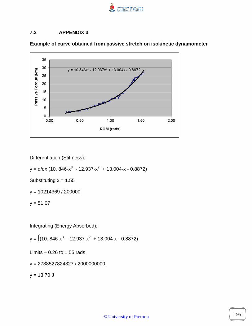
195
7.3 APPENDIX 3
Example of curve obtained from passive stretch on isokinetic dynamometer
Differentiation (Stiffness):
y = d/dx (10. 846·x3 - 12.937·x2 + 13.004·x - 0.8872)
Substituting x = 1.55
y = 10214369 / 200000
y = 51.07
Integrating (Energy Absorbed):
y = ∫(10. 846·x3 - 12.937·x2 + 13.004·x - 0.8872)
Limits – 0.26 to 1.55 rads
y = 2738527824327 / 2000000000
y = 13.70 J
©© UUnniivveerrssiittyy ooff PPrreettoorriiaa
196
7.4 APPENDIX 4
20 March 2013
Pretoria 0002 Republiek van Suid Afrika
Http://www.up.ac.za
FACULTY OF HUMANITIES
Dept Biokinetics, Sport & Leisure Sciences
Tel: 012-420-6040 Fax: 012-420-6099
www.bsl.up.ac.za
INFORMED CONSENT FORM AND INFORMATION SHEET CHAPTER 6 Project title
The title of this project is Flexibility; Passive Biomechanical Responses and
Recovery from Exercise
The student leading the project is Miss Sally Waterworth
The supervisor of the project is Prof PE Krüger
Thank you for showing an interest in this project. Please read all the information
carefully. Think about whether or not you want to take part. If you decide to take part,
you will be asked to sign this form. You do not have to take part. If you decide that you
do not want to participate, there will be no disadvantage to you.
What are the aims of the project?
The main aims of the project are
Determine the between day reliability of an Isokinetic Dynamometer for passive
measurements of muscle stiffness
Establish the effect of an repeated sprint protocol on Perceived Stiffness, Knee Joint
Position Sense, Biomechanical Properties of the Hamstring, Concentric and Eccentric
Quadriceps and Hamstring Strength
©© UUnniivveerrssiittyy ooff PPrreettoorriiaa
197
Determine the effects of static stretching on the above factors
Procedures
If you agree to take part, you will be asked to come for testing on four occasions. The
first and third tests will last for approximately one hour each and the second and fourth
tests approximately 20mins each. On each occasion you will be asked to lie on a
treatment table where a goniometer (device measuring joint angle) will be used to
measure your maximum range for the Active Knee Extension (AKE), Passive Knee
Extension (PKE) and Straight Leg Raise (SLR) tests. You will be seated into the chair
of the Isokinetic Dynamometer while it is adjusted to your measurements. Your leg will
be raised to 45o through the use of a thigh pad. You will be asked to relax completely at
this time and not provide any resistance to the movement of the Dynamometer. You will
then go through a single passive knee extension to a point where you feel a strong
tension in the back of your thigh, similar to the feeling you get when performing a
normal hamstring stretch. After this, the Dynamometer will be adjusted and you will then
be asked to resist the machine in bending and straightening your knee while concentric
and eccentric quadriceps and hamstrings strength is measured. On the first and third
testing occasions you will then go through a multiple sprint protocol before repeating the
above procedure. On the second and forth testing occasions you will go thought the
above procedure alone. After one of the multi-sprint sessions, you will be asked to
perform some static stretching of the lower limb and after the other some static
stretching of the upper limb.
Risks and discomfort
There are no risks in taking part in this study above what you expose yourself to in your
daily training and playing. It is highly unlikely that you will experience any discomfort as
a result of this testing. If you do experience pain or discomfort, please tell the
researcher immediately.
Full details of the risks involved in the procedures are detailed in the risk assessments
A19 and A36 in the department health and safety manual.
©© UUnniivveerrssiittyy ooff PPrreettoorriiaa

198
Safety
General health and safety procedures will be followed as detailed in the department
health and safety manual. The researcher is a qualified First-Aider.
Benefits
You will not get any direct benefit from this study. However, by taking part, you will help
us to increase knowledge of the area being studied.
Can you stop taking part?
You can change your mind and decide not to take part at any time. If you decide to stop,
you do not have to give any reasons for your decision, and you will not be placed at any
disadvantage whatsoever.
What information will be collected, and how will it be used?
Details of your height, weight, age and the results of your flexibility tests will be
recorded. No-one other than the researcher will have access to your specific data.
When testing is finished, data will be coded and grouped together for analysis. Data will
be stored on a password-protected personal computer.
The results of this project may be published, but the information will not be linked to any
specific person. A copy of your results will be given to you if you ask for them. You can
ask questions about the project at any time. Please contact Sally Waterworth at any of
the testing sessions, by email [email protected] or telephone 01695
584686 / 07814 106332.
Statement by subject
I have volunteered to take part in this project
I know I can stop taking part at any time without being disadvantaged
I am satisfied that the results will be stored securely
I know that the results may be published, but they will not be linked to me
I am aware of any possible risks and discomfort
I agree to inform the researcher immediately if I am in pain, or if I feel uncomfortable
©© UUnniivveerrssiittyy ooff PPrreettoorriiaa

199
I have had the chance to ask questions
I know that I will not receive any money for taking part
I have read this form and I understand it. I agree to take part in the project.
Name:
Signed (Subject): Date:
Signed (Witness): Date:
©© UUnniivveerrssiittyy ooff PPrreettoorriiaa
200
7.5 APPENDIX 5
Data Collection Sheet Chapter 5
Date:
Name DOB
Position
Height Weight
Test No
One Two Three
Joint Position Sense
Actual Perceived
Perceived Stiffness
ROM
AKE PKE ASLR PSLR
Worst Stiffness
Ever Experienced No
Stiffness
©© UUnniivveerrssiittyy ooff PPrreettoorriiaa

201
IKD Passive Stretch Eccentric 2.09rads-1
Sprints
½ Speed x 5 ¾ Speed x 5 Full Speed x 20
Condition
Stretch No Stretch
©© UUnniivveerrssiittyy ooff PPrreettoorriiaa

202
7.6 APPENDIX 6
Tests of Normality Chapter 4
Tests of Normality
Kolmogorov-Smirnov
a
Statistic df Sig.
Tor0.99 .168 25 .066
MaxTor .122 25 .200*
Grad0.99 .164 25 .083
FinalGrad .102 25 .200*
Energy0.99 .108 25 .200*
TotalEnergy .092 25 .200*
AKE .119 25 .200*
PKE .142 25 .200*
ASLR .139 25 .200*
PSLR .107 25 .200*
a. Lilliefors Significance Correction
*. This is a lower bound of the true significance.
©© UUnniivveerrssiittyy ooff PPrreettoorriiaa

203
7.7 APPENDIX 7
Sample size calculation Chapter 4
SPSS Sample Power software Power for a test of the null hypothesis One goal of the proposed study is to test the null hypothesis that the correlation in the population is 0.00. The criterion for significance (alpha) has been set at 0.050. The test is 1-tailed, which means that only an effect in the expected direction will be interpreted. With the proposed sample size of 25 the study will have power of 86.0% to yield a statistically significant result. This computation assumes that the correlation in the population is 0.49. The observed value will be tested against a theoretical value (constant) of 0.00 This effect was selected as the smallest effect that would be important to detect, in the sense that any smaller effect would not be of clinical or substantive significance. It is also assumed that this effect size is reasonable, in the sense that an effect of this magnitude could be anticipated in this field of research.
Precision for estimating the effect size A second goal of this study is to estimate the correlation in the population. Based on these same parameters and assumptions the study will enable us to report the this value with a precision (95.0% confidence level) of approximately plus/minus 0.26 points. For example, an observed correlation of 0.49 would be reported with a 95.0% confidence interval of 0.18 to infinity, or (alternatively, per the a priori hypothesis) of minus infinity to 0.71. (Since the confidence interval has been defined as one tailed, only one boundary is meaningful). The precision estimated here is the . Precision will vary as a function of the observed correlation (as well as sample size), and in any single study will be narrower or wider than this estimate.
Notes Power computation: Fisher Z approximation (when null=0, exact formula is used) Precision computation: Fisher Z approximation
©© UUnniivveerrssiittyy ooff PPrreettoorriiaa
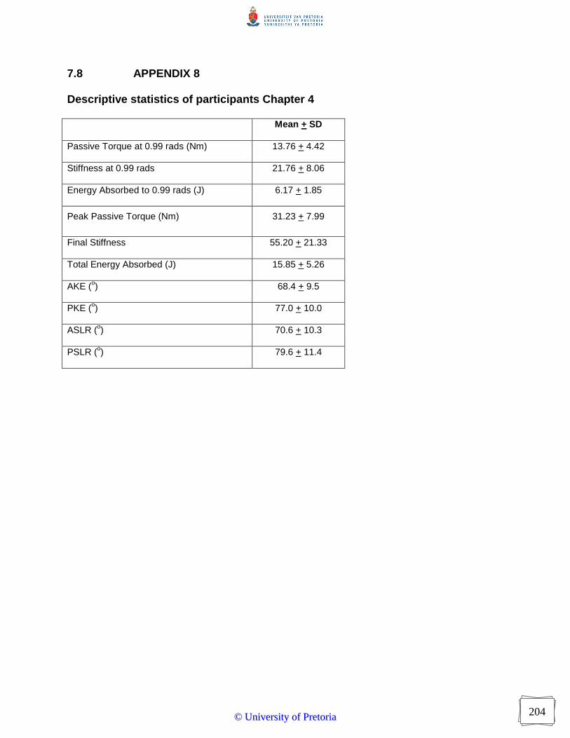
204
7.8 APPENDIX 8 Descriptive statistics of participants Chapter 4
Mean + SD
Passive Torque at 0.99 rads (Nm) 13.76 + 4.42
Stiffness at 0.99 rads 21.76 + 8.06
Energy Absorbed to 0.99 rads (J) 6.17 + 1.85
Peak Passive Torque (Nm) 31.23 + 7.99
Final Stiffness 55.20 + 21.33
Total Energy Absorbed (J) 15.85 + 5.26
AKE (o) 68.4 + 9.5
PKE (o) 77.0 + 10.0
ASLR (o) 70.6 + 10.3
PSLR (o) 79.6 + 11.4
©© UUnniivveerrssiittyy ooff PPrreettoorriiaa
205
7.9 APPENDIX 9 Tests of Normality Chapter 5
Tests of Normality
Kolmogorov-Smirnov
Statistic df Sig.
MaxTorque .202 15 .103
MaxTorque .215 15 .093
FinGrad .153 15 .200*
FinGrad .216 15 .097
TotalEnergy .154 15 .200*
TotalEnergy .164 15 .200*
Grad0.99 .231 15 .080
Grad0.99 .194 15 .134
Energy0.99 .250 15 .082
Energy0.99 .209 15 .076
Torque0.99 .280 15 .082
Torque0.99 .268 15 .125
AKE .257 15 .069
AKE .335 15 .289
PKE .164 15 .200*
PKE .317 15 .127
ASLR .251 15 .092
ASLR .189 15 .154
PSLR .259 15 .148
PSLR .165 15 .200*
a. Lilliefors Significance Correction
©© UUnniivveerrssiittyy ooff PPrreettoorriiaa

206
7.10 APPENDIX 10 Bonferroni adjustment tables for post-hoc testing in Chapter 5 Peak Passive Torque
t Sig. (2-tailed)
Pair 1 Pre-Exercise - Pre-Exercise 2.161 .037
Pair 2 Post-Exercise - Post-Exercise 1.635 .110
Pair 3 18 Hours Post-Exercise - Post-
Exercise 1.279 .209
Pair 4 Pre-Exercise - Post-Exercise -.838 .407
Pair 5 Pre-Exercise - 18 Hours Post-
Exercise -1.812 .078
Passive Energy Absorbed
t Sig. (2-tailed)
Pair 1 Pre-Exercise - Pre-Exercise 1.520 .137
Pair 2 Post-Exercise - Post-Exercise -2.099 .043
Pair 3 18 Hours Post-Exercise - 18 Hours
Post-Exercise -2.291 .028
Pair 4 Pre-Exercise - Post-Exercise -1.467 .151
Pair 5 Pre-Exercise - 18 Hours Post-
Exercise -3.508 .001
Peak Torque during Eccentric Contraction
t Sig. (2-tailed)
Pair 1 Pre-Exercise - Pre-Exercise -.992 .328
Pair 2 Post-Exercise - Post-Exercise -2.761 .009
Pair 3 18 Hours Post-Exercise - 18 Hours
Post-Exercise -3.298 .002
Pair 4 Pre-Exercise - Post-Exercise .208 .836
Pair 5 Pre-Exercise - 18 Hours Post-
Exercise -.152 .880
©© UUnniivveerrssiittyy ooff PPrreettoorriiaa
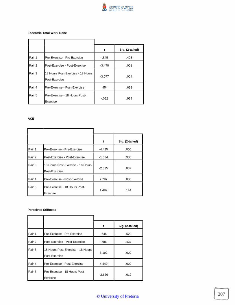
207
Eccentric Total Work Done
t Sig. (2-tailed)
Pair 1 Pre-Exercise - Pre-Exercise -.845 .403
Pair 2 Post-Exercise - Post-Exercise -3.478 .001
Pair 3 18 Hours Post-Exercise - 18 Hours
Post-Exercise -3.077 .004
Pair 4 Pre-Exercise - Post-Exercise .454 .653
Pair 5 Pre-Exercise - 18 Hours Post-
Exercise -.052 .959
AKE
t Sig. (2-tailed)
Pair 1 Pre-Exercise - Pre-Exercise -4.435 .000
Pair 2 Post-Exercise - Post-Exercise -1.034 .308
Pair 3 18 Hours Post-Exercise - 18 Hours
Post-Exercise -2.825 .007
Pair 4 Pre-Exercise - Post-Exercise 7.797 .000
Pair 5 Pre-Exercise - 18 Hours Post-
Exercise 1.492 .144
Perceived Stiffness
t Sig. (2-tailed)
Pair 1 Pre-Exercise - Pre-Exercise .646 .522
Pair 2 Post-Exercise - Post-Exercise .786 .437
Pair 3 18 Hours Post-Exercise - 18 Hours
Post-Exercise 5.192 .000
Pair 4 Pre-Exercise - Post-Exercise 4.449 .000
Pair 5 Pre-Exercise - 18 Hours Post-
Exercise -2.636 .012
©© UUnniivveerrssiittyy ooff PPrreettoorriiaa

208
JPE
t Sig. (2-tailed)
Pair 1 Pre-Exercise - Pre-Exercise 3.106 .004
Pair 2 Post-Exercise - Pre-Exercise 9.319 .000
Pair 3 18 Hours Post-Exercise - 18 Hours
Post-Exercise -.696 .491
Pair 4 Pre-Exercise - Post-Exercise -8.330 .000
Pair 5 Pre-Exercise - 18 Hours Post-
Exercise -2.286 .028
PKE
t Sig. (2-tailed)
Pair 1 Pre-Exercise - Pre-Exercise -7.339 .000
Pair 2 Post-Exercise - Post-Exercise -.357 .723
Pair 3 18 Hours Post-Exercise - 18 Hours
Post-Exercise -2.851 .007
Pair 4 Pre-Exercise - Post-Exercise 9.216 .000
Pair 5 Pre-Exercise - 18 Hours Post-
Exercise 2.741 .009
ASLR
t Sig. (2-tailed)
Pair 1 Pre-Exercise - Pre-Exercise .233 .817
Pair 2 Post-Exercise - Post-Exercise 2.881 .006
Pair 3 18 Hours Post-Exercise - 18 Hours
Post-Exercise .084 .934
Pair 4 Pre-Exercise - Post-Exercise 4.441 .000
Pair 5 Pre-Exercise - 18 Hours Post-
Exercise .018 .986
©© UUnniivveerrssiittyy ooff PPrreettoorriiaa

209
PSLR
t Sig. (2-tailed)
Pair 1 Pre-Exercise - Pre-Exercise -.854 .399
Pair 2 Post-Exercise - Post-Exercise 2.549 .015
Pair 3 18 Hours Post-Exercise - 18 Hours
Post-Exercise -1.183 .244
Pair 4 Pre-Exercise - Post-Exercise 9.580 .000
Pair 5 Pre-Exercise - 18 Hours Post-
Exercise 1.242 .222
©© UUnniivveerrssiittyy ooff PPrreettoorriiaa