Comparative evaluation of 2 resin materials in luting composite inlays
23
Abstracts Abstracts from the Academy of Dental Materials Conference, Siena, Italy, October 2001 1. Effect of orthodontic stripping on enamel demineralization after storage in cariogenic solution. An SEM investigation; M. Zanarini p , S. Marchionni, G. Borea, C. Prati (University of Bologna, Italy) Introduction. Many studies show how orthodontic stripping should be responsible for damaging the enamel structure and interproximal caries. The aim of our study was to evaluate the ultrastructural aspect of the stripped interproximal enamel after demineralization with cariogenic solution (Lactic Acid buffer solution of pH 4.4) by SEM. We also wanted to evaluate role of saliva in the demineralization and its ability to protect enamel from the acid attack. Materials and methods. Nineteen human incisor teeth extracted were selected and stored in distilled water (pH 7.0) at 4 8C for not more than 30 days. Each of the incisors was stripped interproximally with an Horico strip. Teeth were than carefully cleaned under water for 15 s and incubed at 37 8C. Ten of the selected incisors were stored in a cariogenic solution alterned with distilled water buffered (H 2 Ob) by pH 7.0 with Ammonium hydroxide for 3 days. Other five teeth were stored in a cariogenic solution alterned with human saliva (Sal) for the same time. After a storage of 72 h, a vertical section was made to obtain 38 samples. These were fixed in 4% glutaraldeyde in 0.2 M phospate buffer, deydrated in alcohol then gold sputtered before SEM evaluation. Pictures were taken at a magnification of between 200 and 5000 £ . Results and discussion. SEM analysis shows the following differences. Samples stored in H 2 Ob show deep grooves on the enamel surface (2– 3m) but no removal of enamel prisms or exposed interprismatic enamel matrix. It is also possible to see somewhere the smear layer, consequence of stripping procedures. Samples stored in cariogenic solution and H 2 Ob show no grooves or smear layer on the enamel surface. Removed enamel prisms and exposed interprismatic enamel matrix are observable. Samples stored in cariogenic solution and saliva show no grooves or smear layer on the enamel surface, the number of enamel prisms is higher than the one observed in the samples stored in H 2 Ob while the exposed interprismatic enamel matrix is less represented. No stripped samples stored in cariogenic solution and H 2 Ob/saliva show a homogeneous enamel surface. Material groups n Enamel grooves Enamel prisms Interprismatic matrix Stripping-H 2 Ob 4 þþþ 2 2 Stripping-cariogenic sol.-H 2 Ob 20 2 þ þþþ Stripping-cariogenic sol.-saliva 10 2 þþ þþ No stripping -cariogenic sol.-H 2 Ob 4 2 2 2 Conclusions. Our study confirm that orthodontic stripping causes an alteration of the integrity and morphology of the enamel surface. We also observed a different behavior of the stripped enamel stored in acid solution and H 2 Ob with respect to the one stored in cariogenic solution and saliva. Saliva seems to prevent enamel demineralization and to play a protective role on the enamel surface reducing the depth of demineralization and preserving the integrity of prisms. 2. The color of the same shade in different composites: a spectrophotometric analysis; M. Anselmi p , G. Dondi Dall’orologio, R. Lorenzi (University of Bologna, Bologna, Italy) It has long been recognized that the color of the tooth is strongly influenced by the color of the dentin and the thickness of the enamel. The total color effect of natural teeth is a result of the combination of light reflected from the enamel surface and light scattered in the enamel and dentin and finally reflected back. The use of the recommended layering technique with composite resins has provided the means to produce a highly esthetic restoration but has presented some problems to the area of color control. The purpose of this study was to use a spectrophotometric analysis to evaluate colorimetric data of composites of different manufacturers with the same shade designation. Three shades (A3,A3.5,B1) of three different composites were selected: Filtek Z250(Z), (3M); Charisma (C), (Heraeus Kulzer); Esthet X (E) (Dentsply). Samples of the materials were prepared on the same resin tooth, where a cavity, 3.0 mm in diameter, 3 mm thick, was done. Each sample was measured using a reflectance spectrophotometer on black and white backings. For the white background resin tooth cavity had L p 87.8, a p 4.8, b p 26.2, T 27% colorimetric values. For the black background a black varnish was applied on cavity walls. Colorimetric values for the black cavity were: L p 21.8, a p 20.7, b p 23.2 and T 93%. A layer of glycerin was used on the walls for an easier removal of the material. Each sample was light cured from the top for 40 s with a visible light unit, having 7 mm diameter tip. Colorimetric analysis was performed with a reflection spectrophotometer (Pikkio, M.H.T.) with a 358/0 type illuminating and viewing geometry; with a 3 mm measuring port and 1.5 mm detector area: the measure was automatically reported after three identical recordings. All the data were directly measured in CIELAB coordinates, with respect to the standard light source A. The manufacturer states that the precision of the machine in within 1%. The CIELAB color difference metric (CIE 1986) was used to determine the size of color shift. Means for CIE L p a p b p and translucency for the shade A3 are listed in the following table. A3 L p a p b p T (%) Charisma 59.0 4.0 21.0 70 Esthet X 56.1 2.3 20.2 66 Z250 57.9 2.1 18.8 69 DE CE ¼ 5:37; DE CZ ¼ 3:1; DE EZ ¼ 2:69: For the shade A3.5 PII: S0109-5641(02)00044-1 Dental Materials 18 (2002) A1–A23 www.elsevier.com/locate/dental
Transcript of Comparative evaluation of 2 resin materials in luting composite inlays
Abstracts
Abstracts from the Academy of Dental Materials Conference,
Siena, Italy, October 2001
1. Effect of orthodontic stripping on enamel demineralization after
storage in cariogenic solution.
An SEM investigation; M. Zanarinip, S. Marchionni, G. Borea, C. Prati
(University of Bologna, Italy)
Introduction. Many studies show how orthodontic stripping should be
responsible for damaging the enamel structure and interproximal caries.
The aim of our study was to evaluate the ultrastructural aspect of the
stripped interproximal enamel after demineralization with cariogenic
solution (Lactic Acid buffer solution of pH 4.4) by SEM. We also wanted
to evaluate role of saliva in the demineralization and its ability to protect
enamel from the acid attack.
Materials and methods. Nineteen human incisor teeth extracted
were selected and stored in distilled water (pH 7.0) at 4 8C for not
more than 30 days. Each of the incisors was stripped interproximally
with an Horico strip. Teeth were than carefully cleaned under water
for 15 s and incubed at 37 8C. Ten of the selected incisors were stored
in a cariogenic solution alterned with distilled water buffered (H2Ob)
by pH 7.0 with Ammonium hydroxide for 3 days. Other five teeth
were stored in a cariogenic solution alterned with human saliva (Sal)
for the same time. After a storage of 72 h, a vertical section was made
to obtain 38 samples. These were fixed in 4% glutaraldeyde in 0.2 M
phospate buffer, deydrated in alcohol then gold sputtered before SEM
evaluation. Pictures were taken at a magnification of between 200 and
5000 £ .
Results and discussion. SEM analysis shows the following differences.
Samples stored in H2Ob show deep grooves on the enamel surface (2–
3m) but no removal of enamel prisms or exposed interprismatic enamel
matrix. It is also possible to see somewhere the smear layer, consequence of
stripping procedures. Samples stored in cariogenic solution and H2Ob show
no grooves or smear layer on the enamel surface. Removed enamel prisms
and exposed interprismatic enamel matrix are observable. Samples stored
in cariogenic solution and saliva show no grooves or smear layer on the
enamel surface, the number of enamel prisms is higher than the one
observed in the samples stored in H2Ob while the exposed interprismatic
enamel matrix is less represented. No stripped samples stored in cariogenic
solution and H2Ob/saliva show a homogeneous enamel surface.
Material groups n Enamel
grooves
Enamel
prisms
Interprismatic
matrix
Stripping-H2Ob 4 þþþ 2 2
Stripping-cariogenic
sol.-H2Ob
20 2 þ þþþ
Stripping-cariogenic
sol.-saliva
10 2 þþ þþ
No stripping -cariogenic
sol.-H2Ob
4 2 2 2
Conclusions. Our study confirm that orthodontic stripping causes an
alteration of the integrity and morphology of the enamel surface. We also
observed a different behavior of the stripped enamel stored in acid solution
and H2Ob with respect to the one stored in cariogenic solution and saliva.
Saliva seems to prevent enamel demineralization and to play a protective
role on the enamel surface reducing the depth of demineralization and
preserving the integrity of prisms.
2. The color of the same shade in different composites: a
spectrophotometric analysis; M. Anselmip, G. Dondi Dall’orologio,
R. Lorenzi (University of Bologna, Bologna, Italy)
It has long been recognized that the color of the tooth is strongly
influenced by the color of the dentin and the thickness of the enamel. The
total color effect of natural teeth is a result of the combination of light
reflected from the enamel surface and light scattered in the enamel and
dentin and finally reflected back. The use of the recommended layering
technique with composite resins has provided the means to produce a highly
esthetic restoration but has presented some problems to the area of color
control.
The purpose of this study was to use a spectrophotometric analysis to
evaluate colorimetric data of composites of different manufacturers with
the same shade designation.
Three shades (A3,A3.5,B1) of three different composites were selected:
Filtek Z250(Z), (3M); Charisma (C), (Heraeus Kulzer); Esthet X (E)
(Dentsply).
Samples of the materials were prepared on the same resin tooth, where a
cavity, 3.0 mm in diameter, 3 mm thick, was done. Each sample was
measured using a reflectance spectrophotometer on black and white
backings. For the white background resin tooth cavity had L p 87.8, a p 4.8,
b p 26.2, T 27% colorimetric values. For the black background a black
varnish was applied on cavity walls. Colorimetric values for the black
cavity were: L p 21.8, a p 20.7, b p 23.2 and T 93%.
A layer of glycerin was used on the walls for an easier removal of the
material. Each sample was light cured from the top for 40 s with a visible
light unit, having 7 mm diameter tip. Colorimetric analysis was performed
with a reflection spectrophotometer (Pikkio, M.H.T.) with a 358/0 type
illuminating and viewing geometry; with a 3 mm measuring port and
1.5 mm detector area: the measure was automatically reported after three
identical recordings.
All the data were directly measured in CIELAB coordinates, with
respect to the standard light source A. The manufacturer states that the
precision of the machine in within 1%.
The CIELAB color difference metric (CIE 1986) was used to determine
the size of color shift. Means for CIE L p a p b p and translucency for the
shade A3 are listed in the following table.
A3 L p a p b p T (%)
Charisma 59.0 4.0 21.0 70
Esthet X 56.1 2.3 20.2 66
Z250 57.9 2.1 18.8 69
DECE ¼ 5:37; DECZ ¼ 3:1; DEEZ ¼ 2:69: For the shade A3.5
PII: S0 10 9 -5 64 1 (0 2) 00 0 44 -1
Dental Materials 18 (2002) A1–A23
www.elsevier.com/locate/dental
DECE ¼ 9.4, DECZ ¼ 5:7; DEEZ ¼ 4:1; for the shade B1 DECE ¼ 8:5;
DECZ ¼ 6:9; DEEZ ¼ 8:9:
The difference was significant, p , 0.01 for all the shades of the three
systems.
For the brands and the shades measured, the following conclusions can
be made:
1. The CIELAB color system and spectrophotometer provide an objective
technique for evaluating color.
2. Corresponding shades of different composites can produce perceivably
different colors that are mostly a result of differences in all three color
directions.
3. For a more scientific approach to color selection, the clinician should
increase his information about colorimetric data for a color matching
more reproducible.
3. Low-temperature aging of transformation-toughened zirconia, Y-
TZP, for dental inlays, crowns and bridges; B.I. Ardlinp (Department
of Odontology, Umea University, Umea, Sweden)
One concern, for transformation-toughened zirconia, Y-TZP, is their
liability to low temperature aging with accompanying degrading of
properties such as strength and toughness. The loss in strength is related
to the amount of transformation of tetragonal grains to monoclinic. The
transformation is related to factors such as loading of the ceramic
reconstruction, temperature and time of exposure to surrounding media
(aging) and the manufacturing processes of Y-TZP.
Objectives. The purpose of this investigation was to study the flexural
strength, chemical solubility, surface texture, crystal structure and low-
temperature aging (4% acetic acid at 80–83 8C for 168 h) of two shades, P0
and P17, of a Y-TZP ceramic used for dental inlays, crowns and bridges.
The hypotheses to be tested were that both shades of the dental Y-TZP
ceramic have high strength and chemical solubility, and that the strength,
surface texture and crystal structures of the ceramic were not effected by
aging.
Methods. Forty specimens of Y-TZP, 20 of the shade P0 and 20 of
the shade P17 were ground and polished. Ten specimens of each shade
were exposed to low-temperature aging. The flexural strength of all 40
specimen was registered. Surfaces of the specimens were evaluated by
using SEM, XRD and roughness recorder. The chemical solubility was
determined by weight loss. Wilcoxon Signed Rank test was used for
statistical analysis.
Results. As expected, the two shades, P0 and P17, of the studied dental
Y-TZP had high strengths that were not effected by low temperature aging
and high chemical solubility. Contrary to what was assumed the crystal
structure and surface texture of P0 and P17 were effected. Transformation
from tetragonal to monoclinic structures occurred and small elevations on
the ceramic surfaces were observed after aging.
Conclusion. The dental Y-TZP ceramic used for dental inlays, crowns
and bridges has high strength and low chemical solubility.
4. Stability measurements of osseointegrated implants in the treatment
of partially edentulous upper jaws; P. Ballerip, A. Cozzolino, L. Ghelli
(Universita degli studi di Siena)
Introduction and purpose:
In the upper jaw, the risk of failure for implants supporting partial
prostheses, is increased by the property of bone and by anatomical limits,
and this is quite evident when the implants are placed in postero-lateral
regions without using grafts or bone augmentations techniques.
The aim of the present study was to test the stability of implants
supporting partial prosthesis in the upper jaw, after one year of loading,
when the fixtures comes to the steady state.
Materials and methods:
The implants were inserted in according to Branemark protocol.
A group of 7 patients were selected for study and the patients had a total
of 21 implants placed. The stability of each implant was tested with
Resonance Frequency and the analysis was performed by Osstelle
(Integration Diagnostics, Gothenburg, Sweden). The resonance frequency
of a trasducer, connected directly with the implant, can measure the
stability of an implant and the obtained values are reported in a reference
scale called I.S.Q. (implant, stability, quotient).
Results and statistical analyses:
All the I.S.Q. values was up to 60, the critical limit for implant loading.
The level of marginal bone around each implant is investigated by
radiography and there was no sign of bone resorption.
Stability measurements indicate the absence of a direct correlation
between implant length and resonance frequency ðR ¼ 20:026Þ:
Conclusions:
I.S.Q. values are always encouraging and they show a good result in
postero-lateral sites too.
The outcome of the study indicate that, in partially edentulous upper
jaws, safe and predictable results can be obtained with Branemark implant
technique and that the placement of short implants to give a good support to
the prosthesis is possible in poor quality bone.
The results support the hypothesis that the bone augmentations and graft
techniques are not always necessary for implant placement, even if the
width and the quality of bone do not show the right property.
5. Effect of different polymerization methods on marginal leakage of
composite restorations; M.F. Bernardop, J. Portugal, J. Leitao
(University of Lisbon, Lisbon, Portugal)
Stress generated by polymerization shrinkage of composites may, in
some instances, be high enough to produce debonding of the restoration or
even enamel microcracks. Both these events result in marginal leakage.
Several polymerization methods have been proposed in order to minimize
polymerization shrinkage stress.
Objective: The objective of this ‘in vitro’ study was to assess the
marginal leakage occurring at the margins of composite restorations, using
five different polymerization methods.
Materials and methods: Two composite resins, Z100 (3M Dental
Products) and Surefil (Caulk/Dentsply), were used to restore 80 restorations
made in the buccal and lingual surfaces of 40 recently extracted human
permanent molars. Five polymerization methods were tested: (1) low
intensity light—40 s at 300 mW/cm2; (2) medium intensity light—40 s at
650 mW/cm2; (3) high intensity light—PAC—10 s at 1900 mW/cm2; (4)
ramp intensity light—10 s at 100–1000 mW/cm2 þ 10 s at 1000 mW/cm2
and (5) pulse-activation—3 s at 300 mW/cm2 þ 4–5 min delay þ 40 s at
300 mW/cm2. Following restorative procedures, the teeth were kept in de-
ionized water for 24 h and then thermocycled between 5 and 55 8C for 500
cycles with a dwell time of 20 s. After thermocycling teeth were isolated
with bright color nail varnish and dental compound up to a distance of
1 mm to the restoration margins and then, immersed in 2% methylene blue.
Teeth were cut twice, in a buccal-lingual direction, with a slow speed
ultramicrotome. Determination of marginal leakage was performed in each
of the eight cross-sectioned restoration margins, using the following ordinal
scale: 0—no marginal leakage; 1—marginal leakage confined to the
gingival/occlusal wall; 2—marginal leakage extending to the axial wall.
Statistical analysis of the results was performed using a non-parametric
ANOVA (Kruskal–Wallis test) at a 95% significance level.
Results: Median scores for each group are presented in the following table:
Low
intensity
Medium
intensity
High
intensity
Ramp Pulse-
activation
Z100 0.75 0.25 1 0.5 1
Surefil 1 0.25 1 1 1.25
Abstracts / Dental Materials 18 (2002) A1–A23A2
The statistical analysis of the results showed significant differences
between the five polymerization methods ( p , 0.05).
Conclusion: Results suggest that the presence of marginal leakage is
strongly influenced by the method of polymerization.
6. Comparative evaluation of 2 resin materials in luting composite
inlays; M. Biasottoa,p, O. Cotarcaa, F. Rusina, M. Cadenaroa,
G. Merlatib, R. Di lenardaa (aUniversita’degli Studi di Trieste, Trieste,
Italy; bUniversita’degli Studi di Pavia, Pavia, Italy)
The aim of this study was the comparative evaluation of a photocuring
resin composite (Z100, 3M) and a selfcuring resin cement (C&B, Bisco Inc.) in
luting composite inlays on human teeth: microleakage at the tooth/inlay
interface and microhardness of the luting materials were evaluated. For the
infiltration test 28 occlusal cavities on human molars extracted for periodontal
reasons were prepared with butt margins using a diamond bur for inlays. After
the application of two layers of Lab-Separator Coltene on the cavity walls 28
inlays were prepared with an incremental technique using Z100 (3M) resin
composite. Each layer of the material (max thickness 2 mm) was polymerized
exposing the occlusal surface to the Apollo 95E curing light (D.M.D.S. Ltd)
for 9 s. After removal from the cavities, the inlays were accurately rinsed with
water and post-polymerized for 9 min in a Coltene D.I. 500 oven; then they
were lightly reduced on the pulpal and axial surfaces with a diamond bur,
respecting the margins. The inlays were sandblasted with Al2O3 for 5 s at
2 atm (Danville Microetcher) and covered with a layer of One Step (Bisco Inc.)
polymerized with Apollo 95E for 9 s. The cavities were total etched for 30 s
with 37% phosphoric acid (3 M), washed for 30 s, gently dried and covered
with two layers of One Step,polymerized with Apollo 95E for 9 s. The samples
were divided into four groups (group 1 and 2, n ¼ 7; group 3 and 4, n ¼ 5). In
groups 1 and 3 inlays were luted with Z100 while in groups 2 and 4 they were
luted with C&B. Inlays were placed into the cavities and luting materials were
polymerized with the Apollo 95E lamp, applying the curing tip occlusally,
buccally and lingually for 1 min each, after removing the material’s excesses.
Teeth in groups 1 and 2 were thermocycled for 9 12
h (Thermocycler FCA14;
500 5/50 8C cycles). All the 28 teeth were then immersed in 0.05% basic
fuchsine for 48 h, embedded in resin and sectioned in buccal–lingual direction
with a microtome cooled with tap water. The sections were analyzed with a
64X stereomicroscope to assess the microleakage at the tooth/inlay interface.
The depth of dye penetration was evaluated using a scale ranging from 0 to 4.
For the Vickers microhardness (VH) test other 12 M were selected and inlays
prepared as mentioned above. Six inlays were luted with Z100 (group 5), while
the other six were luted with C&B (group 6). Teeth were embedded in resin
and sectioned in buccal–lingual direction with a microtome cooled with tap
water. VH of the luting materials was measured on the bottom of the cavity
sections and on the resin composite in the middle of the inlays; microhardness
was correlated with the cavity depth. All data were statistically analyzed with
the Mann–Whitney test. There was no statistical difference between the two
luting materials in the dye infiltration degree both in thermocycled and no
thermocycled samples, although the highest infiltration was observed in the
samples luted with the self-curing cement in group 4. Z100 showed higher
Vickers microhardness (VH) values than C&B ( p , 0.05), but lower than the
inlay material. A significant correlation ( p , 0.05) between VH and cavity
depth was found only for Z100. Microinfiltration was low for both materials,
while the microhardness was higher in the Z100 samples, associated with an
easier material manipulation than C&B. Resin inlays luted with a photocuring
composite represent an excellent choice for the restoration of posterior teeth.
7. In vivo evaluation of new calcium hydroxide points for intracanal
medications; G. Gambarini, G. Bologninip, A. Lupi, L. Testarelli,
M. Bossu’, Polimeni, A., Ottolenghi, L., Barbato, E., De Biase, A.,
(University of Rome, Dental School)
A new formulation of calcium hydroxide has recently been developed
(CaOH Plus, Roeko, Germany) for easy placement and removal of the
intracanal medicament. Since the clinical properties of calcium hydroxide
can change depending on the different vehicles, the purpose of the present
study was to compare in vivo tissue response with the new material and
chemically pure Ca(OH)2 pastes. Twenty patients scheduled for extraction
for periodontal disease were selected for this study and divided into two
homogeneous groups. The written consent of the patients was obtained after
the nature of the procedures and the possible discomfort and risks had been
fully explained. Chemio-mechanical preparation of the endodontic space
was performed using a crown-down instrumentation technique and
irrigation with 5% NaOCl and 17% EDTA. The two different intracanal
medications were then placed in the root canals. After 14 days all teeth were
extracted and processed for histopathological evaluation. Tissue response
to the intracanal medicament was scored as follow: (1) none/mild; (2)
moderate; (3) severe. Data were collected and assessed using the Mann–
Whitney–Wilcoxon test. Results for Group A (CaOH Plus) were 80%
none/mild and 20% moderate. The same score was reported for Group B
specimens (Ca(OH)2). No statistically significant differences ( p . 0.05)
were noted between the two groups. Overall, histological sections showed
that tissue response to CaOH Plus cones appeared not to be distinguishable
from the response to chemically pure Ca(OH)2.
8. Effect of burnout schedule on thermal expansion of phosphate-
bonded investments; L. Borchersp, J. Heine, P. Ohnmacht, U. Schmitt
(Medical University of Hannover, Hannover, Germany)
Modern phosphate-bonded investments are suited for accelerated
bench-set and burnout schedules offering substantial savings in laboratory
time. Some of these materials may as well be processed according to the
conventional, considerably more time consuming technique. Since the
effective heating rates applied in both cases differ by approximately two
orders of magnitude, the reactions and phase transitions within the
investment might well be completed to a different extent. Thus, the
associated volume changes and the fit of cast restorations would be different
for both techniques. In order to verify this hypothesis and to find an
explanation for recently reported differences in fit of crowns made
according to both methods, we studied the effect of burnout schedule on
thermal expansion of six commonly used and one experimental investment
for high gold crown and bridge alloys.
The materials used in this study are listed in Table 1. Cylindrical
specimens (diameter 5 mm, length 20 mm) were prepared in silicone
molds. The specimens were demolded 60 min after start of mixing, and
drying at (23 ^ 1) 8C for an additional 60 min. Immediately thereafter, they
were transferred to a computer-controlled, calibrated, horizontal quartz rod
dilatometer and heated to 950 8C using one of the following schedules:
constant heating rates of (a) 5 K/min, (b) 50 K/min, and (c) 200 K/min, (d)
thermal shock in dilatometer preheated to 950 8C for simulating accelerated
burnout, and (e) conventional schedule as recommended by the
manufacturer. Final readings of expansion were taken after the end
temperature had been held constant for 15 min. Measurements were
corrected for offset errors and converted to percentage values with respect
to the initial length. Three specimens were tested per material and schedule.
ANOVA and Scheffe tests were performed in order to reveal significant
influences of schedule type on expansion.
The mean values reported in Table 1 are ranging between 0.60 and
1.15% and do not show much variation for a given material. Maximum
expansion is always accomplished either with the slowest heating rate or
with the conventional procedure, whereas shock heating or one of the
higher heating rates leads to lower expansion. Though differences between
shock and conventional heating are significant in all cases with the
exception of Experimental, they amount to only 0.21% at the most, which is
of minor importance clinically. Therefore, with respect to thermal
expansion alone, all materials tested seem to be suited for preparing well
fitting casts using either accelerated or conventional burnout schedule.
Abstracts / Dental Materials 18 (2002) A1–A23 A3

9. Surface analysis of 5 esthetic materials after polishing;
M. Cadenaroa,p, M. Biasottoa, R. Chiesab, R. Di Lenardaa, E. Dorigoa
(aUniversita’degli Studi di Trieste, Trieste, Italy; bPolitecnico di
Milano, Milano, Italy)
Obtaining a smooth surface is a prime objective for composite resin
restorations, in order to avoid plaque retention and surface staining and
improve esthetics. Curing the material against a Mylar strip produces the
smoothest surface, however, the removal of excess material or recontouring
of restorations is often necessary clinically. It has been suggested that a
perfectly smooth surface is virtually unattainable with current polishing
techniques. Bollen et al. (1996) indicated the existence of a threshold
roughness (0.2 mm) below which no influence on the bacterial adhesion
should be expected. The aim of this study was the in vitro evaluation of the
surface roughness of five esthetic materials (Surefil, Dentsply; Definite,
Degussa; Ariston pHc, Vivadent; Solitaire, Kulzer; Z100, 3M) polymerized
with an halogen curing light (VIP, BISCO Inc.) and a plasma arc lamp
(Apollo 95E, Dental/Medical Diagnostic System Inc.), following finishing
and polishing. Using a metal mold, 10 block specimens (5 £ 5 £ 2 mm3)
were prepared from each material. A Mylar strip was placed on both sides
of the mold. Five specimens from each material were cured applying the
curing tip of VIP on the top surface for 40 s (group 1), while the other five
were polymerized with Apollo 95E for 9 s (group 2). In both groups the
surface exposed to curing light was finished and polished with the
ENHANCEw system (Dentsply). The opposite surface was not treated and
served as a control. Both surfaces of each specimen were analyzed with a
laser profilometer (UBM microfocus): the roughness average (Ra) was
evaluated. The Vickers microhardness of the polished surfaces was also
calculated. All data were statistically analyzed with the Mann–Whitney
test. The regression test was carried out to evaluate the possible correlation
between roughness and microhardness. All surfaces polymerized against
the Mylar strip presented in both groups a mean Ra lower than 0.2 mm.
Roughness was significantly increased in both groups in the treated
surfaces, with a mean Ra higher than 0.2 mm. The plasma cured non-
polished surfaces presented lower Ra values than in the halogen light group.
It was observed that the higher the Ra values, the lower the microhardness
values, although a significant correlation was obtained only for Ariston pHc
and Solitaire. These two materials presented very low microhardness
values, clinically inadequate for posterior restorations if compared to the
conventional composite Z100.
10. Effect of sodium hypochloride on dentin: a high resolution SEM
study; L. Breschia, S. Chersonia,p, L. Montebugnolia, M. Toledanob,
R. Osoriob, G. Mazzottia, C. Pratia (aUniversity of Bologna, Italy;bUniversity of Grenada, Spain)
The use of sodium hypochloryde (NaOCl) to remove the collagen fibrils
(CF) on dentin as to improve the dental adhesion is controversial. The
purpose of this study was to evaluate the effect of different times of
application of a 5% solution of sodium hypocloride on the dentin surface
after application of different acids solutions.
Eighteen extracted human molars were selected and randomly assigned
to the tested etching treatments. Dentin disks were obtained with a low
speed diamond saw and treated with: (1) 10% maleic acid, (2) 10% citric
acid, (3) 2.5% oxalic acid, (4) EDTA, (5) 35% phosphoric acid, (6) no
etching. Acids were left on the dentin surface for 15 s, then NaOCl was
applied for 2 and 5 min. Control specimens without the NaOCl application
were also prepared. Dentin disks were then fixed, dehydrated, mounted on
stubs and finally observed under a high resolution SEM (Jeol JSM-890).
The application of sodium hypochloride on dentin showed different
morphological features depending upon the etching agent previously applied
to demineralize the dentin surface. In particular, the application of maleic and
citric acid on the dentin surface revealed similar features, such as complete
removal of the smear layer and smear plugs and the exposure of the acid of
several CF on the intertubular (ITD) and peritubular dentin. After the
application of NaOCl for 2 min most of the CF of the ITD were completely
removed, while residual CF were evident only in small areas; on the other hand
no evidence of CF was present in any of the maleic and citric acid etched
specimens after 5 min application of NaOCl. Oxalic acid resulted in the
formation of several crystal salts on the ITD. The use of NaOCl revealed minor
modifications of the dentin surface. EDTA surface conditioning resulted in
uncompleted opening of the tubules and a very superficial demineralization of
the ITD. After the use of NaOCl at both times of application tested, a very
compact ITD as well as residual CF were observed under a thick mineral
coating. On the other hand the use of NaOCl on phosphoric acid etched dentin
revealed an enlargement of the tubule orifices particularly evident after the
5 min application, while the treatment with NaOCl produced minor changes
on the dentin surface of the unetched specimens.
Conclusions. The application of different acid treatments is responsible
for different morphological modifications of dentin surface that can greatly
influence the effectiveness of NaOCl. The combined application of acids
and NaOCl may represent the correct approach in the preparation of dentin
surface to obtain a thin and effective hybrid layer. After 2 min application
of a solution of 5% NaOCl residual collagen fibrils were evident, while no
organic remants were observed after 5 min application on phosphoric,
maleic and citric acid etched samples.
11. Analysis of leachable monomers of composites by NMR1H in
solution; I.C. Correap, M.I.B. Tavares, W.G. Miranda Jr (Dental
Materials-University of Sao Paulo and IMA-UFRJ-Brazil)
Several studies used infrared spectroscopy to evaluate the degree of
conversion by the detection of remaining CyC. This remaining double bond
was due to non-reacted and pendant monomers. The presence of unreacted
monomers after photo-activation of dental composites causes mechanical
and biological properties to decrease and could be detected by NMR
analysis. The aim of this study was to evaluate the percentage of leachable
monomers of light curing composites under the effect of variations of
exposure time of photoactivation by nuclear magnetic ressonance of
hydrogen in solution (NMR1H). Cilindrical specimens (d ¼ 5 mm and
h ¼ 3 mm) of Z250. FillMagic (hybrid composites), Durafill and A110
(microparticles composite), of shade A3, were light-cured for 20, 40 and
80 s with Optilux 401 curing unit (500 mW/cm2). The unreacted monomers
Table 1
Thermal expansion (%) for different investments and temperature schedules ðn ¼ 3Þ
Material Manufacturer 5 K/min 50 K/min 200 K/min Shock Conventional
Bellavest SH Bego, Germany 0.98 0.82 0.80 0.84 1.04
Fujivest Super GC, Japan 1.02 0.98 1.01 1.00 1.12
Heravest Speed Heraeus, Germany 0.98 0.84 0.81 0.78 0.93
PowerCast WhipMix, USA 1.13 0.92 0.93 0.93 1.10
Presto-Vest Siladent, Germany 1.15 1.13 1.13 1.07 1.15
Experimental Dentaurum, Germany 1.03 0.83 0.88 0.95 1.02
FastFire 15 WhipMix, USA 0.86 0.71 0.60 0.62 0.83
Abstracts / Dental Materials 18 (2002) A1–A23A4
of each specimen were extracted after a 1-day and 7-days immersion in
deuteraded chloroform, and analyzed in a spectrometer VARIAN
MERCURY 300. The 1H spectra of all specimens showed leachable
monomers unreacted after extracting (shown in the table), by the detection
of 5.6 and 6.1 ppm signals of CH2y groups. After 7 days, only the
specimens of manufacturer time of exposure were immersed in a new
deuteraded chloroform for 2 weeks, to optimize the monomers extraction.
The table shows the percentage of non-reacted monomers leached after
different exposure times.
The shadow values correspond to the manufacturer’s time of photo-
activation.
Z250 and FillMagic obtained similar values of unreacted monomers (%) at
photocuring time suggested by the manufacturer and values were also lower
than Durafill and A110 concentrations. A 1-day extraction time was efficient to
quantify the total amount of residual monomers for Z250, FillMagic and A110.
PRONEX-CNPq 0327.00/00 e CAPES
12. Clinical evaluation of esthet-x composite resin; J.R. Dunnp, C.A.
Munoz, R. Kinzer, D.E. Tan, J. Sy-Munoz, A. Wilson (Loma Linda
University, Loma Linda, CA)
This Clinical Trial evaluated the clinical performance of a new
polychromatic high density composite resin (Esthet, X., L.D. Caulk) over a
two year period. Forty-three anterior and posterior restorations were placed
in 27 subjects (36 at 1 Year). Following cavity preparation the teeth were
etched, restored, and polished following manufacturer’s instructions.
Marginal adaptation (MA), Anatomic Form (AF), color match (CM),
secondary caries (SC) marginal discoloration (MD) and polishability (PO)
were evaluated. At 2 years, using a modified USPHS grading systems the
following results were found in percentages (%) (BL ¼ Base Line):
At the 2-year recall: (1) categories MA, AF, and SC were unchanged from
baseline; (2) MD showed a decrease in restoration quality, (3) PO showed that
restorations polished adequately at baseline but showed increase in the surface
polish over time, while restorations polished inadequately at baseline had a
decrease in surface polish and (4) Overall clinical performance is acceptable
for routine clinical use. Study supported by L.D. Caulk/Dentsply.
13. The influence of cold working and thermal treatment of the fit of
implant-supported fixed partial dentures; Salman Lakhani, Carlo
Ercolip, Gerald N. Graser, Ross H. Tallents, Mark E. Moss (University
of Rochester Eastman Dental Center, Rochester, NY, USA)
Purpose. The fit of porcelain-fused-to-metal frameworks has been
shown to substantially change after porcelain application. It has been shown
that a significant reduction in metal distortion was evident, in single
castings, when the oxidation cycle was completed before the prosthesis was
cold worked (finished). This study compared the fit of implant-supported
porcelain-fused-to-metal fixed partial dentures when different thermal
treatments are performed.
Material and methods. Fifteen implant-supported porcelain-fused-to-
metal fixed partial denture frameworks were fabricated on a machined
aluminum base and randomly divided in three groups: Group 1. Cold
working and unrestrained thermal oxidation. Group 2. Unrestrained thermal
oxidation, cold working, unrestrained thermal oxidation. Group 3.
Phosphate-bonded investment-restrained thermal oxidation, cold working,
unrestrained thermal oxidation. After this treatment, porcelain was applied
to all frameworks. Three-dimensional changes (x, y, and z ) were monitored
by measuring the frameworks (three times) after each step (oxidation, cold
working, opaque porcelain application, dentin porcelain application, and
glaze stages) with a digital Mitutoyo caliper (error ¼ ,1 mm). Intraobser-
ver reliability was determined by measuring two fixed partial dentures at 4
weeks interval (coefficient of variation ¼ 0.05–0.13). Data was analyzed
with repeated ANOVA and Tukey studentized range test. Statistical
significance was set at P ¼ ,0.05. Distortion trends in each group
(Positive ¼ away from the measuring references; Negative ¼ toward the
measuring references) were also assessed.
Results. Mean total absolute distortion was not significantly different
among groups (P . 0.05). No significant difference among groups was
found in the distortion after thermal treatment (P . 0.05). Absolute values
of distortion at each step were not significantly different among groups
(P . 0.05). Specific patterns of distortion were found in each group. These
trends of distortion were somehow maintained throughout the experiment.
Indeed, group 1 castings generally deformed in a positive direction (away
from the measuring references) while castings in group 2 deformed in a
negative direction (toward the measuring references). Group 3 frameworks
showed the greatest amount of negative distortion (toward the measuring
references).
Conclusions. Heat treating implant-supported porcelain-fused-to-
metal fixed partial denture frameworks before cold working (finishing)
did not decrease the absolute distortion. Specific patterns of distortion
are found when different heat treatments are performed. The authors
therefore suggest that, if the purpose of the heat treatment is to allow
casting stress relief and recrystallization of the alloy with minimal
distortion, it is important to select a restraining material that has
pattern and magnitude of thermal expansion similar to those of the
metal.
This project was partially supported by a 1999 American Academy of
Fixed Prosthodontics Stanley D. Tylman Research Grant.
Z250 FillMagic Durafill A110
20 s 40 s 80 s 20 s 40 s 80 20 s 40 s 80 s 20 s 40 s 80 s
1 d 2.45 1.98 1.55 4.5 2.48 2.22 7.27 5.9 4.74 7.76 4.24 3.36
7 d 2.34 1.96 1.97 6.25 2.49 2.22 8.30 7.57 6.89 8.57 4.48 3.29
þ14 d 0 – – – 0 – 0 – – – 0
MA AF CM SC MD PO
BL 1Y BL 1Y BL 1Y BL 1Y BL 1Y BL 1Y
Alpha 54 50.0 84 92.1 53 50 100 100 100 73.7 47 73.7
Bravo 46 50.0 16 7.9 47 47.4 0 0 0 23.7 43 21
Charlie 0 0 0 0 0 2.6 0 0 0 2.6 0 0
Delta 0 0 0 0 0 0 0 0 0 0 0 5.2
Abstracts / Dental Materials 18 (2002) A1–A23 A5
14. The influence of bur wear on cutting efficiency and heat production
during osteotomy preparation for dental implants: A study of bur
durability. Ercoli Carlop, P.D. Funkenbusch, T.D. Greene, H.J. Lee,
M.E. Moss, U. Ben-Hanan, G.N. Graser, R.H. Tallents, K.S. Hebel
(Department of Mechanical Engineering, University of Rochester
Eastman Dental Center, Rochester, NY)
Introduction. Excessive surgical trauma is considered an important
cause of implant failure. During bone drilling, most of the energy not used
in the cutting process is transformed into heat, and the amount of heat will
depend on several factors (i.e. drill flute geometry and sharpness, torque,
chip removal rate, bone density etc.). Repeated use of drills progressively
increases their wear and decreases their cutting efficiency, thus producing
more frictional heat. A review of the literature has shown that there are no
scientific established guidelines on the durability of drills used to prepare
osteotomies.
Purposes. (1) To determine the maximum number of osteotomies that
can be prepared with several implant drills without causing a temperature
increase damaging to the bone. (2) To determine the relationship between
osteotomy depth and heat production during consecutive osteotomies.
Cutting efficiency and heat production of different drill designs (i.e. spade,
twist, tri-flute, TiN coated) were evaluated and compared during 100
successive osteotomies. Materials and methods. Bovine ribs were used in
this study. Two positioning holes were drilled at the base of each bone
piece. Two thermocouple holes were also drilled at 1 mm distance from the
future osteotomy site. Two chromega/constantan thermocouples were
inserted into the bone block to the predetermined depths of 5 and 15 mm.
Parallelism between the thermocouple canal, the future implant site and
positioning holes was achieved by positioning the bone to a bi-directional,
micrometer-controlled (x,y ) translation slide secured to a press stand base.
A silicone heat-transfer compound was injected into the thermocouple
canals to facilitate heat transfer. The bone block was positioned in the
cutting apparatus holder by placing metal pins in the positioning holes. The
handpiece was secured to a pneumatic cylinder-driven translation slide so
that the drill was kept in a horizontal position (force ¼ 2 kg). The rotational
speed, torque and amount of room temperature water (external cooling)
were, respectively, 1500 rpm, 37 N cm, and 40 ml/min. An electronic valve
allowed a back and forth motion of the translation slide/handpiece assembly
to simulate the clinical setting. Drill position was monitored with a LVDT.
A computer software program controlled the handpiece back and forth
motion and recorded the handpiece displacement (depth of cutting),
temperatures, and drilling time. Total drilling depth was 15 mm. The
computer-driven assembly performed this drilling in four progressive
cycles (respectively, 2, 3, 5, and 5 mm advancements). Tested drills were:
Nobel Biocare, Implant Innovations, Steri-Oss, Paragon, Implamed,
Lifecore, and ITI. Initial drilling was performed with a 2 mm-diameter
drill. The osteotomy was then enlarged with a 3 mm-diameter drill. When
these bur diameters were not available (Steri-Oss, Paragon, and ITI), the
closest diameter was used. Data analysis. After outliers were removed (they
were defined as observations more than three standard deviations away
from the group mean), single factor ANOVA and Tukey Multiple Range
Test (P ¼ ,0.05) were used to compare removal rates among groups. Data
is reported for the 2 mm burs. Temperature data was analyzed with a T-Test
(start, peak and increase in temperature for all groups) and with ANOVA
and Tukey Studentized Test (among groups). Temperature data in the first
and last five osteotomies is presented.
Results. Nobel Biocare and 3I drills mean removal rate was significantly
greater than the other drills (P , 0.05). TiN-coated drills (Steri-Oss,
Paragon) showed statistically significant (P , 0.05) lower removal rates
than non-coated drills. Two-flute twist drills (Nobel Biocare, Lifecore, 3I,
ITI, Implamed) showed statistically greater removal rates compared to tri-
flute (Paragon) and spade drills (Steri-Oss). Mean starting temperature for
the two thermocouples were 28.46 (15 mm) and 30.45 8C (5 mm). Mean
peak temperatures (15 mm: 30.98 8C; 5 mm: 31.94 8C) and increases in
temperature (15 mm: 2.52 8C; 5 mm: 1.49 8C) were not statistically
different. Temperature increase for Paragon drills at 15 mm depth was
significantly higher than the other groups; this increase (4.57 8C), however,
was not clinically significant. Temperature increase at 5 mm depth was not
statistically different among the groups.
15. Operator variability influence on marginal seal of Class II resin
restorations; A. Fabianellip, M. Ferrari, A. Dagostin, S. Grandini
Aim of this study was to evaluate the effect of the operator variability on
the marginal sealing of resin composite restorations bonded using an
experimental dual-curing bonding system under laboratory conditions.
Eighty freshly extracted sound posterior teeth were prepared according to
standardized procedure obtaining mesio-occlusal Class II cavities. The
samples were randomly divided into four groups and the composite
restorations were made by four operators, having different levels of clinical
experience. An experimental dual-curing adhesive system (Excite DSC)
was used in its self-activating and light-curing version (Subgroup a and b),
in combination with a microhybrid resin composite (Tetric Ceram). A
microleakage evaluation was performed and the scores recorded were
tested for statistical significance by Newman Keuls and Kruskal-Wallis
multi-comparison test at p , 0.001. Three samples of each group were
selected and prepared for SEM observation. Score 0 ( ¼ no leakage) was
detected in a higher number of samples of Subgroup a than of Subgroup b.
In Subgroup a, the non-parametric one-way analysis of variance showed a
non-significant difference in the extent of leakage on enamel site among the
four groups and a significant difference between Group 1 and Group 2, 3
and 4 at the dentin–cementum site. In Subgroup b, there was no significant
difference among groups on enamel; on dentin, Groups 1 and 2 differed
significantly from Groups 3 and 4. The SEM observations showed gap
formation in Groups 1 and 2 at the interface between adhesive resin and
resin composite and/or adhesive resin and dentin. In the samples of Groups
3 and 4 the specimens did not show gaps along the adhesive interface and
rarely a gap was noted between hybrid layer and resin composite. It can be
concluded that even with less technique-sensitive bonding systems of last
generation, like the one tested in this study, a persistent operator variability
existed.
16. Verification jig for implant-supported prosthesis: a comparison of
standard impressions with verification jigs made of different materials;
Jorge E. De La Cruz, Paul D. Funkenbusch, Carlo Ercolip, Mark
E. Moss, Gerald N. Graser, Ross H. Tallents (University of Rochester—
Eastman Dental Center and Department of Mechanical Engineering,
Rochester NY, USA)
Purpose. Non-passive implant prostheses are associated with a greater
occurrence of screw loosening and fracture. Verification jigs are clinically
used to verify the accuracy of the master cast and correct eventual errors
after impression making. While the fabrication of verification jigs is an
additional step during the fabrication of implant-supported prosthesis, jigs
are widely used and several techniques and materials have been suggested
for their fabrication. There is, however, no evidence in the literature
confirming their greater accuracy relative to impression making, nor
comparing the dimensional accuracy of verification jigs made of different
materials. This study compared the dimensional accuracy of verification
jigs with that of conventional impression procedures, and to measure the
dimensional accuracy of three resin materials used to fabricate verification
jigs.
Methods/materials. A master stone base was fabricated with a
preexisting implant-supported fixed partial denture metal framework
(Master Framework). The master base had three implants (Center: C,
Left: L, and Right: R). Thirty verification jigs and 20 impressions
were fabricated from this master base according to the following
groups: Group 1. Jig: GC pattern resin. Group 2. Jig: Duralay resin.
Group 3. Jig: Triad Gel resin. Group 4. Impression: Closed-tray
Abstracts / Dental Materials 18 (2002) A1–A23A6
impression copings. Group 5. Impression: Open-tray impression
copings. X and Y coordinates of each implant center were used to
calculate inter-implant distances (CL, RL, LC) based on Pythagorean
theorem. Z measurements were calculated relative to the master
framework and to the center implant (fixed point). Distortion in the Z-
plane was measured at two locations (ZR; ZL) corresponding to the
right and left terminal implants. Mean three-dimensional accuracy of
the experimental bases obtained from each jig or impression group
was compared with the master base with a traveling microscope and a
Mitutoyo caliper (error ¼ ,1 mm). Data was analyzed with ANOVA
and Tukey Studentized Range test. Statistical significance was set at
p ¼, 0:05:
Results. All the results are expressed in microns with standard
deviations in parenthesis, and groups with the same letter are not
significantly different:
Conclusions. The accuracy provided by verification jigs was not
superior to standard impression procedures. Therefore, we suggest that jig
fabrication does not improve the accuracy of stone casts. Open-tray
impression copings showed a significant greater inaccuracy in the vertical
plane.
This project was supported by the 2000 American Academy of Fixed
Prosthodontics Stanley D. Tylman Research Grant.
17. Hydrolytic instability of dental glass-ionomer cements; L. Fanop,
M. Bonanini, S. Pizzi, V. Fano (University of Parma, Italy)
When restorative materials take up water, their dimensions and
structural integrity may be affected. In this work, glass-ionomer cements
(GIC) and resin modified glass-ionomer cements (RMGIC) were analyzed
by laser beam scanning micrometer during the setting reaction. After
mixing in room atmosphere and storage in water or in lactic acid solution,
the porosity, filler distribution and gel layer, which was formed at the
filler/matrix interface, were tested by stereomicroscope and confocal laser
spectroscope. To observe the microstructure with this last technique, the
specimens were treated with fluorescent dye solution before the test. After
45 days in water, the uptake of water by the matrix clearly showed a
swelling process. The degradation of the materials involved two processes:
one was related to the matrix and induced swelling of the surface, opening
of surface bubbles, diffusion of entangled molecules, and subsequent
formation of gelatinous layers both in water and acid environment; the other
involved the removal of the gel layer and the missing of the filler particles
from the surface. The first process was the same for all maturated matrixes.
The second process was affected by the setting mechanism. For Fuji II,
setting by acid-base mechanism, this lifetime in water was about 45 days,
and was several months longer for other materials. After 45 days in acid
environment at pH3.5, only Vitremer showed unchanged filler structure.
18. Influence of sodium fluoride in watery solution on dentin
permeability; A. Fondip, G. Lorenzini, S. Parrini (University of Siena,
Siena, Italy)
Dentin permeability and its related tooth hypersensitivity is a common
and important problem since it can effort oral comfort.
The purpose of this study was to investigate desensitizing activity of a
sodium fluoride in watery solution to reduce dentin permeability and tooth
pain. The study group involved 29 patients (14 male and 15 female) which
presented spontaneous hypersensibility (cervical wear), or secondary
hypersensitivity (prosthetic preparations or conservative therapy); in total
sixty-five sites were evaluated: 30 were represented by Class I or II cavity
preparation, 24 by prosthetic abutments and 11 by cervical lesions.
Teeth were cleaned using a medical solution to remove smear layer and
then the exposed dentin was abudantly covered with sodium fluoride
emulsion by micro-sponge.
Dentin sensitivity was evaluated by cold air jet before the application of
fluoride varnish, the same test was repeated after 30 s. The degree of tooth
sensibility was expressed by the patient according to a Visual Analogic
Scale (V.A.S.) with 0–10 subdivision. When we used anaesthesia the
evaluation was performed after normal sensibility reappearance.
The results were submitted to statistical analysis by T-Student test. A
critical reduction of dentin sensitivity was observed after the application of
fluoride varnish (P , 0.001).
19. Osteoblast-like cell adhesion on bioactive glasses: surface reactions
and resistance to trypsinization; S. Foppianoa,p, A.P. Tomsiab, G.W.
Marshalla, T. Breuniga, D.J. Rowea, S.J. Marshalla (aUniversity of
California San Francisco; bLawrence Berkeley National Laboratory
Berkeley, CA, USA)
We have developed bioactive glass coatings of Ti alloys that
provide good metal adhesion while retaining bioactivity. Two of the
glasses (6P1 and 6P8) proved to be suitable substrates for the
attachment of osteoblast-like cells. Osteoblast-like cells, when seeded
on glass 6P8, showed remarkable resistance to detachment by
trypsinization. The purpose of this study was to investigate two
possible mechanisms underlying trypsin resistance of MG63 osteoblast-
like cells on glass 6P8: (1) trypsin inactivation by solubility products
released from glass 6P8 in tissue culture medium; (2) differential
protein adsorption on the substrates. Glass discs of the same dimensions
(B ¼ 12 mm) were finished through 0.05 mm alumina slurry, cleaned
by ultra-sonication in alcohol, sterilized in dry heat at 250 8C, and
placed in 12-well tissue culture plates (N ¼ 5 per material). Human
osteosarcoma (osteoblast-like) cells (MG63) were cultured in a-MEM
with 10% fetal calf serum and antibiotics. 4 £ 105 cells in a 20 ml
aliquot were plated on each glass or titanium alloy (Ti6Al4V), as a
control. Cells were allowed to settle for 1 h prior to flooding with
medium. After 30 min cells were treated with 1.5 ml of trypsin, either
fresh or previously incubated for 1 h with a disc of glass 6P8. Cells
were completely detached from Ti6Al4V at 5 min and glass 6P1 at
10 min. After 15 min cells were still adhering onto glass 6P8, as
previously observed. Therefore, cell adherence does not seem to be due
to glass 6P8 reactivity products inactivating trypsin. Then specimens of
the same two glasses and Ti6Al4V (control) (N ¼ 4) were prepared, as
described above, and incubated with 2 ml of fetal calf serum for 2 h at
37 8C. Samples were gently rinsed with PBS to remove weakly
adsorbed proteins and desorbed with 10 rinses of 500 mL of 0.1%
sodium dodecyl sulphate (SDS). The SDS rinses were collected and
analyzed for protein concentration, using a spectrophotometric assay
(BioRad Laboratories, and Molecular Devices n max kinetic microplate
Group C-L L-R R-C ZR ZL
Group 1 0.055(0.042) BA 0.049 (0.028) A 0.038 (0.027) B 0.097 (0.026) B 0.06 (0.025) B
Group 2 0.059 (0.045) BA 0.046 (0.04) A 0.09 (0.035) A 0.10 (0.044) B 0.053 (0.024) B
Group 3 0.078 (0.082) A 0.048 (0.05) A 0.049 (0.043) BA 0.087 (0.045) B 0.07 (0.03) B
Group 4 0.012 (0.013) B 0.04 (0.029) A 0.03 (0.024) B 0.065 (0.055) B 0.05 (0.036) B
Group 5 0.034 (0.034) BA 0.057 (0.031) A 0.039 (0.049) B 0.262 (0.157) A 0.33 (0.189) A
Abstracts / Dental Materials 18 (2002) A1–A23 A7

reader) with serum albumin as the standard. Data normalized to sample
surface area showed significant differences in the amount of protein
adsorbed per unit of surface area among substrates (6P8 . 6P1 .
Ti6Al4V, one-way ANOVA p , 0.001). Aliquots of the same samples were
fractionated by SDS–polyacrylamide gel electrophoresis, and the proteins
visualized by staining with Coomassie blue. The distribution of the bands
indicated differential protein adsorption between the bioactive glasses and
Ti6Al4V. Glass surfaces were analyzed by Fourier transform infrared
spectroscopy (FTIR) before and after the protein adsorption experiments to
identify surface reactions and residual adsorbed proteins. FTIR surface
analysis showed that glass 6P8 readily reacted in solution, forming silanols,
while glass 6P1 did not. These results indicate that resistance to
trypsinization of osteoblast-like cells from glass 6P8 may be due to
differential protein adsorption but not to trypsin inactivation. Supported by
NIH/NIDCR Grant DE 11289.
20. Ability of NiTi instruments to shape endodontic walls: a comparison
with manual and mechanical techniques; F. Foschip, S. Marchionni,
M. Mercuri, C. Prati (Department of Dental Sciences, Alma Mater,
University of Bologna, Italy)
Introduction. The aim of this study was to evaluate the ability to
shape apical and medium third of endodontic dentin walls using two
different mechanical NiTi techniques and one manual crown-down
conventional technique.
Materials and methods. The study was conducted upon a sample of
30 extracted human teeth (incisors with straight or slightly curved,
,58, single canal). After coronal removal, each root was firstly
prepared with a #10 K file for the evaluation of working length. Three
different instruments and techniques were used (10 teeth for each
group): crow-down/K file with manual technique (FKG), mechanical
technique with NiTi Hero 642 (Micromega) and mechanical technique
with NiTi Race (FKG). NaOCl 5% and EDTA irrigations were used in
each technique. After the preparation, each sample was split vertically
into two halves with chisel. Each sample was fixed in 4%
glutaraldeyde in 0.2 M sodium cacodylate buffer, dehydrated in graded
concentration alcohol, air dried, then gold sputtered and observed
with SEM. Observations were obtained at a magnification of
£1000– £ 5000. Each specimen was evaluated in its three main
area (coronal, middle and apical third), comparing its aspect with a
predefined scale of values regarding four different parameters (smear
layer, presence of pulpal and inorganic debris, homogeneous surface
profile) in order to have a score to all the area of each one. Not-
parametric statistic analysis for the type of instrument employed was
made.
Results. No significant difference was found among the two mechanical
instrumentation, instead a significant difference was found among HeRo
642-Race and the manual instrumentation with K file. It was evident that the
main difference could be observed at the apical third: manual instrument
allows a more precise surface profile, showed a thin not-layered smear
layer, the lack of pulpal and inorganic debris in comparison with
mechanical instruments.
Conclusion. Conventional manual technique with K file permits better
results in comparison with mechanical techniques with NiTi instruments
(HeRo 642 and RaCe), because of the well known importance of an
adequate cleansing of canal surface to avoid relapses and failure of the
endodontic treatment.
21. A new histological technique to study the interface between tooth
tissues and adhesives restorative materials; M. Grandep, F. Ricciardi,
L. Cianconi (University of Rome Tor Vergata, Italy)
In the past years, diverse microscopic techniques have been used to
investigate the interfacial relation between restorative materials and dental
tissue. SEM is the most widely used device to study the ultrastructure of the
hybrid layer, but the procedure which is necessary to prepare the specimens
could produce artifacts.
The cofocal microscope allows for an observation of the specimen
under less critical conditions. However, the traditional methodologies
utilized to prepare the hystological sections need procedures like
decalcification, dehydration, embedding and wearing, which can give
artifact as well.
The authors tested this new technique defined ‘fresh cut’ which allowed
the specimen to be cut immediately without its dehydration or embedding.
In this study, class V cavities have been restored with adhesive amalgam,
compomer and flowable composite. The samples were fixed on a stub and
cut longitudinally in three or four sections about 100 mm thick by a
diamond saw (Exact, Bio Optica). Each section was grounded using a
diamond disk (3M, Dental Products St Paul, MN 55144), cleaned with
phoshoric acid 37% for 15 in. and washed with distilled water. The
specimen were stained with toluidine blue (pH 4.4) and methilene blue (pH
7.3), and observed with a cofocal microscope (LSM, C. Zeiss, 73428 Aalen,
Germany) in conventional light, in bright field or in differential interference
contrast (DIC).
From the samples reserved for the SEM observation negative replicas
were prepared using a low viscosity polyether.
The ‘fresh cut technique’ showed a perfect preservation of the soft and
hard tissue of the tooth, and restorative materials. The technique allowed for
the evaluation of gaps, adaptation of the material to the dental structures and
the microscopical morphology of the hybrid layer. The staining technique
allowed for a differentiation of the dental structure, restorative materials,
hybrid layer and resin tags as well.
The SEM observations have been invalidated by the presence of notable
detachments which were not noted in the negative replicas. This is due to
the dehydration and contraction of the dental structure, and to the
observations in high vacuum, which are critical points for the study of
the biological tissues. For these reasons SEM cannot be used for the study
of marginal adaptation and gaps of restorative materials to the dental
structures.
22. SEM investigation of the surface of fiber posts after cutting
procedures; S. Grandinip, P. Narducci, P.F. Porciani, M. Ferrari
(School of Dental Medicine, University of Siena, Italy)
Aim of this study was to evaluate the surface of different types of fiber
posts after cutting with different methods, and to investigate if different
cutting procedures can affect the integrity of the posts. Among those available
in the market, six types of fiber posts were selected for this study: (1) Carbon
Smear layer Pulpal debris Inorganic debris Correct surface profile
Apical Middle Coronal Apical Middle Coronal Apical Middle Coronal Apical Middle Coronal
K file þ 2 2 þ 2 2 þ þ 2 þþ þþ þþþ
Hero 642 þþþ þ þ þþþ þ þ þ þ þ 2 þ þ
RaCe þþ þ þ þþ þ þ þ þ þ - þ þ
Abstracts / Dental Materials 18 (2002) A1–A23A8
Fiber Posts (RTD), (2) Quartz Fiber Posts (RTD), (3) Aesthetic Posts (RTD),
(4) Aesthetic Plus Posts (RTD), (5) Translucent Posts (Dentatus), (6) FRC
Pertac Posts (Vivadent). Fifteen posts of each type were used as a single group.
All fiber posts had a diameter between 1.2 and 1.4 mm and a standardized
length (between 14 and 16 mm). Each single group was divided into three
subgroups, according to the cutting method: (a) Diamond bur, (b)
Carborundum disk and (c) Scissors. The samples were then processed for
SEM investigation. Analysis of fiber posts cut surfaces showed different
features mainly due to the cutting procedures. Uniform results were
found according to the type of cutting procedure. At low magnifications,
no microscopic differences were found among the samples of groups 1–
4 and 6. Only samples of group 5 showed more irregular surfaces after
being cut with the two procedures providing rotary instruments
(subgroups a and b). All groups showed evident differences between
cut surface of subgroup c samples and those of groups a and b. In
subgroup a, the posts showed regular surfaces after cutting with diamond
bur. Cutting with a carborundum disc (subgroup b) brought to an almost
regular surface, but sometimes with some irregularities more evident
close to the surface borders. In subgroup c the cut surfaces of all samples
showed two plane and convergent flanges; also due to formation of
fracture lines, these posts loose their integrity not only at the cutting
surface but also along their length. The results of this study indicated
that fiber posts can be cut using a diamond bur mounted in a handpiece
under copious water coolant. Although carborundum disc cutting
procedure (subgroup b) showed a less regular post surface, it can be
speculated that it might be clinically acceptable. The cutting procedure
using scissors must be avoided.
23. The role of fluoride in the antibacterial activity of Glass Ionomer
cements; A. Guidaa,p, S. Eramob (aUniversity of Limerick Ireland;bUniversity of Perugia, Italy)
Glass-ionomer cements (GICs) have been shown to possess antibacter-
ial properties. Fluoride released by the materials and low pH have been
reported to be the most important factors involved in these properties. The
aim of this study was to investigate whether or not fluoride was responsible
for the antibacterial activity of GICs. The antimicrobial properties of three
groups of GICs (fluoride and non-fluoride containing cements, commercial
GICs and cements with different fluoride release characteristics) were
tested against Streptococcus mutans and Actinomyces Viscous. Materials
were evaluated in both the freshly mixed and pre-set state. The inhibition of
bacterial growth by fluoride and poly(acrylic acid) solutions at different
concentrations was also evaluated. Standard agar diffusion assay method-
ology was applied in the study. The fluoride concentration of the agar
adjacent to the materials was also determined in order to investigate any
correlation between fluoride ion release and inhibition of bacterial growth.
An ion selective electrode was used for the fluoride measurement. Results
indicated that all materials tested were effective against both bacterial
strains in the freshly mixed state. Conventional GICs were totally
ineffective against either micro-organism in the pre-set state. In this state
only a number of light-activated GICs produced inhibition zones. A strong
correlation was observed between the fluoride concentration of the agar
adjacent to the cements and width of the inhibition zones produced by them.
It was concluded that fluoride was one factor directly responsible for the
antibacterial activity of GICs. The low pH of the materials while setting
appeared to play a decisive role in this activity.
24. Fracture strength of 3-unit inlay bridges after thermo-mechanical
fatigue in a chewing simulator; P. Hahnp, A. Peemoller, H.F. Kappert,
E. Hellwig (Department of Operative Dentistry and Periodontology,
University of Freiburg, Freiburg, Germany)
The purpose of this pilot study was to evaluate the fracture strength of
posterior inlay bridges after thermomechanical fatigue. The cavities were
cut with a newly invented device for standardized preparation of teeth
intraorally.
Eighty human caries free premolars and molars were used for the
experiments. Each two teeth were mounted a distance of 10 mm into
an acrylic base, roots coated with an artificial periodontal membrane
made from gum resin. The models were randomly divided into five
groups. In four groups standardized slots (small proximal inlay boxes)
were cut into the teeth with the new preparation device, and 32 slot-
retained bridges were fabricated (Remanium EH, non-precious alloy).
The size of the cavities was 2.5 mm mesio-distally and buccal-orally,
while the occlusal-cervical height, and the angle of diamond bur
differed.
The four slot retained groups were: (1) preparation angle 5.28,
occlusal-cervical height 3.5 mm, (2) preparation angle 5.28, occlusal-
cervical height 2.5 mm, (3) preparation angle 2.88, occlusal-cervical
height 3.5 mm, (4) preparation angle 2.88, occlusal-cervical height
2.5 mm, respectively. The slot bridges were treated tribochemically
(Rocatec), and adhesively fixed with Panavia F cement. In group (5)
eight partial-coverage crown bridges were prepared, and cemented
with zinc oxide phosphate cement. They served as control.
All samples were subjected to a dynamic load with synchronized
thermocycling (5/55 8C) in a dual-axis chewing simulator (250.000
chewing cycles). Afterwards the samples were quasi statically loaded
under 08 loading direction until fracture.
Except for group 4 (one bridge failed), survival rate after testing in the
artificial oral environment was 100%. The mean fracture strengths [N ] were
in: group (1) 2681.29 (^526.56), group (2) 2046.23 (^241.85), group (3)
2953.69 (^504.15), group (4) 2307.76 (^737.85), and group (5) 2722.93N
(^873.55), respectively.
ANOVA revealed significant differences concerning fracture strength
between the five groups ( p ¼ 0.025). There was no significant difference
between the control group (partial crown retained bridges) and the slot
retained bridges. Tukey’s test revealed significant higher fracture strength
values for the bridges with preparation size 2.88 angle and 3.5 mm cavity
height (group 3) than with 5.28 angle and 2.5 mm height (group 2,
p ¼ 0:013).
Within the limitations of this in vitro pilot study, the results indicate that
non precious alloy inlay bridges, adhesively fixed, could be an alternative to
conventional partial-coverage crown retained 3-unit bridges.
25. Class V preparation design and restoration materials analysis
in the first maxillary premolar through the finite element analysis;
Angelica Reis Hannasa,p, Estevam de Las Casasb, Tulimar
Cornacchiaa, Marcos Dias Lanzaa, Fabiano Carvalhob
(aDepartment of Restorative Dentistry, School of Dentistry;bDepartment of Structures, School of Engineering, Federal
University of Minas Gerais, Brazil)
The purpose of this study was to discuss a two-dimensional finite
element analysis in a class V preparation with and without retention groove
and restored either with composite resin or the sandwich technique (glass
ionomer þ composite resin).
Method. A numerical plane strain finite element study was carried
out, for which the maxillary first premolar tooth was used. All tissues
were assumed to be isotropic linear elastic—with the exception of
enamel, which was considered orthotropic. First, a model for the
sound premolar was developed. Then, the cavity class V was modeled
in the cervical area with and without a retention groove in the gingival
wall. The cavity was then filled with microhybrid composite resin and
an additional model was analyzed with glass ionomer under composite
resin restoration, simulating the sandwich technique. The models
contained between 6700 and 7200 linear strain triangular elements. A
unit horizontal load was applied and the resulting distribution of
tensile, compressive and shear stresses were analyzed. Results. Under
Abstracts / Dental Materials 18 (2002) A1–A23 A9
horizontal load, the obtained stress distribution in the cervical region
indicated that, for the empty preparation, there was shear stress
concentration in the retention groove and tensile stress in the class V
axial wall. Comparing the sound tooth and the restored one, the
restoration and the presence of the retention groove made no
significant difference in the stress distribution. When the tooth
restored with composite resin was compared with that restored with
the combination composite resin and glass ionomer, the latter
produced an increase of 18% in tensile stresses. Conclusions. It can
be concluded that either when filled with resin or using the sandwich
technique, did not result in new stress concentration areas. The groove
was not relevant in the stress distribution in the presence of the
restoration, as the mechanical properties of the resin are similar to the
dentine. When analyzing the stresses in the unfilled preparation, the
groove increased the stresses by approximately 18%.
26. Availability as a dental polymers of aromatic dimethacrylates
synthesized from bis(4-hydroxyphenyl)sulfone and bis(4-
hydroxyphenyl)ketone; Y. Hashimotop, M. Kawaguchi, K. Miyazaki,
M. Nakamura (Osaka Dental University, Osaka, Japan, Fukuoka
Dental College, Fukuoka, Japan)
The purpose of this study was to investigate the availability as dental
polymers of aromatic dimethacrylates synthesized from bis(4-hydroxyphe-
nyl)sulfone (BPS) and bis(4-hydroxyphenyl)ketone (BPO), which showed
little estrogenicity in our previous study.
Bis(4-(2-hydroxy-3-methacryloyloxypropyl)phenyl) sulfone (BPS-
GMA) and bis(4-(2-hydroxy-3-methacryloyloxypropyl)phenyl)ketone
(BPO-GMA) were synthesized from glycidyl methacrylate and BPS,
BPO, respectively, and characterized by IR spectrometer. Two monomers,
together with Bis-GMA were dissolved with DMSO and then diluted with
cell culture medium with 1% fetal bovine serum to final concentrations
ranging from 15.6 to 500 mM. HgCl2 as a positive control, was dissolved
with ddH2O and then diluted with cell culture medium with 1% fetal bovine
serum to final concentrations ranging from 1.56 to 50 mM. Human pulp
cells (HPCs) were seeded into 96- and 12-well multi-plate at
5 £ 103/100 ml and 5 £ 104 cells per well/1 ml of cell culture medium
with 10% fetal bovine serum, respectively, and then cultured for 72 h. The
medium in each well was changed to the specimens of six different
concentrations and cultured for another 24 h. The cell survival ratios seeded
into a 96-well multi-plate were calculated by WST-8 assay. IL-6 and IL-8
release from cells seeded into a 12-well multi-plate was investigated using
an ELISA test system according to the manufacturer’s instructions
(BIOSOURCE). Each aromatic dimethacrylate was diluted with triethylane
glycol dimethacrylate in a major ratio of 1/1. Photosensitizer was added to
the monomer to prepare three types of visible light-cured unfilled resin
systems. The unfilled resins were poured into a stainless ring and irradiated
for 40 seconds each. Knoop hardness (KHN) was determined using a
hardness tester.
Cell responses after exposure to BPS-GMA were very similar to those
observed with BPO-GMA. BPS-GMA and BPO-GMA showed cytotoxicity
at concentrations of more than 125 and 250 mM, respectively. An elevated
level of IL-6 was not found in cultures exposed to both types among the
concentrations tested. BPO-GMA slightly elevated IL-8 level only at
concentration of 250 mM compared to untreated control; however, BPS-
GMA showed no effect. Bis-GMA was cytotoxic at the concentration of
more than 3.13 mM. IL-6 level was elevated after exposure to 31.3 and
62.5 mM, compared to untreated control. The highest amounts of IL-8 were
released from the HPCs at a concentration of 31.3 mM among those of
15.6–250 mM, which increased the IL-8 level compared to untreated
control. A positive control HgCl2 was cytotoxic at 25 and 50 mM. IL-6 and
IL-8 levels were elevated after exposure to 1.56–12.5 and 1.56–25 mM
compared to untreated control, respectively, and they were completely
absent at 50 mM, which was severely cytotoxic. BPO-GMA showed the
lowest KHN among the three types, followed by BPS-GMA and Bis-GMA,
the highest, although the latter two were not significantly different.
In summary, the present data suggest that one of the aromatic
dimethacrylates, BPS-GMA, might be available for use as a dental
polymer.
27. Polymerization contraction strain and thermal load of resin based
composites following boost vs. ramp halogen irradiation; N. Hofmannp,
T. Markert, B. Hugo, B. Klaiber (University of Wuerzburg, Germany)
The aim of the present study was to compare polymerization shrinkage
strain and thermal load of resin composites after high energy (‘boost’) as
compared to slow-start (‘ramp’) halogen irradiation.
Shrinkage strain was determined using the ‘deflecting disk technique’
described by Watts and Cash [Dent Mater 1991;7:281–7]. Composite
specimens of standardized volumes (ca. 65 mm3) and 1.5 mm height were
irradiated using two different halogening curing lights (Astralis 10, Vivadent;
Optilux 501, Kerr). In the high-energy mode, the units provide irradiation
intensities of 1200 mW/cm2 (Curing Radiometer, Demetron, equipped with
ND filters) for 10 s [A10 Boost, OLBoost]. In the ‘Pulse’ mode the Astralis
10 unit provides a radiation intensity exponentially increasing to
1200 mW/cm2 during 10 s and subsequently alternating between three
periods of 2 s at 1200 mW/cm2 and two periods of 2 s at 700 mW/cm2 [A10
Ramp]. The ‘Ramp’ mode of the Optilux 501 unit features a similar increase
during 10 s followed by 1200 mW/cm2 for another 10 s [OLRamp]. All
experiments were carried out at a temperature of 37 8C. Polymerization
contraction strain was recorded after 60 min using an inductive displacement
transducer and expressed as a percentage of the original specimen height. In
addition to the original experimental setup described by Watts and Cash, a
thermocouple was incorporated into the resin composite to record the thermal
load during polymerization. The test materials included the fine hybrid
composites Herculite XRV (Kerr) [Herc], Solitaire 2 (Kulzer) [Soli2], Tetric
Ceram (Vivadent) [TC] and Filtek Z250 (3M ESPE) [Z250] and the micro-
filled composite Filtek A110 (3M ESPE) [A110]. For each combination of
resin composite and curing mode, six specimens were tested. Results were
statistically analyzed using two-way ANOVA. Differences between the
levels of the main factors were analyzed using the Tukey test at a level of
p , 0.05. Following results were observed:
The highest shrinkage strain was observed for Soli2 followed by TC and
Herc, which were not significantly different. A110 and Z250 featured lower
strain values, respectively. The boost modes produced a higher shrinkage
strain than the ramp modes, and strain was higher for the Optilux 501 as
Mean(SD) Contraction strain after 60 min (%) Maximum thermal load (8C)
A10Boost A10Ramp OLBoost OLRamp A10Boost A10Ramp OLBoost OLRamp
TC 2.7(0.08) 2.6(0.04) 2.8(0.07) 2.9(0.09) 25.3(1.4) 19.8(1.9) 21.9(1.4) 20.6(1.5)
Herc 2.7(0.10) 2.5(0.10) 2.9(0.08) 2.8(0.08) 25.6(3.4) 20.9(2.2) 19.6(1.1) 18.6(1.5)
Soli2 3.3(0.13) 3.3(0.11) 3.3(0.14) 3.3(0.06) 28.8(1.5) 24.9(2.0) 21.3(2.6) 21.8(2.2)
A110 2.2(0.08) 2.2(0.05) 2.4(0.05) 2.3(0.09) 22.5(1.7) 20.2(1.9) 16.5(1.8) 16.5(2.0)
Z250 2.1(0.06) 2.1(0.05) 2.3(0.04) 2.2(0.09) 21.4(1.1) 17.6(0.9) 16.8(1.7) 14.4(1.5)
Abstracts / Dental Materials 18 (2002) A1–A23A10
compared to the Astralis 10 unit. OLRamp and A10 Boost were not
significantly different. Regarding maximum rise of temperature, the
ranking between the resin composites was the same as stated above. The
highest thermal load was observed for the Astralis 10 unit used in the power
mode, followed by the ramp mode of the same unit. The Optilux 501
produced lower temperatures with both curing modes.
28. Influence of adhesive resin removal procedures on quality of cement
at the margin; L.L. Pace, S.K. Hummelp, V.A. Marker (Baylor College
of Dentistry-TAMUS, Dallas, TX, USA)
Purpose. To compare the effects of cement removal time on the quality
of the cement at the margin using four new adhesive resin cements and one
established resin cement as a control.
Materials and methods. A simulated clinical situation was used to test
the cement removal as a function of time of four new resin adhesives and a
control cement. Stainless steel crowns were fit to ivorine dies to simulate
both open and closed margins. One clinician performed all the cementing
and cleanup procedures. Removal of the excess cement at the margin was
done at two periods: (a) immediately after seating and (b) a delayed time
specified in the handling instructions. Four independent evaluators rated the
cement conditions for: (a) amount of excess cement, (b) quality of the
cement at the margin and (c) number of specimens with cement pull outs.
The results between the time periods and among the cements were
evaluated by both non-parametric and parametric statistics ( p , 0.05).
Paired t-test was used to compare parameters evaluated on the same
specimen.
Results. Handling assessment at the early removal time revealed little
difference among the cements. When the cement removal was delayed,
there were major distinctions among the cements in ease of manipulation.
Variation in the scores obtained for the amount of excess cement showed
little consistency between the two test conditions. Only Panaviae F
specimens in selected comparisons exhibited more excess cement than the
other new resin adhesives. In conclusion, this study demonstrated that the
cements tested could be removed at an early time, immediately after
seating, and there was no apparent adverse affect compared to waiting or
light-curing the cement prior to removal as long as the crown accurately fit.
Clinical significance. Early excess cement removal of adhesive resin
cement may be desirable because of ease of elimination but caution should
be used if the clinical situation dictates seating a crown with an open
margin, as it is likely you will get cement pulling out from these areas.
29. Bond strength of dual cure resin cement to bovine root canal dentin;
T. Kanno, R.M. Foxton, M. Nakajima, J. Tagami, H. Miura (Tokyo
Medical and Dental University, Tokyo, Japan)
Several studies have demonstrated that metallic posts and cores
cemented with no-bonding dentin cements (inorganic cements) were not
able to reinforce endodontically treated teeth. Recently, the use of dentin
adhesive systems have been recommended for post-cementation. However,
there are few studies that have evaluated bond strength to root canal dentin
using dual cure resin cement. The aim of this study was to evaluate the bond
strengths of dual cure resin cement to bovine root canal dentin.
Using bovine teeth, the crowns were removed, and then post cavities
(3 mm in base diameter, 12 mm in depth) in root canal were prepared by
means of a carborundum point (Shofu, HP20) with copious water spray.
Two dual cure resin cements (BistiteII, Tokuyama; Panavia F, Kuraray)
were used in this study. The root canal dentin in the post cavity was treated
with the primer and the cavity then filled with the resin cement according to
the manufacturer’s instructions. The cemented specimens were further
divided into two sub groups; with light-cure (light) and without light-cure
(no-light) groups. The light group specimens were photo-irradiated for 60 s
from a coronal direction, while the no-light group specimens were placed in
darkness for 30 min. After water storage for 24 h at 37 8C, a chemical-cure
resin composite (Clearfil FII; Kuraray) was placed on the cementum to
make grips for testing. The specimens were then serially sectioned
perpendicularly to the long axis of the root into approximately 0.7 mm thick
slabs. The slabs were then trimmed to an hourglass shape with a cross-
sectional area of approximately 1 mm2 for the microtensile bond strength
(mTBS) test. The specimens were attached to a testing apparatus with a
cyanoacrylate adhesive, and subjected to tensile stress in a universal testing
machine at a crosshead speed of 1.0 mm/min. The data were divided into
two groups by the coronal and apex side regions in the cavity.
The data (mean(SD)(n ), MPa) were analyzed by three-way ANOVA
and Fisher’s PLSD test ( p , 0.05).
NS ¼ not significantly different ( p . 0.05).
Three-way ANOVA revealed that there were significant differences
among the material (Panavia, F. vs. Bistite II) ( p ¼ 0.0136) and the
polymerization methods (with vs. without light irradiation) ( p ¼ 0.0079).
On the other hand, there was no significant difference among the regions
(coronal vs. apex side) ( p ¼ 0.1097). For both materials, the mTBS of the
coronal side region with light irradiation were higher than those of the other
groups, although there were no significant differences ( p . 0.05).
30. Setting reaction kinetics of different glass ionomer cements;
U. Lohbauerp, P. Sahrmann, S. Nikolaenko, J. Neubauer, A. Petschelt,
P.Greil (University of Erlangen-Nuremberg, Erlangen, Germany)
The setting reactions of different glass ionomer restorative cements
(GIC) were examined. Starting with the mixing of the components (mixing
time) the setting kinetics were analyzed (net setting time) according to the
criteria specified in ISO standard 9917.
The needle (i ¼ 1 mm) of an indentor (400 g) was set vertically on the
freshly mixed cement surface ðn ¼ 11Þ: The time elapsed between end of
mixing until the needle fails to make a complete circular indentation was
recorded as net setting time.
The grain size was measured by laser light scattering method
(Granulometre 517, Cilas Corp., France) and the reactive surface was
determined by isothermal nitrogen adsorbance method according to
Brunauer–Emmert–Teller (Asap 2000; Micrmeridics Corp., USA).
A microcalorimeter (Kuzel 1984, University of Erlangen, Germany)
was used to observe heat flow during setting of the cement. The measuring
principle of the highly sensitive equipment is based on the determination of
the voltage difference of two thermo columns (71 serial BiTe semi
conductors) which are placed under both, sample and reference. Calibration
of the voltage difference gives the heat flow of the sample. Mixing of the
cement components was done outside the calorimeter. The heat of reaction
Q was determined by integration of the heat flow Q/t from t1 ¼ 45 s (after
mixing was finished) to t2 ¼ 3600 s:
Ketac Molar (hand-mix, 3MEspe Corp., Germany), Ketac Molar Quick
PANAVIA F BISTITE
Coronal side Apex side Coronal side Apex side
Light 22.1(7.0)(10) NS 17.5(7.0)(10) 16.2(5.0)(8) NS 14.0(6.1)(10)
NS NS NS NS
No-light 17.3(6.1)(10) NS 12.3(5.5)(10) 11.1(4.7)(9) NS 13.9(6.3)(10)
Abstracts / Dental Materials 18 (2002) A1–A23 A11
(capsule, 3MEspe Corp., Germany), Fuji IX GP Fast (capsule, GC Corp.,
Japan) and an experimental cement G111/4 (hand-mix, University of
Erlangen, Germany) were examined.
The results are listed below:
The results reveal a direct correlation between the powder grain size,
e.g. BET surface and the reaction kinetics. The larger reactive surface area
of G111/4 show a correlating higher heat flow maximum. Although the heat
flow maximum is a double, the resulting heat of reaction was in the same
range like Ketac Molar cement. The measured net setting times may also be
correlated directly with the heat flow.pKuzel, H.-J., Ein leistungsfahiges Warmeleitungskalorimeter. TIZ—
Fachberichte 1984;108(1):46–51.
31. Development of reactive fiber reinforced glass ionomer cements;
U. Lohbauerp, J. Walker, S. Nikolaenko, A. Petschelt, P.Greil
(University of Erlangen-Nuremberg, Erlangen, Germany)
The main disadvantage of glass ionomer cements (GIC) is the
sensitivity to brittle fracture. Improvement of fracture strength can be
achieved by reinforcement with fibers.
The purpose of this study was to develop a new glass system and to
manufacture reactive glass fibers. The glass fibers were added to the glass
ionomer cements. The compressive strength of the reinforced glass ionomer
cements was measured.
A glass called ‘G111/4’ was melted at 1400 8C (viscosity
h1400 ¼ 102:023 dPa s) for 1 h. The following components were selected:
SiO2: 33.3, Al203: 16.7, AlF3 £ 3 H20: 5.5, CaCO3: 24.97, NaF: 3.35,
Na3AlF6: 16.18 (wt%). For comparison, commercial GIC’s were tested: Ketac
Molar (hand-mix, 3MEspe Corp., Germany), Ketac Molar Quick (capsule,
3MEspe Corp., Germany), Fuji IX GP Fast (capsule, GC Corp., Japan).
The glass powder was milled to a grain size of d50 ¼ 2:4 mm ðd97 ¼
7:3 mmÞ so that a BET surface of 10.4 m2/g was reached. The glass powder
was treated by acid washing and tempering. Fibers were drawn from the
glass melt by a bushing process at 1200 8C (viscosity h1200 , 105 dPa s).
The fibers showed a thickness of 26 mm and a length of 500 mm. A ratio of
length to thickness was approximately 20:1.
The specimens ðn ¼ 15Þ to determine the compressive strength
were manufactured according to ISO standard 9917. The ratio of
powder to liquid for preparation of the cements was varied depending
on the amount of reactive glass surface. The GICs were stored for up
to 30 days in nanopure water at 38 8C. The compressive strength was
determined using a universal testing machine (Instron Mod. 4204, England).
The data were statistically treated by an analysis of variance technique
(ANOVA,mod. LSD test, p , 0.05). The results are listed below:
All results show sufficient compressive strength according to the ISO
standard. The highest experimental strength data were measured for a 20% fiber
loading. Specimens of Ketac Molar and G111/4 leached for 30 days showed a
significant increase of compressive strength compared to the 1 day results.
32. Use of posterior composite resin restorations by Dutch dental
practitioners; B.A.C. Loomansp, N.J.M. Opdam, F.J.M. Roeters,
R.C.W. Burgersdijk (College of Dental Science, University of
Nijmegen, The Netherlands)
Introduction.
Posterior composite resin restorations (PCRs) have been reported to be
of clinical use in controlled clinical trials (review: Hickel Am J Dent
2000;13(spec issue):41—54). Reported reasons for failure are secondary
caries, marginal deficiencies, fracture and wear. Dental practitioners (DPs)
report problems in placing PCRs, which might be related to inadequate
materials and application techniques. Aim of this study was to investigate
the type of materials and application techniques and problems encountered
by Dutch dental practitioners in placing PCRs.
Materials and methods.
A 31 items questionnaire was sent to a random sample of 750 clinicians,
stratified for age and region in the country. Data were collected regarding
the period 1999–2000. A reminder was sent after 6 weeks to increase the
response rate.
Results.
The response rate was 35% ðn ¼ 252Þ: Equally distributed over the
stratification criteria.
Materials and techniques.
Composites. Ten percent of the DPs only used packable composites,
54% only used traditional composites and 31% used packable as well as
traditional composites. Z100, TPH and Herculite XRV were the materials
used most frequently.
Adhesives. One-bottle systems were used by 46%, multi-bottle
systems by 33% and self-etching primers by 21% of the responding
dentists.
Curing lights. Eighty-six percent of the dentists used a conventional
halogen light, 12% used a plasma light.
Rubberdam. Fifty percent of the respondents never used rubberdam,
35% only in selected cases, 17% in almost all restorative procedures.
Matrices. Automatrix 24%, Tofflemire 27%. Sectional matrices 27%.
Seventy-six percent never used special separation rings for improvement of
the proximal contact.
Lining technique. Fifty percent used a total-etch technique and 50%
used a sandwich technique including GIC-lining. Twenty-eight percent
applied a first layer of flowable composite resin.
Composite application. Seventy-four percent used an injection
Ketac Molar Ketac Molar Quick Fuji IX GP Fast G111/4
Grain size (d10/d50/d97) (mm) 0.7/2.7/10.7 0.7/2.7/10.7 1.1/4.2/24.0 0.9/2.4/7.3
BET surface (mm) 2.99 2.99 1.75 10.43
Mixing time (s) 45 10 10 45
Net setting time (s) 243 117 155 105
Heat flow max. Q/t (J/s g) 91.96 – – 221.09
Heat of reaction Q (J/g) 67.53 – – 73.47
Material [Mean (SD)] Comp. strength (1 d) (MPa) Comp. strength (30 d) (MPa)
Ketac Molar Quick (Capsule) – 326.57 (22.57)
Fuji IX GP Fast (Capsule) – 316.91 (22.3)
Ketac Molar (Hand-mix) 154.6 (42.3) 305.0 (29.96)
G111/4 100.0 (7.5) 169.0 (14.11)
F111/4 (20% fiber loading) 134.2 (4.0) –
F111/4 (40% fiber loading) 98.0 (3.7) –
F111/4 (60% fiber loading) 81.7 (6.1) –
Abstracts / Dental Materials 18 (2002) A1–A23A12

technique and 22% used hand instruments for PCRs application. Ninety-
two percent placed PCR’s in layers, 6% placed them in bulk.
Reported problems.
59% reported postoperative sensitivity in more than 5% of the PCRs.
Eight percent reported postoperative sensitivity in more than 25% of the
PCR’s placed. Most DPs (82%) had problems obtaining a good proximal
contact as compared to amalgam.
Conclusion.
Dutch DPs apply a great variety of materials and techniques in placing
PCRs. Dental practitioners report significant problems in placing PCRs
regarding to postoperative sensitivity and creating adequate proximal
contact. These findings are not consistent with results of some controlled
clinical studies.
33. Quartz fiber posts: an in vitro evaluation on fatigue resistance;
S. Malferrarip, P. Baldissara, R. Scotti (Department of Prosthodontics,
University of Bologna, Bologna, IT)
The use offiber posts for the reconstruction of endodontically treated teeth
is nowadays quite common. Different posts and clinical procedures have been
proposed in the literature. A new clinical ‘one step’ post and core
reconstruction procedure has been suggested in order to achieve a simpler
and less time consuming clinical technique, by using a dual composite resin
both for luting the post- and for core reconstruction. By doing this a post-core
uniform complex can be obtained. The aim of this study is to compare the
fatigue resistance of two different quartz fiber posts. Thirty sound human
incisors, canines and premolars extracted for periodontal reasons were
endodontically treated. The coronal portion of the teeth was removed 1 mm
above the CEJ, and a quartz fiber post was inserted to a depth of 9 mm into the
root leaving at least 3 mm of apical guttapercha seal. The teeth were randomly
divided in three groups of 10 samples each according to the type offiber post or
to the luting cement utilized. Group A was assigned to Endo Light-Post (RDT,
St Egreve, France), while group B and C to DT Light Post (RDT, St Egreve,
France). The cementation of the posts in groups A and B was performed with
the adhesive All Bond 2 (Bisco, Schaumburg, IL, USA) and with the
composite resin Bisfil 2B (Bisco, Schaumburg, IL, USA) utilized as a resin
cement. Posts of group C were cemented with the adhesive One Step (Bisco,
Schaumburg, IL, USA) and Duo Link Cement (Bisco, Schaumburg, IL, USA).
Each group was equally subdivided in an experimental sub-group and in a
control one. Specimens from experimental groups underwent two-million
8 Hz frequency load cycles in distilled water at 37 ^ 3 8C. The load applied on
the post with a 458 direction, ranged from 3 to 21 N during each cycle. The
control specimens were stored in distilled water at 37 8C. Failure was defined
as fracture or de-cementation of the post, or fracture of the root. At the end of
the loading cycles all the posts have completed the fatigue test with no
macroscopic failures. According to these results, and considering the limits of
this study, it is possible to conclude that the quartz fiber posts are resistant to
fatigue test and that the one step reconstruction procedure was successful.
34. Marginal quality and microleakage of InTen-S composite in class II
cavities after artificial aging; J. Manhartp, A. Albrecht, R. Hickel
(Restorative Dentistry, LMU University, Munich, Germany)
This study determined the marginal quality and microleakage of a new
composite restorative material (InTen-S, Ivoclar-Vivadent, Schaan, FL) in
class II cavities after artificial aging. Standardized class II inlay cavities
were cut in 24 freshly extracted, caries-free molars with one proximal box
limited within enamel (1-1.5 mm above CEJ) and one proximal box
extending into dentin (1–1.5 mm below CEJ). Teeth were assigned to three
groups ðn ¼ 8Þ; etched with 37% phosphoric acid (total etch), bonded, and
restored with incrementally placed direct composite restorations (Table 1).
After polishing, the teeth were stored for 48 h in Aqua dest. at 37 8C
before subjected to artificial aging by thermal stress (5/55 8C; 2000 £ ;
30 s) and mechanical loading in an artificial oral environment (50 N;
50,000 £ ). Teeth were replicated with an epoxy resin. The replicas were
gold-sputtered and analyzed in the SEM (200 £ ) according to the criteria of
the quantitative and qualitative margin analysis. Segments of marginal gap
formation were recorded as a percentage of the total length of the proximal
margins in enamel and dentin. The original specimens were isolated with
nail polish and immersed in 5% methylene blue for 24 h. After sectioning,
the specimens were evaluated for leakage (ordinal scale: 0–4) at the enamel
and dentin margins under a microscope. SEM (Table 2) and dye penetration
(Table 3) results were analyzed using Kruskal–Wallis H-test and Mann–
Whitney U-tests with adjusted Alpha-level for pairwise multiple compari-
sons. Superscripts indicate statistically different subsets.
Both InTen-S groups showed significantly more perfect margins and
less gap formation in the SEM than the control group Tetric Ceram, in
enamel and dentin, respectively. Both InTen-S groups exhibited signifi-
cantly less microleakage in enamel than the control group, whereas no
significant difference could be detected in dentin. The adhesive properties
of InTen-S in combination with a high power LCU seem to be superior to a
well-established control. Supported by Ivoclar-Vivadent.
35. Effects of amorphous calcium phosphate on dentin bond strength;
A.C.B.F. Martinelip, S.R.L. Teixeira, M.S. Tung, C.E. Francischone,
R.F.L. Mondelli (Bauru School of Dentistry, University of Sao Paulo,
Brazil and Paffenbarger Research Center, NIST, Gaithersburg, MD,
USA)
Solutions of calcium and phosphate have been used to treat dentin
hypersensitivity because they result in the precipitation of amorphous calcium
phosphate (ACP), thus obstructing dentinal tubules. The total or partial
remission of the symptoms of dentin hypersensitivity may not be obtained after
a single treatment with a desensitizing agent, so an adhesive restoration should
be indicated. The aim of this study was to compare the bond strength of a
composite resin to dentin with and without pre-treatment with amorphous
calcium phosphate (ACP) using the microtensile test. Four extracted human
third molars were prepared by reducing the occlusal surface in order to expose
a flat dentin surface. In the control group, the dentin was conditioned with 37%
H3PO4 gel for 15 s, then washed and blot dried (moist technique), followed by
the application of the adhesive system (Single Bond—3M Dental) according
to the manufacturer’s instructions. In the experimental group, the dentin
surface was treated with a solution of 1.5 mol/l calcium chloride ðpH ¼ 5:1Þ
for 5 s, and kept wet for 20 s. Then, a solution of 1.0 mol/l potassium
phosphate ðpH ¼ 12:7Þwas applied for 2 s, and held wet for 10 s. After 1 min,
the dentin surface treated was washed and dried before acid etching, followed
by the application of the adhesive system, as it was done in the control group. A
6 mm ‘crown’ was then built using a composite resin (Z100—3M Dental).
Each 1 mm resin composite increment was light cured for 40 s. After a 24 h
storage in deionized water at 37 8C, the teeth were sectioned transversally to
the bonded interface in both mesio-distal and facial–lingual directions in order
to obtain several bonded beams, with cross-sectional bonded areas of
Table 1
Group Composite DBA Light polymerization
1 InTen-S Excite Astralis 10 LCU (DBA 10 s with low power, each composite increment 10 s with high power)
2 InTen-S Syntac Classic Astralis 10 LCU (DBA 10 s with low power, each composite increment 10 s with high power)
3 (control) Tetric Ceram Syntac Classic Elipar Highlight LCU (DBA 10 s standard, each composite increment 40 s with standard mode)
Abstracts / Dental Materials 18 (2002) A1–A23 A13
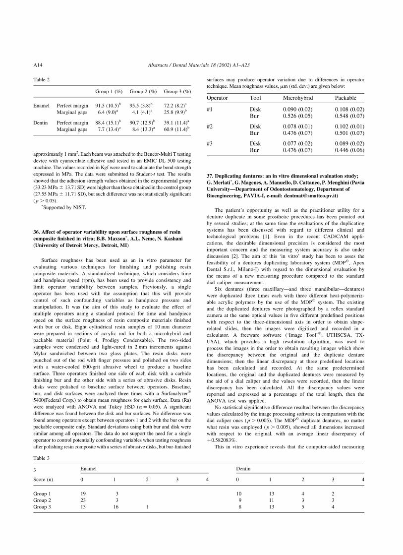
approximately 1 mm2. Each beam was attached to the Bencor-Multi T testing
device with cyanocrilate adhesive and tested in an EMIC DL 500 testing
machine. The values recorded in Kgf were used to calculate the bond strength
expressed in MPa. The data were submitted to Student-t test. The results
showed that the adhesion strength values obtained in the experimental group
(33.23 MPa ^ 13.71 SD) were higher than those obtained in the control group
(27.55 MPa ^ 11.71 SD), but such difference was not statistically significant
( p . 0.05).pSupported by NIST.
36. Affect of operator variability upon surface roughness of resin
composite finished in vitro; B.B. Maxsonp, A.L. Neme, N. Kashani
(University of Detroit Mercy, Detroit, MI)
Surface roughness has been used as an in vitro parameter for
evaluating various techniques for finishing and polishing resin
composite materials. A standardized technique, which considers time
and handpiece speed (rpm), has been used to provide consistency and
limit operator variability between samples. Previously, a single
operator has been used with the assumption that this will provide
control of such confounding variables as handpiece pressure and
manipulation. It was the aim of this study to evaluate the effect of
multiple operators using a standard protocol for time and handpiece
speed on the surface roughness of resin composite materials finished
with bur or disk. Eight cylindrical resin samples of 10 mm diameter
were prepared in sections of acrylic rod for both a microhybrid and
packable material (Point 4, Prodigy Condensable). The two-sided
samples were condensed and light-cured in 2 mm increments against
Mylar sandwiched between two glass plates. The resin disks were
punched out of the rod with finger pressure and polished on two sides
with a water-cooled 600-grit abrasive wheel to produce a baseline
surface. Three operators finished one side of each disk with a carbide
finishing bur and the other side with a series of abrasive disks. Resin
disks were polished to baseline surface between operators. Baseline,
bur, and disk surfaces were analyzed three times with a Surfanalyzerw
5400(Federal Corp.) to obtain mean roughness for each surface. Data (Ra)
were analyzed with ANOVA and Tukey HSD ða ¼ 0:05Þ: A significant
difference was found between the disk and bur surfaces. No difference was
found among operators except between operators 1 and 2 with the bur on the
packable composite only. Standard deviations using both bur and disk were
similar among all operators. The data do not support the need for a single
operator to control potentially confounding variables when testing roughness
after polishing resin composite with a series of abrasive disks, but bur-finished
surfaces may produce operator variation due to differences in operator
technique. Mean roughness values, mm (std. dev.) are given below:
Operator Tool Microhybrid Packable
#1 Disk 0.090 (0.02) 0.108 (0.02)Bur 0.526 (0.05) 0.548 (0.07)
#2 Disk 0.078 (0.01) 0.102 (0.01)Bur 0.476 (0.07) 0.501 (0.07)
#3 Disk 0.077 (0.02) 0.089 (0.02)Bur 0.476 (0.07) 0.446 (0.06)
37. Duplicating dentures: an in vitro dimensional evaluation study;
G. Merlatip, G. Magenes, A. Manuello, D. Cattaneo, P. Menghini (Pavia
University—Department of Odontostomatology, Department of
Bioengineering, PAVIA-I, e-mail: [email protected])
The patient’s opportunity as well as the practitioner utility for a
denture duplicate in some prosthetic procedures has been pointed out
by several studies; at the same time the evaluations of the duplicating
systems has been discussed with regard to different clinical and
technological problems [1]. Even in the recent CAD/CAM appli-
cations, the desirable dimensional precision is considered the most
important concern and the measuring system accuracy is also under
discussion [2]. The aim of this ‘in vitro’ study has been to asses the
feasibility of a dentures duplicating laboratory system (MDPq, Apex
Dental S.r.l., Milano-I) with regard to the dimensional evaluation by
the means of a new measuring procedure compared to the standard
dial caliper measurement.
Six dentures (three maxillary—and three mandibular—dentures)
were duplicated three times each with three different heat-polymeriz-
able acrylic polymers by the use of the MDPq system. The existing
and the duplicated dentures were photographed by a reflex standard
camera at the same optical values in five different predefined positions
with respect to the three-dimensional axis in order to obtain shape-
related slides, then the images were digitized and recorded in a
calculator. A freeware software (‘Image Tool’w, UTHSCSA, TX-
USA), which provides a high resolution algorithm, was used to
process the images in the order to obtain resulting images which show
the discrepancy between the original and the duplicate denture
dimensions; then the linear discrepancy at three predefined locations
has been calculated and recorded. At the same predetermined
locations, the original and the duplicated dentures were measured by
the aid of a dial caliper and the values were recorded, then the linear
discrepancy has been calculated. All the discrepancy values were
reported and expressed as a percentage of the total length, then the
ANOVA test was applied.
No statistical significative difference resulted between the discrepancy
values calculated by the image processing software in comparison with the
dial caliper ones ( p . 0.005). The MDPq duplicate dentures, no matter
what resin was employed ( p . 0.005), showed all dimensions increased
with respect to the original, with an average linear discrepancy of
þ0.582083%.
This in vitro experience reveals that the computer-aided measuring
Table 2
Group 1 (%) Group 2 (%) Group 3 (%)
Enamel Perfect margin 91.5 (10.5)b 95.5 (3.8)b 72.2 (8.2)a
Marginal gaps 6.4 (9.0)a 4.1 (4.1)a 25.8 (9.9)b
Dentin Perfect margin 88.4 (15.1)b 90.7 (12.9)b 39.1 (11.4)a
Marginal gaps 7.7 (13.4)a 8.4 (13.3)a 60.9 (11.4)b
Table 3
3 Enamel Dentin
Score (n) 0 1 2 3 4 0 1 2 3 4
Group 1 19 3 10 13 4 2
Group 2 23 3 9 11 3 3
Group 3 13 16 1 8 13 5 4
Abstracts / Dental Materials 18 (2002) A1–A23A14
system is effective in providing an accurate comparison testing. The
photo taking and the image digitizing may be considered complicated
procedures, but the following image processing analysis is fast and
easy to be handled, moreover many software tools can provide a
surface analysis and more detailed measurements. As a consequence of
this experience it can be concluded that the MDPq duplicating method
seems to be effective in the production of a reliable dentures duplicate
because of it is accepted by the literature a linear discrepancy of not
more than the 1% [3].
References
[1] Lindquist TJ, Ettinger RL. Patient management and decision making
in complete denture fabrication using a duplicate denture procedure:
a clinical report. J Prosthet 1999;82(5):499–503.
[2] Kawahata et al. Trial of duplication procedure for complete dentures
by CAD/CAM. J Oral Rehabil 1997;24(7):540–8.
[3] Kippax et al. How well are complete dentures copied? Br Dent J
1998;185(3):129–33.
38. Microleakage evaluation of the marginal adaptation of different
restorative-adhesive combinations in Class II composite restorations.
Raquel Lima Mouraoa, Angelica Reis Hannasb, Simone Grandinia,
Marco Ferraria (aDepartment of Dental Materials, School of Dental
Medicine, University of Siena, Italy; bDepartment of Restorative
Dentistry, School of Dentistry, Federal University of Minas Gerais,
Brazil)
Objectives. The aim of the present study was to evaluate the sealing
ability of different types of restorative–adhesive combinations in class II
composite restorations and the influence of the consistency of filling
materials on marginal seal. Methods. Fifty extracted human posterior teeth
were selected and stored. A standardized adhesive Class II preparation was
made in the mesial and occlusal surface. The interproximal cervical margin
box was placed 1 mm below the cementum enamel junction. Five groups of
10 samples each were selected and restored with five different combinations
of adhesive-restorative materials: Group 1: Prime & Bond NT, Dyract,
Spectrum TPH, Group 2: Syntac Single Component, Compoglass Flow (as
base) and Tetric Ceram, Group 3: Syntac Single Component, Tetric Flow
and Tetric Ceram, Group 4: One-Step, AelitFlow and Pyramid/ VIP and
Group 5: Excite LC, Tetric Flow and Tetric Ceram. The samples were
immersed in a dye solution and submitted to thermal cycling tests. After
embedding the samples, they were sectioned and were evaluated for
leakage scores at cervical and occlusal margins. The worst score for the
sections of each tooth area was selected for scoring. The results of the
staining measurements were statistically evaluated using the Kruskal–
Wallis non-parametric analysis of variance (ANOVA). Results. In the
cervical wall, group 2 showed better performance, followed by groups 4, 3
and 5 from the least to the highest leakage, respectively. In the occulsal
wall, groups 2 and 4 showed less leakage, followed by groups 5, 1 and 3
from the least to the highest leakage, respectively. Conclusions. Compomer
flowable material in combination with microhybrid resin composite showed
good sealing ability of Class II direct restorations.
39. Cyclic voltammetry an accepted method for corrosion
characterization for anybody W.-D. Mullerp, Chr. Schopf, K.-P. Lange
(Humboldt-University of Berlin, Berlin, Germany)
The modern analytical technique, cyclic voltammetry, is accepted now
for the characterization of the corrosion behavior of dental alloys by
publication of ISO standard 1021 “dental metallic materials—Corrosion
test methods”. It is more complicated to explain this method and the results
caused by the huge amount of data in comparison to the classical immersion
test by watching of the weight loss, but it has more information not only
concerning the corrosion behavior of a specimen. The aim of this work is to
present a special developed equipment for characterization of electro-
chemical behavior of dental alloys using the cyclic voltammetry. With help
of the named Mini-Cell-System it is possible to measure on any interesting
surface of a metallic dental device with a minimum area of 0.008 cm2, so a
special preparation of specimens is no longer necessary. Additional
advantages are the possibility to change the electrolyte very quickly and to
measure fast, with a scan rate of 10 mV/s. The measurements onto pure
elements could demonstrate that the design of the MCS does not produce
any systematic mistakes on the contrary with the developed measurement
routine results were performed comparable with data from reference books.
This special cell was developed for a direct characterization of the
electrochemical behavior of Nd–YAG Laser welding tracks. The
width of a laser track is more or less about 1 mm. By comparing the
results performed onto the surface of the bulks and the tracks it is
possible to make a decision about changes of the corrosion behavior.
The application of this cell starts with pure qualitatively descriptions
of changes in the electrochemical behavior, now it is also possible to
calculate the material loss from the characteristic data Rp (polarization
resistance) and io (exchange current density).
The performed I (current) vs. E (potential) curves can be collected and
summarized to a mean I vs. E curves range for each dental alloy in any
electrolyte to use as a finger print. Deviation and shifts from this reference
curve, spectra, can be interpreted as changes of the electrochemical
behavior with the special view to the corrosion stability. In this way
information about changing of the stability of the oxide layer, activity and
possible adsorption phenomena can be collected.
So different applications are possible besides the characterization of
welding tracks, also the characterization of soldered junctions and the
electrochemical assessment of modified metallic surfaces, like surfaces
of implants.
Last but not the least the application of the MCS can be helpful in
educational training of colleagues and dental students for a better
understanding of corrosion phenomena onto dental alloys and combination
of them.
40. Clinical evaluation of heliomolar HB for posterior restorations;
J.R. Dunn, C.A. Munozp, G. Bernal, J. Torres, A. Wilson (Loma Linda
University, Loma Linda, CA)
This investigation evaluated the clinical performance of a new heavy
body composite resin (Heliomolar, H.B., Ivoclar, N.A.) over a 12-month
period. Fifty posterior MOD and Class II restorations were placed in 32
subjects. Forty-three restorations were evaluated at 12 months. Following
conventional procedures for cavity preparation the teeth were etched,
restored, and polished following manufacturer’s instructions. The teeth
were then evaluated by two examiners for the following variables: Marginal
adaptation (MA), Interproximal anatomic form (IAF), proximal contact
(PC), secondary caries (SC) marginal discoloration (MD) Surface Gloss
(SG) and sensitivity (SE) were evaluated. At 12 months, using a modified
USPHS grading system the following results were found in percentages (%)
(BL ¼ Base Line, IY ¼ one year):
MA IAF PC MD SE
BL 1Y BL 1Y BL 1Y BL 1Y BL 1Y
Alpha 100 88 92 95 78 91 100 88 100 93
Bravo 0 12 8 5 22 7 0 12 0 7
Charlie 0 0 0 0 0 0 0 0 0 0
Delta 0 0 0 0 0 0 0 0 0 0
One year results: (1) Surface gloss on the restorations and secondary
caries remained unchanged (all Alpha) from baseline to one year. (2) Four
of the restorations had a slight degradation of the marginal adaptation
(MA). Those same restorations presented with marginal discoloration
(MD). (3) The interproximal anatomic form (IAF) and the proximal contact
Abstracts / Dental Materials 18 (2002) A1–A23 A15
(PC) showed improvement over time. (4) Surface gloss showed that the
restorations polished adequately at baseline and they maintained the surface
polish over time. (5) Two restorations still had some sensitivity at 1 year.
Overall clinical use of Heliomolar HB as a posterior restoration is
acceptable for routine clinical use.
Study supported by Ivoclar Vivadent.
41. Regional bond strengths to dog cuspid dentin in vivo;
M. Nakajimaa,b,p, I. Urabea,b, R.M. Foxtona,b, J. Tagamia,b, D.H.
Pashleya,b (aTokyo Medical and Dental University, Tokyo, Japan;bMedical College of Georgia, Augusta, GA, USA)
When applying adhesive systems to dentin, the variation of bond
strengths depends not only on the materials but also on many factors about
the substrate such as dentin depth, tubule orientation or with/without pulpal
pressure. The purpose of this study was to evaluate the regional bond
strengths to dentin in dog cuspids in vivo using the microtensile bond test.
Using anesthetized dogs, the cervical surfaces of intact cuspid teeth
were exposed by gingival flap surgery. Long dentin cavities from the incisal
to the gingival of the buccal surface were prepared by means of a diamond
point with copious water spray. The dentin surfaces were bonded with two
self-etching primer adhesive systems (Clearfil LinerBond 2; Kuraray and
Clearfil LinerBond 2V; Kuraray) and two total-etch wet-bonding systems
(Single Bond; 3M and One-Step; Bisco), and then covered with Protect
Liner F. One hour later, the teeth were surgically removed, and then stored
in water at 37 8C. After 24 h, the resin-bonded specimens were serially-
sectioned into 0.7 mm slabs perpendicular to the long axis of the tooth. The
slabs were trimmed to an hourglass shape with a cross-sectional area of
approximately 1 mm2. The specimens were attached to Ciucchi testing
apparatus with a cyanoacrylate adhesive, and subjected to tensile stress in a
universal testing machine at a crosshead speed of 1.2 mm/min.
Statistical analysis was performed using one-way ANOVA and Fisher’s
PLSD post-hoc tests. Statistical significance was considered as p , 0.05.
Materials Incisal Mid-coronal Cervical
LB2 33.8(11.7)(13) 38.2(9.8)(13) 34.8(12.8)(12)
LB2 V 31.0(5.8)(13) 35.8(7.6)(14) 38.6(5.4)(13)
OS 28.3(5.5)(9) 30.9(9.6)(10) 26.2(7.9)(9)
SB 25.5(5.6)(9) 29.0(5.4)(10) 31.6(5.0)(9)
The results were: mTBS(MPa) mean(S.D.)(N)
For mTBS of each material, there were no significant differences
among the three regions ( p . 0.05). For the SEM observation of the
debonded surface, the direction of the dentinal tubules of the incisal
group was relatively parallel, while in the cervical group, the dentinal
tubules were cross-sectioned perpendicular to the adhesive interface.
Therefore, pulpal fluid in the cervical groups may have influenced the
dentin subsurface more than in the incisal groups, although the
remaining dentin of the incisal group was thinner than that of the
cervical group. The condition of the dentin surface as an adhesive
substrate has been thought to be very different at the three regions. On
the other hand, the remaining dentin of the cervical group was
approximately 1.8–2.0 mm thick, through which it might have been
difficult for pulpal fluid to contaminate the dentin subsurface.
Moreover, the dentin surface was prepared using a coarse diamond
bur, resulting in the production of a thicker smear layer. This might be
a reason why the mTBS in this study did not show any differences
among not only the regions but also the materials.
42. Three months evaluation of fluoride release from dentin adhesives;
S. Phanthavonga,p, H. Sonodaa,T. Nikaidoa, P.N.R. Pereirab, J. Tagamia
(aTokyo Medical and Dental University, Tokyo, Japan; bUniversity of
North Carolina at Chapel Hill, USA)
Fluoride has been shown to be an effective agent for prevention of
dental caries. Recently, fluoride-releasing dentin adhesives have been
developed. The purpose of this study was to measure fluoride release
from three single-step adhesives: One-Up Bond F (OB, Tokuyama,
Japan), Reactmer Bond (RB, Shofu, Japan) and Xeno CF Bond (XB,
Sankin, Japan). Resin-modified glass ionomer cement, Fuji II LC-
improved (F LC-imp, GC, Japan), was used as a positive control. Each
material was applied to the vinyl mold (3 mm diameter, 0.36 mm
height), and light-cured for 20 s each on both sides. Specimens were
stored in either distilled water (DW) or demineralizing solution (DS,
pH 4.5) at 37 8C. Amount of fluoride release from each material was
measured with a fluoride ion-specific electrode (combination electrode
fluoride 960900; Orion, USA) up to three months. The data were
statistically analyzed using one way ANOVA and Fisher’s PLSD test
at 95% level of significance. Fluoride concentration was calculated in
ppm. The mean and standard deviation for five samples of each group
were calculated for each interval.
Cumulative amounts of fluoride release are summarized in the Table.
The total amounts of fluoride released from F LC-imp in DS were
significantly higher than that in DW ( p , 0.05). For One-Up Bond F, there
was no significant difference in amount of fluoride release between DW and
in DS ( p . 0.05). For Reactmer Bond, the amount of fluoride release in DS
was significantly higher than that in DW ( p , 0.05). For Xeno CF Bond,
the amount of fluoride release in DW was significantly higher than that in
DS ( p , 0.05). F LC-imp provided the highest amount of fluoride release
among the materials in each solution. For DW groups, the amount of
fluoride release from Xeno CF Bond was significantly higher than those of
One-Up Bond F and Reactmer Bond ( p , 0.05). There was no significant
difference in the amount of fluoride release between One-Up Bond F and
Reactmer Bond ( p . 0.05). For DS groups, the amount of fluoride release
from Reactmer Bond was significantly higher than those of One-Up Bond
and Xeno CF Bond ( p , 0.05). There were no significant differences in
amounts of fluoride release between One-Up Bond F and Xeno CF Bond
( p . 0.05). The single-step adhesives demonstrated the capacity for
fluoride release in both solutions, although these amounts of fluoride-
release were smaller than that of resin-modified GIC. Fluoride released
from each material up to 3 months (ppm) are given below:
Mean(SD), n ¼ 5. S—statistically significant, NS—not statistically
significant.
43. Micro-invasive cavity preparation: an in vitro microleakage study;
C. Poggiop, S. Bianchi, M. Chiesa, U. Genova, G. Merlati (Department
of Odontostomatology, Pavia University, PAVIA-I, e-mail:
The aim of this ‘in vitro’ study was to assess the microleakage behavior
of Class V composite resin restorations performed by means of a micro-
Solution F LC-imp One-Up Bond F Reactmer Bond Xeno CF Bond
DW 1.09(0.28) 0.18(0.04) 0.21(0.02) 0.42(0.06)
S NS S S
DS 1.94(0.35) 0.23(0.06) 0.32(0.04) 0.20(0.03)
Abstracts / Dental Materials 18 (2002) A1–A23A16
invasive cavity preparation system (Durr Vectorw, Durr-Dental GmbH u.
Co.KG) vs. the standard operative technique performed by the usage of
diamond burs. Durr Vectorw improve the ultrasonic cleaning action with
the aid of an abrasive aqueous slurry (silicon carbide particles, 40–50 mm
average grain size).
Thirty sound human teeth, stored in a 0.6% NaCl solution, were
randomly divided into two groups. Group 1: two Class V cavities
(buccal and lingual) were made on each tooth by the means of the
Durr Vectorw system. Group 2 (control group): two Class V cavities
(buccal and lingual) were made on each tooth by the help of diamond
burs. The occlusal boundaries of the cavities were located in the
enamel tissue while the gingival ones were located in dentin. The total
number of specimens was 60 (sixty). After a 10 s acid etching (37%
phosphoric acid) and the placing of two layers of an ‘one bottle’
adhesive system (Scotchbond 1e, 3M, 3M Inc., St Paul, MN, USA),
the samples were restored using three different composite products:
Filtek Z250e (3M), Filtek A110e (3M) and Filtek Flowe (3M).
Following the ISO/TR 11405:1994, the specimens were stored in
water (24 h), then thermocycled (500 cycles, 5–50 8C, 30 s dwell
time), and immersed (24 h at room temperature) in a 2% methylene
blue aqueous solution. Finally, they were rinsed and sectioned in a
bucco-lingual direction at three different locations and the tooth/-
restoration interfaces were evaluated at 32X. The extention of the
microleakage was recorded as a percentage of the total length of the
boundaries.
All the specimens showed no microleakage at the occlusal boundaries
while the Anova test of the scores at the gingival contours showed a highly
significant statistical difference (P , 0,001) between the two groups. There
is no statistically significant difference with regard to the behavior of the
composite products used.
The preliminary in vitro experience revealed that the micro-invasive
cavity preparation by Durr Vectorw is effective in preforming tissue
surfaces available to an adhesive system interaction in comparison with the
usage of a standard diamond bur technique. This result seems to be justified
by the eventually more marked roughness of the treated substrates and the
consequent increase of the total surface available for adhesion, resulting
from the ‘alternative’ treatment. Following the ISO/TR 11405:1994, our
preliminary study has to be considered a laboratory screening research that
should encourage more significant in vivo studies.
Gingival Microleakage is reported in the following table as the
average values (and the standard error of the mean).
Material Durr Vectorw Diamond Burs
A 110 7.2 (6.61) 15.1 (6.88)
Z 250 4.8 (6.52) 14.2 (7.31)
FLOW 8.2 (7.77) 16.8 (6.01)
44. Effect of light curing time on the effectiveness of composite
polymerization; J. Portugalp, M.F. Bernardo, C. Pereira, J. Ortet,
J. Leitao (University of Lisbon, Lisbon, Portugal)
Composite manufacturers generally recommend a minimum 40 s
light curing time to achieve adequate polymerization in 2 mm
increments. For some recent composites, shorter curing times are
being recommended by the manufacturers. Shorter curing times may
have a negative influence on polymerization effectiveness. Several
methods for measuring polymerization effectiveness have been
described. Microhardness determination is a simple, indirect method
to assess the effectiveness of composite polymerization.
Objective. The objective of this study was to compare the
polymerization effectiveness, using the traditional 40 s curing time
and the smaller curing time recommended by some manufacturers, in
three different micro-hybrid composites.
Materials and methods. Silicone molds were used to form thirty
composite slabs with 2 mm of thickness. The composites used were Filtek
Z250 (3M Dental Products), Synergy Duo Shade (Coltene/Whaledent) and
Esthet-X (Dentsply/Caulk). All the specimens had equivalent shades and were
polymerized with a 3M Curing Light 3000XL (600 mW/cm2). For each
composite, half of the specimens were light cured for the time recommended
by their manufacturers (Filtek Z250—20 s; Synergy Duo Shade—30 s;
Esthet-X—20 s) and for 40 s. Six groups with five specimens each were
created. Microhardness (VHN) was determined immediately after light curing
with a Shimadzu Microhardness Tester, on the top and bottom surfaces of the
composite slabs. In each surface three measurements were made and their
mean was used to express the individual sample surface hardness. The
hardness ratio (HR) was calculated using the following formula: HR ¼ VHN
of bottom surface/VHN of top surface. The ideal HR should be equal to 1, as
the hardness of the bottom surface should be similar to that of the top surface, it
has been suggested that an effective polymerization is achieved with a HR
equal or superior to 0.8.
Statistical analysis of the results was performed using ANOVA and
Student’s t-test.
Results. Mean HR for each group is presented in the following table
(Means sharing equal superscript letters are not significantly different):
Light Curing Time Synergy
duo shade
Esthet-X Filtek Z250
Recommended
by manufacturer
0.86 a c 0.88 a b 0.93 b
40 seconds 0.87 c 0.98 d 0.97 d
Statistical analysis showed that both the time of exposure to light and
the composite used have a significant ( p , 0.001) influence on the HR.
Interaction between the two factors was found ( p , 0.05).
Conclusions. Using manufacturers recommended curing times pro-
duced an effective polymerization (HR $ 0.8) for all composites tested at
2 mm depths. Increasing the curing time to 40 s resulted in higher HR for all
composites, except for Synergy Duo Shade.
45. Experimental and numerical analysis of the acting loads on fiber-
reinforced composite resin; N. Borgarelli, G. Rappellip, L. Scalise, A.
Putignano (Universita degli Studi di Ancona, Ancona, Italy)
The main objective of this research is to develop a reliable numerical
model, based on a finite element code in a position to being able to offer an
effective appraisal of loads and deformations on dental prosthesis structures,
when static and dynamic solicitations are present simulating the masticatory
act. In particular, we want to estimate the present characteristics and types of
solicitation on fiber-reinforced composite inlay fixed partial dentures and on
the abutments of the bridge. The surface of a molar and of a premolar have been
reconstructed using a non contact 3D acquisition system. The system is based
on the use of a laser triangulation sensor and generates the grid of points
representing the two teeth surfaces. These clouds of points have been imported
into the finite element model and is used as the geometry of the objects under
simulation. Experimental tests on the composite materials have been carried
out for the determination of their proprieties. Experimental tests have been
conducted for the validation of the finite element model. Compression tests on
in vitro structures have been carried out. Good agreement between the
numerical model and the experimental data have been obtained.
46. Interface amalgam-tooth. Study with six different adhesive systems;
F. Ricciardip, M. Grande, L. Cianconi (University of Rome Tor
Vergata, Italy)
The purpose of this study was to evaluate the morphological relation
existing among amalgam, bonding agents and dental structures.
Ninety-six class V cavities were restored using six different adhesive
systems (Pro-bond, Scotchbond, Amalgambond plus, Amalgambond
Abstracts / Dental Materials 18 (2002) A1–A23 A17

plus þ HPA, All-bond 2, One Step þ Resinomer) and two cavity
varnishes (Copalite, Tubulitec liner), under a dispersed phase and
spherical amalgam. The restored teeth were fixed on a stub and cut
longitudinally towards buccal–lingual direction in two halves by a
diamond saw. This histological technique does not require dehydration
and embedding of the sample. A clear difference between primer and
bonding was allowed by a simple staining technique.
The specimens were observed with a cofocal microscope and under
SEM. All the adhesive systems, with the exception for Amalgambond,
showed a non-homogeneous distribution pattern along the cavity walls. At
the adhesive agent/amalgam interface, Dispersalloy produced a waver
adhesion surface and 2 mm gaps were seldom observed when the adhesive
systems were used. Amalgambond performed no gaps and the best seal of
the tubules. The cavity varnishes instead showed significant detachments
and gaps 10–20 mm large. The study itself demonstrated that the better
interlock between adhesive agent and amalgam is obtained when the
bonding agent is more flowable and the condensation strength is direct and
appropriate.
47. Flexural strength of indirect composites and porcelains; R.P.
Rusinp, R.F. Murphy, P. Baker, M.E. Cavaleri (3M ESPE Dental, St
Paul, MN)
A new composite block material is now available for dental
CAD/CAM. The objective of this study was to compare the flexural
strength of CAD/CAM milled and conventionally fabricated indirect
materials. Surface flaws from machining or finishing can weaken
materials, particularly ceramics. Thus, it is critical to prepare these
materials for strength measurement in the same way as they are used
clinically. 2.5 £ 1.5 £ 12 mm3 bars were milled on a Sironae
CERECe two system from block materials, or molded and cured
from composite pastes; stored 1 d in deionized water at 37 8C; then
broken in a three-point bending fixture. The flexural strengths were:
CEREC-milled Vitae Mark II 118 ^ 15; CEREC-milled Ivoclare
ProCADe 118 ^ 11; 3Me Z100 Restorative 118 ^ 23; Kerre XRV
Lab 99 ^ 16; Jeneric/Pentrone Sculpturee 93 ^ 16; Kulzere Art-
glasse 96 ^ 13; Kerre Belleglasse HP 133 ^ 22; CEREC-milled
3Me Paradigme MZ100 Block for CERECw 146 ^ 15. The data
were analyzed via one-way ANOVA and compared with Tukey’s T-
test ( p , 0.05). The flexural strength of MZ100 is statistically higher
than Mark II, ProCAD, XRV Lab, Sculpture, Artglass, and Z100
Restorative; MZ100 is similar to Belleglass HP.
48. Influence of illuminants on color parameters of enamel shades of
Enamel Plus HFO and Miris composite resins; J.P. Salomona,b,p, J.N.
Monnetc, L. Lapeyred, M. Degrangea, J. Dejoub (aParis V; bFacultes
d’Odontologie de Marseille; cI.U.T de Nımes, France; dMontpellier)
This study investigated the influence of different illuminants on the
color parameters of the incisal shades of two composite resins: ENAMEL
PLUS HFO-MICERIUM- shades Intensiv Milk, White-IM-, IW-, Generic
Enamel 1,2,3-GE1, GE2, GE3-, Opalescent Amber, Blue, Grey,White-OA,
OB, OG, OW- and MIRIS-COLTENE-WHALEDENT-shades Enamel
White Regular-EWR-, Enamel White Bleach-EWB-, Enamel Neutral
Regular-ENR-, Enamel Neutral Translucent-ENT-, Enamel Ivory Regular-
EIR-, Enamel Ivory Translucent-EIT-, Blue-B. Five 1 mm thick samples
were lighted-cured during 80 s (40 s on each side) and stored 24 h at 37 8C.
Five color parameters—L p, a p, b p, whitness index WI BERGER-,
yellowness index YI D1925—were measured with a spectro-colorimeter
(458/0) using 12 different illuminants: A, C, D50, D55, D65, D75, F2, F6,
F7, F8, F10, F11. The comparison of the delta E values between the 12
illuminants showed that the higher values of delta E are the following:
HFOIM:1,03 (between D55 and F6 illuminants), HFOIG:1,02(D75/F6),H-
FOGE1:1,96(D75/A),HFOGE2:1, 56(D75/A),HFOGE3:1,04(D75/A),
HFOOA:5,03(F7/A),HFOOB:2,53(D75/F11),HFOOG:2,76(D75/A),H-
FOOW:1, 57(D75/A), EWR:1,45(D75/A),EWB:1,07(D75/A),ENR:2,
61(F7/A),ENT:3,19(D75/A),EIR:3,29(D75/A),EIT:3,76 (D75/A),B:4,
07(D75/A). These results may explain the difference of colorimetric
perception by the human eye in different illumination conditions.
49. Microleakage around a low-shrinkage composite (InTen-S)
cured with a high-performance light (ASTRALIS 10);
A. Santinip, E Milia. (Edinburgh, UK and University of Sassari,
Sardinia, Italy)
The purpose of the study was to compare microleakage around the low-
shrinkage composite (InTen-S; Ivoclar) cured with the high-performance
light (ASTRALIS 10:Ivoclar) using a bonding agent (Excite; Ivoclar) with a
combination of controls as listed below.
Method and materials. Class V cavities were cut in 18 intact
human teeth. One half of each cavity was in enamel, one half in
cementum/dentine. The teeth were randomly assigned to three groups
ðn ¼ 12Þ: Group 1. Excite, InTen-S, ASTRALIS 10: Group 2. Excite,
InTen-S, Prismetics Lite2 (DeTrey) Group 3. Prime & Bond NT
(DeTrey), Spectrum (DeTrey), Prismetics Lite2; Excite was cured
with ASTRALIS 10 on low setting (700 mV/cm2) for 10 s. InTen-S &
Spectrum were applied in 2.0 mm increments. InTen-S was cured for
10 seconds with ASTRALIS 10 at .1250 mV/cm2 in Group 1 and for
20 s with the Prismetics Lite2 at 400 mV/cm2 in Group 2. In Group 3,
Prime & Bond NT and Spectrum were cured strictly in accordance
with the manufacturer’s instructions. Root apices were sealed with
acrylic resin and the teeth covered with two layers of nail varnish
except for an area of 1 mm around the cavity. The specimens were
stored in water for 6 days and then thermocycled 5000 times between
water baths held at 5 and 55 8C for 10 s at each temperature. The
transport time between baths was 10 s. After thermocycling the
samples were immersed in 5% Procion Brilliant Red buffered to pH 7
at 37 8C for 24 h and rinsed in distilled water for 15 min. A bucco-
lingual cut was made with a diamond disc through each cavity
creating two surfaces along which dye penetration could be measured.
The extent of microleakage was evaluated at £ 10 magnification
using the following criteria.
0 ¼ no leakage;
1 ¼ penetration to 1/3 cavity depth,
2 ¼ penetration to 2/3 cavity depth,
3 ¼ penetration to full cavity depth,
4 ¼ penetration onto axial wall.
The data was analyzed using Kruskal–Wallis non-parametric analysis
of variance test and Dunn’s multiple comparison test.
Results. There was no microleakage around any restorations at the
Enamel margins.
Gingival margins results:
Comparison Mean rank difference Mean rank difference Mean rank difference P value
Test Group 1. vs. Test Group 2. 25.875 ns P . 0.05
Test Group 1. vs Test Group 3. 29.50 Significant * significant * P , 0.05
Test Group 2. vs. Test Group 3. 23.625 ns ns P . 0.05
Abstracts / Dental Materials 18 (2002) A1–A23A18

Group1. (Excite/InTenS/ASTRALIS10) Group3. (P&B.NT/Spectrum/-
Prismatics Lite2)
The results suggest that the low-shrinkage material cured with the high
performance light allows the placement of restorations in less time with
reduced microleakage and therefore offers significant clinical advantages.
50. In vitro erosion of dental luting cements by acidic beverages as a
function of the marginal gap; P. Scheutzelp, D. Lohmann (Department
of Prosthodontics, University of Munster, Germany)
As dissolution and disintegration of dental luting cements may result in
recurrent caries and loosening of the fixed restoration, one of the most
important properties of dental luting agents is their resistance to
environmental factors. In most international standards the values of
solubility and disintegration of dental luting agents are measured in water
and lactic acid solutions. However, these measurements may bear only little
relationship to the nature of in vivo erosion processes by different dietary
acids. Thus, the present study was conducted to investigate the solubility of
different types of dental luting agents immersed in commonly used acidic
beverages as it relates to the size of the cement gap.
The following luting agents were tested: zinc phosphate cements
(Harvard Zement w, Fixodont w), zinc oxide-eugenol cement (Opotow
Trial Zement w), zinc polycaboxylate cement (Carboxylatzement w),
glass ionomer (Aqua Meron w, Ketac Cem w), compomere (Dyract
Cem w), composite (Duo Cement w, Dual Zement w) and resin cements
(F21 w). Using acid-resistant Plexiglasw plates, standardized test
specimens with cement-filled gaps measuring 50, 100, 200, 250 and
300 mm in width were produced. The amount of substance lost at the
cement surface after 3- or 6 h immersion of test pieces in lemon,
orange and apple juice, Coca Cola w, Isostar w, mineral water, white
wine, and buttermilk was quantified profilometrically. In addition to
profilometric analysis, micromorphological surface changes were
assessed qualitatively by scanning electron microscopy.
Clear-cut differences were recorded between the different groups of
luting agents in their susceptibility to erosion. For example, the zinc
polycaboxylate and the zinc phosphate cements (mean erosion depth after
6 h: 2.5–2460 and 1.5–1400 mm, respectively) were eroded distinctly
more strongly under the same conditions than the glass ionomer cements
and the ZOE cement (mean erosion depth after 6 h: 3.0–570 and 5.0–
65 mm, respectively). The least substance loss was recorded in the
composite and the compomere luting agents (mean erosion depth after
6 h: 0–40 mm). Apart from the resin cement, which was not eluated but
rose out of the gap, the erosion depth of all luting agents, especially in case
of compomere and composite cements, underwent a marked increase with
an increasing width of the cement gap (rp ¼ 0:4–0:6; p , 0.001). The
erosion depth of the water-soluble cements was moreover closely correlated
with the acid content of the respective solution (multiple regression
analysis: rp ¼ 0:88–0:98=R2 ¼ 0:78–0:96; p , 0.001). A comparison of
the different beverages revealed clear-cut differences in their capacity to
erode the luting agents tested.
As the glass ionomer, compomere and composite luting cements are
distinctly more resistant to acidic beverages than zinc phosphate cements,
they should be given preference in the dentition damaged by or susceptible
to erosion. However, since the erosion depth especially for compomere and
composite cements were correlated with cement gap size, even in the case
of those largely erosion-resistant cements, every effort should be made to
optimize the marginal fit of fixed restorations and minimize the marginal
gap. The marked material loss of the zinc polycaboxylate cement tested
confirms previous observations which suggested that this type of cement
should be used with caution for long-term luting of fixed restorations.
51. Fluoride-release and secondary caries inhibition by new fluoride-
releasing restorative materials; H. Sonodap, S. Phanthavong,
M. Okuda, Y. Sasafuchi, Y. Kitasako, T. Nikaido, M. Otsuki, J. Tagami
(Tokyo Medical and Dental University, Tokyo, Japan)
This study evaluated the fluoride-releasing properties of a light-cured
one-step bonding resin (Reactmer Bond, Shofu) in vitro and also estimated
the effects of fluoride-releasing restorative materials (Reactmer Bond and
Reactmer Paste, Shofu) on the inhibition of artificial secondary caries
around restorations in monkey teeth in vivo. These materials, Reactmer
Bond and Reactmer Paste, are called ‘GIOMER’ system. GIOMER
comprises pre-reacted glass-ionomer filler (PRG, Ikemura et al., 2000), and
it is a new hybridization of glass-ionomer and composite resin.
Disk-shaped specimens of Reactmer Bond with a diameter of 3.0 mm and
a height of 0.36 mm were prepared. The amount of fluoride released from the
specimens in distilled water was measured up to 3 months (n ¼ 5 for each
group).
One box-shaped cavity (approximately 3 mm long, 4 mm wide, and
1.5 mm deep) was prepared on a buccal root dentin surface restored with
Reactmer. At 7, 30, and 90 days after operation, the teeth were removed
from the experimental animals and the roots were obtained by separating
them from the crown segments (n ¼ 4 for each group). Extracted bovine
roots were used as a control. Two box-shaped cavities on each bovine root
surface were prepared and filled with a bonding system without fluoride-
releasing (Clearfil Liner Bond 2V, Kuraray)/a resin composite without
fluoride-releasing (Clearfil AP-X, Kuraray) ðn ¼ 12Þ: The bovine roots
were then stored for one week in water. All restored roots were stored in
demineralizing solution (pH 4.5) for 3.5 days at 37 8C. A cut through the
center of each restoration was made parallel to the long axis of the root, and
the cut surfaces were polished and observed using a confocal laser scanning
microscope (CLSM, Okuda et al.,2000). Data were analyzed by one-way
ANOVA, Fisher’s PLSD Test at the 95% level of confidence.
Cumulative amounts of fluoride and the widths of the inhibition zone
are summarized in the table. Reactmer created a thick inhibition zone in the
artificial lesion along the restoration–dentin interface. The widths of the
inhibition zones were not significantly different among all groups
( p . 0.05). Inhibition zones were not observed adjacent to LB2V/AP-X
specimens. These results indicated that the fluoride-releasing restorative
materials have the potential to inhibit secondary caries formation around
restorations.
52. Post-operative symptoms after single-visit endodontic retreatment
of periapical lesions; G. Gambarini, L. Testarellip, G. Pongione, C. Di
Paolo, G. Floridi, E. Tucci, R. Pippi, L. Gallottini, M. De Luca
(University of Rome “La Sapienza” Dental School)
Several studies reported the efficacy of laser in disinfecting a contaminated
root canal and consequently reduce post-operative symptoms. The aim of the
present clinical investigation was to determine whether Nd/YAG laser therapy
has a beneficial effect on post-operative pain after single-visit non-surgical
endodontic retreatment of periapical lesions. Thirty retreatment cases
(infected teeth with periapical lesions) were selected and divided into two
homogeneous groups. All teeth (Groups A and B) were prepared using a
crown-down nickel–titanium rotary preparation, irrigated with 5.25% sodium
hypochlorite (Niclor, Ogna, Milan) and, after shaping procedures were
7 days 30 days 90 days
Cumulative amounts of fluoride (mean(SD) ppm) 0.07(0.004) 0.14(0.01) 0.21(0.02)
Widths of the inhibition zone (mean(SD) mm) 26(6) 32(11) 38(18)
Abstracts / Dental Materials 18 (2002) A1–A23 A19
completed, irrigated using ultrasonic irrigation for 2 min. Teeth of Group B
(laser) patients were then irradiated with Nd/YAG laser for 20 s. All canals
were obturated in the same visit with warm guttapercha and sealer. Occurrence
of pain was recorded 7 days after treatment. Data were collected and
statistically analyzed using the Mann–Whitney Wilconxon test. Pain ratings
were: Group A: 0. None 3 (20%), 1. Mild 4 (27%), 2. Moderate 6 (40%), 3.
Severe 2 (13%); Mean 2.2. Group B: 0. None 7 (46%), 1. Mild 4 (27%), 2.
Moderate 4 (27%), 3. Severe 0 (00%); Mean 1.2 Mean values showed that pain
intensity difference was significantly less ( p , 0.05) in the laser treated group
(B) than in the control (A) group. These findings suggest that the clinical
application of Nd/YAG laser migth be advantageous in reducing post-
operative pain following endodontic retreatment of periapical lesions.
53. Role of composite materials to prevent enamel marginal
demineralization; L. Savarinoa, A. Saponara Teutonicob,p, L. Breschib,
M. Visentina, C. Prati b (aIstituti Ortopedici Rizzoli, Bologna;bUniversity of Bologna, Italy)
New composite restorative materials have been recently developed to
prevent secondary caries formations and to reduce the risks for marginal
alterations. Several investigations demonstrated that marginal enamel surfaces
are highly affected by gaps and fractures caused by polymerization shrinkage
stress of composite materials. These gaps may increase the depth and the
extent of enamel demineralization and the consequent enamel caries. The
purpose of this study was to evaluate the role of composite and fluoride
releasing-materials to prevent demineralization after immersion in cariogenic
solution, in an attempt to evaluate their role in preventing secondary caries
formation. Twelve class V restorations were placed in vitro in human extracted
third molars and immersed in demineralizing solution to simulate the
formation of secondary caries and its effect on marginal integrity of composite
restorations. Scotchbond 1 plus Z 250, Scotchbond 1 plus F 2000, SE Bond
plus APX, ABF plus APX, and Fuji IX were used in the study. All restorations
were made by one operator. Each sample was stored in demineralizing solution
(lactic acid, pH 4.5) at 378 for 4 days. The cariogenic solution was changed
three times a day. Enamel marginal demineralization was firstly inspected
under optical microscope ( £ 60 magnifications). Microhardness evaluations
were performed with a Leica VMHT 30A microindenter device working at 50
gr for 15 s and with indentations at 20 mm. Results (mean(SD); n ¼ ; ANOVA
plus t test for paired data)
Conclusions. Exposed enamel was affected by demineralizing-cario-
genic procedures. However, enamel close to restoration margins frequently
showed the highest rate of demineralization, as demonstrated by the lowest
rate of microhardness number.
The materials which claimed fluoride release did not prevent any type of
enamel marginal alteration. The study demonstrated that exposed marginal
enamel shows the highest rate of demineralization and for this reason it may
be rapidly affected by secondary caries formation.
54. Resin bond strength and micromorphology of human teeth prepared
with an erbium/YAG laser; C.P. Trajtenberga,b,p, P.N.R. Pereiraa,b, J.M.
Powersa,b (aUniversity of Texas-Houston Dental Branch; bUniversity of
North Carolina at Chapel Hill, School of Dentistry)
The Er/YAG laser (2940 nm) used for cavity preparation offers advantages
over the rotary instrumentation such as selective caries removal, surface
etching and no need for anesthesia. There are limited data on how these effects
influence bond strength of restorative materials. The purpose of this study was
to determine the effect of two different surface treatments and 3 conditioners
on the bond strength of composite to enamel and dentin. Forty-eight, caries-
free third molars were used. Dentin specimens (D) were sectioned at the
occlusal surface 2 mm from the highest pulp horn. Enamel specimens (E) were
sectioned mesio-distally. Surfaces were polished and treated either with
carbide bur (B, Brasseler, USA) or an Er: YAG Laser (L, DeLight Laser
System, Continuum Biomedical). Laser energy settings for enamel and dentin
were 260 mJ/25 Hz and 160 mJ/10 Hz, respectively. Three different con-
ditions: acid-etch/bonding agent (ASM, Scotchbond Multipurpose, 3M
ESPE); same bonding agent without etching (SM), and an experimental
self-etching primer (SEP, EXL 547, 3M ESPE) were used. Resin composite
(Filtek Z250, 3M ESPE) was used to create composite build-ups. After storage
for 48 h at 37 8C and 100% RH, specimens were prepared in an hourglass
shape for microtensile bond test (ca. 1 mm) and debonded in tension at a
crosshead speed of 0.5 mm/min. Failure modes were classified using SEM into
three types: type 1, complete adhesive failure between bonding resin and
dentin/enamel; type 2, partial adhesive failure between bonding resin and
dentin/enamel, and partial cohesive in bonding resin; type 3, cohesive failure
within bonding resin. Means, standard deviations and failure modes by type
with percentages are listed. Data were analyzed with ANOVA ( p , 0.05).
Morphology of surface treatments were analyzed with SEM.
Scheffe’s intervals ( p , 0.05) for comparison among treatments,
conditioners and tooth substrates were 1.7, 2.7 and 1.7 MPa, respectively.
BD/ASM group had the highest bond strength. LE and LD resulted in the
highest bond strengths when used with ASM. Bur exhibited higher bond
strengths than laser with the use of SEP. Laser showed higher bond strengths
than bur with the use of SM. Most laser-treated groups exhibited a type two-
failure mode with the exception of LE/SM that was a type 1. BE and BD/SM
exhibited a 100% type 1. The remaining bur-cut groups exhibited a type 2
except BD/ASM that showed type 3. SEM analysis of laser-treated enamel and
dentin showed absence of the smear layer when compared to untreated control
surfaces. Laser-treated enamel resulted in a retentive pattern similar to acid-
etched enamel. Micromorphology of laser-treated dentin revealed a good
Material Groups
(N ¼ 8)
Marginal Enamel
Vickers Number
Exposed Enamel
Vickers Number
Protected Enamel
Vickers Number
ABF Bond þ APX 243.8(37.4) P , 0.001 325.5(54.4) 573.0(88.1)
Fuji IX 179.0(64.8) P , 0.001 272.9(101.2) 407.1(98.7)
F 2000 139.5(69.1) P , 0.001 227.9(108.5) 379.0(54.5)
Tensile bond strength (MPa) Predominant failure type and (%)
LE LD BE BD LE LD BE BD
SM 16.7 (5.8) n ¼ 15 9.1 (5.7) n ¼ 19 0a* n ¼ 20 0a n ¼ 20 1(77) 2(79) 1(100) 1(100)
ASM 25.8 (8.0)b n ¼ 20 21.1 (6.1) n ¼ 20 25.4 (10.3)b n ¼ 19 35.7 (10.2)d n ¼ 20 2(84) 2(63) 2(58) 3(68)
SEP 10.8 (4.7)c n ¼ 20 11.9 (2.8)c n ¼ 20 16.9 (9.3)a n ¼ 12 19.6 (5.5)a n ¼ 17 2(70) 2(53) 2(58) 2(80)pGroups with the same superscript letters are statistically the same ( p , 0.05)
Abstracts / Dental Materials 18 (2002) A1–A23A20

definition of dentinal tubules. At lower energy settings, some smear plugs
remained evident. Bonded interfaces showed continuity of the hybrid layer
with the intertubular dentin when the dentin was acid-etched, and a gap when
the self-etching primer was used. The acid-etch and self-etching primers
allowed good resin tag formation. The resin tags produced by ASM and SEP in
the dentin presented a collar-like formation in the first 5 mm, and lateral
filaments due to the effect of the laser and as a result of resin monomer
penetration. The hybrid layer in the enamel disclosed an intimate adaptation of
the bonding agent with both ASM and SEP. Bonding of laser-treated teeth with
adhesive resin without the application of phosphoric acid was possible due to
the ability of the laser to remove the smear layer, which is usually present on
bur-cut surfaces. Acid-etching the tooth surface treated with either the laser or
bur, prior to the application of bonding resin, is necessary to achieve clinically
acceptable bond strengths. Supported in part by 3M ESPE.
55. National survey of adverse reactions to dental materials in the UK;
R. Van Noortp, A. Scott, J. Grummitt, P. Hatton, W. Egner,
D. Gawkrodger, C. Yeoman (University of Sheffield, UK)
Purpose
Dental treatment involves the use of a wide range of materials, and
they are therefore the most extensively employed biomaterials. Many
of these materials or their components pose a potential occupational
risk, as well as a risk to the patient. While the introduction of new
materials brings great benefits, there is always a risk of an adverse
reaction to one or more components by members of the dental team or
the public. Pre-market biocompatibility testing cannot guarantee
absolute safety, so it is important to identify materials that can
cause an adverse reaction when placed on the open market. The
prevalence of adverse reaction to dental materials and surgical
products among the general population and the dental profession has
not been effectively monitored, and have been reported to be up to 4
and 20%, respectively. It is important to identify the true extent and
severity of these reactions so as to be able to respond appropriately.
The aim of this project is to evaluate adverse reactions to dental
materials in the UK.
Method
A simple reporting form has been designed, and is being circulated to
approximately 23,500 dental practices and 2700 dental laboratories on a
regular basis. It has also been made available on the web (http://www.shef.
ac.uk/~arrp/). Dental professionals are requested to complete a reporting
form whenever an adverse reaction is suspected. An adverse reaction is
described as an unusual or unexpected response to contact with a dental
material. By dental material we mean any material found in the dental
practice or sold for dental purposes, such as restorative materials, rubber
gloves, toothpastes and oral rinses. However, it excludes drugs and local
anaesthetic that should be reported to the Yellow Card Adverse Drug
Reactions Reporting Scheme.
Results
To date the number of adverse reactions reported is 600, consisting of
268 and 329 patients and occupational adverse reactions, respectively. The
results show that the dental material suspected of causing most adverse
reactions in patients is metal, specifically amalgam ðn ¼ 64Þ: These adverse
reactions are seen as intra-oral swelling, tenderness and lichenoid type
lesions. Occupational adverse reactions were predominantly associated
with gloves ðn ¼ 213Þ; causing contact dermatitis, irritation and respiratory
problems to dentists, dental nurses and hygienists in the dental practice. In
contrast dental technicians were seen to experience most adverse reactions
to resin ðn ¼ 23Þ; causing contact dermatitis on the hands and fingers.
Conclusion
The results show that different groups of people are susceptible to
adverse reactions to different materials. This national survey has only just
begun and further work is needed to establish trends in relation to the
adverse reactions and whether or not adverse reactions are increasing or
declining.
Acknowledgement
Funded by the NHS National Programme for Primary Dental Care
Research and Development, reference number RDO/90/40.
56. Color stability of two resin-based composites after water aging;
A. Vichip, E. Bertelli, G. Blasi, M. Ferrari (University of Siena and
University of Genova, Italy)
Objectives. It has been recently suggested that some resin composite
systems, initially developed for indirect technique, can be used also for
direct restorations. As the polymerization method is different, this may
result in a different behavior of the resin system. One of the parameters that
may be affected, it is the intrinsic color shifting with time. The aim of this
study was to test the influence of exposure to water on the color stability of
two different resin-based composites.
Methods. Four comparable Vita shades of two different resin-based
composite systems were selected: Gradia (GC, Tokyo, Japan) and Sinfony
(Espe, Seefeld, Germany). For each product at each shade, three disks were
made, 1 mm in thickness and 15 mm in diameter. For each product, the
polymerization was made with its proprietary polymerization device and
with an office polymerization lamp. The samples were studied with a
spectrophotometer equipped with an integrating sphere. For color
determinations, a 50% gray card was used as background, and the data
were recorded in the CIE L pa pb p system. After the initial measurements,
the samples were stored for 30 days in a 60 8C water bath and then
measured again under same conditions. The color shifts were calculated
using the formula: DE ¼ ½ðL1 2 L2Þ2 þ ða1 2 a2Þ
2 þ ðb1 2 b2Þ2�1=2:
Results. If the standard of DE , 3.3 is taken into consideration as reference
for clinical acceptability both materials showed shade shifts that exceed the
reference value. Gradia showed a lower degree of discoloration, with 50%
of the comparisons within an acceptable shade shift (DE , 3.3) after aging
when polymerized in the laboratory device, and 75% of the comparisons
acceptable when polymerized in the laboratory device. Sinfony showed a
higher degree of discoloration with 25% of the comparison within
acceptable shade shift when polymerized with the halogen lamp and 50%
acceptable when polymerized in the laboratory device. However, the
difference of the DE values between the two products was not statistically
significant ðp ¼ 0:575Þ when evaluated with ANOVA statistical analysis.
The difference of the DE values between the two methods of polymerization
was also not statistically significant ðp ¼ 0:827Þ: Significance. From this
work it can be concluded that for these two products aging in water leads to
a certain degree of color instability. The different method of polymerization
does not influence the degree of shade shift of the two products when
submitted to water aging.
57. Carbon fiber post/composite core restoration of flared roots:
adaptation of one or more posts; D. Angerame (University of Trieste,
Trieste, Italy)
The aim of this in vitro qualitative study was to evaluate the adaptation
among materials and between materials and dentin in flared roots restored
with carbon fiber post and composite core build-up. A total of 48 human
monorooted premolars were divided into four groups of 12 teeth; their
crowns were sectioned and the root canals were enlarged by a diamond bur
until the dentin walls became very thin. After acid (20 s) and adhesive (self-
curing mode) conditioning (Scotchbond Multipurpose System, 3M) the
canals were completely filled with a self curing composite resin (Ti-Core N,
EDS, USA). A control group (G1) had only the composite resin into the
canal; the second group (G2) was restored with one carbon fiber post (Tech
2000, Isasan, Italy) inserted into the composite before its polymerization;
the third (G3) and fourth (G4) groups were restored by inserting three and
seven posts, respectively. The specimens were aged by thermocycling (150
cycles at 2–4/90–95 8C), nail coated, and immersed in super-saturated
Abstracts / Dental Materials 18 (2002) A1–A23 A21

methylene blue solution (5 min); then they were sectioned and 10 cross-
sections (A) were obtained from each root; the cervical portion of the
specimen was longitudinally sectioned and four mesio-distal sections (B)
were prepared. The sections were observed by a stereomicroscope and the
dye penetration was scored 0–3; defects of adaptation and voids were also
checked. The data were analyzed by Kruskal–Wallis and Mann–Whitney
tests. The adaptation of materials was found very good and the percentage
of sections without dye penetration was high in all groups (A: G1 ¼ 99:17;
G2 ¼ 100; G3 ¼ 98:33; G4 ¼ 97:5; B: G1 ¼ 93:75; G2 ¼ 95:83; G3 ¼
89:58; G4 ¼ 87:5); there was no statistical difference among the groups.
The three variants of the technique can be useful in treating flared roots;
further in vitro investigations are required to understand which of the
variants has the better resistance to load.
58. The influence of the light source on resin composite polymerization;
P. Casconep, L. Di Palma, G. Montanaro, A. Apicella, S. Rengo
(University of Naples “Federico II”, Italy)
The aim of this study was to investigate the difference in kinetics of
composite polymerization using three types of light source. Four different
composites were tested: two traditional composites (Prodigy–Kerr and
Charisma–Kulzer); and two flow composites (Revolution–Kerr and Flow-
line–Kulzer). The composite samples were positioned in the Differential
Scanner Calorimeter cell (DSC 25 by Mettler Instrumente AG) and then light
cured. The DSC is able to record the heat developed during the polymerization
reaction. From the heat reaction it is possible to calculate the relative degree of
polymerization of composite materials. Each material was cured with three
different light sources: six samples were light cured with a halogen light
(Optilux 500-Kerr) at a constant power of 500 mW/cm2 for 60 s; six samples
were light cured with a halogen light (Astralis 10-Ivoclar Vivadent) using soft
start polymerization: in the first 10 s the intensity light increases from 150 to
650 mW/cm2, in the following 10 s the light works in a pulsating mode
between 650 and 1200 mW/cm2; six samples were light cured with a LED light
source (Zap) using a step polymerization technique: 5 s at low intensity
(50 mW/cm2) and then 12 s at high intensity (300 mW/cm2). The specific
reaction heats (J/g) with standard deviations for all tested composite materials
are shown in the following table:
Material Halogen
(Optilux)
reaction heat
(J/g)
LED (Zap)
reaction heat
(J/g)
Halogen
soft start
(Astralis)
reaction heat
(J/g)
Prodigy (Kerr) 30 (3) 32 (1) 32 (4)
Charisma (Kulzer) 29 (4) 29 (1) 30 (1)
Revolution (Kerr) 62 (4) 61 (2) 60 (7)
Flowline (Kulzer) 65 (9) 52 (1) 52 (2)
ANOVA statistical analysis was performed with p , 0.05. The three
different polymerization techniques showed no statistically different
reaction heats for all the tested materials except for the Flowline (Kulzer);
in this case the reaction heat obtained with the polymerization with halogen
light in one step at high power was statistically significantly higher than the
reaction heat obtained with the soft start and the step polymerization.
In conclusion, step and soft start polymerization are not able to cure all
the composite materials with the same conversion obtained with traditional
halogen polymerization.
59. Wear of posterior composite resins by glass beads abrasion test;
Y. Tanip, K. Kamata, A. Ishikawa (University of East Asia and Nippon
Dental University at Tokyo, Shimonoseki and Tokyo, Japan)
The glass-beads abrasion test reported by Tani et al. (1983) had foreseen
that the coarse-filled composites would be inferior to microfilled
composites in clinical results of wear resistance. The purpose of this
study was undertaken to determine an accurate appraisal of wear resistance
of different types of the composite resins. The composite resins used in this
study consisted of three highly-loaded coarse-filled composites; Clearfil
AP-X (Kuraray), Z250 (3M) and Beautifil (Shofu), two submicrofilled
composites; Palfique Estelite (Tokuyama) and Palfique Toughwell
(Tokuyama), and two microfilled composites; Silux Plus (3M) and Metafil
C (Sun Medical). Five specimens (10 £ 10 £ 1.5 mm3) of each material
were prepared by photo-curing. All specimens were polished with a 1200
grit SiC paper to wear away 200 mm from the surface to be tested. After,
these specimens were kept in a desiccator until they attained a constant
weight, the specific gravity of each material was measured by an electronic
gravity meter. The specimens, fixed in the horizontal drive arm, was
reciprocated with a 400 g load of glass beads at a velocity of 120 strokes per
minute for 10,000 times, which is equivalent to traveling 2000 m. The
degree of wear was measured by weight loss, converted to volume loss with
the specific gravity of each material. Two types of glass beads (Toshiba–
Ballotini) slurries were mixed with 140 g of the surface-treated glass beads
(EGB) to 100 ml water. The other slurry was prepared with the surface-non-
treated glass beads (GB). Both glass beads were 40 mm in diameter. These
were placed in an open-top stainless steel container like a ship. The slurry
was poured into the container. The results of glass bead wear of composite
resins are shown in Table 1.
Data were analyzed by two way ANOVA. It was concluded that the
wear with EGB was much larger than with GB. The microfilled composite
resin showed least wear compared to larger wear of the coarsefilled
composite resins. This result suggests that the filler particle size is relatively
important to wear resistance of the composite resin.
This research was supported in part by the Scientific Research Grant
(B)(1) No. 11470425, Ministry of Education and Science, Japan.
60. Thermal effects on the microstructure of human dentin; Y.K.E.
Ariantop, Na Peng Bo, S.M. Surono Akbar (Graduate Program of
Materials Science, Faculty of Mathematics and Natural Sciences and
Faculty of Dentistry, University of Indonesia, Jakarta, Indonesia)
Human dentin represents a hydrated biological composite structure for
Table 1
Glass bead wear of composite resins (cm2 £ 1023)
Materials Specific gravity Surface treated glass beads (EGB) Surface-untreated glass beads (GB)
Clearfil AP-X 2.03 56.37 (3.23) 22.53 (1.59)
Z250 2.08 61.31 (6.82) 28.63 (5.53)
Beautifil 2.25 71.78 (3.61) 37.48 (2.57)
Palfique Estelite 1.88 34.97 (3.14) 16.11 (2.43)
Palfique Toughwell 2.04 45.12 (1.97) 20.74 (2.88)
Silux Plus 1.60 32.13 (1.90) 15.10 (2.87)
Metafil C 1.47 23.04 (1.69) 7.56 (0.76)
Abstracts / Dental Materials 18 (2002) A1–A23A22

which only limited structure–thermal property relationship are available.
Underlying treated and untreated dentin structural characteristics and
properties are the key considerations for future development of improved
restorative treatment.
The purpose of this study was to investigate the thermal effects on the
microstructure of human dentin by X-ray diffractometry (XRD), differential
scanning calorimetry (DSC), differential thermal analysis (DTA) and
thermogravimetry analysis (TGA).
Dentin samples were taken from sound permanent human third molar
teeth. Discs and powder specimens were heated to different temperatures
from room temperature (RT) to 1000 8C. The heat-energy changes
associated with physical and chemical transformations as a function of
temperature were measured by differential thermal analysis (DTA-50,
Shimadzu Corp., Japan). Dentin disc was placed in an alumina crucible and
heated from room temperature to 900 8C with heating rate of 10 8C/min.
Thermal decomposition behavior of human dentin discs and powder
from RT to 700 8C were determined using Differential Scanning
Calorimetry (DSC-50, Shimadzu Corp, Japan).
The loss of volatile decomposition products were measured by loss of
weight from dentin powder using Thermogravimetry analysis as a function
of temperature (TG-50, Shimadzu Corp, Japan). The heating rate was
10 8C/min from room temperature to 1000 8C.
To identify the structural phase changes, dentin discs were heated in a
Ring furnace at temperature of 150, 600, 750 and 830 8C, respectively. The
normal and heat-treated dentin discs were then examined by X-ray
diffractometry (Philips X-Ray Diffractometer type PW 3710 based with
PC-APD diffraction software) with Co K radiaton, 35 kV, 20 mA and step
scanning type with 1.5 s time/step. The crystallinity of dentin crystals were
calculated after profile fitting ð2u ¼ 288 2 428Þ using reference specimen of
quartz. Peak width (B) was calculated using Full Width at Half Maximum
(FWHM) from peaks of (002), (211) and (112) planes by Warren equation
ðB2 ¼ b2 þ b2Þ and crystallite size by Scherrer equation ðD ¼ kl=b cos uÞ:
The DSC of dentin shows a large endothermic curve at 50–285 8C,
likewise the DTA endothermic peak at 137 8C which represent a material of
less crystallinity and inhomogenous crystals. An exothermic peak was also
shown at 498 8C by DSC. The loss of dentin weight by TG analysis at 35–
330 8C was 8.52% with total weight loss of 21.26% after heated to 900 8C.
The results of XRD revealed that at higher temperature (750 and
830 8C) the amount of diffraction peaks of hydroxyapatite were higher and
more intense with the development of Whitlockite peaks at 2u ¼ 34:72;
36.32; 40.3; 55.48. The crystallite size were also higher showing crystal
growth upon heat-treatment.
It can be concluded that the thermal behavior of human dentin shows
physical and chemical changes with higher temperature, which affect their
microstructure and properties relationship.
Abstracts / Dental Materials 18 (2002) A1–A23 A23


![[Composite Cultures] - CORE](https://static.fdokumen.com/doc/165x107/6325e67de491bcb36c0a86c0/composite-cultures-core.jpg)