The diffusion kinetics of a nanofilled and a midifilled resin composite immersed in distilled water,...
9
ORIGINAL ARTICLE The diffusion kinetics of a nanofilled and a midifilled resin composite immersed in distilled water, artificial saliva, and lactic acid Eduardo Moreira da Silva & Luana Gonçalves & José Guilherme Antunes Guimarães & Laiza Tatiana Poskus & Carlos Eduardo Fellows Received: 13 May 2009 / Accepted: 24 January 2010 / Published online: 9 March 2010 # Springer-Verlag 2010 Abstract This study investigated the diffusion kinetics of a nanofilled (Filtek Z350) and a midifill (Filtek P60) resin composite immersed in distilled water, artificial saliva and lactic acid. Resin composite specimens were desiccated, immersed in the media, weighed at suitable time intervals until they reached sorption equilibrium and were then desiccated again. Sorption and solubility (μg/mm 3 ) were calculated based on ISO 4049:2000(E). The diffusion coefficient (m 2 .s -1 ) was determined according to Flick’ s second law. The degree of conversion (DC%) was evaluated by FT-IR and the action of the media on the surfaces of the resin composite was evaluated by SEM. Z350 immersed in lactic acid presented the highest sorption (25.9±1.3). The highest solubility was presented by Z350 immersed in lactic acid (5.6±0.9), followed by P60 immersed in lactic acid (4.4±0.5). The other groups presented no significant difference among them. The diffusion coefficients of both resin composites immersed in lactic acid and that of Z350 immersed in artificial saliva were significantly higher. The lowest diffusion coefficient was presented by P60 immersed in distilled water. The DC % was not significant, (p >0.05). The SEM analysis showed that the effect of lactic acid on the resin composites was more deleterious than those of water and artificial saliva. Keywords Resin composites . Solubility . Sorption . Diffusion coefficient . Degree of conversion Introduction In an endeavor to gain a better understanding of the resin composite degradation process, several researchers have exhaustively studied the sorption, solubility, and mechan- ical properties of many resin composite after immersion in water, artificial saliva, or ethanol [1–11]. Resin composites are generally constituted of a polymeric matrix, filler particles, and a silane-coupling agent that links the matrix to the fillers. The current resin composites are classified according to the size of their filler particles, as microfilled, hybrid, microhybrid, and nanofilled [12]. Filtek Z350 (the universal restorative that contains the same patented and proprietary nanotechnology used to create Filtek™ Su- preme Universal Restorative, 3M-ESPE) is a typical nano- filled composite, which has a filler particle system that combines silica nanofillers with a primary particle size of 20 nm and zirconia-silica nanoclusters of 0.6–1.4 μm [13]. Although some published studies have shown that this type of resin composite presents mechanical properties similar to those of the hybrid or midifill type [14, 15], the large surface area to volume ratio derived from the silica particles may increase its water uptake, leading to degradation of the filler-polymeric matrix interface [5, 9], and to a probable reduction in some of its mechanical properties [1]. E. M. da Silva : L. Gonçalves : J. G. A. Guimarães : L. T. Poskus Department of Restorative Dentistry, School of Dentistry, Federal Fluminense University, Niterói, Rio de Janeiro, Brazil C. E. Fellows Institute of Physics, Federal Fluminense University, Avenida Litorânea, Niterói, Rio de Janeiro, Brazil E. M. da Silva (*) Universidade Federal Fluminense / Faculdade de Odontologia - Rua São Paulo, nº 28–Campus Valonguinho, Centro, Niterói, Rio de Janeiro 24040-110, Brazil e-mail: [email protected] Clin Oral Invest (2011) 15:393–401 DOI 10.1007/s00784-010-0392-z
-
Upload
independent -
Category
Documents
-
view
5 -
download
0
Transcript of The diffusion kinetics of a nanofilled and a midifilled resin composite immersed in distilled water,...
ORIGINAL ARTICLE
The diffusion kinetics of a nanofilled and a midifilled resincomposite immersed in distilled water, artificial saliva,and lactic acid
Eduardo Moreira da Silva & Luana Gonçalves &
José Guilherme Antunes Guimarães &
Laiza Tatiana Poskus & Carlos Eduardo Fellows
Received: 13 May 2009 /Accepted: 24 January 2010 /Published online: 9 March 2010# Springer-Verlag 2010
Abstract This study investigated the diffusion kinetics of ananofilled (Filtek Z350) and a midifill (Filtek P60) resincomposite immersed in distilled water, artificial saliva andlactic acid. Resin composite specimens were desiccated,immersed in the media, weighed at suitable time intervalsuntil they reached sorption equilibrium and were thendesiccated again. Sorption and solubility (µg/mm3) werecalculated based on ISO 4049:2000(E). The diffusioncoefficient (m2.s!1) was determined according to Flick’ssecond law. The degree of conversion (DC%) wasevaluated by FT-IR and the action of the media on thesurfaces of the resin composite was evaluated by SEM.Z350 immersed in lactic acid presented the highest sorption(25.9±1.3). The highest solubility was presented by Z350immersed in lactic acid (5.6±0.9), followed by P60immersed in lactic acid (4.4±0.5). The other groupspresented no significant difference among them. Thediffusion coefficients of both resin composites immersedin lactic acid and that of Z350 immersed in artificial salivawere significantly higher. The lowest diffusion coefficient
was presented by P60 immersed in distilled water. The DC% was not significant, (p>0.05). The SEM analysis showedthat the effect of lactic acid on the resin composites wasmore deleterious than those of water and artificial saliva.
Keywords Resin composites . Solubility . Sorption .
Diffusion coefficient . Degree of conversion
Introduction
In an endeavor to gain a better understanding of the resincomposite degradation process, several researchers haveexhaustively studied the sorption, solubility, and mechan-ical properties of many resin composite after immersion inwater, artificial saliva, or ethanol [1–11]. Resin compositesare generally constituted of a polymeric matrix, fillerparticles, and a silane-coupling agent that links the matrixto the fillers. The current resin composites are classifiedaccording to the size of their filler particles, as microfilled,hybrid, microhybrid, and nanofilled [12]. Filtek Z350 (theuniversal restorative that contains the same patented andproprietary nanotechnology used to create Filtek™ Su-preme Universal Restorative, 3M-ESPE) is a typical nano-filled composite, which has a filler particle system thatcombines silica nanofillers with a primary particle size of20 nm and zirconia-silica nanoclusters of 0.6–1.4 µm [13].Although some published studies have shown that this typeof resin composite presents mechanical properties similar tothose of the hybrid or midifill type [14, 15], the largesurface area to volume ratio derived from the silica particlesmay increase its water uptake, leading to degradation of thefiller-polymeric matrix interface [5, 9], and to a probablereduction in some of its mechanical properties [1].
E. M. da Silva : L. Gonçalves : J. G. A. Guimarães : L. T. PoskusDepartment of Restorative Dentistry, School of Dentistry,Federal Fluminense University,Niterói, Rio de Janeiro, Brazil
C. E. FellowsInstitute of Physics, Federal Fluminense University,Avenida Litorânea,Niterói, Rio de Janeiro, Brazil
E. M. da Silva (*)Universidade Federal Fluminense / Faculdade de Odontologia -Rua São Paulo,nº 28–Campus Valonguinho, Centro,Niterói, Rio de Janeiro 24040-110, Brazile-mail: [email protected]
Clin Oral Invest (2011) 15:393–401DOI 10.1007/s00784-010-0392-z

The sorption phenomenon in resin composite is adiffusion-controlled process that causes chemical degrada-tion due to residual monomer release and filler-polymericmatrix debonding [4, 10]. Resin composite solubility isreflected by the amount of leachable unreacted monomers.During polymerization, these unreacted monomers aretrapped in microgels between the polymer chains andadsorbed to the surrounding network, or are trapped innanopores. The latter are more susceptible to leaching outof the resin composite than the monomers inside themicrogels [7]. Although sorption and solubility are impor-tant to understanding the behavior of resin composites asregards degradation in the oral environment, both propertiesrepresent the final stage of fluid flow inside the material.During the polymerization reaction, the monomers presentin the polymeric matrix of resin composites are convertedto a rigid-elastic network [16]. However, even when light-activated, the monomer conversion is never immediatelycomplete and the matrix may undergo a post-curing effect[17]. In other words, the polymeric network undergoes re-arrangement until it reaches a maximum degree ofconversion (DC%) [16].
In addition to sorption and solubility, the diffusioncoefficient that represents the rate at which the fluidspenetrate the polymer network could be crucial toincreasing knowledge regarding resin composite degrada-tion [18–21]. Most of the previous researches that haveanalyzed the sorption, solubility and diffusion coefficientof resin composites have used immersion media such aswater [1, 4, 6–11] artificial saliva [3–5, 10, 11] andethanol [2, 4]. However, to date, few studies haveanalyzed the effect of acids produced by human dentalplaque on these properties [22]. Indeed, several earlierstudies have shown that lactic and other acids produced byhuman dental plaque had detrimental effects on softening[23], wear [24], and surface degradation [25] of polymericresins and resin composites. Moreover, some publishedstudies have shown that lactic acid is one of the main acidsproduced by human dental plaque [26, 27]. Thus it isimportant to analyze the sorption kinetics of resincomposites when they are immersed, not only in waterand artificial saliva, but in lactic and other acids producedby human dental plaque.
Therefore, the aim of this research was to analyze thesorption kinetics (sorption, solubility and diffusion coef-ficient) of a nanofilled and a midifill resin compositewhen immersed in saliva artificial and lactic acid 0.01 M.Distilled water was used as control. The researchhypotheses were: (1) the nanofilled resin composite wouldpresent higher sorption, solubility and diffusion coeffi-cient than the midifill one, (2) the lactic acid wouldincrease the sorption, solubility, and diffusion coefficientof the resin composites tested.
Materials and methods
The resin composites Filtek Z350 (nanofilled) and FiltekP60 (midifill), (3M ESPE St. Paul, MN, USA) wereinvestigated in the present study. According to the 3 MESPE, the two resin composites have qualitatively the samepolymeric matrix (Bis-GMA, Bis-EMA, UDMA, andTEGDMA). Their compositions are disclosed in Table 1.
All the resin composite specimens used in theexperimental protocol were light-activated with aquartz-tungsten-halogen unit (Optilux 501, Kerr, Dan-bury, CT, USA) using an irradiance of 650 mW/cm2 for40 s. The radiant exposure (26 J/cm2) was calculated asthe product of the irradiance of the curing unit, by using aradiometer (model 100, Demetron Inc. Danburry, USA),and the time of irradiation.
Degree of conversion—DC%
Spectra of the unpolymerized and polymerized specimensof each resin composite were recorded by a FT-IRspectrometer (Varian 3100 FT-IR, Varian Inc, Palo Alto,CA, USA), equipped with an attenuated total reflectancecrystal—ATR (MIRacle ATR, Pike Technologies, WI,USA) operating with 120 scans at a resolution of 4 cm!1.Standard Increments of each resin composite were com-pressed between two polyethylene strips and two glassslides to produce a thin film. Five films of each resincomposite were then light-activated with the light tip incontact with the glass slide. FT-IR spectra of the polymer-ized specimens were recorded 24 h after dry storage at37 C. The DC% was calculated from the ratio between theheight of absorbance peaks of the aliphatic C = C bond(1,638 cm!1) to the aromatic C = C bond (1,608 cm!1),
Table 1 Composition of the resin composites investigated in this study
Material Composition
Filtek P 60 (A3 shade) Filler: 61 vol.% silica/zirconia fillerwith mean particle size of 0.6 µm
Polymeric matrix: Bis-GMA, Bis-EMA,UDMA, and TEGDMA
Filtek Z350 (A3 shade) Filler: 59.5 vol.% combinationof aggregated zirconia/silica clusterranging from 0.6 to 1.4 µm withprimary particles size of 5–20 nmand nonagglomerated 20 nm silica filler
Polymeric matrix: Bis-GMA,Bis-EMA, UDMA, and TEGDMA
The resin composite compositions are given according to themanufacturer’s information (3M ESPE, St. Paul, MN, USA)
394 Clin Oral Invest (2011) 15:393–401

used as an internal standard, obtained from the polymerizedand unpolymerized specimens by the following equation:
DC %! " # 100 x 1$ Rpolymerized=Runpolymerized! "# $
where R = peak at 1,638 cm!1/ peak at 1,608 cm!1
Diffusion kinetics
Disc-shaped specimens were built up by filling an aluminummold (1 mm thick and 6 mm in diameter). After filling themold to excess, the material surface was covered with apolyester strip and a glass slide, compressed with a device(500 g) for 20 s to avoid porosities, and then light-activatedfrom the top. The discs were assigned in groups of fivespecimens (n=5), placed in a desiccator containing freshlydried silica gel, and transferred to an oven at 37 C. After 24 h,the discs were repeatedly weighed in an analytical balance(AX 220, Shimadzu, Tokyo, Japan) until a constant mass (m1)was attained, i.e., disc mass variation was less than ±0.1 mgin any 24 h period. After that, the discs were individuallyplaced in plastic vials and immersed in 10 ml of distilledwater, artificial saliva (KCl, NaCl, MgCl, CaCl, Nipagin,CNC, Sorbitol, and deionized water - neutral pH) and lacticacid 0.01 M (pH=4) at 37±1 C. Subsequently, the discswere removed from the vials at fixed time intervals, washedin distilled water, blotted dry with absorbent paper, air driedfor 15 s, weighed and returned to the vials containing 10 mlof fresh fluids. On the first day the discs were weighed attime intervals of 5, 10, 15, 30, 60, 90, 120, 150, and180 min. After the first day, the discs were weighed dailyuntil the sorption equilibrium was reached, (mass variationless than ±0.1 mg (m2)). This process lasted 16 days. Afterthis, the discs were placed in a desiccator and weighed dailyuntil the mass variation was less than ±0.1 mg (m3). Thethickness and the diameter of the discs were measured at fourpoints, with a digital caliper (MPI/E-101, Mitutoyo, Tokyo,Japan), and the volume (V) was calculated in mm3.
Sorption and solubility
The sorption (Sp) and the solubility (Sl) during the courseof the 16 days of media immersion were obtained using thefollowing formulae (µg/mm3):
Sp # m2$m3V
Sl # m1$m3V
Where m1 is the specimen mass (mg) after drying, m2 is thespecimen mass (mg) at equilibrium uptake (maximumsorption), m3 is the mass (mg) of redried specimen and Vis the specimen volume (mm3).
Diffusion coefficient
For a solid bounded by two parallel planes, the Fick’ssecond law relates the diffusion coefficient in one dimen-sion (x) as a function of the time, and its solution isexpressed as follows:
@C@t
# D@2C@x2
Here, D is the diffusion coefficient and C is the concentra-tion (%) of the diffusing specimen at time t.
For longer times of diffusion, the solution to thisdifferential equation is expressed as follows:
Mt
M1# 1$ 8
p2X1
n#0
1
2n% 1! "2
" #
e $ 2n% 1! "2p2
L2Dt
" #
Where Mt is the mass uptake (g) at time t, M! is themass uptake (g) at equilibrium, and L is the specimenthickness (m).
However, for the initial stages of uptake (when Mt /M!"0.6), the above equation is reduced to:
Mt
M1# 4
LDtp
% &12
The diffusion coefficientD (m2.s!1) was obtained from theinitial curve slope of the plot of Mt /M! against t1/2.
SEM analysis
In order to characterize the effect of media on the surface ofthe resin composites, scanning electron microscopy (SEM)images of the composite surfaces were taken. For thispurpose, two specimens of each material were analyzedbefore and after immersion in the media. The specimenswere air dried for 48 h in a desiccator containing driedsilica gel, then mounted on aluminum stubs and sputter-coated with Au-Pd (EMITECH model K550, Emitech,Ashford, Kent, UK). The specimen surfaces were observedusing a SEM (JSM 5310, Jeol Ltd, Akishima Tokyo, Japan)operating in the backscattered electron mode.
Statistical analysis
The obtained data were analyzed using Statgraphics 5.1Software (Manugistics, Rockville, MD, USA). Initially, thenormal distribution of the errors and the homogeneity ofvariances were checked, respectively, by Shapiro–Wilk’stest and Levene’s test. Based on these preliminary analyses,the DC% data were analyzed by Student’s t test and thesorption, solubility and diffusion coefficient data wereanalyzed by two-way analysis of variance (ANOVA) and
Clin Oral Invest (2011) 15:393–401 395

Student-Newman–Keuls test for multiple comparisons. Theanalyses were performed at a significance level of !=0.05.In addition, the SEM images were analyzed qualitatively.
Results
Table 2 shows the means and standard deviations of the DC%, sorption and solubility. As regards DC%, Student’s t testdetected no statistically significant difference between theresin composites (p>0.05).
With respect to sorption, two-way ANOVA identifiedstatistical significance for resin-based composite inde-pendent factor (p=0.004) as well as for the doubleinteraction (resin composite vs. immersion medium, p=0.0368). On the other hand, the immersion medium wasshown to be not significant (p=0.2436). Student-Newman–Keuls test showed that the sorption of Z350immersed in lactic acid was significantly higher than thatin the all other groups (p=0.0074).
As regards solubility, two-way ANOVA detected asignificant influence for the two main factors: resin-basedcomposite (p=0.003) and immersion medium (p<0.05).The double interaction was not significant (p=0.7154).According to Student-Newman–Keuls test, Z350 immersedin lactic acid presented the highest solubility, followed byP60 immersed in lactic acid (p<0.05). It was found that theother groups did not differ from each other, (p<0.05).
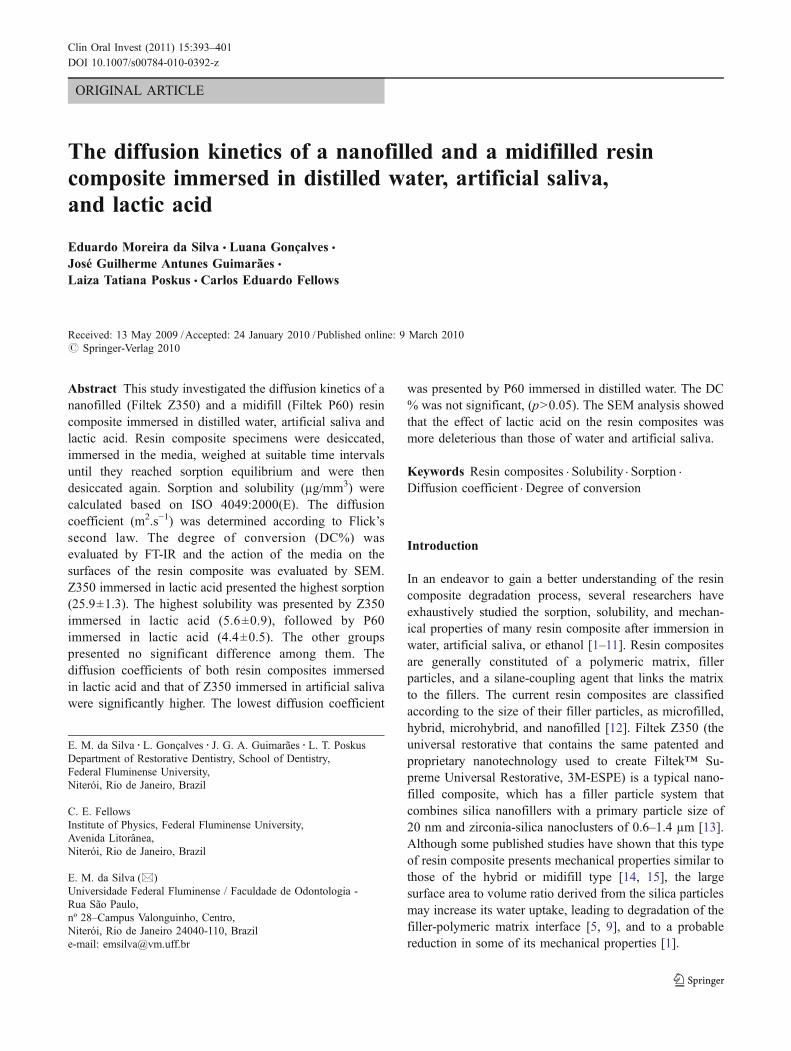
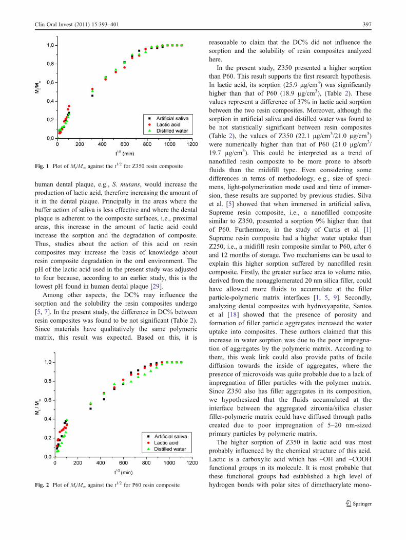
The diffusion coefficients calculated from the plots ofMt/M! against the t1/2 (Figs. 1 and 2) are shown in Table 3.Two-way ANOVA showed a significant influence for thetwo main factors: resin-based composite (p=0.0032) andimmersion medium (p<0.05). According to the Student-Newman–Keuls test both resin composites immersed inlactic acid and Z350 in artificial saliva presented the highestdiffusion coefficient, followed by Z350 immersed indistilled water, P60 immersed in artificial saliva and P60immersed in distilled water (p<0.05).
Figures 3 and 4 show representative SEM images ofresin composites before and after 16 days of immersion inthe tested media.
Discussion
Resin composite degradation is a somewhat complexphenomenon that involves mechanisms such as the poly-meric matrix plasticization, hydrolysis of silane bondsbetween the fillers and the matrix, as well as filler particlecorrosion [7, 10, 28]. These phenomena are stronglyinfluenced by the chemical structure of monomers presentin the polymeric matrix and features of the filler particlesystem [4, 5, 25], and may diminish some mechanicalproperties such as hardness, flexural strength and elasticmodulus [1, 4]. The rationale to analyze Z350 (nanofilled)and P60 (midifill) resin composites in the present study wasdue because, according to their manufacturer (3M ESPE, StPaul, MN, USA) both material have qualitatively the samepolymeric matrix (Bis-GMA, Bis-EMA, UDMA, andTEGDMA). In other words, the findings about theproperties assessed here were focused on the influence ofthe filler particle systems present in these resin composites.
Earlier studies have reported that organic acids producedby the human dental plaque caused adverse effects on resinsand resin-based restorative materials [22, 25]. Asmussen[23] analyzing the effect of propionic, acetic and lacticacids on the softening of dimethacrylate resins showed thatthe former two acids produced a higher softening effectthan lactic acid and claimed that this reflects the similaritybetween the solubility parameters of these acids and thoseof dimethacrylate monomers. At a first glance, this resultleads to accepting that propionic and acetic acids have moredeleterious effects on resin composites than lactic acid has.However, in an in vivo study, Distler and Kröncke [26]showed that in addition to acetic acid, lactic acid accountsfor about 70% of the total acids present in the human dentalplaque of the subjects examined. Furthermore, in the samestudy, there was a steeper increase in lactic acid than aceticand propionic acids after sucrose rinsing. According tothese authors, this result “is due to rapid sucrose degrada-tion by lactate–producing bacteria such as Streptococcusmutans, and is associated with a decrease in the percentagesof the other acids”. Based on this, it is reasonable tospeculate that during sugar insult, the bacteria present in
Media Z350 P60
Degree of conversion
55.7±3.8 59.0±4.6
Solubility Sorption Solubility Sorption
Artificial saliva 3.4±0.7 C 22.1±1.9 b 2.8±0.3 C 21.0±2.7 b
Lactic acid 5.5±0.9 A 25.9±1.3 a 4.5±0.5 B 18.9±3.3 b
Distilled water 3.5±0.9 C 21.0±3.5 b 2.7±0.4 C 19.7±3.1 b
Table 2 Means and standarddeviations of DC%, sorptionand solubility (µg/mm3) of theresin composites
The capital letters represent thestatistical significance for solu-bility values and the lower caseletters represent the statisticalsignificance for sorption values.Means followed by the sameletters are not statistically dif-ferent (ANOVA/Student-New-man–Keuls test, !=0.05)
396 Clin Oral Invest (2011) 15:393–401
human dental plaque, e.g., S. mutans, would increase theproduction of lactic acid, therefore increasing the amount ofit in the dental plaque. Principally in the areas where thebuffer action of saliva is less effective and where the dentalplaque is adherent to the composite surfaces, i.e., proximalareas, this increase in the amount of lactic acid couldincrease the sorption and the degradation of composite.Thus, studies about the action of this acid on resincomposites may increase the basis of knowledge aboutresin composite degradation in the oral environment. ThepH of the lactic acid used in the present study was adjustedto four because, according to an earlier study, this is thelowest pH found in human dental plaque [29].
Among other aspects, the DC% may influence thesorption and the solubility the resin composites undergo[5, 7]. In the present study, the difference in DC% betweenresin composites was found to be not significant (Table 2).Since materials have qualitatively the same polymericmatrix, this result was expected. Based on this, it is
reasonable to claim that the DC% did not influence thesorption and the solubility of resin composites analyzedhere.
In the present study, Z350 presented a higher sorptionthan P60. This result supports the first research hypothesis.In lactic acid, its sorption (25.9 µg/cm3) was significantlyhigher than that of P60 (18.9 µg/cm3), (Table 2). Thesevalues represent a difference of 37% in lactic acid sorptionbetween the two resin composites. Moreover, although thesorption in artificial saliva and distilled water was found tobe not statistically significant between resin composites(Table 2), the values of Z350 (22.1 µg/cm3/21.0 µg/cm3)were numerically higher than that of P60 (21.0 µg/cm3/19.7 µg/cm3). This could be interpreted as a trend ofnanofilled resin composite to be more prone to absorbfluids than the midifill type. Even considering somedifferences in terms of methodology, e.g., size of speci-mens, light-polymerization mode used and time of immer-sion, these results are supported by previous studies. Silvaet al. [5] showed that when immersed in artificial saliva,Supreme resin composite, i.e., a nanofilled compositesimilar to Z350, presented a sorption 9% higher than thatof P60. Furthermore, in the study of Curtis et al. [1]Supreme resin composite had a higher water uptake thanZ250, i.e., a midifill resin composite similar to P60, after 6and 12 months of storage. Two mechanisms can be used toexplain this higher sorption suffered by nanofilled resincomposite. Firstly, the greater surface area to volume ratio,derived from the nonagglomerated 20 nm silica filler, couldhave allowed more fluids to accumulate at the fillerparticle-polymeric matrix interfaces [1, 5, 9]. Secondly,analyzing dental composites with hydroxyapatite, Santoset al [18] showed that the presence of porosity andformation of filler particle aggregates increased the wateruptake into composites. These authors claimed that thisincrease in water sorption was due to the poor impregna-tion of aggregates by the polymeric matrix. According tothem, this weak link could also provide paths of facilediffusion towards the inside of aggregates, where thepresence of microvoids was quite probable due to a lack ofimpregnation of filler particles with the polymer matrix.Since Z350 also has filler aggregates in its composition,we hypothesized that the fluids accumulated at theinterface between the aggregated zirconia/silica clusterfiller-polymeric matrix could have diffused through pathscreated due to poor impregnation of 5–20 nm-sizedprimary particles by polymeric matrix.
The higher sorption of Z350 in lactic acid was mostprobably influenced by the chemical structure of this acid.Lactic is a carboxylic acid which has –OH and –COOHfunctional groups in its molecule. It is most probable thatthese functional groups had established a high level ofhydrogen bonds with polar sites of dimethacrylate mono-Fig. 2 Plot of Mt/M! against the t1/2 for P60 resin composite
Fig. 1 Plot of Mt/M! against the t1/2 for Z350 resin composite
Clin Oral Invest (2011) 15:393–401 397

mers present in Z350 polymeric matrix, i.e., -OH- in Bis-GMA, -O- in TEGDMA and Bis-EMA and –NH- inUDMA, thus increasing the acid uptake by the matrix.The micromorphology showed in the Fig. 3c reinforces thispossibility. In other words, the large surface area and themicrovoids formed in the Z350 polymeric matrix afterimmersion in lactic acid could have increased the trappingof acid molecules that established hydrogen bonds withmethacrylate monomers. Taking into account the resultspresented in the study of Bagheri et al. [25], it is reasonableto claim that this higher lactic acid sorption will increases
the degradation of Z350. Analyzing the surface degradationof some resin-based materials, these authors showed thatSupreme resin composite immersed in lactic acid presenteda higher silver penetration depth than it presented when itwas immersed in distilled water. According to these authorsthe possible factor that influenced this finding was thedegree of sorption suffered by Supreme resin composite.
The solubility phenomenon in resin composites reflects therelease of residual monomers and oligomers as well as fillerparticles and ions from its surfaces [30]. In the present study,the highest solubility was presented by Z350 (5.5 µg/mm3)
Fig. 3 Representative SEM micrographs of the Z350 specimens. aBefore immersion; and after immersion b in distilled water; c in lacticacid; and d in artificial saliva. Severe superficial damage, suggestingaggregated zirconia/silica cluster filler loss can be seen in the
specimen after lactic acid and artificial saliva immersion (whitecircles). Microcracks between filler-polymeric matrix can be notedin the specimens after immersion in distilled water and artificial saliva(white arrows)
Table 3 Means± standard deviations of the diffusion coefficient (10!13!m2.s!1)
RBC Immersion media
Artificial saliva Lactic acid Distilled water
Filtek Z350 7.6±0.4 (0.98) a 7.7±0.4 (0.99) a 5.6±0.3 (0.97) b
Filtek P60 4.9±0.1 (0.98) c 7.7±0.2 (0.92) a 3.6±0.1 (0.97) d
The r parameter values for each condition are presented in parentheses. Mean values with identical letters indicate no statistically significantdifference (Student-Newman–Keuls, p>0.05)
398 Clin Oral Invest (2011) 15:393–401

immersed in lactic acid. This supports the first researchhypothesis. In addition, the solubility of both resin compo-sites in lactic acid was higher than those in distilled waterand artificial saliva (Table 2). Thus, the second researchhypothesis was partially proved. It seems obvious that thisaction of lactic acid on the solubility of resin composites wasinfluenced by its low pH (= 4). Initially, the low pH mayhave acted in the polymeric matrixes through catalysis ofester groups from dimethacrylate monomers. The hydrolysisof ester groups leads to the formation of alcohols andcarboxylic molecules that may accelerate degradation due tolowering the pH inside the matrix [31]. On the other hand,the low pH may also have caused erosion on the fillersurfaces, accelerating their debonding or, at least, increasingthe release of ions from their surfaces [30]. The high level ofporosities shown in Fig. 3c could reinforce this possibility.
Specifically in the case of the highest solubility presentedby Z350 in lactic acid, discussed for sorption, here the role thegreater surface area to volume ratio derived from thenonagglomerated 20 nm silica filler plays in this property isclear. The larger amount of silane-coupling agent probablypre-mixed with the filler particle system of this resincomposite, e.g., "-methacryloxypropyltrimethoxysilane, cer-
tainly suffered a high level of hydrolysis via ester linkages,contributing to an increase in its solubility.
The diffusion coefficient values obtained in the presentstudy (ranging from 3.6!10!13m2.s!1 to 7.7!10!13m2.s!1),(Table 3) are in good agreement with other values publishedin the literature [18–20, 32]. Based on Figs. 1 and 2 theauthors assumed that for all the media the plots of Mt /M!against t1/2 were linear in the initial stages of sorption. InTable 2 the goodness-of-fit parameter for the linear approx-imation (r parameter), are shown for each linear fit. It can beobserved that all r values are higher than 0.9, showing thatthe sorption processes showed a Fickian behavior, confirmingthe authors’ assumption. Coincidently, the two resin compo-sites presented equal diffusion coefficient when immersed inlactic acid (7.7!10!13m2.s!1). Most probably, this was due tothe fact that the two resin composites have similar polymericmatrixes (Bis-GMA, Bis-EMA, UDMA, and TEGDMA).Santos et al. [18] found similar values of diffusion coef-ficients for experimental composites based on BisGMA/TEGDMA matrixes (66.5/33.5 wt.%) irrespective of presenceof hydroxyapatite filler particles (3.7!10!13m2.s!1 for un-filled resin and 3.1 to 4.5!10!13m2.s!1 for filled materials).According to these authors this suggests that water diffuses
Fig. 4 Representative SEMmicrographs of the P60 specimens. a Before immersion; and after immersion b in distilled water; c in lactic acid; and d inartificial saliva. Microcracks between filler particle-polymeric matrix can be seen in all the specimens after immersion in the media (white arrows)
Clin Oral Invest (2011) 15:393–401 399

principally through the resin phase of composite fillingmaterials.
The two resin composites presented the highest diffusioncoefficient when immersed in lactic acid (Table 2). Thisfinding contributes to acceptance of the second researchhypothesis and may be interpreted as a faster diffusion oflactic acid inside the resin composites than distilled waterand artificial saliva. We hypothesized that this behavior wasinfluenced by the plasticizing effect of lactic acid on thepolymeric matrixes of materials [33]. Again, the devastat-ing effect seen in Fig. 3c could be interpreted as a result ofplasticization in the polymeric matrix of Z350. Further-more, Z350 presented a higher diffusion coefficient thanP60 when immersed in distilled water and artificial saliva(Table 2). Although the magnification of SEM micrographsseen in Fig. 3 (!5.000) is not sufficiently high to identifydetails at the filler-matrix interface, it is possible that thesehigher diffusion coefficients could have been influenced bysome areas of ineffective impregnation of nanofillers andnanoclusters by the polymeric matrix of Z350 nanocompo-site [18].
The Figs. 3 and 4 highlight the effects of media on resincomposite surfaces. It is worth noting that the image seen inFig. 4a (P60 before immersion) shows cracks at some fillerparticle-polymeric matrix interfaces. At a first glance, thiscould suggest that the specimen processing produced somealterations in the surfaces of this resin composite. However,when making a comparison between Figs. 3a and 4a, it canbe noted that the surface of Z350 resin composite did notsuffer any damage during the specimen processing. Basedon this, the authors assumed that most of the aspects seenin Figs. 3b, c, d and 4b, c, d were produced or, at least,increased after immersion in the media. For both materials,the worst damage was produced by lactic acid and artificialsaliva (Figs. 3c, d, 4c and d). Although the ISO 4049:2000(E) for polymer-based filling, restorative and luting materialsdetermines that the sorption and solubility phenomena inresin composites must be conducted using water, thesefindings suggest that the behavior of resin composites whenimmersed in oral-like fluids, i.e. artificial saliva and acidsproduced by human dental plaque, are also crucial forunderstanding their degradation in the oral environment.
Conclusion
Within the limits of the experimental protocol used in thisstudy it was concluded that due to the greater surface areato volume ratio, the nanofilled resin composite may suffer ahigher degradation than the midifill type in the oralenvironment. In addition to sorption and solubility, thediffusion coefficient must be taken into account in theanalysis of resin composite degradation. The effect of acid
lactic on resin composites may be more deleterious thanthat of distilled water and artificial saliva in the degradationprocess of resin composites. Therefore, further investiga-tions using other acids produced by human plaque areneeded to increase the knowledge base about resincomposite degradation.
Acknowledgements This study was supported by a grant (E-26/171.432/2004) from Rio de Janeiro Research Foundation (FAPERJ). Theauthors would like to thank 3M ESPE for supplying the Z350 and P60dental composites; the Institute of Macromolecules (IMA) of the FederalUniversity of Rio de Janeiro (UFRJ)—for performing the degree ofconversion measurements, and the Electronic Microscopy Laboratory/PEMM of UFRJ—for performing the SEM analysis.
Conflict interest declaration The authors declare that they have noconflict of interest.
References
1. Curtis AR, Shortall AC, Marquis PM, Palin WM (2008) Wateruptake and strength characteristics of a nanofilled resin-basedcomposite. J Dent 36:186–193
2. Sideridou ID, Karabela MM, Vouvoudi EC (2008) Dynamic thermo-mechanical properties and sorption characteristics of two commerciallight cured dental resin composites. Dent Mater 24:737–743
3. Gonçalves L, Noronha Filho JD, Guimarães JGA, Poskus LT,Silva EM (2008) Solubility, salivary sorption and degree ofconversion of dimethacrylate-based polymeric matrixes. J BiomedMater Res B Appl Biomater 85:320–325
4. Zhang Y, Xu J (2008) Effect of immersion in various media on thesorption, solubility, elution of unreacted monomers, and flexuralproperties of two model dental composite compositions. J MaterSci Mater Med 19:2477–2483
5. Silva EM, Almeida GS, Poskus LT, Guimarães JGA (2008)Relationship between the degree of conversion, solubility andsalivary sorption of a hybrid and a nanofilled resin composite:influence of the light-activation mode. J Appl Oral Sci 16:161–166
6. Toledano M, Osorio R, Osorio E, Fuentes V, Prati C, Garcia-Godoy F (2003) Sorption and solubility of resin-based restorativedental materials. J Dent 31:43–50
7. Sideridou I, Tserki V, Papanastasiou G (2003) Study of watersorption, solubility and modulus of elasticity of light-cureddimethacrylate-based dental resins. Biomaterials 24:655–665
8. Hofmann N, Renner J, Hugo B, Klaiber B (2002) Elution ofleachable components from resin composite after plasma arc vs.standard or soft-start halogen light irradiation. J Dent 30:223–232
9. Kalachandra S, Wilson TW (1992) Water sorption and mechanicalproperties of light-cured proprietary composite tooth restorativematerials. Biomaterials 13:105–109
10. Söderholm KJ, Mukherjee R, Longmate J (1996) Filler leachabil-ity of composites stored in distilled water or artificial saliva. JDent Res 75:1692–1699
11. Musanje L, Darvell BW (2003) Aspects of water sorption from theair, water and artificial saliva in resin composite restorativematerials. Dent Mater 19:414–422
12. Rawls HR, Esquivel-Upshaw J (2005) Resinas restauradoras. In:Anusavice KJ (ed) Phillips materiais dentários, 3rd edn. Elsevier,Rio de Janeiro, pp 375–418
13. Mitra SB, Wu D, Holmes BN (2003) An application of nanotechnol-ogy in advanced dental materials. J Am Dent Assoc 134:1382–1390
400 Clin Oral Invest (2011) 15:393–401

14. Beun S, Glorieux T, Devaux J, Vreven J, Leloup G (2007)Characterization of nanofilled compared to universal and micro-filled composites. Dent Mater 23:51–59
15. Rodrigues SAJ, Zanchi CH, Carvalho RV, Demarco FF (2007)Flexural strength and modulus of elasticity of different types ofresin-based composites. Braz Oral Res 21:16–21
16. Dickens SH, Stansbury JW, Choi KM, Floyd CJE (2003)Photopolymerization kinetics of methacrylate dental resins.Macromolecules 36:6043–6053
17. Schneider LFJ, Consani S, Ogliari F, Correr AB, Sobrinho LC,SinhoretiMAC (2006) Effect of time and polymerization cycle on thedegree of conversion of a resin composite. Oper Dent 31:489–495
18. Santos C, Clarke RL, Braden M, Guitian F, Davy KWM (2002)Water absorption characteristics of dental composites incorporat-ing hydroxyapatite filler. Biomaterials 23:1897–1904
19. Asaoka K, Hirano S (2003) Diffusion coefficient of water throughdental composite resin. Biomaterials 24:975–979
20. Palin WM, Fleming GJP, Burke FJT, Marquis PM, Randall RC(2005) The influence of short and medium-term water immersionon the hydrolytic stability of novel low-shrink dental composites.Dent Mater 21:852–863
21. Lagouvardos PE, Pissis P, Kyritsis A, Daoukaki D (2003) Watersorption and water-induced molecular mobility in dental compos-ite resins. J Mater Sci Mater Med 14:753–759
22. Nicholson JW, Millar BJ, Czarnecka H, Limanowska-Shaw H(1999) Storage of Polyacid-modified resin composites(“compomers”) in lactic acid solution. Dent Mater 15:413–416
23. Asmussen E (1984) Softening o BISGMA-based polymers by ethanoland by organic acids of plaque. Scand J Dent Res 92:257–261
24. De Gee AJ, Wendt SL, Werner A, Davidson CL (1996) Influenceof enzymes and plaque acids on in vitro wear of dentalcomposites. Biomaterials 17:1327–1332
25. Bagheri R, Tyas MJ, Burrow MF (2007) Subsurface degradationof resin-based composites. Dent Mater 23:944–951
26. Distler W, Kröncke A (1983) The acid pattern in human dentalplaque. J Dent Res 62:87–91
27. Geddes DA (1975) Acids produced by human dental plaquemetabolism in situ. Caries Res 9:98–109
28. Sarkar NK (2000) Internal corrosion in dental composite wear: itssignificance and simulation. J Biomed Mater Res B ApplBiomater 53:371–380
29. Beech DR, Bandyopadhyai S (1983) A new laboratory method forevaluating the relative solubility and erosion of dental cements. JOral Rehabil 10:57–63
30. Ferracane JL (2006) Hygroscopic and hydrolytic effects in dentalpolymer networks. Dent Mater 22:211–222
31. Göpferich A (1996) Mechanisms of polymer degradation anderosion. Biomaterials 17:103–114
32. Flemming GJP, Awan M, Cooper PR, Sloan AJ (2008) Thepotential of a resin-composite to be cured to a 4 mm depth. DentMater 24:522–529
33. Sideridou I, Achilias DS, Spyroudi C, Karabela M (2004) Watersorption characteristics of light-cured dental resins and compositesbased on Bis-EMA/PCDM. Biomaterials 25:367–376
Clin Oral Invest (2011) 15:393–401 401