Correlation and toxicological inference of trace elements in tissues from stranded and free-ranging...
13
Correlation and toxicological inference of trace elements in tissues from stranded and free-ranging bottlenose dolphins (Tursiops truncatus) Hui-Chen W. Stavros a , Megan Stolen b , Wendy Noke Durden b , Wayne McFee a , Gregory D. Bossart c,d , Patricia A. Fair a,⇑ a National Oceanic and Atmospheric Administration, National Ocean Service, Center for Coastal Environmental Health & Biomolecular Research, 219 Fort Johnson Road, Charleston, SC 29412, USA b Hubbs-SeaWorld Research Institute, 3830 South Highway A1A #4-181, Melbourne Beach, FL 32951, USA c Harbor Branch Oceanographic Institution, Florida Atlantic University, 5600 US 1 North, Ft. Pierce, FL 34946, USA d Georgia Aquarium, 225 Baker Street, NW Atlanta, GA 30313, USA article info Article history: Received 17 June 2010 Received in revised form 3 November 2010 Accepted 4 November 2010 Available online 3 December 2010 Keywords: Trace metals Marine mammals Bottlenose dolphins Tursiops truncatus Mercury abstract The significance of metal concentrations in marine mammals is not well understood and relating concen- trations between stranded and free-ranging populations has been difficult. In order to predict liver con- centrations in free-ranging dolphins, we examined concentrations of trace elements (Al, As, Ba, Be, Cd, Co, Cu, Fe, Li, Mn, Ni, Pb, Sb, Se, Sn, total Hg (THg), V, Zn) in skin and liver of stranded bottlenose dolphins (Tursiops truncatus) from the South Carolina (SC) coast and the Indian River Lagoon, Florida (FL) during 2000–2008. Significantly higher concentrations of Zn, Fe, Se, Al, Cu and THg were found in skin while liver exhibited significantly higher Cu, Fe, Mn and THg concentrations for both study sites. Mean skin concen- trations of Cu and Mn were significantly higher in SC dolphins while higher concentrations of THg and V were found in FL dolphins. In addition, liver tissues in SC dolphins exhibited significantly higher As con- centrations while higher Fe, Pb, Se, THg, and V levels were found in FL dolphins. Two elements (Cu and THg) showed significant age-related correlations with skin concentration while five elements (Cu, Se, THg, Zn and V) showed age-related correlations with liver concentrations. Geographic location influenced age-related accumulation of several trace elements and age-related accumulation of THg in hepatic tissue was observed for both sites to have the highest correlations (r 2 = 0.90SC; r 2 = 0.69FL). Mean THg concen- tration in liver was about 10 times higher in FL dolphins (330 lgg 1 dw) than those samples from SC dol- phins (34.3 lgg 1 dw). The mean molar ratio of Hg to Se was 0.93 ± 0.32 and 1.08 ± 0.38 for SC and FL dolphins, respectively. However, the Hg:Se ratio varied with age as much lower ratios (0.2–0.4) were found in younger animals. Of the 18 measured elements, only THg was significantly correlated in skin and liver of stranded dolphins and skin of free-ranging dolphins from both sites suggesting that skin may be useful in predicting Hg concentrations in liver tissue of free-ranging dolphins. Results indicate that 33% of the stranded and 15% of the free-ranging dolphins from FL exceed the minimum 100 lgg 1 wet weight (ww) (400 dw) Hg threshold for hepatic damage while none from SC reached this level. Hepatic concentrations of As in SC dolphins and V in FL dolphins were also highly correlated with skin concentrations which may have some regional specificity predictive value. The present study provides the first application of trace element concentrations derived from stranded bottlenose dolphins to predict liver concentrations in free-ranging populations. Published by Elsevier Ltd. 1. Introduction Increasing human populations and related anthropogenic activ- ities have resulted in the release of trace elements into the envi- ronment with temporal trends indicating increases in certain elements both regionally and globally (Riget and Dietz, 2000; Riget et al., 2004; Braune et al., 2005; Braune, 2007). Many of these metals have toxic properties and the toxicological significance of metals found in tissues for marine mammals is more problematic than other species due to the limited available information (Law, 1996). Metals may adversely affect mammalian health by modula- tion of immune homeostasis and metal-induced alterations of im- mune function have been demonstrated in captive and wild harbor seals (Phoca vitulina) providing insight into mechanisms of these toxicants (Kakuschke et al., 2005, 2008, 2009). In response to these concerns, numerous studies have been conducted over the last several decades on trace metals in marine mammals. Due to its 0045-6535/$ - see front matter Published by Elsevier Ltd. doi:10.1016/j.chemosphere.2010.11.019 ⇑ Corresponding author. Tel.: +1 843 762 8533. E-mail address: [email protected] (P.A. Fair). Chemosphere 82 (2011) 1649–1661 Contents lists available at ScienceDirect Chemosphere journal homepage: www.elsevier.com/locate/chemosphere
-
Upload
independent -
Category
Documents
-
view
0 -
download
0
Transcript of Correlation and toxicological inference of trace elements in tissues from stranded and free-ranging...

Chemosphere 82 (2011) 1649–1661
Contents lists available at ScienceDirect
Chemosphere
journal homepage: www.elsevier .com/locate /chemosphere
Correlation and toxicological inference of trace elements in tissuesfrom stranded and free-ranging bottlenose dolphins (Tursiops truncatus)
Hui-Chen W. Stavros a, Megan Stolen b, Wendy Noke Durden b, Wayne McFee a, Gregory D. Bossart c,d,Patricia A. Fair a,⇑a National Oceanic and Atmospheric Administration, National Ocean Service, Center for Coastal Environmental Health & Biomolecular Research,219 Fort Johnson Road, Charleston, SC 29412, USAb Hubbs-SeaWorld Research Institute, 3830 South Highway A1A #4-181, Melbourne Beach, FL 32951, USAc Harbor Branch Oceanographic Institution, Florida Atlantic University, 5600 US 1 North, Ft. Pierce, FL 34946, USAd Georgia Aquarium, 225 Baker Street, NW Atlanta, GA 30313, USA
a r t i c l e i n f o a b s t r a c t
Article history:Received 17 June 2010Received in revised form 3 November 2010Accepted 4 November 2010Available online 3 December 2010
Keywords:Trace metalsMarine mammalsBottlenose dolphinsTursiops truncatusMercury
0045-6535/$ - see front matter Published by Elsevierdoi:10.1016/j.chemosphere.2010.11.019
⇑ Corresponding author. Tel.: +1 843 762 8533.E-mail address: [email protected] (P.A. Fair).
The significance of metal concentrations in marine mammals is not well understood and relating concen-trations between stranded and free-ranging populations has been difficult. In order to predict liver con-centrations in free-ranging dolphins, we examined concentrations of trace elements (Al, As, Ba, Be, Cd, Co,Cu, Fe, Li, Mn, Ni, Pb, Sb, Se, Sn, total Hg (THg), V, Zn) in skin and liver of stranded bottlenose dolphins(Tursiops truncatus) from the South Carolina (SC) coast and the Indian River Lagoon, Florida (FL) during2000–2008. Significantly higher concentrations of Zn, Fe, Se, Al, Cu and THg were found in skin while liverexhibited significantly higher Cu, Fe, Mn and THg concentrations for both study sites. Mean skin concen-trations of Cu and Mn were significantly higher in SC dolphins while higher concentrations of THg and Vwere found in FL dolphins. In addition, liver tissues in SC dolphins exhibited significantly higher As con-centrations while higher Fe, Pb, Se, THg, and V levels were found in FL dolphins. Two elements (Cu andTHg) showed significant age-related correlations with skin concentration while five elements (Cu, Se,THg, Zn and V) showed age-related correlations with liver concentrations. Geographic location influencedage-related accumulation of several trace elements and age-related accumulation of THg in hepatic tissuewas observed for both sites to have the highest correlations (r2 = 0.90SC; r2 = 0.69FL). Mean THg concen-tration in liver was about 10 times higher in FL dolphins (330 lg g�1 dw) than those samples from SC dol-phins (34.3 lg g�1 dw). The mean molar ratio of Hg to Se was 0.93 ± 0.32 and 1.08 ± 0.38 for SC and FLdolphins, respectively. However, the Hg:Se ratio varied with age as much lower ratios (0.2–0.4) werefound in younger animals. Of the 18 measured elements, only THg was significantly correlated in skinand liver of stranded dolphins and skin of free-ranging dolphins from both sites suggesting that skinmay be useful in predicting Hg concentrations in liver tissue of free-ranging dolphins. Results indicatethat 33% of the stranded and 15% of the free-ranging dolphins from FL exceed the minimum 100 lg g�1
wet weight (ww) (�400 dw) Hg threshold for hepatic damage while none from SC reached this level.Hepatic concentrations of As in SC dolphins and V in FL dolphins were also highly correlated with skinconcentrations which may have some regional specificity predictive value. The present study providesthe first application of trace element concentrations derived from stranded bottlenose dolphins to predictliver concentrations in free-ranging populations.
Published by Elsevier Ltd.
1. Introduction
Increasing human populations and related anthropogenic activ-ities have resulted in the release of trace elements into the envi-ronment with temporal trends indicating increases in certainelements both regionally and globally (Riget and Dietz, 2000; Rigetet al., 2004; Braune et al., 2005; Braune, 2007). Many of these
Ltd.
metals have toxic properties and the toxicological significance ofmetals found in tissues for marine mammals is more problematicthan other species due to the limited available information (Law,1996). Metals may adversely affect mammalian health by modula-tion of immune homeostasis and metal-induced alterations of im-mune function have been demonstrated in captive and wild harborseals (Phoca vitulina) providing insight into mechanisms of thesetoxicants (Kakuschke et al., 2005, 2008, 2009). In response to theseconcerns, numerous studies have been conducted over the lastseveral decades on trace metals in marine mammals. Due to its

1650 Hui-Chen W. Stavros et al. / Chemosphere 82 (2011) 1649–1661
persistence and high mobility in the marine ecosystem, mercury isof particular concern as it biomagnifies in the upper levels of thefood web (Leonzio et al., 1992b; Cardellicchio et al., 2002; Brauneet al., 2005). Ecosystem-scale simulations suggest that anthropo-genic mercury pollution is a global problem with increased dis-posal and release of mercury, primarily from the US and Chinathat will continue to affect future generations in all regions ofthe world (Booth and Zeller, 2005).
Elevated mercury levels have been observed in several mamma-lian species including river otters (Lontra canadensis) (Basu et al.,2005), mink (Mustela vison) (Wobeser and Swift, 1976), Floridapanthers (Facemire et al., 1995b) and polar bears (Urus maritimus)(Dietz et al., 2006). The high concentrations of some non-essentialmetals, such as mercury, in wildlife species have raised concernsregarding their toxicity. Mercury is a potent developmental andneural toxin that can readily cross placental and brain barriersand has been shown to affect the brain, nervous system, kidneysand developing fetuses (ASTDR, 1999). In mammals, mercury hasbeen documented to cause adverse physiological effects, endocrine(thyroid) function, reproductive problems and death (Facemireet al., 1995b; Basu et al., 2005).
Marine mammals occupy a high trophic level in the marine foodchain, have relatively long life spans and have been suggested to bea representative sentinel for monitoring spatial and temporaltrends of contaminants and environmental health (Fair and Becker,2000; Das et al., 2003; Wells et al., 2004; Bossart, 2006). As awidely distributed cetacean that exhibits site fidelity to near coast-al environments, along with a well-characterized life history and acapacity for bioaccumulating metals, bottlenose dolphins mayserve as a bell weather species indicating changes in the environ-ment. Accumulations of trace elements, such as cadmium and mer-cury, have been reported primarily in liver, kidney and muscletissue from stranded dolphins (Leonzio et al., 1992a; Kemperet al., 1994b; Kuehl and Haebler, 1995; Beck et al., 1997; Meadoret al., 1999; Durden et al., 2007). Recent studies evaluating tracemetals in free-ranging dolphins along the US coasts have estab-lished that skin can be used as a non-invasive sampling methodto evaluate trace element levels in these populations (Bryanet al., 2007; Stavros et al., 2007).
Exposure assessment in dolphins may indicate geographic areaswhere dolphins may face the greatest risk from metal toxicity andis an important aspect of the management and conservation ofcetacean wildlife species. Most studies report metal concentrationsin tissues of marine mammals as it relates to age, sex, tissue type,and geographic area, which can vary greatly among marine mam-mals (Wagemann and Muir, 1984; Kemper et al., 1994a; Law,1996; Das et al., 2003). However, studies investigating tissue con-centrations of elements as measures of health outcome are neededto develop tissue residue criteria for prediction of toxicological riskin these species. The majority of studies on levels of trace metals inmarine mammals are based on samples obtained from strandedanimals and liver tissue is one of the most frequently reported.Generally, the liver has the highest concentrations of mercuryand other metals and it has been used as the indicator organ formetals (Lockhart et al., 2005). It is important to assess trace ele-ments in free-ranging dolphins that have the potential to causetoxicity. However, this is not possible using skin biopsy samplesfrom free-ranging dolphins as there are no toxicological indicatorsavailable to assess metal concentrations in skin. Liver tissue accu-mulates the highest levels of some elements such as mercurywhich causes liver damage at high concentrations (ASTDR, 1999).Further, liver abnormalities have been reported to be associatedwith chronic mercury accumulation in Atlantic bottlenose dolphins(Rawson et al., 1993) and there is an estimated mercury limitthreshold for hepatic damage in liver tissue of marine mammals(Wagemann and Muir, 1984).
Our previous studies investigated the concentrations of tracemetals in skin and blood from free-ranging dolphins during2003–2005 from South Carolina (SC) and the Indian River Lagoon,Florida (FL) (Stavros et al., 2007, 2008). In order to compare liverconcentrations in dolphins from these same areas, we measuredtrace elements in liver and skin samples from stranded dolphinsinhabiting similar locations during 2000–2008. The objectives ofthis study were to: (1) examine the relationship of trace elementconcentrations between skin and liver in stranded bottlenose dol-phins and compare to levels in skin of free-ranging dolphins withthe aim of predicting liver concentrations; (2) assess age-relatedassociations of trace elements in skin and liver tissues fromstranded dolphins; and (3) compare mercury levels in dolphinswith mercury toxicological reference levels.
2. Materials and methods
2.1. Sample collection
Samples of liver and skin tissues were collected from strandedbottlenose dolphins between 2000 and 2008 from two study sites,South Carolina coastal area (n = 12) and the Indian River Lagoon, FL(n = 15) by the South Carolina Marine Mammal Stranding Networkand Hubbs-Sea World Research Institute, FL, respectively (Table 1).Tissue samples were collected during necropsies from fresh dead(code 2) or early moderate decomposition (code 3) carcasses.Approximately 250 g of liver was collected from the mid-lateralsection of one lobe and �3 g of skin tissue was collected for tracemetal analysis from the mid-lateral side in the lumbar region. Sam-ples were stored at �80 �C until further analysis. Total length wasmeasured as straight-line length from the tip of the upper jaw tothe fluke notch. Sex was determined by external and internalexamination. Teeth were decalcified, sectioned using a freezingmicrotome, and stained with hematoxylin using standard methods(Myrick et al., 1983). Age was determined by counting post-nataldentine layers in an extracted tooth (Hohn et al., 1989).
2.2. Sample preparation
Liver and skin tissues were cleaned with deionized water(18 MX Milli-Pore Billerica, MA) before being processed as detailedin Stavros et al. (2007). Samples of skin and liver tissue (approxi-mately 1–2 g) were dried for 12 h at 80 �C. Sub-samples of ca0.1–0.05 g of both tissues were prepared for total mercury (THg)analysis. Dry tissue samples were digested in a Teflon digestivevessel with ultrapure nitric acid (J.T. Baker, Philipsburg, NJ) in amicrowave (CEM MDS-2100, Matthews, NC). After the first diges-tion, ultrapure 30% hydrogen peroxide (J.T. Baker) was added foran additional 10 min microwave digestion to complete oxidationof organic matter. The final solution was diluted with deionizedwater for trace element analyses.
2.3. Trace element determination
Concentrations of 17 trace elements (Al, As, Ba, Be, Cd, Co, Cu,Fe, Li, Mn, Ni, Pb, Sb, Se, Sn, V, Zn) were measured as describedin Stavros et al. (2007) using Perkin Elmer ICP-MS 6100 (Wellesley,MA). Internal standards (45Sc, 72Ge, 103Rh, 175Lu) were added toeach sample and calibration standard solutions. Total mercury(THg) concentration was determined by Milestone direct mercuryanalyzer (DMA-80, Shelton, CT). Methods were previously de-scribed for trace metal analysis in skin and blood (Stavros et al.,2007, 2008). Several reference materials including 1566b (oystertissue, NIST, USA), TORT-2 (lobster hepatopancreas), DOLT-3 (dog-fish liver) and DORM2 (dogfish muscle, NRC Canada) were used for

Table 1Biological characteristics on stranded bottlenose dolphins from South Carolina coast and Indian River Lagoon, Florida from 2000 to 2008.
Sample ID Code Date year-m d�1 Sex Total length (cm) Age (yr) Age class
South Carolina (n = 12)SC0034 2 2000–07/26 F 121 0.25 JuvenileSC0051 2 2000–11/23 M 99 0.1 JuvenileSC0216 2 2002–07/23 F 220 10 AdultSC0223 1 2002–08/22 F 201 6 JuvenileSC0224 1 2002–08/23 F 152 1a JuvenileSC0322 1 2003–08/17 M 175 4 JuvenileSC0334 2 2003–11/14 F 109 0 JuvenileSC0414 2 2004–0/15 F 190 4.75 JuvenileSC0429 2 2004–07/27 F 111 0 JuvenileSC0545 2 2005–12/11 M 210 5a JuvenileSC0729 1 2007–07/25 F 114 0.1a JuvenileSC0740 1 2007–08/18 M 250 29 Adult
Indian River Lagoon (n = 15)Hubbs-0407-Tt 2 2004–01/16 F 243 25a AdultHubbs-0458-Tt 3 2004–06/18 F 210 8 AdultHubbs-0489-Tt 2 2004–12/23 M 220 15 AdultHubbs-0517-Tt 3 2005–06/14 M 259 20 AdultHubbs-0523-Tt 2 2005–07/20 F 175 2.5 JuvenileHubbs-0541-Tt 2 2005–12/26 M 245 15 AdultHubbs-0630-Tt 2 2006–05/24 F 236 27 AdultHubbs-0657-Tt 2 2006–12/28 M 213 5.5 JuvenileHubbs-0701-Tt 3 2007–01/01 M 249 21 AdultHubbs-0714-Tt 3 2007–02/03 F 226 17 AdultHubbs-0717-Tt 2 2007–02/11 M 250 22 AdultHubbs-0723-Tt 3 2007–03/12 M 205 6 JuvenileHubbs-0783-Tt 2 2007–12/27 M 98 0 JuvenileHubbs-0805-Tt 2 2008–01/30 F 231 18 AdultHubbs-0808-Tt 3 2008–02/04 F 236 12 Adult
a Ages estimated using Gompertz equations calculated for SC and FL dolphins (McFee et al., 2010).
Hui-Chen W. Stavros et al. / Chemosphere 82 (2011) 1649–1661 1651
quality assurance. Recovery of trace elements in these referencematerials ranged from 85.3% to 127.7%, averaging 104.6 ± 9.87%SD. All plasticware was pre-cleaned with reagent grade nitric acidand hydrochloric acid to eliminate contamination. Moisture con-tent in liver tissue was 73.0 ± 3.7% SD and in skin tissue65.4 ± 5.3% SD.
2.4. Data analysis
Concentrations of trace elements in this study are presented inlg g�1 dry weight (dw). For comparison to other published studies,dw data presented in this study were converted into wet weight(ww) using a conversion factor (CF) of 0.27 and 0.35 for liver andskin tissues, respectively. Descriptive statistics (mean, standarddeviation and range) were calculated for each element levels. Dif-ferences between means of trace element concentrations in liverand skin tissues as well as study sites were assessed by the Stu-dent’s t-test. Sexual maturity in bottlenose dolphins has been cat-egorized from 5 to 12 years for females and 10–13 years for males(Mead and Potter, 1990). In our study, adults were defined as fe-males age 7 and older and males age 10 and older and juvenilescategorized as less than these ages. In four cases, age was not avail-able and age was estimated based on gompertz equations derivedfor age and length of SC and FL dolphins (McFee et al., 2010). Nodifferences in the growth curves were found between dolphinsfrom the two locations; females reached asymptomic growth at240 cm and males at �255 cm. Potential relationships betweentrace element level and dolphin age were examined using regres-sion models. Since there were significant differences in skin con-centrations of Cu, Mn and THg in dolphins between these twosites (Table 2), regressions were performed separately for deter-mining age-related associations with these trace elements in skin.Similarly, since hepatic concentrations of As, Fe, Pb, Se, THg and Vwere significantly different in dolphins between the two sitesregression analysis for these trace elements in liver tissue were
also performed separately for dolphins from the two sites. For allother elements data from both sites were combined to determineage-related associations with trace element concentration. Datawere log transformed when necessary to meet the assumption ofnormality for Student’s t-test and regression models. Results wereconsidered significant when p 6 0.05. Interpretation of statisticalsignificance testing should consider the small sample size fromstranded dolphins for the two sites.
3. Results and discussion
3.1. Biological factors
Skin and liver samples were analyzed from 12 dolphins thatstranded along the SC coast (8 females, 4 males) and 15 dolphinsfrom the FL Indian River Lagoon (7 females, 8 males) (Table 1). Themean age of SC dolphins is 5.0 y (±SD 8.2) and 14.3 y (±SD 8.3) forFL dolphins. The majority of the tissue samples were collected pri-marily from code 2 (freshly dead) animals in addition to several fromcode 3 (moderate decomposition) animals. Estimated ages on ani-mals ranged from less than 1 year to 27 years. The dolphins fromSC had a greater proportion of juvenile animals (2 adults, 10 juve-niles) while FL dolphins had a larger number of adult animals (11adults, 4 juveniles). Biological data on each dolphin including animalidentification, stranding code, sex, total length, age and age class arepresented in Table 1. It is well-known that trace metal accumulationmay be influenced by various biological and ecological factorsincluding geographic area, age, sex, tissue type, diet, temporal distri-bution (Das et al., 2003; O’Hara et al., 2003).
3.2. Trace element liver and skin concentrations from dolphins in SCand FL
The elements found with the highest mean levels in skin tissueof stranded dolphins in descending order were: Zn, Fe, Se, Al, Cu,

Table 2Trace element concentrations (mean ± SD; ranges in parenthesis; lg g�1 dw) in skin and liver tissues of stranded bottlenose dolphins collected from South Carolina coast andIndian River Lagoon, Florida.
South Carolina coast Indian River Lagoon, Florida
Skin n Liver n Skin n Liver n
Al 21.2 ± 22.4 (2.30–80.8) 12 19.6 ± 14.2 (6.19–55.8) 12 10.5 ± 10.2 (2.61–44.8) 15 12.0 ± 8.81 (3.16–30.9) 15Asb,d 1.66 ± 1.23 (0.506–4.45) 12 1.70 ± 1.34 (0.628–5.22) 12 1.29 ± 0.841 (0.559–4.01) 15 0.821 ± 0.271 (0.414–1.29) 15Ba 0.323 ± 0.356 (0.026–1.02) 11 0.274 1 0.218 ± 0.065 (0.146–0.295) 4 BDLBeb BDL BDL 0.056 ± 0.010 (0.043–0.075) 15 0.019 ± 0.001 (0.017–0.021) 15Cda 0.041 ± 0.066 (0.004–0.202) 8 0.266 ± 0.364 (0.003–1.09) 8 0.013 ± 0.008 (0.005–0.025) 4 0.142 ± 0.228 (0.002–0.948) 15Co 0.036 1 0.033 ± 0.014 (0.014–0.058) 9 0.054 ± 0.004 (0.051–0.057) 2 0.026 ± 0.004 (0.021–0.029) 6Cua,b,c 3.81 ± 1.51 (1.78–7.60) 12 43.7 ± 31.5 (13.0–132) 12 2.67 ± 0.725 (1.67–4.23) 15 27.5 ± 15.2 (11.2–57.9) 15Fea,b,d 104 ± 80.8 (48.3–286) 11 732 ± 320 (182–1365) 12 62.7 ± 10.2 (46.4–77.7) 15 1125 ± 474 (427–2184) 15Lib BDL BDL 0.033 ± 0.008 (0.018–0.046) 15 0.064 ± 0.014 (0.043–0.097) 15Mna,b,c 0.823 ± 0.486 (0.35–2.10) 12 15.0 ± 5.96 (7.21–27.2) 12 0.511 ± 0.190 (0.349–1.15) 15 13.7 ± 5.22 (5.74–27.3) 15Nia 0.128 ± 0.089 (0.047–0.349) 12 0.041 ± 0.052 (0.008–0.182) 12 BDL BDLPbb,d 0.063 ± 0.096 (0.010–0.360) 12 0.031 ± 0.017 (0.006–0.056) 12 0.028 ± 0.019 (0.009–0.076) 13 0.196 ± 0.158 (0.016–0.615) 15Sba,b 0.410 ± 0.338 (0.172–1.19) 12 0.087 ± 0.031 (0.043–0.168) 11 0.296 ± 0.195 (0.175–0.981) 15 0.090 ± 0.014 (0.074–0.127) 14Sea,b,d 26.2 ± 19.2 (3.77–70.1) 12 14.5 ± 18.9 (1.14–63.0) 12 15.9 ± 8.96 (3.19–41.3) 15 109 ± 99.5 (1.33 – 380) 15Sna 0.039 ± 0.027 (0.021–0.087) 5 0.595 ± 0.879 (0.047–3.17) 12 0.052 ± 0.014 (0.042–0.061) 2 0.743 ± 0.531 (0.097–2.04) 15THga,b,c,d 1.81 ± 1.81 (0.169–5.65) 12 34.3 ± 58.9 (0.977–205) 12 8.57 ± 7.04 (1.52–22.6) 15 300 ± 242 (6.21–744) 15Va,b,c,d 0.430 ± 0.365 (0.219–1.22) 12 0.207 ± 0.135 (0.064–0.440) 12 0.673 ± 0.145 (0.351–0.898) 15 0.501 ± 0.197 (0.294–0.872) 15Zna,b 976 ± 1108 (326–4435) 12 253 ± 233 (54.5–860) 12 731 ± 181 (432–1101) 15 211 ± 132 (62.7–542) 15
BDL – below detection limit.a Significant difference between skin and liver tissue of dolphins from South Carolina.b Significant difference between skin and liver tissue of dolphins from Indian River Lagoon, FL.c Significant difference of skin concentrations between South Carolina and Indian River Lagoon, FL.d Significant difference of liver concentrations between South Carolina and Indian River Lagoon, FL.
1652 Hui-Chen W. Stavros et al. / Chemosphere 82 (2011) 1649–1661
THg, As, Mn, V, Sb, Ba, Ni, Pb, Cd, Sn and Co in SC dolphins (n = 12)and Zn, Fe, Se, Al, THg, Cu, As, V, Mn, Sb, Ba, Be, Co, Sn, Li, Pb, and Cdin FL dolphins (n = 15) (Table 2). For free-ranging dolphins, a sim-ilar order occurred in the trace elements with the highest values inskin tissues of SC dolphins (n = 74; Zn > Fe > Se > Al > As > -Cu > THg) and FL dolphins (n = 75; Zn > Fe > Se > THg > Al > -Cu > As) with slight variations (Stavros et al., 2007). Traceelements such as Se and Zn have been reported to accumulate inskin of cetaceans (Yang et al., 2002). These two elements, Se andZn, have several important protective roles including photo-oxida-tion from solar radiation, tumor progression, skin inflammationand wound healing (Emonet et al., 1998; Gonul et al., 1998; Lecciaet al., 1999). As observed in this study, cutaneous Zn levels in ceta-ceans can be extremely high often occurring at concentrations tenor a hundred fold higher than other elements (Monaci et al., 1998;Yang et al., 2002; Stavros et al., 2007). Mean skin concentrations ofCu and Mn were significantly higher in SC dolphins while signifi-cantly higher THg and V concentrations were found in FL dolphins(Table 2). Site-related findings from free-ranging dolphins foundstatistically higher levels in skin from SC dolphins for several ele-ments (As, Co, Mn, Pb, Sb, Se, TI) while only THg was higher in FLdolphins (Stavros et al., 2007). Significant age and sex related dif-ferences in skin concentrations of Cu, THg and Mn were found inthe above study on the same populations of free-ranging dolphins(Stavros et al., 2007).
Similarly, those elements in liver tissues with the highest meanlevels (lg g�1 dw) in descending order were: Fe, Zn, Cu, THg, Al,Mn, Se, As, Sn, Ba, Cd, V, Sb, Ni, Co and Pb in SC dolphins and Fe,THg, Zn, Se, Cu, Mn, Al, As, Sn, V, Pb, Cd, Sb, Li, Co and Be in FL dol-phins (Table 2). Several of the above elements found in high con-centrations, such as Cu, Fe, Se, Mn and Zn, are generally regardedas essential although these can also exert toxic effects at high lev-els (Liu et al., 2008). In contrast, other elements including As andTHg have no known essential role and are considered toxic ele-ments. The levels of As, Cd, Cu, and Zn, in liver from SC dolphinsin this study were comparable to earlier reported levels by Becket al. (1997) on dolphins stranded along the SC coast althoughPb, Se and THg were higher in our study. In addition, liver tissuesin SC dolphins exhibited significantly higher As concentrations
while higher Fe, Pb, Se, THg, and V levels were found in FL dolphins(Table 2).
Mean THg liver concentration was 34.3 lg g�1 dw (range 0.98–205 lg g�1 dw) in SC dolphins while FL dolphins had300 lg g�1 dw (range 6.21–744 lg g�1 dw), representing a 10-foldhigher concentration than in SC dolphins. Comparison of wetweight (ww) tissue concentrations in the current study (meanTHg = 81 lg g�1 ww) for FL dolphins found similar levels to thosepreviously reported from stranded dolphins (73 lg g�1 ww;n = 19) from the same region during 2001–2003 with a similarupper limit of 200 lg g�1 ww (Durden et al., 2007). Mercury con-centrations varied greatly among FL dolphins in both of these stud-ies. Concentrations of Hg liver concentrations in marine mammalsvary depending upon species, geographic location, as well as othervariables and high levels are reported from areas contaminated byindustrial discharges and input from natural sources (Law, 1996).Several studies have reported high liver Hg concentrations(lg g�1 dw) in stranded bottlenose dolphins from various locationsincluding Florida (mean: 134; range: below detection-443; Raw-son et al., 1993), French Atlantic (mean 461; range 24–783 l Hols-beek et al., 1998), Thyrrhenian Sea (mean 270; range 12–13155;Leonzio et al., 1992); and the Israeli Mediterranean (mean 671;range 4–2210; (Roditi-Elasar et al., 2003). Liver is the most impor-tant organ for Hg accumulation in cetaceans (Wagemann and Muir,1984). Over 95% of the Hg burden in spotted dolphins (Stenellaattenuata) was attributed to three (liver, skeletal muscle, and blub-ber) of 18 tissues analyzed (Andre et al., 1991a).
In our study, skin THg concentrations reflected the same trendfound in liver tissue with higher levels in FL dolphins (8.57lg g�1 dw, ranging from 1.52 to 22.6 lg g�1 dw) compared to SCdolphins (1.81 lg g�1 dw, ranging from 0.17 to 6.65 lg g�1 dw).As a highly toxic element with a long biological half-life, Hg isaccumulated in cetaceans through the diet mostly as methyl-Hg,the more toxic form, with reported half-life estimates of 1000 dfor striped dolphins (Itano and Kawai, 1981). Hg accumulation inmarine mammal liver is mainly as the inorganic form (Dietzet al., 1990) which suggests the liver as a site for demethylation.Selenium plays an important role in the demethylation of Hg andan equimolar Hg:Se ratio has been documented (Koeman et al.,

Hui-Chen W. Stavros et al. / Chemosphere 82 (2011) 1649–1661 1653
1973). Demethylation likely occurs when methyl-Hg exceeds athreshold value (Palmisano et al., 1995). Tiemannite, an insolubleform of HgSe, is thought to be the final product of methyl-Hgdemethylation which immobilizes Hg and prevents cytotoxic ef-fects (Nigro and Leonzio, 1996). Thus, the high levels of Hg in tis-sues without overt evidence of deleterious effects are due toefficient Hg detoxification mechanisms, particularly in cetaceans.
Selenium, in contrast to Hg, is both an essential and toxic ele-ment, depending upon the concentration, and functions as anantagonist against the toxicity of several heavy metals (Lemly,1996). Mean selenium liver concentrations were seven times high-er in FL dolphins (109 lg g�1 dw; 29.4 lg ww�1) compared tothose in SC (14.5 lg g�1 dw; 3.90 lg g�1 ww, Table 2). The meanSe liver ww concentrations found in dolphins from both sites werecomparable to previous cetacean studies which reported Se con-centrations generally <30 mg kg�1 ww in liver (Mackay et al.,1995). The upper Se limit of 103 lg g�1 ww (or mg kg�1) foundin liver of IRL dolphins greatly exceed this concentration whilethe upper limit in SC dolphins (17 lg g�1 ww) was only half of thislevel. Comparison of Se in liver of IRL adult dolphins to a previouslyreported study of dolphins in this region found similar mean valueof 29.8 lg g�1 ww although the range was less variable (1.20–90.70 lg g�1 ww) (Durden et al., 2007). As an essential element,Se is integral to the enzyme glutathione peroxidase and protectsmembrane lipids, proteins and nucleic acids from damage by oxi-dants and free-radicals (Liu et al., 2008). Selenium has various bio-inorganic interactions and the selenide form can react with metalsto form insoluble selenides, as mentioned above for Hg, thus reduc-ing metal toxicities. The protective role that Se exerts along withthe key role of glutathione towards ameliorating the neurotoxic ef-fects of Hg has long been noted (Liu et al., 2008).
Cadmium, another non-essential toxic element, accumulates inthe liver and concentrations found in both SC and FL dolphins weregenerally low with the upper range of 1.09 lg g�1 dw (0.29 lg ww)(Table 2). Hepatic Cd concentrations in other marine mammalsranged from 0.3 to 0.9 lg g�1 ww in white-sided dolphins (Lagen-orhychus sp) and harbor porpoises (Phoceoena phoceoena) (Mackayet al., 1995). High concentrations of Cd are found in marine mam-mals ingesting prey from polluted areas and renal tissues typicallyhigher in Cd levels than hepatic tissues (Wagemann and Muir,1984).
The hepatic As mean concentration in SC dolphins (1.70 lgg�1 ww) was approximately twice as high compared to FL dolphins(0.82 lg g�1 ww). The levels in SC dolphins were also highercompared to previously reported levels in SC dolphins (0.28mg kg�1 ww) (Beck et al., 1997) which may be attributed to differ-ences in the geographical locations of the stranded dolphins anddifferent prey or perhaps reflect higher environmental contamina-tion of this element over time. The levels of As in marine mammalsrarely exceed 1.0 mg kg�1 ww, in any tissue (Thompson, 1990) andhepatic levels in marine mammals are generally below this level(Mackay et al., 1995).
Both Cu and Zn are essential elements involved in metabolismand are highly bioregulated with homeostatic ranges of 3–30mg kg�1 ww and 20–100 mg kg�1 ww, respectively (Law, 1996).Concentrations of Cu and Zn were similar between dolphins fromboth sites and were within these ranges, albeit slightly higherranges in SC dolphins (Table 2). Copper hepatic concentrations inSC dolphins were generally similar to earlier reports in dolphinsfrom this area while Zn was somewhat higher (Beck et al., 1997).Decreasing concentrations of Cu and Zn with age were reportedin liver of cetaceans (Zhou et al., 2001) which is also consistentwith findings reported in skin of free-ranging dolphins (Stavroset al., 2007). Young animals are normally richer in Cu than adultsreflective of transplacental transfer of Cu while there is less trans-placental transfer for Zn in marine mammals (Fujise et al., 1988;
Law et al., 1992; Law, 1996). The significantly higher hepatic Cuconcentrations found in SC dolphins compared to those in FL mightalso be influenced by the higher proportion of younger animals.
3.3. Associations of trace element concentrations and age
Trace elements (Al, As, Cd, Fe, Mn, Pb, Sb, Se, THg, V and Zn)measured in the liver and skin of all dolphins from both sites werecompared to age. For all elements in which no site-related differ-ences in trace element concentrations occurred (Table 2) data fromboth sites were combined to determine age-related associationswith concentration. Analysis of growth curves between dolphinsin SC and IRL found no site-differences for either males or females(McFee et al., 2010) supporting combining samples sets from thetwo sites for comparison of age-related trace element concentra-tions. Among those 11 elements, concentrations of only five ele-ments (Cu, Se, THg, V and Zn) showed significant correlationswith respect to dolphin age (Fig. 1).
Of all elements, age-related accumulation of THg in liver tissuewas observed to have the highest correlations for dolphins fromboth SC and FL, r2 = 0.90, 0.69, respectively (Fig. 1). These findingsconfirm the well-known relationship between Hg accumulationand age as observed in cetaceans, including bottlenose dolphins(Durden et al., 2007; Pompe-Gotal et al., 2009). For SC dolphins,THg in skin (r2 = 0.76) was also positively associated with age. SkinHg concentrations have been shown in other cetaceans to be highlycorrelated with age and even suggested to be a possible alternativemethod for age determination (Yang et al., 2002). Interestingly,skin THg concentration was not significantly correlated with ageof FL dolphins which is similar to the previous study with free-ranging dolphins from these two sites (Stavros et al., 2007).
Hepatic Se concentration was positively and highly correlatedwith total Hg hepatic concentration (r2 = 0.89SC; r2 = 0.81FL). Themolar ratio of Hg to Se was 0.93 ± 0.32 and 1.08 ± 0.38 for SC andFL dolphins, respectively. These findings are similar to severalstudies indicating that the molar Hg:Se ratio in liver tissue of mar-ine mammals was close to 1, particularly those with high Hg levels,including bottlenose and Risso’s (Grampus griseus) dolphins(Leonzio et al., 1992; Nigro and Leonzio, 1996; Meador et al.,1999; Durden et al., 2007). Interestingly, a 1:1 Hg:Se ratio is notobserved in a large number of fish that constitute the diets ofmarine mammals, thus, it is thought that the 1:1 ratio occurs inthe mammals themselves (Cuvin-Aralar and Furness, 1991). Itshould be noted that when we examined the Hg:Se ratios by agefor dolphins at each site, we found much lower ratios (0.2–0.4)for younger animals (Fig. 2). It would appear that an Hg:Se molarratio of 1 does not occur in liver tissue of these dolphins until theyreach adulthood. Also, this ratio was observed to decrease slightlyin IRL dolphins with age perhaps reflecting changes in Hg and Semetabolism with increasing Hg in the tissue. Further investigationwould be needed to examine this in more detail. While the meanHg:Se molar ratio in liver was �1 for dolphins both in SC and FL,this ratio in blood of free-ranging dolphins was 4.8-fold higher inSC dolphins (7.2) compared to IRL dolphins (1.5) correspondingto circulating levels of these elements. Generally, interactionbetween Hg2+ and selenite occurs in the blood to form a highmolecular weight complex which is then taken up in the liverwhere processes form an insoluble Hg–Se complex (Yang et al.,2008). The lower Hg concentration found in the skin and livertissue of SC dolphins denotes lower Hg exposures in these dolphinsand perhaps also some protective role attributed to their higher Seconcentrations in blood and liver.
Hepatic Zn concentrations and Cu skin concentration were theonly element with a significant negative association with age (Cuin SC dolphins r2 = 0.48; Zn in SC and FL dolphins r2 = 0.30). Thefindings of decreasing concentrations of Cu and Zn with age were

Fig. 1. Relationship between age and trace element concentrations in tissues collected from stranded bottlenose dolphins from South Carolina (SC) and the Indian RiverLagoon, Florida (FL) during 2000–2008.
1654 Hui-Chen W. Stavros et al. / Chemosphere 82 (2011) 1649–1661
consistent with reports of these elements in hepatic tissue of ceta-ceans (Zhou et al., 2001). This is also similar to previous reportedresults in bottlenose dolphins indicating a higher Cu concentrationin livers of juvenile animals compared to adults (Beck et al., 1997).Additionally, Beck et al. (1997) found significantly higher Cd, Seand Hg in livers of adult dolphins from SC compared to juveniledolphins.
Also, V levels in livers of SC were found to increase with age(r2 = 0.87; Fig. 1) which is similar to those reported in four pinni-ped species (Mackey et al., 1996; Saeki et al., 1999; Anan et al.,2002). The high correlation found between hepatic V and Hg accu-mulation in the pinnipeds was attributed to an increase of reten-
tion in nuclei and mitochondria fraction in the cell (Saeki et al.,1999).
3.4. Comparison and correlations between liver and skin traceelements
In comparing trace element concentrations in dolphin skin andliver tissues, significant differences were found for the followingtrace elements in dolphins at both sites: Cu, Fe, Mn, Sb, Se, THg,V, and Zn (Table 2). Additionally, significant differences were ob-served for Cd, Ni, and Sn levels between these two tissues in SCdolphins while As, Be, Li and Pb were noted to differ significantly

Fig. 1 (continued)
Hui-Chen W. Stavros et al. / Chemosphere 82 (2011) 1649–1661 1655
between skin and liver in FL dolphins. Significant differences be-tween skin and liver concentrations of Be and Li occurred in dol-phins from FL although these elements were below detectionlimit in tissues of the SC dolphins. Skin and liver Ni concentrationswere low in SC dolphins, however, this element was below thedetection limit in FL dolphins.
Consistently higher liver concentrations of Cu, Fe, Mn, and THgcompared skin concentrations were observed in dolphins at bothsites (Table 2). In contrast, Sb, and Zn were more concentrated inskin tissue compared to liver tissue for dolphins at both sites. Addi-tionally, Se was also found in higher levels in skin compared to li-ver tissue in SC dolphins while the opposite was found in FLdolphins. Concentrations of Co were similar in both skin and livertissues in SC and FL dolphins while concentrations of other ele-ments (i.e., Al, As, and Pb) varied between sites as to what tissuehad higher mean concentrations. Some of the differences observed
between dolphins in these two sites may be attributed to differ-ences between adults and young animals as samples analyzed fromFL dolphins were predominately adults while the majority of thosefrom SC were juveniles.
In SC dolphins, skin and liver tissues exhibited significant posi-tive (p < 0.01) relationships with As (r2 = 0.60), and THg (r2 = 0.89)concentrations. Significant positive relationships were found be-tween THg (r2 = 0.53) and V (r2 = 0.28) concentrations in skin andliver tissues in FL dolphins. Only THg was significantly correlatedin tissues from dolphins at both sites.
3.5. Predicting liver concentrations in free-ranging populations
In assessing contaminant exposures in free-ranging popula-tions, biological samples are generally confined to skin samplesfrom biopsy studies (Querouil et al., 2010) and blood, skin and
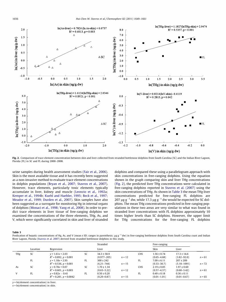
Fig. 2. Comparison of trace element concentration between skin and liver collected from stranded bottlenose dolphins from South Carolina (SC) and the Indian River Lagoon,Florida (FL) in SC and FL during 2000–2008.
1656 Hui-Chen W. Stavros et al. / Chemosphere 82 (2011) 1649–1661
urine samples during health assessment studies (Fair et al., 2006).Skin is the most available tissue and it has recently been suggestedas a non-invasive method to evaluate trace element concentrationsin dolphin populations (Bryan et al., 2007; Stavros et al., 2007).However, trace elements, particularly toxic elements typicallyaccumulate in liver, kidney and muscle (Leonzio et al., 1992a;Kemper et al., 1994b; Kuehl and Haebler, 1995; Beck et al., 1997;Meador et al., 1999; Durden et al., 2007). Skin samples have alsobeen suggested as a surrogate for monitoring Hg in internal organsof dolphins (Monaci et al., 1998; Yang et al., 2008). In order to pre-dict trace elements in liver tissue of free-ranging dolphins weexamined the concentrations of the three elements, THg, As, andV, which were significantly correlated in skin and liver of stranded
Table 3Predication of hepatic concentrations of Hg, As, and V (mean ± SD; ranges in parenthesis;River Lagoon, Florida (Stavros et al. 2007) derived from stranded bottlenose dolphins in th
Stranded
Location Regression Liver
THg SC y = 1.61x + 2.03 SC 34.3 ± 58.9R2 = 0.892, p < 0.001 (0.977–205)
FL y = 1.18x + 2.95 FL 300 ± 242R2 = 0.536, p < 0.001 (6.21–744)
As SC y = 0.78x + 0.07 SC 1.70 ± 1.34R2 = 0.601, p = 0.003 (0.63–5.22)
V FL y = 0.82x � 0.41 FL 0.50 ± 0.20R2 = 0.281, p = 0.0042 (0.29–0.87)
y = ln(element concentration) in liver.x = ln(element concentration) in skin.
dolphins and compared these using a parallelogram approach withskin concentrations in free-ranging dolphins. Using the equationshown in the graph comparing skin and liver THg concentrations(Fig. 2), the predicted liver THg concentrations were calculated infree-ranging dolphins reported in Stavros et al. (2007) using theskin concentrations of THg. As shown in Table 3 the mean THg liverconcentrations predicted for free-ranging FL dolphins are207 lg g�1 dw, while 17.3 lg g�1 dw would be expected for SC dol-phins. The mean THg concentrations predicted in free-ranging pop-ulations in these two areas are very similar to what was found instranded liver concentrations with FL dolphins approximately 10times higher levels than SC dolphins. However, the upper limitfor THg concentrations for the free-ranging FL dolphins
lg g�1 dw) in free-ranging bottlenose dolphins from South Carolina coast and Indianis study.
Free-ranging
Skin Liver
CHS 1.56 ± 0.74 17.3 ± 14.6n = 12 (0.65–4.68) (3.82–92.0) n = 61
FL 7.09 ± 6.11 207 ± 209n = 15 (0.33–30.7) (5.18–1091) n = 72
CHS 2.19 ± 0.85 1.97 ± 0.60n = 12 (0.57–4.37) (0.60–3.42) n = 61
FL 0.49 ± 0.18 0.36 ± 0.11n = 15 (0.01–1.01) (0.01–0.67) n = 65
Table 4Comparison of trace element concentrations (mean ± SD, ranges in parenthesis; lg g�1 dw) in skin tissue of free-ranging (Stavros et al., 2007) andstranded bottlenose dolphins in this study.
South Carolina coast n Significant Indian River Lagoon, FL n Significant
Free-ranging n Stranded Free-ranging n Stranded
Al 9.00 ± 22.6 21.2 ± 22.4 3.79 ± 5.58 10.5 ± 10.2(1.06–157) 61 (2.30–80.8) 12 *** (0.81–48.4) 72 (2.61–44.8) 15 ***
As 2.19 ± 0.85 1.66 ± 1.23 0.76 ± 0.24 1.29 ± 0.84(0.57–4.37) 61 (0.51–4.45) 12 * (0.24–1.47) 65 (0.56–4.01) 15 ***
Ba 0.08 ± 0.11 0.32 ± 0.36 0.07 ± 0.07 0.22 ± 0.06(0.02–0.68) 33 (0.03–1.02) 11 ** (0.01–0.42) 65 (0.15–0.30) 4 ***
Be BDL BDL 0.06 ± 0.04 0.06 ± 0.01(0.02–0.19) 49 (0.04–0.08) 15
Cd 0.016 ± 0.007 0.041 ± 0.066 0.011 ± 0.013 0.013 ± 0.008(0.002–0.032) 22 (0.004–0.202) 8 (0.001–0.075) 56 (0.005–0.025) 4
Co 0.70 ± 0.36 0.036 0.29 ± 0.16 0.06 ± 0.01(0.06–1.82) 52 1 (0.03–0.83) 63 (0.05–0.06) 2 *
Cu 1.57 ± 0.34 3.81 ± 1.51 1.44 ± 0.30 2.67 ± 0.72(0.86–2.48) 61 (1.78–7.60) 12 *** (0.71–2.31) 72 (1.67–4.23) 15 ***
Fe 91.1 ± 174 104 ± 80.7 87.3 ± 128 62.7 ± 10.3(7.00–820) 46 (48.3–286) 11 (18.0–658) 68 (46.0–78.0) 15
Li 0.14 ± 0.05 BDL 0.12 ± 0.07 0.03 ± 0.01(0.08–0.26) 14 (0.04–0.36) 64 (0.02–0.05) 15 ***
Mn 0.50 ± 0.35 0.82 ± 0.49 0.46 ± 0.16 0.51 ± 0.19(0.07–1.46) 52 (0.35–2.10) 12 * (0.19–1.07) 72 (0.35–1.15) 15
Ni 0.06 ± 0.06 0.13 ± 0.09 0.04 ± 0.08 BDL(0.01–0.30) 34 (0.05–0.35) 12 *** (0.01–0.57) 50
Pb 0.40 ± 0.34 0.06 ± 0.10 0.17 ± 0.13 0.03 ± 0.02(0.01–1.28) 53 (0.01–0.35) 12 *** (0.04–0.76) 70 (0.01–0.08) 13 ***
Sb 0.96 ± 0.35 0.41 ± 0.34 0.44 ± 0.44 0.30 ± 0.20(0.36–1.74) 18 (0.17–1.19) 12 *** (0.09–1.54) 30 (0.18–0.98) 15
Se 25.2 ± 7.42 26.2 ± 19.2 17.3 ± 5.95 15.9 ± 8.96(13.1–43.9) 61 (3.77–70.1) 12 (7.20–31.6) 72 (3.19–41.3) 15
Sn 0.15 ± 0.15 0.04 ± 0.03 0.05 ± 0.02 0.05 ± 0.01(0.02–0.49) 12 (0.02–0.09) 5 (0.02–0.08) 14 (0.04–0.06) 2
THg 1.56 ± 0.74 1.81 ± 1.82 7.09 ± 6.11 8.56 ± 7.04(0.65–4.68) 62 (0.17–5.65) 12 (0.33–30.7) 72 (1.52–22.6) 15
V 0.73 ± 0.45 0.43 ± 0.36 0.49 ± 0.18 0.67 ± 0.14(0.09–2.04) 61 (0.22–1.22) 12 * (0.01–1.01) 64 (0.35–0.90) 15 ***
Zn 685 ± 234 976 ± 1108 602 ± 209 731 ± 181(302–1384) 61 (326–4435) 12 (299–1470) 72 (432–1101) 15 **
Student’s t-test was used to determine differences between free-ranging and stranded dolphins.* p < 0.05.
** p < 0.01.*** p < 0.001.
Hui-Chen W. Stavros et al. / Chemosphere 82 (2011) 1649–1661 1657
(1091 lg g�1 dw; n = 72) were considerably higher than that foundin stranded FL dolphins (744 lg g�1 dw; n = 15). Once again, ageand location may contribute to these differences and it would ap-pear that the larger sample set derived from free-ranging dolphinsmay better reflect the sampled populations. Predicted mean liverconcentrations for both As and V in free-ranging dolphin popula-tions were similar to those found in stranded dolphins from theserespective locations (Table 3). A similar outcome was found in astudy with bowhead whales (Balaena mysticetus) in that epidermalconcentrations were found to have predictive value for liver con-centrations for only three elements (Hg, As, and Mg) and in mostcases epidermis could not predict concentrations in four key tis-sues of the stranded animals (O’Hara et al., 2008).
3.6. Comparison of trace element concentrations in skin of free-ranging and stranded bottlenose dolphins
We compared trace element concentrations between skin tis-sues reported for free-ranging dolphins in SC and FL (Stavroset al., 2007) and in stranded dolphins from SC and FL in this studyand found significant differences in Al, As, Ba, Cu, Pb and V for bothsites (Table 4). Additionally, concentrations of Mn, Ni, and Sb dif-fered significantly in SC dolphins while FL dolphins had significantdifferences in Co, Li, and Zn concentrations. Initially, it may appearthat the significant differences shown in Table 4 between stranded
and free-ranging dolphins may indicate that skin may not be com-parable between stranded and free-ranging dolphins. However, itis important to recognize that factors such as age, gender andfine-scale location differences can affect the concentrations ofthese metals. These factors, coupled with the limited number ofsamples from stranded dolphins in this study may help explainand account for these differences. Comparisons made using a largenumber of free-ranging dolphins from SC and FL found significantbetween-site differences in concentrations of As, Co, Mn, Sb, Se,THg, and TI suggesting these elements may be useful indices to dis-criminate these two populations (Stavros et al., 2007).
3.7. Hg concentrations and toxicological benchmarks
Mercury is one of the most serious environmental threats towildlife in the southeastern US (Facemire et al., 1995a). Mercuryalso poses a risk to humans who regularly consume fish resultingin 67% of fish consumption advisories in the US during 2004 duein part to Hg and also constitutes the majority of new fish adviso-ries related to Hg (EPA, 2005). Consumption of contaminated fish isthe predominant exposure pathway for Hg accumulation in wild-life and human populations (EPA, 2006). The toxic effects of Hgincluding neurotoxicity, impaired growth and development, re-duced reproductive success, hepatic and renal damage and immu-nomodulation have been observed in fish, birds, and mammals
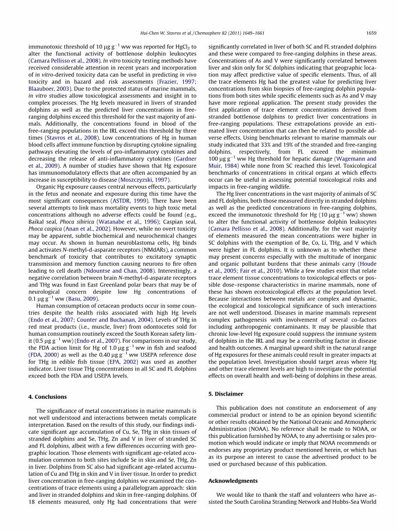
Fig. 3. Total mercury (THg) concentrations (lg g�1 wet wt) in liver of stranded bottlenose dolphins from South Carolina (SC) and the Indian River Lagoon, Florida (FL)compared to thresholds (1Wagemann and Muir, 1984; 2FDA, 2000; and 3EPA, 2002).
1658 Hui-Chen W. Stavros et al. / Chemosphere 82 (2011) 1649–1661
(ASTDR, 1999; Weiner et al., 2003). Of particular concern are po-tential neurotoxic effects of methyl-Hg on the developing fetus.Heavy metals, such as Hg and Cd, typically accumulate and exerttoxic effects in vital organs (e.g., liver, kidney, and brain). Toxico-logical benchmarks of concentrations in critical organs at which ef-fects occur can be useful in assessing potential toxicological risksand impacts in free-ranging wildlife. Fig. 3 illustrates the liver con-centrations of THg in stranded dolphins from SC and FL and severaltoxicological benchmarks, including the estimated Hg limit for he-patic damage in liver tissue of marine mammals at 100–400 lg g�1 ww (approx 400–1600 lg g�1 dw) (Wagemann andMuir, 1984). While the mean liver THg levels in SC and FL strandeddolphins were below the value associated with occurrence of hepa-tic damage in marine mammals, 3 of 15 dolphins (33%) from FLranged between 100 and 200 lg g�1 ww; none of the SC dolphinsexceeded this threshold. Further concern is warranted regardingthe free-ranging population in the IRL as the predicted liver Hgconcentrations found 11 of 72 (15%) of IRL dolphins above100 lg g�1 ww and the highest individual concentration was798 lg g�1 ww, although this may be ameliorated by higher levelsof Se as a protective agent in these dolphins.
The higher Hg levels found in IRL dolphins is likely due to ele-vated levels of environmental Hg (Trocine and Trefry, 1996; Cantilloet al., 1999) resulting in the higher range of exposure for dolphinsinhabiting these areas. Florida dolphins had 10 times higher liverconcentrations compared to SC dolphins, suggesting that they con-sumed prey with higher Hg levels. Atmospheric deposition, onesource of Hg to the aquatic environment, was reported to be consis-tently higher in FL (Fuylkerson and Nnadi, 2006) compared to SC.Interestingly, non-marine mammals with 30 lg g�1 ww liver Hgconcentrations were likely to suffer Hg intoxication (Thompson,1996), a level much lower than the estimated 100–400 lg g�1 wwHg limit for damage in marine mammals (Wagemann and Muir,1984). Mercury increases progressively through the food chainand with a mammal’s growth the amount of food eaten increasesalong with prey size (Andre et al., 1991b). Thus, the body burdenof Hg increases with age as observed in our study and others. Acomparison of THg concentrations in six fish species between IRLand Charleston, SC found that IRL fish ranged from 3 to 12 timeshigher (Adams et al., 2003; Stavros et al., 2007).
Apparently marine mammals are able to tolerate high Hg bodyburdens without overt effects and no intoxication has been re-ported in ringed (Pusa hispida) and bearded seals (Erignathus barb-atus) with liver concentrations up to 420 mg kg�1 (Smith andArmstrong, 1978). Striped dolphins (Stenella coeruleoalba) alsohad no symptoms of Hg poisoning although their total Hg burdenwas 2.5 times as high as fatal concentration for humans (Itano andKawai, 1980). The remarkable tolerance of marine mammals toheavy metals has been attributed to detoxification processes andstorage in non-toxic physico-chemical form. Toxicological assess-ments of specific metals such as Hg are known to cause liver dam-age at high concentrations (ASTDR, 1999). Liver abnormalitiesconsisting of lipofuscinosis, central necrosis and lymphocytic infil-trates have been reported to be associated with chronic Hg accu-mulation > 234 lg g�1 dw in Atlantic bottlenose dolphins(Rawson et al., 1993) and also in a single beaked whale Mesoplodondensirostris with Hg of 248 lg g�1 ww (�1000 lg g�1 dw) (Lawet al., 1997). Additionally, an acute feeding study in seals providesevidence that a high intake rate of methyl-Hg may exceed thedetoxification and mineralization processes (Ronald et al., 1977).A higher accumulation of methyl-Hg has also been observed in sickanimals suggesting that these animals may lack ability to effi-ciently detoxify organic Hg as healthy animals (Dietz et al.,1990). Mean liver concentrations of Hg, selenium (Se), the Hg:Semolar ratio, and zinc (Zn) were significantly higher in harbor por-poises (Phocoena phocoena) that died of infectious disease com-pared to healthy porpoises that died from physical trauma(Bennett et al., 2001). Trace element profiles in diseased and ema-ciated sea otters suggest that oxidative stress mediates the pertur-bation of essential element concentrations, thus, other elements inaddition to Hg may contribute to disease (Kannan et al., 2006).Health evaluation studies of the free-ranging dolphin populationsin both the IRL and Charleston, SC found that over half of the ani-mals were considered not healthy (Reif et al., 2008). It may beplausible that chronic low-level Hg exposure could suppress theimmune system of dolphins in these areas, particularly IRL dol-phins, and we are conducting further analysis to evaluate whetherHg may be a causative factor in diseases and health outcomes.
Limited information exists for assessing toxicological inferenceof trace elements, especially in marine mammals. An in vitro
Hui-Chen W. Stavros et al. / Chemosphere 82 (2011) 1649–1661 1659
immunotoxic threshold of 10 lg g�1 ww was reported for HgCl2 toalter the functional activity of bottlenose dolphin leukocytes(Camara Pellisso et al., 2008). In vitro toxicity testing methods havereceived considerable attention in recent years and incorporationof in vitro-derived toxicity data can be useful in predicting in vivotoxicity and in hazard and risk assessments (Frazier, 1997;Blaauboer, 2003). Due to the protected status of marine mammals,in vitro studies allow toxicological assessments and insight in tocomplex processes. The Hg levels measured in livers of strandeddolphins as well as the predicted liver concentrations in free-ranging dolphins exceed this threshold for the vast majority of ani-mals. Additionally, the concentrations found in blood of thefree-ranging populations in the IRL exceed this threshold by threetimes (Stavros et al., 2008). Low concentrations of Hg in humanblood cells affect immune function by disrupting cytokine signalingpathways elevating the levels of pro-inflammatory cytokines anddecreasing the release of anti-inflammatory cytokines (Gardneret al., 2009). A number of studies have shown that Hg exposurehas immunomodulatory effects that are often accompanied by anincrease in susceptibility to disease (Moszczyzski, 1997).
Organic Hg exposure causes central nervous effects, particularlyin the fetus and neonate and exposure during this time have themost significant consequences (ASTDR, 1999). There have beenseveral attempts to link mass mortality events to high toxic metalconcentrations although no adverse effects could be found (e.g.,Baikal seal, Phoca sibirica (Watanabe et al., 1996); Caspian seal,Phoca caspica (Anan et al., 2002). However, while no overt toxicitymay be apparent, subtle biochemical and neurochemical changesmay occur. As shown in human neuroblastoma cells, Hg bindsand activates N-methyl-d-asparate receptors (NMdARs), a commonbenchmark of toxicity that contributes to excitatory synaptictransmission and memory function causing neurons to fire oftenleading to cell death (Ndountse and Chan, 2008). Interestingly, anegative correlation between brain N-methyl-d-asparate receptorsand THg was found in East Greenland polar bears that may be ofneurological concern despite low Hg concentrations of0.1 lg g�1 ww (Basu, 2009).
Human consumption of cetacean products occur in some coun-tries despite the health risks associated with high Hg levels(Endo et al., 2007; Counter and Buchanan, 2004). Levels of THg inred meat products (i.e., muscle, liver) from odontocetes sold forhuman consumption routinely exceed the South Korean safety lim-it (0.5 lg g�1 ww) (Endo et al., 2007). For comparisons in our study,the FDA action limit for Hg of 1.0 lg g�1 ww in fish and seafood(FDA, 2000) as well as the 0.40 lg g�1 ww USEPA reference dosefor THg in edible fish tissue (EPA, 2002) was used as anotherindicator. Liver tissue THg concentrations in all SC and FL dolphinsexceed both the FDA and USEPA levels.
4. Conclusions
The significance of metal concentrations in marine mammals isnot well understood and interactions between metals complicateinterpretation. Based on the results of this study, our findings indi-cate significant age accumulation of Cu, Se, THg in skin tissues ofstranded dolphins and Se, THg, Zn and V in liver of stranded SCand FL dolphins, albeit with a few differences occurring with geo-graphic location. Those elements with significant age-related accu-mulation common to both sites include Se in skin and Se, THg, Znin liver. Dolphins from SC also had significant age-related accumu-lation of Cu and THg in skin and V in liver tissue. In order to predictliver concentration in free-ranging dolphins we examined the con-centrations of trace elements using a parallelogram approach: skinand liver in stranded dolphins and skin in free-ranging dolphins. Of18 elements measured, only Hg had concentrations that were
significantly correlated in liver of both SC and FL stranded dolphinsand these were compared to free-ranging dolphins in these areas.Concentrations of As and V were significantly correlated betweenliver and skin only for SC dolphins indicating that geographic loca-tion may affect predictive value of specific elements. Thus, of allthe trace elements Hg had the greatest value for predicting liverconcentrations from skin biopsies of free-ranging dolphin popula-tions from both sites while specific elements such as As and V mayhave more regional application. The present study provides thefirst application of trace element concentrations derived fromstranded bottlenose dolphins to predict liver concentrations infree-ranging populations. These extrapolations provide an esti-mated liver concentration that can then be related to possible ad-verse effects. Using benchmarks relevant to marine mammals ourstudy indicated that 33% and 19% of the stranded and free-rangingdolphins, respectively, from FL exceed the minimum100 lg g�1 ww Hg threshold for hepatic damage (Wagemann andMuir, 1984) while none from SC reached this level. Toxicologicalbenchmarks of concentrations in critical organs at which effectsoccur can be useful in assessing potential toxicological risks andimpacts in free-ranging wildlife.
The Hg liver concentrations in the vast majority of animals of SCand FL dolphins, both those measured directly in stranded dolphinsas well as the predicted concentrations in free-ranging dolphins,exceed the immunotoxic threshold for Hg (10 lg g�1 ww) shownto alter the functional activity of bottlenose dolphin leukocytes(Camara Pellisso et al., 2008). Additionally, for the vast majorityof elements measured the mean concentrations were higher inSC dolphins with the exemption of Be, Co, Li, THg, and V whichwere higher in FL dolphins. It is unknown as to whether thesemay present concerns especially with the multitude of inorganicand organic pollutant burdens that these animals carry (Houdeet al., 2005; Fair et al., 2010). While a few studies exist that relatetrace element tissue concentrations to toxicological effects or pos-sible dose–response characteristics in marine mammals, none ofthese has shown ecotoxicological effects at the population level.Because interactions between metals are complex and dynamic,the ecological and toxicological significance of such interactionsare not well understood. Diseases in marine mammals representcomplex pathogenesis with involvement of several co-factorsincluding anthropogenic contaminants. It may be plausible thatchronic low-level Hg exposure could suppress the immune systemof dolphins in the IRL and may be a contributing factor in diseaseand health outcomes. A marginal upward shift in the natural rangeof Hg exposures for these animals could result in greater impacts atthe population level. Investigation should target areas where Hgand other trace element levels are high to investigate the potentialeffects on overall health and well-being of dolphins in these areas.
5. Disclaimer
This publication does not constitute an endorsement of anycommercial product or intend to be an opinion beyond scientificor other results obtained by the National Oceanic and AtmosphericAdministration (NOAA). No reference shall be made to NOAA, orthis publication furnished by NOAA, to any advertising or sales pro-motion which would indicate or imply that NOAA recommends orendorses any proprietary product mentioned herein, or which hasas its purpose an interest to cause the advertised product to beused or purchased because of this publication.
Acknowledgments
We would like to thank the staff and volunteers who have as-sisted the South Carolina Stranding Network and Hubbs-Sea World

1660 Hui-Chen W. Stavros et al. / Chemosphere 82 (2011) 1649–1661
Research Institute, FL. The authors would like to thank the follow-ing reviewers for their critical review of the manuscript: Drs. Mal-colm Meaburn, Natasha Henry and Mr. James Powell.
References
Adams, D.H., McMichael, R.H., Jr., Henderson, G.E., 2003. Mercury Levels in Marineand Estuarine Fishes of Florida 1989–2001. Florida Marine Research InstituteTechnical Report.
Anan, Y., Kunito, T., Ikemoto, T., Kubota, R., Watanabe, I., Tanabe, S., Miyazaki, N.,Petrov, E.A., 2002. Elevated concentrations of trace elements in Caspian seals(Phoca caspica) found stranded during the mass mortality events in 2000. Arch.Environ. Contam. Toxicol. 42, 354–362.
Andre, J.M., Ribeyre, R., Boudou, A., 1991a. Mercury contaminantion levels anddistribution in tissues and organs of delphinids (Stenella attenuata) from theeastern tropical pacific, in relation to biological and ecological factors. Mar.Environ. Res. 30, 43–72.
Andre, J.M., Boudou, A., Ribeyre, F., Bernhard, M., 1991b. Comparative study ofmercury accumulation in dolphins (Stenella coeruleoalba) from French Atlanticand Mediterranean coasts. Sci. Total Environ. 104, 191–209.
ASTDR, 1999. Toxicological Profile for Mercury. Toxicological Profiles. USDepartment of Health and Human Services, Public Health Service Agency forToxic Substances and Disease Registry, Atlanta, Georgia 30333.
Basu, N., 2009. Is dietary mercury of neurotoxicological concern to wild polar bears(Ursus maritimus)? Environ. Toxicol. Chem. 28, 133–140.
Basu, N., Scheuhammer, A., Grochowina, N., Klenavic, K., Evans, D., O’Brien, M., Chan,H., 2005. Effects of mercury on neurochemical receptors in wild river otters(Lontra canadensis). Environ. Sci. Technol. 39, 3585–3591.
Beck, K.M., Fair, P., McFee, W., Wolf, D., 1997. Heavy metals in livers of bottlenosedolphins stranded along the South Carolina coast. Mar. Pollut. Bull. 34, 734–739.
Bennett, P.M., Jepson, P.D., Law, R.J., Jones, B.R., Kuiken, T., Baker, J.R., Rogan, E.,Kirkwood, J.R., 2001. Exposure to heavy metals and infectious disease mortalityin harbour porpoises from England and Wales. Environ. Pollut. 112, 33–40.
Blaauboer, B.J., 2003. The integration of data on physico-chemical properties,in vitro-derived toxicity data and physiologically based kinetic and dynamic asmodelling a tool in hazard and risk assessment. A commentary. Toxicol. Lett.138, 161–171.
Booth, S., Zeller, D., 2005. Mercury, food webs, and marine mammals: implicationsof diet and climate change for human health. Environ. Health Perspect. 113,521–526.
Bossart, G.D., 2006. Marine mammals as sentinel species for oceans and humanhealth. Oceanography 19, 44–47.
Braune, B., 2007. Temporal trends of organochlorines and mercury in seabird eggsfrom the Canadian Arctic, 1975–2003. Environ. Pollut. 148, 599–613.
Braune, B., Outridge, P., Fisk, A., Muir, D., Helm, P., Hobbs, K., Hoekstra, R., Kuzyk, A.,Kwan, M., Letcher, R., Lockhart, W., Norstrom, R., Stern, G., Stirling, I., 2005.Persistent organic pollutants and mercury in marine biota of the CanadianArctic: an overview of spatial and temporal trends. Sci. Total Environ. 4, 351–352.
Bryan, C., Christopher, S., Balmer, B., Wells, R., 2007. Establishing baseline levels oftrace elements in blood and skin of bottlenose dolphins in Sarasota Bay, Florida:implications for non-invasive monitoring. Sci. Total Environ. 388, 325–342.
Camara Pellisso, S., Munoz, M.J., Carballo, M., Sanchez-Vizcaino, J.M., 2008.Determination of the immunotoxic potential of heavy metals on thefunctional activity of bottlenose dolphin leukocytes in vitro. Vet. Immunol.Immunopathol. 121, 189–198.
Cantillo, A.Y., Lauenstein, G.G., O’Connor, T.P., Johnson, W.E., 1999. Status andTrends of Contaminant Levels in Biota and Sediments of South Florida. RegionalReports Series 2. National Oceanic and Atmospheric Administration, SilverSpring, MD. pp. 1–40.
Cardellicchio, N., Decataldo, A., Di Leo, A., Misino, A., 2002. Tissue distribution ofmercury and selenium in striped dolphins (Stenella coeruleoalba) from theMediterranean Sea (Southern Italy). Environ. Pollut. 116, 265–271.
Counter, S.A., Buchanan, L.H., 2004. Mercury exposure in children: a review. Toxicol.Appl. Pharmacol. 198, 209–230.
Cuvin-Aralar, M.L.A., Furness, R.W., 1991. Mercury and selenium interaction: areview. Ecotoxicol. Environ. Safe. 21, 348–364.
Das, K., Debacker, V., Pillet, S., Bouquegneau, J.-M., 2003. Heavy metals in marinemammals. In: Vos, J., Bossart, G., Fournier, M., O’Shea, T.J. (Eds.), Toxicology ofMarine Mammals. Taylor Francis, New York, NY, pp. 135–167.
Dietz, R., Nielson, C.O., Hansen, M.M., Hansen, C.T., 1990. Organic mercury inGreenland birds and mammals. Sci. Total Environ. 95, 31–51.
Dietz, R., Riget, F., Born, E., Sonne, C., Grandjean, P., Kirkegaard, M., Olsen, M.,Asmund, G., Renzoni, A., Baagøe, H., Andreasen, C., 2006. Trends in mercury hairof Greenlandic polar bears (Ursus maritimus) during 1892–2001. Environ. Sci.Technol. 40, 1120–1125.
Durden, W.N., Stolen, M.K., Adams, D.H., Stolen, E.D., 2007. Mercury and seleniumconcentrations in stranded bottlenose dolphins from the Indian River LagoonSystem, Florida. Bull. Mar. Sci. 81, 37–54.
Emonet, N., Leccia, M.T., Favier, A., Beani, J.C., Richard, M.J., 1998. Thiols andselenium: protective effect on human skin fibroblasts exposed to UVA radiation.J. Photochem. Photobiol. B 40, 84–90.
Endo, T., Yong-un, M., Baker, C.S., Funahashi, N., Lavery, S., Dalebout, M.L.,Lukoschek, V., Haraguchi, K., 2007. Contaminantion level of mercury in red
meat products from cetaceans available from South Korea markets. Mar. Pollut.Bull. 54, 669–677.
Facemire, C., Augspruger, T., Bateman, D., Brim, M., Conzelmann, P., Delchamps, S.,Douglas, E., Inmon, L., Looney, F., Masson, G., Morrison, D., Morse, N., Robison,A., 1995a. Impacts of mercury contamination in the southeastern United States.Water Air Soil Pollut. 80, 923–926.
Facemire, C.F., Gross, T., Guillette, L., 1995b. Reproductive impairment in the Floridapanther: nature or nurture? Environ. Health Perspect. 03, 79–86.
Fair, P.A., Becker, P.R., 2000. Review of stress in marine mammals. J. Aquat. Ecosyst.Stress Recov. 7, 335–354.
Fair, P., Adams, J., Zolman, E., McCulloch, S., Goldstein, J., Murdoch, M., Varela, R.,Hansen, L., Townsend, F., Kucklick, J., Bryan, C., Christopher, S., Pugh, R., Bossart,G., 2006. Protocols for Conducting Dolphin Capture-release Health AssessmentStudies. NOAA Technical Memorandum, pp. 1–83.
Fair, P.A., Adams, J., Mitchum, G., Hulsey, T.C., Reif, J., Houde, M., Muir, D., Wirth, E.,Wetzel, D., Zolman, E.Z., McFee, W., Bossart, G.D., 2010. Contaminant blubberburdens in Atlantic bottlenose dolphins (Tursiops truncatus) from two southeastUS estuarine areas: Concentrations and patterns of PCBs, Pesticides, PBDEs,PFCs, and PAHs. Sci. Total Environ. 408, 1577–1597.
Frazier, J.M., 1997. Predictive toxicodynamics: empirical/mechanistic approaches.Toxicol. In Vitro 11, 465–472.
Fujise, Y., Honda, K., Tatsukawa, R., Mishima, S., 1988. Tissue distribution of heavymetals in Dall’s porpoise in the northwestern Pacific. Mar. Pollut. Bull. 19, 226–230.
Fuylkerson, M., Nnadi, F.N., 2006. Predicting mercury wet depostion in Florida: asimple approach. Atmos. Environ. 40, 3962–3968.
Gardner, R.M., Nyland, J.F., Evans, S.L., Wang, S.B., Doyle, K.M., Crainiceanu, C.M.,Silbergeld, E.K., 2009. Mercury induces an unopposed inflammatory response inhuman peripheral blood mononuclear cells in vitro. Environ. Health Perspect.117, 1932–1938.
Gonul, B., Soylemezoglu, T., Babul, A., Celebi, N., 1998. Effects of epidermal growthfactor dosage forms on mice full-thickness skin wound zinc levels and relationto wound strength. J. Pharm. Pharmacol. 50, 641–644.
Hohn, A.A., Scott, M.D., Wells, R.S., Sweeney, J.C., Irvine, A.B., 1989. Growth layers inteeth from known-age, free-ranging bottlenose dolphins. Mar. Mammal Sci. 5,315–342.
Holsbeek, L., Siebert, U., Joiris, C.R., 1998. Heavy metals in dolphins stranded on theFrench Atlantic Coast. Sci. Total Environ. 217, 241–249.
Houde, M., Wells, R., Fair, P., Bossart, G., Hohn, A., Rowles, T., Sweeney, J., Solomon,K., Muir, D., 2005. Polyfluoroalkyl compounds in free-ranging bottlenosedolphins (Tursiops truncatus) from the Gulf of Mexico and the Atlantic Ocean.Environ. Sci. Technol. 39, 6591–6598.
Itano, K., Kawai, S., 1980. Changes of Mercury and Selenium Contents and BiologicalHalf-life of Mercury in the Striped Dolphins. Univ of the Ryukyus, Okinawa,Japan. pp. 49–72.
Itano, K., Kawai, S., 1981. Changes of mercury contents and biological half-life ofmercury in the striped dolphins. In: Fujiyama, N. (Ed.), Studies on the Levels andOrganochlorine Compounds and Heavy Metals in the Marine Organisms.University of the Ryukyus, Nishihara, Okinawa, pp. 49–73.
Kakuschke, A., Valentine-Thon, E., Griesel, S., Fonfara, S., Siebert, U., Prange, A., 2005.Immunological impact of metals in harbor seals (Phoca vitulina) of the NorthSea. Environ. Sci. Technol. 39, 7568–7575.
Kakuschke, A., Valentine-Thon, E., Griesel, S., Rosenberger, T., Mundry, R., Siebert, U.,Prange, A., 2008. Blood metal levels and metal-influenced immune functions ofharbour seals in captivity. Mar. Pollut. Bull. 56, 764–769.
Kakuschke, A., Valentine-Thon, E., Fonfara, S., Kramer, K., Prange, A., 2009. Effects ofmethyl-, phenyl-, ethylmercury and mercurychlorid on immune cells of harborseals (Phoca vitulina). J. Environ. Sci. 21, 1716–1721.
Kannan, K., Agusa, T., Perrotta, E., Thomas, N.J., Tanabe, S., 2006. Comparison of traceelement concentrations in livers of diseased, emaciated and non-diseasedsouthern sea otters from the California coast. Chemosphere 11, 2160–2167.
Kemper, C., Gibbs, P., Obendorf, D., Marvanek, S., Lenghaus, C., 1994. A review ofheavy metal and organochlorine levels in marine mammals in Australia. Sci.Total Environ. 154, 129–139.
Koeman, J.H., Peters, W.H.M., Koudstall-Hol, C.H.M., Tjoe, P.S., DeGoeij, J.J.M.,1973. Mercury–selenium correlations in marine mammals. Nature 245, 385–386.
Kuehl, D.W., Haebler, R., 1995. Organochlorine, organbromine, metal, and seleniumresidues in bottlenose dolphins (Tursiops truncatus) collected during an unusualmortality event in the Gulf of Mexico, 1990. Arch. Environ. Contam. Toxicol. 28,494–499.
Law, R.J., 1996. Metals in marine mammals. In: Beyer, W., Heinz, G., Redmon-Norwood, A. (Eds.), Environmental Contaminants in Wildlife Interpreting TissueConcentrations. CRC Lewis Publishers, Boca Raton, FL, pp. 357–376.
Law, R.J., Jones, B.R., Baker, J.R., Kennedy, S., Milne, R., Morris, R.J., 1992. Trace metalsin the livers of marine mammals from the Welsh coast and Irish Sea. Mar.Pollut. Bull. 22, 183–191.
Law, R.J., Allchin, C.R., Jones, B.R., Jepson, P.D., Baker, J.R., Spurrier, C.J.H., 1997.Metals and organochlories in tissues of a Blainville’s Beaked Whale (Mesoplodondensirostris) and a killer whale (Orcinus orca) stranded in the United Kingdom.Mar. Pollut. Bull. 34, 208–212.
Leccia, M.T., Richard, M.J., Favier, A., Beani, J.C., 1999. Zinc protects againstultraviolet Al-induced DNA damage and apoptosis in cultured humanfibroblasts. Biol. Trace Elem. Res. 69, 177–190.
Lemly, D., 1996. Selenium in aquatic organisms. In: Beyer, W., Heinz, G., Redmon-Norwood, A. (Eds.), Environmental Contaminants in Wildlife Interpreting TissueConcentrations. CRC Lewis Publishers, Boca Raton, FL, pp. 357–376.

Hui-Chen W. Stavros et al. / Chemosphere 82 (2011) 1649–1661 1661
Leonzio, C., Focardi, S., Fossi, F., 1992. Heavy metals and selenium in strandeddolphins of the northern Tyrrhenian (NW Mediterranean). Sci. Total Environ.119, 77–84.
Liu, J., Goyer, R.A., Waalkes, M.P., 2008. Toxic effects of metals. In: Klaassen, C.D.(Ed.), Casarett and Doull’s Toxicology: The Basic Sci. of Poisons. McGraw Hill,New York, pp. 931–979.
Lockhart, W., Stern, G.A., Wagemann, R., Hunt, R., Metner, D., DeLaronde, J., Dunn, B.,Stewart, R., Hyatt, C., Harwood, L., Mount, K., 2005. Concentrations of mercuryin tissues of beluga whales (Delphinapterus leucas) from several communities inthe Canadian Arctic from 1981 to 2002. Sci. Total Environ. 351–352, 391–412.
Mackay, E.A., Demiralp, R., Becker, P.R., Greenberg, R.R., Koster, B.J., Wise, S.A., 1995.Trace element concentrations in cetacean liver tissues archived in the nationalmarine mammal tissue bank. Sci. Total Environ. 175, 25–41.
Mackey, E.A., Becker, P.R., Demiralp, R., Greenberg, R.R., Koster, B.J., Wise, S.A., 1996.Bioaccumulation of vanadium and other trace metals in livers of Alaskancetaceans and pinnipeds. Arch. Environ. Contam. Toxicol. 30, 503–512.
McFee, W. E., Adams, J.D., Fair, P.A., Bossart, G.D., 2010. Age Distribution and Growthof Two Bottlenose Dolphin (Tursiop truncatus) from Capture-release Studies inthe Southeastern United States, submitted for publication.
Mead, J.G., Potter, C.W., 1990. Natural History of bottlenose dolphins along thecentral Atlantic coast of the United States. In: Leatherwood, S., Reeves, R.R.(Eds.), The Bottlenose Dolphin. Academic Press, San Diego, pp. 165–195.
Meador, J.P., Ernest, D., Hohn, A.A., Tilbury, K., Gorzelany, J., Worthy, G., Stein, J.E.,1999. Comparison of elements in bottlenose dolphins stranded on the beachesof Texas and Florida in the Gulf of Mexico over a one-year period. Arch. Environ.Contam. Toxicol. 36, 87–98.
Monaci, F., Borrel, A., Leonzio, C., Marsili, L., Calzada, N., 1998. Trace elements instriped dolphin (Stenella coeruleoalba) from the western Mediterranean.Environ. Pollut. 99, 61–68.
Moszczyzski, P., 1997. Mercury compounds and the immune system: a review. Int. J.Occup. Med. Environ. Health 10, 247–258.
Myrick, A.C., Hohn, A.A., Sloan, P.A., Kimura, M., Stanley, D.D., 1983. Estimating Ageof Spotted and Spinner Dolphins (Stenella attenuata and Stenella longirostris)from Teeth. NOAA Technical Memorandum. Southwest Fisheries Service Center.National Marine Fisheries Service, La Joaa, CA. pp. 1–17.
Ndountse, L.T., Chan, H.M., 2008. Methylmercury increases receptors on human SH-SY 5Y neuroblastoma cells leading to neurotoxicity. Toxicology 249, 251–255.
Nigro, M., Leonzio, C., 1996. Intracellular storage of mercury and selenium indifferent marine vertebrates. Mar. Ecol. Prog. Ser. 135, 137–143.
O’Hara, T.M., Woshner, V.M., Bratton, G., 2003. Inorganic pollutants in Arctic marinemammals. In: Vos, J., Bossart, G., Fournier, M., O’Shea, T.J. (Eds.), Toxicology ofMarine Mammals. Taylor Francis, New York, pp. 206–246.
O’Hara, T.M., Hanns, C., Woshner, V.M., Zeh, J., Bratton, G., Taylor, R., 2008. Essentialand non-essential elements in the bowhead whale: epidermis-basedpredictions of blubber, kidney, liver and muscle tissue concentrations. J. Cet.Res. Manage. 10, 107–117.
Palmisano, F., Cardellicchio, N., Zambonin, P.G., 1995. Speciation of mercury indolphin liver. A two-stage mechanism for the demethylation accumulationprocess and role of selenium. Mar. Environ. Res. 40, 109–121.
Pompe-Gotal, J., Srebocan, E., Gomercic, H., Crnic, P., 2009. Mercury concentrrationsin tissues of bottlenose dolphins (Tursiops truncatus) and striped dolphins(Stenella coeruloalba) stranded on the Croatian Adriatic coast. Vet. Med. 54, 598–604.
Querouil, S., Freitas, L., Dinis, A., Alves, F., Cascao, I., Prieto, R., Silva, M.A., Magalhaes,S., Matos, J.A., Santos, R.S., 2010. Sex bias in biopsy samples collected from free-ranging dolphins. Eur. J. Wildlife Res. 56, 151–158.
Rawson, A., Patoon, G., Hofman, S., Petra, G., Johns, L., 1993. Liver abnormalitesassociated with chronic mercury accumulation in stranded bottlenose dolphins.Ecotox. Environ. Safe. 25, 41–47.
Reif, J.S., Fair, P.A., Adams, J., Joseph, B., Kilpatrick, D.K., Sanchez, R., Goldstein, J.D.,Townsend, F.I., McCulloch, S.D., Mazzoil, M., Zolman, E., Hansen, L.J., Bossart,G.D., 2008. Health status of Atlantic Bottlenose Dolphins (Tursiops truncatus)from the Indian River Lagoon, FL and Charleston, SC. J Am. Vet. Assoc. 233, 299–307.
Riget, F., Dietz, R., 2000. Temporal trends of cadmium and mercury in Greenlandmarine biota. Sci. Total Environ. 245.
Riget, F., Dietz, R., Vorkamp, K., Johansen, P., Muir, D., 2004. Spatial and temporaltrends of contaminants in Greenland terrestrial, fresh water and marine biota:an update. Sci. Total Environ. 331, 29–52.
Roditi-Elasar, M., Kerem, D., Hornung, H., Kress, N., Shoham-Frider, E., Goffman, O.,Spanier, E., 2003. Heavy metal levels in bottlenose and striped dolphins off theMediterranean coast of Israel. Mar. Pollut. Bull. 46, 503–512.
Ronald, K., Tessaro, S.V., Uthe, J.F., Freeman, C., Frank, R., 1977. Methylmercurypoisoning in the harp seal (Pagophilus groenlandicus). Sci. Total Environ. 8,153–166.
Saeki, K., Nakajima, M., Noda, K., Loughlin, T.R., Baba, N., Kiyota, M., Tatsukawa, R.,Calkins, D.G., 1999. Vanadium accumulation in pinnipeds. Arch. Environ.Contam. Toxicol. 36, 81–86.
Smith, T.G., Armstrong, F.A.J., 1978. Mercury and selenium in ringed and beardedseal tissues from Arctic Canada. Arctic 31, 75–84.
Stavros, H.W., Bossart, G., Hulsey, T., Fair, P.A., 2007. Trace element concentrationsin skin of free-ranging bottlenose dolphins (Tursiops truncatus) from thesoutheast Atlantic coast. Sci. Total Environ. 300–315, 300–315.
Stavros, H.W., Bossart, G., Hulsey, T., Fair, P., 2008. Trace element concentrations inblood of free-ranging bottlenose dolphins (Tursiops truncatus): influence of age,sex and location. Mar. Pollut. Bull. 56, 348–379.
Thompson, D.R., 1990. Heavy metals in marine vertebrates. In: Furness, R.W.,Rainbow, P.S. (Eds.), Heavy Metals in the Marine Environment. CRC Press, BocaRaton, FL, pp. 143–182.
Thompson, D.R., 1996. Mercury in birds and terresterial mammals. In: Beyer, W.,Heinz, G., Redmon-Norwood, A. (Eds.), Environmental Contaminants in WildlifeInterpreting Tissue Concentrations. CRC Lewis Publishers, Boca Raton, FL, pp.341–356.
Trocine, R.P., Trefry, J.H., 1996. Metal concentrations in sediment, water and clamsfrom the Indian River Lagoon, Florida. Mar. Pollut. Bull. 32, 754–759.
USEPA, 2002. Risk Assessment Forum. A Review of the Reference Dose andReference Concentration Process. Washington, DC.
USEPA, 2005. 2004 National Listing of Fish Advisories. Environmental ProtectionAgency, Washington, DC. <http://www.epa.gov/waterSci./fish/advisories/2004/> (accessed 20.05.10).
USEPA, 2006. Mercury Transport and Fat through a Watershed. Synthesis Report ofResearch from EPAs Sci. to Achieve Results (STAR) Grant Program.Environmental Protection Agency, Washington, DC, pp. 1–24.
USFDA, 2000. Guidance for Industry: Action Levels for Poisonous or DeleteriousSubstances in Human Food and Animal Feed. Washington, DC. <http://www.fda.gov/Food/GuidanceComplianceRegulatoryInformation/GuidanceDocuments/ChemicalContaminantsandPesticides/ucm077969.htm> (accessed 19.05.10).
Wagemann, R., Muir, D., 1984. Concentrations of heavy metals and organochlorinesin marine mammals of Northern waters: overview and evaluation. CanadianTech. Rpts. Fish. Aquatic. Sci. 1279, 1–97.
Watanabe, I., Ichihashi, H., Tanabe, S., Amano, M., Miyazaki, N., Petrov, E.A.,Tatsukawa, R., 1996. Trace element accumulation in Baikal seal (Phoca sibirica)from the Lake Baikal. Environ. Pollut., 169–179.
Weiner, J., Krabbenhoft, D., Heinz, G., Scheuhammer, A., 2003. Exoctoxicology ofmercury. In: Hoffman, D., Rattner, B.A., Burton, G.A.J., Cairns, J.J. (Eds.),Handbook of Ecotoxicology. CRC Press, Boca Raton, FL, pp. 409–463.
Wells, R.S., Rhinehardt, H.L., Hansen, L.J., Sweeney, J.C., Townsend, F.I., Stone, R.,Casper, D.R., Scott, M.D., Hohn, A.A., Rowles, T.K., 2004. Bottlenose dolphins asmarine ecosystem sentinels: developing a health monitoring system. EcoHealth1, 246–254.
Wobeser, G., Swift, M., 1976. Mercury poisoning in a wild mink. J. Wildlife Dis., 335–340.
Yang, J., Kunito, T., Tanabe, S., Amano, M., Miyazaki, N., 2002. Trace elements in skinof Dall’s porpoises (Phocoenoides dalli) from the northern waters of Japan: anevaluation for utilization as non-lethal tracers. Mar. Pollut. Bull. 45, 230–236.
Yang, D.-Y., Chen, Y.-W., Gunn, J.M., Belzile, N., 2008. Selenium and mercury inorganisms: interactions and mechanisms. Environ. Rev. 16, 71–92.
Zhou, L., Salvador, S.M., Liu, Y.P., Sequeira, M., 2001. Heavy metals in the tissues ofcommon dolphins (Delphinus delphis) stranded on the Portuguese coast. Sci.Total Environ. 271, 61–76.