GROWTH LAYERS IN TEETH FROM KNOWN-AGE, FREE-RANGING BOTTLENOSE DOLPHINS
28
MARINE MAMMAL SCIENCE, 5(4):3 15-342 (October 1989) 0 1989 by the Society for Marine Mammalogy GROWTH LAYERS IN TEETH FROM KNOWN-AGE, FREE-RANGING BOTTLENOSE DOLPHINS ALETA A. HOHN Southwest Fisheries Center, National Marine Fisheries Service, P.O. Box 27 1, La Jolla, California 92038 MICHAEL D. Scott Inter-American Tropical Tuna Commission, % Scripps Institution of Oceanography, La Jolla, California 92037 RANDALL S. WELLS University of California, Long Marine Laboratory, Santa Cruz, California 95060 and Dolphin Biology Research Associates, 163 Siesta Drive, Sarasota, Florida 34242 JAY C. SWEENEY A. BLAIR IRVINE Dolphin Biology Research Associates, 163 Siesta Drive, Sarasota, Florida 34242 ABSTRACT Growth layers were examined in teeth collected from free-ranging bottlenose dolphins, Tursiops truncatus, from Florida that have been part of a long-term study begun in 1970; 26 of the dolphins were of known or approximately known age, and 19 were of minimum known age. A second tooth was extracted from 6 animals for examination of growth that had taken place in the interval following the initial extraction. The teeth were read for age estimates without knowledge of any data pertaining to the animals. Most of the estimated ages were the same as or close to the known and approximately known ages of the animals, ranging from 2 to 16 yr. We conclude that the structures we define as dentinal growth layer groups (GLGs) are annual, we describe sources of error in age estimates, and we provide a description of the GLG pattern that can be used by others to estimate age for dolphins. Key words: age determination, growth layers, GLGs, teeth, Tursiops truncatus, bottlenose dolphin, known age, long-term studies. The ability to estimate the age of individuals has been an important tool in the study of population biology. For most species of mammals, we have been able to count growth layers in teeth or bones to obtain age estimates. The use of these structures has generally been accepted and applied for age estimation 315 Help Volumes Main Menu
Transcript of GROWTH LAYERS IN TEETH FROM KNOWN-AGE, FREE-RANGING BOTTLENOSE DOLPHINS
MARINE MAMMAL SCIENCE, 5(4):3 15-342 (October 1989) 0 1989 by the Society for Marine Mammalogy
GROWTH LAYERS IN TEETH FROM KNOWN-AGE, FREE-RANGING
BOTTLENOSE DOLPHINS
ALETA A. HOHN Southwest Fisheries Center, National Marine Fisheries Service, P.O. Box 27 1,
La Jolla, California 92038
MICHAEL D. Scott
Inter-American Tropical Tuna Commission, % Scripps Institution of Oceanography, La Jolla, California 92037
RANDALL S. WELLS University of California, Long Marine Laboratory, Santa Cruz, California 95060 and
Dolphin Biology Research Associates, 163 Siesta Drive, Sarasota, Florida 34242
JAY C. SWEENEY A. BLAIR IRVINE
Dolphin Biology Research Associates, 163 Siesta Drive, Sarasota, Florida 34242
ABSTRACT
Growth layers were examined in teeth collected from free-ranging bottlenose dolphins, Tursiops truncatus, from Florida that have been part of a long-term study begun in 1970; 26 of the dolphins were of known or approximately known age, and 19 were of minimum known age. A second tooth was extracted from 6 animals for examination of growth that had taken place in the interval following the initial extraction. The teeth were read for age estimates without knowledge of any data pertaining to the animals. Most of the estimated ages were the same as or close to the known and approximately known ages of the animals, ranging from 2 to 16 yr. We conclude that the structures we define as dentinal growth layer groups (GLGs) are annual, we describe sources of error in age estimates, and we provide a description of the GLG pattern that can be used by others to estimate age for dolphins.
Key words: age determination, growth layers, GLGs, teeth, Tursiops truncatus, bottlenose dolphin, known age, long-term studies.
The ability to estimate the age of individuals has been an important tool in the study of population biology. For most species of mammals, we have been able to count growth layers in teeth or bones to obtain age estimates. The use of these structures has generally been accepted and applied for age estimation
315
Help Volumes Main Menu

316 MARINE MAMMAL SCIENCE, VOL. 5, NO. 4, 1989
even when they have not been calibrated for a species because (1) growth layers are very common, having been identified in most mammalian species examined, (2) the appearance and structure of growth layers have been similar among different species within similar tissues, such as teeth or bones, and (3) in each species for which data have been available, growth layers have been calibrated to real time showing the existence of annually occurring layers (see Klevezal’ and Kleinenberg 1967, Grue and Jensen 1979).
Evidence for annual growth layers in teeth of dolphins has come primarily from bottlenose dolphins, Tursiops truncatus, especially those which were born in captivity or were captured young and spent the remainder of their lives in captivity (e.g., Sergeant 1959, Sergeant et al. 1973, Hui 1980). When these animals died, their teeth were sectioned and dentinal growth layers were counted. The number of layers equalled the known age or approximated the suspected age of the dolphins and, thus, growth layers were interpreted to be annual. These interpretations have since been applied to non-captive animals, with the assumption that the annual layering patterns seen in teeth from captive dolphins pertain as well to dolphins in the wild.
A long-term study of a community of bottlenose dolphins on the west coast of Florida has provided the opportunity to calibrate age-estimation techniques on free-ranging, known-age dolphins. A large portion of this community of approximately 100 animals is identifiable through the use of tags, freeze brands, and natural marks (Irvine and Wells 1972; Irvine et al. 1981; Wells et al. 1980, 1987; Wells 1986). Tagging operations and surveys of the community were conducted periodically during 1970-1971, 1975-1976, and since 1980. A total of 91 individuals from the Sarasota dolphin community were captured, sexed, measured, and tagged during these periods, with many of these individuals being captured several times.
The long-term study has provided data for obtaining known or approximate ages of identifiable dolphins. The known-age dolphins are individuals which were observed as calves, newly born to identifiable mothers. In subsequent years, many of these calves were identified with their mothers and captured and marked for later re-identification. Approximately known-age animals are those for which, although the birth date was not known, a minimum or approximate age could be estimated on the basis of when the dolphin was first captured or identified from photographs as a naturally marked animal. For example, if an individual was captured while it was still a calf, the age could be estimated from length- at-age data. These data are available for specimens whose age and length are known and from growth curves (Harrison et al. 1972, Hohn 1980) and are precise for estimating age during the first l-2 yr (Hohn and Hammond 1985). In some cases a maximum age could also be determined; for example, if an identifiable adult female was seen without a calf in the winter of 1980 and with a small calf in the summer of 198 1, the age of the calf could be pinpointed to within a 6-mo period.
This relatively large, unique sample provides an opportunity for examining a number of questions regarding age estimation in dolphins. This study focuses on the following questions:
Help Volumes Main Menu

HOHN ET AL.: AGE DETERMINATION 317
1. Can annual growth layers be identified in teeth from free-ranging, known- age bottlenose dolphins?
2. Do ages estimated from growth layers in teeth correspond to known ages? 3. Are there some individuals in which an obvious growth-layer pattern that
matches the known or approximate age cannot be found? 4. Is it possible to identify one or more factors responsible for most of the
error in age estimation? 5. Can the similarities and differences between the annual patterns be cate-
gorized to allow for the development of a written guide (or “model”) to standardize age estimation from dolphin teeth?
The term growth layer group (GLG) has been used to describe the structures which comprise the repetitive layering pattern seen in dolphin teeth (Perrin and Myrick 1980). The amount of time represented by a GLG depends on how each investigator defines the repeating pattern being examined. Although GLGs do not automatically refer to annual layers, in this study we were interested in a repeating pattern (GLG) that represents one year’s growth.
MATERIALS AND METHODS
Teeth were collected during eight field seasons (five summers and three winters) in 1984-1989, using extraction techniques described by Ridgway et al. (1975). Generally, tooth number 15 was taken from the lower left jaw. Teeth were collected from as many individuals as possible (19 in 1984, 15 in 1985, 9 in 1986, 4 during winter and 10 during summer in 1987, 1 during winter and 6 during summer in 1988, and 5 during winter in 1989). Of these specimens, 14 were of known age and 12 of approximately known age. For the rest, at most only a minimum age was known, From 6 animals, a second tooth was extracted in years following the initial extraction; 1 animal was of known age, 2 of approximately known age, and 3 of minimum or unknown age. Included in the sample were teeth from eight beach-cast specimens with no known history that stranded in the study area. Teeth extracted from live animals were randomly mixed with teeth from beach-cast specimens, and all field numbers were coded before processing.
The teeth were preserved in formalin for up to a month then stored in ethanol. The method for decalcifying, sectioning, staining, and mounting teeth is described in detail in Myrick et al. (1983) for teeth from Stenella spp, The only significant difference is that the smaller teeth from Stenella can be decalcified whole, while the larger teeth from Tursiops must be thick-sectioned first and these 2-3 mm sections then decalcified. The thick-sections were cut using an Isomet l low-speed saw with a diamond-embedded blade. Decalcification time using RDO’, a commercially produced mixture of acids, ranged from 7-17 h, with the greater times required for older animals. Thin-sections were cut to 25 pm on a freezing microtome, stained in hematoxylin, and mounted in 100% glycerin.
1 Reference to trade names does not imply endorsement by the authors’ institutions.
Help Volumes Main Menu

318 MARINE MAMMAL SCIENCE, VOL. 5, NO. 4, 1989
18
16
14
12
10
8
6
4
2
0 0 2 4 6 8 10 12 14 16 18
KNOWN OR APPROXIMATE AGE Figure 1. Relationship between the estimated ages and known or approximate ages
of those animals for whom the age was known. The horizontal bars represent the range of ages possible for an individual (see Tables 1, 2, and 4). Squares represent known-age individuals and diamonds represent approximately known-age individuals. The line rep- resents the one-to-one relationship between estimated and known age for comparison.
Age estimates were made without reference to collaborative data, such as age or length, or knowledge that a tooth represented the second extraction for the six individuals in which this occurred. There were two exceptions to this “blind” reading of tooth sections. Before the study began the tooth from one known- age animal was examined with knowledge of the animal’s age to identify the annual layering pattern (No. 13). (A second tooth from this animal taken 2.5 yr later was read in the blind.) The other exception was for an animal from which a second tooth was collected after the animal was found beach-cast and the tooth prepared after other teeth from that season had been completed (No. 11). For the remaining specimens, age was estimated from counts of GLGs in both dentine and cement.
There were two components to the study. First, ages were estimated in the blind, as described above. These estimated ages were compared to the known or approximately known ages for examination of accuracy, or, in the event that age estimates were inaccurate, for possible explanations why they were inaccurate. Second, the tooth sections were examined again, this time with knowledge of the known ages of animals, for the purpose of demarcating and measuring probable GLGs in dentine. On the basis of the known ages, probable GLGs were marked on an 8” x 10” photographic print of the section. GLG-thickness measurements were taken simultaneously at the location where the GLG was marked on the photograph. The measurements were made using an ocular micrometer in a compound microscope at 100 x with transmitted light.
Help Volumes Main Menu
HOHN ET AL.: AGE DETERMINATION 319
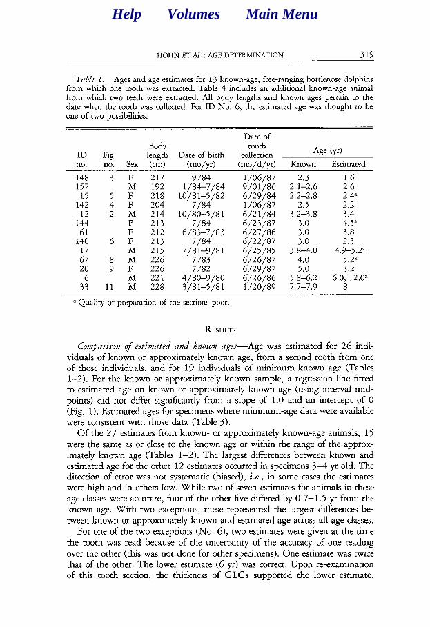
Table 1. Ages and age estimates for 13 known-age, free-ranging bottlenose dolphins from which one tooth was extracted. Table 4 includes an additional known-age animal from which two teeth were extracted. All body lengths and known ages pertain to the date when the tooth was collected. For ID No. 6, the estimated age was thought to be one of two possibilities.
ID Fig. Body length Date of birth
Date of tooth
collection Age (yd
no. no. Sex (cm) (mohd (mo/d/yr) Known Estimated
148 3 F 217 157 M 192
15 5 F 218 142 4 F 204
12 2 M 214 144 F 213 61 F 212
140 6 F 213
:: M M 215 226 20 ; F 226
6 M 221 33 11 M 228
9/84 l/84-7/84
10/81-5/82 7/84
10/80--5/S 1
6&,83 7/84
7/8 l-9/8 1 7/83 7/82
4/80-9/80 3/81-5/81
l/06/87 9/O l/86 6/29/X4 l/06/87 6/2 l/84 6/23/X7 6/27/86 6/22/87 C/25/85 6/26/87 6/29/87 6/26/86 l/20/89
2.3 2.1-2.6 2.2-2.8
2.5 3.2-3.8
;::
3.z.o 4.0 5.0
5.8-6.2 7.7-7.9
1.6 2.6 2.4a 2.2 3.4 4.5” 3.8 2.3
4.9-5.2a 5.2”
6.03’:2.0a A
a Quality of preparation of the sections poor.
RESULTS
Comparison of estimated and known ages--Age was estimated for 26 indi- viduals of known or approximately known age, from a second tooth from one of those individuals, and for 19 individuals of minimum-known age (Tables l-2). For the known or approximately known sample, a regression line fitted to estimated age on known or approximately known age (using interval mid- points) did not differ significantly from a slope of 1.0 and an intercept of 0 (Fig. 1). Estimated ages for specimens where minimum-age data were available were consistent with those data (Table 3).
Of the 27 estimates from known- or approximately known-age animals, 15 were the same as or close to the known age or within the range of the approx- imately known age (Tables 1-2). The largest differences between known and estimated age for the other 12 estimates occurred in specimens 3-4 yr old. The direction of error was not systematic (biased), i.e., in some cases the estimates were high and in others low. While two of seven estimates for animals in these age classes were accurate, four of the other five differed by 0.7-1.5 yr from the known age. With two exceptions, these represented the largest differences be- tween known or approximately known and estimated age across all age classes.
For one of the two exceptions (No. 6), two estimates were given at the time the tooth was read because of the uncertainty of the accuracy of one reading over the other (this was not done for other specimens). One estimate was twice that of the other. The lower estimate (6 yr) was correct. Upon re-examination of this tooth section, the thickness of GLGs supported the lower estimate.
Help Volumes Main Menu
320 MARINE MAMMAL SCIENCE, VOL. 5. NO. 4. 1989
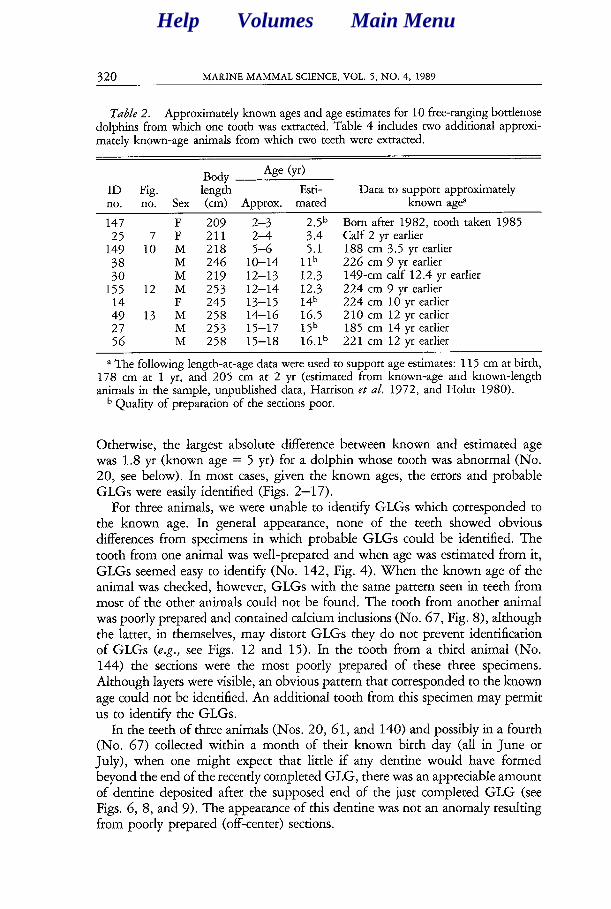
Table 2. Approximately known ages and age estimates for 10 free-ranging bottlenose dolphins from which one tooth was extracted. Table 4 includes two additional approxi- mately known-age animals from which two teeth were extracted.
Body Age (yr)
ID Fig. length Esti- Data to support approximately no. no. Sex (cm) Approx. mated known age
147 F 209 2-3 ::: b Born after 1982, tooth taken 1985 25 7 F 211 2-l Calf 2 yr earlier
149 10 M 218 5-6 5.1 188 cm 3.5 yr earlier
;i M M 246 219 lo-14 12-13 lib 12.3 226 149-cm cm 9 calf yr 12.4 earlier yr earlier 155 12 M 253 12-14 12.3 224 cm 9 yr earlier
i; 13 F M 245 258 13-15 14-16 14b 16.5 224 2 10 cm cm 10 12 yr yr earlier earlier 27 M 253 15-17 15b 185 cm 14 yr earlier 56 M 258 15-18 16.1b 22 1 cm 12 yr earlier
a The following length-at-age data were used to support age estimates: 115 cm at birth, 178 cm at 1 yr, and 205 cm at 2 yr (estimated from known-age and known-length animals in the sample, unpublished data, Harrison et al. 1972, and Hohn 1980).
b Quality of preparation of the sections poor.
Otherwise, the largest absolute difference between known and estimated age was 1.8 yr (known age = 5 yr) for a dolphin whose tooth was abnormal (No. 20, see below). In most cases, given the known ages, the errors and probable GLGs were easily identified (Figs. 2-l 7).
For three animals, we were unable to identify GLGs which corresponded to the known age. In general appearance, none of the teeth showed obvious differences from specimens in which probable GLGs could be identified. The tooth from one animal was well-prepared and when age was estimated from it, GLGs seemed easy to identify (No. 142, Fig. 4). When the known age of the animal was checked, however, GLGs with the same pattern seen in teeth from most of the other animals could not be found. The tooth from another animal was poorly prepared and contained calcium inclusions (No. 67, Fig. S), although the latter, in themselves, may distort GLGs they do not prevent identification of GLGs (e.g., see Figs, 12 and 15). In the tooth from a third animal (No. 144) the sections were the most poorly prepared of these three specimens. Although layers were visible, an obvious pattern that corresponded to the known age could not be identified. An additional tooth from this specimen may permit us to identify the GLGs.
In the teeth of three animals (Nos. 20, 61, and 140) and possibly in a fourth (No. 67) collected within a month of their known birth day (all in June or July), when one might expect that little if any dentine would have formed beyond the end of the recently completed GLG, there was an appreciable amount of dentine deposited after the supposed end of the just completed GLG (see Figs. 6, 8, and 9). The appearance of this dentine was not an anomaly resulting from poorly prepared (off-center) sections.
Help Volumes Main Menu
HOHN ET AL.: AGE DETERMINATION 321
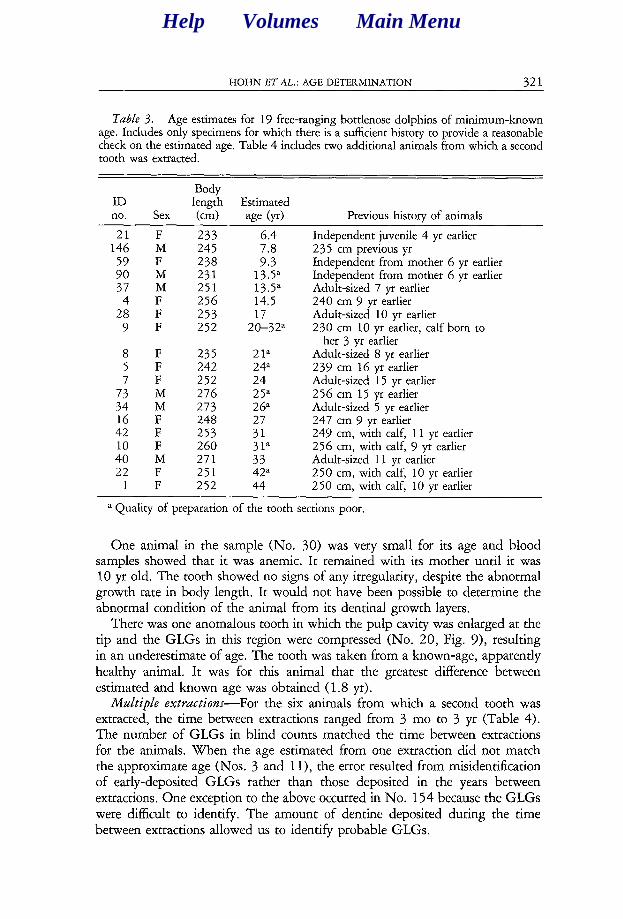
Table 3. Age estimates for 19 free-ranging bottlenose dolphins of minimum-known age. Includes only specimens for which there is a sufficient history to provide a reasonable check on the estimated age. Table 4 includes two additional animals from which a second tooth was extracted.
ID no. Sex
Body length Estimated km) age (yr) Previous history of animals
21 F 233 146 M 245
:: F M 238 231 37 M 251
4 F 256 28 F 253
9 F 252
6.4
::; 13.5” 13.5” 14.5 17
20-32”
8 F 235 21a 5 F 242 24a 7 F 252 24
:: M M 273 276 26” 25”
16 F 248 27 42 F 253 31 10 F 260 40 M 271 :ia 22 F 251 42”
1 F 252 44
Independent juvenile 4 yr earlier 235 cm previous yr Independent from mother 6 yr earlier Independent from mother 6 yr earlier Adult-sized 7 yr earlier 240 cm 9 earlier yr Adult-sized 10 yr earlier 230 cm 10 yr earlier, calf born to
her 3 yr earlier Adult-sized 8 yr earlier 239 cm 16 earlier yr Adult-sized 15 yr earlier 256 cm 15 earlier yr Adult-sized 5 yr earlier 247 cm 9 earlier yr 249 cm, with calf, 11 earlier yr 256 cm, with calf, 9 earlier yr Adult-sized 11 yr earlier 250 cm, with calf, 10 earlier yr 250 cm, with calf, 10 earlier yr
a Quality of preparation of the tooth sections poor.
One animal in the sample (No. 30) was very small for its age and blood samples showed that it was anemic. It remained with its mother until it was 10 yr old. The tooth showed no signs of any irregularity, despite the abnormal growth rate in body length. It would not have been possible to determine the abnormal condition of the animal from its dentinal growth layers.
There was one anomalous tooth in which the pulp cavity was enlarged at the tip and the GLGs in this region were compressed (No. 20, Fig. 9), resulting in an underestimate of age. The tooth was taken from a known-age, apparently healthy animal. It was for this animal that the greatest difference between estimated and known age was obtained (1.8 yr).
Multiple extractions- For the six animals from which a second tooth was extracted, the time between extractions ranged from 3 mo to 3 yr (Table 4). The number of GLGs in blind counts matched the time between extractions for the animals. When the age estimated from one extraction did not match the approximate age (Nos. 3 and 1 l), the error resulted from misidentification of early-deposited GLGs rather than those deposited in the years between extractions. One exception to the above occurred in No. 154 because the GLGs were difficult to identify. The amount of dentine deposited during the time between extractions allowed us to identify probable GLGs.
Help Volumes Main Menu

322 MARINE MAMMAL SCIENCE, VOL. 5, NO. 4, 1989
Figure 2. A good section of a bottlenose dolphin tooth. The section was cut on thebuccal-lingual, mid-longitudinal (“on-center”) plane rather than oblique to that plane(“off-center”), evident because the apices of the layers are pointed rather than round. Theneonatal line is well-defined, picking up little, if any, stain. Accessory layers are apparentbut not disruptively conspicuous. The boundary layer between the first and second GLGs
Help Volumes Main MenuHelp Volumes Main Menu
HOHN ET AZ,.: AGE DETERMINATION 323
The GLGs were easy to distinguish even in older animals in which the GLGs had become narrow (Figs. 14-17). The appearance of layers (GLG boundary layers as well as accessory layers) and of areas with different stainability was virtually identical (disregarding preparation variability) in the two teeth from each individual, except for the additional years’ growth.
One specimen was found dead and slightly decomposed. Although external identifying marks in the specimen were no longer visible, tooth number 15 was missing from the left lower jaw and we were able to help identify the individual using the dentinal layering pattern as a “fingerprint.”
Main s0zLyce.r of ewor in age estimates-The greatest absolute errors in all age classes were due to misinterpretation of layers and to poorly prepared sections, with the latter condition contributing greatly to the occurrence of the former. Misinterpretation occurred when prominent, narrow, stained layers (accessory layers, see Hohn, in press) or distinct boundaries between lightly and darkly stained zones within GLGs were erroneously taken to be boundary layers between GLGs (e.g., Fig. 3). The errors in age estimation in 3-4-yr-old specimens may be the result of too few GLGs having been deposited for the annual pattern to be clear, yet enough dentine having been deposited to cause confusion between accessory layers and GLG boundary layers. Even in well-prepared sections, accessory layers could be quite prominent (e.g., Figs. 7 and 13).
Poorly prepared sections most often influenced interpretation of layers when the orientation of the cut was other than mid-longitudinal along the buccal- lingual axis of the tooth. One difficulty in obtaining an on-center cut was that most of the teeth were curved in two directions, so that a cut that was on-center in one part of the tooth would be off-center somewhere else. A decision prior to cutting had to be made as to whether to minimize distortion near the tip of the tooth, which is more important for estimating age in younger animals, or near the pulp cavity, which may be more important for older animals. In some sections, probable GLGs could not be identified until we first determined the orientation of the cut.
When the cut was oblique to or laterally displaced from the mid-longitudinal axis (“off-center”), the GLGs were skewed, appearing wider than with a mid- longitudinal cut (because the layers are concentric, any orientation of cut other than through the center will produce a wider layer). The skewing accentuated the accessory layers, because they also become wider (e.g., Figs. 5 and 14b). The skewing or widening was most evident in the first 2 GLGs. In addition,
c is also not conspicuous, a condition common in many Tursiops and other delphinid teeth. The subsequent boundary layers between GLGs are well-defined. This tooth is from a male (No. 12) known to be 3.2-3.8 years old when the tooth was extracted. Symbols: NNL-neonatal line, numbers4LG number, where the GLGs defined represent one year’s growth. The bars in the postnatal dentine mark likely GLGs. The bar in the prenatal dentine marks the region at which the thickness of the prenatal zone was measured to use as a guide for identifying the first GLG (see Appendix). For GLG-thickness mea- surements see Table 5. The indentations on the lower, outer edges of the section occurred during tooth extraction.
Help Volumes Main Menu

324 MARINE MAMMAL SCIENCE, VOL. 5, NO. 4, 1989
Figure 3. Tooth section from a known-age, 2.3-yr-old female (No. 148). The bound-ary layer between GLGs 1 and 2 is unusually well-defined for this sample of bottlenosedolphins. A very distinct accessory layer appears near the center of the second GLG. SeeFigure 2 for explanation of symbols and Table 5 for GLG-thickness measurements.
Figure 4. Tooth section from a known-age, 2.5-yr-old female (No. 142). No GLGswere marked in this section because the correct GLGs were not obvious (although 2.5GLG-type layers can be seen, viewed from the edge of the pulp cavity towards the neonatalline). It is possible that in this animal, born in July, the distinctive layers were depositedduring the winter, 6 mo out of phase with the time of birth (see text for discussion). SeeFigure 2 for explanation of symbols.
in GLGs cut obliquely the upper portion (near the apices) appears concaverather than, as in mid-longitudinal buccal-lingual sections, straight or slightlyconvex on the convex side of the tip of the tooth and slightly concave on theconcave side of the tip of the tooth (illustrated in Figs. 2, 4, 6, and 13). Thecombination of wider-than-expected GLGs and enhanced accessory layers re-sulted in errors of misinterpreting layers. When the age estimate from thesesections was inaccurate, generally age was overestimated. Because of the curvatureof the tooth (with the pulp cavity also curving), within a tooth section some ofthe GLGs appeared on-center while others appeared off-center.
Teeth cut mid-longitudinally yet rotated sagittally relative to the buccal-lingual axis also caused errors, because the resulting GLGs were narrower thanwith a strictly buccal-lingual cut (because of the non-radially symmetric thinning
Help Volumes Main MenuHelp Volumes Main Menu

HOHN ET AL.: AGE DETERMINATION 325
Figure 5. Tooth section from a female (No. 15) known to be 2.2–2.8 yr old. Adistinctive accessory layer is apparent in about the center of the first GLG. The GLGsare relatively wide in this tooth, due somewhat to an off-center cut (note that tips ofGLGs are blunt rather than sharply pointed). See Figure 2 for explanation of symbolsand Table 5 for GLG-thickness measurements.
Figure 6. Tooth section from a known-age 3.0-yr-old female (No. 140). The GLGboundaries are relatively well-defined. There are numerous accessory layers, with someparticularly distinct about mid-way through the GLGs. There is a darkly stained narrowlayer immediately following the neonatal line. Although this animal was known to be3.0 yr old at the time the tooth was extracted, there is clearly additional dentinal depositionbeyond the apparent end of the third GLG. A new GLG appears to have been forming.See Figure 2 for explanation of symbols and Table 5 for GLG-thickness measurements.
at the tip of the tooth). The narrowing was most evident in the first 2 GLGs,with the other GLGs affected by having their upper portion (near the apices)appear convex. Errors in age estimation appeared to be a result of underestimatingdue to misinterpreting the end of the relatively narrow first GLG as an accessorylayer (e.g., Figs. 12 and l6b).
GLG pattern— The general pattern of GLGs in these bottlenose dolphin teethis similar to that described in teeth from other dolphins (e.g., from Stenella spp.,Myrick et al. 1983, Perrin and Myrick 1980). Although there is much individualvariation, there are characteristics, such as the appearance of boundary andaccessory layers and the relative widths of GLGs, that were useful for providingconsistency in age estimation, Absence of these guidelines would have producedan incorrect age estimate (e.g., Fig. 7). Conversely, adhering too strictly to the
Help Volumes Main MenuHelp Volumes Main Menu
326 MARINE MAMMAL SCIENCE, VOL. 5, NO. 4, 1989
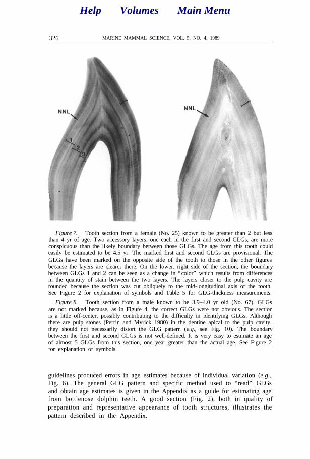
Figure 7. Tooth section from a female (No. 25) known to be greater than 2 but lessthan 4 yr of age. Two accessory layers, one each in the first and second GLGs, are moreconspicuous than the likely boundary between those GLGs. The age from this tooth couldeasily be estimated to be 4.5 yr. The marked first and second GLGs are provisional. TheGLGs have been marked on the opposite side of the tooth to those in the other figuresbecause the layers are clearer there. On the lower, right side of the section, the boundarybetween GLGs 1 and 2 can be seen as a change in “color” which results from differencesin the quantity of stain between the two layers. The layers closer to the pulp cavity arerounded because the section was cut obliquely to the mid-longitudinal axis of the tooth.See Figure 2 for explanation of symbols and Table 5 for GLG-thickness measurements.
Figure 8. Tooth section from a male known to be 3.9–4.0 yr old (No. 67). GLGsare not marked because, as in Figure 4, the correct GLGs were not obvious. The sectionis a little off-center, possibly contributing to the difficulty in identifying GLGs. Althoughthere are pulp stones (Perrin and Myrick 1980) in the dentine apical to the pulp cavity,they should not necessarily distort the GLG pattern (e.g., see Fig. 10). The boundarybetween the first and second GLGs is not well-defined. It is very easy to estimate an ageof almost 5 GLGs from this section, one year greater than the actual age. See Figure 2for explanation of symbols.
guidelines produced errors in age estimates because of individual variation (e.g.,Fig. 6). The general GLG pattern and specific method used to “read” GLGsand obtain age estimates is given in the Appendix as a guide for estimating agefrom bottlenose dolphin teeth. A good section (Fig. 2), both in quality ofpreparation and representative appearance of tooth structures, illustrates thepattern described in the Appendix.
Help Volumes Main MenuHelp Volumes Main Menu
HOHN ET AL.: AGE DETERMINATION 327
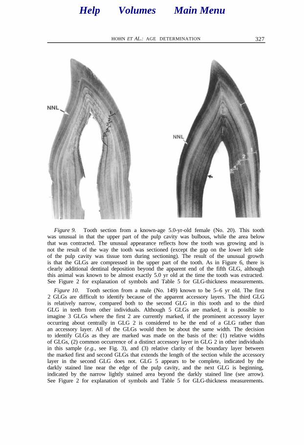
Figure 9. Tooth section from a known-age 5.0-yr-old female (No. 20). This toothwas unusual in that the upper part of the pulp cavity was bulbous, while the area belowthat was contracted. The unusual appearance reflects how the tooth was growing and isnot the result of the way the tooth was sectioned (except the gap on the lower left sideof the pulp cavity was tissue torn during sectioning). The result of the unusual growthis that the GLGs are compressed in the upper part of the tooth. As in Figure 6, there isclearly additional dentinal deposition beyond the apparent end of the fifth GLG, althoughthis animal was known to be almost exactly 5.0 yr old at the time the tooth was extracted.See Figure 2 for explanation of symbols and Table 5 for GLG-thickness measurements.
Figure 10. Tooth section from a male (No. 149) known to be 5–6 yr old. The first2 GLGs are difficult to identify because of the apparent accessory layers. The third GLGis relatively narrow, compared both to the second GLG in this tooth and to the thirdGLG in teeth from other individuals. Although 5 GLGs are marked, it is possible toimagine 3 GLGs where the first 2 are currently marked, if the prominent accessory layeroccurring about centrally in GLG 2 is considered to be the end of a GLG rather thanan accessory layer. All of the GLGs would then be about the same width. The decisionto identify GLGs as they are marked was made on the basis of the: (1) relative widthsof GLGs, (2) common occurrence of a distinct accessory layer in GLG 2 in other individualsin this sample (e.g., see Fig. 3), and (3) relative clarity of the boundary layer betweenthe marked first and second GLGs that extends the length of the section while the accessorylayer in the second GLG does not. GLG 5 appears to be complete, indicated by thedarkly stained line near the edge of the pulp cavity, and the next GLG is beginning,indicated by the narrow lightly stained area beyond the darkly stained line (see arrow).See Figure 2 for explanation of symbols and Table 5 for GLG-thickness measurements.
Help Volumes Main MenuHelp Volumes Main Menu

328 MARINE MAMMAL SCIENCE, VOL. 5, NO. 4, 1989
Figure 11. Tooth section from a male (No. 33) known to be 7.7–7.9 yr old. GLGsare countable but difficult to see. Numerous subannual incremental layers can be seenwithin many of the GLGs. The boundary layers of adjacent GLGs are different fromthose in many other individuals in that the darkly stained (i.e., relatively hypercalcified)layer is relatively narrow or absent. The prenatal zone, however, has stained similar tothat of other individuals, i.e., uniformly and relatively dark compared to much of thepostnatal dentine. The unusual appearance of the GLGs apical to the pulp cavity is dueto the orientation of the tooth when it was sectioned. See Figure 2 for explanation ofsymbols and Table 5 for GLG-thickness measurements.
Figure 12. Tooth section from a male (No. 155) known to be 12–14 yr old. Theunusual concave appearance at the apex of many of the GLGs results from the toothbeing rotated somewhat towards its sagittal axis during sectioning. This rotation facilitatesobtaining a section which contains as much of the center of the tooth as possible whenthe tooth is recurved along its longitudinal axis. As in many strictly mid-longitudinalsections, the boundary layers between GLGs are well-defined and the conspicuous accessorylayers about mid-way through the first and second GLGs are visible. Unlike sections cutoff-center, the layers are not skewed in such a way as to interfere with the correctidentification of GLGs. Possible errors in age estimation may result, however, because thefirst few GLGs are thinner than they would be if the section were mid-longitudinal. Inthis section, the accessory layer mid-way through the second GLG could be counted asthe end of GLG 1 and the end of GLG 2 could be counted as the end of GLG 3. Theresult would be an underestimate of age by 1 yr. The last, partial, GLG is barely visiblein this photograph. The bar indicating GLG 12 encompasses the last, partial GLG also.GLG 13 is marked, at the circle, at its widest point near the apex of the pulp cavity. Aportion of the cementum is indicated (C). See Figure 2 for explanation of other symbolsand Table 5 for GLG-thickness measurements.
Help Volumes Main MenuHelp Volumes Main Menu

HOHN ET AL.: AGE DETERMINATION 329
The thicknesses of temporally corresponding GLGs in longitudinal, on-center sections were similar among individuals of known and approximately known age (where we had the most certainty of identifying the correct GLGs) (Table 5). Differences in orientation of the cut (see above section on sources of error) affected the apparent thickness of layers. An example of differences in mea- surements can be seen in equivalent GLGs when two teeth were extracted from an individual (Table 6), especially when differences in the orientation of the cut can be observed (Figs. 14-17).
DISCUSSION
This sample of known-age, free-ranging dolphins has provided a unique opportunity to examine the annual layering pattern in teeth and verify methods for estimating age. During that process, we discovered some unexpected results in GLG deposition rates and patterns and were able to investigate the main sources of error in age estimates.
Because for most of the animals age estimates were the same as or close to the known or approximately known age, these results confirm the prior as- sumption that annual growth layers (= GLGs in this study) occur in the teeth of free-ranging bottlenose dolphins. In addition, most of the GLGs were easily identifiable.
Errors in age estimates were predominantly due to accessory layers which were enhanced by sections that were poorly prepared, suggesting that in many cases it is possible to minimize error by using only well-prepared sections. When sections appear to be poorly prepared, another tooth should be sectioned. For teeth curved in two directions, it can be helpful to prepare two teeth, to minimize distortion of GLGs near the tip of the tooth and near the pulp cavity, especially for old animals. When preparing another tooth is not possible, the degree of deviation of a section from the mid-longitudinal buccal-lingual plane should be considered when counting GLGs. We had only one tooth from most of the animals in our sample so we attempted to estimate age even when the quality of the preparation was poorer than we preferred.
The cause of deposition of GLGs is still unknown, An understanding of this process may help us determine the basis for individual variation in deposition of GLGs and the reason why for three animals we were unable to identify GLGs that corresponded to age. For two of the three (No. 142, Fig. 4 and No. 67, Fig. S), the correct number of GLGs can be identified if the first layer represents only half a year rather than a full year’s growth. All three animals were born in July. Three other animals born in July, however, appear to have a fully formed GLG following the neonatal line. The differences between estimated and known age for these specimens were 0.3, 1.2, and 1.5 yr, the latter two representing some of the greatest differences between known and estimated age.
One interesting and unexpected result was the occurrence of dentine after the end of the last complete GLG in teeth from three animals collected within a month of their date of birth. If GLGs begin at birth and the deposition rate of dentine remains constant throughout the year, these teeth would contain little,
Help Volumes Main Menu
330 MARINE MAMMAL SCIENCE, VOL. 5, NO. 4, 1989
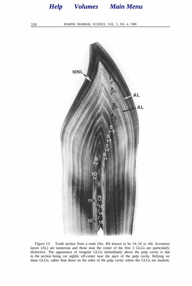
Figure 13. Tooth section from a male (No. 49) known to be 14–16 yr old. Accessorylayers (AL) are numerous and those near the center of the first 2 GLGs are particularlydistinctive. The appearance of irregular GLGs immediately above the pulp cavity is dueto the section being cut sightly off-center near the apex of the pulp cavity. Relying onthese GLGs, rather than those on the sides of the pulp cavity where the GLGs are marked,
Help Volumes Main MenuHelp Volumes Main Menu

HOHN ET AL.: AGE DETERMINATION 331
Figure 14. Tooth sections from a known-age female (No. 13) from which two teethwere extracted. The section on the left was extracted in June 1984 when the animal was3.8 yr old. The section on the right was extracted in January 1987 when the animal was6.2 yr old. The dashed, vertical line on the second tooth represents the time when thefirst tooth was extracted. The rounded apices of layers closer to the tip of the tooth andmore pointed layers closer to the pulp cavity show that the upper part of the latter sectionwas cut off-center. This orientation skews the GLGs and emphasizes accessory layers inthe section of the tooth that is off-center. Aside from artifacts due to preparation differences,the first GLGs in the two teeth are similar in pattern. See Figure 2 for explanation ofsymbols and Table 6 for GLG-thickness measurements,
if any, dentine forming the next GLG. All of the animals were born in summer,while, on the basis of the relative amount of dentine comprising the new GLG,the last complete GLG appeared to have ended the previous spring. It may bethat the onset of layering was triggered by factors other than the calf’s birthdate. For example, deposition might be influenced in the fetus by the physiologyof the mother or an increased light intensity during the spring. Alternatively,new GLGs may begin annually near the date of birth, with dentine depositedat a higher rate in summer than in winter.
could result in an overestimated age because accessory layers are visible and easily mistakenfor annual layers. See Figure 2 for explanation of other symbols and Table 5 for GLG-thickness measurements.
Help Volumes Main MenuHelp Volumes Main Menu
332 MARINE MAMMAL SCIENCE, VOL. 5, NO. 4, 1989
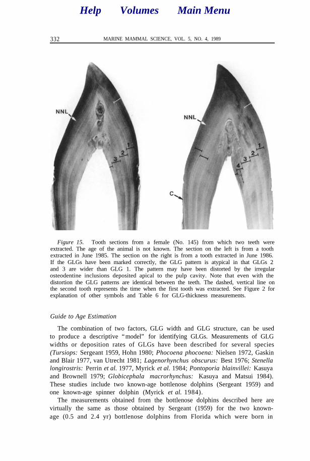
Figure 15. Tooth sections from a female (No. 145) from which two teeth wereextracted. The age of the animal is not known. The section on the left is from a toothextracted in June 1985. The section on the right is from a tooth extracted in June 1986.If the GLGs have been marked correctly, the GLG pattern is atypical in that GLGs 2and 3 are wider than GLG 1. The pattern may have been distorted by the irregularosteodentine inclusions deposited apical to the pulp cavity. Note that even with thedistortion the GLG patterns are identical between the teeth. The dashed, vertical line onthe second tooth represents the time when the first tooth was extracted. See Figure 2 forexplanation of other symbols and Table 6 for GLG-thickness measurements.
Guide to Age Estimation
The combination of two factors, GLG width and GLG structure, can be usedto produce a descriptive “model” for identifying GLGs. Measurements of GLGwidths or deposition rates of GLGs have been described for several species(Tursiops: Sergeant 1959, Hohn 1980; Phocoena phocoena: Nielsen 1972, Gaskinand Blair 1977, van Utrecht 1981; Lagenorhynchus obscurus: Best 1976; Stenellalongirostris: Perrin et al. 1977, Myrick et al. 1984; Pontoporia blainvillei: Kasuyaand Brownell 1979; Globicephala macrorhynchus: Kasuya and Matsui 1984).These studies include two known-age bottlenose dolphins (Sergeant 1959) andone known-age spinner dolphin (Myrick et al. 1984).
The measurements obtained from the bottlenose dolphins described here arevirtually the same as those obtained by Sergeant (1959) for the two known-age (0.5 and 2.4 yr) bottlenose dolphins from Florida which were born in
Help Volumes Main MenuHelp Volumes Main Menu
HOHN ET AL.: AGE DETERMINATION 333
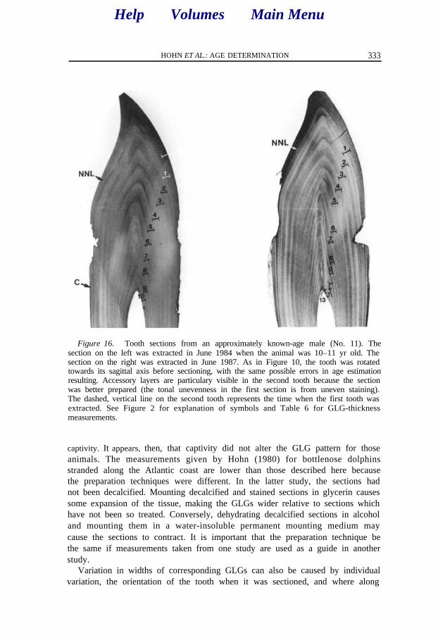
Figure 16. Tooth sections from an approximately known-age male (No. 11). Thesection on the left was extracted in June 1984 when the animal was 10–11 yr old. Thesection on the right was extracted in June 1987. As in Figure 10, the tooth was rotatedtowards its sagittal axis before sectioning, with the same possible errors in age estimationresulting. Accessory layers are particulary visible in the second tooth because the sectionwas better prepared (the tonal unevenness in the first section is from uneven staining).The dashed, vertical line on the second tooth represents the time when the first tooth wasextracted. See Figure 2 for explanation of symbols and Table 6 for GLG-thicknessmeasurements.
captivity. It appears, then, that captivity did not alter the GLG pattern for thoseanimals. The measurements given by Hohn (1980) for bottlenose dolphinsstranded along the Atlantic coast are lower than those described here becausethe preparation techniques were different. In the latter study, the sections hadnot been decalcified. Mounting decalcified and stained sections in glycerin causessome expansion of the tissue, making the GLGs wider relative to sections whichhave not been so treated. Conversely, dehydrating decalcified sections in alcoholand mounting them in a water-insoluble permanent mounting medium maycause the sections to contract. It is important that the preparation technique bethe same if measurements taken from one study are used as a guide in anotherstudy.
Variation in widths of corresponding GLGs can also be caused by individualvariation, the orientation of the tooth when it was sectioned, and where along
Help Volumes Main MenuHelp Volumes Main Menu
334 MARINE MAMMAL SCIENCE, VOL. 5, NO. 4, 1989
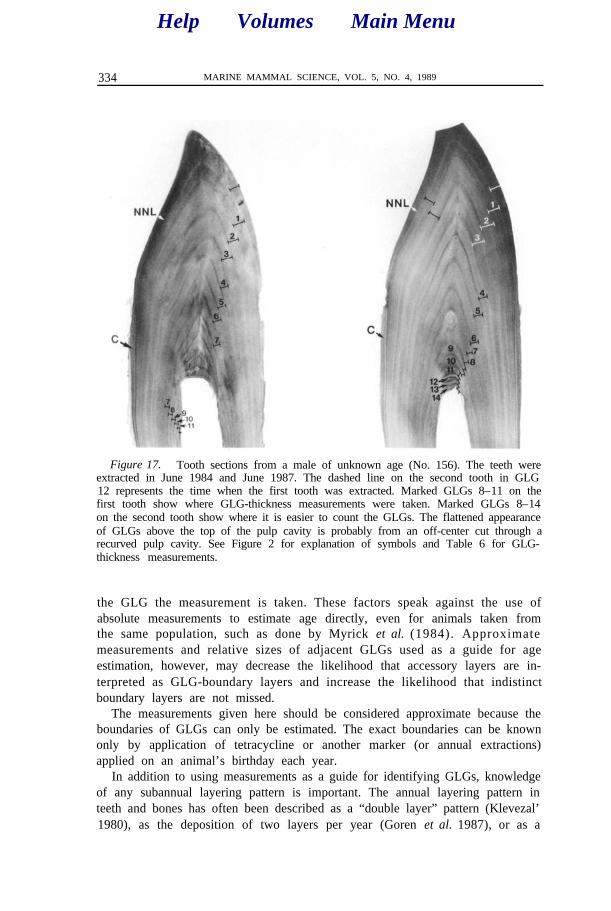
Figure 17. Tooth sections from a male of unknown age (No. 156). The teeth wereextracted in June 1984 and June 1987. The dashed line on the second tooth in GLG12 represents the time when the first tooth was extracted. Marked GLGs 8–11 on thefirst tooth show where GLG-thickness measurements were taken. Marked GLGs 8–14on the second tooth show where it is easier to count the GLGs. The flattened appearanceof GLGs above the top of the pulp cavity is probably from an off-center cut through arecurved pulp cavity. See Figure 2 for explanation of symbols and Table 6 for GLG-thickness measurements.
the GLG the measurement is taken. These factors speak against the use ofabsolute measurements to estimate age directly, even for animals taken fromthe same population, such as done by Myrick et al. (1984). Approximatemeasurements and relative sizes of adjacent GLGs used as a guide for ageestimation, however, may decrease the likelihood that accessory layers are in-terpreted as GLG-boundary layers and increase the likelihood that indistinctboundary layers are not missed.
The measurements given here should be considered approximate because theboundaries of GLGs can only be estimated. The exact boundaries can be knownonly by application of tetracycline or another marker (or annual extractions)applied on an animal’s birthday each year.
In addition to using measurements as a guide for identifying GLGs, knowledgeof any subannual layering pattern is important. The annual layering pattern inteeth and bones has often been described as a “double layer” pattern (Klevezal’1980), as the deposition of two layers per year (Goren et al. 1987), or as a
Help Volumes Main MenuHelp Volumes Main Menu

Table 4. Age estimates for free-ranging bottlenose dolphins from which a second tooth was extracted after a known time-interval following the first extraction. The second tooth from specimens 154 and 11 was taken after the animals were found beach-cast. Specimen 13 was of known age, 3 and 11 of approximately known age, and 154 and 156 of minimum known age, while 145 had no prior age data available.
ID no.
13 145 154 3 11 156
Figure no. 16 17 Date of birth (mo/yr) &, ” - 212-cm calf in 1976 190-cm calf in 1976 -
First tooth extraction: Date (mo/d/yr) 6/28/84 6/25/85 6/22/84 6/22/84 6/22/84 6/2 l/84 Body length (cm) 219 218 245 253 256 261 Estimated age (yr) -a 3.7 3.4 10.5 9.5b 11.5b Probable ageC (yr) 3.8 3.6 4.4 10.5 10.5 11.5
Second tooth extraction: Time between extractions 2.5 yr 1 Yr 0.4 yr 3 yr 3.1 yr Date (mo/d/yr) l/06/87 6/24/86 1 l/05/84 6/22/87 ;&l/87 :;;6,87 Body length (cm) 23t 220 252 265 263 Estimated age (yr) 4.8 4.8b 14.5 4.d 14.1 Probable ageC (yr) 2:; 4.6 4.8 13.5 13.5 14.5
a The first tooth collected from animal No. 13 was used to examine the growth layer pattern before the other teeth were read. The second tooth collected from this animal was read in the blind.
b Quality of preparation of the tooth sections poor. c Probable age was determined by examining the GLGs in both teeth with knowledge of the time between extractions after the original estimates
had been made. d The second tooth from this animal was received after the other teeth had been completed. Age was not estimated.
Help Volumes Main Menu
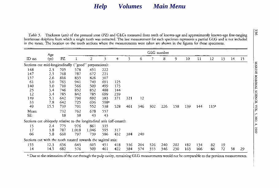
Table 5. Thickness (Km) of the prenatal zone (PZ) and GLGs measured from teeth of known-age and approximately known-age free-ranging bottlenose dolphins from which a single tooth was extracted. The last measurement for each specimen represents a partial GLG and is not included in the mean. The location on the tooth sections where the measurements were taken are shown in the figures for these specimens.
43 GLG number
ID no. (v) PZ 1 2 3 4 5 6 7 8 9 10 11 12 13 14 15
Sections cut mid-longitudinally (“good” preparations): 148 2.3 705 578 451 222 147 2.5 768 787 672 221 157 2.6 816 835 826 307 61 3.0 763 941 749 691 125 140 3.0 710 566 509 499 173 25 3.4 746 852 852 488 144 12 3.4 785 842 785 699 239
149 5.1 642 790 692 383 371 321 12 33 7.8 642 725 696 598" 49 15.5 739 701 552 538 528 461 346 302 226 158 139 144 115a
Mean: 732 762 678 557 SE: 18 38 43 43
Sections cut obliquely relative to the longitudinal axis (off-center):
15 2.4 775 976 861 335 17 3.8 787 1,018 1,046 595 317 06 5.8 660 797 739 586 432 384 240
Sections cut with the tooth rotated towards the sagittal axis:
155 12.3 656 643 605 451 418 336 264 326 240 202 182 134 14 14.5 682 576 509 461 422 384 374 355 346 230 163 106 2 :; 58 29
a Due to the orientation of the cut through the pulp cavity, remaining GLG measurements would not be comparable to the previous measurements.
Help Volumes Main Menu
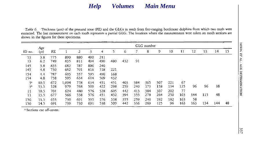
Table 6. Thickness (pm) of the prenatal zone (PZ) and the GLGs in teeth from free-ranging bottlenose dolphins from which two teeth were extracted. The last measurement on each tooth represents a partial GLG.. The locations where the measurements were taken on tooth sections are shown in the figures for these specimens.
Age GLG number
ID no. (yr) PZ 1 2 3 4 5 6 7 8 9 10 11 12 13 14 15
13 3.8 775 890 880 490 211 13 6.2 749 835 811 494 490 480 432 91
145 3.8 835 682 787 806 240 145 4.8 730 662 701 816 518 221 154 4.4 787 605 557 595 490 168 154 4.8 758 595 634 614 509 432
3” 10.5 672 1,094 778 634 451 451 403 384 365 307 221 67 3” 13.5 528 979 768 509 422 298 259 240 173 158 134 125 96 96 38
11 10.5 701 624 480 576 528 605 442 413 384 307 202 11 13.5 677 566 538 576 451 432 394 355 278 264 250 1:; 144 115 48
156 11.5 653 730 691 595 576 538 355 259 240 192 182 163 58 156 14.5 691 739 730 691 538 509 442 336 269 125 96 163 163 134 144 48
a Sections cut off-center.
Help Volumes Main Menu
338 MARINE MAMMAL SCIENCE, VOL. 5, NO. 4, 1989
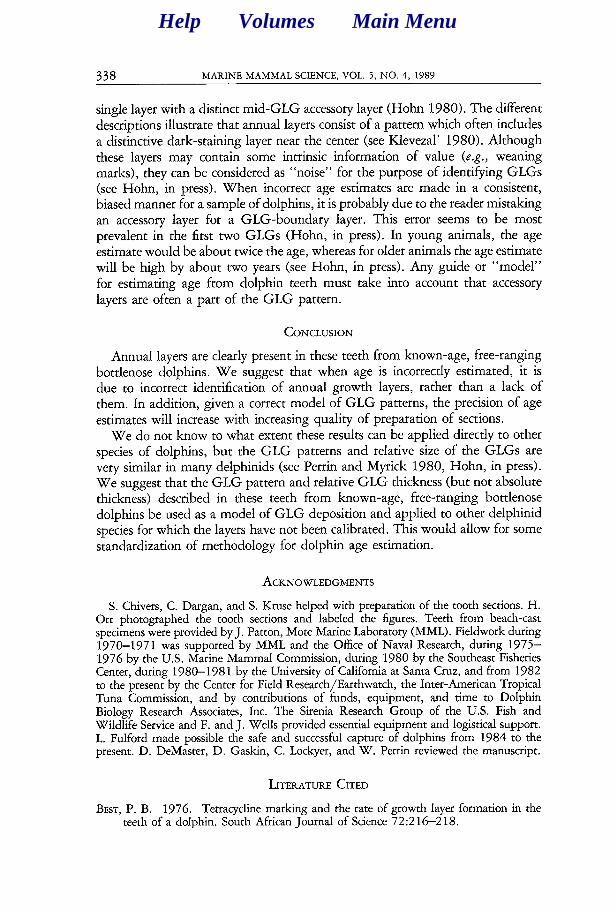
single layer with a distinct mid-GLG accessory layer (Hohn 1980). The different descriptions illustrate that annual layers consist of a pattern which often includes a distinctive dark-staining layer near the center (see Klevezal’ 1980). Although these layers may contain some intrinsic information of value (e.g., weaning marks), they can be considered as “noise” for the purpose of identifying GLGs (see Hohn, in press). When incorrect age estimates are made in a consistent, biased manner for a sample of dolphins, it is probably due to the reader mistaking an accessory layer for a GLG-boundary layer. This error seems to be most prevalent in the first two GLGs (Hohn, in press). In young animals, the age estimate would be about twice the age, whereas for older animals the age estimate will be high by about two years (see Hohn, in press). Any guide or “model” for estimating age from dolphin teeth must take into account that accessory layers are often a part of the GLG pattern.
CONCLUSION
Annual layers are clearly present in these teeth from known-age, free-ranging bottlenose dolphins. We suggest that when age is incorrectly estimated, it is due to incorrect identification of annual growth layers, rather than a lack of them. In addition, given a correct model of GLG patterns, the precision of age estimates will increase with increasing quality of preparation of sections.
We do not know to what extent these results can be applied directly to other species of dolphins, but the GLG patterns and relative size of the GLGs are very similar in many delphinids (see Perrin and Myrick 1980, Hohn, in press). We suggest that the GLG pattern and relative GLG thickness (but not absolute thickness) described in these teeth from known-age, free-ranging bottlenose dolphins be used as a model of GLG deposition and applied to other delphinid species for which the layers have not been calibrated. This would allow for some standardization of methodology for dolphin age estimation.
ACKNOWLEDGMENTS
S. Chivers, C. Dargan, and S. Kruse helped with preparation of the tooth sections. H. Orr photographed the tooth sections and labeled the figures. Teeth from beach-cast specimens were provided by J. Patton, Mote Marine Laboratory (MML). Fieldwork during 1970-1971 was supported by MML and the Office of Naval Research, during 1975- 1976 by the U.S. Marine Mammal Commission, during 1980 by the Southeast Fisheries Center, during 1980-1981 by the University of California at Santa Cruz, and from 1982 to the present by the Center for Field Research/Earthwatch, the Inter-American Tropical Tuna Commission, and by contributions of funds, equipment, and time to Dolphin Biology Research Associates, Inc. The Sirenia Research Group of the U.S. Fish and Wildlife Service and F. and J. Wells provided essential equipment and logistical support. L. Fulford made possible the safe and successful capture of dolphins from 1984 to the present. D. DeMaster, D. Gaskin, C. Lockyer, and W. Perrin reviewed the manuscript.
LITERATURE CITED
BEST, P. B. 1976. Tetracycline marking and the rate of growth layer formation in the teeth of a dolphin. South African Journal of Science 72:216-218.
Help Volumes Main Menu
HOHN ET AL.: AGE DETERMINATION 339
GASKIN, D. E., AND B. A. BLAIR. 1977. Age determination of harbour porpoise, Phocoena phocoena (L.), in the western North Atlantic. Canadian Journal of Zoology 55: 18-30.
GOREN, A. D., P. F. BRODIE, S. SPOtTE, G. C. RAY, H. W. KAUFMAN, A. J, GWINNETT, J. J. SCIUBBA AND J. D. BUCK. 1987. Growth layer groups (GLGs) in the teeth of an adult belukha whale (Delphinapterus leucas) of known age: evidence for two annual layers. Marine Mammal Science 3: 14-2 1.
GRUE, H., AND B. JENSEN. 1979. Review of the formation of incremental lines in tooth cementum of terrestrial mammals. Danish Review of Game Biology 11: 148.
HARRISON, R. J., R. L. BROWNELL, JR. AND R. C. BOICE. 1972. Reproduction and gonadal appearances in some odontocetes. Pages 362429 in R. J. Harrison, ed. Functional anatomy of marine mammals. Vol. 1. Academic Press, New York.
HOHN, A. A. 1980. Age determination and age-related factors in the teeth of western North Atlantic bottlenose dolphins. Scientific Reports of the Whales Research In- stitute, Tokyo 32:39-66.
HOHN, A. A. in press. Reading between the lines: an analysis of age estimation in dolphins. In S. Leatherwood and R. R. Reeves, eds. The bottlenose dolphin. Academic Press, New York.
HOHN, A. A., AND P. S. HAMMOND. 1985. Early postnatal growth of the spotted dolphins. Stenella attenuata, in the offshore eastern tropical Pacific. Fishery Bulletin 83:553-566.
HUI, C. A. 1980. Variability of dentin deposits in Tursiops truncatus. Canadian Journal of Fisheries and Aquatic Science 37:712-716.
IRVINE, A. B., AND R. S. WELLS. 1972. Results of attempts to tag Atlantic bottlenosed dolphins, Tursiops truncatus. Cetology 13: l-5.
IRVINE, A. B., M. D. Scott, R. S. WELLS AND J. H. KAUFMANN. 1981. Movements and activities of the Atlantic bottlenose dolphin, Tursiops truncatus, near Sarasota, Florida. Fishery Bulletin 79:67 l-688.
KASUYA, T., AND R. L. BROWNELL, JR. 1979. Age determination, reproduction, and growth of franciscana dolphin, Pontoporia blainvillei. Scientific Reports of the Whales Research Institute, Tokyo 3 1:45-67.
KASUYA, T., AND S. MATSUI. 1984. Age determination and growth of the short-finned pilot whale off the Pacific coast of Japan. Scientific Reports of the Whales Research Institute, Tokyo 3 5 : 5 7-9 1.
KLEVEZAL’, G. A. 1980. Layers in the hard tissues of mammals as a record of growth rhythms of individuals. Reports of the International Whaling Commission (Special Issue 3): 161-164.
KLEVEZAL’, G. A., AND S. E. KLEINENBERG. 1967. Opredelenie vozrasta mlekopita- yushchikh po sloistym strukturam zubov i kosti. Izdatel’stvo “Nauka,” Moscow, 144 pp. [Translation] 1969. Age determination of mammals from annual layers in teeth and bone. Israeli Program for Scientific Translation, Jerusalem, 128 pp.
MYRICK, A. C., JR., A. A. HOHN, P. A. SLOAN, M. KIMURA AND D. D. STANLEY. 1983. Estimating age of spotted and spinner dolphins (Stenella attenuata and Stenella longirostris) from teeth. National Oceanic and Atmospheric Administration Technical Report NMFS 30. 17 pp.
MYRICK, A. C., JR., E. W. SHALLENBERGER, I. KANG AND D. B. MACKAY. 1984. Cal- ibration of dental layers in seven captive Hawaiian spinner dolphins, Stenella Lon- girostris, based on tetracycline labeling. Fishery Bulletin 82:207-225.
NIELSEN, H. G. 1972. Age determination of the harbour porpoise Phocoena phocoena (L.) (Cetacea) Videnskabelige Meddelelser fra dansk Naturhistoric Forening 13 5 : 61-84.
PERRIN, W. F., D. B. HOLTS AND R. B. MILLER. 1977. Growth and reproduction of the eastern spinner dolphin, a geographical form of Stenella longirostris in the eastern tropical Pacific. Fishery Bulletin 75:725-750.
PERRIN, W. F., AND A. C. MYRICK, JR., EDS. 1980. Age determination of toothed whales
Help Volumes Main Menu
340 MARINE MAMMAL SCIENCE. VOL. 5. NO. 4. 1989
and sirenians. Reports of the International Whaling Commission (Special Issue 3), Cambridge. 229 pp.
RIDGWAY, S. H., R. F. GREEN AND J. C. SWEENEY. 1975. Mandibular anesthesia and tooth extraction in the bottlenosed dolphin. Journal of Wildlife Diseases 11:415- 418.
SERGEANT, D. E. 1959. Age determination in odontocete whales from dentinal growth layers. Norwegian Whaling Gazette 6:273-288.
SERGEANT, D. E., D. K. CALDWELL AND M. C. CALDWELL. 1973. Age, growth, and maturity of bottlenosed dolphin (Tursiops truncatus) from northeast Florida. Journal of the Fisheries Research Board of Canada 30:1009-1011.
UTRECHT, W. L. VAN. 1981. Comparison of accumulation patterns in layered dentinal tissue of some Odontoceti and corresponding patterns in baleen plates and ear plugs of Balaenopteridae. Beaufortia 3 1: 11 l-122.
WELLS, R. S. 1986. Structural aspects of dolphin societies. Ph.D. dissertation. University of California, Santa Cruz. 234 pp.
WELLS, R. S., A. B. IRVINE AND M. D. SCOTT. 1980. The social ecology of inshore odontocetes. Pages 263-318 in L. M. Herman, ed. Cetacean behavior: mechanisms and functions. Wiley-Interscience Press, New York.
WELLS, R. S., M. D. Scan AND A. B. IRVINE. 1987. The social structure of free-ranging bottlenose dolphins. Pages 247-305 in H. Genoways, ed. Current mammalogy. Vol. 1. Plenum Publishing Corp., New York.
Received: February 22, 1988 Accepted: June 8, 1989
Help Volumes Main Menu

HOHN ET AL.: AGE DETERMINATION 341
APPENDIX 1
READER’S GUIDE TO DOLPHIN TEETH
Documentation of the procedure used to estimate the age of known-age dolphins. Provided is a step-by-step guide to estimating age from mid-longitudinal sections of dolphin teeth which have been decalcified and stained. The user is required to have some basic knowledge of the structures found in dolphin teeth and terminology for those structures, a primer for which can be found in Perrin and Myrick (1980). See Figure 2 as a guide for the structures discussed.
Step-By-Step-Procedure Explanation
1. Examine the entire section, tip to base and on both sides of the pulp cavity, for the gen- eral GLG pattern for that spec- imen.
2. Find the buccal (convex) side of the section and the neo- natal line.
3. Identify a point about one- third the distance from the tip of the tooth to the base of the prenatal zone. (The base of the prenatal zone is where the neo- natal line converges with the outside edge of the tooth.)
4. Measure or approximate the thickness of the prenatal zone (perpendicular from the out- side edge of tooth to the neo- natal line) at this point.
5. Find an area in the post- natal dentine above the base of the prenatal zone where the thickness of the layers is rela- tively constant.
6. Use the previously deter- mioned prenatal zone thick- ness as a guide to find the end of the first GLG.
7. Look for the end of the first GLG as a thin, stained layer which may be readily visible only near the tip of the section or as an abrupt change in den- tinal color.
A. Getting started: Where is the first annual growth layer? (We will equate annual growth layer with a growth layer group, GLG, for this sample). This GLG is the most difficult and important to identify correctly. It is the most difficult because often (a) the boundary layer between the first and second GLGs is not distinct, and (b) there may be accessory layers near the centers of the first and second GLGs that are more distinct than the boundary layer between those GLGs. It is the most important because the subsequent GLGs are found rel- ative to it. It is helpful to start the process of age esti- mation by examining the entire section to become fa- miliar with the GLG pattern for that specimen.
A guideline for finding the first GLG is that the thickness of this layer is approximately the same thickness as the prenatal zone at a point in the prenatal zone about one- third the distance from the tip of the tooth to the base of the prenatal zone on the buccal (convex) side of the tooth. The first GLG is then determined in the upper part of the tooth in an area where the width does not change over a short distance, e.g., about half-way from the tip of the tooth to the base of the prenatal zone. The area chosen for these approximations is important since the prenatal zone is widest at the tip and narrows quickly as it approaches its base. In addition, in sections cut off-center or obliquely, the GLGs appear wider or narrower close to the tip of the tooth.
B. Finding the end of the first GLG: Ideally, and often, the boundary layers of the GLGs appear as darkly stained narrow layers. In the first GLG, this layer can be difficult to locate and it may appear almost undifferentiated from the surrounding dentine. Sometimes the boundary layer for the first GLG is readily apparent only in the upper part of the tooth section as a fine line or in the root of the tooth as an abrubt change in color resulting from
Help Volumes Main Menu
342 MARINE MAMMAL SCIENCE, VOL. 5, NO. 4, 1989
APPENDIX 1
Continued.
Step-By-Step-Procedure Explanation
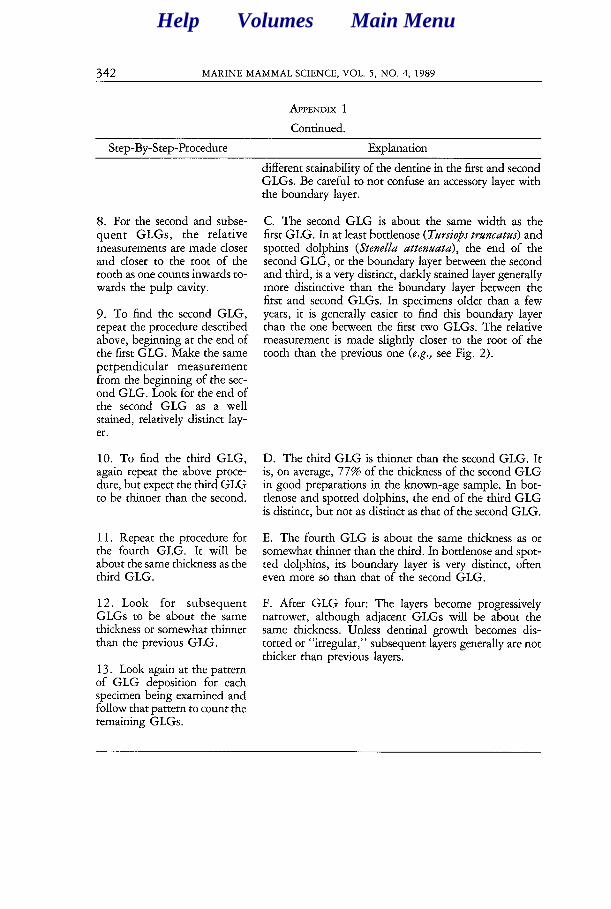
8. For the second and subse- quent GLGs, the relative measurements are made closer and closer to the root of the tooth as one counts inwards to- wards the pulp cavity.
9. To find the second GLG, repeat the procedure described above, beginning at the end of the first GLG. Make the same perpendicular measurement from the beginning of the sec- ond GLG. Look for the end of the second GLG as a well stained, relatively distinct lay- er.
C. The second GLG is about the same width as the first GLG. In at least bottlenose (Tursiops truncatus) and spotted dolphins (Stenella attenuata), the end of the second GLG, or the boundary layer between the second and third, is a very distinct, darkly stained layer generally more distinctive than the boundary layer between the first and second GLGs. In specimens older than a few years, it is generally easier to find this boundary layer than the one between the first two GLGs. The relative measurement is made slightly closer to the root of the tooth than the previous one (e.g., see Fig. 2).
10. To find the third GLG, again repeat the above proce- dure, but expect the third GLG to be thinner than the second.
D. The third GLG is thinner than the second GLG. It is, on average, 77% of the thickness of the second GLG in good preparations in the known-age sample. In bot- tlenose and spotted dolphins, the end of the third GLG is distinct, but not as distinct as that of the second GLG.
11. Repeat the procedure for E. The fourth GLG is about the same thickness as or the fourth GLG. It will be somewhat thinner than the third. In bottlenose and spot- about the same thickness as the ted dolphins, its boundary layer is very distinct, often third GLG. even more so than that of the second GLG.
12. Look for subsequent GLGs to be about the same thickness or somewhat thinner than the previous GLG.
F. After GLG four: The layers become progressively narrower, although adjacent GLGs will be about the same thickness. Unless dentinal growth becomes dis- torted or “irregular,” subsequent layers generally are not thicker than previous layers.
13. Look again at the pattern of GLG deposition for each specimen being examined and follow that pattern to count the remaining GLGs.
different stainability of the dentine in the first and second GLGs. Be careful to not confuse an accessory layer with the boundary layer.
Help Volumes Main Menu