Heavy metal toxicity of kidney and bone tissues in South Australian adult bottlenose dolphins (...
30
Accepted Manuscript Heavy metal toxicity of kidney and bone tissues in South Australian adult bot- tlenose dolphins (Tursiops aduncus) Trish J. Lavery, Catherine M. Kemper, Ken Sanderson, Christopher G. Schultz, Peter Coyle, James G. Mitchell, Laurent Seuront PII: S0141-1136(08)00214-6 DOI: 10.1016/j.marenvres.2008.09.005 Reference: MERE 3290 To appear in: Marine Environmental Research Received Date: 23 April 2008 Revised Date: 28 August 2008 Accepted Date: 25 September 2008 Please cite this article as: Lavery, T.J., Kemper, C.M., Sanderson, K., Schultz, C.G., Coyle, P., Mitchell, J.G., Seuront, L., Heavy metal toxicity of kidney and bone tissues in South Australian adult bottlenose dolphins (Tursiops aduncus), Marine Environmental Research (2008), doi: 10.1016/j.marenvres.2008.09.005 This is a PDF file of an unedited manuscript that has been accepted for publication. As a service to our customers we are providing this early version of the manuscript. The manuscript will undergo copyediting, typesetting, and review of the resulting proof before it is published in its final form. Please note that during the production process errors may be discovered which could affect the content, and all legal disclaimers that apply to the journal pertain. peer-00563057, version 1 - 4 Feb 2011 Author manuscript, published in "Marine Environmental Research 67, 1 (2008) 1" DOI : 10.1016/j.marenvres.2008.09.005
Transcript of Heavy metal toxicity of kidney and bone tissues in South Australian adult bottlenose dolphins (...

Accepted Manuscript
Heavy metal toxicity of kidney and bone tissues in South Australian adult bot-
tlenose dolphins (Tursiops aduncus)
Trish J. Lavery, Catherine M. Kemper, Ken Sanderson, Christopher G. Schultz,
Peter Coyle, James G. Mitchell, Laurent Seuront
PII: S0141-1136(08)00214-6
DOI: 10.1016/j.marenvres.2008.09.005
Reference: MERE 3290
To appear in: Marine Environmental Research
Received Date: 23 April 2008
Revised Date: 28 August 2008
Accepted Date: 25 September 2008
Please cite this article as: Lavery, T.J., Kemper, C.M., Sanderson, K., Schultz, C.G., Coyle, P., Mitchell, J.G.,
Seuront, L., Heavy metal toxicity of kidney and bone tissues in South Australian adult bottlenose dolphins (Tursiops
aduncus), Marine Environmental Research (2008), doi: 10.1016/j.marenvres.2008.09.005
This is a PDF file of an unedited manuscript that has been accepted for publication. As a service to our customers
we are providing this early version of the manuscript. The manuscript will undergo copyediting, typesetting, and
review of the resulting proof before it is published in its final form. Please note that during the production process
errors may be discovered which could affect the content, and all legal disclaimers that apply to the journal pertain.peer
-005
6305
7, v
ersi
on 1
- 4
Feb
2011
Author manuscript, published in "Marine Environmental Research 67, 1 (2008) 1" DOI : 10.1016/j.marenvres.2008.09.005

ACCEPTED MANUSCRIPT
1
Heavy metal toxicity of kidney and bone tissues in South Australian adult bottlenose
dolphins (Tursiops aduncus).
Trish J. Lavery a*, Catherine M. Kemperb, Ken Sandersona, Christopher G. Schultzc, Peter
Coyled, James G. Mitchella, Laurent Seuronta.
a School of Biological Sciences, Flinders University, GPO Box 2100, Adelaide, 5001.
b South Australian Museum, North Terrace, Adelaide, 5000.
c Nuclear Medicine, PET and Bone Densitometry, Royal Adelaide Hospital, North
Terrace, Adelaide, 5000.
d Institute of Medical and Veterinary Science, North Terrace, Adelaide, 5000.
Trish Lavery. Phone: 0011 6141 3031576. Fax: 0011 618 8201 3015. Email:
Trish Lavery
School of Biological Sciences
Flinders University of South Australia
GPO Box 2100
Adelaide, S.A., 5001
AUSTRALIA
peer
-005
6305
7, v
ersi
on 1
- 4
Feb
2011

ACCEPTED MANUSCRIPT
2
ABSTRACT
Metallothioneins (MT) concentration, renal damage, and bone malformations were
investigated in 38 adult Tursiops aduncus carcasses to determine any associations with
cadmium, copper, zinc, mercury, lead and selenium. Significantly higher concentrations
of cadmium, copper, and zinc in the liver were observed in dolphins showing evidence of
more advanced renal damage. No significant differences in metal or selenium
concentrations in the liver were observed between groups differing in level of bone
malformations. Some dolphins displayed evidence of toxicity and knowledge of metal
toxicity pathways were used to elucidate the cause of these abnormalities. Two dolphins
had high metal burdens, high MT concentrations, renal damage, and evidence of bone
malformations, indicating possible severe and prolonged metal toxicity. One dolphin
showed evidence of renal damage, but the lack of any other symptoms suggests that this
was unlikely to be caused by metal toxicity. We recommend examining a range of metal
toxicity symptoms simultaneously to aid in distinguishing metal toxicity from unrelated
aetiologies.
Keywords: Heavy Metals; Toxicity; Mammals; Dolphins; Metallothionein; Kidney;
Bone; Cadmium; Mercury; Lead; Zinc
1. INTRODUCTION
Metal concentrations have been recorded in over 60 species of cetaceans (O’Shea, 1999)
but few studies have explored the relationship between metals and toxicity. Metal
toxicity causes irregular metallothioneins (MT) protein synthesis, renal damage, and
peer
-005
6305
7, v
ersi
on 1
- 4
Feb
2011
ACCEPTED MANUSCRIPT
3
disruption of bone structure in humans and wildlife (Beiglbock et al., 2002; Jarup, 2002;
Sato and Kondoh, 2002). Studies of metal toxicity in cetaceans, particularly beach-
stranded carcasses, are hindered by difficulties arising from examining tissues that have
undergone post-mortem autolysis. Since these are usually the only specimens available,
it is important that methods of measuring metal toxicity in decomposed carcasses are
developed to allow determination of dolphin health, even in degraded specimens.
Metallothioneins (MT) are metal-binding proteins that preferentially bind to Cd
(cadmium) and Zn (zinc), reducing the bioavailability of these elements (Sato and
Kondoh, 2002; Das et al., 2006) and providing an early indicator of cellular responses to
metal toxicity (Petering et al., 1990). Although MT are often correlated with liver and
kidney metal burdens, few attempts have been made to correlate them with markers of
heavy metal toxicity in cetaceans. This is an absolute prerequisite to support the use of
MT overexpression as a bioindicator of metal toxicity.
Metals are toxic to mammalian renal cells (Wang and Pfeiffer, 2001), causing damage
that leads to leakage of phosphates, calcium, glycogen and proteins (proteinuria) from the
kidney (Loghman-Adham, 1997). Histological indicators of proteinuria have been used
as a preliminary measure of renal pathology in Tursiops aduncus in South Australia
(Long et al., 1997) but may have been impacted by post-mortem autolysis. In contrast,
metal-induced swelling of the Bowman’s capsule, and protein leakage within an intact
capsule, are not affected by post-mortem decomposition (Beiglbock et al., 2002).
peer
-005
6305
7, v
ersi
on 1
- 4
Feb
2011
ACCEPTED MANUSCRIPT
4
While MT and renal structure swelling may determine toxicity in mildly degraded
specimens, bone structure is resistant to post-mortem degradation. Loss of bone mineral
structure occurs in cases of Cd, Pb (lead), and Hg (mercury) toxicity in humans
(Escribano et al., 1997; Jarup, 2002; Suzuki et al., 2004) while increased and decreased
bone mineral density have been observed in response to Zn and Se (selenium) excess and
deficiency respectively (Turan et al., 2000; Yun and Zeng-Li, 2002). Loss of bone
density is a clinical endpoint marker of chronic metal toxicity and indicates
accompanying sub-clinical behavioural and immunological deficits (Staessen et al., 1999)
which are difficult to examine directly in free-living cetaceans.
The causes of elevated MT, renal damage, and loss of bone density and complexity are
probably interrelated. MT induced by metal toxicants form a large metal-MT complex
which leads to damage of renal structures. This results in leakage of calcium, phosphate
and proteins from the kidney, hindering bone remodelling and leading to a loss of bone
density (bone mineral density) and complexity (bone histomorphometry) (Alfven et al.,
2002). Thus, dolphins displaying metal-induced bone malformations should show
symptoms of elevated MT synthesis and damage of renal structures, assuming these
markers are reflecting metal toxicity and not extraneous influences (e.g., post-mortem
cellular degradation, unrelated parasitism, unrelated disease, normal mammalian ageing
process). Consistency between MT, renal damage and bone structure should provide a
valuable tool for helping to distinguish metal-induced toxicity from other aetiologies.
peer
-005
6305
7, v
ersi
on 1
- 4
Feb
2011

ACCEPTED MANUSCRIPT
5
South Australian adult Indo-Pacific bottlenose dolphins, Tursiops aduncus, have been
shown previously to have high concentrations of Cd, Hg, and Se in the liver, moderate
concentrations of Pb, Cu, and Zn in the liver and moderate concentrations of Cd and Pb
in bone compared to dolphins elsewhere (Lavery et al., 2008). This study aims to explore
any links between MT, renal damage, bone density and structure, and liver tissue
concentrations of Cd, Hg, Pb, Zn, Cu and Se in adult bottlenose dolphins.
2. MATERIALS AND METHODS
2.1 Necropsy procedures and sample selection
Necropsy procedures and determination of Se and metal concentrations by Inductively
Coupled Plasma Atomic Emission Spectrometry in 71 T. aduncus from South Australia
are detailed elsewhere (Lavery et al., 2008). All dolphins were collected by the South
Australian Museum (SAMA) and given a museum identification number. Some dolphins
had multiple analyses conducted on one tissue, and the mean metal or Se value was used
in these cases. Since animal age impacted metal accumulation in these dolphins, only a
subset of 38 sexually and / or physically mature adults of tooth category 3 and above (see
Kemper and Gibbs, 2001, for details of dolphin development compared to tooth category)
were examined in the present study. This sample of 38 adult dolphins consisted of 17
male and 21 female adult dolphins, 16 of which had kidney samples with intact nuclei
and glomeruli available for histopathological examination (Geraci and Lounsbury 1993,
decomposition code 2 and 3). Kidney (N = 15) and liver (N = 14) samples from a
subsample of these animals were analysed for MT. Bone density was measured in four
peer
-005
6305
7, v
ersi
on 1
- 4
Feb
2011

ACCEPTED MANUSCRIPT
6
vertebrae of 30 animals and the histomorphometry of costal rib tips examined from 15
animals.
2.2 Metallothionein quantification
Soft tissues were collected following procedures that minimise cross contamination
(Geraci and Lounsbury, 1993). Subsampling of frozen (-20 oC) tissue entailed collecting
approximately 0.5 g of liver and kidney for MT analysis. MT was quantified using the
modified cadmium-haemoglobin affinity assay (Eaton and Toal, 1982). Briefly, this
methodology requires adding radioactively labelled 109Cd to the tissue supernatant,
allowing it to bind to MT present. A ‘Cobra Auto-gamma’ counter (Packard Company)
was used to determine the Cd content of the resultant supernatant, which allows for
determination of the equivalent MT concentration, expressed as nmol of Cd bound g-1
wet weight.
2.3 Histological examination
Histology was carried out on 16 formalin-fixed kidney tissues: 15 samples were
previously frozen (-20 oC) and one fixed without prior freezing (SAMA: M21243).
Haematoxylin and eosin (H&E) preparations were carried out on each of the 16 kidney
samples using standard histological methodology (Brancroft and Stevens, 1982). An
Olympus BH-2 light microscope was used with 20× magnification. Digital microscope
images of 60 glomeruli and Bowman’s capsules observed were captured, and software
(Motic Images Plus, Version 2.0) was used to measure the area of 20 randomly selected
glomerulus and Bowman’s capsules from each slide. The area of the space between the
peer
-005
6305
7, v
ersi
on 1
- 4
Feb
2011

ACCEPTED MANUSCRIPT
7
glomeruli and Bowman’s capsule, and the presence of proteins within each Bowman’s
capsule was recorded.
2.4 Quantification of bone density and structure
Bone strength depends on the amount of bone and the organisational structure of the
trabeculae (Kleerekoper et al., 1985), so two measures of bone density were obtained;
Dual energy X-ray Absorptiometry (DXA) and histomorphometry. DXA measures the
total amount of bone (cortical and trabecular bone), but skeletal metal stores can bias
results (Puzas et al., 2002). Histomorphometry directly quantifies the 3-dimensional
micro-architectural structure and organisation of bone and is not biased by metal burden.
DXA scans were performed on a GE Lunar Prodigy Vision Dual X-ray Absorptiometry,
GE Lunar Madison, Wisconsin. Four caudal vertebrae from each of 30 animals were
analysed to obtain measurements of bone mineral density, BMD (g cm-2), and bone
mineral content, BMC (g). Caudal vertebrae were selected on the basis of being the most
posterior four caudal vertebrae with small transverse processes. Small animal software
(version 8.10.027) on standard settings was used to examine a maximum area of 25 × 25
cm. Vertebrae were laid longitudinally to best simulate the longitudinal stress placed on
the caudal region of the spine by powerful dorsal and ventral muscles associated with
swimming. Bones were laid on a 1 cm thick Lucite tissue equivalent block and scanned
with parameters set at 76 kilovolts and 150 �A. Pixel size was 1.05 × 0.6 mm. BMD and
BMC of the vertebrae and posterior process (but not the transverse processes) were
determined.
peer
-005
6305
7, v
ersi
on 1
- 4
Feb
2011
ACCEPTED MANUSCRIPT
8
Histomorphometry was performed using a Skyscan 1072 X-ray Microtomograph on the
largest costal rib available from each of 15 animals. A piece 2 cm long was sampled
from the largest end of the bone and placed in the X-ray Microtomograph with the cut
facing down. Pixel size was 18.95 �m while the X-ray parameters were set at 16×
magnification, 100 kilovolts and 98 �A. A ring artefact correction was used and a beam
hardening correction of 100% was set. Cone reconstruction software was used to
reconstruct the 3-dimensional structure of the bone from 1,000 separate 2-dimensional
cross section images. Bone density parameters from the 3-dimensional reconstructions
were calculated using CTan software (Skyscan). Density parameters calculated by CTan
are shown in Table 1.
2.5 Statistical analyses
Our primary aim was to determine if metal concentrations were associated with markers
of toxicity. To examine the influence of metal concentrations on kidney structures a
hierarchical cluster analysis (data not shown) was employed which sorted 14 individuals
into two groups based on their concentrations of MT and their levels of renal toxicity.
Between groups linkage was used to designate groups based on all available information
and squared Euclidian distances were used to derive proximity measurements. Values
were transformed using Z scores due to variables being measured on different scales.
Once groups were designated, an independent samples t-test was then used to determine
any significant differences in log transformed metal concentrations, dolphin length, or
blubber thickness between the two groups. This process was then repeated, using cluster
peer
-005
6305
7, v
ersi
on 1
- 4
Feb
2011
ACCEPTED MANUSCRIPT
9
analysis to sort bone structure variables of 15 individuals into two groups, and a t-test to
determine differences in log transformed metal concentrations, dolphin length, and
blubber thickness between the groups. All analyses were conducted using SPSS version
14.0.
2.6 Qualitative analyses
Our second aim was to examine the datasets for outlier animals with extreme levels of
toxicity which could be examined to shed light on the aetiology of the observed toxicity.
Three outlier animals were clearly identifiable from scatter plots (data not shown) and
examined. We assume here that metal toxicity aetiology will be highlighted by
consistency along the metal toxicity pathway (high metal concentrations, high MT
concentrations, advanced renal damage and advanced bone malformations). We assume
one of these indicators alone suggests an aetiology that is unrelated to metal toxicity.
3. RESULTS
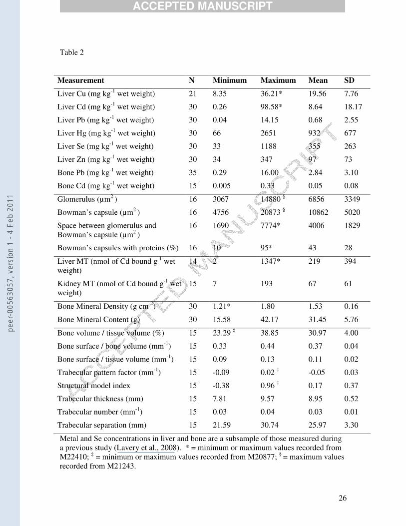
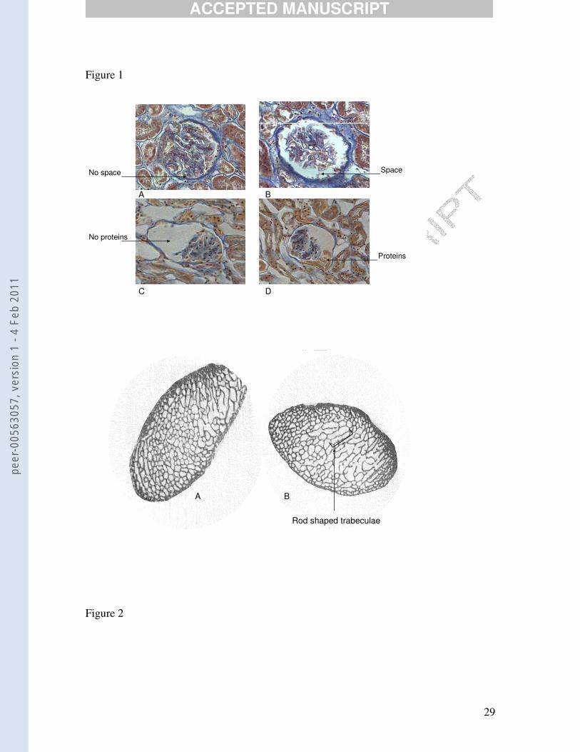
High levels of variation among individuals were observed in all toxicity markers (Table
2) and qualitative differences among animals were observed for histological indicators
(Figure 1) and costal rib histomorphometry (Figure 2).
3.1 Renal damage
Cluster analysis classified individuals into two groups based on differences in their
measured renal parameters (renal structure swelling, protein leakage, and MT
peer
-005
6305
7, v
ersi
on 1
- 4
Feb
2011

ACCEPTED MANUSCRIPT
10
concentrations). Renal Group 1 had low scores for indicators of renal damage, whereas
Renal Group 2 was characterised by high levels of renal damage, with advanced renal
structure (glomerulus, Bowman’s capsule, and the space between glomerulus and
Bowman’s capsule) swelling, higher levels of protein leakage, and higher MT (liver and
kidney) concentrations (Table 3). Renal Group 2 had significantly higher levels of Cd (p
= 0.045), Cu (p = 0.047) and Zn (p = 0.047) than Renal Group 1. No significant
difference in dolphin length or blubber thickness existed between the groups (p > 0.05).
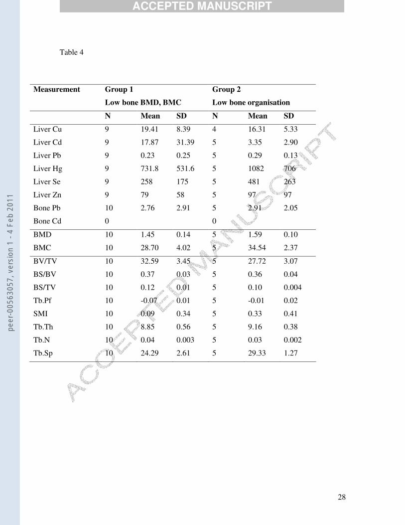
3.2 Bone malformations
Classifications based on bone parameters were less clear than those based on renal
parameters. Bone Group 1 was defined by lower spinal bone mineral density whereas
Bone Group 2 had lower levels of rib bone trabecular organisation (Table 4). Metal
concentrations, animal length, and blubber thickness were not significantly different
between the groups (p > 0.05).
3.3 Consistency among toxicity markers
Three dolphins showed extreme values in one or more of the measured markers (MT,
liver histology, bone density or bone organisation). A 2 m female dolphin (SAMA:
M22410) from lower Spencer Gulf showed consistency among the measured parameters.
This dolphin had the lowest recorded spinal bone mineral density, highest tissue
concentrations of Cd and Cu (Table 2), moderately high levels of Zn in the liver (209.35
mg kg-1), the highest concentration of liver MT, the largest area between Bowman’s
peer
-005
6305
7, v
ersi
on 1
- 4
Feb
2011

ACCEPTED MANUSCRIPT
11
capsule and glomeruli, the highest percentage of protein leakage within the Bowman’s
capsule (Table 2), the second highest Bowman’s capsule area (19477 �m2) and third
highest glomerulus area (11703 �m2) recorded. Examination of the skeleton revealed
serious malformations in the lumbar-caudal region. The last lumbar vertebra had no
transverse processes on the right side. The first caudal vertebra had no transverse
processes at all, while the second and third caudal vertebrae were missing transverse
processes on the left side. Neural spines were thickened on the second, third and fourth
caudal vertebrae.
A 2.1 m male dolphin (SAMA: M20877) from upper Spencer Gulf had the lowest
recorded bone complexity values (BV/TV, Tb.Pf, SMI) (Table 2) and also showed
consistency among measured parameters. This male dolphin had the second highest
recorded scores for concentrations of liver MT (876 nmol of Cd bound g-1 wet weight),
glomerulus area (11922 �m2), and area of space between Bowman’s capsule and
glomerulus (6675 �m2). It had the third largest Bowman’s capsule area (18597 �m2), and
second highest recorded liver Zn burden (270 mg kg-1).
A 2.3 m male (SAMA: M21243) from Gulf St Vincent had the highest recorded
glomerulus and Bowman’s capsule areas (Table 2) but this was not consistent with other
measured parameters. Values for tissue metal burdens and other markers of toxicity were
very close to the mean and there were no other signs of toxicity in this dolphin.
peer
-005
6305
7, v
ersi
on 1
- 4
Feb
2011

ACCEPTED MANUSCRIPT
12
4. DISCUSSION
4.1 Renal damage
Concentrations of Cu and Cd in the liver were significantly higher in the cluster of
animals defined by high levels of renal damage. Cadmium is a known nephrotoxicant
(Wang and Pfeiffer, 2001). Exposure to Cd is known to alter Cu metabolism (Peraza et
al., 1998) and the close association between Cd, Cu and renal damage in the present study
raises the possibility that alteration of Cu metabolism may be a mechanism for the
observed damage. The relationship between MT and renal damage provides support for
the use of MT overexpression as a suitable biomarker of Cd-induced renal damage in
cetaceans.
The cluster defined by high levels of renal damage also had significantly higher levels of
hepatic Zn. Zinc is an essential metal but it can be toxic in high concentrations (Walsh et
al., 1994). Large anthropogenic quantities of Zn (> 30,000 kg) are released into the
marine environment annually in Spencer Gulf, South Australia (NEPC, 2003), and in the
long term could result in unusually high Zn accumulation in dolphin tissues and toxic
impacts. The significance of renal structure swelling has not been studied in cetaceans
specifically; however, laboratory mammal and human studies have found an increase in
annual mortality risk of 40 – 80% in human patients with metal-induced renal pathology
(Iwata et al., 1992; Nakagawa et al., 1993; Nishijo et al., 1999).
4.2 Bone malformations
peer
-005
6305
7, v
ersi
on 1
- 4
Feb
2011
ACCEPTED MANUSCRIPT
13
The bone demineralising effects of environmental contaminants are well documented.
East Greenland polar bear and Baltic grey seal populations show evidence of bone
mineral disruption associated with high body burdens of organochlorine contaminants
(Lind et al., 2003; Sonne et al., 2004). While laboratory studies suggest that metals also
influence bone structure and mineralisation (Suzuki et al., 2004), associations between
metals and bone in wildlife studies are less readily observed (Sonne-Hansen et al., 2002;
although see Beiglbock et al., 2002)
Rather than sorting individuals based on high or low levels of bone damage, cluster
analysis sorted dolphins into groups based on different types of bone organisation. Bone
Group 1 was characterised by low levels of spinal bone density and Bone Group 2 were
characterised by low levels of rib bone trabecular organisation. Perhaps due to this
method of clustering, and despite the known impacts of metals on bone, there were no
significant differences in metal concentrations between groups, suggesting metals may be
exerting no influence on bone structure in this study (although see sections 4.3 below).
Alternatively, it is known that Pb preferentially accumulates in the skeleton, leading to
overestimates of bone mineral density and complexity (Puzas et al., 2002). Thus, animals
with high bone mineral density scores here may be biased by bone metal content.
Interpretation of these results is hindered by the fact that measurements were taken in
different bones. Histomorphometry was measured in peripheral rib bones while bone
mineral density was measured in the axial spinal skeleton. The axial skeleton is more
sensitive to the effects of toxicants than the peripheral skeleton (Laan et al., 1993).
peer
-005
6305
7, v
ersi
on 1
- 4
Feb
2011

ACCEPTED MANUSCRIPT
14
Future increases in microtomograph scanning size may allow for quantification of spinal
histomorphometry and would aid in clarifying the relationship between metals, bone
density, and bone organisation.
4.3 Consistency among toxicity markers
Three dolphins had evidence of toxicity and these animals were examined more closely
to reveal possible aetiologies. A 2.3 m male (SAMA: M21243) from Gulf St Vincent had
high levels of renal structure swelling but no other markers of toxicity suggesting this
animal was probably not suffering from metal toxicity, but from an undetermined renal
condition.
A female dolphin from Spencer Gulf (SAMA: M20877) had high Cd and Cu
concentrations in the liver, MT overexpression, renal damage and extreme bone
malformations. While difficult to prove, the consistency amongst toxicity markers may
suggest this dolphin was suffering from severe metal toxicity. This animal had a tooth
category of five, indicating that it should be both physically and sexually mature (Kemper
and Gibbs, 1997). However, examination of ovaries and skeleton revealed that this
animal was physically and sexually immature. It is possible, therefore, that the high
levels of metal toxicants have delayed sexual and physical maturity in this animal.
However, due to the non-experimental nature of these observations, it is impossible to
rule out the presence of an extraneous influence that either caused the observed
pathology, or rendered this dolphin more susceptible to the effects of metal toxicity.
peer
-005
6305
7, v
ersi
on 1
- 4
Feb
2011

ACCEPTED MANUSCRIPT
15
A 2.1 m male dolphin (SAMA: M20877) from upper Spencer Gulf with high Zn tissue
concentrations showed decreased bone complexity, high concentrations of liver MT, and
renal structure swelling. This dolphin had a tooth category of 5 and was sexually and
physically mature (Kemper and Gibbs, 1997). The consistency between toxicity markers
suggests this dolphin may have been suffering from metal toxicity. Zinc toxicity in
humans occurs above a threshold level of 465 mg kg-1 dry weight (Anon, 1998), which is
well below the dry weight Zn concentration of 999 mg kg-1 recorded in this animal.
The relationship between metals, toxicity of the kidney, and bone malformations, has
been demonstrated in experimental and natural studies of humans and wildlife (Staessen
et al., 1999; Alfven et al., 2002; Jarup and Alfven, 2004). A study of 100 ringed seals
(Phoca hispida) in Greenland, however, found no relationship between cadmium, degree
of bone mineralisation, and nephropathy (Sonne-Hansen et al., 2002) and the authors
concluded that high dietary intakes of vitamin D and calcium (among other minerals) in
these ringed seals may protect seals from cadmium toxicity. However, the sample of
ringed seals consisted mainly of animals younger than 10 years. Cadmium-induced renal
damage typically occurs only after 10 years exposure (Friberg et al., 1986) and the effect
of Cd on bone is more pronounced in older bone (Alfven et al., 2002). While accurate
ages of the bottlenose dolphins examined here are unknown, all are tooth category 3 and
above, and would be older than 10 years (Kemper and Gibbs, 2001).
Experimental studies of metal toxicity in dolphins are undesirable, necessitating the use
of natural experiments to infer relationships between health and toxicants. Natural
peer
-005
6305
7, v
ersi
on 1
- 4
Feb
2011
ACCEPTED MANUSCRIPT
16
experiments must contend with a lack of experimental control and the presence of
confounding factors (e.g., reproductive status, nutritive condition etc). Results obtained
from natural studies must be viewed cautiously but can provide important information
that can guide future research despite the absence of controlled experimental data. The
influence of age on metal accumulation (Lavery et al., 2008) and bone mineral density
(Butti et al., 2007) have been minimised in the present study by including only adult
animals but not eliminated as knowledge of the accurate ages of each dolphin included in
this study is not known.
There was a large hiatus between the highest Cd levels (M22410, 98.58 mg kg-1) which
seem to be associated with toxicity, and the second highest (M21231, 22 mg kg-1) which
showed no association with toxicity. Thus, toxicity thresholds for T. aduncus could not be
determined. Future discoveries of such intermediate Cd tissue burdens in South
Australian T. aduncus may allow for the establishment of a toxicity threshold which will
aid in interpreting the relevance of metal concentrations already determined throughout
the world (see O’Shea, 1999, for a review).
5. CONCLUSION
Higher metal concentrations were found in a group of animals suffering from renal
damage. No differences in metal concentrations were found between groups sorted on
the basis of bone density and organisation. Knowledge of the toxicological pathway of
metals can aid in distinguishing symptoms of metal toxicity from other, unrelated
aetiologies. While only adult dolphins were examined here, the influence of fine scale
peer
-005
6305
7, v
ersi
on 1
- 4
Feb
2011

ACCEPTED MANUSCRIPT
17
changes in dolphin age and undetected pathologies represent a methodological limitation
in carcasses studies such as this. Extraneous influences are difficult to determine or
overcome but we suggest that, by examining a range of metal toxicity symptoms
concurrently, future studies can better allow for determination of whether observed health
deficits are related to metal toxicity, or an unrelated aetiology.
6. ACKNOWLEDGEMENTS
Funding was provided by Flinders University, University of Adelaide, and Environmental
Protection Agency, Adelaide. Carcass collection, management, and necropsies were
carried out by staff and volunteers from the South Australian Museum, including Lynette
Queale, Bob Hamilton-Bruce, David Stemmer, Ross Goble, Sue Gibbs, Martine Long
and members of the Dolphin Trauma Group. Officers of the National Parks and Wildlife
SA and Department of Primary Industries Fisheries are thanked for collecting many of
the carcasses. Martine Long and Nicole Butterfield were instrumental in laying the
groundwork for this project by their preliminary work on metal burdens in South
Australian marine mammals. Sam Gaylard was extremely helpful in ensuring that the
results of previous researchers were available for inclusion in this study. Ben Roudnew
provided invaluable assistance with collecting and preparing samples. Helpful advice
and assistance was given by Mike Bossley, Michelle Lewis, Robert Reid, Roger Byard,
Nick Fazzalari, Helen Beard, Ian Parkinson, John Stirling, John Edwards, David Duncan,
Peter Self, Brian Woodward, Alan McLennan, Kylie Lange, Nardi Cribb and Karen
peer
-005
6305
7, v
ersi
on 1
- 4
Feb
2011
ACCEPTED MANUSCRIPT
18
Stockin. Anonymous reviewers greatly improved the quality of earlier manuscripts. We
are greatly indebted to all these people.
peer
-005
6305
7, v
ersi
on 1
- 4
Feb
2011
ACCEPTED MANUSCRIPT
19
REFERENCES
Alfven, T., Jarup, L., Elinder, C.G., 2002. Cadmium and lead in blood in relation to low bone mineral density and tubular proteinuria. Environmental Health Perspectives, 110, 699–702. Anon., 1998. National irrigation water quality program. Information report No. 3, US Department of Interior. Beiglbock, C., Steineck, T., Tataruch, F., Ruf, T., 2002. Environmental cadmium induces histopathological changes in kidneys of roe deer. Environmental Toxicology and Chemistry, 21, 1811 – 1816. Brancroft, J., Stevens, A., 1982. Theory and practice of histological techniques. 2nd Edition. New York: Churchill-Livingstone. Butti, C., Corain, L., Cozzi, B., Podesta, M., Pirone, A., Affronte, M., Zotti, A., 2007. Age estimation in the Mediterranean bottlenose dolphin Tursiops truncatus (Montagu 1821) by bone density of thoracic limb. Journal of Anatomy, 211, 639 – 646. Das, K., De Groff, A., Jauniaux, T., Bouquegneau J.-M., 2006. Zu, Cu, Cd and Hg binding to metallothioneins in harbour porpoises Phocoena phocoena from the southern North Sea. BMC Ecology, 6, 2. Eaton, D.L., Toal, B.F., 1982. Evaluation of the Cd/hemoglobin affinity assay for the rapid determination of metallothionein in biological tissues. Toxicology and Applied Pharmacology, 66, 134 – 142. Escribano, A., Revilla, M., Hernandez, E.R., Seco, C., Gonzalez-Riola, J., Villa, L.F., Rico, H., 1997. Effect of lead on bone development and bone mass: A morphometric, densitometric, and histomorphometric study in growing rats. Calcified Tissue International, 60, 200 – 203. Friberg, L., Elinder, C.-G., Kjellstrom, T., Nordberg, G.F., 1986. Cadmium and health: A toxicological and epidemiological appraisal, vol. II. Florida: CRC Press Inc. Geraci, J.R., Lounsbury, V.J., 1993. Marine Mammals Ashore: A field guide for strandings. Texas: A&M Sea Grant Publications. Iwata, H., Saito, H., Moriyama, M., Nakano, A., 1992. Follow up study of renal tubular dysfunction and mortality among residents of an area polluted with cadmium. British Journal of Industrial Medicine, 49, 736 – 737. Jarup, L., 2002. Cadmium overload and toxicity. Nephrology Dialysis Transplantation, 17, 35 – 39.
peer
-005
6305
7, v
ersi
on 1
- 4
Feb
2011

ACCEPTED MANUSCRIPT
20
Jarup, L., Alfven, T., 2004. Low level cadmium exposure, renal and bone effects – the OSCAR study. Biometals, 17, 505 – 509. Kemper C.M., and Gibbs, S.E, 1997. A study of the life history parameters of dolphins and seals entangled in tuna farms near Port Lincoln, and comparisons with information from other South Australian dolphin carcasses. Report to Environment Australia, pp. 98. Kemper, C., Gibbs, S., 2001. Dolphin interactions with tuna feedlots at Port Lincoln, South Australia and recommendations for minimising entanglements. Journal of Cetacean Research and Mangement, 3, 283 – 292. Kleerekoper, M., Villanueva, A.R., Stanciu, J., Sudhaker Rao, D., Parfitt, A.M., 1985. The role of three-dimensional trabecular microstructure in the pathogenesis of vertebral compression fractures. Calcified Tissue International, 37, 594 – 597. Laan, R.F.J.M., Buijs, W.C.A.M., van Erning, L.J.T.O., Lemmens, J.A.M., Corstens, F.H.M., Ruijs, S.H.J., van de Putte, L.B.A., van Riel, P.L.C.M., 1993. Differential effects of glucocorticoids on cortical appendicular and cortical vertebral bone mineral content. Calcified Tissue International, 52, 5 – 9. Lavery, T.J., Butterfield, N., Kemper, C.M., Reid, R.J., Sanderson, K., 2008. Metals and selenium in the liver and bone of three dolphin species from South Australia, 1988 – 2004. Science of the Total Environment, 390, 77 – 85. Lind, P.M., Bergman, A., Olsson, M., Orberg, J., 2003. Bone mineral density in male Baltic grey seal. Ambio, 32, 385 – 388. Loghman-Adham, M., 1997. Renal effects of environmental and occupational lead exposure. Environmental Health Perspectives, 105, 928 – 938. Long, M., Reid, R.J., Kemper, C.M., 1997. Cadmium accumulation and toxicity in the bottlenose dolphin, the common dolphin, and some dolphin prey species in South Australia. Australian Mammalogy, 20, 25 – 33. Nakagawa, H., Nishijo, M., Morikawa, Y., Tabata, M., Senma, M., Kitagawa, Y., Kawano, S., Ishizaki, M., Sugita, N., Nishi, M., Kido, T., Nogawa, K., 1993. Urinary B2- microglobulin and mortality in a cadmium polluted area. Archives of Environmental Health, 48, 428 – 435. NEPC, National Environment Protection Council., 2003. The national pollutant inventory. A public database on pollutant emissions. Environment Australia. Nishijo, M., Nakagawa, H., Morikawa, M., Tabata, M., Miura, T., Yoshita, K., Higasgiguchi, K., Seto, T., Kido, T., Nogawa, K., Mizukoshi, K., Nishi, M., 1999.
peer
-005
6305
7, v
ersi
on 1
- 4
Feb
2011

ACCEPTED MANUSCRIPT
21
Relationship between urinary cadmium and mortality among inhabitants living in a cadmium polluted area in Japan. Toxciology Letters, 108, 321 – 327. O’Shea, T.J., 1999. Environmental contaminants and marine mammals. (pp. 485 – 564) In: Reynolds, III J.E., Rommel, S.A., editors. Biology of Marine Mammals. Melbourne: Melbourne University Press. Peraza, M.A., Ayala-Fierro, F., Barber, D.S., Casarez, E., Rael, L.T., 1998. Effects of micronutrients on metal toxicity. Environmental Health Perspectives, 106, 203 – 216. Petering, D.H., Goodrich, M., Hodgman, W., Krezoski, S., Weber, D., Shaw III, C.F., Spieler, R., Zettergren, L., 1990. Metal-binding proteins and peptides for the detection of heavy metals in aquatic organisms, In: McCarthy, J.F., Shugart, L.R., Biomarkers of Environmental Contamination, Florida: Lewis Publishers. Puzas, E., Campbell, J., O'Keefe, R.J., Schwarz, E.M., Rosier, R.N., 2002. Lead in the skeleton interferes with bone mineral density measurements. Journal of Bone and Mineral Research, 17, S314. Sato, M., Kondoh, M., 2002. Recent studies on metallothionein: Protection against toxicity of heavy metals and oxygen free radicals. Tohoku Journal of Experimental Medicine, 196, 9 – 22. Sonne, C., Dietz, R., Born, E.W., Riget, F.F., Kirkegaard, M., Hyldstrup, L., Letcher, R.J., Muir, D.C.G., 2004. Is bone mineral composition disrupted by organochlorines in East Greenland polar bears (Ursus maritimus)? Environmental Health Perspectives, 112, 1711 – 1716. Sonne-Hansen, C., Dietz, R., Leifsson, P.S., Hyldstrup, L., Riget, F.F., 2002. Cadmium toxicity to ringed seals (Phoca hispida) – an epidemiological study of possible cadmium induced nephropathy and osteodystrophy in ringed seals (Phoca hispida) from Qaanaaq in Northwest Greenland. Science of the Total Environment, 295, 167 – 181. Staessen, J.A., Roels, H.A., Emelianov, D., Kuznetsova, T., Thijs, L., Vangronsveld, J., Fagard, R., 1999. Environmental exposure to cadmium, forearm bone density and risk of fractures: prospective population study. Lancet, 353, 1140 – 1144. Suzuki, N., Yamamoto, M., Watanabe, K., Kambegawa, A., Hattori, A., 2004. Both mercury and cadmium directly influence calcium homeostasis resulting from suppression of scale bone cells: The scale is a good model for the evaluation of heavy metals in bone metabolism. Journal of Bone and Mineral Research, 22, 439 – 446. Turan, B., Bayari, S., Balcik, C., Severcan, F., Akkas, N., 2000. A biomechanical and spectroscopic study of bone from rats with selenium deficiency and toxicity. BioMetals, 13, 113 – 121.
peer
-005
6305
7, v
ersi
on 1
- 4
Feb
2011
ACCEPTED MANUSCRIPT
22
Walsh, C.T., Sandstead, H.H., Prasad, A.S., Newberne, P.M., Fraker, P.J., 1994. Zinc: Health effects and research priorities for the 1990s. Environmental Health Perspectives, 102, Suppl 2, 5 – 46. Wang, A., Pfeiffer, C.J., 2001. Cytopathology induced by mercuric chloride and methylmercury in cultured renal cells of Atlantic spotted dolphin (Stenella plagiodon). Journal of Submicroscopic Cytology & Pathology, 33, 7 – 16. Yun, L., Zeng-Li, Y., 2002. Effect of zinc on bone metabolism in fetal mouse limb culture. Biomedical and Environmental Science, 15, 323 – 329.
peer
-005
6305
7, v
ersi
on 1
- 4
Feb
2011
ACCEPTED MANUSCRIPT
23
Figure Captions.
Table 1 Description of trabecular microarchitectural parameters acquired by
histomorphometric analyses.
Table 2 Copper, cadmium, lead, mercury, selenium and zinc concentrations in the
liver, cadmium and lead concentrations in the bone, MT (metallothioneins)
concentrations in liver and kidney, and markers of health (renal damage and bone
structure) measured in adult Indo-Pacific bottlenose dolphin (Tursiops aduncus)
carcasses collected from South Australia, 1989 – 2004.
Table 3 Copper, cadmium, lead, mercury, selenium and zinc concentrations in the
liver, cadmium and lead concentrations in the bone, and renal parameters measured in
two groups of adult bottlenose dolphins (Tursiops aduncus) differentiated by cluster
analysis on the basis of renal damage.
Table 4 Copper, cadmium, lead, mercury, selenium and zinc concentrations in the
liver, cadmium and lead concentrations in the bone, and bone parameters measured in
two groups of adult bottlenose dolphins (Tursiops aduncus) in two groups differentiated
on the basis of bone structure.
Figure 1 Examples of renal parameters measured in adult bottlenose dolphin
(Tursiops aduncus) carcasses collected from South Australia. A: no space between
glomeruli and surrounding Bowman’s capsule. B: enlarged space between the glomeruli
and surrounding Bowman’s capsule. C: enlarged space between glomeruli and
Bowman’s capsule with no proteins enclosed. D: glomeruli with protein leakage inside
the Bowman’s capsule.
peer
-005
6305
7, v
ersi
on 1
- 4
Feb
2011

ACCEPTED MANUSCRIPT
24
Figure 2 Cross sectional image of two costal ribs of adult bottlenose dolphins
(Tursiops aduncus) generated by �CT (×16) showing trabecular microarchitecture. A:
SAMA M21282 had low liver cadmium (0.49 mg kg-1) and plate shaped trabeculae. B:
SAMA M22410 had high liver cadmium (98.58 mg kg-1) and rod shaped trabeculae
characteristic of osteoporosis.
peer
-005
6305
7, v
ersi
on 1
- 4
Feb
2011

ACCEPTED MANUSCRIPT
25
Table 1
Name Abbreviation Description References.
Bone volume /
tissue volume
BV/TV Trabecular bone volume relative to marrow
volume (%).
(Parfitt et al.,
1983)
Bone surface /
bone volume
BS/BV Bone surface to bone volume ratio. Describes
complexity (mm-1)
(Parfitt et al.,
1983)
Bone surface /
total bone
BS/TB Total surface of the bone over total amount of
bone (mm-1).
(Parfitt et al.,
1983)
Trabecular
thickness
Tb.Th Mean dimensions of individual trabeculae
(mm).
(Parfitt et al.,
1983)
Trabecular
separation
Tb.Sp Thickness of the marrow cavities between
trabeculae (mm).
(Parfitt et al.,
1983)
Trabecular
pattern factor
Tb.Pf Measures trabecular bone connectivity (mm-
1).
(Hahn et al.,
1992)
Trabecular
number
Tb.N The number of traversals across trabeculae
(mm-1).
(Parfitt et al.,
1983)
Structural
model index
SMI Relative frequency of rods and plates (no
units).
(Hildebrand and
Ruegsegger,
1997)
peer
-005
6305
7, v
ersi
on 1
- 4
Feb
2011
ACCEPTED MANUSCRIPT
26
Table 2
Measurement N Minimum Maximum Mean SD
Liver Cu (mg kg-1 wet weight) 21 8.35 36.21* 19.56 7.76
Liver Cd (mg kg-1 wet weight) 30 0.26 98.58* 8.64 18.17
Liver Pb (mg kg-1 wet weight) 30 0.04 14.15 0.68 2.55
Liver Hg (mg kg-1 wet weight) 30 66 2651 932 677
Liver Se (mg kg-1 wet weight) 30 33 1188 355 263
Liver Zn (mg kg-1 wet weight) 30 34 347 97 73
Bone Pb (mg kg-1 wet weight) 35 0.29 16.00 2.84 3.10
Bone Cd (mg kg-1 wet weight) 15 0.005 0.33 0.05 0.08
Glomerulus (µm2 ) 16 3067 14880 § 6856 3349
Bowman’s capsule (µm2 ) 16 4756 20873 § 10862 5020
Space between glomerulus and Bowman’s capsule (µm2 )
16 1690 7774* 4006 1829
Bowman’s capsules with proteins (%) 16 10 95* 43 28
Liver MT (nmol of Cd bound g-1 wet weight)
14 2 1347* 219 394
Kidney MT (nmol of Cd bound g-1 wet weight)
15 7 193 67 61
Bone Mineral Density (g cm-2) 30 1.21* 1.80 1.53 0.16
Bone Mineral Content (g) 30 15.58 42.17 31.45 5.76
Bone volume / tissue volume (%) 15 23.29 ‡ 38.85 30.97 4.00
Bone surface / bone volume (mm-1) 15 0.33 0.44 0.37 0.04
Bone surface / tissue volume (mm-1) 15 0.09 0.13 0.11 0.02
Trabecular pattern factor (mm-1) 15 -0.09 0.02 ‡ -0.05 0.03
Structural model index 15 -0.38 0.96 ‡ 0.17 0.37
Trabecular thickness (mm) 15 7.81 9.57 8.95 0.52
Trabecular number (mm-1) 15 0.03 0.04 0.03 0.01
Trabecular separation (mm) 15 21.59 30.74 25.97 3.30
Metal and Se concentrations in liver and bone are a subsample of those measured during a previous study (Lavery et al., 2008). * = minimum or maximum values recorded from M22410; ‡ = minimum or maximum values recorded from M20877; § = maximum values recorded from M21243.
peer
-005
6305
7, v
ersi
on 1
- 4
Feb
2011

ACCEPTED MANUSCRIPT
27
Table 3
Measurement Renal Group 1
Low renal damage
Renal Group 2
High renal damage
N Mean SD N Mean SD
Liver Cu * 9 16.02 5.55 2 29.72 9.18
Liver Cd * 11 4.55 6.27 3 37.00 53.33
Liver Pb 11 0.17 0.13 3 0.12 0.09
Liver Hg 11 800 637 3 1359 687
Liver Se 11 291 255 3 446 175
Liver Zn * 11 73 37 3 178 111
Bone Pb 11 2.85 2.83 3 1.47 0.46
Bone Cd 4 0.10 0.16 0
Glomerulus 11 5546 1461 3 12835 1774
Bowman’s capsule 11 9065 2657 3 19649 1148
Space between glomerulus and Bowman’s capsule
11 3519 1279 3 6814 898
Protein leakage 11 43 28 3 52 38
Liver MT 11 76 66 3 745 678
Kidney MT 11 60 58 3 101 81
* = metal concentrations are significantly different between groups (p < 0.05) peer
-005
6305
7, v
ersi
on 1
- 4
Feb
2011
ACCEPTED MANUSCRIPT
28
Table 4
Measurement Group 1
Low bone BMD, BMC
Group 2
Low bone organisation
N Mean SD N Mean SD
Liver Cu 9 19.41 8.39 4 16.31 5.33
Liver Cd 9 17.87 31.39 5 3.35 2.90
Liver Pb 9 0.23 0.25 5 0.29 0.13
Liver Hg 9 731.8 531.6 5 1082 706
Liver Se 9 258 175 5 481 263
Liver Zn 9 79 58 5 97 97
Bone Pb 10 2.76 2.91 5 2.91 2.05
Bone Cd 0 0
BMD 10 1.45 0.14 5 1.59 0.10
BMC 10 28.70 4.02 5 34.54 2.37
BV/TV 10 32.59 3.45 5 27.72 3.07
BS/BV 10 0.37 0.03 5 0.36 0.04
BS/TV 10 0.12 0.01 5 0.10 0.004
Tb.Pf 10 -0.07 0.01 5 -0.01 0.02
SMI 10 0.09 0.34 5 0.33 0.41
Tb.Th 10 8.85 0.56 5 9.16 0.38
Tb.N 10 0.04 0.003 5 0.03 0.002
Tb.Sp 10 24.29 2.61 5 29.33 1.27
peer
-005
6305
7, v
ersi
on 1
- 4
Feb
2011
ACCEPTED MANUSCRIPT
29
Figure 1
A
D
B
C
No space
No proteins
Space
Proteins
A B
Rod shaped trabeculae
Figure 2
peer
-005
6305
7, v
ersi
on 1
- 4
Feb
2011