The claustrum of the bottlenose dolphin Tursiops truncatus (Montagu 1821)
8
SYSTEMS NEUROSCIENCE ORIGINAL RESEARCH ARTICLE published: 28 March 2014 doi: 10.3389/fnsys.2014.00042 The claustrum of the bottlenose dolphin Tursiops truncatus (Montagu 1821) Bruno Cozzi 1 *, Giulia Roncon 1 , Alberto Granato 2 , Maristella Giurisato 1 , Maura Castagna 3 , Antonella Peruffo 1 , Mattia Panin 1 , Cristina Ballarin 1 , Stefano Montelli 1 and Andrea Pirone 4 1 Department of Comparative Biomedicine and Food Science, University of Padova, Legnaro, Italy 2 Department of Psychology, Catholic University, Milan, Italy 3 Department of Translational Research on New Technologies in Medicine and Surgery, University of Pisa, Pisa, Italy 4 Department of Veterinary Sciences, University of Pisa, Pisa, Italy Edited by: Brian N. Mathur, University of Maryland School of Medicine, USA Reviewed by: Preston E. Garraghty, Indiana University, USA Stefan Huggenberger, University of Cologne, Germany *Correspondence: Bruno Cozzi, Department of Comparative Biomedicine and Food Science, University of Padova, Viale dell’Università 16, 35020 Legnaro, Italy e-mail: [email protected] The mammalian claustrum is involved in processing sensory information from the environment. The claustrum is reciprocally connected to the visual cortex and these projections, at least in carnivores, display a clear retinotopic distribution. The visual cortex of dolphins occupies a position strikingly different from that of land mammals. Whether the reshaping of the functional areas of the cortex of cetaceans involves also modifications of the claustral projections remains hitherto unanswered. The present topographic and immunohistochemical study is based on the brains of eight bottlenose dolphins and a wide array of antisera against: calcium-binding proteins (CBPs) parvalbumin (PV), calretinin (CR), and calbindin (CB); somatostatin (SOM); neuropeptide Y (NPY); and the potential claustral marker Gng2. Our observations confirmed the general topography of the mammalian claustrum also in the bottlenose dolphin, although (a) the reduction of the piriform lobe modifies the ventral relationships of the claustrum with the cortex, and (b) the rotation of the telencephalon along the transverse axis, accompanied by the reduction of the antero-posterior length of the brain, apparently moves the claustrum more rostrally. We observed a strong presence of CR-immunoreactive (-ir) neurons and fibers, a diffuse but weak expression of CB-ir elements and virtually no PV immunostaining. This latter finding agrees with studies that report that PV-ir elements are rare in the visual cortex of the same species. NPY- and somatostatin-containing neurons were evident, while the potential claustral markers Gng2 was not identified in the sections, but no explanation for its absence is currently available. Although no data are available on the projections to and from the claustrum in cetaceans, our results suggest that its neurochemical organization is compatible with the presence of noteworthy cortical inputs and outputs and a persistent role in the general processing of the relative information. Keywords: claustrum, bottlenose dolphin, calcium binding proteins, NPY, somatostatin, insular cortex INTRODUCTION Dolphins (Delphinidae) are carnivore marine mammals belong- ing to the Infraorder Odontoceti (toothed whales). Together with the Infraorder Mysticeti (baleen whales) toothed whales belong to the taxon Cetacea which is today grouped together with Artio- dactyla (even-toed hooved mammals) into the single order Cetar- tiodactyla (Geisler and Uhen, 2005; Thewissen et al., 2007). This relationship may explain some of their anatomical conformities and common evolutionary adaptations with domestic animals like the cow and the pig (for general reference see Slijper, 1979). However the brain of dolphins possesses some strikingly unique features, including compression of the longitudinal axis and expansion of the temporal width, pronounced rotation along the transverse (inter-insular) axis, essential absence of the olfactory lobe and nerves, intense folding of the cerebral cortex accompa- nied by reduced thickness, and general uniformity of columnar organization in the different topographical areas. On the other hand, the virtual absence of a typical layer IV in cetaceans (for a comprehensive review see Manger, 2006) is common also to other Cetartiodactyla and Ungulates in general (Hof et al., 1999, 2000). Physiological studies on brain functions and internal connections are obviously restricted by ethical reasons and by the conse- quent limited number of published studies. However, a review of the available information suggests that functional localizations differ from terrestrial mammals (Oelschläger and Oelschläger, 2009). The visual cortex is not located in the occipital pole, but shifted dorsally and placed longitudinally, separated from the inter-hemispheric scissure by the peculiar paralimbic lobe, and accompanied laterally by the elongated acoustic cortex (Lende and Akdikmen, 1968; Kesarev and Malofeeva, 1969; for review see Morgane et al., 1986). Motor and somatosensory cortices are pushed rostrally almost entirely on the frontal aspect of the Frontiers in Systems Neuroscience www.frontiersin.org March 2014 | Volume 8 | Article 42 | 1
-
Upload
uni-hannover -
Category
Documents
-
view
0 -
download
0
Transcript of The claustrum of the bottlenose dolphin Tursiops truncatus (Montagu 1821)
SYSTEMS NEUROSCIENCEORIGINAL RESEARCH ARTICLE
published: 28 March 2014doi: 10.3389/fnsys.2014.00042
The claustrum of the bottlenose dolphin Tursiopstruncatus (Montagu 1821)Bruno Cozzi1*, Giulia Roncon1, Alberto Granato2, Maristella Giurisato1, Maura Castagna3,Antonella Peruffo1, Mattia Panin1, Cristina Ballarin1, Stefano Montelli1 and Andrea Pirone4
1 Department of Comparative Biomedicine and Food Science, University of Padova, Legnaro, Italy2 Department of Psychology, Catholic University, Milan, Italy3 Department of Translational Research on New Technologies in Medicine and Surgery, University of Pisa, Pisa, Italy4 Department of Veterinary Sciences, University of Pisa, Pisa, Italy
Edited by:Brian N. Mathur, University ofMaryland School of Medicine, USA
Reviewed by:Preston E. Garraghty, IndianaUniversity, USAStefan Huggenberger, University ofCologne, Germany
*Correspondence:Bruno Cozzi, Department ofComparative Biomedicine and FoodScience, University of Padova, Vialedell’Università 16, 35020 Legnaro, Italye-mail: [email protected]
The mammalian claustrum is involved in processing sensory information from theenvironment. The claustrum is reciprocally connected to the visual cortex and theseprojections, at least in carnivores, display a clear retinotopic distribution. The visual cortexof dolphins occupies a position strikingly different from that of land mammals. Whetherthe reshaping of the functional areas of the cortex of cetaceans involves also modificationsof the claustral projections remains hitherto unanswered. The present topographic andimmunohistochemical study is based on the brains of eight bottlenose dolphins and a widearray of antisera against: calcium-binding proteins (CBPs) parvalbumin (PV), calretinin (CR),and calbindin (CB); somatostatin (SOM); neuropeptide Y (NPY); and the potential claustralmarker Gng2. Our observations confirmed the general topography of the mammalianclaustrum also in the bottlenose dolphin, although (a) the reduction of the piriform lobemodifies the ventral relationships of the claustrum with the cortex, and (b) the rotationof the telencephalon along the transverse axis, accompanied by the reduction of theantero-posterior length of the brain, apparently moves the claustrum more rostrally. Weobserved a strong presence of CR-immunoreactive (-ir) neurons and fibers, a diffusebut weak expression of CB-ir elements and virtually no PV immunostaining. This latterfinding agrees with studies that report that PV-ir elements are rare in the visual cortexof the same species. NPY- and somatostatin-containing neurons were evident, while thepotential claustral markers Gng2 was not identified in the sections, but no explanation forits absence is currently available. Although no data are available on the projections to andfrom the claustrum in cetaceans, our results suggest that its neurochemical organizationis compatible with the presence of noteworthy cortical inputs and outputs and a persistentrole in the general processing of the relative information.
Keywords: claustrum, bottlenose dolphin, calcium binding proteins, NPY, somatostatin, insular cortex
INTRODUCTIONDolphins (Delphinidae) are carnivore marine mammals belong-ing to the Infraorder Odontoceti (toothed whales). Together withthe Infraorder Mysticeti (baleen whales) toothed whales belongto the taxon Cetacea which is today grouped together with Artio-dactyla (even-toed hooved mammals) into the single order Cetar-tiodactyla (Geisler and Uhen, 2005; Thewissen et al., 2007). Thisrelationship may explain some of their anatomical conformitiesand common evolutionary adaptations with domestic animalslike the cow and the pig (for general reference see Slijper, 1979).However the brain of dolphins possesses some strikingly uniquefeatures, including compression of the longitudinal axis andexpansion of the temporal width, pronounced rotation along thetransverse (inter-insular) axis, essential absence of the olfactorylobe and nerves, intense folding of the cerebral cortex accompa-nied by reduced thickness, and general uniformity of columnar
organization in the different topographical areas. On the otherhand, the virtual absence of a typical layer IV in cetaceans (for acomprehensive review see Manger, 2006) is common also to otherCetartiodactyla and Ungulates in general (Hof et al., 1999, 2000).Physiological studies on brain functions and internal connectionsare obviously restricted by ethical reasons and by the conse-quent limited number of published studies. However, a reviewof the available information suggests that functional localizationsdiffer from terrestrial mammals (Oelschläger and Oelschläger,2009). The visual cortex is not located in the occipital pole, butshifted dorsally and placed longitudinally, separated from theinter-hemispheric scissure by the peculiar paralimbic lobe, andaccompanied laterally by the elongated acoustic cortex (Lendeand Akdikmen, 1968; Kesarev and Malofeeva, 1969; for reviewsee Morgane et al., 1986). Motor and somatosensory corticesare pushed rostrally almost entirely on the frontal aspect of the
Frontiers in Systems Neuroscience www.frontiersin.org March 2014 | Volume 8 | Article 42 | 1
Cozzi et al. The claustrum of the dolphin
brain, in relation also to the particularly advanced position ofthe cruciate sulcus (Lende and Akdikmen, 1968; Kesarev andMalofeeva, 1969).
The claustrum is considered to be reciprocally connected toseveral cortical areas, and to possess direct involvement in theprocessing of sensorimotor information (Crick and Koch, 2005),with a special relationship to the visual cortex in the cat (Olsonand Graybiel, 1980; Minciacchi et al., 1995) and monkey (Reme-dios et al., 2010). The topographic shift of the dolphin visualcortex to a location parallel to the inter-hemispheric cleft, and theprofound modifications of several modalities of somatosensoryinputs (i.e., related to the virtual absence of taste buds in thetongue, reduction of the hand, and disappearance of the hindlimb) suggest possible functional adaptative modifications of theclaustrum. The claustrum of dolphins (and cetaceans in general)has never been specifically described, although references to itsposition and relationship with the complex insular cortex andpocket (Jacobs et al., 1984) indicate a predominance of theinfrainsular part, corresponding to the central core of the insularcortex in parasagittal section. In fact the same authors reportthat in the posterior and dorsal insular regions of the bottlenosedolphin (Tursiops truncatus), the claustrum is “either absent orpresent as a discontinuous cell band beneath the insular cortex”,and in their study the diagrams show a close proximity, if notcontiguity, of the ventral claustrum with the sylvian cleft in themore caudal extension (Jacobs et al., 1984). The contiguity of thecetacean claustrum with the cerebral cortex was noted by otherauthors in the harbor porpoise (Jelgersma, 1934) and reported ina comprehensive review (Jansen and Jansen, 1969).
In the present study we intend to investigate the topographyand selected neurochemical characteristics of the claustrum in thebottlenose dolphin, the most widely studied member of the Fam-ily Delphinidae. The neurochemical organization of the claus-trum, as defined by the expression of calcium-binding proteins(CBPs), selected modulators (neuropeptide Y, NPY; somatostatin,SOM), and the recognized claustral marker Gng2 (Mathur et al.,2009) may help understand whether this structure maintains indolphins the organization now considered typical of terrestrialmammals, and whether its neurochemical characteristics are sim-ilar to those of other mammalian models.
MATERIALS AND METHODSTISSUE SAMPLESIn this study we used samples of the claustrum obtained fromthe brains of eight bottlenose dolphins (see Table 1) stored in
Table 1 | Details of the sampled bottlenose dolphins.
Specimen Sex Origin Age (years)/length/weight Formalin Frozen
ID # 95 F wild 11 (pregnant adult)/285 cm xID # 107 M captive 9 /250 cm x xID # 110 M wild >2/190 cm /74 kg xID # 114 M captive newborn/115 cm xID # 133 F captive adult (age uncertain) /248 cm xID # 139 M captive 12 /268 cm/198 kg x xID # 146 M captive 3.5/226 cm xID # 159 M captive >40 /328 cm x
the Mediterranean marine mammal tissue bank (MMMTB) of theUniversity of Padova at Legnaro, Italy. The MMMTB is a CITESrecognized (IT020) research center and tissue bank (Ballarin et al.,2005), sponsored by the Italian Ministry of the Environment andthe University of Padova, with the aim of harvesting tissues fromwild and captive cetaceans and distributing them to qualifiedresearch centers worldwide.
Tissue samples consisted of blocks approximately 1 cm thick,including the claustrum, surrounded by portions of the adjoin-ing structures (extreme and external capsules, insular cortex,putamen), carefully dissected (Figure 1) during post-mortemprocedures performed in the necropsy room of the Departmentof Comparative Biomedicine and Food Science of the Universityof Padova at Legnaro. Post-mortem delay before actual samplingvaried between 18 and 40 h. The samples were fixed by immersionin 4% buffered formalin, washed in phosphate saline buffer (PBS)0.1 M, pH 7.4 and subsequently either processed for paraffinembedding or frozen by immersion in liquid nitrogen-chilledisopentane at −30◦C. Sections were cut either with a microtome(6 µm) or a cryostat (20 µm) and subsequently processed forimmunohistochemistry (see below). For each tissue block, onesection out of ten was treated for Nissl stain for general outline,reference and topography.
IMMUNOHISTOCHEMISTRYA rabbit polyclonal anti-calretinin (CR) antibody (sc-50453;Santa Cruz Biotech., Inc., Santa Cruz, CA; dilution 1:200), amouse monoclonal anti-parvalbumin (PV) antibody (Clone PA-235, Cat. # P-3171; Sigma-Aldrich, St. Louis, MO, USA; dilution
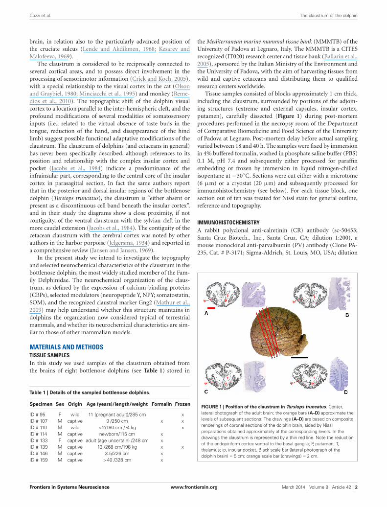
FIGURE 1 | Position of the claustrum in Tursiops truncatus. Center,lateral photograph of the adult brain; the orange bars (A–D) approximate thelevels of subsequent sections. The drawings (A–D) are based on compositerenderings of coronal sections of the dolphin brain, sided by Nisslpreparations obtained approximately at the corresponding levels. In thedrawings the claustrum is represented by a thin red line. Note the reductionof the endopiriform cortex ventral to the basal ganglia; P, putamen; T,thalamus; ip, insular pocket. Black scale bar (lateral photograph of thedolphin brain) = 5 cm; orange scale bar (drawings) = 2 cm.
Frontiers in Systems Neuroscience www.frontiersin.org March 2014 | Volume 8 | Article 42 | 2
Cozzi et al. The claustrum of the dolphin
1:3000), a mouse monoclonal anti-calbindin-D-28K (CB) anti-body (Clone CB-955, Cat. # C9848, Sigma-Aldrich, St. Louis,MO, USA; dilution 1:3000), a rabbit polyclonal anti-CB-D-28Kantibody (Cat. # CB38A, Swant, Bellinzona, Switzerland; diluition1:10000), a rabbit polyclonal anti-NPY antibody (ab30914;abcam; dilution 1:3000), a rabbit polyclonal anti-Gng2 anti-body (HPA003534; Sigma-Aldrich, dilution 1:100), and a rab-bit polyclonal anti-SOM antibody (ab103790; abcam; dilution1:700) were used in this study. Epitope retrieval was carried outat 120◦C in a pressure cooker for 5 min using a Tris/EDTAbuffer pH 9.0. Sections were rinsed in PBS and incubated in1% H2O2-PBS for 10 min, then pre-incubated in PBS with0.3% Triton X-100 (TX) (Sigma-Aldrich, St. Louis, MO, USA)and 5% normal goat serum (Vector Labs, Burlingame, CA) toreduce non-specific staining. Sections were subsequently incu-bated overnight in a humid chamber at 4◦C with the primaryantibody diluted in PBS with 0.3% TX and 1% normal goatserum. After several washings in PBS, sections were incubatedfor 1 h at room temperature with biotinylated goat anti-rabbit(for CR, NPY and SOM) or biotinylated goat anti-mouse sec-ondary antibodies (for PV and CB) (Vector Labs, Burlingame,CA), diluted 1:300 in PBS. Sections were then washed for 3 ×10 min in PBS, and incubated for 1 h at room temperature inavidinbiotin-horseradish peroxidase complex (PK-6100; VectorLabs, Burlingame, CA). After washing for 3× 10 min in Tris/HCl(pH 7.6), peroxidase activity was detected by incubation in asolution of 0.125 mg/ml diaminobenzidine (Sigma-Aldrich, St.Louis, MO, USA) and 0.1% H2O2 in the same buffer for 10 min orby a VIP substrate Kit for peroxidase (Cat. # SK-4600, Vector Labs,Burlingame, CA).
The amino acid sequence of the proteins investigated in thisarticle in the claustrum of Tursiops truncatus were compared withthose of other mammals (and especially the rat). For this aimwe used the Ensembl genomic database.1 The sequence of NPY,SOM, CB and CR is shared for over 93%, whereas correspondencefor Gng2 and PV is over 70%. The specificity of the immuno-histochemical staining was tested in repeated trials as follows:substitution of either the primary antibody, the anti-rabbit oranti-mouse IgG, or the ABC complex by PBS or non-immuneserum. Under these conditions the staining was abolished.
RESULTSGENERAL TOPOGRAPHY AND SHAPEBased on the examinations of macroscopic slices of the brain,followed by analysis of Nissl-stained sections, the claustrum wasdetected in all the examined specimens, including newborns. Thetopography of the claustrum observed in our experimental seriesreflects the general orientation of the dolphin brain, in whichthe lateral (temporal) lobes grow considerably, and the lower(partially olfactory) divisions of the telencephalon are reduced.The position of the claustrum was clearly identified lateral to theconspicuous putamen and medial to the insular formation andrelative insular pocket (Figure 1).
In Nissl-stained sections the claustrum appeared thin (notthicker than 1–2 mm) and dorso-ventrally elongated (up to
1www.ensembl.org
3.5 cm), without an evident endopiriform root as commonlyfound in terrestrial mammals. The ventralmost limits of theclaustrum apparently touch the insular cortex with virtual disap-pearance of the capsula extrema.
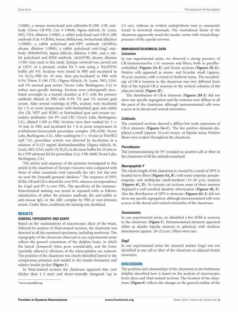
IMMUNOHISTOCHEMICAL DATACalretininIn our experimental series, we observed a strong presence ofCR-immunoreactive (-ir) neurons and fibers, both in paraffin-embedded (Figures 2A–E) and frozen sections (Figures 2F, G).Positive cells appeared as mono- and bi-polar small (approx.10 µm) neurons, with a round or fusiform soma. The morphol-ogy of CR-ir neurons in the claustrum was very different fromthat of the typical CR-ir neurons in the cortical columns of theadjacent insula (Figure 2E).
The distribution of CR-ir elements (Figures 2H–J) did notshow any specific segregation and the neurons were diffuse in allthe parts of the claustrum, although immunostained cells werescarcer at its dorsal and ventral extremities.
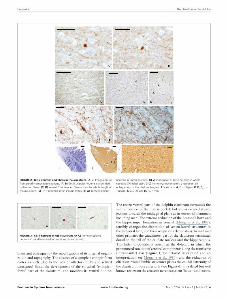
CalbindinThe examined sections showed a diffuse but weak expression ofCB-ir elements (Figures 3A–C). The few positive elements dis-played a small (approx. 10 µm) mono- or bipolar soma. Positivefibers were evident throughout the claustrum.
ParvalbuminThe immunostaining for PV revealed no positive cell or fiber inthe claustrum of all the animals examined.
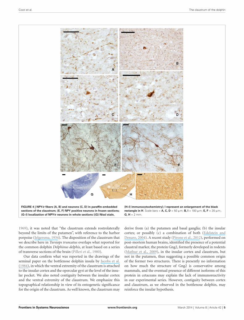
Neuropeptide YThe whole length of the claustrum is crossed by a mesh of NPY-irbeaded nerve fibers (Figures 4A, B), with some unipolar, pseudo-unipolar, and multipolar small-medium (15–20 µm) neurons(Figures 4C, D). In cryostat cut sections some of these neuronsdisplayed a well-ramified dendrite arborization (Figures 4E, F).Also the distribution of NPY-ir elements (Figures 4G–I) did notshow any specific segregation, although immunostained cells werescarcer at the dorsal and ventral extremities of the claustrum.
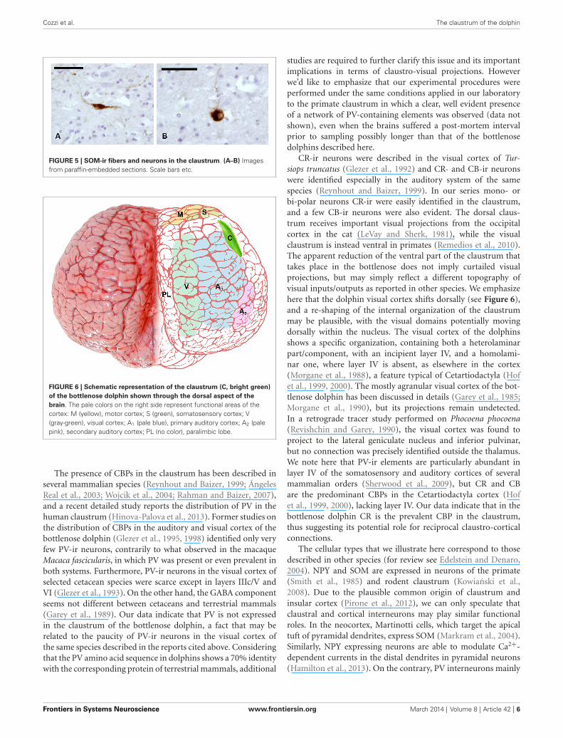
SomatostatinIn our experimental series, we identified a few SOM-ir neuronsin the claustrum (Figure 5). Immunostained elements appearedeither as slender bipolar neurons or spherical, with mediumdimensions (approx. 20–25 µm). Fibers were rare.
Gng2In our experimental series the claustral marker Gng2 was notidentified in any cell or fiber of the claustrum or adjacent brainsstructures.
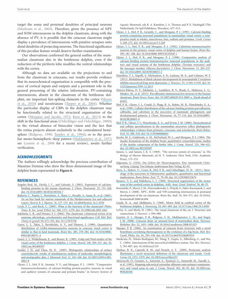
DISCUSSIONThe position and relationships of the claustrum in the bottlenosedolphin described here is based on the analysis of macroscopicbrain slices and Nissl stained sections. The location of the claus-trum (Figure 6) reflects the changes in the general outline of the
Frontiers in Systems Neuroscience www.frontiersin.org March 2014 | Volume 8 | Article 42 | 3
Cozzi et al. The claustrum of the dolphin
FIGURE 2 | CR-ir neurons and fibers in the claustrum. (A–D) Images derivefrom paraffin-embedded sections. (A, B) Small unipolar neurons surroundedby beaded fibers; (C, D) several CR-ir beaded fibers cross the whole length ofthe claustrum. (E) CR-ir neurons in the insular cortex. (F, G) Immunostained
neurons in frozen sections; (H–J) localization of CR-ir neurons in wholesections ((H) Nissl stain, (I–J) immunocytochemistry); J represent anenlargement of the black rectangle in I Scale bars: A, B = 50 µm; C, D, E, J =100 µm; F, G = 20 µm; H, I = 2 mm.
FIGURE 3 | CB-ir neurons in the claustrum. (A–C) Immunoreactiveneurons in paraffin-embedded sections. Scale bars etc.
brain and consequently the modifications of its internal organi-zation and topography. The absence of a complete endopiriformcortex as such (due to the lack of olfactory bulbs and relatedstructures) limits the development of the so-called “endopiri-form” part of the claustrum, and modifies its ventral outline.
The rostro-ventral part of the dolphin claustrum surrounds theventral borders of the insular pocket, but shows no medial pro-jections towards the midsagittal plane as in terrestrial mammalsincluding man. The extreme reduction of the Ammon’s horn andthe hippocampal formation in general (Morgane et al., 1982),sensibly changes the disposition of ventro-lateral structures inthe temporal lobe, and their reciprocal relationships. In man andother primates the caudalmost part of the claustrum terminatesdorsal to the tail of the caudate nucleus and the hippocampus.This latter disposition is absent in the dolphin, in which thepronounced rotation of cerebral components along the transverse(inter-insular) axis (Figure 1; for detailed description and aninterpretation see Morgane et al., 1980), and the reduction ofolfaction-related limbic structures places the caudal extremity ofthe claustrum more anteriorly (see Figure 6). In a dated but wellknown review on the cetacean nervous system (Jansen and Jansen,
Frontiers in Systems Neuroscience www.frontiersin.org March 2014 | Volume 8 | Article 42 | 4
Cozzi et al. The claustrum of the dolphin
FIGURE 4 | NPY-ir fibers (A, B) and neurons (C, D) in paraffin-embeddedsections of the claustrum; (E, F) NPY positive neurons in frozen sections;(G–I) localization of NPY-ir neurons in whole sections ((G) Nissl stain,
(H–I) immunocytochemistry); I represent an enlargement of the blackrectangle in H. Scale bars = A, C, D = 50 µm; B, I = 100 µm; E, F = 20 µm;.G, H = 2 mm.
1969), it was noted that “the claustrum extends rostrolaterallybeyond the limits of the putamen”, with reference to the harborporpoise (Jelgersma, 1934). The disposition of the claustrum thatwe describe here in Tursiops truncatus overlaps what reported forthe common dolphin Delphinus delphis, at least based on a seriesof transverse sections of the brain (Pilleri et al., 1980).
Our data confirm what was reported in the drawings of theseminal paper on the bottlenose dolphin insula by Jacobs et al.(1984), in which the ventral extremity of the claustrum is attachedto the insular cortex and the opercular gyri at the level of the insu-lar pocket. We also noted contiguity between the insular cortexand the ventral extremity of the claustrum. We emphasize thistopographical relationship in view of its ontogenetic significancefor the origin of the claustrum. As well known, the claustrum may
derive from (a) the putamen and basal ganglia; (b) the insularcortex; or possibly (c) a combination of both (Edelstein andDenaro, 2004). A recent study (Pirone et al., 2012), performed onpost-mortem human brains, identified the presence of a potentialclaustral marker, the protein Gng2, formerly developed in rodents(Mathur et al., 2009), in the insular cortex and claustrum, butnot in the putamen, thus suggesting a possible common originof the former two structures. There is presently no informationon how much the structure of Gng2 is conservative amongmammals, and the eventual presence of different isoforms of thisprotein in cetaceans may explain the lack of immunoreactivityin our experimental series. However, contiguity between cortexand claustrum, as we observed in the bottlenose dolphin, mayreinforce the insular hypothesis.
Frontiers in Systems Neuroscience www.frontiersin.org March 2014 | Volume 8 | Article 42 | 5
Cozzi et al. The claustrum of the dolphin
FIGURE 5 | SOM-ir fibers and neurons in the claustrum. (A–B) Imagesfrom paraffin-embedded sections. Scale bars etc.
FIGURE 6 | Schematic representation of the claustrum (C, bright green)of the bottlenose dolphin shown through the dorsal aspect of thebrain. The pale colors on the right side represent functional areas of thecortex: M (yellow), motor cortex; S (green), somatosensory cortex; V(gray-green), visual cortex; A1 (pale blue), primary auditory cortex; A2 (palepink), secondary auditory cortex; PL (no color), paralimbic lobe.
The presence of CBPs in the claustrum has been described inseveral mammalian species (Reynhout and Baizer, 1999; ÁngelesReal et al., 2003; Wojcik et al., 2004; Rahman and Baizer, 2007),and a recent detailed study reports the distribution of PV in thehuman claustrum (Hinova-Palova et al., 2013). Former studies onthe distribution of CBPs in the auditory and visual cortex of thebottlenose dolphin (Glezer et al., 1995, 1998) identified only veryfew PV-ir neurons, contrarily to what observed in the macaqueMacaca fascicularis, in which PV was present or even prevalent inboth systems. Furthermore, PV-ir neurons in the visual cortex ofselected cetacean species were scarce except in layers IIIc/V andVI (Glezer et al., 1993). On the other hand, the GABA componentseems not different between cetaceans and terrestrial mammals(Garey et al., 1989). Our data indicate that PV is not expressedin the claustrum of the bottlenose dolphin, a fact that may berelated to the paucity of PV-ir neurons in the visual cortex ofthe same species described in the reports cited above. Consideringthat the PV amino acid sequence in dolphins shows a 70% identitywith the corresponding protein of terrestrial mammals, additional
studies are required to further clarify this issue and its importantimplications in terms of claustro-visual projections. Howeverwe’d like to emphasize that our experimental procedures wereperformed under the same conditions applied in our laboratoryto the primate claustrum in which a clear, well evident presenceof a network of PV-containing elements was observed (data notshown), even when the brains suffered a post-mortem intervalprior to sampling possibly longer than that of the bottlenosedolphins described here.
CR-ir neurons were described in the visual cortex of Tur-siops truncatus (Glezer et al., 1992) and CR- and CB-ir neuronswere identified especially in the auditory system of the samespecies (Reynhout and Baizer, 1999). In our series mono- orbi-polar neurons CR-ir were easily identified in the claustrum,and a few CB-ir neurons were also evident. The dorsal claus-trum receives important visual projections from the occipitalcortex in the cat (LeVay and Sherk, 1981), while the visualclaustrum is instead ventral in primates (Remedios et al., 2010).The apparent reduction of the ventral part of the claustrum thattakes place in the bottlenose does not imply curtailed visualprojections, but may simply reflect a different topography ofvisual inputs/outputs as reported in other species. We emphasizehere that the dolphin visual cortex shifts dorsally (see Figure 6),and a re-shaping of the internal organization of the claustrummay be plausible, with the visual domains potentially movingdorsally within the nucleus. The visual cortex of the dolphinsshows a specific organization, containing both a heterolaminarpart/component, with an incipient layer IV, and a homolami-nar one, where layer IV is absent, as elsewhere in the cortex(Morgane et al., 1988), a feature typical of Cetartiodactyla (Hofet al., 1999, 2000). The mostly agranular visual cortex of the bot-tlenose dolphin has been discussed in details (Garey et al., 1985;Morgane et al., 1990), but its projections remain undetected.In a retrograde tracer study performed on Phocoena phocoena(Revishchin and Garey, 1990), the visual cortex was found toproject to the lateral geniculate nucleus and inferior pulvinar,but no connection was precisely identified outside the thalamus.We note here that PV-ir elements are particularly abundant inlayer IV of the somatosensory and auditory cortices of severalmammalian orders (Sherwood et al., 2009), but CR and CBare the predominant CBPs in the Cetartiodactyla cortex (Hofet al., 1999, 2000), lacking layer IV. Our data indicate that in thebottlenose dolphin CR is the prevalent CBP in the claustrum,thus suggesting its potential role for reciprocal claustro-corticalconnections.
The cellular types that we illustrate here correspond to thosedescribed in other species (for review see Edelstein and Denaro,2004). NPY and SOM are expressed in neurons of the primate(Smith et al., 1985) and rodent claustrum (Kowianski et al.,2008). Due to the plausible common origin of claustrum andinsular cortex (Pirone et al., 2012), we can only speculate thatclaustral and cortical interneurons may play similar functionalroles. In the neocortex, Martinotti cells, which target the apicaltuft of pyramidal dendrites, express SOM (Markram et al., 2004).Similarly, NPY expressing neurons are able to modulate Ca2+-dependent currents in the distal dendrites in pyramidal neurons(Hamilton et al., 2013). On the contrary, PV interneurons mainly
Frontiers in Systems Neuroscience www.frontiersin.org March 2014 | Volume 8 | Article 42 | 6
Cozzi et al. The claustrum of the dolphin
target the soma and proximal dendrites of principal neurons(Markram et al., 2004). Therefore, given the presence of NPYand SOM interneurons in the dolphin claustrum, along with theabsence of PV, it is possible that the cetacean claustrum mightdisplay a prevalence of interneurons with putative synapses ontodistal dendrites of projecting neurons. The functional significanceof this peculiar feature would deserve further examination.
Our observations confirmed the general outline of the mam-malian claustrum also in the bottlenose dolphin, even if thereduction of the piriform lobe modifies the ventral relationshipswith the cortex.
Although no data are available on the projections to andfrom the claustrum in cetaceans, our results provide evidencethat its neurochemical organization is compatible with the pres-ence of cortical inputs and outputs and a persistent role in thegeneral processing of the relative information. PV-containinginterneurons, absent in the claustrum of the bottlenose dol-phin, are important fast-spiking elements in the cortex (Mooreet al., 2010) and neostriatum (Tepper et al., 2004). Whetherthe particular display of CBPs in the dolphin claustrum maybe functionally related to the structural organization of thecortex (Morgane and Jacobs, 1972; Kern et al., 2011); to theshift in the functional areas (Oelschläger and Oelschläger, 2009);to the virtual absence of binocular vision and the fact thatthe retina projects almost exclusively to the contralateral hemi-sphere (Ridgway, 1990; Tarpley et al., 1994); or to the pecu-liar mono-hemispheric sleep pattern (Mukhametov et al., 1977;see Lyamin et al., 2008 for a recent review), awaits furtherverification.
ACKNOWLEDGMENTSThe Authors willingly acknowledge the precious contribution ofMassimo Demma who drew the three-dimensional image of thedolphin brain represented in Figure 6.
REFERENCESÁngeles Real, M., Dávila, J. C., and Guirado, S. (2003). Expression of calcium-
binding proteins in the mouse claustrum. J. Chem. Neuroanat. 25, 151–160.doi: 10.1016/s0891-0618(02)00104-7
Ballarin, C., Papini, L., Bortolotto, A., Butti, C., Peruffo, A., Sassu, R., et al. (2005).An on-line bank for marine mammals of the Mediterranean Sea and adjacentwaters. Hystrix It. J. Mamm. 16, 127–133. doi: 10.4404/hystrix-16.2-4350
Crick, F. C., and Koch, C. (2005). What is the function of the claustrum? Philos.Trans. R. Soc. Lond. B Biol. Sci. 360, 1271–1279. doi: 10.1098/rstb.2005.1661
Edelstein, L. R., and Denaro, F. J. (2004). The claustrum: a historical review of itsanatomy, physiology, cytochemistry and functional significance. Cell. Mol. Biol.(Noisy-le-grand) 50, 675–702. doi: 10.1170/T558
Garey, L. J., Takás, J., Revishchin, A. V., and Hámori, J. (1989). Quantitativedistribution of GABA-immunoreactive neurons in cetacean visual cortex issimilar to that in land mammals. Brain Res. 485, 278–284. doi: 10.1016/0006-8993(89)90571-4
Garey, L. J., Winkelmann, E., and Brauer, K. (1985). Golgi and Nissl studies of thevisual cortex of the bottlenose dolphin. J. Comp. Neurol. 240, 305–321. doi: 10.1002/cne.902400307
Geisler, J. H., and Uhen, M. D. (2005). Phylogenitic relationships of extinctcetartiodactyls: results of simultaneous analyses of molecular, morphological,and stratigraphic data. J. Mammal. Evol. 12, 145–160. doi: 10.1007/s10914-005-4963-8
Glezer, I. I., Hof, P. R., Istomin, V. V., and Morgane, P. J. (1995). “Comparativeimmunocytochemistry of calcium-binding protein-positive neurons in visualand auditory systems of cetacean and primate brains,” in Sensory Systems of
Aquatic Mammals, eds R. A. Kastelein, J. A. Thomas and P. E. Nachtigall (TheNetherlands: De Spil Publishers, Woerden), 477–513.
Glezer, I. I., Hof, P. R., Leranth, C., and Morgane, P. J. (1993). Calcium-bindingprotein-containing neuronal populations in mammalian visual cortex: a com-parative study in whales, insectivores, bats, rodents and primates. Cereb. Cortex3, 249–272. doi: 10.1093/cercor/3.3.249
Glezer, I. I., Hof, P. R., and Morgane, P. J. (1992). Calretinin-immunoreactiveneurons in the primary visual cortex of dolphin and human brains. Brain Res.595, 181–188. doi: 10.1016/0006-8993(92)91047-i
Glezer, I. I., Hof, P. R., and Morgane, P. J. (1998). Comparative analysis ofcalcium-binding protein-immunoreactive neuronal populations in the audi-tory and visual systems of the bottlenose dolphin (Tursiops truncatus) andthe macaque monkey (Macaca fascicularis). J. Chem. Neuroanat. 15, 203–237.doi: 10.1016/s0891-0618(98)00022-2
Hamilton, T. J., Xapelli, S., Michaelson, S. D., Larkum, M. E., and Colmers, W. F.(2013). Modulation of distal calcium electrogenesis by neuropeptide Y receptorsinhibits neocortical long-term depression. J. Neurosci. 33, 11184–11193. doi: 10.1523/jneurosci.5595-12.2013
Hinova-Palova, D. V., Edelstein, L., Landzhov, B. V., Braak, E., Malinova, L. G.,Minkov, M., et al. (2013). Parvalbumin-immunoreactive neurons in the humanclaustrum. Brain Struct. Funct. doi: 10.1007/s00429-013-0603-x. [Epub ahead ofprint].
Hof, P. R., Glezer, I. I., Condé, F., Flagg, R. A., Rubin, M. B., Nimchinsky, E. A.,et al. (1999). Cellular distribution of the calcium-binding proteins parvalbumin,calbindin, and calretinin in the neocortex of mammals: phylogenetic anddevelopmental patterns. J. Chem. Neuroanat. 16, 77–116. doi: 10.1016/s0891-0618(98)00065-9
Hof, P. R., Glezer, I. I., Nimchinsky, E. A., and Erwin, J. M. (2000). Neurochemicaland cellular specializations in the mammalian neocortex reflect phylogeneticrelationships: evidence from primates, cetaceans, and artiodactyls. Brain Behav.Evol. 55, 300–310. doi: 10.1159/000006665
Jacobs, M. S., Galaburda, A. M., McFarland, W. L., and Morgane, P. J. (1984). Theinsular formations of the dolphin brain: quantitative cytoarchitectonic studiesof the insular component of the limbic lobe. J. Comp. Neurol. 225, 396–432.doi: 10.1002/cne.902250307
Jansen, J., and Jansen, J. K. S. (1969). “The nervous system of cetacean,” in TheBiology of Marine Mammals, ed H. T. Andersen (New York, USA: AcademicPress), 175–252.
Jelgersma, G. (1934). Das Gehirn der Wassersäugetiere. Eine Anatomische Unter-suchung. Leipzig: Von Johann Ambrosium Bart Verlag, 82–91.
Kern, A., Siebert, U., Cozzi, B., Hof, P. R., and Oelschläger, H. H. (2011). Stere-ology of the neocortex in Odontocetes: qualitative, quantitative and functionalimplications. Brain Behav. Evol. 77, 79–90. doi: 10.1159/000323674
Kesarev, V. S., and Malofeeva, L. I. (1969). Structural organization of the motorzone of the cerebral cortex in dolphins. Arkh. Anat. Gistol. Embriol. 56, 48–55.
Kowianski, P., Morys, J. M., Dziewiatkowski, J., Wójcik, S., Sidor-Kaczmarek, J., andMorys, J. (2008). NPY-, SOM- and VIP-containing interneurons in postnataldevelopment of the rat claustrum. Brain Res. Bull. 76, 565–571. doi: 10.1016/j.brainresbull.2008.04.004
Lende, R. A., and Akdikmen, S. (1968). Motor field in cerebral cortex of thebottlenose dolphin. J. Neurosurg. 29, 495–499. doi: 10.3171/jns.1968.29.5.0495
LeVay, S., and Sherk, H. (1981). The visual claustrum of the cat. I. Structure andconnections. J. Neurosci. 1, 956–980.
Lyamin, O. I., Manger, P. R., Ridgway, S. H., Mukhametov, L. M., and Siegel,J. M. (2008). Cetacean sleep: an unusual form of mammalian sleep. Neurosci.Biobehav. Rev. 32, 1451–1484. doi: 10.1016/j.neubiorev.2008.05.023
Manger, P. R. (2006). An examination of cetacean brain structure with a novelhypothesis correlating thermogenesis to the evolution of a big brain. Biol. Rev.Camb. Philos. Soc. 81, 293–338. doi: 10.1017/s1464793106007019
Markram, H., Toledo-Rodriguez, M., Wang, Y., Gupta, A., Silberberg, G., and Wu,C. (2004). Interneurons of the neocortical inhibitory system. Nat. Rev. Neurosci.5, 793–807. doi: 10.1038/nrn1519
Mathur, B. N., Caprioli, R. M., and Deutch, A. Y. (2009). Proteomic analysisilluminates a novel structural definition of the claustrum and insula. Cereb.Cortex 19, 2372–2379. doi: 10.1093/cercor/bhn253
Minciacchi, D., Granato, A., Antonini, A., Tassinari, G., Santarelli, M., Zanolli, L.,et al. (1995). Mapping subcortical extrarelay afferents onto primary somatosen-sory and visual areas in cats. J. Comp. Neurol. 362, 46–70. doi: 10.1002/cne.903620104
Frontiers in Systems Neuroscience www.frontiersin.org March 2014 | Volume 8 | Article 42 | 7
Cozzi et al. The claustrum of the dolphin
Moore, C. I., Carlen, M., Knoblich, U., and Cardin, J. A. (2010). Neocorticalinterneurons: from diversity, strength. Cell 142, 189–193. doi: 10.1016/j.cell.2010.07.005
Morgane, P. J., Glezer, I. I., and Jacobs, M. S. (1988). Visual cortex of thedolphin: an image analysis study. J. Comp. Neurol. 273, 3–25. doi: 10.1002/cne.902730103
Morgane, P. J., Glezer, I. I., and Jacobs, M. S. (1990). “Comparative anatomy of thevisual cortex of the dolphin,” in Cerebral Cortex (Vol. 8B), eds E. G. Jones and A.Peters (New York, USA: Plenum Publishing Co.), 215–262.
Morgane, P. J., and Jacobs, M. S. (1972). “Comparative anatomy of the cetaceannervous system,” in Functional Anatomy of Marine Mammals (Vol. 1), ed R. J.Harrison (London, UK: Academic Press), 117–244.
Morgane, P. J., Jacobs, M. S., and Galaburda, A. (1986). “Evolutionary morphologyof the dolphin brain,” in Dolphin Cognition and Behavior. A ComparativeApproach, eds R. Schusterman, J. Thomas and F. Wood (Hillsdale, New Jersey:Lawrence Erlbaum Associates), 5–28.
Morgane, P. J., Jacobs, M. S., and McFarland, W. L. (1980). The anatomy ofthe brain of the bottlenose dolphin (Tursiops truncatus). Surface configurationof the telencephalon of the bottlenose dolphin with comparative anatomicalobservations in four other cetacean species. Brain Res. Bull. 5(Suppl. 3), 1–107.doi: 10.1016/0361-9230(80)90272-5
Morgane, P. J., McFarland, W. L., and Jacobs, M. S. (1982). The limbic lobe of thedolphin brain: a quantitative cytoarchitectonic study. J. Hirnforsch. 23, 465–552.
Mukhametov, L. M., Supin, A. Y., and Polyakova, I. G. (1977). Interhemisphericasymmetry of the electroencephalographic sleep pattern in dolphins. Brain Res.134, 581–584. doi: 10.1016/0006-8993(77)90835-6
Oelschläger, H. H. A., and Oelschläger, J. S. (2009). “Brain,” in Encyclopedia ofMarine Mamamals (2nd Edn.), eds W. F. Perrin, B. Würsing and J. G. M.Thewissen (Amsterdam: Academic Press), 134–149.
Olson, C. R., and Graybiel, A. M. (1980). Sensory maps in the claustrum of the cat.Nature 288, 479–481. doi: 10.1038/288479a0
Pilleri, G., Peixun, C., and Zuohua, S. (1980). Concise Macroscopical Atlas of theBrain of the Common Dolphin (Delphinus delphis Linnaeus, 1758). Waldau-BerneSwitzerland: Brain Anatomy Institute, University of Berne.
Pirone, A., Cozzi, B., Edelstein, L., Peruffo, A., Lenzi, C., Quilici, F., et al. (2012).Topography of Gng2- and NetrinG2-expression suggests an insular origin of thehuman claustrum. PLoS One 7:e44745. doi: 10.1371/journal.pone.0044745
Rahman, F. E., and Baizer, J. S. (2007). Neurochemically defined cell types inthe claustrum of the cat. Brain Res. 1159, 94–111. doi: 10.1016/j.brainres.2007.05.011
Remedios, R., Logothetis, N. K., and Kayser, C. (2010). Unimodal responsesprevail within the multisensory claustrum. J. Neurosci. 30, 12902–12907. doi: 10.1523/JNEUROSCI.2937-10.2010
Revishchin, A. V., and Garey, L. J. (1990). The thalamic projection to the sensoryneocortex of the porpoise, Phocoena phocoena. J. Anat. 169, 85–102.
Reynhout, K., and Baizer, J. S. (1999). Immunoreactivity for calcium-bindingproteins in the claustrum of the monkey. Anat. Embryol. (Berl) 199, 75–83.doi: 10.1007/s004290050211
Ridgway, S. H. (1990). “The central nervous system of the bottlenose dolphin,” inThe Bottlenose Dolphin, eds S. Leatherwood and R. R. Reeves (San Diego, USA:Academic Press), 69–97.
Sherwood, C. C., Stimpson, C. D., Butti, C., Bonar, C. J., Newton, A. L., Allman,J. M., et al. (2009). Neocortical neuron types in Xenarthra and Afrotheria:implications for brain evolution in mammals. Brain Struct. Funct. 213, 301–328.doi: 10.1007/s00429-008-0198-9
Slijper, E. J. (1979). Whales 2nd Edn. London: Hutchinson. 511pp.Smith, Y., Parent, A., Kerkérian, L., and Pelletier, G. (1985). Distribution of
neuropeptide Y immunoreactivity in the basal forebrain and upper brainstemof the squirrel monkey (Saimiri sciureus). J. Comp. Neurol. 236, 71–89. doi: 10.1002/cne.902360107
Tarpley, R. Y., Gelderd, J. B., Bauserman, S., and Ridgway, S. H. (1994). Dolphinperipheral visual pathway in chronic unilateral ocular atrophy: complete decus-sation apparent. J. Morphol. 222, 91–102. doi: 10.1002/jmor.1052220109
Tepper, J. M., Koós, T., and Wilson, C. J. (2004). GABAergic microcircuits in theneostriatum. Trends Neurosci. 27, 662–669. doi: 10.1016/j.tins.2004.08.007
Thewissen, J. G. M., Cooper, L. N., Clementz, M. T., Bajpai, S., and Tiwari, B. N.(2007). Whales originated from aquatic artiodactyls in the Eocene epoch ofIndia. Nature 450, 1190–1194. doi: 10.1038/nature06343
Wojcik, S., Dziewiatkowski, J., Spodnik, E., Ludkiewicz, B., Domaradzka-Pytel, B.,Kowianski, P., et al. (2004). Analysis of calcium binding protein immunoreactiv-ity in the claustrum and the endopiriform nucleus of the rabbit. Acta Neurobiol.Exp. (Wars) 64, 449–460.
Conflict of Interest Statement: The authors declare that the research was con-ducted in the absence of any commercial or financial relationships that could beconstrued as a potential conflict of interest.
Received: 25 January 2014; accepted: 10 March 2014; published online: 28 March 2014.Citation: Cozzi B, Roncon G, Granato A, Giurisato M, Castagna M, Peruffo A, PaninM, Ballarin C, Montelli S and Pirone A (2014) The claustrum of the bottlenose dolphinTursiops truncatus (Montagu 1821). Front. Syst. Neurosci. 8:42. doi: 10.3389/fnsys.2014.00042This article was submitted to the journal Frontiers in Systems Neuroscience.Copyright © 2014 Cozzi, Roncon, Granato, Giurisato, Castagna, Peruffo, Panin,Ballarin, Montelli and Pirone. This is an open-access article distributed under theterms of the Creative Commons Attribution License (CC BY). The use, distribution orreproduction in other forums is permitted, provided the original author(s) or licensorare credited and that the original publication in this journal is cited, in accordance withaccepted academic practice. No use, distribution or reproduction is permitted whichdoes not comply with these terms.
Frontiers in Systems Neuroscience www.frontiersin.org March 2014 | Volume 8 | Article 42 | 8


![TAXONOMY AND BIOLOGY OF SIPUNCULANS, WITH EMPHASIS ON THE MORPHOLOGY OF PHASCOLION STROMBUS (MONTAGU, 1804). ANNOTATED BIBLIOGRAPHY 1555-1995 APPENDED AS ANNEX. [1995]](https://static.fdokumen.com/doc/165x107/6322f412050768990e10167c/taxonomy-and-biology-of-sipunculans-with-emphasis-on-the-morphology-of-phascolion.jpg)