Copper(I) and copper(II) binding to β-amyloid 16 (Aβ16) studied by electrospray ionization mass...
6
Copper(I) and copper(II) binding to b-amyloid 16 (Ab16) studied by electrospray ionization mass spectrometryw Yu Lu, Michel Prudent, Liang Qiao, Manuel A. Mendez and Hubert H. Girault* Received 27th March 2010, Accepted 12th May 2010 First published as an Advance Article on the web 26th May 2010 DOI: 10.1039/c004693k Copper-b-amyloid 16 (Ab16) complexes were investigated by electrospray ionization mass spectrometry (ESI-MS). Copper(I) and (II) complexes were formed on-line in a microchip electrospray emitter by using a sacrificial copper electrode as the anode in positive ionization mode. In the presence of ascorbic acid in the peptide solution, the amount of Cu(I)-Ab16 generated electrochemically was even higher. A kinetic model is proposed to account for the generation of copper complexes. The structure of Cu(I)-Ab16 was investigated by tandem mass spectrometry (MS/MS), and the binding site of Cu(I) to Ab16 was identified at the His13, His14 residues. Cu(II)-Ab16 was also investigated by MS/MS and, based on the unusual observations of a-ions, the two binding residues of His13 and His14 of Ab16 to Cu(II) were also confirmed. This approach provides direct information on Cu(I)-Ab16 complexes generated in solution from metallic copper and gives evidence that both His13 and His14 are involved in the coordination of both Cu(I)- and Cu(II)-Ab16 complexes. Introduction Beta-amyloid (Ab) deposits in tissues are one of the main pathological characteristics of Alzheimer’s disease (AD), and many studies have been devoted to understanding the aggre- gation mechanism. 1–3 Indeed, Ab is usually present in healthy brains in a soluble form, but amyloid plaques are often observed in AD patients. In vivo, the most usual forms of Ab consist of 40 amino-acids (Ab40), which at physiological pH are largely random coils and of 42 amino-acids (Ab42), which have a strong tendency to aggregate. 4 Beta-amyloid plaques have been found to contain large amounts of transi- tion metal cations such as Zn, Cu, and Fe (mM range) and it has also been suggested that the altered homeostasis of these transition metal ions is related to degenerative diseases. 5–7 For this reason, many groups have investigated the oxidative damage of proteins and peptides induced by transition metals. 8–10 Considering the redox activity of metal ions such as copper and iron, these ions play a crucial role in generating reactive oxygen species (ROS) responsible for the oxidative stress in the brain and for neuronal toxicity. A basic knowledge of the coordination of these metal ions at different oxidation states with amyloid peptides is therefore essential to apprehend their reactivity and reveal their potential role in the degeneration process. Furthermore, understanding the coordination of Cu(Ab) and Zn(Ab) may provide a strategy to rationalize novel therapeutics. 11,12 It has been proposed that the metal binding site lies in the N-terminus domain, more precisely in the first 16 amino acids of Ab (Ab16). This peptide shows no tendency to aggregate or to form fibrils under moderate concentrations, and represents a model for soluble metallated Ab peptides. Currently, most studies on the structure of Cu(Ab) complexes are mainly focused on the binding sites of Cu(II) to Ab peptides. A recently published review 13 recapitulates the various proposi- tions reported in the literature and summarized the most reasonable coordination environments for Cu(II) binding to Ab peptides considering the experimental differences and the limitation of each method. Two likely complexations involving the N-terminus, His6 and His13 or His14, Asp1-COO on one hand, and His6, His13, His14 and Asp1-COO on the other hand are usually considered. By comparison, the binding of the reduced Cu(I) with Ab has not yet been fully characterized. Extended X-ray absorption fine structure (EXAFS) spectro- scopy has been used to speculate a linear two-coordinate geometry with two imidazole ligands as recently reported by two different groups. 14,15 Most copper(I) compounds are known for their instability in aerobic environments. It has also been reported that Cu(I)-Ab complexes have a limited stability in air i.e. 20 min and are only stable in inert atmo- spheres or in the presence of strong reducing agents. 14,15 Therefore, the preparation of pure copper(I) complexes is usually an experimental challenge. Copper(I) ions are involved in oxidation damages (Fenton and Haber-Weiss reactions) and since there is some evidence that Cu(I) could play an important role in the aggregation of amyloid peptide and is biologically relevant to the oxidative stress in the brain, we report here a mass spectrometric study of copper(I) and copper(II) binding to Ab16. Mass spectrometry (MS) coupled to electrospray ionization (ESI) is a powerful tool widely used in studying biological molecules and their complexes. Usually, the study of metal– protein interactions is carried out by mixing the biomolecules of interest with an electrolyte solution of the metal salt. 16 Laboratoire d’Electrochimie Physique et Analytique, Station 6, Ecole Polytechnique Fe´de ´rale de Lausanne, CH-1015 Lausanne, Switzerland. E-mail: hubert.girault@epfl.ch; Fax: +41 216933667; Tel: +41 216933151 w Electronic supplementary information (ESI) available: Details of the kinetic model and the whole simulation. See DOI: 10.1039/c004693k 474 | Metallomics, 2010, 2, 474–479 This journal is c The Royal Society of Chemistry 2010 PAPER www.rsc.org/metallomics | Metallomics
Transcript of Copper(I) and copper(II) binding to β-amyloid 16 (Aβ16) studied by electrospray ionization mass...

Copper(I) and copper(II) binding to b-amyloid 16 (Ab16) studiedby electrospray ionization mass spectrometryw
Yu Lu, Michel Prudent, Liang Qiao, Manuel A. Mendez and Hubert H. Girault*
Received 27th March 2010, Accepted 12th May 2010
First published as an Advance Article on the web 26th May 2010
DOI: 10.1039/c004693k
Copper-b-amyloid 16 (Ab16) complexes were investigated by electrospray ionization mass
spectrometry (ESI-MS). Copper(I) and (II) complexes were formed on-line in a microchip
electrospray emitter by using a sacrificial copper electrode as the anode in positive ionization
mode. In the presence of ascorbic acid in the peptide solution, the amount of Cu(I)-Ab16generated electrochemically was even higher. A kinetic model is proposed to account for the
generation of copper complexes. The structure of Cu(I)-Ab16 was investigated by tandem mass
spectrometry (MS/MS), and the binding site of Cu(I) to Ab16 was identified at the His13, His14
residues. Cu(II)-Ab16 was also investigated by MS/MS and, based on the unusual observations
of a-ions, the two binding residues of His13 and His14 of Ab16 to Cu(II) were also confirmed.
This approach provides direct information on Cu(I)-Ab16 complexes generated in solution from
metallic copper and gives evidence that both His13 and His14 are involved in the coordination
of both Cu(I)- and Cu(II)-Ab16 complexes.
Introduction
Beta-amyloid (Ab) deposits in tissues are one of the main
pathological characteristics of Alzheimer’s disease (AD), and
many studies have been devoted to understanding the aggre-
gation mechanism.1–3 Indeed, Ab is usually present in healthy
brains in a soluble form, but amyloid plaques are often
observed in AD patients. In vivo, the most usual forms of
Ab consist of 40 amino-acids (Ab40), which at physiological
pH are largely random coils and of 42 amino-acids (Ab42),which have a strong tendency to aggregate.4 Beta-amyloid
plaques have been found to contain large amounts of transi-
tion metal cations such as Zn, Cu, and Fe (mM range) and it
has also been suggested that the altered homeostasis of these
transition metal ions is related to degenerative diseases.5–7 For
this reason, many groups have investigated the oxidative
damage of proteins and peptides induced by transition metals.8–10
Considering the redox activity of metal ions such as copper
and iron, these ions play a crucial role in generating reactive
oxygen species (ROS) responsible for the oxidative stress in the
brain and for neuronal toxicity. A basic knowledge of the
coordination of these metal ions at different oxidation states
with amyloid peptides is therefore essential to apprehend their
reactivity and reveal their potential role in the degeneration
process. Furthermore, understanding the coordination of
Cu(Ab) and Zn(Ab) may provide a strategy to rationalize
novel therapeutics.11,12
It has been proposed that the metal binding site lies in the
N-terminus domain, more precisely in the first 16 amino acids
of Ab (Ab16). This peptide shows no tendency to aggregate or
to form fibrils under moderate concentrations, and represents
a model for soluble metallated Ab peptides. Currently, most
studies on the structure of Cu(Ab) complexes are mainly
focused on the binding sites of Cu(II) to Ab peptides. A
recently published review13 recapitulates the various proposi-
tions reported in the literature and summarized the most
reasonable coordination environments for Cu(II) binding to
Ab peptides considering the experimental differences and the
limitation of each method. Two likely complexations involving
the N-terminus, His6 and His13 or His14, Asp1-COO� on one
hand, and His6, His13, His14 and Asp1-COO� on the other
hand are usually considered. By comparison, the binding of
the reduced Cu(I) with Ab has not yet been fully characterized.
Extended X-ray absorption fine structure (EXAFS) spectro-
scopy has been used to speculate a linear two-coordinate
geometry with two imidazole ligands as recently reported by
two different groups.14,15 Most copper(I) compounds are
known for their instability in aerobic environments. It has
also been reported that Cu(I)-Ab complexes have a limited
stability in air i.e. 20 min and are only stable in inert atmo-
spheres or in the presence of strong reducing agents.14,15
Therefore, the preparation of pure copper(I) complexes is
usually an experimental challenge. Copper(I) ions are involved
in oxidation damages (Fenton and Haber-Weiss reactions)
and since there is some evidence that Cu(I) could play an
important role in the aggregation of amyloid peptide and is
biologically relevant to the oxidative stress in the brain, we
report here a mass spectrometric study of copper(I) and
copper(II) binding to Ab16.Mass spectrometry (MS) coupled to electrospray ionization
(ESI) is a powerful tool widely used in studying biological
molecules and their complexes. Usually, the study of metal–
protein interactions is carried out by mixing the biomolecules
of interest with an electrolyte solution of the metal salt.16
Laboratoire d’Electrochimie Physique et Analytique, Station 6,Ecole Polytechnique Federale de Lausanne, CH-1015 Lausanne,Switzerland. E-mail: [email protected]; Fax: +41 216933667;Tel: +41 216933151w Electronic supplementary information (ESI) available: Details of thekinetic model and the whole simulation. See DOI: 10.1039/c004693k
474 | Metallomics, 2010, 2, 474–479 This journal is �c The Royal Society of Chemistry 2010
PAPER www.rsc.org/metallomics | Metallomics

Alternatively, soluble anodes can be used both to apply the
high voltage to the electrospray emitter and to generate metal
ions in the absence of counter ions, therefore preventing the
charge neutralization effect induced by counter anions when
using a salt.17,18 Here a microchip emitter has been used as the
sensitivity is increased by two orders of magnitude compared
to the commercial ESI source. We have shown that a sacrificial
copper anode coupled to an electrospray emitter can generate
a mixture of both Cu(I) and Cu(II) ions.19–21 Indeed, the
dissolution of copper metal proceeds first by the generation
of Cu(I) ions that can be further oxidized to Cu(II). How-
ever, in the presence of adequate ligands, Cu(I) ions can be
scavenged to form complexes.20 In the present work, we apply
this methodology to investigate the binding of both Cu(I) and
Cu(II) to Ab16.
Experimental
Chemicals
Ab16 peptide (DAEFRH6DSGYEVH13H14QK, M =
1955.1 g mol�1) was purchased from Bachem (Bubendorf,
Switzerland). Cupric chloride dihydrate (CuCl2�2H2O) and
zinc chloride (ZnCl2) were bought from Acros Organics (Geel,
Belgium). Ascorbic acid was bought from AppliChem GmbH
(Darmstadt Germany) and methanol from Riedel-de-Haen
(Seelze, Germany). Deionized water (18.2 MO cm) was from
aMilli-Q system fromMillipore (Bedford, MA). The lyophilized
Ab16 peptide was dissolved with deionized water at a final
concentration of 1 mg mL�1 as a stock solution stored at�20 1C.Ab16 solution was diluted at a final concentration of 10 mM in
50/50 (vol/vol) MeOH/H2O before each experiment. All the
solutions were prepared fresh daily.
MS setup and microspray interface
Metal ion on-line complexation was carried out using a
microspray interface described previously.19 In brief, it consists
of a single microchannel (45 mm � 120 mm � 1 cm) polyimide
microchip developed by DiagnoSwiss SA (Monthey, Switzerland).
The tip of the end of microchannel was cut by a blade as
shown in Scheme 1 in order to form a stable Taylor cone under
a high voltage. Therefore, the nanospray was stably produced
by this well-shaped emitter, which could retain the stability for
half an hour at least and be reused at least tens of times. A
reservoir of polycarbonate (+ = 3 mm, h = 5 mm) was
glued at the inlet of the microchannel. The sample was loaded
(V = 20 mL) in the reservoir in which a metallic electrode was
immersed. MeOH/H2O (50/50) and methanol were used for
the cleaning and rinse of the reservoir and the microchannel
for three times before new sample loading respectively. A
platinum wire was used for the mixture of the peptide and
Cu(II) salt solution while a copper plate electrode (S= 30 mm2)
were used for the peptide solution in the absence and presence
of ascorbic acid. Both electrodes were sanded and rinsed with
methanol before each experiment.
A LCQ DUO ion trap mass spectrometer (Thermo, San
Jose, CA) was used in positive ion mode. The heated capillary
was kept at 200 1C. The commercial ESI interface was
removed and the microchip was mounted on a plate fixed on
the probe slide adapter of the mass spectrometer. After the MS
power supply onset (U= 3.5 kV) the chip was moved closer to
the entrance of the MS. The use of high voltage is to be
handled cautiously. The current was set between 30 and 50 nA
by adjusting the distance between the microspray outlet and
the entrance of the MS, and monitored by a nano-ammeter.
The ion optics parameters were kept constant for each experi-
ment. The MS fragments were assigned based on the calcula-
tion of a web-based software, MS-Products from UCSF
(http://prospector.ucsf.edu/prospector/mshome.htm).
Results and discussion
Soluble copper anode for the formation of copper(I) complexes
The probable mechanism for the generation of copper(I) ions
by using a sacrificial copper electrode as an anode during
the electrospray process has been proposed and discussed
previously in reference.19 Briefly, the detachment of Cu+ ions
from the copper electrode into the solution is immediately
followed by a complexation by proper ligands, which com-
petes with the second oxidation on the electrode to form Cu2+
ions. Indeed, in the absence of suitable ligands, free Cu+ ions
are thermodynamically unstable in aqueous solution. In the
case of the presence of Ab16 peptide in the solution, which is
known to bind both Cu+ and Cu2+ ions, the formation of
Ab16 to Cu+ ions implies that the complexation reaction has a
rate of the same order as that of the oxidation of Cu+ ions.
Here, we have considered a simple kinetic model to predict
the generation of copper complexes using a sacrificial electrode
following the reaction Scheme 1. Using the steady-state approxi-
mation, we assume that the rate of production of Cu+ ions is
equal to the combined rate of the complexation and oxidation
of Cu+ ions. We also neglect the reverse reactions of the
complexation of both Cu+ and Cu2+ ions. Furthermore, it
has been reported that the reduction potential of Cu(I)-Ab16/Cu(II)-Ab16 is much higher than Cu(I)/Cu(II), and therefore
the oxidation of Cu(I)-Ab16 on the electrode was not con-
sidered.22 Considering the steric hindrance of the large Ab16peptide, we can anyway assume that the rate of oxidation of
Cu(I)-Ab16 complex on the electrode is very slow. The present
kinetic model was used both in the absence and presence of
reducing agents in solution. The simulated time evolution for
the concentrations of Cu(I)-Ab16, Cu(II)-Ab16, Cu(II) and
Scheme 1 Mechanism for the generation of Cu(I)- and Cu(II)-Ab16complexes in the absence and presence of reducing agent.
This journal is �c The Royal Society of Chemistry 2010 Metallomics, 2010, 2, 474–479 | 475

Ab16 in the electrode reservoir are illustrated in Fig. 1. The
details of the mathematical model are given in the ESI.wFig. 1a shows that the concentrations of the complexes of
both Cu+ and Cu2+ increased gradually as the electrospray
went on until the depletion of Ab16 according to the simulated
curves. The concentration of free Cu2+ ions did not increase
until the depletion of Ab16 in the reservoir. The fact that
about 55% � 10% copper(I) complex was experimentally
observed on-line shown below is consistent with the simulated
results and corroborate the assumptions made. In this case, the
ratio between the rates of the complexation and oxidation of
Cu+ is found to be close to 0.5, i.e. about the same order of
magnitude.
In the presence of reducing agents in excess, we have
neglected the oxidation at the electrode of the reducing agent
as here we have used ascorbic acid that can only be electro-
chemically oxidized at potentials higher than that of the
formation of Cu+, and we have also neglected the reduction
of bare Cu2+. Fig. 1b shows the time evolution of the different
species showing that only Cu(I)-Ab16 was produced until the
depletion of Ab16 peptide, which is also consistent with the
dominant amount of copper(I) complex observed experimentally
by mass spectrometry. The model indicates that the fast rate of
the reduction of Cu(II)-Ab16 by the reducing agent plays an
important role to ensure a high yield of formation of copper(I)
complexes.
All in all, this simple kinetic model accounts well for the
trends observed experimentally, and confirms that copper(I)
complexes can be formed in solution.
Copper-Ab16 complexes
Copper-Ab16 complexes were generated on-line by using a
sacrificial copper electrode as anode in ESI-MS. After 10 min
of electrospray, the doubly and triply charged complexes of
Ab16 bound with copper ions were observed. As shown in
Fig. 2, the complex bound with one copper ion Cu1(Ab16)1was observed at m/z = 672.9 Th and m/z = 1009.4 Th,
respectively, and the complex bound with two copper ions
Cu2(Ab16)1 was observed at m/z = 694.0 Th and m/z =
1040.3 Th. As the spray time increased, the number of copper
ions bound to Ab16 increased as well. Up to six copper ions
were observed to bind to a single Ab16 peptide. Several studiesusing Electron paramagnetic resonance (EPR), Circular
Dichroism Spectroscopy (CD) and Isothermal Titration
Calorimetry (ITC) showed that the Ab peptide can bind two
equivalents of Cu(II) in a sequential way where the first Cu(II)
equivalent shows about a 100 times stronger affinity for the Abpeptide than the second one.23 The observation of Cun(Ab16)1may stem from the extremely soft ionization provided by the
microchip24 and the formation of copper complexes with the
amide backbone of the peptide as in the Biuret reaction or may
stem from the formation of the clusters of copper ions
generated by the copper sacrificial electrode.
Thereafter, we focus on the Cu1(Ab16)1 complex i.e. that
with the highest affinity. Given that both Cu(I) and Cu(II)
ions can be produced in solution when using a sacrificial
copper electrode,19,20 the isotopic distribution of each charged
peak was used to identify the Ab16 oxidation states of the
copper ions involved and their concentrations as detailed
Fig. 1 Simulated concentrations of Cu(I)-Ab16, Cu(II)-Ab16, Cu(II)and Ab16 (a) in the absence of reducing agent and (b) in the presence
of reducing agents. The assignments of the rate constants for the
calculation are given as follows: k = 1.6 mM min�1, k1 = k2 =
85 mM�1 min�1, kox = 400 min�1, kred[AA] = 5 min�1.
Fig. 2 Mass spectrum of Ab16 (10 mM in 50/50 (vol/vol MeOH/H2O)
obtained using a Cu electrode as an anode for 10 min. The insert
(a) shows the isotopic distribution of doubly charged copper-Ab16complexes generated from copper electrode in the presence of ascorbic
acid and the insert (b) shows the isotopic distribution of doubly
charged Cu(II)-Ab16 complex obtained from the addition of a Cu(II)
salt as a reference.
476 | Metallomics, 2010, 2, 474–479 This journal is �c The Royal Society of Chemistry 2010

previously.19 Shortly, in the present work, Cu(I)-Ab16 com-
plex displays its isotopic distribution (+2 charged ions
are taken as an example) as follows: 1009.0 Th, 1009.5 Th,
1009.9 Th, 1010.4 Th and 1010.9 Th while the isotopic peaks
of Cu(II)-Ab16 complex ranges at 1008.5 Th, 1009.0 Th,
1009.5 Th, 1009.9 Th and 1010.4 Th. In this case, the isotopic
distribution of the mixture of Cu(I)- and Cu(II)-Ab complexes
should theoretically be the sum of the isotopic distribution of
each Cu(I)- and Cu(II)-Ab complexes. Therefore, the ratio at
which the calculated isotopic peaks of mixture match the
experimental best in the whole isotopic distribution range is
considered as the component ratio of Cu(I)- and Cu(II)-Abcomplexes. In this way, it was found that 55% � 10% of the
complexes contain Cu(I).19 To favor further the formation of
Cu(I) complexes, ascorbic acid solution was added before
spraying. As predicted by the kinetic model, the amount of
Cu(I) complexes observed on-line increased greatly in the
presence of ascorbic acid. As shown in Fig. 2a, the iso-
topic distribution of the doubly charged copper complex at
m/z = 1009.4 Th obtained with a sacrificial copper electrode
in the presence of ascorbic acid was quite similar to the
theoretical isotopic distribution of a Cu(I) complex (data not
shown) but different from that of a Cu(II) complex. In
comparison, the peak at m/z = 1009.4 Th obtained by
analyzing a solution of a Cu(II) salt mixed with Ab16 showed
an isotopic distribution corresponding to that of a Cu(II)
complex (see Fig. 2b), confirming that a great amount of
Cu(I) complex observed with a soluble copper anode in the
presence of ascorbic acid was generated in solution and not in
the gas phase. Furthermore, according to the calculation it was
confirmed that 86% � 4% of Cu(I)-Ab16 complex was
observed by using a soluble copper electrode in the presence
of ascorbic acid. It was reported that Cu(I) complex could also
be formed by mixing off-line the reducing agent with the Cu(II)
complexes25 and we have tested this off-line reduction method
using ascorbic acid to reduce Cu(II) complexes before the
analysis of mass spectrometry. However, Cu(I) complexes were
observed only one or two minutes in the same experimental
conditions partially due to the exposition to the air and
immediate re-oxidization into Cu(II) complex. However, in
the case of using sacrificial copper electrode, a great propor-
tion of Cu(I) complexes was observed and found to be stable
for about half an hour. Therefore, the present system provide
an easy methodology to investigate Cu(I)-Ab16, which are
otherwise difficult to generate and remain stable in aerobic
conditions.25
Cu(I)-Ab16 complex
To investigate the structure of monometallated Cu(I)-Ab16complex, collision-induced dissociation (CID) was applied to
generate fragments of Cu(I)-Ab16 complex to determine the
binding site between copper(I) ion and Ab16. As shown in
Fig. 3, the CID spectrum of [M+CuI+H]2+ at 30% of
collision energy displayed many product ions. Although there
was a little of overlap of [M+CuII]2+ and [M+CuI+H]2+ in
this selected m/z region, CID spectrum of [M+CuI+H]2+
displayed distinct fragments from that of [M+CuII]2+ and
main fragments of tandem mass spectrum of [M+CuI+H]2+
resulted from its fragmentation. The most intense peak in CID
spectrum of [M+CuII]2+ only possess 26% abundance in
tandem mass spectrum of [M+CuI+H]2+ because of the high
abundance of the parent ion Cu(I)-Ab16 complex. After
subtraction of all fragments shown in the CID spectrum of
[M+CuII]2+ from CID spectrum of [M+CuI+H]2+, the
fragments resulting from the fragmentation of [M+CuI+H]2+
were confirmed. As shown in Fig. 3, most fragments obtained
are conventional b-ions and complexes of b-ions and y-ions
bound to Cu+. The b-ions present are b5, b6, b72+, b11, b12, b13
while the b-ions bound to Cu+ are [b13+CuI–H]+, [b14+CuI]2+,
[b15+CuI]2+. All the y-ions displayed in CID spectrum are
bound to Cu+ such as [y3+CuI–H]+, [y5+CuI–H]+,
[y9+CuI–H]+, [y10+CuI–H]+, [y11+CuI–H]+, [y13+CuI]2+,
and [y15+CuI]2+. According to these fragments, the binding site
of Cu+ to Ab16 was deduced sequentially. Due to the presence
of [y9+CuI–H]+, [y10+CuI–H]+, [y11+CuI–H]+, [y13+CuI]2+,
[y15+CuI]2+ and b5, b6, b7, [b14+CuI]2+, [b15+CuI]2+, the
binding site was confined to the region between Ser8 and
His14. Furthermore, the presence of b11, b12, [y5+CuI–H]+
revealed the binding site of Cu+ as His13-His14. Since histidine
residues are common ligands for Cu(I), these results indicated
that Cu(I) should be coordinated to two imidazole ligands of
both His13 and His14 of the Ab16 peptide (see Fig. 3 insert),
which is quite consistent with structural studies of Cu(I)-Abcomplex in which extended X-ray absorption fine structure
(EXAFS) spectroscopy.14,15 The observation of [y5+CuI–H]+,
[b13+CuI–H]+ and b12 all together proves that not only His14
but also His13 are involved in the coordination of Cu(I) further.
To the best of our knowledge, these results represent the first
mass spectrometric evidence that the binding site of Cu(I) to
Ab16 is located as the His13–His14 residues.
In order to verify the binding site of Cu(I) further, comparison
of tandem mass spectrometry between [M+CuI+H]2+ and
[M+ZnII]2+ was processed because it has been reported that
copper and zinc possess very similar coordination environ-
ments and the binding structure of Zn(II) to Ab16 has been
well investigated. The CID spectra of [M+CuI+H]2+ and
Fig. 3 Tandem mass spectrum of copper-Ab16 complexes obtained
with a Cu electrode (t = 10 min), in which the selected parent ion is at
m/z= 1009 Th � 2.5 Th at 30% of collision energy and possible mode
of Cu+ coordinating to Ab16 schematically drawn by software
Pymol.26
This journal is �c The Royal Society of Chemistry 2010 Metallomics, 2010, 2, 474–479 | 477
[M+ZnII]2+ obtained in the absence of copper showed
the similarity to some extent and only displayed the differences
in the intensities and patterns of some peaks between each
other (data not shown). As shown in Fig. 4, the diagrams
summarized all the fragment assignments stemming from
[M+CuI+H]2+ and [M+ZnII]2+ schematically. The com-
plexes of y-ions (y9, y10, y11, y13, y15) and b-ions (b14, b15)
bound to Cu+ (or Zn2+) and bare b-ions (b5, b6, b7) indicated
that the similar binding sites of Cu+ and Zn2+ were both
involved in the region between Ser8 and His14. The coexisting
presence of y10, [y10+ZnII–2H]+ only in the CID spectrum of
[M+ZnII]2+ indicates His6 is also involved in the coordina-
tion to Zn2+ while the coordination of Cu+ is exclusive in
the region of Ser8-His14. Moreover, the presence of y3and [b12+ZnII–2H]+ only in CID spectrum of [M+ZnII]2+
instead of [y3+CuI–H]+, [y5+CuI–H]+, b11 and b12 in CID
spectrum of [M+CuI+H]2+ showed essential differences of
the binding sites, which indicated that the binding site of Cu+
was just located in His13–His14 while Zn2+ bound more
residues besides this region. As discussed above and according
to the published literature,14,15 this results directly from the
tandem spectrum was consistent to two-coordinate geometry
with two imidazole ligands and the possible binding mode of
Cu+ to Ab16 is shown schematically in Fig. 3 insert.
Cu(II)-Ab16 complex
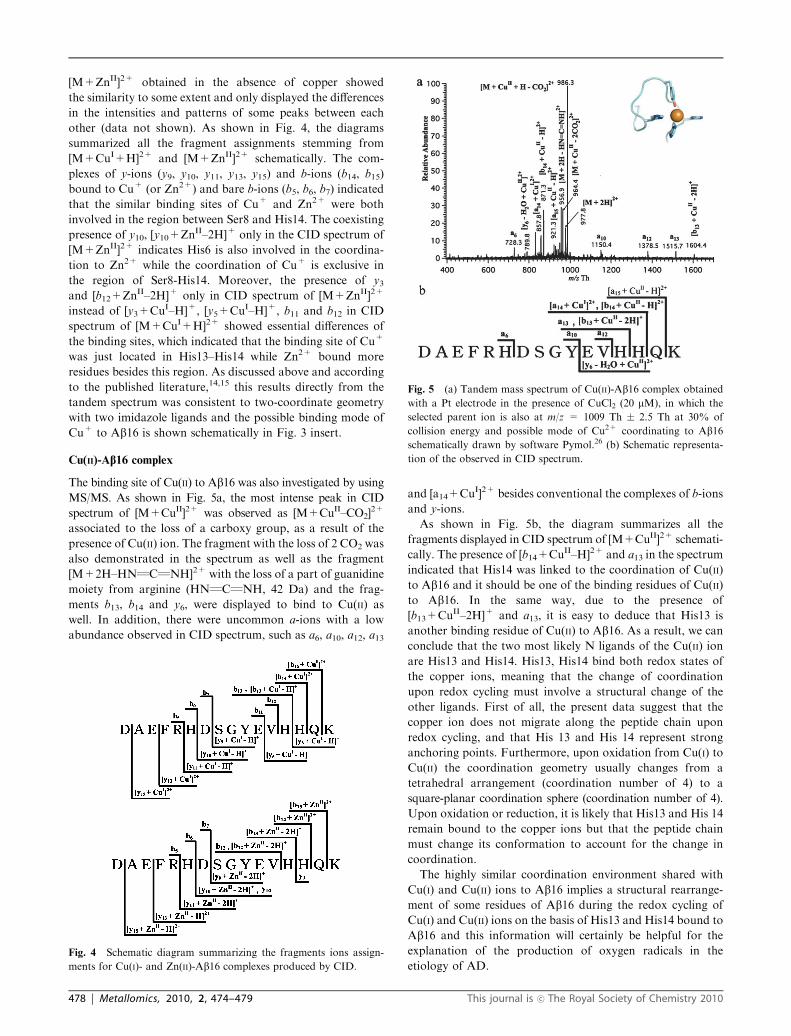
The binding site of Cu(II) to Ab16 was also investigated by using
MS/MS. As shown in Fig. 5a, the most intense peak in CID
spectrum of [M+CuII]2+ was observed as [M+CuII–CO2]2+
associated to the loss of a carboxy group, as a result of the
presence of Cu(II) ion. The fragment with the loss of 2 CO2 was
also demonstrated in the spectrum as well as the fragment
[M+2H–HNQCQNH]2+ with the loss of a part of guanidine
moiety from arginine (HNQCQNH, 42 Da) and the frag-
ments b13, b14 and y6, were displayed to bind to Cu(II) as
well. In addition, there were uncommon a-ions with a low
abundance observed in CID spectrum, such as a6, a10, a12, a13
and [a14+CuI]2+ besides conventional the complexes of b-ions
and y-ions.
As shown in Fig. 5b, the diagram summarizes all the
fragments displayed in CID spectrum of [M+CuII]2+ schemati-
cally. The presence of [b14+CuII–H]2+ and a13 in the spectrum
indicated that His14 was linked to the coordination of Cu(II)
to Ab16 and it should be one of the binding residues of Cu(II)
to Ab16. In the same way, due to the presence of
[b13+CuII–2H]+ and a13, it is easy to deduce that His13 is
another binding residue of Cu(II) to Ab16. As a result, we can
conclude that the two most likely N ligands of the Cu(II) ion
are His13 and His14. His13, His14 bind both redox states of
the copper ions, meaning that the change of coordination
upon redox cycling must involve a structural change of the
other ligands. First of all, the present data suggest that the
copper ion does not migrate along the peptide chain upon
redox cycling, and that His 13 and His 14 represent strong
anchoring points. Furthermore, upon oxidation from Cu(I) to
Cu(II) the coordination geometry usually changes from a
tetrahedral arrangement (coordination number of 4) to a
square-planar coordination sphere (coordination number of 4).
Upon oxidation or reduction, it is likely that His13 and His 14
remain bound to the copper ions but that the peptide chain
must change its conformation to account for the change in
coordination.
The highly similar coordination environment shared with
Cu(I) and Cu(II) ions to Ab16 implies a structural rearrange-
ment of some residues of Ab16 during the redox cycling of
Cu(I) and Cu(II) ions on the basis of His13 and His14 bound to
Ab16 and this information will certainly be helpful for the
explanation of the production of oxygen radicals in the
etiology of AD.Fig. 4 Schematic diagram summarizing the fragments ions assign-
ments for Cu(I)- and Zn(II)-Ab16 complexes produced by CID.
Fig. 5 (a) Tandem mass spectrum of Cu(II)-Ab16 complex obtained
with a Pt electrode in the presence of CuCl2 (20 mM), in which the
selected parent ion is also at m/z = 1009 Th � 2.5 Th at 30% of
collision energy and possible mode of Cu2+ coordinating to Ab16schematically drawn by software Pymol.26 (b) Schematic representa-
tion of the observed in CID spectrum.
478 | Metallomics, 2010, 2, 474–479 This journal is �c The Royal Society of Chemistry 2010

The generation of unusual a-ions in CID spectrum of
[M+CuII]2+ might be ascribed to the high oxidation ability
of Cu2+ in the gas phase since it has been reported that
the generation of a-ions are highly linked to the oxidation
environment of the dissociation.19,27 The observation of
[a14+CuI]2+ also corroborated that the reduction of Cu2+
occurred during the process of CID. Moreover, CO2 loss from
[M+CuII]2+ also represents the crucial feature of electron
detachment dissociation,28 which process results in the disso-
ciation of Ca–C and the generation of a-ions. Therefore, the
reduction of bound Cu2+ in the gas phase during the process
of CID may lead to the transfer of electrons to the backbone
of the peptide, which then resulted in Ca–C fragmentation and
the generation of a-ions. Also, the fact that as the collision energy
increased, the intensity of all a-ions increased subsequently
along with the decrease of intensity of [M+CuII–CO2]2+ con-
firmed that these a-ions did come from the dissociation of
[M+CuII–CO2]2+, which also means these a-ions were the result
of high oxidation environment of CID of [M+CuII–CO2]2.
Conclusion
Copper-Ab16 complexes with two oxidation states were
generated on-line by using a sacrificial copper electrode as
an anode of mass spectrometry and Cu(I)-Ab16 complex in
high abundance was formed especially in the presence of
ascorbic acid. A kinetic model was further built and simulated
the process of the production of both Cu(I)- and Cu(II)-Ab16complexes. The binding sites of Cu+ and Cu2+ to Ab16 were
well studied with the help of tandem mass spectrometry. The
binding site of Cu+ was successfully identified as His13, His14
residues, which were further found to be involved in the
binding site of Cu2+ to Ab16. The same coordination environ-
ment shared with both Cu(I) and Cu(II) to Ab16 implies the
conformational change of the peptide chains on the redox
cycling of copper ions bound to Ab16.
Acknowledgements
The authors thank the China Scholarship Council (CSC) for
financial support. Dr Xiaofeng Liu is acknowledged for the
producing of the insets of Fig. 3 and 5(a) with software Pymol.
The authors also thank Swiss National Science Fundation
(SNCF) for the funding for the project of Analytical tools for
proteome analysis and redoxomics (grant no 200020_127142/1).
Notes and references
1 H. A. Lashuel, S. R. LaBrenz, L. Woo, L. C. Serpell andJ. W. Kelly, J. Am. Chem. Soc., 2000, 122, 5262–5277.
2 H. A. Lashuel, D. Hartley, B. M. Petre, T. Walz andP. T. Lansbury, Nature, 2002, 418, 291–291.
3 P. T. Lansbury and H. A. Lashuel, Nature, 2006, 443,774–779.
4 K. Takano, Curr. Alzheimer Res., 2008, 5, 540–547.5 A. I. Bush, Curr. Opin. Chem. Biol., 2000, 4, 184–191.6 A. I. Bush, Trends Neurosci., 2003, 26, 207–214.7 E. Gaggelli, H. Kozlowski, D. Valensin and G. Valensin, Chem.Rev., 2006, 106, 1995–2044.
8 D. G. Smith, R. Cappai and K. J. Barnham, Biochim. Biophys.Acta, Biomembr., 2007, 1768, 1976–1990.
9 L. Guilloreau, S. Combalbert, A. Sournia-Saquet, H. Mazarguiland P. Faller, ChemBioChem, 2007, 8, 1317–1325.
10 M. Brzyska, K. Trzesniewska, A. Wieckowska, A. Szczepankiewiczand D. Elbaum, ChemBioChem, 2009, 10, 1045–1055.
11 P. Zatta, D. Drago, S. Bolognin and S. L. Sensi, Trends Pharmacol.Sci., 2009, 30, 346–355.
12 G. Meloni, V. Sonois, T. Delaine, L. Guilloreau, A. Gillet,J. Teissie, P. Faller and M. Vasak, Nat. Chem. Biol., 2008, 4,366–372.
13 P. Faller and C. Hureau, Dalton Trans., 2009, 1080–1094.14 R. A. Himes, G. Y. Park, G. S. Siluvai, N. J. Blackburn and
K. D. Karlin, Angew. Chem., Int. Ed., 2008, 47, 9084–9087.15 J. Shearer and V. A. Szalai, J. Am. Chem. Soc., 2008, 130,
17826–17835.16 G. Drochioiu, M. Manea, M. Dragusanu, M. Murariu,
E. S. Dragan, B. A. Petre, G. Mezo and M. Przybylski, Biophys.Chem., 2009, 144, 9–20.
17 M. Prudent and H. H. Girault, Analyst, 2009, 134, 2189–2203.18 U. A. Mirza and B. T. Chait, Anal. Chem., 1994, 66,
2898–2904.19 M. Prudent and H. H. Girault, J. Am. Soc. Mass Spectrom., 2008,
19, 560–568.20 M. Prudent, C. Roussel and H. H. Girault, Electrochem. Commun.,
2007, 9, 2067–2074.21 M. Prudent and H. H. Girault, Metallomics, 2009, 1,
157–165.22 V. A. Streltsov and J. N. Varghese, Chem. Commun., 2008,
3169–3171.23 C. S. Atwood, R. C. Scarpa, X. D. Huang, R. D. Moir,
W. D. Jones, D. P. Fairlie, R. E. Tanzi and A. I. Bush,J. Neurochem., 2000, 75, 1219–1233.
24 L. Deng, N. Sun, E. N. Kitova and J. S. Klassen, Anal. Chem.,2010, 82, 2170–2174.
25 N. C. Maiti, D. L. Jiang, A. J. Wain, S. Patel, K. L. Dinh andF. M. Zhou, J. Phys. Chem. B, 2008, 112, 8406–8411.
26 http://www.pymol.org/.27 L. Qiao, H. Y. Bi, J. M. Busnel, J. Waser, P. Y. Yang,
H. H. Girault and B. H. Liu, Chem.–Eur. J., 2009, 15,6711–6717.
28 F. Kjeldsen, O. A. Silivra, I. A. Ivonin, K. F. Haselmann,M. Gorshkov and R. A. Zubarev, Chem.–Eur. J., 2005, 11,1803–1812.
This journal is �c The Royal Society of Chemistry 2010 Metallomics, 2010, 2, 474–479 | 479