Connective Tissue Growth Factor Regulates Interneuron Survival and Information Processing in the...
16
Neuron Article Connective Tissue Growth Factor Regulates Interneuron Survival and Information Processing in the Olfactory Bulb Konstantin Khodosevich, 1,2 Franc ¸ oise Lazarini, 4,5,7 Jakob von Engelhardt, 3,6,7 Hiroshi Kaneko, 1,2 Pierre-Marie Lledo, 4,5 and Hannah Monyer 1,2, * 1 Department of Clinical Neurobiology, Heidelberg University Medical Center, 69120 Heidelberg, Germany 2 Department of Clinical Neurobiology/A230 3 Synaptic Signaling and Neurodegeneration/A300 German Center for Cancer Research (DKFZ), 69120 Heidelberg, Germany 4 Institut Pasteur, Laboratory for Perception and Memory, F-75015 Paris, France 5 Centre National de la Recherche Scientifique (CNRS), Unite ´ Mixte de Recherche (UMR3571), F-75015 Paris, France 6 Synaptic Signalling and Neurodegeneration, German Center for Neurodegenerative Diseases (DZNE), 53127 Bonn, Germany 7 These authors contributed equally to this work *Correspondence: [email protected] http://dx.doi.org/10.1016/j.neuron.2013.07.011 SUMMARY Neurogenesis underlies plastic changes in defined neuronal circuits in the postnatal and adult brain. Here we identify connective tissue growth factor (CTGF) as a critical factor in the mouse olfactory bulb (OB) in determining the efficiency of incorpora- tion of postnatally born inhibitory neurons, thus gating the output of glomeruli, the first relay station of olfactory processing in the brain. In the OB, CTGF expression was restricted to prenatally born external tufted cells. CTGF enhanced the proapopto- tic activity of glial-derived TGF-b2, decreasing the survival of periglomerular inhibitory neurons. Changes in CTGF expression levels in the OB led to modifications in local neuronal circuitry and olfactory behaviors. We show that the odorant-specific re- cruitment of distinct glomeruli resulted in enhanced local CTGF expression levels in the activated glomeruli. Collectively our data reveal a molecular mechanism controlling the survival of defined post- natally born neurons, thus adapting neuronal integra- tion to the sensory experiences. INTRODUCTION In many mammals, including rodents, olfaction is a key sensory modality governing vital behavior such as food seeking, spatial orientation, and sexual and maternal behavior. Olfactory stimuli are conveyed by the olfactory epithelium to the olfactory bulb (OB), the first central relay station of the olfactory system that sends processed information to the cortex (Mombaerts, 2006; Shipley and Ennis, 1996). The two main excitatory neuronal cell types in the OB, namely mitral and tufted cells, are born embry- onically (Hinds, 1968; Imamura et al., 2011). In sharp contrast, the vast majority of OB interneurons are born postnatally. Inter- estingly, the generation of OB interneurons is not restricted to early postnatal stages but persists in the adult (Lledo et al., 2006). Thus, the postnatal OB continues to be invaded by thousands of new neurons daily. Postnatally generated OB interneurons originate in the subventricular zone (SVZ) of the lateral ventricles and migrate via the rostral migratory stream (RMS) to the OB. There, the majority matures into distinct inter- neuron subtypes, namely granule and periglomerular cells (Lledo et al., 2008). The life-long addition of postnatally generated OB interneu- rons into pre-existing local circuits contributes to olfactory infor- mation processing and olfactory learning (Kageyama et al., 2012; Lazarini and Lledo, 2011). The survival of neuroblasts that are integrating into the pre-existing circuitry depends not only on bottom-up and top-down inputs to the OB (Yokoyama et al., 2011), but also on intrinsic cellular programs, including regulation of pro-apoptotic gene expression (Kim et al., 2007). Importantly, at the site of their destination, the survival of OB interneurons is activity-dependent (Petreanu and Alvarez-Buylla, 2002; Saghatelyan et al., 2005), and can be modulated by factors provided by neighboring cells (Lin et al., 2010). In microarray studies we identified connective tissue growth factor (CTGF) whose expression is high in the OB but not detectable in the SVZ, the site of origin for neuroblasts, and the migratory pathway (Khodosevich et al., 2007, 2009). CTGF is a small extracellular protein (38 kDa) encoded by an early response gene (Bradham et al., 1991). Both Ctgf mRNA and pro- tein have a short half-life (Kroening et al., 2009). The various modes of CTGF action have been mapped to distinct functional domains. For instance, CTGF binds and modulates the activity of other growth factors, including IGF, TGF, and BMP (Abreu et al., 2002; Kim et al., 1997), and also interacts with cell receptors, e.g., integrins (Schober et al., 2002). CTGF was shown to have a pleiotropic action during pre- and postnatal development (Frie- drichsen et al., 2005; Ivkovic et al., 2003; Stritt et al., 2009). In the adult, CTGF is expressed in some cell types of cardiovascular and reproductive tissues, and in the brain its expression is Neuron 79, 1–16, September 18, 2013 ª2013 Elsevier Inc. 1 Please cite this article in press as: Khodosevich et al., Connective Tissue Growth Factor Regulates Interneuron Survival and Information Processing in the Olfactory Bulb, Neuron (2013), http://dx.doi.org/10.1016/j.neuron.2013.07.011
-
Upload
independent -
Category
Documents
-
view
2 -
download
0
Transcript of Connective Tissue Growth Factor Regulates Interneuron Survival and Information Processing in the...

Please cite this article in press as: Khodosevich et al., Connective Tissue Growth Factor Regulates Interneuron Survival and Information Processing inthe Olfactory Bulb, Neuron (2013), http://dx.doi.org/10.1016/j.neuron.2013.07.011
Neuron
Article
Connective Tissue Growth Factor RegulatesInterneuron Survival and InformationProcessing in the Olfactory BulbKonstantin Khodosevich,1,2 Francoise Lazarini,4,5,7 Jakob von Engelhardt,3,6,7 Hiroshi Kaneko,1,2 Pierre-Marie Lledo,4,5
and Hannah Monyer1,2,*1Department of Clinical Neurobiology, Heidelberg University Medical Center, 69120 Heidelberg, Germany2Department of Clinical Neurobiology/A2303Synaptic Signaling and Neurodegeneration/A300
German Center for Cancer Research (DKFZ), 69120 Heidelberg, Germany4Institut Pasteur, Laboratory for Perception and Memory, F-75015 Paris, France5Centre National de la Recherche Scientifique (CNRS), Unite Mixte de Recherche (UMR3571), F-75015 Paris, France6Synaptic Signalling and Neurodegeneration, German Center for Neurodegenerative Diseases (DZNE), 53127 Bonn, Germany7These authors contributed equally to this work
*Correspondence: [email protected]
http://dx.doi.org/10.1016/j.neuron.2013.07.011
SUMMARY
Neurogenesis underlies plastic changes in definedneuronal circuits in the postnatal and adult brain.Here we identify connective tissue growth factor(CTGF) as a critical factor in the mouse olfactorybulb (OB) in determining the efficiency of incorpora-tion of postnatally born inhibitory neurons, thusgating the output of glomeruli, the first relay stationof olfactory processing in the brain. In the OB,CTGF expression was restricted to prenatally bornexternal tufted cells. CTGF enhanced the proapopto-tic activity of glial-derived TGF-b2, decreasing thesurvival of periglomerular inhibitory neurons.Changes in CTGF expression levels in the OB led tomodifications in local neuronal circuitry and olfactorybehaviors. We show that the odorant-specific re-cruitment of distinct glomeruli resulted in enhancedlocal CTGF expression levels in the activatedglomeruli. Collectively our data reveal a molecularmechanism controlling the survival of defined post-natally born neurons, thus adapting neuronal integra-tion to the sensory experiences.
INTRODUCTION
In many mammals, including rodents, olfaction is a key sensory
modality governing vital behavior such as food seeking, spatial
orientation, and sexual and maternal behavior. Olfactory stimuli
are conveyed by the olfactory epithelium to the olfactory bulb
(OB), the first central relay station of the olfactory system that
sends processed information to the cortex (Mombaerts, 2006;
Shipley and Ennis, 1996). The two main excitatory neuronal cell
types in the OB, namely mitral and tufted cells, are born embry-
onically (Hinds, 1968; Imamura et al., 2011). In sharp contrast,
the vast majority of OB interneurons are born postnatally. Inter-
estingly, the generation of OB interneurons is not restricted to
early postnatal stages but persists in the adult (Lledo et al.,
2006). Thus, the postnatal OB continues to be invaded by
thousands of new neurons daily. Postnatally generated OB
interneurons originate in the subventricular zone (SVZ) of the
lateral ventricles and migrate via the rostral migratory stream
(RMS) to the OB. There, the majority matures into distinct inter-
neuron subtypes, namely granule and periglomerular cells (Lledo
et al., 2008).
The life-long addition of postnatally generated OB interneu-
rons into pre-existing local circuits contributes to olfactory infor-
mation processing and olfactory learning (Kageyama et al., 2012;
Lazarini and Lledo, 2011). The survival of neuroblasts that are
integrating into the pre-existing circuitry depends not only on
bottom-up and top-down inputs to the OB (Yokoyama et al.,
2011), but also on intrinsic cellular programs, including
regulation of pro-apoptotic gene expression (Kim et al., 2007).
Importantly, at the site of their destination, the survival of OB
interneurons is activity-dependent (Petreanu and Alvarez-Buylla,
2002; Saghatelyan et al., 2005), and can bemodulated by factors
provided by neighboring cells (Lin et al., 2010).
In microarray studies we identified connective tissue growth
factor (CTGF) whose expression is high in the OB but not
detectable in the SVZ, the site of origin for neuroblasts, and
the migratory pathway (Khodosevich et al., 2007, 2009). CTGF
is a small extracellular protein (38 kDa) encoded by an early
response gene (Bradham et al., 1991). Both Ctgf mRNA and pro-
tein have a short half-life (Kroening et al., 2009). The various
modes of CTGF action have been mapped to distinct functional
domains. For instance, CTGF binds andmodulates the activity of
other growth factors, including IGF, TGF, and BMP (Abreu et al.,
2002; Kim et al., 1997), and also interacts with cell receptors,
e.g., integrins (Schober et al., 2002). CTGF was shown to have
a pleiotropic action during pre- and postnatal development (Frie-
drichsen et al., 2005; Ivkovic et al., 2003; Stritt et al., 2009). In the
adult, CTGF is expressed in some cell types of cardiovascular
and reproductive tissues, and in the brain its expression is
Neuron 79, 1–16, September 18, 2013 ª2013 Elsevier Inc. 1
Neuron
CTGF Regulates Interneuron Survival
Please cite this article in press as: Khodosevich et al., Connective Tissue Growth Factor Regulates Interneuron Survival and Information Processing inthe Olfactory Bulb, Neuron (2013), http://dx.doi.org/10.1016/j.neuron.2013.07.011
restricted to layer VI of the cortex and the mitral cell and
glomerular layers of the OB (Friedrichsen et al., 2005; Williams
et al., 2007).
In contrast to the wealth of information regarding the involve-
ment of CTGF in a number of pathogenic processes, e.g.,
fibrosis, wound healing, or cancer (de Winter et al., 2008;
Shi-Wen et al., 2008), little is known so far about its function
under physiological conditions in the postnatal and adult organ-
ism. The lack of studies is not surprising, given the scarce
expression of CTGF postnatally and the perinatal lethality of
Ctgf knockout mice (Ivkovic et al., 2003).
In this study we overcame the drawback of the global
knockout by using virus-mediated overexpression and knock-
down approaches in vivo, and demonstrated activity-dependent
regulation of CTGF expression in prenatally born external tufted
cells. Furthermore, we provided evidence that, in conjunction
with glial-derived TGF-b2, CTGF controls the survival of newly
generated neurons, thus modifying local network activity and
olfactory behavior.
RESULTS
CTGF Expression in the Postnatal BrainTo determine the regional expression pattern of CTGF in the
postnatal brain, we performed in situ hybridization experiments
on sagittal brain sections from 2-month-old wild-typemice using
38 nt oligoprobes complementary to Ctgf mRNA. As previously
shown (Stritt et al., 2009; Williams et al., 2007), Ctgf mRNA
was detected in layer VI of the cortex as well as in the mitral
cell and glomerular layers of the main and accessory OB (Fig-
ure 1A). At the immunohistochemical level, cortical CTGF
expression was confined to a thin layer just above the corpus
callosum, most likely comprising layer VIb neurons, also known
as layer VII or subplate neurons (Figure 1B). In the OB, CTGF
immunolabeling was restricted to the glomerular layer (Fig-
ure 1C). In the somata of individual cells, CTGF expression
was more intense in the vicinity of the major process (Figure 1D).
CTGF was barely detectable in the mitral cell layer (Figure 1C).
Since the glomerular layer of the OB comprises different excit-
atory and inhibitory neuronal subtypes (Batista-Brito et al., 2008;
Kiyokage et al., 2010), we analyzed the cell-type-specific
expression of CTGF. CTGF-positive cells were colabeled exclu-
sively by cholecystokinin (CCK) antibodies (Figure 1E), but not
interneuron- (calretinin, calbindin, tyrosine hydroxylase, GAD)
or glia (Olig2 and GFAP)-specific antibodies (see Figures S1A–
S1D online, or data not shown, respectively). Since it was
previously shown that in the OB CCK positivity can be detected
by and large only in the external tufted cells (Liu and Shipley,
1994; Shipley and Ennis, 1996), it can be inferred from our
colabeling experiments that CTGF expression is restricted to
this cell type.
Next, we analyzed the time of birth for CTGF-positive cells by
taking recourse to labeling with 5-bromo-20-deoxyuridine (BrdU),a marker of dividing cells, and injected BrdU at several time
points before and after birth of the animals. We found that
CTGF-positive neurons were generated mainly around E16-18
(37% of CTGF-positive cells were labeled by BrdU at E16.5
and 42% at E17.5) (see Figure 1F for E17.5). The production of
2 Neuron 79, 1–16, September 18, 2013 ª2013 Elsevier Inc.
CTGF-positive neurons was completed by the time of birth
(none of the analyzed neurons generated at P0 (n > 500) and
P7 (n > 500) coexpressed CTGF) (Figures S1E and S1F, respec-
tively). This profile corresponds to the one reported for external
tufted cells (Hinds, 1968).
Finally, to further confirm the glutamatergic nature of CTGF-
positive cells, we injected adeno-associated virus (AAV) ex-
pressing tdTomato into the OB (to visualize cell processes) and
analyzed 1 week later the expression of CTGF and of the vesic-
ular glutamate transporter 1 (vGluT1), known to be expressed in
tufted cells (Ohmomo et al., 2009). All virus-labeled CTGF-
positive cells (26/26) coexpressed vGluT1 (Figure S1G).
Together these data provide evidence that CTGF-positive cells
in the glomerular layer are prenatally born excitatory external
tufted cells.
The postnatal developmental expression profile revealed that
CTGF began to be detectable around P3 and was expressed in
the glomerular and mitral cell layers by P5 (Figure 1G). While
CTGF expression in the mitral cell layer gradually decreased
and was barely detectable by P12, expression in the glomerular
layer remained stable throughout postnatal development and
persisted in the adult (Figure 1G).
CTGF Regulates Neuronal Survival in the GlomerularLayer of the OBGiven the postnatal lethality ofCtgf�/�mice around birth (Ivkovic
et al., 2003), we used an alternative approach and knocked down
CTGF expression in the OB (Figure 2A). We produced AAVs ex-
pressing tdTomato together with control shRNA or any of two
shRNAs against CTGF and infected the entire OB of P3-old
wild-type mice (Figure 2A1). Simultaneously, we injected a retro-
virus expressing EGFP into the SVZ to label newborn neurons
around P3, and analyzed the survival of labeled neurons in the
OB at P31 and P59. Retroviruses infect only fast-dividing cells,
the majority of which migrate from the SVZ into the OB within
7 days (Khodosevich et al., 2011). This approach allows the
labeling of SVZ-generated neuroblasts that are born around
the same time and will thus have approximately the same
‘‘age’’ whenmaturing in theOB.We confirmed efficient OB infec-
tion by AAVs 4 weeks postinjection (Figure 2B; note the high
intensity of red fluorescence visible even in normal daylight). At
the same time, we were able to track EGFP-positive infected
cells that had migrated from the SVZ into the OB and had inte-
grated into local circuits. CTGF expression knockdown was
confirmed by western blot (Figure 2C) and immunohistochem-
istry in the OB (Figure 2D).
Injection with any of the two CTGF knockdown viruses led to
an increase in the number of infected EGFP-positive cells
located in the glomerular layer (Figures 2E and 2F). The effect
of CTGF knockdown mediated by shRNA Ctgf-2 AAV was
completely rescued when coinjecting another AAV expressing
a modified CTGF that was resistant to shRNA Ctgf-2 (see Fig-
ure 2A2 for the rationale and Figure 2F for the results). Similar re-
sults were obtained when AAV infections were restricted to the
glomerular layer (data not shown).
Since the SVZ produces granule and periglomerular cells, an
increase in the number of periglomerular cells might result from
one of two causes. (1) CTGF knockdown resulted in mistargeted
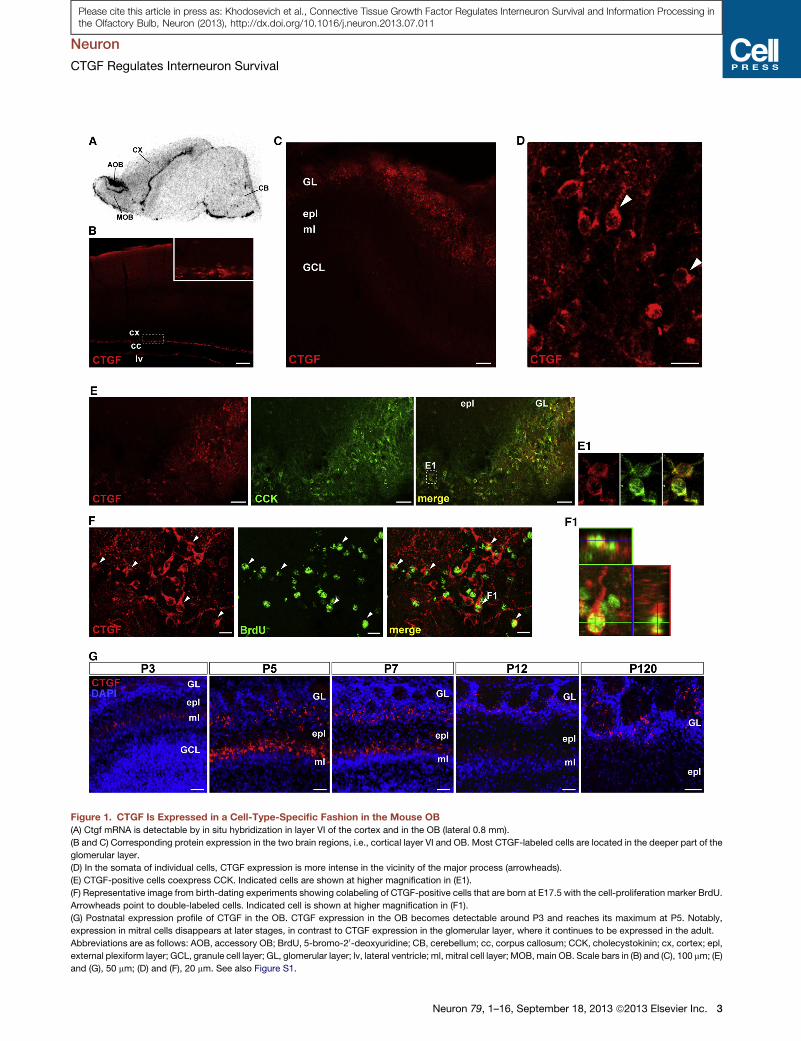
Figure 1. CTGF Is Expressed in a Cell-Type-Specific Fashion in the Mouse OB
(A) Ctgf mRNA is detectable by in situ hybridization in layer VI of the cortex and in the OB (lateral 0.8 mm).
(B and C) Corresponding protein expression in the two brain regions, i.e., cortical layer VI and OB. Most CTGF-labeled cells are located in the deeper part of the
glomerular layer.
(D) In the somata of individual cells, CTGF expression is more intense in the vicinity of the major process (arrowheads).
(E) CTGF-positive cells coexpress CCK. Indicated cells are shown at higher magnification in (E1).
(F) Representative image from birth-dating experiments showing colabeling of CTGF-positive cells that are born at E17.5 with the cell-proliferation marker BrdU.
Arrowheads point to double-labeled cells. Indicated cell is shown at higher magnification in (F1).
(G) Postnatal expression profile of CTGF in the OB. CTGF expression in the OB becomes detectable around P3 and reaches its maximum at P5. Notably,
expression in mitral cells disappears at later stages, in contrast to CTGF expression in the glomerular layer, where it continues to be expressed in the adult.
Abbreviations are as follows: AOB, accessory OB; BrdU, 5-bromo-20-deoxyuridine; CB, cerebellum; cc, corpus callosum; CCK, cholecystokinin; cx, cortex; epl,
external plexiform layer; GCL, granule cell layer; GL, glomerular layer; lv, lateral ventricle; ml, mitral cell layer; MOB, main OB. Scale bars in (B) and (C), 100 mm; (E)
and (G), 50 mm; (D) and (F), 20 mm. See also Figure S1.
Neuron
CTGF Regulates Interneuron Survival
Neuron 79, 1–16, September 18, 2013 ª2013 Elsevier Inc. 3
Please cite this article in press as: Khodosevich et al., Connective Tissue Growth Factor Regulates Interneuron Survival and Information Processing inthe Olfactory Bulb, Neuron (2013), http://dx.doi.org/10.1016/j.neuron.2013.07.011
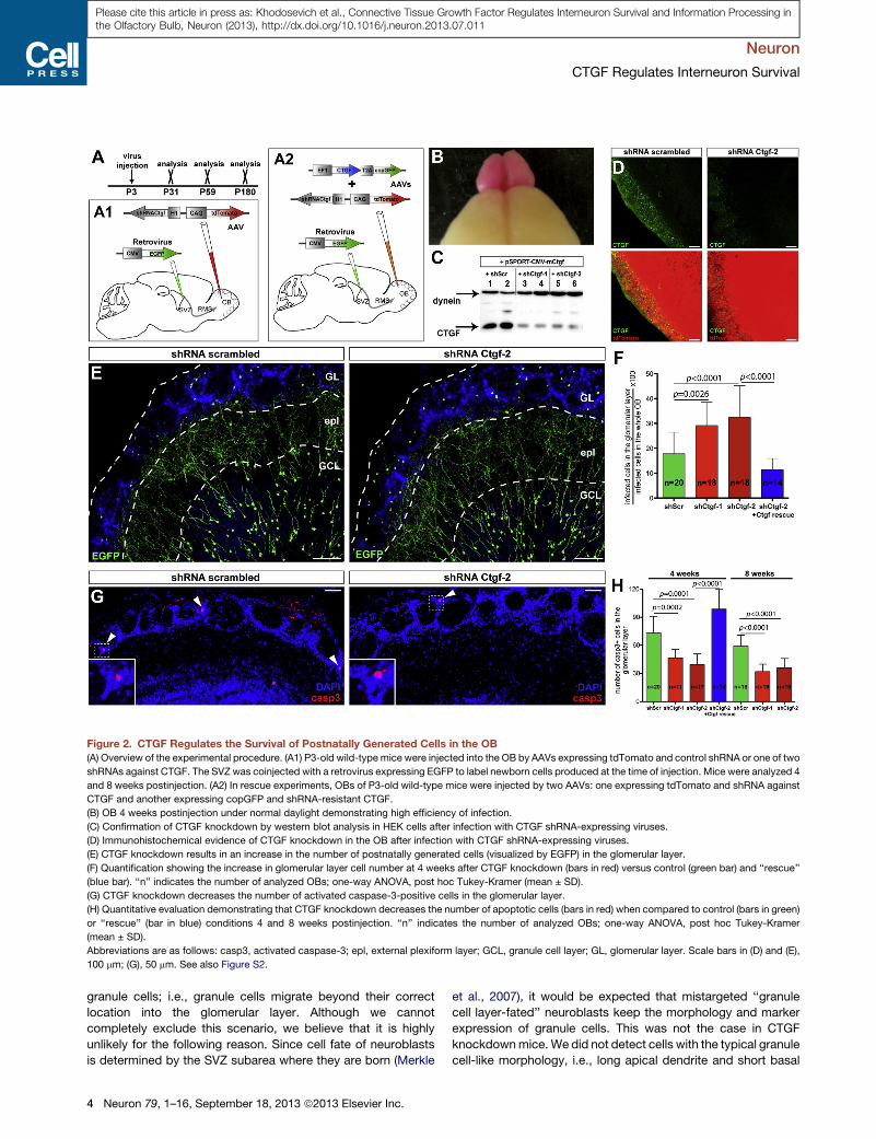
Figure 2. CTGF Regulates the Survival of Postnatally Generated Cells in the OB(A) Overview of the experimental procedure. (A1) P3-old wild-type mice were injected into the OB by AAVs expressing tdTomato and control shRNA or one of two
shRNAs against CTGF. The SVZ was coinjected with a retrovirus expressing EGFP to label newborn cells produced at the time of injection. Mice were analyzed 4
and 8 weeks postinjection. (A2) In rescue experiments, OBs of P3-old wild-type mice were injected by two AAVs: one expressing tdTomato and shRNA against
CTGF and another expressing copGFP and shRNA-resistant CTGF.
(B) OB 4 weeks postinjection under normal daylight demonstrating high efficiency of infection.
(C) Confirmation of CTGF knockdown by western blot analysis in HEK cells after infection with CTGF shRNA-expressing viruses.
(D) Immunohistochemical evidence of CTGF knockdown in the OB after infection with CTGF shRNA-expressing viruses.
(E) CTGF knockdown results in an increase in the number of postnatally generated cells (visualized by EGFP) in the glomerular layer.
(F) Quantification showing the increase in glomerular layer cell number at 4 weeks after CTGF knockdown (bars in red) versus control (green bar) and ‘‘rescue’’
(blue bar). ‘‘n’’ indicates the number of analyzed OBs; one-way ANOVA, post hoc Tukey-Kramer (mean ± SD).
(G) CTGF knockdown decreases the number of activated caspase-3-positive cells in the glomerular layer.
(H) Quantitative evaluation demonstrating that CTGF knockdown decreases the number of apoptotic cells (bars in red) when compared to control (bars in green)
or ‘‘rescue’’ (bar in blue) conditions 4 and 8 weeks postinjection. ‘‘n’’ indicates the number of analyzed OBs; one-way ANOVA, post hoc Tukey-Kramer
(mean ± SD).
Abbreviations are as follows: casp3, activated caspase-3; epl, external plexiform layer; GCL, granule cell layer; GL, glomerular layer. Scale bars in (D) and (E),
100 mm; (G), 50 mm. See also Figure S2.
Neuron
CTGF Regulates Interneuron Survival
Please cite this article in press as: Khodosevich et al., Connective Tissue Growth Factor Regulates Interneuron Survival and Information Processing inthe Olfactory Bulb, Neuron (2013), http://dx.doi.org/10.1016/j.neuron.2013.07.011
granule cells; i.e., granule cells migrate beyond their correct
location into the glomerular layer. Although we cannot
completely exclude this scenario, we believe that it is highly
unlikely for the following reason. Since cell fate of neuroblasts
is determined by the SVZ subarea where they are born (Merkle
4 Neuron 79, 1–16, September 18, 2013 ª2013 Elsevier Inc.
et al., 2007), it would be expected that mistargeted ‘‘granule
cell layer-fated’’ neuroblasts keep the morphology and marker
expression of granule cells. This was not the case in CTGF
knockdownmice.We did not detect cells with the typical granule
cell-like morphology, i.e., long apical dendrite and short basal
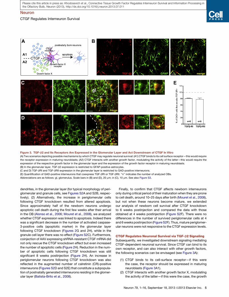
Figure 3. TGF-b2 and Its Receptors Are Expressed in the Glomerular Layer and Act Downstream of CTGF In Vitro
(A) Two scenarios depicting possible mechanisms by which CTGFmay regulate neuronal survival: (A1) CTGF binds to its cell surface receptor—this would require
the receptor expression in maturing neuroblasts; (A2) CTGF interacts with another growth factor, modulating the activity of the latter—this would require the
expression of the respective growth factor in the glomerular layer and the expression of the growth factor receptor in maturing neuroblasts.
(B) In the glomerular layer, TGF-b2 expression is restricted to GFAP-positive astrocytes.
(C and D) TGF-bRI and TGF-bRII expression in the glomerular layer is restricted to GAD-positive interneurons.
(E) Quantification of GAD-positive interneurons that coexpress TGF-bRI or TGF-bRII. ‘‘n’’ indicates the number of analyzed OBs.
Abbreviations are as follows: gl, glomerulus. Scale bars in (B) and (D), 20 mm; in (C), 10 mm. See also Figure S3.
Neuron
CTGF Regulates Interneuron Survival
Please cite this article in press as: Khodosevich et al., Connective Tissue Growth Factor Regulates Interneuron Survival and Information Processing inthe Olfactory Bulb, Neuron (2013), http://dx.doi.org/10.1016/j.neuron.2013.07.011
dendrites, in the glomerular layer (for typical morphology of peri-
glomerular and granule cells, see Figures S2A and S2B, respec-
tively). (2) Alternatively, the increase in periglomerular cells
following CTGF knockdown resulted from altered apoptosis.
Since approximately half of the newborn neurons undergo
apoptotic cell death during the first few weeks after their arrival
in the OB (Alonso et al., 2006; Mouret et al., 2008), we analyzed
whether CTGF expression was linked to apoptosis. Indeed there
was a significant decrease in the number of activated caspase-
3-positive cells (apoptotic marker) in the glomerular layer
following CTGF knockdown (Figures 2G and 2H), while in the
granule cell layer there was no effect (Figure S2C). Furthermore,
coinjection of AAV expressing shRNA-resistant CTGFmRNA did
not only rescue the CTGF knockdown effect but even increased
the number of apoptotic cells (Figure 2H). Reduction in the num-
ber of apoptotic cells following CTGF knockdown was still
significant 8 weeks postinjection (Figure 2H). An increase in
periglomerular neurons following CTGF knockdown was also
reflected in the augmented number of calretinin (CR)-positive
interneurons (Figures S2D and S2E) that constitute a subpopula-
tion of postnatally generated interneurons residing in the glomer-
ular layer (Batista-Brito et al., 2008).
Finally, to confirm that CTGF affects newborn interneurons
only during critical period of their maturationwhen they are prone
to cell death, around 10–25 days after birth (Mouret et al., 2008),
but not when these neurons become mature, we extended
our analysis of newborn cell survival after CTGF knockdown
to 6 weeks postinjection and compared the data with those
obtained at 4 weeks postinjection (Figure S2F). There were no
differences in the number of survived periglomerular cells at 4
and6weekspostinjection (FigureS2F). Thus,mature periglomer-
ular neurons were not responsive to the CTGF expression levels.
CTGF Regulates Neuronal Survival via TGF-b2 SignalingSubsequently, we investigated downstream signaling mediating
CTGF-dependent neuronal survival. Since CTGF can bind to its
own receptor, and can also interact with other growth factors,
the following scenarios can be envisaged (see Figure 3A):
(1) CTGF binds to its cell-surface receptor—if this were
the case, the receptor should be expressed in maturing
neuroblasts (Figure 3A1).
(2) CTGF interacts with another growth factor X, modulating
the activity of the latter—if this were the case, the growth
Neuron 79, 1–16, September 18, 2013 ª2013 Elsevier Inc. 5
Neuron
CTGF Regulates Interneuron Survival
Please cite this article in press as: Khodosevich et al., Connective Tissue Growth Factor Regulates Interneuron Survival and Information Processing inthe Olfactory Bulb, Neuron (2013), http://dx.doi.org/10.1016/j.neuron.2013.07.011
factor X should be expressed in the glomerular layer and
the corresponding receptor in maturing neuroblasts (Fig-
ure 3A2).
To identify potential candidates that are expressed in the
glomerular layer, we took recourse to published data and the
Allen Mouse Brain In Situ Atlas (http://www.brain-map.org).
The expression of several genes was analyzed in the glomerular
layer by western blot analysis and immunohistochemistry. The
first hypothesis could be refuted, since cell receptors mediating
direct interaction with CTGF (i.e., TrkA, integrins aMb2, avb3,
a6b1, and a5b1) were not expressed in maturing neuroblasts
(data not shown).
Pursuing the second hypothesis (Figure 3A2), we identified
insulin-like growth factor 1 (IGF1) expression in a subpopulation
of TH-positive interneurons and in CCK-positive external tufted
cells. However, we could not detect IGF1 receptor expression
in glomerular layer interneurons (data not shown).
Another potential candidate that might be involved in CTGF
downstream signaling is the transforming growth factor b
(TGF-b). CTGF was shown to interact via its N-terminal domain
with both TGF-b1 and TGF-b2, enhancing their binding to
TGF-b receptors and thus augmenting their activity (Abreu
et al., 2002; Khankan et al., 2011). Furthermore, TGF-b signaling
was demonstrated to activate apoptosis via a caspase-3-depen-
dent pathway (Jang et al., 2002). We did not detect TGF-b1
expression in the glomerular layer of the OB by western blot or
immunohistochemistry (data not shown). In contrast, TGF-b2
and its receptors TGF-bRI and TGF-bRII were all expressed in
the glomerular layer (Figure S3A). Interestingly, TGF-b2 was
expressed exclusively in GFAP-positive astrocytes in the
glomerular layer (Figure 3B). TGF-bRs, on the other hand, were
found in a subpopulation of GAD-positive interneurons of the
glomerular layer (Figures 3C–3E). Furthermore, TGF-bRI-
expressing cells colocalized 100% with TGF-bRII-expressing
cells (Figure S3B). Thus, at least at the expression level, the
TGF-b signaling components fulfill the requirements to mediate
CTGF-dependent responses in the newly born glomerular layer
neurons: CTGF and TGF-b2 are expressed in the glomerular
layer, whereas TGF-bRI and TGF-bRII can be detected in newly
born neurons (see scheme in Figure 3A2).
To further substantiate the hypothesis of CTGF-TGF-b2
coupling, we quantified activated caspase-3-positive cells in
the glomerular layer of organotypic cultures obtained from
coronal OB sections of 1-month-old wild-type mice that were
cultured for 16 hr (Figure S3C). Addition of neutralizing anti-
CTGF antibody decreased apoptosis in the glomerular layer,
whereas recombinant CTGF increased it (Figure S3D). Anti-
TGF-b2, but not anti-TGF-b1, neutralizing antibody abolished
the effect of recombinant CTGF. Furthermore, both recombinant
TGF-bRI-Fc and TGF-bRII-Fc (fusions of TGF-bRs with the Fc
immunoglobulin domain that bind to TGF-b and block its activity)
abolished the CTGF effect. Likewise, neutralizing antibody to
TGF-b2, but not to TGF-b1, reduced glomerular layer apoptosis,
and recombinant TGF-b2 enhanced it. When CTGF and TGF-b2
were both added to the medium, there was a dramatic increase
in the number of apoptotic cells in the glomerular layer. Blocking
TGF-b signaling by TGF-bRI-Fc or TGF-bRII-Fc did not decrease
6 Neuron 79, 1–16, September 18, 2013 ª2013 Elsevier Inc.
apoptosis in the granule cell layer (Figure S3E). The intracellular
apoptotic effect of TGF-b is mediated by SMADproteins (Shi and
Massague, 2003). SMAD3 inhibitor completely abrogated the
enhanced apoptosis resulting from treatment with recombinant
CTGF or with CTGF+TGF-b2 (Figure S3D). Thus, CTGF potenti-
ates TGF-b2 activity in the glomerular layer and regulates
apoptosis of newly generated cells via the TGF-bR-SMAD
pathway.
To obtain in vivo evidence that CTGF acts via the TGF-b
pathway, we knocked down TGF-bRI expression in postnatally
generated neuroblasts (Figure 4A). For these knockdown exper-
iments, we employed microRNAs (miRNAs) rather than shRNAs,
since they enable the use of RNA-polymerase II-specific
promoters (e.g., synapsin promoter). P3-old wild-type mice
were injected into the SVZ with retroviruses expressing EmGFP
(Emerald GFP) and control miRNA or any of two miRNAs against
TGF-bRI and were analyzed 4 weeks postinjection (Figure 4A1).
The synapsin promoter assured the onset of miRNA expression
only during neuroblast maturation in the OB, thus leading to
restricted EmGFP fluorescence in the postnatally born OB inter-
neurons. TGF-bRI expression knockdown was confirmed by
western blot (Figure 4A3). Knockdown of TGF-bRI in maturing
neurons of the OB increased the number of infected cells in
the glomerular layer, mimicking the effect of CTGF knockdown
in the OB (Figures 4B and 4C). Together these results demon-
strated that the effects observed in vitro in organotypic cultures
could be replicated in vivo.
To show that CTGF activity is TGF-b dependent in vivo, P3-old
wild-type mice were injected into the SVZ with retroviruses ex-
pressing EmGFP and control miRNA or any of two miRNAs
against TGF-bRI. Simultaneously, we injected AAV to knock
down CTGF in the OB (Figure 4A2). If CTGF acted via a different
receptor than TGF-bRI, then TGF-bRI-knockdown cells should
continue to be responsive to changed CTGF levels in the glomer-
ular layer. However, CTGF knockdown did not affect the survival
of TGF-bRI-knockdown neurons (Figure 4C), demonstrating that
CTGF regulates neuronal survival via TGF-b signaling.
To substantiate our finding that TGF-bRs act downstream of
CTGF, we employed P5-old Tgfbr2 fl/fl mice that were injected
into the SVZ with two retroviruses: one expressing tdTomato
and another expressing Cre recombinase together with EGFP
(Figure 4D, D1). This ensures that Tgfbr2 knockout cells in the
OB are EGFP positive, whereas control cells are tdTomato pos-
itive. To increase CTGF-dependent apoptosis in maturing neuro-
blasts, OBs were simultaneously injected with AAV that ex-
pressed CTGF and copGFP (since EGFP and copGFP are
unrelated proteins, they can be distinguished by immunohisto-
chemistry). As yet another control, we used the same injection
protocol in wild-type mice. Mice were analyzed 7, 14, 28, and
40 days postinjection. Up to 14 days postinjection, the ratio
between EGFP (= knockout) and tdTomato (= control) cells
was around 1 for both granule cell and glomerular layers of the
Tgfbr2 fl/fl and wild-type mice (Figure 4F). However, at 28 days
postinjection and later, the EGFP:tdTomato cell ratio increased
in the glomerular layer of Tgfbr2 fl/fl mice in comparison to
wild-type mice (Figures 4E and 4F), indicating that Tgfbr2
knockout led to enhanced cell survival. Furthermore, this effect
was observed only in the glomerular layer, but not in the granule
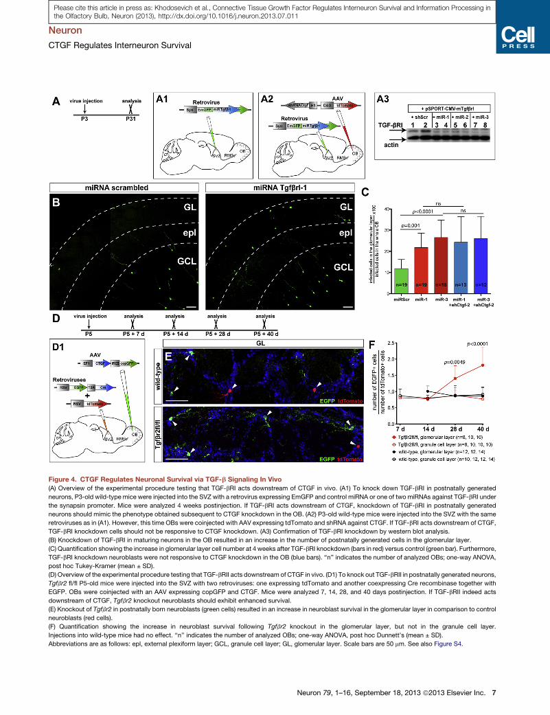
Figure 4. CTGF Regulates Neuronal Survival via TGF-b Signaling In Vivo
(A) Overview of the experimental procedure testing that TGF-bRI acts downstream of CTGF in vivo. (A1) To knock down TGF-bRI in postnatally generated
neurons, P3-old wild-type mice were injected into the SVZ with a retrovirus expressing EmGFP and control miRNA or one of two miRNAs against TGF-bRI under
the synapsin promoter. Mice were analyzed 4 weeks postinjection. If TGF-bRI acts downstream of CTGF, knockdown of TGF-bRI in postnatally generated
neurons should mimic the phenotype obtained subsequent to CTGF knockdown in the OB. (A2) P3-old wild-type mice were injected into the SVZ with the same
retroviruses as in (A1). However, this time OBs were coinjected with AAV expressing tdTomato and shRNA against CTGF. If TGF-bRI acts downstream of CTGF,
TGF-bRI knockdown cells should not be responsive to CTGF knockdown. (A3) Confirmation of TGF-bRI knockdown by western blot analysis.
(B) Knockdown of TGF-bRI in maturing neurons in the OB resulted in an increase in the number of postnatally generated cells in the glomerular layer.
(C) Quantification showing the increase in glomerular layer cell number at 4 weeks after TGF-bRI knockdown (bars in red) versus control (green bar). Furthermore,
TGF-bRI knockdown neuroblasts were not responsive to CTGF knockdown in the OB (blue bars). ‘‘n’’ indicates the number of analyzed OBs; one-way ANOVA,
post hoc Tukey-Kramer (mean ± SD).
(D) Overview of the experimental procedure testing that TGF-bRII acts downstream of CTGF in vivo. (D1) To knock out TGF-bRII in postnatally generated neurons,
Tgfbr2 fl/fl P5-old mice were injected into the SVZ with two retroviruses: one expressing tdTomato and another coexpressing Cre recombinase together with
EGFP. OBs were coinjected with an AAV expressing copGFP and CTGF. Mice were analyzed 7, 14, 28, and 40 days postinjection. If TGF-bRII indeed acts
downstream of CTGF, Tgfbr2 knockout neuroblasts should exhibit enhanced survival.
(E) Knockout of Tgfbr2 in postnatally born neuroblasts (green cells) resulted in an increase in neuroblast survival in the glomerular layer in comparison to control
neuroblasts (red cells).
(F) Quantification showing the increase in neuroblast survival following Tgfbr2 knockout in the glomerular layer, but not in the granule cell layer.
Injections into wild-type mice had no effect. ‘‘n’’ indicates the number of analyzed OBs; one-way ANOVA, post hoc Dunnett’s (mean ± SD).
Abbreviations are as follows: epl, external plexiform layer; GCL, granule cell layer; GL, glomerular layer. Scale bars are 50 mm. See also Figure S4.
Neuron
CTGF Regulates Interneuron Survival
Neuron 79, 1–16, September 18, 2013 ª2013 Elsevier Inc. 7
Please cite this article in press as: Khodosevich et al., Connective Tissue Growth Factor Regulates Interneuron Survival and Information Processing inthe Olfactory Bulb, Neuron (2013), http://dx.doi.org/10.1016/j.neuron.2013.07.011
Neuron
CTGF Regulates Interneuron Survival
Please cite this article in press as: Khodosevich et al., Connective Tissue Growth Factor Regulates Interneuron Survival and Information Processing inthe Olfactory Bulb, Neuron (2013), http://dx.doi.org/10.1016/j.neuron.2013.07.011
cell layer (Figure 4F, Figure S4A). Notably, enhanced cell survival
following Tgfbr2 knockout was also observed when CTGF-
dependent apoptosis was not boosted by CTGF overexpression
(Figure S4B). Together, these experiments imply TGF-bRII as the
downstream effector of CTGF in postnatally generated periglo-
merular cells.
Next, we sought direct proof that astrocytes are the source of
TGF-b2 that mediates CTGF signaling in vivo (Figure S4C). Wild-
type P3-old mice were injected into the SVZ with retrovirus
expressing tdTomato to label newborn neurons around P3 (Fig-
ure S4C1). Simultaneously, OBs were injected with a mix of two
AAVs: one AAV overexpressing CTGF (to increase CTGF-
dependent apoptosis) and another AAV to selectively knock-
down TGF-b2 expression in astrocytes. To this end, we used
an AAV expressing EmGFP and control miRNA or any of two
miRNAs against TGF-b2 under the control of the astrocyte-
specific promoter GFAP (Figure S4C1). In addition, we pseudo-
typed AAV particles with rh43 nonhuman AAV serotype that was
shown to ensure glial cell tropism (Lawlor et al., 2009). The result-
ing AAV indeed guaranteed astrocyte-specific infection in the
OB (98% of total infected cells were astrocytes; Figure S4D).
TGF-b2 expression knockdown was confirmed by western blot
(Figure S4C2). Using this approach, we showed that TGF-b2
knockdown resulted in a significant increase of tdTomato-posi-
tive cells in the glomerular layer (Figures S4E and S4F). TGF-b2
knockdown rescued CTGF-induced neuronal apoptosis, thus
identifying astrocytes as the source that provides TGF-b2.
CTGF Regulates Local Circuit Activity in the GlomerularLayerTo evaluate the potential impact of CTGF-induced changes on
olfactory information processing, we first tested whether an
increase in the number of neurons in the glomerular layer
changed local circuit activity in the OB. P3-old wild-type mice
were injected into the OB by AAVs expressing tdTomato
together with control shRNA or any of the two shRNAs against
CTGF (Figure 5A). Simultaneously, a retrovirus expressing
EGFP was injected into the SVZ. Around 4–5 weeks postin-
jection, we tested electrophysiological parameters of cells
belonging to the two cell populations: EGFP-labeled neurons
representing periglomerular neurons produced postnatally
around P3, and tdTomato-labeled neurons born before P3
(only interneurons in the glomerular layer of the OB were
recorded, identified by visual and electrophysiological charac-
teristics). The frequency of spontaneous inhibitory postsynaptic
currents (sIPSCs) recorded from control periglomerular cells
(0.37 and 0.27 Hz, for Figures 5B and 5C, respectively) was
similar to what was published before (0.36 Hz) (Grubb et al.,
2008). However, after CTGF knockdown, EGFP-labeled inter-
neurons exhibited an increase in the sIPSC frequency, indicating
an increase in the network inhibition on periglomerular cells,
while the amplitude of sIPSCs was not affected (Figure 5B, Fig-
ure S5A). Frequency and amplitude of spontaneous excitatory
postsynaptic currents (sEPSCs) were not changed (Figure 5B,
S5A). The same effect was observed for tdTomato-labeled inter-
neurons (the bulk of this population is born prenatally) (Figure 5C,
Figure S5B). As a consequence, there was a considerable
decrease in the sEPSC/sIPSC ratio for both cell populations.
8 Neuron 79, 1–16, September 18, 2013 ª2013 Elsevier Inc.
The glomerular layer contains at least five interneuronal subtypes
(Parrish-Aungst et al., 2007) that are born at different prenatal/
postnatal ages (Batista-Brito et al., 2008). The decrease in exci-
tation/inhibition ratio appeared to affect all cell types. The elec-
trophysiological results demonstrate that CTGF expression
levels have a profound role in the regulation of local circuit
activity by shifting the excitation/inhibition ratio in periglomerular
interneurons.
We then tested if the increase in periglomerular cell number
affects inhibition of the two main excitatory neuron types of the
OB, i.e., mitral cells and external tufted cells. P3-old wild-type
mice were injected into the OB with AAVs expressing tdTomato
together with control shRNA or any of the two shRNAs against
CTGF (Figure 5D, D1), and were analyzed around 30 or 45 days
postinjection. At 30 days postinjection there was no difference
in sIPSC frequency betweenCTGFknockdown and controlmitral
cells. However, at 45 days postinjection, mitral cells in CTGF
knockdown mice exhibited significantly increased sIPSC fre-
quency (Figure 5E). In contrast, CTGF knockdown did not in-
crease significantly sIPSC frequency of external tufted cells
that were recorded 45 days postinjection (Figure S5F). Neither
mitral nor external tufted cell sIPSC amplitudes were modified
by CTGF knockdown (Figures S5C and S5D for mitral cells and
Figure S5G for external tufted cells). Mitral cells receive inhibition
via periglomerular cells onto the most distal parts of their apical
dendrites in the glomeruli, whereas granule cells impinge on
mitral cell lateral dendrites considerably more proximally in the
external plexiform layer (Liu and Shipley, 1994; Shipley and
Ennis, 1996). Since dendritic filtering slows the kinetics of re-
corded synaptic inputs, we investigated if the increase in the
electrotonically more distal inhibition of mitral cell dendrites pro-
vided by periglomerular cells leads to a slowing of sIPSC decay
kinetics. There was indeed an increase in the sIPSC tdecay in
mitral cells ofCTGFknockdownanimals compared to that in con-
trol animals around 45 days postinjection (Figures 5F and 5G).
Activation of dopamine and GABAB receptors on olfactory
nerve reduces the probability of glutamate release (Aroniadou-
Anderjaska et al., 2000; Kageyama et al., 2012). We tested if
the periglomerular cell number increase affects the release prob-
ability by analyzing paired-pulse ratios of EPSCs evoked by two
subsequent stimuli delivered on olfactory nerve. Paired-pulse
ratios of EPSCs recorded in mitral and external tufted cells
were around 0.7 for both control and CTGF knockdown condi-
tions (Figures S5E and S5H) and were in accordance with pub-
lished data (Aroniadou-Anderjaska et al., 2000; Grubb et al.,
2008). Thus, unaltered paired-pulse ratios indicate that presyn-
aptic properties of olfactory nerve input to the glomeruli were
not affected by the genetic manipulation.
CTGF Expression Regulates Mouse Olfactory BehaviorOdorant detection, discrimination, and memory (Figure 6A) were
tested in control and CTGF knockdown wild-type mice (Fig-
ure 6A1) 2 months postinjection (n control = 6, n shCtgf-2 = 11)
using anolfactometer. Following theprotocol shown in Figure 6A,
we investigated olfactory sensitivity by determining the detection
threshold for two odorants, namely pyridazine and 1-decanol,
using the descending method of limits in two-odorant rewarded
discrimination tasks (rewarded odorant, stimulus [S+]; solvent,
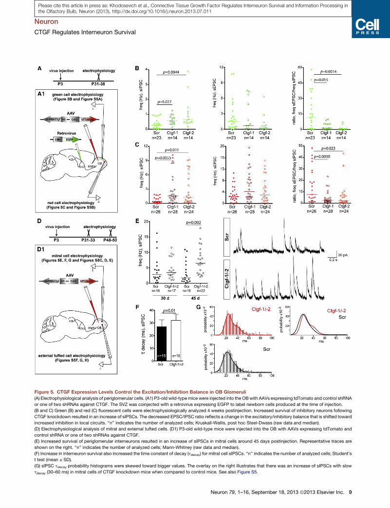
Figure 5. CTGF Expression Levels Control the Excitation/Inhibition Balance in OB Glomeruli
(A) Electrophysiological analysis of periglomerular cells. (A1) P3-old wild-typemice were injected into the OBwith AAVs expressing tdTomato and control shRNA
or one of two shRNAs against CTGF. The SVZ was coinjected with a retrovirus expressing EGFP to label newborn cells produced at the time of injection.
(B and C) Green (B) and red (C) fluorescent cells were electrophysiologically analyzed 4 weeks postinjection. Increased survival of inhibitory neurons following
CTGF knockdown resulted in an increase of sIPSCs. The decreased EPSC/IPSC ratio reflects a change in the excitatory/inhibitory balance that is shifted toward
increased inhibition in local circuits. ‘‘n’’ indicates the number of analyzed cells; Kruskall-Wallis, post hoc Steel-Dwass (raw data and median).
(D) Electrophysiological analysis of mitral and external tufted cells. (D1) P3-old wild-type mice were injected into the OB with AAVs expressing tdTomato and
control shRNA or one of two shRNAs against CTGF.
(E) Increased survival of periglomerular interneurons resulted in an increase of sIPSCs in mitral cells around 45 days postinjection. Representative traces are
shown on the right. ‘‘n’’ indicates the number of analyzed cells; Mann-Whitney (raw data and median).
(F) Increase in interneuron survival also increased the time constant of decay (tdecay) for mitral cell sIPSCs. ‘‘n’’ indicates the number of analyzed cells; Student’s
t test (mean ± SD).
(G) sIPSC tdecay probability histograms were skewed toward bigger values. The overlay on the right illustrates that there was an increase of sIPSCs with slow
tdecay (30–60 ms) in mitral cells of CTGF knockdown mice when compared to control mice. See also Figure S5.
Neuron
CTGF Regulates Interneuron Survival
Neuron 79, 1–16, September 18, 2013 ª2013 Elsevier Inc. 9
Please cite this article in press as: Khodosevich et al., Connective Tissue Growth Factor Regulates Interneuron Survival and Information Processing inthe Olfactory Bulb, Neuron (2013), http://dx.doi.org/10.1016/j.neuron.2013.07.011

Figure 6. CTGF Knockdown Enhances Olfactory Discrimination, and CTGF Expression Is Dependent upon Olfactory Experience
(A) Overview of the experimental protocol. OBs of P3-old wild-type mice were infected by AAVs expressing tdTomato and control shRNA or shRNA Ctgf-2 (A1).
Two-month-old mice were analyzed on olfactometers (n control = 6, n shCtgf-2 = 11).
(B and D) CTGF knockdown resulted in a decrease of the detection threshold for pyridazine and 1-decanol, respectively, Student’s t test (mean ± SEM).
(C and E) CTGF knockdown mice reached criterion performance (R90% correct responses in the block) at 10–100 lower odorant concentrations than controls,
two-way ANOVA (mean ± SEM).
(F) CTGF knockdown mice needed fewer blocks of learning to discriminate between odorants, two-way ANOVA, post-hoc Bonferroni (mean ± SEM).
(G) For some odorants, CTGF knockdown mice had a decreased reaction time for the negative (S�) odorant, Student’s t test (mean ± SEM).
(H) Overview of the experimental protocol for olfactory ablation. P30-old wild-type mice were i.p. injected with the olfactotoxin dichlobenil that impairs olfactory
sensory neuronal input into the OB, and CTGF expression was analyzed at 4, 8, 12, and 20 days postinjection.
(I) CTGF expression was dramatically decreased already 4 days postinjection. Twelve days postinjection, CTGF expression was close to background. CTGF
expression was restored to some extent 20 days postinjection, correlating with partial reinnervation of olfactory sensory input as previously reported (Alonso
et al., 2008).
(J) TGF-b2 expression was already increased 4 days postinjection. Eight days postinjection, TGF-b2 expression level was highest and gradually decreased
thereafter.
Scale bars, 50 mm. See also Figure S6.
Neuron
CTGF Regulates Interneuron Survival
Please cite this article in press as: Khodosevich et al., Connective Tissue Growth Factor Regulates Interneuron Survival and Information Processing inthe Olfactory Bulb, Neuron (2013), http://dx.doi.org/10.1016/j.neuron.2013.07.011
nonrewarded [S�]). Mice were given two sessions (eight blocks
each) per day with one decimal dilution of the odorant per ses-
sion. CTGF knockdown resulted in a decrease of the detection
threshold for both odorants (Figures 6B and 6D, respectively)
10 Neuron 79, 1–16, September 18, 2013 ª2013 Elsevier Inc.
and in shifting criterion performance (i.e., R90% correct re-
sponses per block) to lower odorant concentration (Figures 6C
and 6E, respectively). The same paradigmwas used for olfactory
discrimination between limonene pair (+ and� enantiomers) and

Neuron
CTGF Regulates Interneuron Survival
Please cite this article in press as: Khodosevich et al., Connective Tissue Growth Factor Regulates Interneuron Survival and Information Processing inthe Olfactory Bulb, Neuron (2013), http://dx.doi.org/10.1016/j.neuron.2013.07.011
their binary mixtures. Overall, CTGF knockdown mice needed
fewer blocks of trials to reach criterion performance (Figure 6F)
and spent less time at negative (S�) odorant identification
when discriminating between limonene enantiomers (Figure 6G).
Analysis of long-termmemory did not show adifference between
CTGF knockdown and controls (data not shown). Thus, CTGF
knockdown mice performed better in odorant detection and
olfactory discrimination than did controls, but their olfactory
memory remained unchanged.
CTGF Expression Is Dependent on Olfactory ActivityFinally, we investigated whether CTGF expression is sensitive to
the degree of olfactory experience. To this end, we injected P30-
old wild-type mice i.p. with the olfactotoxin dichlobenil, thus
ablating sensory neurons of the main olfactory epithelium target-
ing the OB (Alonso et al., 2008), and analyzed CTGF expression
4, 8, 12, and 20 days postinjection (Figure 6H). Ablation of olfac-
tory receptor neurons was confirmed by impairment in food
search of olfactotoxin-injected mice (Figures S6A and S6B)
and by decreased expression of the olfactory marker protein
(OMP) (Figures S6C and S6D). Suppression of the sensory input
led to a significant decrease in CTGF expression already 4 days
postinjection, and at 12 days postinjectionCTGF expressionwas
barely detectable (Figure 6I and Figure S6E for quantification).
CTGF expression was partially restored 20 days postinjection,
correlating with partial reinnervation of olfactory sensory input
as reported (Alonso et al., 2008). Decreased CTGF expression
levels following olfactotoxin treatment did not result from a
demise of external tufted cells (i.e., CTGF-expressing cells), as
was evidenced by an unchanged number of CCK-positive cells
(data not shown). Conversely, TGF-b2 expression was strongly
elevated during the period of olfactory input impairment and
reached a maximum 8 days postinjection (Figure 6J).
Ctgf is an immediate-early gene with a short half-life (Kroening
et al., 2009) and is thus suited to test whether environmental
changes at a fast timescale alter CTGF expression levels. The
first indication for the activity-dependent regulation of CTGF
expression derives from its coexpression with c-fos, another
activity-dependent immediate-early gene (Figure S6F). Since
suppression of olfactory input resulted in a reduction of CTGF
expression, we hypothesized that olfactory enrichment aug-
ments CTGF expression. Mice were presented daily with three
distinct odors for three weeks (63 distinct odorants in total) (Fig-
ure S6G), and CTGF expression levels were evaluated thereafter
in 200 glomeruli (Figures S6H and S6I). Glomeruli were subdi-
vided according to their intensity of CTGF expression into four
categories: very high (>10 units), high (5–10 units), intermediate
(1–5 units), and low (0–1 units) (for details, see the Supplemental
Experimental Procedures). There was no difference in CTGF
expression levels between controls and mice subjected to an
odor-enriched environment (Figure S6J). This is not too surpris-
ing, as one would expect to see a potential augmentation of the
short-lived CTGF only in few glomeruli that were activated by the
last few odorants. We also measured whether the enrichment
paradigm resulted in changes of neuronal survival in the glomer-
ular layer (see BrdU treatment rationale in Figure S6G) and found
no difference in BrdU-positive cell number between control and
odor-enriched mice (Figure S6K).
To investigate the possible activity dependence of CTGF
expression in specific glomeruli, we employed transgenic
mice, namely MOR23-IRES-tauGFP, in which only two glomeruli
per OB are EGFP labeled and hence can be surveyed over time
(Vassalli et al., 2002). MOR23 receptors (and thus MOR23-GFP
glomeruli) are activated by the odorant lyral (Grosmaitre et al.,
2006). In the first set of experiments, we presented lyral or ace-
tophenone (unrelated to lyral odorant) to P60-old transgenic
mice for 10 min or 8 hr and analyzed CTGF expression 3 hr post-
exposure or immediately thereafter, respectively (Figure 7A).
Lyral exposure increased CTGF expression levels in EGFP-
labeled glomeruli in comparison to acetophenone (Figures 7B
and 7C). We also investigated whether adjacent glomeruli might
be affected, keeping in mind though that glomeruli detecting
odorants with similar chemical functional groups might cluster
together (Mori et al., 2006). Despite this caveat, differences in
CTGF levels evoked by the two odorants were not significant
(Figures S7A and S7B). Overall, our results indicate that
olfactory activity indeed enhances CTGF expression precisely
in specific odor-activated glomeruli. Thus, CTGF expression
levels undergo rapid modifications in response to changes in
olfactory activity.
Most previous studies employed ‘‘broad-spectrum’’ modifica-
tions of sensory input induced either by olfactory enrichment or
sensory deprivation in order to study survival of postnatally
generated OB neurons in the whole circuitry. Here, we aimed
at studying activity-dependent modulation of neuronal survival
in distinct glomeruli. To this end, we employed MOR23-IRES-
tauGFP transgenic mice and labeled postnatally generated cells
by adding BrdU in the drinking water from P20 to P27 (Fig-
ure S7C). Three weeks later, mice were exposed to lyral or ace-
tophenone for different time periods—1 min, 10 min, 1 hr, 8 hr,
and 24 hr—and analyzed 7 days postexposure. Exposure to lyral
for 1 min had no effect on neuronal survival in MOR23 glomeruli,
but all other treatments from 10 min onward decreased neuronal
survival by 20% (Figures S7D and S7E). Thus, stimulation of ol-
factory activity by a distinct odorant decreases neuronal survival
in the odorant-specific glomeruli.
Finally, we analyzedwhether this decrease of neuronal survival
is mediated by CTGF. We performed a similar experiment as the
one above, in MOR23-IRES-tauGFP transgenic mice that were
injected into the OBs by control or CTGF knockdown AAVs (Fig-
ure 7D, D1). CTGF knockdownmice exhibited higher cell survival
in the glomerular layer in comparison to control mice, again con-
firming our data that CTGF stimulates neuronal apoptosis (Fig-
ures 7E and 7F). As expected, lyral reduced neuronal survival
across MOR23 glomeruli in control AAV-injected mice (Fig-
ure 7F). However, CTGF knockdown completely abolished
lyral-dependent reduction of neuronal survival (Figure 7F). These
experiments demonstrate that modifications in CTGF expres-
sion levels in response to olfactory activity adjust the survival
of postnatally born neurons in an odorant-specific fashion in
the odorant-responsive glomeruli.
DISCUSSION
In this study we identified CTGF as a modulator of postnatal/
adult OB circuitry. Based on our results, we propose that
Neuron 79, 1–16, September 18, 2013 ª2013 Elsevier Inc. 11

Figure 7. Activity-Dependent Expression of CTGF Regulates Survival of Interneurons in the Glomerular Layer
(A) Overview of the experimental protocol. P60-old MOR23-IRES-tauGFP mice were exposed to lyral for 10 min or 8 hr, and CTGF expression levels were
measured.
(B) CTGF expression was increased after activation of MOR23 glomeruli, indicating that olfactory sensory activity enhanced CTGF expression.
(C) Quantitative evaluation demonstrating that lyral enhances CTGF expression in MOR23 glomeruli after 10 min and 8 hr exposure. ‘‘n’’ indicates the number of
analyzed glomeruli; Student’s t test (mean ± SD).
(D) Overview of the experimental protocol. P3-old MOR23-IRES-tauGFP mice were injected into the OB with AAVs expressing tdTomato and control shRNA or
shRNA against CTGF. Three weeks postinjection mice were given BrdU in the drinking water for 1 week. Mice were given normal water for 3 weeks more and
exposed to lyral or acetophenone for 10 min. Mice were analyzed 1 week following odorant exposure.
(E) CTGF knockdown increased the number of BrdU-positive cells in MOR23 glomeruli in comparison to control mice, and this increase in survival was not
reduced following lyral exposure.
(F) Quantification showing that CTGF knockdown increased neuronal survival in the glomerular layer (green bars, controls; red bars, CTGF knockdown).
Furthermore, whereas in controls lyral exposure reduced cell survival, in CTGF knockdown mice lyral did not affect cell survival. ‘‘n’’ indicates the number of
analyzed glomeruli; Student’s t test (mean ± SD).
Abbreviations are as follows: AC, acetophenone; LY, lyral. Scale bars, 50 mm. See also Figure S7.
Neuron
CTGF Regulates Interneuron Survival
Please cite this article in press as: Khodosevich et al., Connective Tissue Growth Factor Regulates Interneuron Survival and Information Processing inthe Olfactory Bulb, Neuron (2013), http://dx.doi.org/10.1016/j.neuron.2013.07.011
CTGF derived from prenatally born excitatory neurons regulates
the survival of postnatally born inhibitory neurons in the glomer-
ular layer in the following fashion (Figure 8): CTGF is expressed in
external tufted cells that control the output of the glomerular
layer, the first relay station of olfactory information processing
(De Saint Jan et al., 2009; Hayar et al., 2004). CTGF acts via
glial-derived TGF-b2, whose activity it potentiates, promoting
SMAD-dependent apoptosis of newborn neurons in the glo-
merular layer via TGF-bRs. CTGF expression is enhanced by
12 Neuron 79, 1–16, September 18, 2013 ª2013 Elsevier Inc.
olfactory stimulation, thus leading to an activity-dependent
potentiation of TGF-b2 signaling. At the functional level, changes
in inhibitory neuron number modify the excitation/inhibition
balance in stimulated glomeruli and OB output cells (i.e., mitral
cells), thus affecting olfactory behavior. It is of note that the regu-
lation of CTGF-mediated cell survival occurs in a region- and
cell-type-specific manner. Thus, within the OB, only the survival
of periglomerular cells, but not granule cells, is subject to CTGF
regulation.
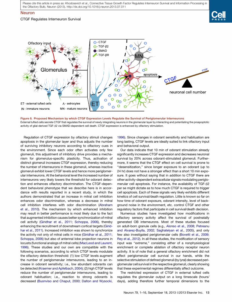
Figure 8. Proposed Mechanism by which CTGF Expression Levels Regulate the Survival of Periglomerular Interneurons
External tufted cells secrete CTGF that regulates the survival of newly integrating neurons in the glomerular layer by interacting and potentiating the proapoptotic
activity of glial-derived TGF-b2 via SMAD-dependent cell death. CTGF expression is enhanced by olfactory stimulation.
Neuron
CTGF Regulates Interneuron Survival
Please cite this article in press as: Khodosevich et al., Connective Tissue Growth Factor Regulates Interneuron Survival and Information Processing inthe Olfactory Bulb, Neuron (2013), http://dx.doi.org/10.1016/j.neuron.2013.07.011
Regulation of CTGF expression by olfactory stimuli changes
apoptosis in the glomerular layer and thus adjusts the number
of surviving inhibitory neurons according to olfactory cues in
the environment. Since each odor often activates only few
glomeruli, this adjustment of inhibitory drive provides a mecha-
nism for glomerulus-specific plasticity. Thus, activation of
distinct glomeruli increases CTGF expression, thereby reducing
the number of interneurons in these glomeruli, whereas inactive
glomeruli exhibit lower CTGF levels and hence more periglomer-
ular interneurons. At the behavioral level the increased number of
interneurons very likely lowers the threshold for odorant detec-
tion and enhances olfactory discrimination. The CTGF-depen-
dent behavioral phenotype that we describe here is in accor-
dance with results reported in a recent study, in which the
authors demonstrated that an increase in mitral cell inhibition
enhances odor discrimination, whereas a decrease in mitral
cell inhibition interferes with odor discrimination (Abraham
et al., 2010). The mechanism by which enhanced inhibition
may result in better performance is most likely due to the fact
that augmented inhibition causes better synchronization ofmitral
cell activity (Giridhar et al., 2011; Schoppa, 2006), thereby
enhancing the recruitment of downstream cortical targets (Girid-
har et al., 2011). Increased inhibition was shown to synchronize
the activity not only of mitral cells in mice (Giridhar et al., 2011;
Schoppa, 2006) but also of antennal lobe projection neurons in
locusts (functional analogs ofmitral cells) (MacLeod and Laurent,
1996). These studies and our own are compatible with the
following scenarios, according to which CTGF levels modulate
the olfactory detection threshold: (1) low CTGF levels augment
the number of periglomerular interneurons, leading to an in-
crease in odorant sensitivity; i.e., low abundant odorants can
be detected (Kraemer and Apfelbach, 2004); (2) high CTGF levels
reduce the number of periglomerular interneurons, leading to
odorant habituation; i.e., reaction to strong odorants is
decreased (Buonviso and Chaput, 2000; Dalton and Wysocki,
1996). Since changes in odorant sensitivity and habituation are
long lasting, CTGF levels are ideally suited to link olfactory input
and behavioral output.
Our data indicate that 10 min of odorant stimulation already
significantly increases CTGF expression and decreases neuronal
survival by 20% across odorant-stimulated glomeruli. Further-
more, it seems that the CTGF effect on cell survival is prone to
‘‘desensitization,’’ since longer exposure to an odorant (up to
24 hr) does not have a stronger effect than a short 10 min expo-
sure. It goes without saying that in addition to CTGF there are
other activity-dependent extracellular signalsmodulating periglo-
merular cell apoptosis. For instance, the availability of TGF-b2
per se might dictate as to how much CTGF is required to trigger
cell apoptosis. Each of these signals very likely exhibits different
kinetics of cell survival/death regulation. Little is known so far on
how time of odorant exposure, odorant intensity, level of back-
ground noise in the environment, etc. control CTGF and other
regulatory factors that participate in cell survival/death decision.
Numerous studies have investigated how modifications in
olfactory sensory activity affect the survival of postnatally
generated OB interneurons. Most of these studies focused
on adult-born granule cells (e.g., Alonso et al., 2008; Petreanu
and Alvarez-Buylla, 2002; Saghatelyan et al., 2005), and only
few also investigated periglomerular cells (Bovetti et al., 2009;
Rey et al., 2012). In all these studies, the modification of sensory
input was ‘‘extreme,’’ consisting either of a nonphysiological
enrichment or complete ablation of olfactory receptor neuron
activity. It is of note that a general olfactory enrichment did not
affect periglomerular cell survival in our hands, while the
selective stimulationof definedglomeruli (by lyral) decreasedperi-
glomerularcell survival in the respectiveglomeruli, clearly showing
that these experimental regimes differentially affect outcome.
The restricted expression of CTGF in external tufted cells
regulates the glomerular output on a long timescale (hours/
days), adding therefore further temporal dimensions to the
Neuron 79, 1–16, September 18, 2013 ª2013 Elsevier Inc. 13

Neuron
CTGF Regulates Interneuron Survival
Please cite this article in press as: Khodosevich et al., Connective Tissue Growth Factor Regulates Interneuron Survival and Information Processing inthe Olfactory Bulb, Neuron (2013), http://dx.doi.org/10.1016/j.neuron.2013.07.011
well-described short timescale (millisecond range) regulation.
External tufted cells exert a control of local synaptic processing
in a glomerulus at several levels. Thus, the axons of external
tufted cells connect intrabulbar isofunctional odor columns
(Liu and Shipley, 1994), whereas intraglomerular connections
between external tufted cells and periglomerular cells as well
as short axon cells amplify the sensory input and synchronize
glomerular output (De Saint Jan et al., 2009; Hayar et al.,
2004). Low threshold activation and the propensity of sponta-
neous rhythmic burst firing designate external tufted cells to be
excitatory pacemaker-like cells that drive glomerular output
activity. External tufted cells exert this control at a fast timescale
via chemical and electrical synapses. In contrast, we demon-
strate here a mechanism by which external tufted cells regulate
glomerular output at a much longer timescale.
CTGF responsiveness is reminiscent of two other immediate-
early genes, c-fos and Egr1 (also known as Zif268 protein).
Expression of c-fos and Egr1 in the glomerular layer already sig-
nificantly increases 45 min after odor exposure (Johnson et al.,
1995). This regulation contrasts with the activity-dependent
regulation of tyrosine hydroxylase (TH) in periglomerular neurons
that decreases significantly only after several days following
sensory deprivation (Baker et al., 1993), again highlighting the
diversity of temporal regulations that take place in glomeruli.
In summary, we here identified CTGF as a proapoptotic factor
whose activity-dependent increase of expression eliminated
newborn neurons in a locally restrictedmanner. Our experiments
showed that even a small increase in the number of surviving
cells dramatically changed olfactory behavior. Survival/death
choice is regulated by external stimuli, and the number of surviv-
ing cells is ‘‘adapted’’ according to the animal’s local environ-
ment. Since olfaction is the most important sensory mode for
many mammals, ‘‘olfactory competition’’ for food, mating, and
predator versus prey relationship plays a decisive role during
the life of an animal. Hence tight regulation of newly added
neurons is a crucial mechanism enabling an adaptive response
to environmental changes.
EXPERIMENTAL PROCEDURES
Materials and Reagents
All antibodies and chemicals are listed in the Supplemental Experimental
Procedures.
Animals
All animal procedures were performed according to the regulations of Heidel-
berg University/German Cancer Research Center or Pasteur Institute Animal
Care Committees.
Plasmid Cloning
To obtain miRNA cassettes expressed under the synapsin or GFAP promoter,
we used the BLOCK-iT PolII system (Invitrogen, Germany) and subcloned
miRNA cassettes to viral vectors containing the synapsin or GFAP promoter,
respectively. shRNA constructs were cloned as previously described (Khodo-
sevich et al., 2009). For details of cloning, see the Supplemental Experimental
Procedures.
Analysis of mi/shRNA Silencing Efficiency
The efficiency of mi/shRNA silencing was tested as previously described
(Khodosevich et al., 2009). For details, see the Supplemental Experimental
Procedures.
14 Neuron 79, 1–16, September 18, 2013 ª2013 Elsevier Inc.
Production of Recombinant Viruses
Recombinant retroviruses and AAVs were produced as previously described
(Khodosevich et al., 2009, 2012).
Injection of Recombinant Viruses into the Mouse Brain
The titer of the injected virus had been adjusted such as to be equal for all
experiments—4 3 108 units/ml for AAVs and 108 units/ml for retroviruses.
For double SVZ/OB injections, mice were first injected into the OB using glass
capillary and immediately after into the SVZ by a Hamilton syringe (Hamilton,
Switzerland). Injection procedure is described in the Supplemental Experi-
mental Procedures.
Data Analysis and Statistics
Data were analyzed using GraphPad Prism (GraphPad Software, USA) and R
software. Normally distributed data were analyzed by t test, ANOVA, Tukey, or
Tukey-Kramer tests; non-normally distributed data were analyzed by
Mann-Whitney, Kruskal-Wallis, Dunn’s, Dunnett’s, or Steel-Dwass tests. See
the Supplemental Experimental Procedures for details of analysis procedures
and statistics.
Olfactory Enrichment
Animals were exposed to odor-enriched environment for 3 weeks as
described previously (Alonso et al., 2008). For details, see the Supplemental
Experimental Procedures.
Chemical Ablation of Olfactory Epithelium
Mice were given one i.p. injection of dichlobenil (2,6-dichlobenzonitrile,
100 mg/kg body weight) or DMSO and analyzed 4, 8, 12, and 20 days
postinjection.
Lyral Stimulation
MOR23-IRES-tauGFP mice were presented with tissue soaked in 4-
(4-hydroxy-4-methylpentyl)-3-cyclohexene-1-carboxaldehyde (= lyral) for dif-
ferent time periods. For details, see the Supplemental Experimental
Procedures.
Electrophysiology
Whole-cell recordings were performed at room temperature using sagittal OB
slices of 250 mm thickness from P31- to P50-old mouse brains. For details, see
the Supplemental Experimental Procedures.
Olfactory Behavior
Mice were partially water deprived for 1 week and then trained using an oper-
ant conditioning go-out procedure in computer-controlled olfactometers. In
this paradigm, mice were trained to respond to the presence of a positive
stimulus odorant (S+) by licking the water delivery tube and to refrain from
responding to the presence of negative stimulus odorant (S�). Odorant detec-
tion threshold, odorant discrimination, and long-term olfactory memory were
analyzed. See the Supplemental Experimental Procedures for details.
Additional experiments are described in the Supplemental Experimental
Procedures.
SUPPLEMENTAL INFORMATION
Supplemental Information includes seven figures, Supplemental Experimental
Procedures, and Supplemental References and can be found with this article
online at http://dx.doi.org/10.1016/j.neuron.2013.07.011.
ACKNOWLEDGMENTS
We thank U. Amtmann, R. Hinz-Herkommer, I. Preugschat-Gumprecht, D. van
der Giezen, and N. Torquet for technical assistance; C. Le Magueresse and
P. Seeburg for critical reading of the manuscript; R. Goldschmeding for Ctgf
knockout mice; K. Krieglstein and Bjorn Spittau for Tgfbr2 knockout mice;
P. Mombaerts for MOR23-IRES-tauGFP mice; M. Ehlers for the pSPORT-
mTgfbr1 plasmid; W. Kelsch for the retroviral EGFP-T2A-Cre plasmid; and
Neuron
CTGF Regulates Interneuron Survival
Please cite this article in press as: Khodosevich et al., Connective Tissue Growth Factor Regulates Interneuron Survival and Information Processing inthe Olfactory Bulb, Neuron (2013), http://dx.doi.org/10.1016/j.neuron.2013.07.011
University of Pennsylvania Vector Core team for the AAV rh43 helper plasmid.
The GFAP promoter was provided by M. Brenner (Alabama Neuroscience
Blueprint Core, NIH grants NS39055 andNS057098). This work was supported
by the Schilling Foundation and DFG (SFB488 and FOR643 grants) to H.M. The
laboratory of P.-M.L. is supported by the life insurance company ‘‘AG2R-La
Mondiale,’’ the Agence Nationale de la Recherche ‘‘ANR-BLAN-SVSE4-LS-
110624,’’ and ‘‘ANR-09-NEUR-004’’ in the frame of ‘‘ERA-NET NEURON’’ of
the FP7 program by the European Commission.
Accepted: July 11, 2013
Published: August 29, 2013
REFERENCES
Abraham, N.M., Egger, V., Shimshek, D.R., Renden, R., Fukunaga, I.,
Sprengel, R., Seeburg, P.H., Klugmann, M., Margrie, T.W., Schaefer, A.T.,
and Kuner, T. (2010). Synaptic inhibition in the olfactory bulb accelerates
odor discrimination in mice. Neuron 65, 399–411.
Abreu, J.G., Ketpura, N.I., Reversade, B., and De Robertis, E.M. (2002).
Connective-tissue growth factor (CTGF) modulates cell signalling by BMP
and TGF-beta. Nat. Cell Biol. 4, 599–604.
Alonso, M., Viollet, C., Gabellec, M.M., Meas-Yedid, V., Olivo-Marin, J.C., and
Lledo, P.M. (2006). Olfactory discrimination learning increases the survival of
adult-born neurons in the olfactory bulb. J. Neurosci. 26, 10508–10513.
Alonso, M., Ortega-Perez, I., Grubb, M.S., Bourgeois, J.P., Charneau, P., and
Lledo, P.M. (2008). Turning astrocytes from the rostral migratory stream into
neurons: a role for the olfactory sensory organ. J. Neurosci. 28, 11089–11102.
Aroniadou-Anderjaska, V., Zhou, F.M., Priest, C.A., Ennis, M., and Shipley,
M.T. (2000). Tonic and synaptically evoked presynaptic inhibition of sensory
input to the rat olfactory bulb via GABA(B) heteroreceptors. J. Neurophysiol.
84, 1194–1203.
Baker, H., Morel, K., Stone, D.M., and Maruniak, J.A. (1993). Adult naris
closure profoundly reduces tyrosine hydroxylase expression in mouse olfac-
tory bulb. Brain Res. 614, 109–116.
Batista-Brito, R., Close, J., Machold, R., and Fishell, G. (2008). The distinct
temporal origins of olfactory bulb interneuron subtypes. J. Neurosci. 28,
3966–3975.
Bovetti, S., Veyrac, A., Peretto, P., Fasolo, A., and De Marchis, S. (2009).
Olfactory enrichment influences adult neurogenesis modulating GAD67 and
plasticity-related molecules expression in newborn cells of the olfactory
bulb. PLoS ONE 4, e6359, http://dx.doi.org/10.1371/journal.pone.0006359.
Bradham, D.M., Igarashi, A., Potter, R.L., and Grotendorst, G.R. (1991).
Connective tissue growth factor: a cysteine-rich mitogen secreted by human
vascular endothelial cells is related to the SRC-induced immediate early
gene product CEF-10. J. Cell Biol. 114, 1285–1294.
Buonviso, N., and Chaput, M. (2000). Olfactory experience decreases respon-
siveness of the olfactory bulb in the adult rat. Neuroscience 95, 325–332.
Dalton, P., and Wysocki, C.J. (1996). The nature and duration of adaptation
following long-term odor exposure. Percept. Psychophys. 58, 781–792.
De Saint Jan, D., Hirnet, D., Westbrook, G.L., and Charpak, S. (2009). External
tufted cells drive the output of olfactory bulb glomeruli. J. Neurosci. 29, 2043–
2052.
de Winter, P., Leoni, P., and Abraham, D. (2008). Connective tissue growth
factor: structure-function relationships of a mosaic, multifunctional protein.
Growth Factors 26, 80–91.
Friedrichsen, S., Heuer, H., Christ, S., Cuthill, D., Bauer, K., and Raivich, G.
(2005). Gene expression of connective tissue growth factor in adult mouse.
Growth Factors 23, 43–53.
Giridhar, S., Doiron, B., and Urban, N.N. (2011). Timescale-dependent shaping
of correlation by olfactory bulb lateral inhibition. Proc. Natl. Acad. Sci. USA
108, 5843–5848.
Grosmaitre, X., Vassalli, A., Mombaerts, P., Shepherd, G.M., and Ma, M.
(2006). Odorant responses of olfactory sensory neurons expressing the
odorant receptor MOR23: a patch clamp analysis in gene-targeted mice.
Proc. Natl. Acad. Sci. USA 103, 1970–1975.
Grubb, M.S., Nissant, A., Murray, K., and Lledo, P.M. (2008). Functional matu-
ration of the first synapse in olfaction: development and adult neurogenesis.
J. Neurosci. 28, 2919–2932.
Hayar, A., Karnup, S., Ennis, M., and Shipley, M.T. (2004). External tufted cells:
a major excitatory element that coordinates glomerular activity. J. Neurosci.
24, 6676–6685.
Hinds, J.W. (1968). Autoradiographic study of histogenesis in themouse olfac-
tory bulb. I. Time of origin of neurons and neuroglia. J. Comp. Neurol. 134,
287–304.
Imamura, F., Ayoub, A.E., Rakic, P., and Greer, C.A. (2011). Timing of neuro-
genesis is a determinant of olfactory circuitry. Nat. Neurosci. 14, 331–337.
Ivkovic, S., Yoon, B.S., Popoff, S.N., Safadi, F.F., Libuda, D.E., Stephenson,
R.C., Daluiski, A., and Lyons, K.M. (2003). Connective tissue growth factor
coordinates chondrogenesis and angiogenesis during skeletal development.
Development 130, 2779–2791.
Jang, C.W., Chen, C.H., Chen, C.C., Chen, J.Y., Su, Y.H., and Chen, R.H.
(2002). TGF-beta induces apoptosis through Smad-mediated expression of
DAP-kinase. Nat. Cell Biol. 4, 51–58.
Johnson, B.A., Woo, C.C., Duong, H., Nguyen, V., and Leon, M. (1995). A
learned odor evokes an enhanced Fos-like glomerular response in the olfac-
tory bulb of young rats. Brain Res. 699, 192–200.
Kageyama, R., Imayoshi, I., and Sakamoto, M. (2012). The role of neurogene-
sis in olfaction-dependent behaviors. Behav. Brain Res. 227, 459–463.
Khankan, R., Oliver, N., He, S., Ryan, S.J., and Hinton, D.R. (2011). Regulation
of fibronectin-EDA through CTGF domain-specific interactions with TGFb2
and its receptor TGFbRII. Invest. Ophthalmol. Vis. Sci. 52, 5068–5078.
Khodosevich, K., Inta, D., Seeburg, P.H., andMonyer, H. (2007). Gene expres-
sion analysis of in vivo fluorescent cells. PLoS ONE 2, e1151, http://dx.doi.org/
10.1371/journal.pone.0001151.
Khodosevich, K., Seeburg, P.H., and Monyer, H. (2009). Major signaling
pathways in migrating neuroblasts. Front. Mol. Neurosci. 2, 7.
Khodosevich, K.,Watanabe, Y., andMonyer, H. (2011). EphA4 preserves post-
natal and adult neural stem cells in an undifferentiated state in vivo. J. Cell Sci.
124, 1268–1279.
Khodosevich, K., Zuccotti, A., Kreuzberg, M.M., LeMagueresse, C., Frank, M.,
Willecke, K., andMonyer, H. (2012). Connexin45 modulates the proliferation of
transit-amplifying precursor cells in the mouse subventricular zone. Proc. Natl.
Acad. Sci. USA 109, 20107–20112.
Kim, H.S., Nagalla, S.R., Oh, Y., Wilson, E., Roberts, C.T., Jr., and Rosenfeld,
R.G. (1997). Identification of a family of low-affinity insulin-like growth
factor binding proteins (IGFBPs): characterization of connective tissue growth
factor as a member of the IGFBP superfamily. Proc. Natl. Acad. Sci. USA 94,
12981–12986.
Kim, W.R., Kim, Y., Eun, B., Park, O.H., Kim, H., Kim, K., Park, C.H., Vinsant,
S., Oppenheim, R.W., and Sun, W. (2007). Impaired migration in the rostral
migratory stream but spared olfactory function after the elimination of pro-
grammed cell death in Bax knock-out mice. J. Neurosci. 27, 14392–14403.
Kiyokage, E., Pan, Y.Z., Shao, Z., Kobayashi, K., Szabo, G., Yanagawa, Y.,
Obata, K., Okano, H., Toida, K., Puche, A.C., and Shipley, M.T. (2010).
Molecular identity of periglomerular and short axon cells. J. Neurosci. 30,
1185–1196.
Kraemer, S., and Apfelbach, R. (2004). Olfactory sensitivity, learning and
cognition in young adult and aged male Wistar rats. Physiol. Behav. 81,
435–442.
Kroening, S., Solomovitch, S., Sachs, M., Wullich, B., and Goppelt-Struebe,
M. (2009). Regulation of connective tissue growth factor (CTGF) by hepatocyte
growth factor in human tubular epithelial cells. Nephrol. Dial. Transplant. 24,
755–762.
Lawlor, P.A., Bland, R.J., Mouravlev, A., Young, D., and During, M.J. (2009).
Efficient gene delivery and selective transduction of glial cells in the
Neuron 79, 1–16, September 18, 2013 ª2013 Elsevier Inc. 15
Neuron
CTGF Regulates Interneuron Survival
Please cite this article in press as: Khodosevich et al., Connective Tissue Growth Factor Regulates Interneuron Survival and Information Processing inthe Olfactory Bulb, Neuron (2013), http://dx.doi.org/10.1016/j.neuron.2013.07.011
mammalian brain by AAV serotypes isolated from nonhuman primates. Mol.
Ther. 17, 1692–1702.
Lazarini, F., and Lledo, P.M. (2011). Is adult neurogenesis essential for
olfaction? Trends Neurosci. 34, 20–30.
Lin, C.W., Sim, S., Ainsworth, A., Okada, M., Kelsch, W., and Lois, C. (2010).
Genetically increased cell-intrinsic excitability enhances neuronal integration
into adult brain circuits. Neuron 65, 32–39.
Liu, W.L., and Shipley, M.T. (1994). Intrabulbar associational system in the rat
olfactory bulb comprises cholecystokinin-containing tufted cells that synapse
onto the dendrites of GABAergic granule cells. J. Comp. Neurol. 346, 541–558.
Lledo, P.M., Alonso,M., andGrubb, M.S. (2006). Adult neurogenesis and func-
tional plasticity in neuronal circuits. Nat. Rev. Neurosci. 7, 179–193.
Lledo, P.M., Merkle, F.T., and Alvarez-Buylla, A. (2008). Origin and function of
olfactory bulb interneuron diversity. Trends Neurosci. 31, 392–400.
MacLeod, K., and Laurent, G. (1996). Distinct mechanisms for synchronization
and temporal patterning of odor-encoding neural assemblies. Science 274,
976–979.
Merkle, F.T., Mirzadeh, Z., and Alvarez-Buylla, A. (2007). Mosaic organization
of neural stem cells in the adult brain. Science 317, 381–384.
Mombaerts, P. (2006). Axonal wiring in themouse olfactory system. Annu. Rev.
Cell Dev. Biol. 22, 713–737.
Mori, K., Takahashi, Y.K., Igarashi, K.M., and Yamaguchi, M. (2006). Maps of
odorant molecular features in the Mammalian olfactory bulb. Physiol. Rev. 86,
409–433.
Mouret, A., Gheusi, G., Gabellec, M.M., de Chaumont, F., Olivo-Marin, J.C.,
and Lledo, P.M. (2008). Learning and survival of newly generated neurons:
when time matters. J. Neurosci. 28, 11511–11516.
Ohmomo, H., Ina, A., Yoshida, S., Shutoh, F., Ueda, S., and Hisano, S. (2009).
Postnatal changes in expression of vesicular glutamate transporters in the
main olfactory bulb of the rat. Neuroscience 160, 419–426.
Parrish-Aungst, S., Shipley, M.T., Erdelyi, F., Szabo, G., and Puche, A.C.
(2007). Quantitative analysis of neuronal diversity in the mouse olfactory
bulb. J. Comp. Neurol. 501, 825–836.
Petreanu, L., and Alvarez-Buylla, A. (2002). Maturation and death of adult-born
olfactory bulb granule neurons: role of olfaction. J. Neurosci. 22, 6106–6113.
16 Neuron 79, 1–16, September 18, 2013 ª2013 Elsevier Inc.
Rey, N.L., Sacquet, J., Veyrac, A., Jourdan, F., and Didier, A. (2012).
Behavioral and cellular markers of olfactory aging and their response to enrich-
ment. Neurobiol. Aging 33, 626.e9–626.e23.
Saghatelyan, A., Roux, P., Migliore, M., Rochefort, C., Desmaisons, D.,
Charneau, P., Shepherd, G.M., and Lledo, P.M. (2005). Activity-dependent
adjustments of the inhibitory network in the olfactory bulb following early post-
natal deprivation. Neuron 46, 103–116.
Schober, J.M., Chen, N., Grzeszkiewicz, T.M., Jovanovic, I., Emeson, E.E.,
Ugarova, T.P., Ye, R.D., Lau, L.F., and Lam, S.C. (2002). Identification of integ-
rin alpha(M)beta(2) as an adhesion receptor on peripheral bloodmonocytes for
Cyr61 (CCN1) and connective tissue growth factor (CCN2): immediate-early
gene products expressed in atherosclerotic lesions. Blood 99, 4457–4465.
Schoppa, N.E. (2006). Synchronization of olfactory bulb mitral cells by pre-
cisely timed inhibitory inputs. Neuron 49, 271–283.
Shi, Y., and Massague, J. (2003). Mechanisms of TGF-b signaling from cell
membrane to the nucleus. Cell 113, 685–700.
Shipley, M.T., and Ennis, M. (1996). Functional organization of olfactory
system. J. Neurobiol. 30, 123–176.
Shi-Wen, X., Leask, A., and Abraham, D. (2008). Regulation and function of
connective tissue growth factor/CCN2 in tissue repair, scarring and fibrosis.
Cytokine Growth Factor Rev. 19, 133–144.
Stritt, C., Stern, S., Harting, K., Manke, T., Sinske, D., Schwarz, H., Vingron,M.,
Nordheim, A., and Knoll, B. (2009). Paracrine control of oligodendrocyte
differentiation by SRF-directed neuronal gene expression. Nat. Neurosci. 12,
418–427.
Vassalli, A., Rothman, A., Feinstein, P., Zapotocky, M., and Mombaerts, P.
(2002). Minigenes impart odorant receptor-specific axon guidance in the olfac-
tory bulb. Neuron 35, 681–696.
Williams, E.O., Xiao, Y., Sickles, H.M., Shafer, P., Yona, G., Yang, J.Y., and Lin,
D.M. (2007). Novel subdomains of the mouse olfactory bulb defined by molec-
ular heterogeneity in the nascent external plexiform and glomerular layers.
BMC Dev. Biol. 7, 48, http://dx.doi.org/10.1186/1471-213X-7-48.
Yokoyama, T.K., Mochimaru, D., Murata, K., Manabe, H., Kobayakawa, K.,
Kobayakawa, R., Sakano, H., Mori, K., and Yamaguchi, M. (2011).
Elimination of adult-born neurons in the olfactory bulb is promoted during
the postprandial period. Neuron 71, 883–897.