The estimation of patient-specific cardiac diastolic functions from clinical measurements
Upload
independentCategory
view
0download
0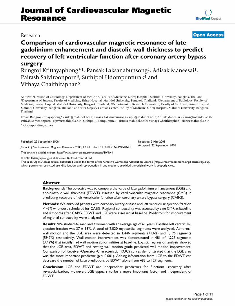
BioMed Central
Journal of Cardiovascular Magnetic Resonance
ss
Open AcceResearchComparison of cardiovascular magnetic resonance of late gadolinium enhancement and diastolic wall thickness to predict recovery of left ventricular function after coronary artery bypass surgeryRungroj Krittayaphong*1, Pansak Laksanabunsong2, Adisak Maneesai1, Pairash Saiviroonporn3, Suthipol Udompunturak4 and Vithaya Chaithiraphan5Address: 1Division of Cardiology, Department of Medicine, Faculty of Medicine, Siriraj Hospital, Mahidol University, Bangkok, Thailand, 2Department of Surgery, Faculty of Medicine, Siriraj Hospital, Mahidol University, Bangkok, Thailand, 3Department of Radiology, Faculty of Medicine, Siriraj Hospital, Mahidol University, Bangkok, Thailand, 4Department of Research Promotion, Faculty of Medicine, Siriraj Hospital, Mahidol University, Bangkok, Thailand and 5Her Majesty Cardiac Center, Faculty of Medicine, Siriraj Hospital, Mahidol University, Bangkok, Thailand
Email: Rungroj Krittayaphong* - [email protected]; Pansak Laksanabunsong - [email protected]; Adisak Maneesai - [email protected]; Pairash Saiviroonporn - [email protected]; Suthipol Udompunturak - [email protected]; Vithaya Chaithiraphan - [email protected]
* Corresponding author
AbstractBackground: The objective was to compare the value of late gadolinium enhancement (LGE) andend-diastolic wall thickness (EDWT) assessed by cardiovascular magnetic resonance (CMR) inpredicting recovery of left ventricular function after coronary artery bypass surgery (CABG).
Methods: We enrolled patients with coronary artery disease and left ventricular ejection fraction< 45% who were scheduled for CABG. Regional contractility was assessed by cine CMR at baselineand 4 months after CABG. EDWT and LGE were assessed at baseline. Predictors for improvementof regional contractility were analyzed.
Results: We studied 46 men and 4 women with an average age of 61 years. Baseline left ventricularejection fraction was 37 ± 13%. A total of 2,020 myocardial segments were analyzed. Abnormalwall motion and the LGE area were detected in 1,446 segments (71.6%) and 1,196 segments(59.2%) respectively. Wall motion improvement was demonstrated in 481 of 1,227 segments(39.2%) that initially had wall motion abnormalities at baseline. Logistic regression analysis showedthat the LGE area, EDWT and resting wall motion grade predicted wall motion improvement.Comparison of Receiver-Operator-Characteristic (ROC) curves demonstrated that the LGE areawas the most important predictor (p < 0.001). Adding information from LGE to the EDWT candecrease the number of false predictions by EDWT alone from 483 to 127 segments.
Conclusion: LGE and EDWT are independent predictors for functional recovery afterrevascularization. However, LGE appears to be a more important factor and independent ofEDWT.
Published: 22 September 2008
Journal of Cardiovascular Magnetic Resonance 2008, 10:41 doi:10.1186/1532-429X-10-41
Received: 3 May 2008Accepted: 22 September 2008
This article is available from: http://www.jcmr-online.com/content/10/1/41
© 2008 Krittayaphong et al; licensee BioMed Central Ltd. This is an Open Access article distributed under the terms of the Creative Commons Attribution License (http://creativecommons.org/licenses/by/2.0), which permits unrestricted use, distribution, and reproduction in any medium, provided the original work is properly cited.
Page 1 of 11(page number not for citation purposes)
Journal of Cardiovascular Magnetic Resonance 2008, 10:41 http://www.jcmr-online.com/content/10/1/41
IntroductionMyocardial hibernation is the state of left ventricularsystolic dysfunction from chronic myocardial ischemia.Revascularization usually results in an improvement ofthis condition [1,2]. However, it may be irreversible if themyocardium is permanently damaged. Accurate selectionof revascularization candidates is important since coro-nary artery bypass surgery (CABG) has higher morbidityand mortality in patients with more severe left ventriculardysfunction, especially in those without significant myo-cardial viability [3]. On the other hand, CABG can be life-saving, as the annual mortality rate is more than 4-foldgreater in patients with a significant viable myocardiumwho were treated medically compared to those whounderwent revascularization [3].
End-diastolic wall thickness (EDWT) is an importantparameter of myocardial viability that can predict recov-ery of myocardial function [3,4]. In clinical practice thin-ning of the myocardial wall as observed fromechocardiograms in patients with coronary disease usu-ally raises concerns about the possibility of recovery inregional function after CABG. Cardiovascular magneticresonance (CMR) has been shown to be an accurate tech-nique for the assessment of global and regional ventricu-lar dysfunction and myocardial viability by lategadolinium enhancement (LGE) and EDWT assessment[5]. It has been shown that the likelihood of improvementin regional contractility after CABG decreased as theextent of the LGE area increased [6]. Although LGE andassessment of EDWT [4,7] can be used to predict recoveryof wall motion after CABG, there has been no data com-paring these 2 parameters in the prediction of wall motionimprovement after CABG.
The primary objective of this study was to assess the accu-racy of CMR in determining the recovery of abnormal wallmotion after CABG by measuring the extent of the LGEarea and EDWT. A secondary objective was to assess pre-dictors of global improvement in left ventricular ejectionfraction (LVEF).
MethodsStudy populationWe studied male and female patients 30–80 years of agewho had coronary artery disease (CAD) confirmed by acoronary angiogram with left ventricular dysfunctiondefined as LVEF of < 45%, stable symptoms, and werescheduled for CABG. Patients were excluded if they hadcontraindications for CMR (such as those with a ferro-magnetic prosthesis, pacemakers or an internal defibrilla-tor implantation), previous CABG, an allergy togadolinium, were pregnant, had unstable hemodynamics,or had a requirement for urgent revascularization as well
as those with a history of acute myocardial infarctionwithin 3 months.
Study proceduresThis study was approved by the Ethics Committee of Sir-iraj Hospital. Informed consent was obtained prior to par-ticipation in all patients. Baseline demographic data andpatient characteristics as well as ECG data were recorded.The presence of a Q-wave from the ECG was evaluated bythe Minnesota code criteria [8]. CMR was performed forthe assessment of overall left ventricular function,regional wall motion, EDWT and LGE at baseline. CMRwas performed 4 months after CABG to assess overall car-diac function and wall motion improvement. Recordingparameters also included CABG outcomes and complica-tions, improvement of symptoms, hospitalization andclinical events after CABG also were recorded.
CMR protocolCMR was performed with the Gyroscan NT Intera 1.5Tesla Philips scanner (Philips Medical Systems, Best, theNetherlands). After obtaining the scout images, and spinecho for structural evaluation, the functional study wasperformed with the 2D-balanced-fast-field echo (FFE)technique in the vertical long axis, 4-chamber view, andmultiple slice short axis view covering the whole left ven-tricle. Cine images were obtained by using cardiac gatedsequences. Parameters for functional images were as fol-lows: repetition time/echo time/number of excitations(TR/TE/NEX) = 3.7/1.8/2, 390 × 312 mm field of view,256 × 240 matrix, 1.52 × 1.3 reconstruction pixel, 8 mmslice thickness, and 70 degree flip angle.
LGE images were acquired 7–10 minutes after a 0.2mmol/kg gadolinium injection (Magnevist, Schering AG,Berlin, Germany). The inversion time was adjusted to nullnormal myocardium. The viability study was assessed inthe short axis view, 2 chamber and 4 chamber views. Theacquisition of short axis views began at the level of mitralvalve insertion and continued through the left ventricle.The LGE images were obtained with the same number ofslices and same positions as the cine functional images onall views. The images were acquired with the use of 3Dsegmented-gradient-echo inversion-recovery sequencewith TR/TE = 4.1/1.25 ms, 303 × 384 mm field of view,240 × 256 matrix, 1.26 × 1.5 mm reconstruction pixel, 8mm slice thickness, 15 degree flip angle, and 1.5 SENSitiv-ity Encoding (SENSE) factor. The total study period tookapproximately 40 minutes.
Analysis of CMRImage analysis was performed on an independent work-station. Segmentation of each slice was performed accord-ing to the recommendation of the American HeartAssociation with the exclusion of segment 17 (most apical
Page 2 of 11(page number not for citation purposes)

Journal of Cardiovascular Magnetic Resonance 2008, 10:41 http://www.jcmr-online.com/content/10/1/41
part)[9]. Segments with a suboptimal image quality wereexcluded from analysis. Wall motion of each myocardialsegment before and after CABG was recorded by a 5-gradesystem as follows: 1 = normal, 2 = mild or moderate hypo-kinesia, 3 = severe hypokinesia, 4 = akinesia or 5 = dyski-nesia.
The epicardial, endocardial contours and LGE area in eachof the short-axis images were manually delineated. Thepresence and extent of a LGE area was divided into 5grades: 0%, 1–25%, 26–50%, 51–75%, and 76–100%according to the extent of the area of LGE as a percentageto the myocardial area of each segment
All data were interpreted by 2 cardiologists blinded to thepatient name and timing of CMR. Images before and afterCABG in the same patient were separately analyzed. LGEimages were read in a blinded fashion to cine CMRimages. Any disagreement was solved by a consensus.LVEF was assessed by using end-systolic and end-diastolicvolume calculated from the multiple slice short axisimages.
Functional data assessment by CMR was repeated 4months after CABG. Recovery of wall motion abnormalitywas defined as an improvement in wall motion abnor-mality by at least 1 grade.
Segmental analysis data from the first 20 patients wereassessed for intraobserver and interobserver variability bythe methods of Bland and Altman. Mean differences forintraobserver and interobserver variability of percentagesof LGE area in each segment were 1.9% (limits of agree-ment ± 5.1%) and 2.8% (limits of agreement ± 9.2%).Assessment of Intraobserver and interobserver variabilityfor EDWT revealed a mean difference of 0.07 mm (limitsof agreement ± 0.7 mm) and 0.05 mm (limits of agree-ment ± 0.9 mm). Intra- and interobserver agreement forthe presence of a LGE area were k = 0.94, p < 0.001 and k= 0.97, p < 0.001 respectively.
Statistical analysisContinuous variables were described as mean ± standarddeviation (SD) and categorical variables were described asfrequencies and percentages. A comparison of continuousvariables was made by the unpaired t-test and comparisonof categorical variables was made by the chi-square test.Chi-square test for trend and logistic regression analysis of'enter' method with a repeated measure variable for thepatient, to adjust for the non-independence of data, wasused for the assessment of predicting the recovery of wallmotion abnormality after CABG. Variables with a p value< 0.1 from the univariate analysis were selected for logisticregression analysis for both segmental wall motionimprovement and global improvement in LVEF. Receiver-
operating-characteristic (ROC) analysis was performed bythe method of Metz [10] to assess the appropriate cut offvalue of the predictors derived from the logistic regressionanalysis and to acquire the area under the curve. The sta-tistical significance of differences between the areas underROC curves was evaluated by a univariate z-score test.
ResultsFifty-two patients were screened. One patient wasexcluded due to recent myocardial infarction and anotherdue to pacemaker implantation. Fifty patients wereenrolled. There were 46 men and 4 women with an aver-age age of 60.8 ± 9 years. Patients were enrolled duringNovember 2003 to June 2005. Baseline characteristics areshown in Table 1. Thirty-three patients (66%) had a his-tory of myocardial infarction. The average time after myo-cardial infarction was 36.3 ± 46.9 months.
Results of baseline CMRAll patients had wall motion abnormalities from baselineCMR. Average LVEF was 37.1 ± 12.8%. An area of LGE wasdemonstrated in 49 patients (98%). A total of 2,020 myo-cardial segments were analyzed. Abnormal wall motionwas detected in 1,446 segments (71.6%). An LGE area wasdemonstrated in 1,196 segments (59.2%). Grading of theLGE area and wall motion is shown in Table 2. AverageEDWT was 5.9 ± 2.0 mm. Average EDWT was greater insegments without LGE compared to those with LGE (6.2± 1.9 versus 5.6 ± 2.0, p < 0.001).
Table 1: Baseline patient characteristics. Values are numbers (percentages) unless otherwise stated.
Characteristics Number (%)
Male 46 (92)Mean (SD) age (years) 60.8 (9)Smoking 23 (46)Hypercholesterolemia 46 (92)Hypertension 30 (60)History of myocardial infarction 33 (66)History of percutaneous coronary intervention 8 (16)History of heart failure 45 (90)Presence of angina 43 (86)Mean (SD) frequency of angina (per month) 31.8 (42)NYHA classification
I 1 (2)II 23 (46)III 26 (52)
Q-wave from ECG 22 (44)Coronary angiogram
2-vessel disease 9 (18)At least 3-vessel disease 41 (82)
Mean (SD) left ventricular ejection fraction (%) 37.1 (12.8)
NYHA = New York Heart Association
Page 3 of 11(page number not for citation purposes)
Journal of Cardiovascular Magnetic Resonance 2008, 10:41 http://www.jcmr-online.com/content/10/1/41
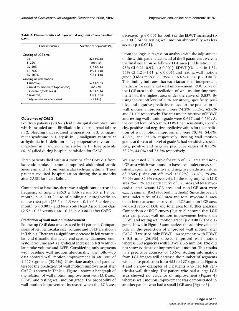
Outcomes of CABGFourteen patients (28.6%) had in-hospital complicationswhich included atrial fibrillation in 4, acute renal failurein 2, bleeding that required re-operation in 3, compart-ment syndrome in 1, sepsis in 1, malignant ventriculararrhythmia in 1, delirium in 1, perioperative myocardialinfarction in 1 and ischemic stroke in 1. Three patients(6.1%) died during hospital admission for CABG.
Three patients died within 4 months after CABG: 1 fromischemic stroke, 1 from a ruptured abdominal aorticaneurysm and 1 from ventricular tachyarrhythmia. Threepatients required hospitalization during the 4 monthsafter CABG for heart failure.
Compared to baseline, there was a significant decrease infrequency of angina (35.3 ± 43.6 versus 0.5 ± 1.8 permonth, p < 0.001), use of sublingual nitroglycerin torelieve chest pain (27.7 ± 41.3 versus 0.1 ± 0.3 tablets permonth, p < 0.001), and New York Heart Association class(2.52 ± 0.55 versus 1.48 ± 0.55, p < 0.001) after CABG.
Prediction of wall motion improvementFollow-up CMR data was assessed in 44 patients. Compar-isons of left ventricular size, volume and LVEF are shownin Table 3. There was a significant decrease in left ventricu-lar end-diastolic diameter, end-systolic diameter, end-systolic volume and a significant increase in left ventricu-lar stroke volume and LVEF. Considering only segmentswith baseline wall motion abnormality, the follow-updata showed wall motion improvement in 481 out of1,227 segments (39.2%). Univariate analysis of parame-ters for the prediction of wall motion improvement afterCABG is shown in Table 4. Figure 1 shows a bar graph ofthe relation of wall motion improvement with LGE area,EDWT and resting wall motion grade. The probability ofwall motion improvement increased when the LGE area
decreased (p < 0.001 for both) or the EDWT increased (p< 0.001) or the resting wall motion abnormality was lesssevere (p < 0.001).
From the logistic regression analysis with the adjustmentof the within patient factor, all of the 3 parameters were inthe final equation as follows: LGE area (Odds ratio 0.92,95% CI 0.91–0.93, p < 0.001), EDWT (Odds ratio 1.31,95% CI 1.21–1.41, p < 0.001) and resting wall motiongrade (Odds ratio 8.29, 95% CI 6.62–10.34, p < 0.001).This finding indicates that each factor is an independentpredictor for segmental wall improvement. ROC curve ofthe LGE area in the prediction of wall motion improve-ment had the highest area under the curve of 0.837. Byusing the cut off level of 25%, sensitivity, specificity, pos-itive and negative predictive values for the prediction ofwall motion improvement were 74.2%, 83.2%, 62.6%and 81.1% respectively. The area under the curve of EDWTand resting wall motion grade were 0.647 and 0.591. Atthe cut off level of 5.5 mm, EDWT had sensitivity, specifi-city, positive and negative predictive values for the predic-tion of wall motion improvement were 70.1%, 54.6%,49.8%, and 73.9% respectively. Resting wall motiongrade, at the cut off level of grade 3, had sensitivity, specif-icity, positive and negative predictive values of 81.5%,33.2%, 44.0% and 73.5% respectively.
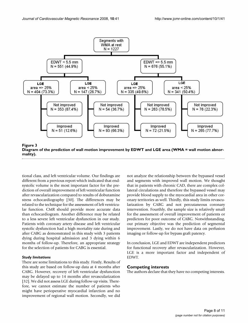
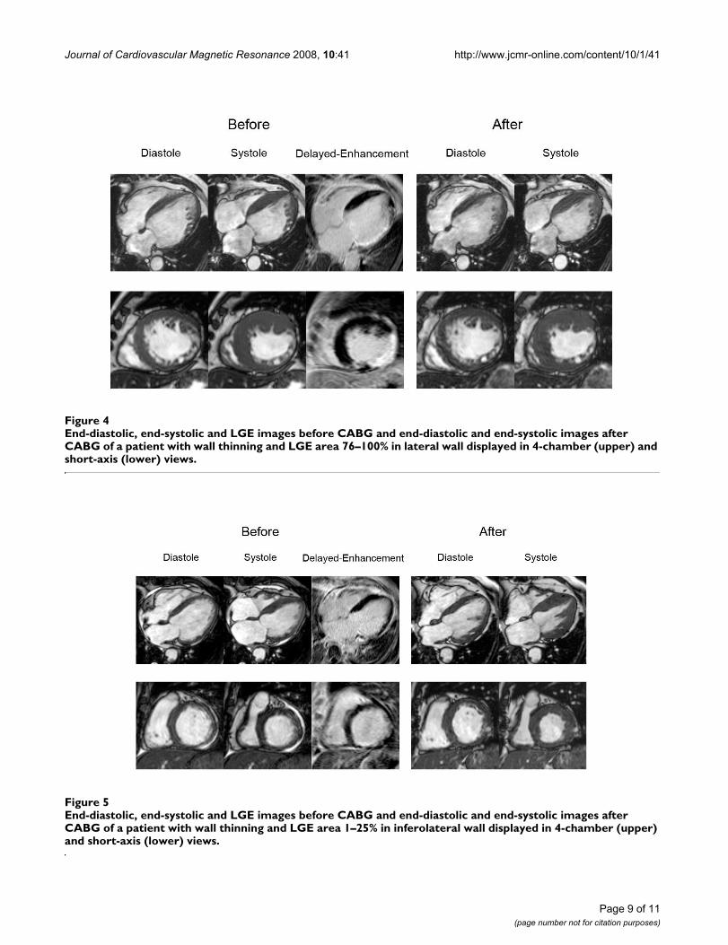
We also tested ROC curve for ratio of LGE area and non-LGE area which was found to have area under curve, sen-sitivity, specificity, positive and negative predictive valuesof 0.805 (using cut off level 32.02%), 74.6%, 79.4%,70.0% and 82.9% respectively. In the subgroup with LGEarea 25–75%, area under curve of LGE area and total myo-cardial area versus LGE area and non-LGE area wereexactly similar (0.638 for both methods). Since the overallarea under curve of LGE area and total myocardial areahad a better area under curve than LGE and non-LGE area,we used ratio of LGE and total area for further analysis.Comparison of ROC curves (Figure 2) showed that LGEarea can predict wall motion improvement better thanEDWT and resting wall motion grade (p < 0.001). The dia-gram shown in Figure 3 summarizes the additive value ofLGE in the prediction of improved wall motion afterCABG. If we used only EDWT, 144 segments with EDWT< 5.5 mm (26.1%) showed improved wall motionwhereas 339 segments with EDWT ≥ 5.5 mm (50.1%) didnot show evidence of improved wall motion. This resultsin a predictive accuracy of 60.6%. Adding informationfrom LGE images will decrease the number of segmentswith a false prediction from 483 to 127 segments. Figures4 and 5 show examples of 2 patients who had left ven-tricular wall thinning. The patient who had a large LGEarea showed no evidence of improvement (Figure 4)whereas wall motion improvement was demonstrated inanother patient who had a small LGE area (Figure 5).
Table 2: Characteristics of myocardial segments from baseline CMR
Characteristics Number of segments (%)
Grading of LGE area0% 824 (40.8)1–25% 201 (10)26–50% 417 (20.6)51–75% 340 (16.8)76–100% 238 (11.8)
Grading of wall motion1 (normal) 574 (28.4)2 (mild to moderate hypokinesia) 566 (28)3 (severe hypokinesia) 476 (23.6)4 (akinesia) 332 (16.4)5 (dyskinesia or aneurysm) 72 (3.6)
Page 4 of 11(page number not for citation purposes)

Journal of Cardiovascular Magnetic Resonance 2008, 10:41 http://www.jcmr-online.com/content/10/1/41
Page 5 of 11(page number not for citation purposes)
Relation of wall motion improvement and LGE area (upper), end-diastolic wall thickness (middle) and resting wall motion (lower)(WM = wall motion)Figure 1Relation of wall motion improvement and LGE area (upper), end-diastolic wall thickness (middle) and resting wall motion (lower)(WM = wall motion).
Journal of Cardiovascular Magnetic Resonance 2008, 10:41 http://www.jcmr-online.com/content/10/1/41
Prediction of LVEF improvementAmong 44 patients who had follow-up data, 22 patients(50%) had LVEF improvement defined as an increase inLVEF of at least 5%. Univariate analysis demonstrated thatonly baseline NYHA class, average LGE area and percentof segments with wall motion improvement are predictorsof LVEF improvement (Table 5). There was no significantdifference in number of diseased vessels and number ofgrafts between patients with and without LVEF improve-ment. There were also no significant differences in percentof segments with wall thinning (EDWT < 5.5 mm) andpercent of segments with LGE area between patients withand without LVEF improvement. However, there weresome trends of a higher chance of LVEF improvement inpatient with fewer segments with wall thinning or LGEarea.
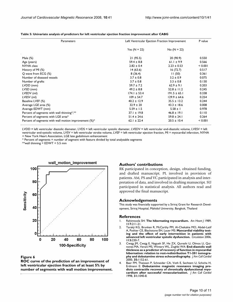
Logistic regression analysis showed that only the numberof segments with wall motion improvement (Odds ratio1.276, 95% CI 1.04–1.567, p = 0.020) remained in thefinal equation. ROC analysis demonstrated the area underthe curve of 0.958 with the cut off at 26% (patientsneeded to have an improvement of wall motion abnor-mality of more than 26% of dysfunctional segments inorder to have LVEF improvement of at least 5%) whichresults in a sensitivity of 95.5% and specificity of 86.4%
(Figure 6). For percent of segments with wall thinning,using the cut off of ≤25%, the sensitivity, specificity andarea under curve were 45.5%, 86.4% and 0.640 respec-tively. For the cut off of ≤37.5% of segments with LGEarea, the sensitivity, specificity and area under curve were36.4%, 86.7% and 0.591 respectively.
DiscussionThe main result of this study showed that extent of LGEarea is the most important predictor for recovery ofregional contractility after revascularization. EDWT andresting wall motion grade can also predict recovery ofregional dysfunction but to a much lesser extent.
Increased signal intensity on LGE images is basicallyrelated to the altered sarcolemmal membrane integrityduring acute injury and extracellular matrix structure aswell as an expanded volume of distribution of gadoliniumin scar tissue late after injury [11]. LGE is a very accuratetechnique in the assessment of acute and chronic myocar-dial infarction [12], determining infarct size after angi-oplasty [13] and is superior to SPECT in detectingmyocardial necrosis after reperfusion in acute myocardialinfarction [14]. LGE can be used to differentiate differentetiologies of cardiomyopathy [15]. Although infarct masscalculated from LGE may be slightly decreased early aftermyocardial infarction [16], this technique has beenshown to be highly reproducible [17]. Among 181 seg-ments with subendocardial infarction identified by CMR,SPECT could detect an abnormality in only 47% [18].Unrecognized myocardial infarction has been detectedmore than expected in a high-risk group with this tech-nique [19]. It provides prognostic data both in patientswith ischemic [20] and nonischemic cardiomyopathy[21]. Because of its high spatial resolution, reproducibilityof functional CMR imaging is much better than thosederived from echocardiography [22]. Therefore, CMRshould be the appropriate technique for the assessment ofthe extent of LGE area, EDWT and global and regional leftventricular function.
It has been shown that LGE can predict improvement ofregional wall motion after occlusion of the left anteriordescending artery in dogs [23]. Earlier studies have shownthat the best predictor of improvement of left ventricularfunction after revascularization is the extent of myocardialinfarction less than 25% of left ventricular wall thickness[11,24]. The positive and negative predictive value of LGEarea less than 25% of wall thickness in the prediction ofrecovery of regional function were 71 and 79% and noneof 57 segments with LGE area more than 75% had anincrease contractility after revascularization [6]. In ourstudy, sensitivity and specificity of 25% of LGE area inidentifying improvement of regional contractility were73.2% and 83.2% respectively.
Table 3: CMR parameters before and after CABG (n = 44)
Baseline After CABG P value
LVDD (mm) 61.3 ± 8.3 59.2 ± 7.2 0.015LVSD (mm) 51 ± 10.1 48.7 ± 11.8 0.035LVEDV (ml) 182.8 ± 59.5 179.7 ± 70.8 0.469LVESV (ml) 119.4 ± 60.1 110.7 ± 67.4 0.04LVSV (ml) 63.3 ± 13.7 68.5 ± 16 0.006LVEF (%) 37.9 ± 13.1 42.5 ± 15.3 0.001
LVDD = left ventricular diastolic diameter, LVDS = left ventricular systolic diameter, LVEDV = left ventricular end-diastolic volume, LVESV = left ventricular end-systolic volume, LVSV = left ventricular stroke volume, LVEF = left ventricular ejection fraction
Table 4: Univariate analysis of predictors for wall motion improvement after CABG (LGE – late gadolinium enhancement)
Parameters Wall motion improvement P value
YesN = 481
NoN = 746
LGE area (%) 15.2 ± 22 54.4 ± 31 < 0.001LGE area/non-LGE area (%) 34.6 ± 105.3 142.8 ± 378.0 <0.001EDWT (mm) 6.35 ± 1.78 5.41 ± 1.85 < 0.001Resting wall motion grade 2.72 ± 0.79 3.04 ± 0.94 < 0.001
Page 6 of 11(page number not for citation purposes)

Journal of Cardiovascular Magnetic Resonance 2008, 10:41 http://www.jcmr-online.com/content/10/1/41
It has been shown that EDWT < 6 mm from echocardio-gram virtually excludes the potential for recovery of func-tion and EDWT > 6 mm had a sensitivity and specificity of94% and 48% respectively for recovery of function [3].Reversible wall thinning in hibernation can be predictedby CMR even in the patient with ejection fraction less than30% [25]. In an CMR study, only 9.6% of segments withEDWT < 5.5 mm showed improvement in contractilityafter revascularization. In contrast, 48% of segments withpreserved EDWT did not improve, thus indicating thatEDWT should not be a good predictor for functionalrecovery [4]. Our study demonstrated that with the cut offof 5.5 mm, EDWT had a sensitivity and specificity of70.1% and 81.5% in the prediction of functional recov-ery. Functional recovery was demonstrated in 26% of seg-ments with EDWT < 5.5 mm and 49.9% of segments withEDWT ≥ 5.5 mm. LGE area is a better predictor for func-tional recovery than EDWT and has a good predictivepower even in patients with an EDWT < 5.5 mm. The areaof viable myocardium can be thinned and retain reservefor functional recovery after revascularization [26]. Theo-retically, the area of thinned and viable myocardium canbe developed during the process of ventricular remode-
ling after an acute event. It has been shown that assess-ment of ratio of non-viable to viable myocardium mayhave additional power for the prediction of functionalrecovery [26,27]. From our additional analysis, we foundthat the 2 methods were not different.
Dobutamine CMR can also be used to predict recovery ofwall motion after revascularization. Recent data suggestedthat it may be even better than LGE [28] since low dosedobutamine can assess contractile reserve by demonstrat-ing an increase in systolic wall thickness at low dose. LGEdo not provide data on contractile reserve. However, ear-lier studies [29] showed that dobutamine CMR had alower sensitivity for the prediction of recovery of wallmotion especially in patients with severe left ventriculardysfunction.
We demonstrated that increased contractility of at leastone-fourth of dysfunctional segments is needed in orderto improve overall left ventricular function. The use ofLVEF improvement of at least 5% was based on previouspublications [30,31]. This is independent of other factorssuch as baseline left ventricular function, NYHA func-
Comparison of ROC curves for the prediction of wall motion improvement by LGE area, end-diastolic wall thickness and rest-ing wall motionFigure 2Comparison of ROC curves for the prediction of wall motion improvement by LGE area, end-diastolic wall thickness and resting wall motion.
Page 7 of 11(page number not for citation purposes)
Journal of Cardiovascular Magnetic Resonance 2008, 10:41 http://www.jcmr-online.com/content/10/1/41
tional class, and left ventricular volume. Our findings aredifferent from a previous report which indicated that end-systolic volume is the most important factor for the pre-diction of overall improvement of left ventricular functionafter revascularization compared to results of dobutaminestress echocardiography [30]. The differences may berelated to the technique for the assessment of left ventricu-lar function. CMR should provide more accurate datathan echocardiogram. Another difference may be relatedto a less severe left ventricular dysfunction in our study.Patients with coronary artery disease and left ventricularsystolic dysfunction had a high mortality rate during andafter CABG as demonstrated in this study with 3 patientsdying during hospital admission and 3 dying within 6months of follow-up. Therefore, an appropriate strategyfor the selection of patients for CABG is essential.
Study limitationsThere are some limitations to this study. Firstly, Results ofthis study are based on follow-up data at 4 months afterCABG. However, recovery of left ventricular dysfunctionmay be delayed up to 14 months after revascularization[32]. We did not assess LGE during follow-up visits. There-fore, we cannot estimate the number of patients whomight have perioperative myocardial infarction and noimprovement of regional wall motion. Secondly, we did
not analyze the relationship between the bypassed vesseland segments with improved wall motion. We thoughtthat in patients with chronic CAD, there are complex col-lateral circulations and therefore the bypassed vessel mayprovide blood supply to the myocardial area in other cor-onary territories as well. Thirdly, this study limits revascu-larization by CABG and not percutaneous coronaryintervention. Fourthly, the sample size is relatively smallfor the assessment of overall improvement of patients orpredictors for poor outcome of CABG. Notwithstanding,our primary objective was the prediction of segmentalimprovement. Lastly, we do not have data on perfusionimaging or follow-up for bypass graft patency.
In conclusion, LGE and EDWT are independent predictorsfor functional recovery after revascularization. However,LGE is a more important factor and independent ofEDWT.
Competing interestsThe authors declare that they have no competing interests.
Diagram of the prediction of wall motion improvement by EDWT and LGE area (WMA = wall motion abnormality)Figure 3Diagram of the prediction of wall motion improvement by EDWT and LGE area (WMA = wall motion abnor-mality).
Page 8 of 11(page number not for citation purposes)
Journal of Cardiovascular Magnetic Resonance 2008, 10:41 http://www.jcmr-online.com/content/10/1/41
Page 9 of 11(page number not for citation purposes)
End-diastolic, end-systolic and LGE images before CABG and end-diastolic and end-systolic images after CABG of a patient with wall thinning and LGE area 76–100% in lateral wall displayed in 4-chamber (upper) and short-axis (lower) viewsFigure 4End-diastolic, end-systolic and LGE images before CABG and end-diastolic and end-systolic images after CABG of a patient with wall thinning and LGE area 76–100% in lateral wall displayed in 4-chamber (upper) and short-axis (lower) views.
End-diastolic, end-systolic and LGE images before CABG and end-diastolic and end-systolic images after CABG of a patient with wall thinning and LGE area 1–25% in inferolateral wall displayed in 4-chamber (upper) and short-axis (lower) viewsFigure 5End-diastolic, end-systolic and LGE images before CABG and end-diastolic and end-systolic images after CABG of a patient with wall thinning and LGE area 1–25% in inferolateral wall displayed in 4-chamber (upper) and short-axis (lower) views.
Journal of Cardiovascular Magnetic Resonance 2008, 10:41 http://www.jcmr-online.com/content/10/1/41
Authors' contributionsRK participated in conception, design, obtained funding,and drafted manuscript. PL involved in provision ofpatients. AM, PS and VC participated in analysis and inter-pretation of data, and involved in drafting manuscript. SUparticipated in statistical analysis. All authors read andapproved the final manuscript.
AcknowledgementsThis study was financially supported by a Siriraj Grant for Research Devel-opment, Siriraj Hospital, Mahidol University, Bangkok, Thailand
References1. Rahimtoola SH: The hibernating myocardium. Am Heart J 1989,
117:211-21.2. Tarakji KG, Brunken R, McCarthy PM, Al-Chekakie MO, Abdel-Latif
A, Pothier CE, Blackstone EH, Lauer MS: Myocardial viability test-ing and the effect of early intervention in patients withadvanced left ventricular systolic dysfunction. Circulation 2006,113:230-7.
3. Cwajg JM, Cwajg E, Nagueh SF, He ZX, Qureshi U, Olmos LI, Qui-nones MA, Verani MS, Winters WL, Zoghbi WA: End-diastolic wallthickness as a predictor of recovery of function in myocardialhibernation: relation to rest-redistribution T1-201 tomogra-phy and dobutamine stress echocardiography. J Am Coll Cardiol2000, 35:1152-61.
4. Baer FM, Theissen P, Schneider CA, Voth E, Sechtem U, Schicha H,Erdmann E: Dobutamine magnetic resonance imaging pre-dicts contractile recovery of chronically dysfunctional myo-cardium after successful revascularization. J Am Coll Cardiol1998, 31:1040-8.
Table 5: Univariate analysis of predictors for left ventricular ejection fraction improvement after CABG
Parameters Left Ventricular Ejection Fraction Improvement P value
Yes (N = 22) No (N = 22)
Male (%) 21 (95.5) 20 (90.9) 0.550Age (years) 59.4 ± 8.8 61.1 ± 9.9 0.566NYHA class 2.82 ± 0.4 2.23 ± 0.53 < 0.001History of MI (%) 14 (63.6) 16 (72.7) 0.517Q wave from ECG (%) 8 (36.4) 11 (50) 0.361Number of diseased vessels 3.7 ± 0.8 3.2 ± 0.9 0.075Number of grafts 3.7 ± 0.8 3.3 ± 0.8 0.130LVDD (mm) 59.7 ± 7.2 62.9 ± 9.1 0.203LVSD (mm) 49.2 ± 8.8 52.8 ± 11.2 0.245LVEDV (ml) 174.1 ± 53.4 191.5 ± 65.1 0.338LVESV (ml) 109 ± 54.7 129.9 ± 64.6 0.254Baseline LVEF (%) 40.2 ± 12.9 35.5 ± 13.2 0.244Average LGE area (%) 32.9 ± 20 43.3 ± 18.6 0.008Average EDWT (mm) 5.59 ± 1.5 5.58 ± 1 0.978Percent of segments with wall thinning*,** 37.1 ± 19.8 46.8 ± 19.1 0.110Percent of segments with LGE area* 51.4 ± 24.6 59.8 ± 24.1 0.264Percent of segments with wall motion improvement (%)* 62.1 ± 22.4 20.5 ± 10.4 < 0.001
LVDD = left ventricular diastolic diameter, LVDS = left ventricular systolic diameter, LVEDV = left ventricular end-diastolic volume, LVESV = left ventricular end-systolic volume, LVSV = left ventricular stroke volume, LVEF = left ventricular ejection fraction, MI = myocardial infarction, NYHA = New York Heart Association, LGE late gadolinium enhancement* Percent of segments = number of segment with feature divided by total analyzable segments**wall thinning = EDWT < 5.5 mm
ROC curve of the prediction of an improvement of left ven-tricular ejection fraction of at least 5% by number of seg-ments with wall motion improvementFigure 6ROC curve of the prediction of an improvement of left ventricular ejection fraction of at least 5% by number of segments with wall motion improvement.
Page 10 of 11(page number not for citation purposes)
Journal of Cardiovascular Magnetic Resonance 2008, 10:41 http://www.jcmr-online.com/content/10/1/41
Publish with BioMed Central and every scientist can read your work free of charge
"BioMed Central will be the most significant development for disseminating the results of biomedical research in our lifetime."
Sir Paul Nurse, Cancer Research UK
Your research papers will be:
available free of charge to the entire biomedical community
peer reviewed and published immediately upon acceptance
cited in PubMed and archived on PubMed Central
yours — you keep the copyright
Submit your manuscript here:http://www.biomedcentral.com/info/publishing_adv.asp
BioMedcentral
5. Sandstede JJ, Lipke C, Beer M, Harre K, Pabst T, Kenn W, NeubauerS, Hahn D: Analysis of first-pass and delayed contrast-enhancement patterns of dysfunctional myocardium on MRimaging: use in the prediction of myocardial viability. AJR AmJ Roentgenol 2000, 174:1737-40.
6. Kim RJ, Wu E, Rafael A, Chen EL, Parker MA, Simonetti O, Klocke FJ,Bonow RO, Judd RM: The use of contrast-enhanced magneticresonance imaging to identify reversible myocardial dysfunc-tion. N Engl J Med 2000, 343:1445-53.
7. Kim RJ, Fieno DS, Parrish TB, Harris K, Chen EL, Simonetti O, BundyJ, Finn JP, Klocke FJ, Judd RM: Relationship of MRI delayed con-trast enhancement to irreversible injury, infarct age, andcontractile function. Circulation 1999, 100:1992-2002.
8. Blackburn H, Keys A, Simonson E, Rautaharju P, Punsar S: The elec-trocardiogram in population studies: A classification system.Circulation 1960, 21:1160-75.
9. Cerqueira MD, Weissman NJ, Dilsizian V, Jacobs AK, Kaul S, LaskeyWK, Pennell DJ, Rumberger JA, Ryan T, Verani MS: Standardizedmyocardial segmentation and nomenclature for tomo-graphic imaging of the heart: a statement for healthcare pro-fessionals from the Cardiac Imaging Committee of theCouncil on Clinical Cardiology of the American Heart Asso-ciation. Circulation 2002, 105:539-42.
10. Metz CE: Basic principles of ROC analysis. Semin Nucl Med 1978,8:283-98.
11. Thomson LE, Kim RJ, Judd RM: Magnetic resonance imaging forthe assessment of myocardial viability. J Magn Reson Imaging2004, 19:771-88.
12. Saeed M, Weber O, Lee R, Do L, Martin A, Saloner D, Ursell P, Rob-ert P, Corot C, Higgins CB: Discrimination of myocardial acuteand chronic (scar) infarctions on delayed contrast enhancedmagnetic resonance imaging with intravascular magneticresonance contrast media. J Am Coll Cardiol 2006, 48:1961-8.
13. Baks T, van Geuns RJ, Biagini E, Wielopolski P, Mollet NR, Cade-martiri F, Giessen WJ van der, Krestin GP, Serruys PW, Duncker DJ,et al.: Effects of primary angioplasty for acute myocardial inf-arction on early and late infarct size and left ventricular wallcharacteristics. J Am Coll Cardiol 2006, 47:40-4.
14. Ibrahim T, Bulow HP, Hackl T, Hornke M, Nekolla SG, Breuer M,Schomig A, Schwaiger M: Diagnostic value of contrast-enhancedmagnetic resonance imaging and single-photon emissioncomputed tomography for detection of myocardial necrosisearly after acute myocardial infarction. J Am Coll Cardiol 2007,49:208-16.
15. Shah DJ, Judd RM, Kim RJ: Technology insight: MRI of the myo-cardium. Nat Clin Pract Cardiovasc Med 2005, 2:597-605. quiz 06
16. Fieno DS, Hillenbrand HB, Rehwald WG, Harris KR, Decker RS,Parker MA, Klocke FJ, Kim RJ, Judd RM: Infarct resorption, com-pensatory hypertrophy, and differing patterns of ventricularremodeling following myocardial infarctions of varying size.J Am Coll Cardiol 2004, 43:2124-31.
17. Thiele H, Kappl MJ, Conradi S, Niebauer J, Hambrecht R, Schuler G:Reproducibility of chronic and acute infarct size measure-ment by delayed enhancement-magnetic resonance imag-ing. J Am Coll Cardiol 2006, 47:1641-5.
18. Wagner A, Mahrholdt H, Holly TA, Elliott MD, Regenfus M, Parker M,Klocke FJ, Bonow RO, Kim RJ, Judd RM: Contrast-enhanced MRIand routine single photon emission computed tomography(SPECT) perfusion imaging for detection of subendocardialmyocardial infarcts: an imaging study. Lancet 2003, 361:374-9.
19. Barbier CE, Bjerner T, Johansson L, Lind L, Ahlstrom H: Myocardialscars more frequent than expected: magnetic resonanceimaging detects potential risk group. J Am Coll Cardiol 2006,48:765-71.
20. Kwong RY, Chan AK, Brown KA, Chan CW, Reynolds HG, Tsang S,Davis RB: Impact of unrecognized myocardial scar detectedby cardiac magnetic resonance imaging on event-free sur-vival in patients presenting with signs or symptoms of coro-nary artery disease. Circulation 2006, 113:2733-43.
21. Assomull RG, Prasad SK, Lyne J, Smith G, Burman ED, Khan M, Shep-pard MN, Poole-Wilson PA, Pennell DJ: Cardiovascular magneticresonance, fibrosis, and prognosis in dilated cardiomyopa-thy. J Am Coll Cardiol 2006, 48:1977-85.
22. Bellenger NG, Burgess MI, Ray SG, Lahiri A, Coats AJ, Cleland JG,Pennell DJ: Comparison of left ventricular ejection fractionand volumes in heart failure by echocardiography, radionu-
clide ventriculography and cardiovascular magnetic reso-nance; are they interchangeable? Eur Heart J 2000, 21:1387-96.
23. Hillenbrand HB, Kim RJ, Parker MA, Fieno DS, Judd RM: Earlyassessment of myocardial salvage by contrast-enhancedmagnetic resonance imaging. Circulation 2000, 102:1678-83.
24. Choi KM, Kim RJ, Gubernikoff G, Vargas JD, Parker M, Judd RM:Transmural extent of acute myocardial infarction predictslong-term improvement in contractile function. Circulation2001, 104:1101-7.
25. John AS, Dreyfus GD, Pennell DJ: Images in cardiovascular med-icine. Reversible wall thinning in hibernation predicted bycardiovascular magnetic resonance. Circulation 2005,111:e24-5.
26. Kim RJ, Shah DJ: Fundamental concepts in myocardial viabilityassessment revisited: when knowing how much is "alive" isnot enough. Heart 2004, 90:137-40.
27. Ichikawa Y, Sakuma H, Suzawa N, Kitagawa K, Makino K, Hirano T,Takeda K: Late gadolinium-enhanced magnetic resonanceimaging in acute and chronic myocardial infarction.Improved prediction of regional myocardial contraction inthe chronic state by measuring thickness of nonenhancedmyocardium. J Am Coll Cardiol 2005, 45:901-9.
28. Wellnhofer E, Olariu A, Klein C, Grafe M, Wahl A, Fleck E, Nagel E:Magnetic resonance low-dose dobutamine test is superior toSCAR quantification for the prediction of functional recov-ery. Circulation 2004, 109:2172-4.
29. Kim RJ, Manning WJ: Viability assessment by delayed enhance-ment cardiovascular magnetic resonance: will low-dose dob-utamine dull the shine? Circulation 2004, 109:2476-9.
30. Schinkel AF, Poldermans D, Rizzello V, Vanoverschelde JL, Elhendy A,Boersma E, Roelandt JR, Bax JJ: Why do patients with ischemiccardiomyopathy and a substantial amount of viable myocar-dium not always recover in function after revascularization?J Thorac Cardiovasc Surg 2004, 127:385-90.
31. White JA, Yee R, Yuan X, Krahn A, Skanes A, Parker M, Klein G,Drangova M: Delayed enhancement magnetic resonanceimaging predicts response to cardiac resynchronizationtherapy in patients with intraventricular dyssynchrony. J AmColl Cardiol 2006, 48:1953-60.
32. Bax JJ, Visser FC, Poldermans D, Elhendy A, Cornel JH, Boersma E,van Lingen A, Fioretti PM, Visser CA: Time course of functionalrecovery of stunned and hibernating segments after surgicalrevascularization. Circulation 2001, 104:I314-8.
Page 11 of 11(page number not for citation purposes)
Copyright © 2022 FDOKUMEN