Outcomes After Laparoscopic Roux-en-Y Gastric Bypass for ...
Upload
khangminh22Category
view
0download
0
Biliary Bypass with Laparoscopic Choledochoduodenostomy
Short Title: Lap CDD
Joshua K. Kays, Leonidas G. Koniaris, Daniel P. Milgrom, Attila Nakeeb
Department of Surgery, Indiana University School of Medicine, 545 Barnhill Drive, Emerson Hall, Indianapolis, IN 46202
Corresponding Author:
Joshua K. Kays, MD
545 Barnhill Drive, EH 202
Indianapolis, IN 46202
(317) 274-5771
Funding Sources: NIH grant R02DK096167 and the Lily Endowment, Inc. to LK
Author Contributions: JKK – data collection, data analysis and interpretation, original manuscript and revisions, development of figures and images, final approval of manuscript for publications, and agrees to be responsible for all aspects of the work. DPM – data collection, data analysis and interpretation, revision of critically important content of the manuscript and figures, final approval of manuscript, and agrees to be accountable for all aspects of the work. LGK – conception and design of the work, revision of critically important content of the manuscript and final figures, final approval of manuscript, and agrees to be accountable for all aspects of the work. AN – conception and design of the work, revision of critically important content of the manuscript and figures, final approval of manuscript, and agrees to be accountable for all aspects of the work.
___________________________________________________________________
This is the author's manuscript of the article published in final edited form as:
Kays, J. K., Koniaris, L. G., Milgrom, D. P., & Nakeeb, A. (2018). Biliary Bypass with Laparoscopic Choledochoduodenostomy. Journal of Gastrointestinal Surgery, 22(5), 928–933. https://doi.org/10.1007/s11605-017-3663-z

Abstract
Introduction: Laparoscopic choledochoduodenostomy (LCDD) is employed to treat
many benign biliary diseases when endoscopic or percutaneous techniques are not
feasible.
Technique: We describe our technique for LCDD, which utilizes common bile duct
transection and an end-to-side biliary-enteric anastomosis. This procedure includes the
following elements: isolation and transection of the common bile duct, mobilization of
the duodenum (Kocher maneuver), inspection of the common bile duct, and end-to-side
biliary-enteric anastomosis. Key details and pitfalls are discussed.
Results: Over a 5-year period LCDD was performed on eighteen patients. Indications
included intractable abdominal pain (10) and choledocholithiasis (8). The majority of
patients, 83%, tolerated the operation well with no complications. There was one post-
operative intra-abdominal abscess and two anastomotic strictures, one in the immediate
post-operative period and the other nine months after the operation. The median length
of stay was four days (IQR: 3.0-5.3), and there was minimal blood loss.
Conclusion: Based on our experience, LCDD with transection and end-to-side biliary-
enteric anastomosis is a safe and effective biliary bypass technique.

Introduction
The first choledochoduodenostomy (CDD), performed in 1888 by Bernhard Riedel, was
used to extract retained bile duct stones and utilized a side-to-side anastomosis
between the common bile duct and the duodenum[1]. In the years since, CDD has been
used to successfully treat many benign biliary diseases including choledocholithiasis,
cholangitis, recurrent/chronic pancreatitis, and biliary tree strictures[1-3]. With the
advent of laparoscopic surgery and increasing surgeon experience and technical
expertise, laparoscopy is a practical approach for many biliary operations including
CDD.
In recent years, however, endoscopic techniques and, in select settings, image-guided
percutaneous approaches have largely replaced surgery in the management of benign
biliary tract diseases[4-6]. Today, surgery is mainly reserved for instances when
endoscopic or percutaneous approaches are not available or not viable options. When
biliary bypass is indicated three options are available: CDD with side-to-side biliary-
enteric anastomosis, CDD with end-to-side biliary-enteric anastomosis, and biliary-
jejunal anastomosis either as a hepaticojejunostomy or a Roux-en-Y
choledochojejunostomy. All three can be accomplished via a laparoscopic approach,
however, there is no consensus to which is best due to a paucity of data comparing the
techniques. Choice of operation is largely left up to the individual surgeon.
Although performed infrequently for benign biliary disease, biliary bypass operations are
fundamental for any gastrointestinal surgeon and are considered a core operation by
the American College of Surgeons for any general surgery resident [7]. Herein, we

describe in detail our technique for laparoscopic choledochoduodenostomy (LCDD) and
outcomes when used for benign biliary disease.
Materials and Methods
Human subjects research approval for this study was obtained from the institutional
review board of the Indiana University School of Medicine and was carried out in
compliance with the IU Standard Operating Procedures for Research Involving Human
Subjects.
The electronic medical record was queried for all patients undergoing laparoscopic
choledochoduodenostomy at Indiana University Hospital between January 1, 2011 to
January 1, 2016. The data presented represents the experience of two surgeons
experienced in both laparoscopy and biliary surgery. Selection of laparoscopic
choledochoduodenostomy as the biliary bypass operation was at the discretion of the
treating surgeon. Operative notes were reviewed for all patients to ensure a
laparoscopic approach was used and to record the details of the procedure. Patient
charts were reviewed and age, sex, body mass index (BMI), operative time, operative
blood loss, length of stay, American Society of Anesthesiologists (ASA) classification,
operative indication, and follow up data were recorded.
Operative Technique
The patient is placed in supine position with arms extended. Appropriate preoperative
antibiotic prophylaxis is administered and intermittent compression stockings are placed
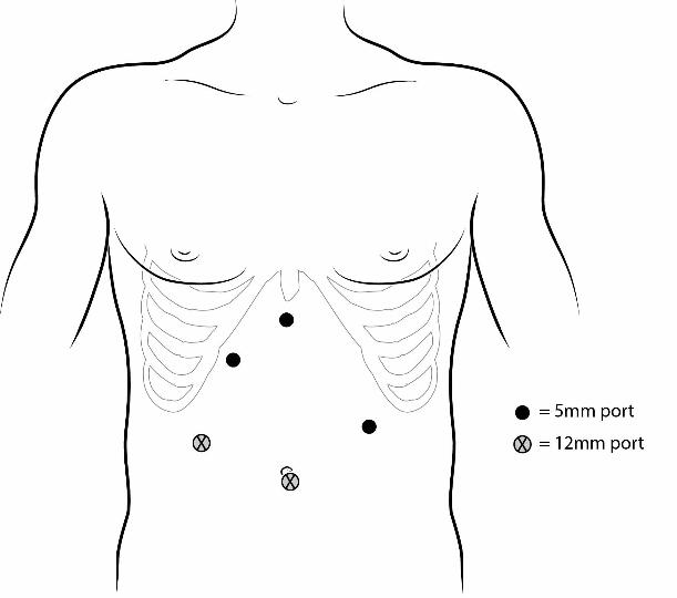
for deep vein thrombosis prophylaxis. Port placement is diagrammed in Figure 1. The
Hassan technique is used to gain entry into the abdomen and placement of the first

port. The method of initial entry, however, may be adjusted based on surgeon
preference. The lateral right abdomen port and the camera ports are 12 mm. All other
ports are 5 mm. A 30o laparoscope is used. As is standard with all operations, the
abdomen is inspected for visceral organ injury during entry and other abdominal
pathology.
Upon entry into the abdomen some degree of adhesiolysis is often necessary due to
patient history of previous cholecystectomy or inflammation from biliary disease. A
Nathanson retractor is placed through the 5 mm epigastric port to elevate the left lateral
segment of the liver. A harmonic scalpel is used to open the port hepatis and
circumferentially dissect out the common bile duct from the hepatic artery and portal
vein (Figure 2). The common bile duct is then divided just above the duodenum with
electrocautery (Figure 3). At this point the distal bile duct is over-sewn with interrupted
2-0 vicryl or PDS sutures. Any biliary stents that are present from previous endoscopic
procedures are removed. In patients with biliary stents or suspected or confirmed bile
duct stones a biliary fogarty catheter is passed through the proximal common bile duct
to clear any stones or debris.
Next, a Kocher maneuver is performed to mobilize the duodenum. A longitudinal
duodenotomy is made. Care must be taken to create this duodenotomy at a point that
will ensures a tension-free anastomosis is achieved. A primary end-to-side
choledochoduodenostomy is then created with a single-layer of 4-0 vicryl sutures in a
running or interrupted fashion.
The right upper quadrant is copiously irrigated with warm normal saline and the
anastomosis is carefully inspected for a bile leak. A 15 mm blake drain may be placed

posterior to the choledochoduodenostomy and brought out through the right abdomen 5
mm port. The pneumoperitoneum is then reversed and all port sites are closed in the
standard fashion.
Results
The described technique has been used in the treatment of eighteen patients. Median
age was 46.8 years (IQR:37.2-67.4) and BMI was 27.2 kg/m2 (IRQ:21.7-30.6). There
were 13 females and 5 males. Median follow up was 68 days (IQR: 36-116) with one
patient lost to follow up. Median common bile duct diameter was 10 mm (IRQ:9.0-13.0),
operative time was 165.5 minutes (IRQ:127.0-195.3), blood loss was 35.0 mL
(IQR:20.0-100.0), and length of stay was 4.0 days (IRQ:3.0-5.3). Ten patients had
intractable abdominal pain and eight had choledocholithiasis. All patients failed
endoscopic treatment with ERCP prior to LCDD with 10 (55.6%) undergoing multiple
ERCPs without resolution of symptoms. All patients were low to moderate surgical risk
with ASA classifications of 2 (33%) or 3 (67%).
The majority of patients, 15, had favorable outcomes with no known complications. One
patient developed an intra-abdominal abscess on post-operative day 11, which required
percutaneous drainage and a course of antibiotics. The abscess resolved, and the
patient recovered with no further complications.
Two patients developed strictures, one in the immediate post-operative period and one
at nine months. The patient with immediate CDD stricture was managed with
percutaneous transhepatic cholangiographic (PTC) drain placement followed by a
recovery period and eventual anastomotic revision with a hepaticojejunostomy. This

patient has recovered and is currently without any further issues. The patient with
stricture development at nine months has required PTC with stent placement and two
subsequent endoscopic retrograde cholangiopancreatographies (ERCP), one for stent
removal and one for CDD dilation. This patient has had symptomatic improvement with
these interventions.
Discussion
Although infrequently used due to the emergence of endoscopic and image-guided
percutaneous approaches, laparoscopic choledochoduodenostomy (LCDD) remains a
necessary and fundamental technique for the gastrointestinal surgeon and is
considered a core operation for general surgery residency training[7]. This series details
our technique for LCDD and demonstrates that it is a safe and effective operation for
patients with benign biliary pathology.
Endoscopic and image-guided percutaneous approaches have largely replaced surgical
procedures for benign disease of the biliary tract. There are situations, however, when
surgery is necessary. Lack of endoscopist or radiologist with the technical expertise,
abnormal anatomy such as after Roux-en-Y gastric bypass surgery, and multiple failed
endoscopic or percutaneous interventions all call for a surgical approach to
management[8-11].
Preoperative assessment for patients is similar to that for any other biliary operation,
including laboratory testing (complete blood count, serum electrolytes, and liver function
tests). Imaging is performed at the treating physician’s discretion and may include CT
scan, magnetic resonance cholangiopancreatography (MRCP), and endoscopic

retrograde cholangiopancreatography (ERCP). Patients with intractable abdominal pain
are only taken to the operating room for LCDD if the symptoms are biliary in nature and
CT scan, MRCP, and ERCP have been performed and failed to identify an alternative
origin. An argument can be made that common bile duct diameter should be determined
prior to operation as experience with the side-to-side anastomosis technique has
indicated that common bile duct diameter > 15 mm is associated with less
complications[12-14]. All patients in this study were noted to have dilated common bile
duct intra-operatively. Fourteen patients had common bile duct measurements with the
median diameter being 10 mm (IQR:9.0-13.0).
Postoperative care is similar to that of a laparoscopic cholecystectomy. Patients are
allowed to advance diet as tolerated, adequate pain control is provided, and early
ambulation is encouraged. A follow up appointment is schedule for approximately 30
days after hospital discharge. If the patient has recovered well at the initial post-
operative visit no additional follow up is necessary.
The technique described above differs from the technique for most data published on
LCDD due to the use of an end-to-side biliary-enteric anastomosis. Data published on
the side-to-side LCDD report hospital length of stay ranging from 4-10 days and
complications as high as 19%[2,10,13-15]. This series has similar results, as the
median length of stay was 4.0 days (IQR:3.0-5.3) and the complication rate was less
than 20%. The end-to-side anastomosis technique is advantageous in that it eliminates
of the potential development of sump syndrome. Sump syndrome occurs as a result of
bile stasis and debris accumulation in the infra-anastomotic bile duct resulting in
bacterial overgrowth. Sump syndrome has been reported to occur in up to 2.5% of

LCDD utilizing side-to-side anastomoses[1,16]. While rare, sump syndrome is a serious
complication that causes continued pain and increases the risk for cholangitis and
hepatic abscesses.
Long-term results of open transection choledochoduodenostomy have been excellent.
Cuschieri et al. demonstrated greater than 5-years of follow up on 26 patients without a
single anastomotic stricture[17]. Our series had two incidences anastomotic stricture.
One occurred in the immediate post-operative period and was likely due to a technical
error during the operation, as this patient never experienced pain relief after surgery and
the stenosis was diagnosed fewer than 30 days after surgery. The other stricture was in
a patient who underwent the operation for intractable abdominal pain of biliary origin
and it is likely that some degree of chronic inflammation due to the underlying pathology
played a role in the development of this stricture. Stricture is a potential long-term
complication with any biliary reconstruction. The majority of the patients presented here
have only had short-term follow up and it will be important to continue to follow these
patients to fully assess the long-term stricture rate of this operation.
A direct comparison between side-to-side CDD, end-to-side CDD, and Roux-en Y
choledochojejunostomy has been difficult due to the rarity of patients who fail or are not
amendable to endoscopic therapy[18,19]. It has been shown that reconstruction with
biliary-duodenal anastomosis can be safely performed and has the added benefit of
preserving endoscopic access to the biliary tree. Thus, a strong argument can be made
that CDD should be utilized before choledochojejunostomy if possible. Roux-en-Y
hepaticojejunostomy is also another options for these patients, however, LCDD has one
less anastomosis and is less divergent from normal anatomy and physiology.

Patient factors must always be considered in any operation and LCDD is no exception.
All patients in this study were relatively low-risk surgical candidates with ASA scores of
either 2 or 3. High-risk surgical candidates, ASA >3, requiring repeated endoscopic
interventions should be considered for LCDD. Open surgery has been shown to have
no increased risk in morbidity or mortality compared to endoscopic sphincterotomy in
this patient population and LCDD may provide a definitive solution[20]. Additionally, the
port site placement outlined here may need modified based on the physical and
anatomic characteristics of individual patients as well as cosmetic consideration[21,22].
A disadvantage of this technique is the higher technical difficulty of the end-to-side
anastomosis compared to the side-to-side approach. Robotic assisted surgery has the
potential to resolve this issue. Robotic assisted choledochoduodenostomy has been
described in the literature and has been shown to be safe and feasible[23,24].
Additional training, however, is required to develop proficiency using the robot and this
training not currently a standard part of a general surgeon’s training. Laparoscopy is a
mandatory part of a general surgeon’s skills set and currently is the most widely used
minimally invasive approach. Choice of biliary bypass surgery should ultimately be
determined by the surgeon with attention to patient factors, plans for future access to
the biliary tree, and surgeon expertise and comfort.
Conclusion
Currently there is not adequate data to demonstrate the superiority of any one biliary-
enteric anastomotic technique. While endoscopic intervention is the first choice for
treatment of benign biliary disease, there are instances where it is not feasible, and
thus, choledochoduodenostomy remains a fundamental operation for the

gastrointestinal surgeon. The choice of biliary bypass technique depends on several
factors and is ultimately up to the operating surgeon. The technique for laparoscopic
choledochoduodenostomy described above can be accomplished safely and provides
acceptable outcomes in patients with benign biliary disease, while eliminating the risk of
sump syndrome.
Conflicts of interest: Drs. Joshua K. Kays, Leonidas G. Koniaris, Daniel P. Milgrom, and
Attila Nakeeb have no conflicts of interest or financial ties to disclose.
Acknowledgements: The authors would like to acknowledge Barbara Sturonas-Brown
and the Visual Media Department at Indiana University School of Medicine for
assistance with creation of the figures that accompany this manuscript.
References
1. Leppard WM, Shary TM, Adams DB, Morgan KA. Choledochoduodenostomy: is
it really so bad? J Gastrointest Surg. 2011;15:754-757.
2. Khajanchee YS, Cassera MA, Hammill CW, Swanstrom LL, Hansen PD.
Outcomes following laparoscopic choledochoduodenostomy in the management of
benign biliary obstruction. J Gastrointestin Surg. 2012;16:801-805.

3. Malik AA, Rather SA, Bari SUL, Wani KA. Long-term results of
choledochoduodenostomy in benign billiary obstruction. World J Gastrointest Surg.
2012;4(2):36-40.
4. Cotton PB, Geenen JE, Sherman S, Cunningham JT, Howell DA, Carr-Locke DL,
Nickl NJ, Hawes RH, Lehman GA, Ferrari A, Lichtenstein DR, Baillie J, Jowell PS, Lail
LM, Evangelou H, Bosco JJ, Hanson BL, Hoffman BJ, Rahaman SM, Male R.
Endoscopic sphincterotomy for stones by experts is safe, even in younger patients with
normal ducts. Ann Surg. 1998;227(2):201-204.
5. Ray AA, Davies ET, Duvdevani M, Razvi H, Denstedt JD. The management of
treatment-resistant biliary calculi using percutaneous endourologic techniques. Can J
Surg. 2009;52(5):407-412.
6. Sherman S, Ruffolo TA, Hawes RH, Lehman GA. Complications of endoscopic
sphincterotomy: a prospective series with emphasis on the increased risk associated
with sphincter of Oddi dysfunction and nondilated bile ducts. Gastroenterology.
1991;101(4):1068-1075.
7. He J, Choti MA. Bile duct injury – acute repair. Score.
https://link.springer.com/article/10.1007/s11605-017-3642-4. Updated December 10,
2016. Accessed December 10, 2017.
8. Shelat VG, Chan CY, Liau KH, Ho CK. Laparoscopic exploration can salvage
failed endoscopic bile duct stone extraction. Singapore Med J. 2012;53(3):313-317.
9. DuCoin C, Moon RC, Teixeira AF, Jawad MA. Laparoscopic
choledochoduodenostomy as an alternate treatment for common bile duct stones after
Roux-en-Y gastric bypass.Surg Obes Relat Dis. 2014;10:647-653.

10. Overby DW, Richardson W, Fanelli R. Choledocholithiasis after gastric bypass: a
growing problem. Surg Obes Relat Dis. 2014;10(4):652-653.
11. Franceschi D, Brandt C, Margolin D, Szopa B, Ponsky J, Priebe P, Stellato T,
Eckhauser M. The management of common bile duct stones in patients undergoing
laparoscopic cholecystectomy. Am Surg. 1993;59(8):525-532.
12. Tinoco R, El-Kadre L, Tinoco A. Laparoscopic choledochoduodenostomy. J
Laparoendosc Adv Surg Tech. 1999;9(2):123-126.
13. Chander J, Mangla V, Vindal A, Lal P, Ramteke VK. Laparoscopic
choledochoduodenostomy for biliary stone disease: a single-center 10-year experience.
J Laparoendosc Adv Surg Tech. 2012;22(1):81-84.
14. Jeyapalan M, Almeida JA, Michaelson RL, Franklin Jr. ME. Laparoscopic
choledochoduodenostomy: review of a 4-year experience with an uncommon problem.
Surg Laparosc Endosc Percutan Tech. 2002;12(3):148-153.
15. Tang CN, Siu WT, Ha JPY, Li MKW. Laparoscopic choledochoduodenostomy: an
effective drainage procedure for recurrent pyogenic cholangitis. Surg Endosc.
2003;17:1590-1594.
16. Qadan M, Clarke S, Morrow E, Triadafilopoulos G, Visser B. Sump syndrome as a
complication of choledochoduodenostomy. Dig Dis Sci. 2012;57(8):2011-2015.
17. Cuschieri A, Wood RAB, Metcalf MJ, Cumming JGR. Long-term experience with
transection choledochoduodenostomy. World J Surg. 1983;7:502-504.
18. Rose JB, Bilderback P, Raphaeli T. Use the duodenum, it's right there: a
retrospective cohort study comparing biliary reconstruction using either the jejunum or
the duodenum. JAMA Surg. 2013;148(9):860-865.

19. Panis Y, Fagniez PL, Brisset D, Lacaine F, Levard H, Hay J. Long term results of
choledochoduodenostomy versus choledochojejunostomy for choledocholithiasis. The
French Association for Surgical Research. Surg Gynecol Obstet. 1993;177(1):33-37.
20. Targarona EM, Pros I, Martinez J, Trias M, Ayuso RMP, Ros E, Tres J, Bordas
JM. Randomised trial of endoscopic sphincterotomy with gallbladder left in situ versus
open surgery for common bile duct calculi in high-risk patients. Lancet.
1996;347(9006):926-929.
21. Cruz-Munoz Ndl, Koniaris LG. Alternative port site selection (APSS) for improved
cosmesis in laparoscopic surgery. J Gastrointest Surg. 2010;14(12):2004-2008.
22. Koniaris LG, Zimmers-Koniaris TA, Lillemoe KD, Sachs SM, Riggle K. A method
for easier laparoscopic cholangiography and common bile duct exploration. J Am Coll
Surg. 2000;190(6):752-756.
23. Chan OCY, Tang CN, Lai ECH, Yang GPC, Li MKW. Robotic hepatobiliary and
pancreatic surgery: a cohort study. J Hepatobiliary Panceat Sci. 2011;18(4):471-480.
24. Gilbert A, Doussot A, Ortega-Deballon P, Rostain F, Rat P, Facy O. Robot
assisted choledochoduodenostomy: a safe and reproducible procedure for benign
common bile duct obstruction. Dig Surg. 2017;34:177-179.

Figure 1. Laparoscopic Port Placement. A 12 mm port is placed in the umbilicus and
used for the camera. An additional 12 mm working port is placed in the right abdomen
just lateral to the midclavicular line. Three 5 mm ports are placed: the epigastric port is
used for placement of a Nathanson liver retractor, while the right and left upper
quadrant ports are working ports.
Figure 2. Dissection of the porta hepatis. A) Schematic and B) intra-operative view of
the dissection of the porta hepatis. A Harmonic scalpel is used to incise the
hepatoduodenal ligament and enter the porta hepatis. The common bile duct (CBD) is
then circumferentially dissected out and isolated from the proper hepatic artery (PHA)
and the portal vein (not in view).
Figure 3. Transection of the common bile duct. The common bile duct is transected with
electrocautery just above the duodenum ensuring to leave enough of the distal common
bile duct to oversew.
Figure 4. Longitudinal duodenostomy. A) A longitudinal duodenostomy is made using a
harmonic scalpel. B) The duodenostomy size will depend on the diameter of the
common bile duct. It is important to create the duodenostomy distal to the pylorus and
at a point where a tension free choledochoduodenstomy can be created.

Figure 5. Creation of the anastomosis. A) Schematic and B) intra-operative view of final
end-to-side anastomosis. The choledochoduodenostomy (CDD) is made in an end-to-
side fashion between the common bile duct (CBD) and the duodenum using an
absorbable suture. This can be done in a running or interrupted fashion.

Table 1 Patient and Surgery Characteristics Age, years 46.8 (37.2-67.4) BMI, kg/m2 27.2 (21.7-30.6) Sex Male Female
5
13 Common Bile Duct diameter, mm 10 (9.0-13.0) Operative time, minutes 165.5 (127.0-195.3) Blood loss, mL 35.0 (20.0-100.0) Length of stay, days 4.0 (3.0-5.3) Indication for surgery Intractable abdominal pain Choledocholithiasis
10 8
ASA 2 3
6
12 Complications None Intra-abdominal abscess CDD stricture
15 1 2
Values are reported as median (interquartile range) or the number of patients.
ASA American Society of Anesthesiologists, CDD choledochoduodenstomy


Copyright © 2022 FDOKUMEN