Blood Brain Barrier: Structure, Function and Bypass by ...
31
Blood Brain Barrier: Structure, Function and Bypass by Microorganisms Presented by: Syeda Kashfi Qadri
-
Upload
khangminh22 -
Category
Documents
-
view
3 -
download
0
Transcript of Blood Brain Barrier: Structure, Function and Bypass by ...

Blood Brain Barrier: Structure,
Function and Bypass by
Microorganisms
Presented by: Syeda Kashfi Qadri
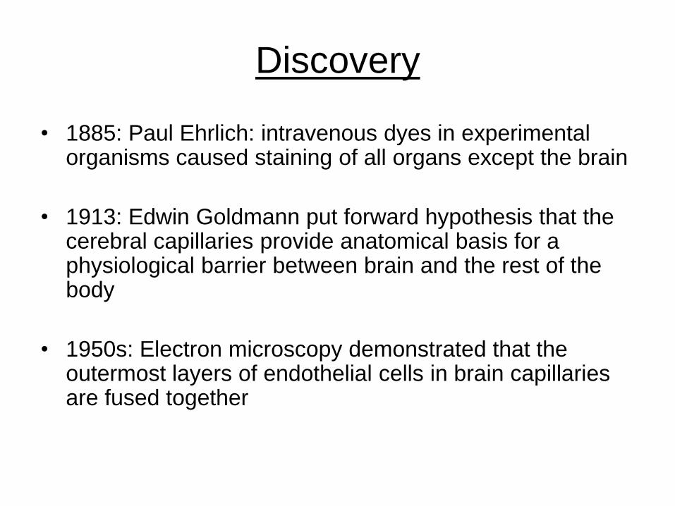
Discovery
• 1885: Paul Ehrlich: intravenous dyes in experimental organisms caused staining of all organs except the brain
• 1913: Edwin Goldmann put forward hypothesis that the cerebral capillaries provide anatomical basis for a physiological barrier between brain and the rest of the body
• 1950s: Electron microscopy demonstrated that the outermost layers of endothelial cells in brain capillaries are fused together
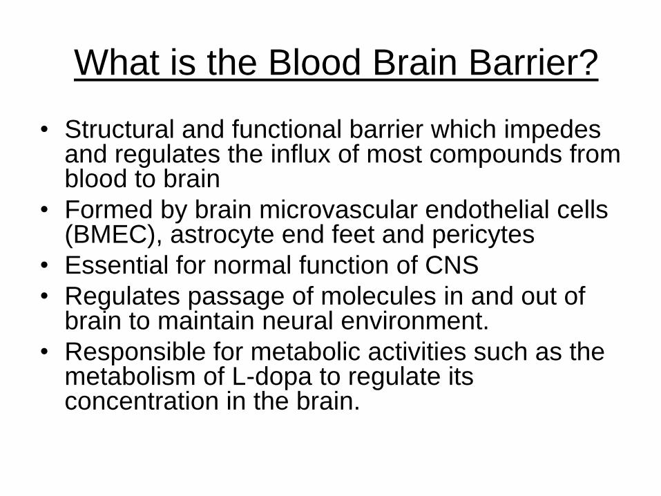
What is the Blood Brain Barrier?
• Structural and functional barrier which impedes and regulates the influx of most compounds from blood to brain
• Formed by brain microvascular endothelial cells (BMEC), astrocyte end feet and pericytes
• Essential for normal function of CNS
• Regulates passage of molecules in and out of brain to maintain neural environment.
• Responsible for metabolic activities such as the metabolism of L-dopa to regulate its concentration in the brain.
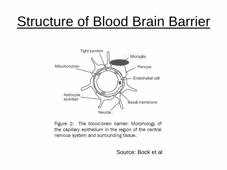
Structure of Blood Brain Barrier
Source: Bock et al

Differences between BMEC and normal
endothelial cells
• Structural differences:
– Absence of fenestrations
– More extensive tight junctions (TJ)
• Functional differences:
– Impermeable to most substances
– Sparse pinocytic vesicular transport
– Increased expression of transport and carrier
proteins: receptor mediated endocytosis
– No gap junctions, only tight junctions
– Limited paracellular and transcellular transport

Integrity of BBB
• Tight Junctions
• Adherens Junctions
• Pericytes
• Astrocyte end feet
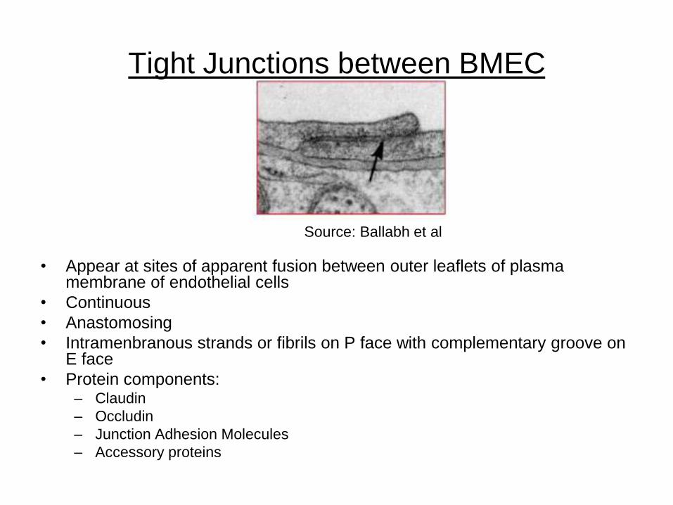
Tight Junctions between BMEC
• Appear at sites of apparent fusion between outer leaflets of plasma membrane of endothelial cells
• Continuous
• Anastomosing
• Intramenbranous strands or fibrils on P face with complementary groove on E face
• Protein components:– Claudin
– Occludin
– Junction Adhesion Molecules
– Accessory proteins
Source: Ballabh et al
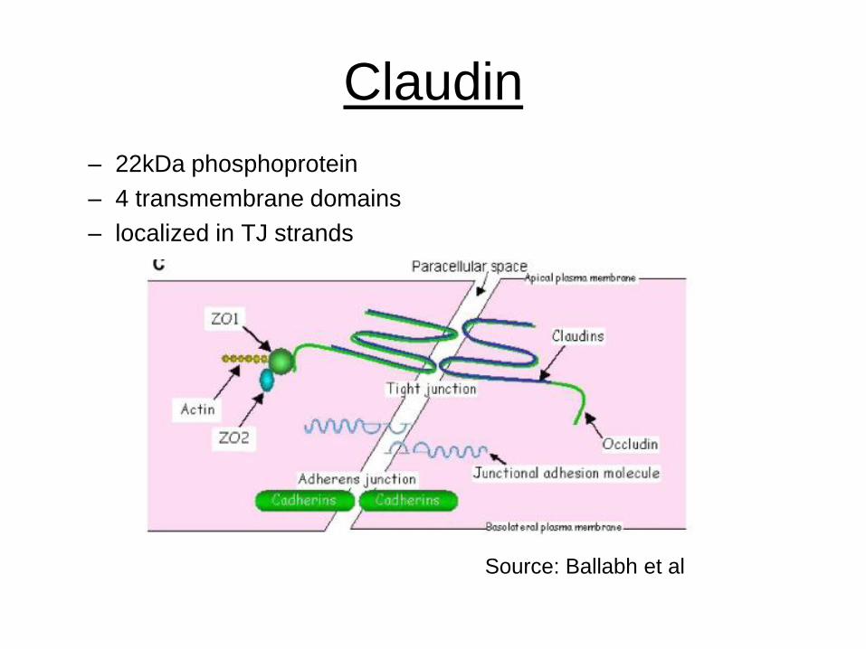
Claudin
– 22kDa phosphoprotein
– 4 transmembrane domains
– localized in TJ strands
Source: Ballabh et al
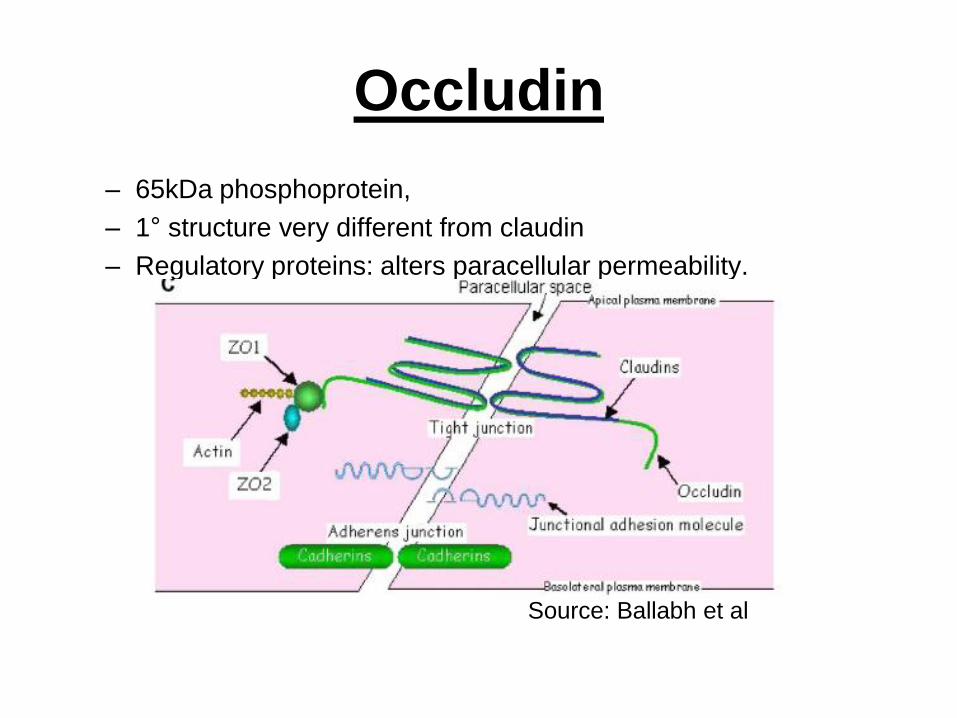
Occludin
– 65kDa phosphoprotein,
– 1° structure very different from claudin
– Regulatory proteins: alters paracellular permeability.
Source: Ballabh et al
Barrier Function of Occludin and
Claudin
• Assemble into heteropolymers and form
intramembranous strands which contain
channels allowing selective diffusion of ions and
hydrophilic molecules.
• Breakdown of BBB in tissue surrounding brain
tumors occurs with concomitant loss of 55kDa
occludin expression
Junction Adhesion Molecules:
• 40kDa
• Integral membrane protein, single transmembrane region
• Belongs to immunoglobulin superfamily
• Localizes at tight junctions
• Involved in cell-to-cell adhesion and monocyte transmigration through BBB
• Regulates paracellular permeability and leukocyte migration
• Also found on circulating leukocytes, platelets and lymphoid organs.
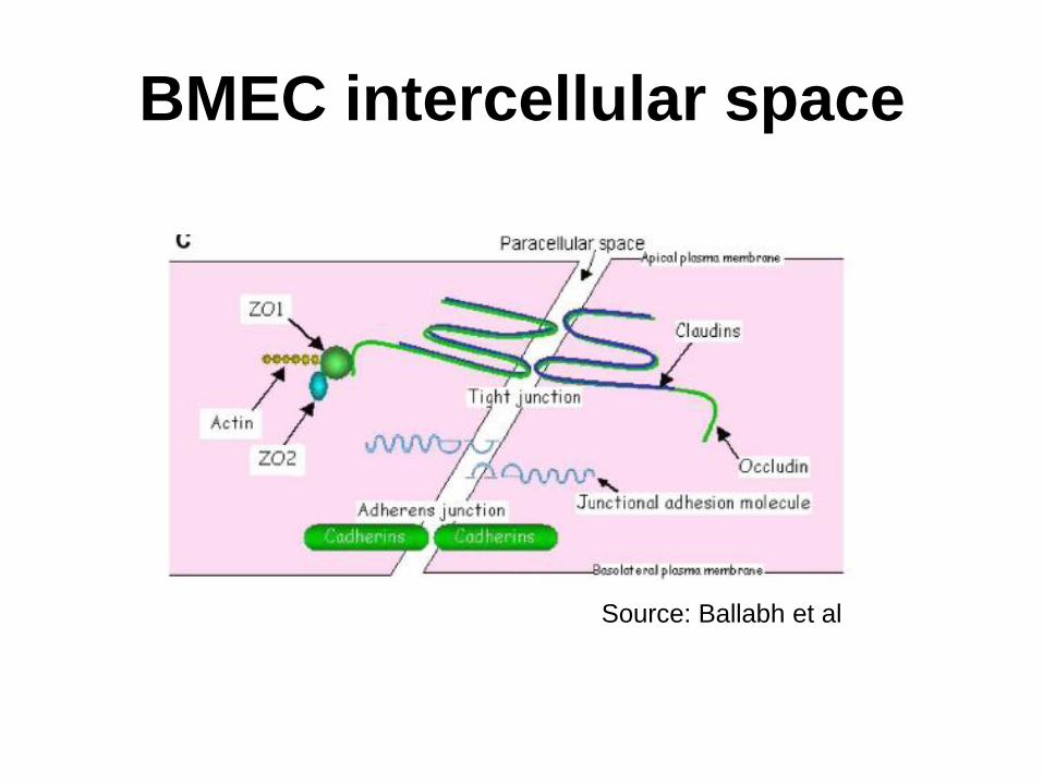
BMEC intercellular space
Source: Ballabh et al
Barrier function of JAM
• Homotypic binding between JAM molecules on
adjacent endothelial cells acts as a barrier for
circulating leukocytes
• Heterotypic binding of endothelial JAM to
leukocyte JAM might guide transmigration of
leukocytes across interendothelial junctions
• So factors that decrease leukocyte migration
must either strengthen homotypic interactions or
weaken heterotypic interactions.
Cytoplasmic accessory proteins
• (ZO-1, ZO-2, ZO-3, cingulin etc)
– These link membrane proteins to actin
– maintenance of structural and functional integrity of endothelium
– crosslink transmembrane proteins.
• Membrane associated guanylate kinase-like proteins (MAGUKS)
– subunits function as protein binding molecules
– role in organization the plasma membrane
Adherens Junction
• Complex between membrane protein cadherin
and intermediary proteins called catenins
• Cadherin-catenin complex joins to actin
cytoskeleton
• Form adhesive contacts between cells.
• Assemble via homophilic interactions between
extracellular domains of calcium ion dependent
cadherins on surface of adjacent cells
Pericytes:
• Cells of microvessels including capillaries, venules, and arterioles that wrap around endothelial cells.
• Provide structural support and vasodynamic capacity to microvasculature.
• Role in structural stability of vessel wall
• Endothelial cells associated with pericytes are more resistance to apoptosis than isolated endothelial cells
– Indicates role of PC in structural integrity and genesis of the BBB
• Phagocytic activity
Astrocyte end feet
• Star shaped glial cells
• Provides biochemical support for BMEC
• Influence of morphogenesis and organization of vessel wall
• Factors released by astrocytes involved in postnatal maturation of BBB
• Direct contact between endothelial cells and astrocytes necessary to generate BBB (Rubin et al, 1991)
• Co-regulate function by the secretion of soluble cytokines such as (LIF, leukemia inhibiting factor), Ca2+ dependent signals by intracellular IP-3 and gap junction dependent pathways, and second messenger pathways involving extracellular diffusion of purinergic messenger.

Regions of brain not enclosed by BBB
• Circumventricular organs
– area postrema,
– median eminence,
– neurohypophysis,
– pineal gland,
– subfornical organ and
– lamina terminalis
These are regions which need to respond to factors present in systemic circulation

Circumventricular organ
functions:
• Pineal gland - secretes melatonin and is associated with circadian rhythms
• Subfornical organ - regulates body fluids, fluid and electrolyte imbalance
• Organum vasculosum of the lamina terminalis – detects peptides
• Choroid Plexus
• Area Postrema - the “vomiting centre” of the brain
• Median eminence - regulates the anterior pituitary through the release of neurohormones
• Neurohypophysis - detects levels of oxytocin and ADH in the blood

Normal BBB transport
• Diffusion
• Facilitated transport by carrier systems
• Receptor mediated endocytosis
• Paracellular transfer more common than
transcellular transfer

Diffusion
• Phospholipid bilayer
• Movement of substances down diffusion
gradient
• Transfer of lipophilic substances
– alcohol, nicotine, oxygen, carbon dioxide
Facilitated transport
• Carrier systems– particular essential amino acids, glucose, these are
extremely specific• transport D-glucose only,
• large neutral amino acids which act as precursors for neurotransmitters,
• only which the brain cannot make,
• glycine: it can block the transmission of nerve signals, hence special carrier which ensures that glycine can be removed from brain
• Receptor mediated endocytosis – Leptin, insulin, overlaps with carrier systems

Factors which cause increase in BBB during
pathophysiology
• Factors produced by astrocytes
– Glutamate,
– Aspartate
– Taurine
– ATP
– Endothelin-1
– NO
– MIP-2
– Tumor necrosis factor alpha TNF-α
– Interleukin beta IL-β
• Paracrine signals secreted by endothelium cells or nerve terminals of neurons running close to blood vessels
– Bradykin
– 5HT
– Histamine
– Thrombin
– UTP
– UMP
– Substance P
– Qionolonic acid
– Platelet activating factor
• Free radicals
E. Coli model
• Requirements for BBB translocation and
successful infection
– High degree of bacteremia
– E Coli invading BMEC
– Rearrangements of actin cytosleleton
– Traversal of BBB as live bacteria

The Consensus
The basis for microbial host interactions
contributing to bacterial invasion of human
BMEC and relevant signaling mechanisms
has not been fully elucidated
Transfer of microbes across BBB:
• Physical damage of BBB
• Ligand receptor interactions followed
by host cell actin cytoskeletal
rearrangements
• Transcellular transport while
maintaining integrity of BMEC

Physcial damage of BBB
• microhemmorage or necrosis of surrounding
tissue
• mechanical obstruction of microvessels by
parasitized red blood cells (PRBC), platelets or
leukocytes in cerebral malaria,
• overproduction of cytokines Borrelia bugdorferi :
fibrinolytic system linked by activation cascade
may lead to focal and transient degradation of
tight junction proteins.

Ligand receptor interactions followed by
host cell actin cytoskeletal
rearrangements
• E.Coli binding to BMEC type I fimbriae, outer
membrane protein A, Ibe proteins, cytotoxic
necrotizing factor 1 (CNF 1)
• S. pneumoniae cell wall phosphorylcholine and
BMEC platelet activating factor receptor

Microbe-specific interaction with BBB
• Bacteria– bind to BMEC, invade BMEC, induce actin cytoskeletal
rearrangement, traverse BBB as live bacteria
• Mycobacteria– unclear, although DNA microarray results show that gene
expression profile of M. tuberculosis associated with human BMEC showed at least 33 genes that were 8X or more upregelated and 147 genes that were 8X or more down-regulated.
• Spirochetes and Fungi– largely unknown, poorly understood: they are able to bind, be
internalized and traverse human BMEC without obvious change in integrity of BMEC (Borrelia burgdorferi, C neoformans, C. albicans.

Conclusion
• Knowledge of the morphology and physiology of the
blood brain barrier has come a long way, but there are
many questions that are still unanswered
References
• Ballabh P, Braun A, Nedergaard M. The blood-brain barrier: an overview: structure, regulation, and clinical implications. Neurobiol Dis. 2004 Jun;16(1):1-13. Review.
• Bock U, Haltner E. Porcine cerebral capillary endothelial cells to study blood brain barrier permeability. Across Barriers publication, May 2003, Int J Parasitol. 2006 May 1;36:541-546.
• Combes V, Coltel N, Faille D, Wassmer SC, Grau GE. Cerebral Malaria: role of microparticles and platelets in alterations of the blood-brain barrier
• Reichel A. The role of blood-brain barrier studies in the pharmaceutical industry.Curr Drug Metab. 2006 Feb;7(2):183-203. Review.
• Kim KS. Microbial translocation of the blood-brain barrier.Int J Parasitol. 2006 May 1;36(5):607-14. Epub 2006 Mar 6. Review.