Comparison between the IR spectra and the structure of the two conformations of a diazabicyclanol
23
PLEASE SCROLL DOWN FOR ARTICLE This article was downloaded by: [Palafox, M Alcolea] On: 15 December 2010 Access details: Access Details: [subscription number 931246444] Publisher Taylor & Francis Informa Ltd Registered in England and Wales Registered Number: 1072954 Registered office: Mortimer House, 37- 41 Mortimer Street, London W1T 3JH, UK Spectroscopy Letters Publication details, including instructions for authors and subscription information: http://www.informaworld.com/smpp/title~content=t713597299 Comparison Between the IR Spectra and the Structure of the Two Conformations of a Diazabicyclanol M. Alcolea Palafox a a Departamento de Química-Física I (Espectroscopia), Facultad de Ciencias Químicas, Universidad Complutense, Madrid, SPAIN To cite this Article Palafox, M. Alcolea(1994) 'Comparison Between the IR Spectra and the Structure of the Two Conformations of a Diazabicyclanol', Spectroscopy Letters, 27: 9, 1165 — 1186 To link to this Article: DOI: 10.1080/00387019408006974 URL: http://dx.doi.org/10.1080/00387019408006974 Full terms and conditions of use: http://www.informaworld.com/terms-and-conditions-of-access.pdf This article may be used for research, teaching and private study purposes. Any substantial or systematic reproduction, re-distribution, re-selling, loan or sub-licensing, systematic supply or distribution in any form to anyone is expressly forbidden. The publisher does not give any warranty express or implied or make any representation that the contents will be complete or accurate or up to date. The accuracy of any instructions, formulae and drug doses should be independently verified with primary sources. The publisher shall not be liable for any loss, actions, claims, proceedings, demand or costs or damages whatsoever or howsoever caused arising directly or indirectly in connection with or arising out of the use of this material.
-
Upload
independent -
Category
Documents
-
view
1 -
download
0
Transcript of Comparison between the IR spectra and the structure of the two conformations of a diazabicyclanol

PLEASE SCROLL DOWN FOR ARTICLE
This article was downloaded by: [Palafox, M Alcolea]On: 15 December 2010Access details: Access Details: [subscription number 931246444]Publisher Taylor & FrancisInforma Ltd Registered in England and Wales Registered Number: 1072954 Registered office: Mortimer House, 37-41 Mortimer Street, London W1T 3JH, UK
Spectroscopy LettersPublication details, including instructions for authors and subscription information:http://www.informaworld.com/smpp/title~content=t713597299
Comparison Between the IR Spectra and the Structure of the TwoConformations of a DiazabicyclanolM. Alcolea Palafoxa
a Departamento de Química-Física I (Espectroscopia), Facultad de Ciencias Químicas, UniversidadComplutense, Madrid, SPAIN
To cite this Article Palafox, M. Alcolea(1994) 'Comparison Between the IR Spectra and the Structure of the TwoConformations of a Diazabicyclanol', Spectroscopy Letters, 27: 9, 1165 — 1186To link to this Article: DOI: 10.1080/00387019408006974URL: http://dx.doi.org/10.1080/00387019408006974
Full terms and conditions of use: http://www.informaworld.com/terms-and-conditions-of-access.pdf
This article may be used for research, teaching and private study purposes. Any substantial orsystematic reproduction, re-distribution, re-selling, loan or sub-licensing, systematic supply ordistribution in any form to anyone is expressly forbidden.
The publisher does not give any warranty express or implied or make any representation that the contentswill be complete or accurate or up to date. The accuracy of any instructions, formulae and drug dosesshould be independently verified with primary sources. The publisher shall not be liable for any loss,actions, claims, proceedings, demand or costs or damages whatsoever or howsoever caused arising directlyor indirectly in connection with or arising out of the use of this material.

SPECTROSCOPY LETTERS, 27(9), 1165-1 186 (1994)
COMPARISON BETWEEN THE IR SPECTRA AND THE STRUCTURE OF THE TWO CONFORMATIONS OF A DIAZABICYCLANOL
KEY WORDS: Vibrational frequencies, diazabicyclanol, Infrared
spectra, scaling frequencies, AM1
H. Alcolea Palafox
Departamento de Quimica-Fisica I (Espectroscopia), Facultad de
Ciencias Quimicas, Universidad Complutense, 28040-Madrid, SPAIN
ABSTRACT
In the two stable conformations of the diazabicyclanol 3,7-
dimethyl-3,7-diazabicyclo[3.3.llnonan-9-01, chair-chair (Val and
chair-boat (Vb), the infrared spectra (200 - 4000 cm-*) were
recorded, compared and their vibrations analysed. Using the AM1 semiempirical method, the geometry was fully optimized in both
forms, and the theoretical Infrared spectra were calculated and
compared. In the (Vb) conformation, the IR spectra were recorded
in CCl,D, CC1, and S,C solvents. Some correlations were
established.
INTRODUCTION
The bicyclo[3.3.llnonane skeleton may be viewed as two cyclohexane
rings fused together, with three ring carbon atoms in common. The
1165
Copyright 0 1994 by Marcel Dekker, Inc
Downloaded By: [Palafox, M Alcolea] At: 17:53 15 December 2010

1166 ALCOLEA PALAFOX
conformation of compounds having the bicyclo[3.3.llnonane
carbocyclic ring structure has been relatively well-established,
and their chair-chair form was found experimentally1P2 and
the~retically**~ to be the most stable conformer. Conformations of the 3.7-diaza analogues have been, in contrast, less well
studied4. They offer additional facets to consider, namely, (i)
the possibility of exo and endo disposition of the N-alkyl
substituents, ( i i ) possible dipole-dipole repulsion of the
heteroatoms in the chair-chair conformation, and (iii) possible
direct overlap of the nitrogen lone-pair orbitals, also in the
chair-chair conformation5.
As part of a research program related to the structures and
vibrational pattern of pharmacologically active compounds, several
aza-bicyclo-nonane systems were examined in previous A
representative example of diazabicyclanols, 3,7-dimethyl-3,7-
diazabicyclo[3.3.llnonan-9-ol, in its chair-chair (Val and
chair-boat (Vb) conformations is examined (Fig. 1) in the present
paper. This kind of compounds, with the two nitrogen atoms in
close proximity and with an alcoholic group, have a remarkable
potential anticholinergic action. The aim of this work is to investigate the vibrational spectra and the structure of the Vb
form, and to compare with the Va conformation. To assist in the
interpretation of the spectroscopic data, theoretical methods were
used.
EXPERIMENTAL
The synthesis and purification of Va and Vb have been previously
reported9. Infrared spectra were recorded on a Perkin Elmer 599 B
spectrophotometer. Indene and polystyrene were used for instrument
calibration. Spectra of Va were recorded in KBr pellets and Nujol,
while spectra of Vb were registered in deuterated chloroform,
carbon tetrachloride and carbon disulfide solvents with
spectroscopic purity. The infrared frequencies are accurate to ?
1 cm-1.
Downloaded By: [Palafox, M Alcolea] At: 17:53 15 December 2010

IR SPECTRA AND STRUCTURE OF DIAZABICYCLANOL 1167
chair-chair (exo, exo) chair-boat (exo,endo)
Fig. 1 Drawing of the two preferred conformations of 3 , 7 -
dimethyl-3,7-diazabicyclo 13.3.11 nonan-9-01.
COIBUTATIONAL METHODS
Two quantum mechanic approaches were employed: semiempirical and
ab initio. The semiempirical approach used was the improved
semiempirical molecular orbital method developed by Dewar et
al.lolll, i.e. AM1. This method has shown reasonable reproduction
of the geometric parameters (bond lengths, bond angles, and
torsional angle^)'^-'^, the vibrational ~ p e c t r u m ~ ~ ~ ~ ~ , and a
remarkable promise in a wide variety of
including studies of chemical reactions of various kinds. The
calculations with AM1 were carried out with the AMPAC program
package11s19 using the PRECISE keyword, and with the GAUSSIAN 90
program package20 using the OPT keyword. Geometries were
completely optimized without fixing any parameters, thus bringing
all geometric variables to their equilibrium values. Force-
constant, vibrational frequency and intensity calculations were
performed.
Downloaded By: [Palafox, M Alcolea] At: 17:53 15 December 2010
1168 ALCOLEAPALAFOX
For the ab initio calculations, the minimal basis STO-3G in
the GAUSSIAN 90 package was selected due to the large size of the
molecule.
The DRAW programz1 was also used several times to evaluate graphically the correctness of starting geometries prior to
calculation, to review the resulting optimized geometries after
the calculation, and to help in the identification of all the
normal vibration modes computed by AM1. The drawings were observed
in high-resolution graphics computer terminals, Tektronic 4105 model..
RESULTS AND DISCUSSION Geometry optimization
Figure 2 shows a view of the molecule of Va and the labeling of
the atoms plotted with the BALL and STICK programz2. The final bond lengths, bond angles and torsional angles calculated by AM1
and sto-3G methods in Va are shown in the z-matrix of Table 1,
while the optimum geometric data of Vb are listed in Table 2. In columns 3 and 8 of these Tables appear the bond lengths in i; in columns 5 and 10 the bond angles in degrees, and in columns 7 and
12 the torsional angles also in degrees. The other columns: 2, 4
and 6 with sto-X, and 2, 9 and 1 1 with AM1, show the connectivity
of the atom considered (Ist column). The computed values
correspond to a single molecule, without the environment of the
crystal structure. Two molecules A and B were experimentally
determined by X-ray diffractiong in the crystal of Va.
According to the values of Tables 1 and 2 , in Vb a larger r N5...N9 interatomic distance together with shorter r N5-a-012
and r N5..-H30 distances than in Va are identified. The latter
distance is relatively short; this fact is attributed to the
existence of intramolecular 0-H.0.N bonding when passing from
structure Va to Vb. Examples of compounds displaying O-H.-.X
bonding are somewhat rare owing to unfavorable energy differences
usually existing between boat and chair conformers. However, a
Downloaded By: [Palafox, M Alcolea] At: 17:53 15 December 2010

IR SPECTRA AND STRUCTURE OF DIAZABICYCLANOL 1169
Fig. 2 Two viewpoints of the optimized geometry and labelling of
the atoms in the chair-chair conformation Va of 3.7-
Dimethyl-3,7-diazabicyclo r3.3.11 nonan-9-01,
number of examples in mono- and bicyclic systems have been
reported23~ 24.
The L N5-C3-C2, ,!. N5-C6-C4 bond angles in Vb structure are
larger by about 4O than in Va, and the torsional angles on C3 and
C6 differ remarkably. Concerning the hydroxylic group, in Va the H30 hydrogen in
the gauche position by AM1 (cis with sto-3G) relative to H13 (see
Fig 31, changes to trans in Vb, in accordance with a shorter
H30***N5 distance, corresponding to the presence of an
intramolecular hydrogen bond on H30. Thus different values of the
L H13-C1-012-H30, /!, CZ-C1-012-H30 and L C4-C1-012-H30 torsional
angles are obtained. These features are in agreement with the
absence of intramolecular bonds in the crystal9 of Va.
Downloaded By: [Palafox, M Alcolea] At: 17:53 15 December 2010

1170 ALCOLEA PALAFOX
Table 1 . Z-Matrix with the optimum geometric values obtained by AM1 and
by sto-3G ab initio level in the Va conformation-
c1
c2
c3
c4
N5
C6
c7
C8
N9
c10
c11 012
H13
H14
H15
H16
H17
H18
H19
H20
H21
H22
H23
H24
H25
H26
H27
H28
H29
H30
1
2
1
3
4
2
4
8
5
9
1
1
2
4
3
6
8
7
3
6
8
7
10
10
10
11
11
1 1
12
sto-3G
1.5521
1.5483
1.5472
1.4870
1.5482
1.5489
1.5490
1.4867
1.4824
1.4818
1.4346
1.0979
1.0906
1 .0896
1.0962
1.0956
1.0968
1.0970
1.0907
1.0906
1.0905
1.0906
1.0886
1.0942
1.0886
1.0886
1.0944
1.0885
0.9908
1
2
2
1
1
1
4
3
8
2
2
1
1
2
4
4
2
2
4
4
2
5
5
5
9
9
9
1
109.22
106.25
111.99
109.28
108.56
108.68
112.08
110.28
110.54
113.27
109.02
109.67
109.20
108.10
108.05
108.32
108.25
109.65
109.58
109.63
109.65
109.30
113.02
109.26
109.29
113.03
109.27
104.07
3
1
2
4
2
1
2
4
3
3
4
2
1
1
1
1
1
1
1
1
3
3
3
8
8
8
2
-61.61
58.95
61.60
62.84
-62.83
58.94
-177.28
-177.24
57.63
180.81
180.34
179.45
-64.31
64.35
-64.55
64.53
178.70
-178.59
178.58
-178.66
-58.87
61.90
-177.36
-176.89
62.35
-58.38
62.94
1.5301
1.5310 1
1.5302 2
1.4554 2
1.5313 1
1.5317 1
1.5317 1
1.4532 4
1.4455 3
1.4444 8
1.4196 2
1.1270 2
1.1230 1
1.1236 1
1.1315 2
1.1324 4
1.1324 4
1.1321 2
1.1267 2
1.1266 4
1.1272 4
1.1272 2
1.1213 5
1.1256 5
1.1213 5
1.1213 9
1.1258 9
1.1214 9
0.9644 1
AM1
110.00
106.77
114.54
109.88
108.37
108.66
114.82
112.03
112.24
108.10
110.34
109.68
110.13
107.09
107.08
107.47
107.61
108.71
108.78
108.57
108.47
108.72
113.48
108.80
108.77
113.43
108.82
106.73
-
3
1
2
4
2
1
2
4
3
3
4
2
1
1
1
1
1
1 1
1
3
3
3
8
8
8
2 -
-60.38
53.50
60.62
63.40
-63.16
53.12
-174.09
-173.04
61.57
180.13
181.43
178.98
-70.49
69.94
-71.39
70.80
172.93
-173.66
172.52
-173.07
-55.52
65.28
-173.84
-173.16
66.04
-54.79
-166.78
Downloaded By: [Palafox, M Alcolea] At: 17:53 15 December 2010
IR SPECTRA AND STRUCTURE OF DIAZABICYCLANOL 1171
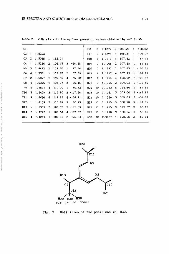
Table 2. 2-Matrix with the optimum geometric values obtalned by AM1 In Vb.
C1
C2 1 1.5292
C3 2 1.5365 1 112.91
C4 I 1.5286 2 106.45 3 -56.38
N5 3 1.4472 2 118.50 1 17.66
C6 4 1.5351 1 112.87 2 57.39
C7 2 1.5371 1 107.05 4 65.78
C8 4 1.5375 1 107.07 2 -65.46
N9 8 1.4561 4 113.70 1 56.52
C10 5 1.4409 3 114.80 2 -117.26
C11 9 1.4450 8 112.25 4 -176.91
012 1 1.4109 2 113.98 3 70.23
HI3 1 1.1303 2 109.75 3 -175.09
H14 2 1.1223 1 109.51 4 -177.37
HI5 4 1.1229 1 109.46 2 178.04
H28
H16 3 1.1299 2 108.29
HI7 6 1.1294 4 108.31
HI8 8 1.1310 4 107.82
H19 7 1.1306 2 107.88
H20 3 1.1292 2 107.43
H21 6 1.1297 4 107.43
H22 8 1.1266 4 108.92
H23 7 1.1264 2 109.03
H24 10 1.1253 5 114.66
H25 10 1.1221 5 109.00
H26 10 1.1226 5 108.68
H27 11 1.1215 9 108.78
H28 1 1 1.1255 9 113.37
H29 1 1 1.1210 9 108.86
H30 12 0.9627 1 108.35
1 138.02
1 -139.87
1 -67.74
1 67.12
1 -106.71
1 104.79
1 175.87
1 -176.45
3 68.84
3 -169.89
3 -52.04
8 -174.05
8 65.15
8 -55.66
2 -63.04
N9
H13 N5
012 H25
cis gauche trans
Fig. 3 Definition of the positions in H30.
Downloaded By: [Palafox, M Alcolea] At: 17:53 15 December 2010

1172 ALCOLEAPALAFOX
The L C2-C1-012 and L C4-C1-012 angles have nearly the
same value in Vb, with a slight variation of 0.07O. This means a
symmetric position of 012 with respect to C2 and C4, in
contrast to those computed for Va (108.10 and 113.09O,
respectively), corresponding to a gauche conformation of H30.
This conformation of the H30 atom in Va impedes the formation of
an intramolecular bond with N5; hence H30 is not aligned in this
way, being asymmetric with C2 and C4. Similar values for C1-C2
and Cl-C4 bond lengths calculated for Vb corroborate this fact.
The preferred conformation of 3,7-dimethyl-3,7-diazabicyclo
[3.3.11 nonan-9-01. as the diazabicyclanols in general, depends on
the phase and on the polarity of the solvent9. Thus in solid and
dimethylsulfoxide (dipole moment pD = 4.5 Debye), the compound
adopts a distorted chair-chair conformation Va (the computed pD by
AM1 is 1.58 D) with H30 in gauche, whereas in CC1, (pD = 0.28 D ) ,
CDC1, (pD = 1.16 Dl and S,C (pD = 0.45 D) solutions with low
dipole moment, an intramolecular hydrogen bond stabilizes the
chair-boat conformation Vb with lower dipole moment (0.76 D) than Va, and with H30 in trans to form this intramolecular bond.
Scarse in the bibliography are examples of bicyclic systems
where the boat conformation is stable and exists
e~perimentally~~~~. Several of them, however, are shown in Fig. 4a. The exolendo notation is explained in Fig. 4b.
The chair-boat conformation seems to be stable and in some cases the preferred form in the bicyclo[3.3.llnonane system if
there is a heteroatom at 3-position (endo) as well as a
substituent at 7-(endo), while a 3-exo/7-exo stabilizes the
chair-chair Substituents at these positions therefore
govern the conformation of the bicyclo[3.3.llnonane system and
chair-boat forms must play a predominant role in the
conformational equilibrium. Moreover, the chair-boat form would
not be possible unless either a very bulky substituent or an intramolecular hydrogen bonding or both are present. The effects
of substituents at 2 and 4 positions are less well understood.
Downloaded By: [Palafox, M Alcolea] At: 17:53 15 December 2010

IR SPECTRA AND STRUCTURE OF DIAZABICYCLANOL 1173
? N-R
R- N R-N
Fig. 4a Several stable experimentally chair-boat conformations in
the bicyclo[3.3.llnonane system.
CH3 (exo) \ \ ',
Chair -Chair (endo, exo) Chair- boat (endo, ex01
Fig. 4b The exo/endo notation refers to the disposition of the N- methyl groups.
Downloaded By: [Palafox, M Alcolea] At: 17:53 15 December 2010
e c
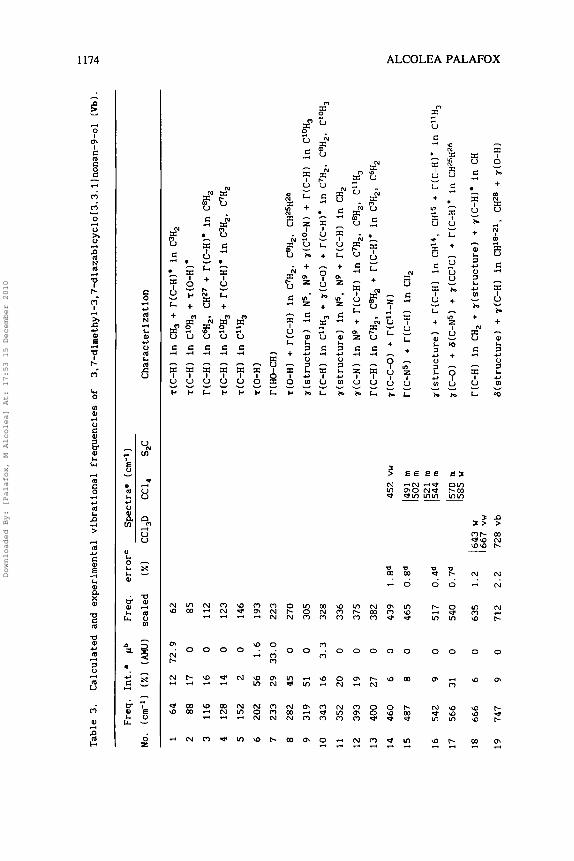
Table 3.
Calculated and e
xperimental
vibr
atio
nal fr
eque
ncie
s of 3.7-dirnethyl-3,7-diazabicyclo[3.3.llnonan-9-ol
(Vb).
Freq
. Int.'
pb
Freq.
errorC
Spec
tra=
(cm-1)
No. (crn-ll
(XI
(AM
U)
scaled
(XI
CC1,D
CC1,
S,C
Char
acte
riza
tion
1 64
12 72.9
62
T(C-H) in C
H, + r(C-H)*
in C3H,
2 88
17
0 85
7CC-H) in C10H3
+ T(O-H)*
3
116
16
0
112
r(C-H) in C6H,, CH27 +
T(C-H)* in C8H,
4 128
14
0
123
T(C-H) in CIOH, +
T(C-H)* in C3H,, C7H,
5 152
2 0
146
7cC-H) i
n C"H,
6 202
56
1.6
193
~(0-H)
7 233
29 33.0
223
r ( HO-CH
1 8
282
45
0
270
r(0-HI
+ T(C-H) in
C7H,, C8H,.
CH25H26
9
319
51
0 305
r(st
ruct
ure)
in N5,
N9
+ r(CIO-N) +
T(C-H)
in CIOH,
10
343
16
3.3
328
T(C-H) in C"H,
+ r(C-0)
+ r(C-H)*
in C7H,, C8H2, CL0H,
11
352
20
0
336
r(st
ruct
ure1
in
N5,
N9 +
r(C-HI
in CH,
12
393
19
0
375
r(C-N) i
n N9 +
T(C-H)
in C7H,, C8H,,
C"H,
13
400
27
0 382
T(C-H) i
n C7H,,
C8H,
+ T(C-H)*
in C3H2, C6H,
2 14
460
6 0
439
1.8d
452 vw
r(C-C-0) +
r(C"-N)
15
487
8 0
465
O.Sd
16
542
9 0
517
0.4d
491
m 502
m 521
m I 54
4 m
h 0
r(C-N5)
+ r(C-H) in
CH,
r(st
ruct
ure)
+ r(C-H)
in CH14, CH15
+ T(C-H)*
in C"H,
cd
17
566
31
0
540
0.7d
570
m r(C-0) +
6(C-N5) +
;I(CC~C) +
r(C-H)* in CH25H26
585 w
2 18
666
6
0
635
1.2
643 w
1667 v
w
19
747
9 0
712
2.2
728 vb
T(C-H) in CH,
+ r(
stru
ctur
e1 +
r(C-H)* in CH
G(st
ruct
ure)
+ r(C-H)
in CH18-21, CH28
+ ~(0-H)
0 x
Downloaded By: [Palafox, M Alcolea] At: 17:53 15 December 2010

20
21
22
23-
25
26
27
28
29
30
31
32
33
34
35
36
37
38
39
40
41
840
944
947
972
1016
1030
1037
1068
1075
1108
1115
1123
1140
1157
1185
1196
1201
1206
1229
1264
1295
815.846
vw
25
0 801
1.9d
856
m
776
mb
4
0
900
29
0 903
1.3
915
vb
909 w
6
0
927
8
0
969
1.9
951
vw
3
0
982
1.3
995
vw
22
0
989
2.3d 1014 m
16
0
1019
1.6
1036 s
8
0.5
1025
14
1.2
1057
0.3
1054 vs
24
0.8
1063
0.6
1069 v
s
27
0.6
1071
1.6
1089 v
s
5
0.8
1087
3
0.5
1103
7
0.4
1130
1.3
1145 s
h
19
0.4
1141
0.3
1138 m
5 0.4
1145
1.5
1162 w
47
0.8
1150
3.0
1127 s
1 0.5
1172
1.3
1187 w
9
0.5
1205
0.9
1216 v
w
34
1.0
1235
0 1235 m
3
m
v1 a
6(CC1C)
+ r(C-H)
in C3H,, C6H, +
r(C-H)'
in CH
? T(C-H) in C7H,, C8H,
+ T(C-H)* in C3H,. C6H,
r(0-H)
+ G(s
truc
ture
1 +
r(C-H) in CHI3, CHZO-23
r(C-H) in CH,
+ r(
stru
ctur
e)*
+ r(C-H)*
in CH,
r(C-H) i
n C
HI4
, CHI5 +
G(s
truc
ture
) U
r(C-H) in C
HI4
, CH15. C%,,
C6H,
+ G(
stru
ctur
e)
r(C-H) in CHI4. CHI5
+ r(C-H)* in CH,
4 > Z rA
r(C-H) in C
HI3,
C3H2, C6H, +
6(C4-C6)
+ r(C-H)* in CH,
2 2 z c1
T(C-H) in CH,
+ 6(C-H) i
n CH
r(C-H) in CH,
+ 6(C-H)* in CH18, CHI9
r(C-H) in CH,
r(C-H) in C'OH,
+ 6(C-H)*
in CH, CH20-,,
6(C-H) in CH
+ r(C-H) in C"H,
% i2 U
r(C-H) in C7H2. C8H2
+ 6(C-H)*
in CH
in CH,
+ 6(C-H)*
in CHI4. CHI5
6(C-H) in CHI4, CH15
+ T(C-H)*
in CH,
E r
4
c1 > Z
T(C-H)
6(C-HI in CH14. CH15
+ r(C-H) i
n C3H,,
C6H,
r(C-H) in C7H2, C8H,
+ 7(C-H)'
in C3H,, C6H2
6(C-H) in CH
+ T(C-H) in C3H,, C6H,
6(C-HI in CHI4, CHI5
+ r(C-H)*
in CH,
6(C-H) in CHI3
+ a(0-H)
+ 6(C-H)*
in CHI4, CHI5
e
(continued)
Downloaded By: [Palafox, M Alcolea] At: 17:53 15 December 2010
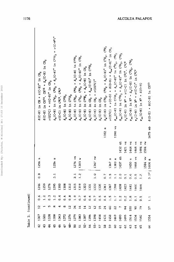
Table 3. (continued)
1.0
42 1307
43 1320
44 1338
45 1358
46 1363
47 1372
48- 1376
50 1379
51
1383
52- 1387
54 1396
55- 1398
57 1404
58 1431
59 1458
60 1469
61 1480
62 1495
63 1514
64 1534
65 1540
1347 v
w
12
0.,6
1 1.2
5
0.3
8 1.2
4
0.4
8 0.6
12
0.8
34
1.0
16
0.7
14
0.6
12
0.6
12
0.7
15
0.5
24
0.6
60
1.5
22
1.7
7
1.2
84
0.2
100
1.8
90
0.1
79
0.5
66 1554
57
1.1
1246
1259
1276
1295
1300
1308
1312
1315
1319
1323
1331
1333
1335
1361
1387
1398
1408
1423
1441
1460
1466
0.8 1256 s
3.1
1256 s
1352 r
n
1390 vs
1432 s
h
1421 v
s
1444 vs
1468 vs
1484 v
s 1584 vw
1675 r
nb
6(C-H1 in CH
+ T(C-H)'
in CH,
b(C-H) in CHI4, CHI5
+ A,(C-H)
in CH,
v(cC'C)
+ T(c-H)*
in a,
A,(C-H)
in C7Hz, C8H2
+ 6,(C-H1*
in CI1H3 +
u(C-Ng)'
v(C-C) in CzC3. C4C6
6,(C-H)
in C"JH3
6,(C-H)
in C"H3
A,(C-H)
in C3H,. C6H,
+ 6,(C-H)
in C10H3
A,(C-H)
in C7H2. C8H,
+ A,(C-H)*
in C3H,
6,(C-H)
in Ci0H3 +
A,(C-H)
in CH,
A,(C-H)
in CH,
+ d,,(C-H)*
in C10H3
A,(C-H)
in CH,
+ v(CCIC)*
A,(C-H)
in CH,
A,(C-H)
in C3Hz. C6H,
+ A,(C-H)*
in C'H,,
C8H,
v(CCICI
+ v(C-0)
+ a(0-H) +
6,,(C-H)*
in CH,
6,,(C-H)
in CiiH, + A,(C-H)* in C7H,, C8H,
6,,(C-H)
in C10H3 +
A,(C-HI*
in C3H,, C6H,
v,(C-N)
in N
9 + u(C-C) in C2C7. C4C8
u,,(C-N)
in N9
+ A,(C-H) in C7H2, C8H,
v,(C-N)
in N
5 + u(C-Cl'
in C2C3
u,,(C-N)
in N
5 + a(0-H)
a(0-H) +
6(C-H) in CHI3
>
cl
0
r F w F % 0
X
Downloaded By: [Palafox, M Alcolea] At: 17:53 15 December 2010
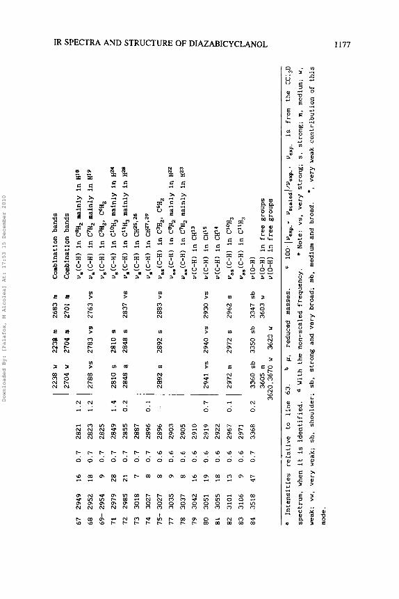
67 2949
16
68 2952
18
69- 2954
9
71 2979
28
72 2985
21
0.7
73 3018
74 3027
75- 3027
77 3035
78 3037
79 3042
80 3051
81 3055
82 3101
83 3106
2941 vs
2940 vs
2930 vs
7
8
8 9 8 6 9 8 3 9
84 3518
47
0.7 2821
0.7 2823
0.7
2825
0.7 2849
0.7 2855
0.7 2887
0.7 2896
0.6
2896
0.6
2903
0.6
2905
0.6
2910
0.6 2919
0.6
2922
0.6
2967
0.6
2971
0.7 3368
2238 w
2238 m
2683 m
2704 w
2704 m
2701 m
2788 vs
2783 v
s 2763 vs
1.4
2810 $
2810 8
0.2 2848 8
2848 s
2837 vs
0.1
1 I 2892 s
2892 s 2883 vs
Combination bands
Combination bands
u,(C-H)
in C8H2 mainly in H1*
u,(C-H)
in C7H2 mainly in H'9
u,(C-H)
in C3H2, C6H2
u,(C-H)
in @OH3
mainly In H*'
u,(C-H)
in Cl1H3 mainly in H28
u,(C-H)
in CH25126
u,(C-H)
in CH27*2q
u,,(C-H)
in CJH,, C6H2
u,,(C-H)
in C8H2 mainly in H22
u,,(C-H)
in C7H2 mainly in HZ3
u(C-H) in CH13
u(C-H) in CH15
u(C-H) in CH14
u,,(C-H)
in C'OH,
u,,(C-H)
in C1'H3
u(0-H)
u(0-HI in free groups
u(0-H) in free groups
a Intensities
relative
to line 63.
p, reduced
masses.
' lo
o'
IVe
xp
.-
~s
ca
~e
d~
/~e
xp
.,
Ue
Xp
. IS
from the
CC1,D
spectrum, when it
is identified.
With the non-scaled frequency.
* Note: vs, very strong; s, strong; m
, medium; w,
weak;
vw, very weak; sh, shoulder; s
b, strong and very broad; mb, medium and broad.
*, very weak contribution of this
mode.
Downloaded By: [Palafox, M Alcolea] At: 17:53 15 December 2010

1178 ALCOLEAPALAFOX
To the best of our knowledge the boat-boat conformation has
not been proposed with certainty for any molecule having the
bicyclo[3.3.l]nonane skeleton.
Vibrational frequencies
Compound Vb crystallizes without water. This fact is reflected in
a different IR spectra, especially in the 0-H modes, with respect to Va which crystallizes with water and alcohol molecules binding
through the hydroxylic group, because in Va the lone nitrogen
electrons pairs in both rings of the bicyclic structure are
involved in the bonding with either of these water or alcohol
moleculesg.
In Table 3 are shown for Vb the assignment of the observed IR bands in CCl,D, CC1, and S,C solvents, and the computed and scaled
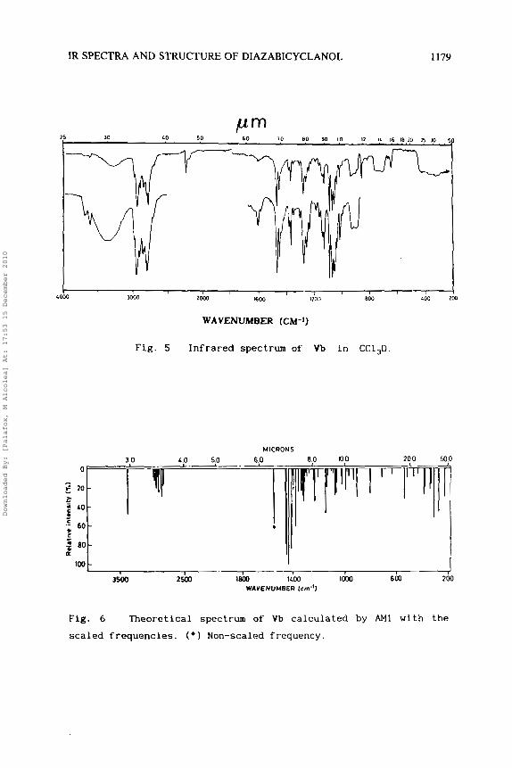
frequencies by AM1 with their intensities. Fig. 5 records the IR spectrum of Vb in CC1,D. In CC1, the spectrum was registered only
in the 450-1000 cm-' and 1300-4000 cm-I ranges, and in S,C in
the 1300-4000 cm-' region. The relative IR intensities of the
third column were calculated by dividing the value of the computed
intensity by the intensity of the strongest line obtained (in the
study, line number 6 3 ) . The characterization of the bands is shown
in the last column. To correct the deficiency of the AM1 method, a
scaling equationz7 was used:
= 0.7 + 0.9528 ~,,,,(cm-~)
However with frequencies higher than 1400 cm-l, a lower error is
obtained using another scaling equation27:
u,,,~~~(c~-~) = -14.8 + 0.9614 ~,,,(cm-~)
The scaled frequencies obtained are listed in the fifth column.
The theoretical spectrum computed with these new frequencies is
shown in Fig. 6 . The % error values determined in this way with
Downloaded By: [Palafox, M Alcolea] At: 17:53 15 December 2010
IR SPECTRA AND STRUCTURE OF DIAZABICYCLANOL 1179
10 80 90 10 I ? IL 16 la ?O B 3 ' 40 5 0 KO 15 30
I I , 1600 1100 B c a LOO 1 3000 2000 LOO0
WAVENUMBER (CM-'1
Fig. 5 Infrared spectrum of Vb in CC1,D
MICRONS
80 100 200 500 30
3500 2500 leal 1400 IOW 600 200
a
WAVENUMBER (cm-')
Fig. 6 Theoretical spectrum of Vb calculated by AM1 with the
scaled frequencies. ( * I Non-scaled frequency.
Downloaded By: [Palafox, M Alcolea] At: 17:53 15 December 2010

1180 ALCOLEAPALAFOX
respect to the experimental data in CC1,D are collected in the
sixth column. In general, a good concordance between the observed
and calculated frequencies is obtained, with errors lower than 3%. The modes undetected in the IR spectra are those with the lowest calculated IR intensities. In the low frequency range ( < 900
cm-l). with modes characteristic of the bicyclic structure, the
possibility of the use of scaling equations is, in general,
reduced due to the specificity of the structure, and they normally
give unsatisfactory results. However in this case the use of the
non-scaled frequencies computed by AM1 gives good concordance with
the experimental.
In Table 4 are collected several selected calculated and
experimental frequencies in Va, especially in the 0 - H and C-N
groups, while in Fig. 7 is registered its theoretical spectrum
with the scaled frequencies and the IR spectrum in KBr pellets.
The characteristics of the spectra are discussed in the
following sections:
0-H vibrations. The IR spectra of vb in very dilute CC1,
solution show a strong 0 - H band at ca. 3350 cm-I, corresponding to
a stretching 0-H vibration. The position of the maximum is
unaltered in solution of CC1,D indicating the existence of an
intramolecular 0-H.e.N bond. This fact is corroborate by the AM1
predictions. Thus a strong 0-H band is computed at 3368 cm-'
(scaled), with a very small error, only 0.2%. A weak band at
3620 cm-l is also observed in CC1,. This band indicates a small
proportion of free 0-H groups of chair-chair forms in Vb.
In the IR spectra of Va in KBr pellets and in Nujol, a broad absorption at 3200-3400 cm-' is identified corresponding to 0 - H
groups, which are bonded with water and alcohol molecules. A broad
absorption band in the 3100-3600 cm-I range with bands at 3480
and 3520 cm-l attributed to water of crystallization9, is also
observed.
The IR spectrum of Vb in CC1,D solution shows a strong and broad band centered at 3360 cm-', which suggests the coexistence
Downloaded By: [Palafox, M Alcolea] At: 17:53 15 December 2010

IR SPECTRA AND STRUCTURE OF DIAZABICYCLANOL 1181
Table 4. Several selected calculated by AM1 and experimental vibrational
frequencies of Va.
Freq. Int.' Freq. errorb SpectraC
No. (cm-') (%) scaled ( X I in KBr Characterization
6 188 79 180 l(0-H) 7 204 17 195 238 m T(O-H) 8 294 19 281 1.1 284 vw T(CH,) + ~ ( 0 - 1 0
9 330 47 315 3.3 305 vw r(C-N) in N9 16 527 17 503 0.2 502 w a(structure)
17 569
26 1015
28 1067
31- 1112
41 1265
44 1345
51- 1378
57 1397
59 1443
60 1468
62 1504 63 1508
64 1513
65 1519
66 1543
72 2984
20
34
19
26
25
38
19
11
85
25
97 65
100
67
40
22
543
968
1017
1060
1206
1282
1314
1331
1373
1397
1431 1435
1440
1446 -
2854
4.4d 595 m-b
2.7 995 s
2.9 1047 s
0.5 1065 vs
0.7 1214 vw
0.9 1270 vs
0 . 3 1310 m
1.5 1352 m
0.2 1370 s
0.5 1390 w
- 1655 mb
1.6 2808 s
7(C-0) + G(ring1 + 7(C-H) in C'OH, 7(C-H) in CH". CHI5 + 6(ring)
a(C-H) in C1P4 and CHI5 l-(C-H) in CH, and CHI3
a(0-H) + 6(C-H) in CH
u(C-C) in C4C8 +6(C-H) in CH15."3s21 6,(C-H) in CH, u(C-C) in C4C6 + 6(C-H) V,(OCC) + 6(C-I t )
6,,(C-H) in CH, u,(C-N) in N5 + B(C-H) in CHI4. CHI5 u,(C-NI in N9 + 6(C-H) in CHI4. CHI5 u,,(C-N) + 6,,(C-H) in CH, u,,(C-N) + 6,,(C-H) in CH, d(0-H) v,(C-H) in C'OH,, mainly in HZ5
73- 3026 8 2895 u,(C-H) in CH, 79 3034 5 2902 u,,(C-H) in C3H, 82- 3105 10 2971 0.3 2980 w u,,(C-H) in CH, 84 3498 47 3348 3300 vs-b u(O-HI
a Intensities relative to line 64. 100. Iuexp.-uscaledl/uexp, Note: vs.
very strong; s , strong; rn, medium; w, weak; vw, very weak; m-b, medium and
broad; vs-b, very strong and broad. d W i t h the non-scaled frequency
Downloaded By: [Palafox, M Alcolea] At: 17:53 15 December 2010

1182 ALCOLEA PALAFOX
0 '
5 2 0 - > -
L O -
' 6 0 - u - > - 2 B O - (L
100
*
( b ) -
I I I 1
Fig. 7 (a) Infrared spectrum of Va in KBr pellets and (bl theo-
retical spectrum with the scaled frequencies by AM1.
of intrarnolecularly-bonded and solvent-bonded molecules, probably
in chair-boat conformations. Moreover, a weak band at 3605 cm-' indicates the presence of free 0-H groups of chair-chair
conformations as in polar solvent solutions. Results with Vb in CS, are similar. Thus in the 0-H region a broad absorption with a maximum at ca. 3347 cm-' is observed.
The a(0-H) mode is predicted with strong intensity at 1554
cm-1 in Vb and 1543 cm-1 in Va , which corresponds to the medium intensity IR band at 1604 cm-' in Vb (in CC1,D) and at 1655 cm-'
Downloaded By: [Palafox, M Alcolea] At: 17:53 15 December 2010

IR SPECTRA AND STRUCTURE OF DIAZABICYCLANOL 1183
in Va. Because AM1 computes a hydroxy group without intermolecular
hydrogen bonds with the environment, the calculated frequencies
are far from the experimental, especially in the Va molecule. The
strong bands in Vb at 1540, 1458 and 1295 cm-I are also computed
with a slight contribution of the mode 6(0-HI.
The out-of-plane bending z(0-H) is determined in Vb at 903
cm-l (scaled), in good agreement with the very broad IR band at 915 cm-1 in CC1,D and the weak absorption at 909 cm-' in CC1,.
The torsional mode r(0-H) is predicted in the far-infrared range,
at 202 cm-l in Vb and at 188 cm-' in Va.
Other vibrations: The C-0 stretching is predicted as a very
strong band at 1387 cm-1 (scaled) in Vb and at 1373 cm- '
(scaled) in Va, which corresponds to the strong IR absorption at 1367 cm-' (in CC1,D) and at 1370 cm-1 (in KBr pellets)
respectively, with slight errors. The out-of-plane bendings r(C-
0) also show close values between the Va and Vb forms. They are
calculated as strong bands at 566 cm-1 (Vb) and at 569 cm-I
(Val, and at 460, 455 cm-I in Vb and Va respectively. The C-H stretchings appear in the 2700-3000 cm-' range of
the IR spectra as very strong Bohlmann bands. They are well
predicted by AM1 with errors, in general, lower than 1.5%. The
frequencies do not change appreciably from Va to Vb form. The most intense computed bands are due to the u(C-N)
stretching vibration. The frequencies corresponding to N9 are very
close between the Va and Vb conformations, but they differ about 25-30 cm-1 in N5 due to the boat form of Vb. Thus the
antisymmetric mode is determined and scaled in N9 at 1446 cm-l
(Val close to the value at 1441 cm-I (Vb); while in N5 it is computed at 1440 cm-' (Val far from the value at 1466 cm-1 in
Vb. A similar result is obtained with the symmetric mode with a scaled frequency in N5 and Vb at 1460 cm-l that differs from the
other v,(C-N) vibrations, about 30-40 cm-l. The strong-very
strong IR bands observed in Vb at 1450 and 1468 cm-l in CC1,D and
at 1421, 1444. 1468 and 1484 cm-1 in CC1, are assigned to this
v(C-N) stretching mode.
Downloaded By: [Palafox, M Alcolea] At: 17:53 15 December 2010
1184 ALCOLEAPALAFOX
CONCLUSIONS
The molecular geometry of the diazabicyclanol studied is
accurately determined by the AM1 theoretical method in its two
stable conformations, Va (chair-chair) and Vb (chair-boat), the
differences being in the standard deviation of this method. The
discrepancias with the x-ray study of Va are attributed to the
fact that, in the crystal, the molecules through their nitrogen
atoms, are implicated in bonding with either water or alcohol
molecules.
The conformer Va is slightly more stable than Vb, and Va is
the conformer that predominates in the solid state. In solution in
nonpolar solvents, the conformer Vb with lower dipole moment (0.75
D by AM11 is stabilized and it is the only form present; while in polar solvents the V a form with higher dipole moment (1 .58 D by
AM11 is adopted. In the transformation from V a to Vb, a
simultaneous change in the position of the hydrogen of the
hydroxylic group from gauche to trans is produced to form an
intramolecular hydrogen bond with N5 which stabilizes this boat-
chair structure.
The frequencies corresponding to N9 are very close between
the Va and Vb conformations, but differ about 25-30 cm-l in N5
due to the boat form of Vb.
Good reproduction of the experimental frequencies is obtained with AM1. Thus the X error obtained with the use of a scaling
equation is very small, in the majority of cases less than 3.5%.
Concerning the intensity of the vibrations, it is noted that, in general, the modes not detected in the spectra are those having
the lowest calculated intensities. In general the coincidence
grade is satisfactory between the different spectra.
ACKNOWLEDGMENTS
The author wishes to thank J. Bellanato and E. Galvez for the experimental spectra supplied. All calculations were performed on
Downloaded By: [Palafox, M Alcolea] At: 17:53 15 December 2010

IR SPECTRA AND STRUCTURE OF DIAZABICYCLANOL 1185
the CRAY Y-MP81864 computer at the Center for High Performance
Computing of the University of Texas.
REFERENCES
1.
2.
3.
4.
5.
6.
7.
8 .
9.
C. Jaime, E. Osawa, Y. Takeuchi and P. Camps, J . Org. Chem., 48, 4514 (1983).
(a) V.S. Mastryukov, E.L. Osina, O.V. Dorofeeva, M.V. Popik, L.V. Vilkov and N.A. Belikova, J. Mol. S t r u c t . , 52, 211 (1979); (b) V.S. Mastryukov, M.V. Popik, O.V. Dorofeeva, A.V. Golubinskii, L.V. Vilkov, N.A. Belikova and N.L. Allinger. J . Am. Chem. S O C . , 103, 1333 (1981); Tetrahedron L e t t s . , 45, 4339 (1979).
P.N. Skancke, J. Mol. S t r u c t . (Theochem.), 151, 11 (1987)
M.S. Arias, E. Galvez, J.C. del Castillo, J.J. Vaquero and J. Chicharro, J . Mol. Struct.. 156, 239 (1987).
P. Livant, K.A. Roberts, M.D. Eggers and S.D. Worley, Tetrahedron, 37, 1853 (1981).
M. Alcolea Palafox. M. Fernanc S t r u c t . (Theochem. ), 249, 313
M. Alcolea Palafox, J. Mol. (1992); and 262, 21 (1992).
M. Alcolea Palafox and J (Theochem. ) , 2 8 5 , 33 ( 1993 1 .
'z Nunez and E. Galvez, J. Mol. 1991 1.
S t r u c t . (Theochem. ), 257, 259
E. Boggs, J. Mol. S t r u c t .
E. Galvez, M.S. Arias, J. Bellanato, J.V. Garcia-Ramos, F. Florencio, P. Smith-Verdier and S. Garcia-Blanco, J. Mol. S t r u c t . , 127, 185 (1985).
10. (a) M.J.S. Dewar, J . Phys. Chem., 89, 2145 (1985); (b) J.J.P. Stewart, J . Cornput.- Aided Mol. Design, 4, 1 (19901, and references cited.
11. M.J.S. Dewar, E.G. Zoebisch, E.F. Healy and J.J.P. Stewart, J . Am, Chem. SOC., 107. 3902 (1985).
12. W.M.F. Fabian, J . Mol. S t r u c t . (Theochem. ), 206, 295 (1990).
13. E. Pop, M.-J. Huang, N. Bodor, S. Bercovici and S. Shatzmiller, J. Mol. Struct . (Theochem. ), 235, 343 (1991).
14. J.E. Stewart, J . Comput. Chem., 10, 209, (1989); J . Am. Chem. S O C . , 10, 221 (1989).
15. M. Alcolea Palafox, J . Mol. S t r u c t . (Theochem. ), 236, 161 (1991).
Downloaded By: [Palafox, M Alcolea] At: 17:53 15 December 2010
ALCOLEA PALAFOX 1186
16. M.B. Coolidge, J.E. Marlin and J.J.P. Stewart, J. Comput. Chem., 12, 8, 948 (1991).
17. (a) M.J.S. Dewar and K.M. Dieter, J. Am. Chem. SOC., 108, 8075 (1986); (b) M.J.S. Dewar and C. Jie. J. Am. Chem. SOC., 109, 5893 (1987).
18. F.M.L.G. Stamato, E. Longo, M.A. Perez, M.A.P. Guimaraes and Y.G. Smeyers, J. H o l . Struct. (Theochem.), 254, 505 (1992).
19. (a) D.A. Liotard, E.F. Healy, J.M. Ruiz and M.J.S. Dewar. AMPAC MANUAL. Version 2.1. A General Molecular Orbital Package. R.D. Dennington and E.F. Healy (Eds. 1, University of Texas, Austin, TX, 1989. (b) M.J.S. Dewar and J.J.P. Stewart, Q.C.P.E. B u l l . . 6, 506 (1986).
20. M. J. Frisch, M. Head-Gordon, G.W. Trucks, J.B. Foreman, H.B. Schlegel, K. Raghavachari, M. Robb, J.S. Binkley, C. Gonzalez, D.J. DeFrees, D.J. Fox, R.A. Whiteside, R. Seeger, C.F. Melius, J. Baker, R.L. Martin, L.R. Kahn, J.J.P. Stewart, S . Topiol and J.A. Pople, Gaussian 90, Revision H. Gaussian, Inc., Pittsburgh, PA, 1990.
21. D.M. Storch, DRAW: Molecule Drawing Program, Dewar group, University of Texas at Austin, 1984.
22. N. Muller and A. Falk, BALL AND STICK program, Version 2.21-4, Austria, 1988.
23. E.E. Smissman and P.C. Ruenitz, J. Ned. Chem., 19, 184 (1976).
24. R.G. Lingard, A.H. Beckett and A.E.E. Theobald, J. Med. Chem., 12, 569 (1969).
25. (a) J.E. Douglass and T.B. Ratliff, J. Org. Chem., 33, 1, 355 (1968); (b) P. Smith-Verdier, F. Florencio and S. Garcia- Blanco, Acta Cryst., C39, 101 (1983); (c) I. Iriepa, M.S. Arias, A. Lorente, E. Galvez, F. Florencio and J. Sanz- Aparicio, J. Mol. Struct., 192, 15 (1989); (d) P. Scheiber and K. Nador, Acta Chim. Acad. Scient. Hung., 84, 2, 193 (1975).
26. N.S. Zefirov and V.A. Palyulin, Topics in Stereochemistry, E.L. Eliel and S.H. Wilen (eds. 1, Wiley & Sons, New York, vol. 20, p. 171 (1991).
27. M. Alcolea Palafox, paper in preparation
Date Received: June 7 , 1994 Date Accepted: July 11, 1994
Downloaded By: [Palafox, M Alcolea] At: 17:53 15 December 2010