Clinical studies monitoring circulating and disseminated tumor cells in gastrointestinal cancers
13
FOLIA HISTOCHEMICA ET CYTOBIOLOGICA Vol. 51, No. 4, 2013 pp. 265–277 ©Polish Society for Histochemistry and Cytochemistry Folia Histochem Cytobiol. 2013 10.5603/FHC.2013.0037 www.fhc.viamedica.pl REVIEW Correspondence address: V. Bobek, Department of Tumor Biology Third Faculty of Medicine, Charles University Ruska 87, 100 97 Prague, Czech Republic Tel.: +420 267 102 108, fax: + 420 267 102 650; e-mail: [email protected] Clinical studies monitoring circulating and disseminated tumor cells in gastrointestinal cancers Petra Eliasova 1, 2 , Katarina Kolostova 1 , Christopher Kobierzycki 3 , Vladimir Bobek 1, 2 1 Department of Tumor Biology, Third Faculty of Medicine, Charles University, Prague, Czech Republic 2 Department of Surgery, Third Faculty of Medicine, Charles University and University Hospital, Kralovske Vinohrady, Prague, Czech Republic 3 Department of Histology and Embryology, Wroclaw Medical University, Wroclaw, Poland Abstract: Circulating tumor cells (CTCs) and disseminated tumor cells (DTCs) are responsible for the deve- lopment of metastatic disease, and may also hold the key to determining tailored therapies of advanced cancer disease. Our review summarizes the prognostic significance of the detection of CTCs and DTCs in various gastrointestinal cancers with an overview of their possible use as prognostic biomarkers. This could be used in the future as a starting point for new clinical trials focusing on the predictive potential of circulating and disse- minated tumor cells. (Folia Histochemica et Cytobiologica 2013, Vol. 51, No. 4, 265–277) Key words: circulating tumor cells; gastrointestinal cancer; esophageal cancer; colorectal cancer; gastric cancer; plastin3; prognosis Abbreviations AFP — alpha fetoprotein; BM — bone marrow; CD — cluster of differentiation; CEA — carcinoembry- onic antigen; CHT — chemotherapy; CI — confiden- ce interval; CTC — circulating tumor cell; CRC — colorectal carcinoma; CVB — central venous blood; CK — cytokeratin; DAPI — 4,6-diamidino-2-pheny- lindole; DFS — disease free survival; DTC — dissemi- nated tumor cell; EpCAM — epithelial cell adhesion molecule; FISH — fluorescent in situ hybridization; 5-FU — 5-fluorouracil; HCC — hepatocellular carci- noma; HR — hazard ratio; ISET — isolation by size of epithelial tumor; ITC — isolated tumor cells; MACS — magnetic activated cell sorting; MFS — metastasis free survival; MSP — methylation specific polymerase chain reaction; MVB — mesenteric venous blood; NA — not available; OS — overall survival; PB — peri- pheral blood; PFS — progression-free survival; qPCR — quantitative real-time polymerase chain reaction; RFA — radiofrequency ablation; RT — radiotherapy; RT-PCR — reverse transcription polymerase chain reaction; TGFb1 — transforming growth factor b1; TRC method — transcription reverse-transcription concerted method Introduction Single tumor cells occurring in blood circulation are called circulating tumor cells (CTCs), while the single tumor cells seeding distant organs prior to detection of metastasis are termed DTCs (disseminated tumor cells) [1]. CTCs and DTCs are believed to be respon- sible for the development of metastatic disease, as shown in the parallel-progression model of metastatic cascade [1, 2]. Over the last decade, various methods and systems have been developed to isolate and characterize CTCs and DTCs. The presence of these cells accompanies tumor invasion through the bloodstream and dissemi- nation into other distant sites. Much effort has been necessary to understand the biology of cancer disse- mination and to make clinical use of CTCs and DTCs. Our review summarizes the prognostic significance of
-
Upload
independent -
Category
Documents
-
view
0 -
download
0
Transcript of Clinical studies monitoring circulating and disseminated tumor cells in gastrointestinal cancers
FOLIA HISTOCHEMICAET CYTOBIOLOGICAVol. 51, No. 4, 2013pp. 265–277
©Polish Society for Histochemistry and CytochemistryFolia Histochem Cytobiol. 201310.5603/FHC.2013.0037
www.fhc.viamedica.pl
REVIEW
Correspondence address: V. Bobek, Department of Tumor Biology Third Faculty of Medicine, Charles University Ruska 87, 100 97 Prague, Czech Republic Tel.: +420 267 102 108, fax: + 420 267 102 650; e-mail: [email protected]
Clinical studies monitoring circulating and disseminated tumor cells in gastrointestinal cancers
Petra Eliasova1, 2, Katarina Kolostova1, Christopher Kobierzycki3, Vladimir Bobek1, 2
1Department of Tumor Biology, Third Faculty of Medicine, Charles University, Prague, Czech Republic 2Department of Surgery, Third Faculty of Medicine, Charles University and University Hospital, Kralovske Vinohrady, Prague, Czech Republic 3Department of Histology and Embryology, Wroclaw Medical University, Wroclaw, Poland
Abstract: Circulating tumor cells (CTCs) and disseminated tumor cells (DTCs) are responsible for the deve-lopment of metastatic disease, and may also hold the key to determining tailored therapies of advanced cancer disease. Our review summarizes the prognostic significance of the detection of CTCs and DTCs in various gastrointestinal cancers with an overview of their possible use as prognostic biomarkers. This could be used in the future as a starting point for new clinical trials focusing on the predictive potential of circulating and disse-minated tumor cells. (Folia Histochemica et Cytobiologica 2013, Vol. 51, No. 4, 265–277)
Key words: circulating tumor cells; gastrointestinal cancer; esophageal cancer; colorectal cancer; gastric cancer; plastin3; prognosis
Abbreviations
AFP — alpha fetoprotein; BM — bone marrow; CD — cluster of differentiation; CEA — carcinoembry-onic antigen; CHT — chemotherapy; CI — confiden-ce interval; CTC — circulating tumor cell; CRC — colorectal carcinoma; CVB — central venous blood; CK — cytokeratin; DAPI — 4,6-diamidino-2-pheny-lindole; DFS — disease free survival; DTC — dissemi-nated tumor cell; EpCAM — epithelial cell adhesion molecule; FISH — fluorescent in situ hybridization; 5-FU — 5-fluorouracil; HCC — hepatocellular carci-noma; HR — hazard ratio; ISET — isolation by size of epithelial tumor; ITC — isolated tumor cells; MACS — magnetic activated cell sorting; MFS — metastasis free survival; MSP — methylation specific polymerase chain reaction; MVB — mesenteric venous blood; NA — not available; OS — overall survival; PB — peri-pheral blood; PFS — progression-free survival; qPCR
— quantitative real-time polymerase chain reaction; RFA — radiofrequency ablation; RT — radiotherapy; RT-PCR — reverse transcription polymerase chain reaction; TGFb1 — transforming growth factor b1; TRC method — transcription reverse-transcription concerted method
Introduction
Single tumor cells occurring in blood circulation are called circulating tumor cells (CTCs), while the single tumor cells seeding distant organs prior to detection of metastasis are termed DTCs (disseminated tumor cells) [1]. CTCs and DTCs are believed to be respon-sible for the development of metastatic disease, as shown in the parallel-progression model of metastatic cascade [1, 2].
Over the last decade, various methods and systems have been developed to isolate and characterize CTCs and DTCs. The presence of these cells accompanies tumor invasion through the bloodstream and dissemi-nation into other distant sites. Much effort has been necessary to understand the biology of cancer disse-mination and to make clinical use of CTCs and DTCs. Our review summarizes the prognostic significance of
266 Petra Eliasova et al.
©Polish Society for Histochemistry and CytochemistryFolia Histochem Cytobiol. 201310.5603/FHC.2013.0037
www.fhc.viamedica.pl
the detection of circulating and disseminated tumor cells in various gastrointestinal cancers with a view of their future use in testing processes in clinical studies.
A cancer cell in circulation: a rare event
Recently, our understanding of cancer has conside-rably improved. While the basic definition of cancer remains unchanged, it is now considered a complex disease. CTCs and DTCs may be rare events of pri-mary tumor progression. Many clinical studies have been conducted showing the utility of CTC detection in the peripheral blood as a valuable predictor of the clinical outcome for patients with solid tumors [3–5]. Detection, monitoring, and molecular analysis of these extremely rare cancer cells (estimated as one tumor cell per billion normal blood cells in patients with diagnosed metastatic cancer) could provide new possibilities in cancer treatment [6].
The methodology used for CTCs studies in gastro-intestinal cancer has been reviewed in depth by Negin et al. [6]. There is no doubt that the development of new more sensitive detection techniques is crucial, and is aimed at gaining higher counts of CTCs and DTCs to make these methods into powerful tools of prediction. We have tried to produce a useful overview of recent methods of detection, isolation, and charac-terization of CTCs, such as immunomagnetic separa-tion, flow cytometry, fluorescent in situ hybridization (FISH), and reverse transcription polymerase chain reaction (RT-PCR).
Nowadays, the only predictive marker used in co-lorectal carcinoma (CRC) is the KRAS gene, tested by gene mutational analysis. It is believed that we are close to discovering other genes for predictive purpo-ses. This can also be achieved using CTCs, but their counts seem currently to be insufficient for proper analysis. CTC counts in analyzed peripheral blood in gastrointestinal cancers (e.g., esophageal and gastric cancer), are low compared with other malignancies such as breast and prostate cancer. The absolute num-bers in gastrointestinal cancers (such as metastatic colorectal cancer) are reported as 1–2 CTCs/7.5 mL of blood, while in metastatic prostate and breast cancer, counts are on the level of 3–5 and 6–7 CTCs/7.5 mL of blood, respectively [7–10]. It has been discussed that that liver could filter the blood coming in from the peritoneum, so CTCs may remain in the liver and occupy hepatic tissue, developing local metastasis [6]. This could be the reason that significantly higher rates of CTCs can be found analyzing mesenteric venous blood (MVB) in comparison to the peripheral blood [11]. This fact should be reflected in clinical studies, where perioperative blood sampling might be a source of CTCs for predictive analysis.
The range of possible diagnostic and therapeutic uses of CTCs is very wide. Firstly, monitoring cancer disease and demonstrating the therapeutic success achieved by molecular testing of CTCs (which in future may be known as ‘liquid biopsy’) are possible applications. Secondly, useful methods for inoperable patients where there is no other possibility of obta-ining information about the tumor character (which could be called ‘real-time tumor biopsy’) is another option. In addition, it seems that CTCs and DTCs could provide a very good source of information about the chemosensitivity and chemoresistance of the pri-mary tumor and about distant sites of metastasis [12].
However, very little is still known about the exact number of tumor cells released into the bloodstream by tumors in humans. It is hypothesized that 1 g of primary tumor may release 106 cells into the bloodstream every 24 hours [13]. It has been shown in orthotopic meta-static tumor animal models that surgical manipulation during oncological procedures may enhance the release of cancer cells from the primary tumor site into the circulation. Pressure, biopsy, and laser treatments can all dramatically increase CTC counts (up to sixty-fold), whereas proper tumor resection significantly decreases CTC count [14]. Similarly, increases in CTC counts have been show in human clinical studies of radiofrequ-ency ablation (RFA) — a method of tissue destruction that uses the heat generated by high-frequency alter-nating current. CTCs from patients with CRC liver metastases were quantified prior to and immediately after open surgery, laparoscopic resection, and open or percutaneous RFA. Surgical procedures led to a stati-stically significant decrease in CTC counts measured at multiple sites (peripheral vein and artery, hepatic portal vein, hepatic vein). Conversely, RFA, whether open or percutaneous, was associated with a significant increase in CTC count [15]. It may be expected that in vivo detection of intervention-amplified CTCs could be used in the future for early diagnosis of small tumors undetectable with conventional methods [14].
Clinical impact of CTCs in clinical studies in patients with esophageal, gastric, and colorectal cancer
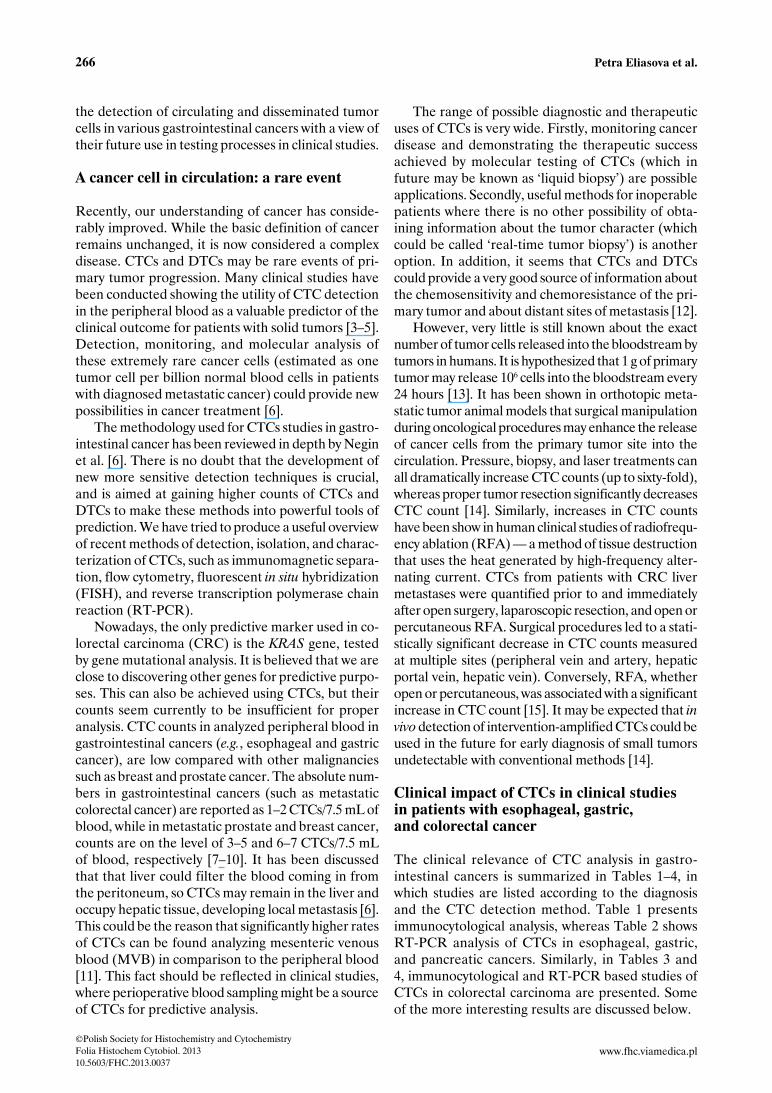
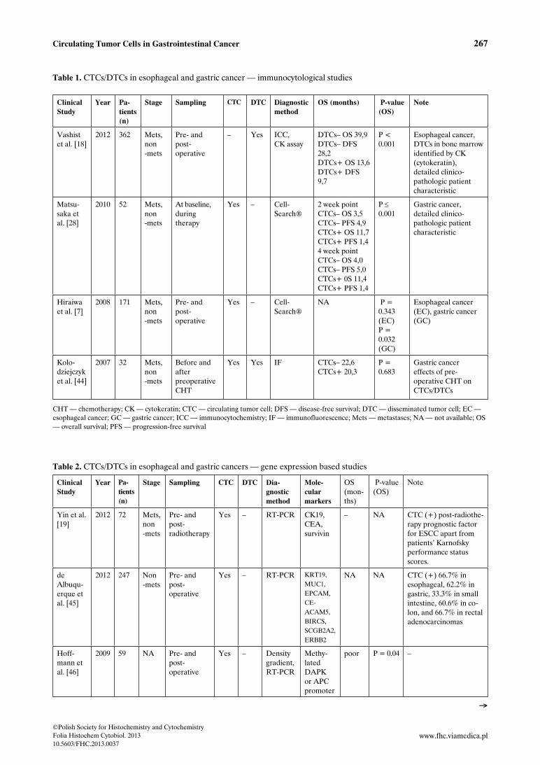
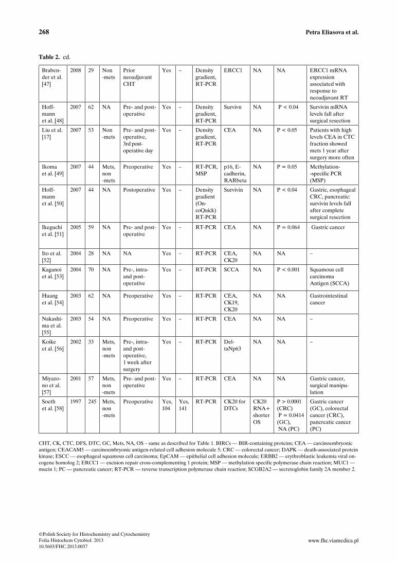
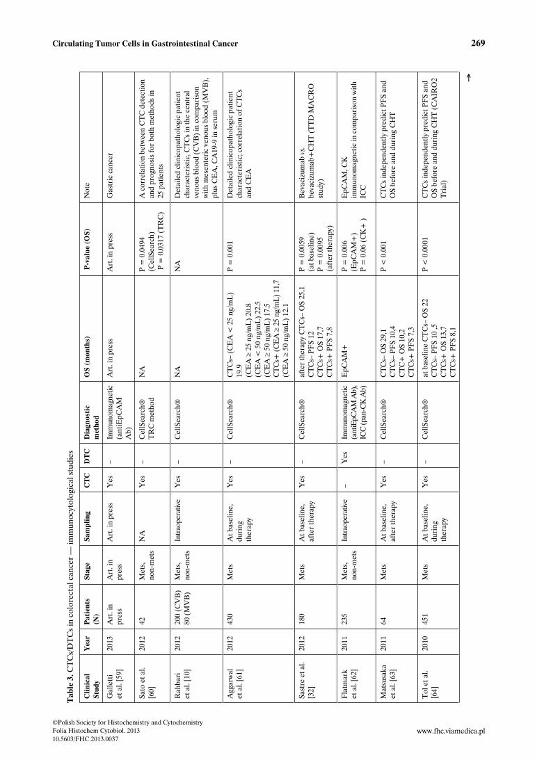
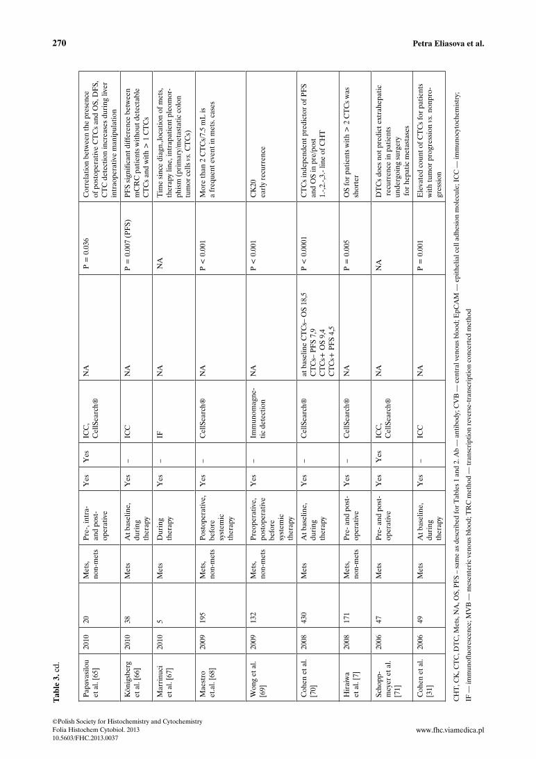
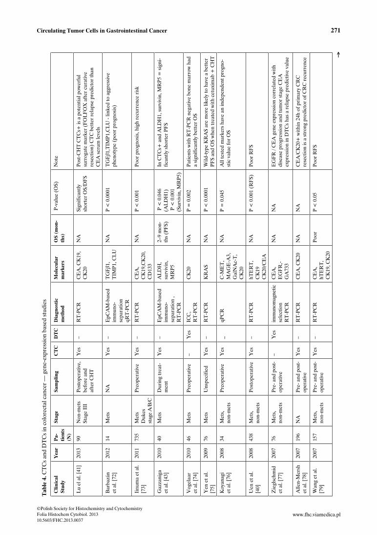
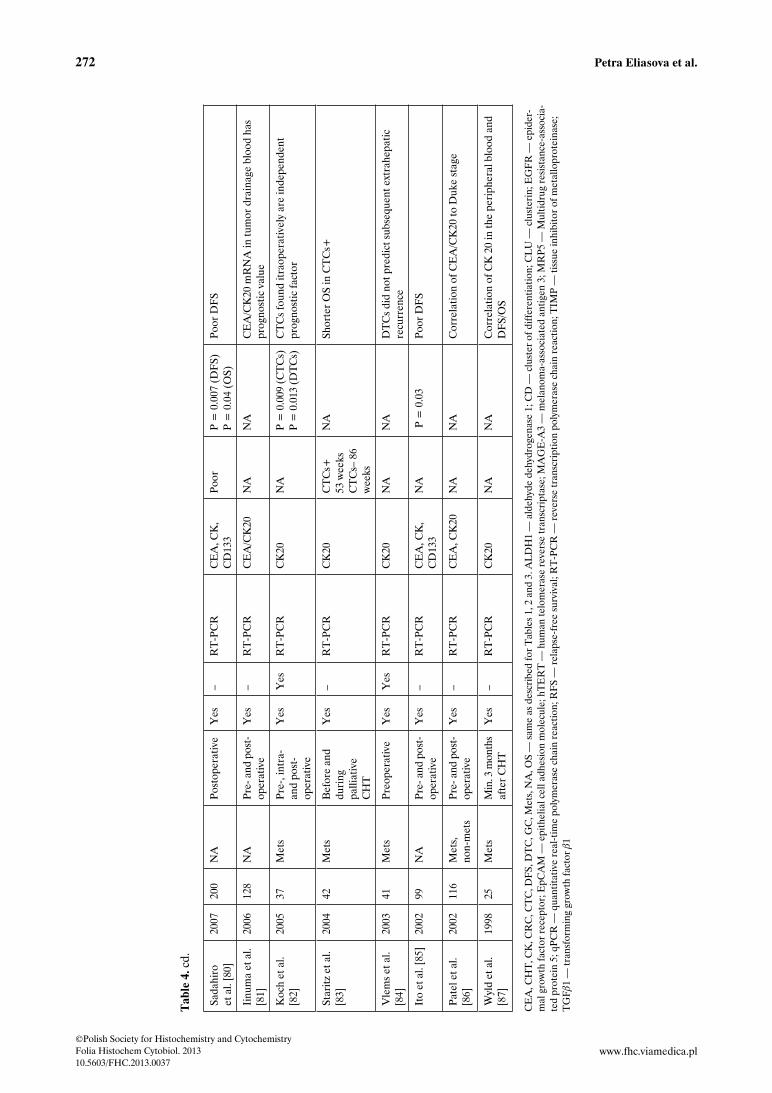
The clinical relevance of CTC analysis in gastro-intestinal cancers is summarized in Tables 1–4, in which studies are listed according to the diagnosis and the CTC detection method. Table 1 presents immunocytological analysis, whereas Table 2 shows RT-PCR analysis of CTCs in esophageal, gastric, and pancreatic cancers. Similarly, in Tables 3 and 4, immunocytological and RT-PCR based studies of CTCs in colorectal carcinoma are presented. Some of the more interesting results are discussed below.
267Circulating Tumor Cells in Gastrointestinal Cancer
©Polish Society for Histochemistry and CytochemistryFolia Histochem Cytobiol. 201310.5603/FHC.2013.0037
www.fhc.viamedica.pl
Table 1. CTCs/DTCs in esophageal and gastric cancer — immunocytological studies
Clinical Study
Year Pa-tients (n)
Stage Sampling CTC DTC Diagnostic method
OS (months) P-value (OS)
Note
Vashist et al. [18]
2012 362 Mets, non-mets
Pre- and post- operative
– Yes ICC, CK assay
DTCs– OS 39,9 DTCs– DFS 28,2 DTCs+ OS 13,6 DTCs+ DFS 9,7
P < 0.001
Esophageal cancer, DTCs in bone marrow identified by CK (cytokeratin), detailed clinico- pathologic patient characteristic
Matsu-saka et al. [28]
2010 52 Mets, non-mets
At baseline, during therapy
Yes – Cell- Search®
2 week point CTCs– OS 3,5 CTCs– PFS 4,9 CTCs+ OS 11,7 CTCs+ PFS 1,4 4 week point CTCs– OS 4,0 CTCs– PFS 5,0 CTCs+ 0S 11,4 CTCs+ PFS 1,4
P ≤ 0.001
Gastric cancer, detailed clinico- pathologic patient characteristic
Hiraiwa et al. [7]
2008 171 Mets, non-mets
Pre- and post- operative
Yes – Cell- Search®
NA P = 0.343 (EC) P = 0.032 (GC)
Esophageal cancer (EC), gastric cancer (GC)
Kolo-dziejczyk et al. [44]
2007 32 Mets, non-mets
Before and after preoperative CHT
Yes Yes IF CTCs– 22,6 CTCs+ 20,3
P = 0.683
Gastric cancer effects of pre- operative CHT on CTCs/DTCs
CHT — chemotherapy; CK — cytokeratin; CTC — circulating tumor cell; DFS — disease-free survival; DTC — disseminated tumor cell; EC — esophageal cancer; GC — gastric cancer; ICC — immunocytochemistry; IF — immunofluorescence; Mets — metastases; NA — not available; OS — overall survival; PFS — progression-free survival
Table 2. CTCs/DTCs in esophageal and gastric cancers — gene expression based studies
Clinical Study
Year Pa-tients (n)
Stage Sampling CTC DTC Dia-gnostic method
Mole-cular markers
OS (mon-ths)
P-value (OS)
Note
Yin et al. [19]
2012 72 Mets, non-mets
Pre- and post- radiotherapy
Yes – RT-PCR CK19, CEA, survivin
– NA CTC (+) post-radiothe-rapy prognostic factor for ESCC apart from patients’ Karnofsky performance status scores.
de Albuqu-erque et al. [45]
2012 247 Non-mets
Pre- and post- operative
Yes – RT-PCR KRT19, MUC1, EPCAM, CE-ACAM5, BIRCS, SCGB2A2, ERBB2
NA NA CTC (+) 66.7% in esophageal, 62.2% in gastric, 33.3% in small intestine, 60.6% in co-lon, and 66.7% in rectal adenocarcinomas
Hoff-mann et al. [46]
2009 59 NA Pre- and post- operative
Yes – Density gradient, RT-PCR
Methy-lated DAPK or APC promoter
poor P = 0.04 –
Æ
268 Petra Eliasova et al.
©Polish Society for Histochemistry and CytochemistryFolia Histochem Cytobiol. 201310.5603/FHC.2013.0037
www.fhc.viamedica.pl
Table 2. cd.
Braben-der et al. [47]
2008 29 Non-mets
Prior neoadjuvant CHT
Yes – Density gradient, RT-PCR
ERCC1 NA NA ERCC1 mRNA expression associated with response to neoadjuvant RT
Hoff-mann et al. [48]
2007 62 NA Pre- and post- operative
Yes – Density gradient, RT-PCR
Survivn NA P < 0.04 Survivin mRNA levels fall after surgical resection
Liu et al. [17]
2007 53 Non-mets
Pre- and post- operative, 3rd post- operative day
Yes – Density gradient, RT-PCR
CEA NA P < 0.05 Patients with high levels CEA in CTC fraction showed mets 1 year after surgery more often
Ikoma et al. [49]
2007 44 Mets, non-mets
Preoperative Yes – RT-PCR, MSP
p16, E-cadherin, RARbeta
NA P = 0.05 Methylation- -specific PCR (MSP)
Hoff-mann et al. [50]
2007 44 NA Postoperative Yes – Density gradient (On-coQuick) RT-PCR
Survivin NA P < 0.04 Gastric, esophageal CRC, pancreatic: survivin levels fall after complete surgical resection
Ikeguchi et al. [51]
2005 59 NA Pre- and post- operative
Yes – RT-PCR CEA NA P = 0.064 Gastric cancer
Ito et al. [52]
2004 28 NA NA Yes – RT-PCR CEA, CK20
NA NA –
Kaganoi et al. [53]
2004 70 NA Pre-, intra- and post- operative
Yes – RT-PCR SCCA NA P < 0.001 Squamous cell carcinoma Antigen (SCCA)
Huang et al. [54]
2003 62 NA Preoperative Yes – RT-PCR CEA, CK19, CK20
NA NA Gastrointestinal cancer
Nakashi-ma et al. [55]
2003 54 NA Preoperative Yes – RT-PCR CEA NA NA –
Koike et al. [56]
2002 33 Mets, non-mets
Pre-, intra- and post- operative, 1 week after surgery
Yes – RT-PCR Del-taNp63
NA NA –
Miyazo-no et al. [57]
2001 57 Mets, non-mets
Pre- and post- operative
Yes – RT-PCR CEA NA NA Gastric cancer, surgical manipu-lation
Soeth et al. [58]
1997 245 Mets, non-mets
Preoperative Yes, 104
Yes, 141
RT-PCR CK20 for DTCs
CK20 RNA+ shorter OS
P > 0.0001 (CRC) P = 0.0414 (GC), NA (PC)
Gastric cancer (GC), colorectal cancer (CRC), pancreatic cancer (PC)
CHT, CK, CTC, DFS, DTC, GC, Mets, NA, OS – same as described for Table 1. BIRCs — BIR-containing proteins; CEA — carcinoembryonic antigen; CEACAM5 — carcinoembryonic antigen-related cell adhesion molecule 5; CRC — colorectal cancer; DAPK — death-associated protein kinase; ESCC — esophageal squamous cell carcinoma; EpCAM — epithelial cell adhesion molecule; ERBB2 — erythroblastic leukemia viral on-cogene homolog 2; ERCC1 — excision repair cross-complementing 1 protein; MSP — methylation specific polymerase chain reaction; MUC1 — mucin 1; PC — pancreatic cancer; RT-PCR — reverse transcription polymerase chain reaction; SCGB2A2 — secretoglobin family 2A member 2.
269Circulating Tumor Cells in Gastrointestinal Cancer
©Polish Society for Histochemistry and CytochemistryFolia Histochem Cytobiol. 201310.5603/FHC.2013.0037
www.fhc.viamedica.pl
Tab
le 3
. CT
Cs/
DT
Cs
in c
olor
ecta
l can
cer
— im
mun
ocyt
olog
ical
stu
dies
Cli
nica
l St
udy
Yea
rP
atie
nts
(N)
Stag
eSa
mpl
ing
CT
CD
TC
Dia
gnos
tic
met
hod
OS
(mon
ths)
P
-val
ue (
OS)
Not
e
Gal
lett
i et
al.
[59]
2013
Art
. in
pres
sA
rt. i
n pr
ess
Art
. in
pres
sY
es–
Imm
unom
agne
tic
(ant
iEpC
AM
A
b)
Art
. in
pres
sA
rt. i
n pr
ess
Gas
tric
can
cer
Sato
et a
l. [6
0]20
1242
Met
s,
non-
met
sN
AY
es–
Cel
lSea
rch®
T
RC
met
hod
NA
P =
0.0
494
(Cel
lSea
rch)
P
= 0
.031
7 (T
RC
)
A c
orre
latio
n be
twee
n C
TC
det
ectio
n an
d pr
ogno
sis
for
both
met
hods
in
25 p
atie
nts
Rah
bari
et
al.
[10]
2012
200
(CV
B)
80 (
MV
B)
Met
s,
non-
met
sIn
trao
pera
tive
Yes
–C
ellS
earc
h®
NA
NA
Det
aile
d cl
inic
opat
holo
gic
patie
nt
char
acte
rist
ic, C
TC
s in
the
cent
ral
veno
us b
lood
(C
VB
) in
com
pari
son
with
mes
ente
ric
veno
us b
lood
(M
VB
),
plus
CE
A, C
A19
-9 in
ser
um
Agg
arw
al
et a
l. [6
1]20
1243
0M
ets
At b
asel
ine,
du
ring
th
erap
y
Yes
–C
ellS
earc
h®
CT
Cs–
(C
EA
< 2
5 ng
/mL
) 19
.9
(CE
A ≥
25
ng/m
L)
20.8
(C
EA
< 5
0 ng
/mL
) 22
.5
(CE
A ≥
50
ng/m
L)
17.5
C
TC
s+ (C
EA
≥ 2
5 ng
/mL
) 11,
7 (C
EA
≥ 5
0 ng
/mL
) 12
.1
P =
0.0
01D
etai
led
clin
icop
atho
logi
c pa
tient
ch
arac
teri
stic
; cor
rela
tion
of C
TC
s an
d C
EA
Sast
re e
t al.
[32]
2012
180
Met
sA
t bas
elin
e,
afte
r th
erap
yY
es–
Cel
lSea
rch®
af
ter
ther
apy
CT
Cs–
OS
25,1
C
TC
s– P
FS
12
CT
Cs+
OS
17,7
C
TC
s+ P
FS
7,8
P =
0.0
059
(a
t bas
elin
e)
P =
0.0
095
(a
fter
ther
apy)
Bev
aciz
umab
vs.
be
vaci
zum
ab+
CH
T (
TT
D M
AC
RO
st
udy)
Fla
tmar
k
et a
l. [6
2]20
1123
5M
ets,
no
n-m
ets
Intr
aope
rativ
e–
Yes
Imm
unom
agne
tic
(ant
iEpC
AM
Ab)
, IC
C (p
an-C
K A
b)
EpC
AM
+
P =
0.0
06
(EpC
AM
+)
P
= 0
.06
(CK
+ )
EpC
AM
, CK
im
mun
omag
netic
in c
ompa
riso
n w
ith
ICC
Mat
susa
ka
et a
l. [6
3]20
1164
Met
sA
t bas
elin
e,
afte
r th
erap
yY
es–
Cel
lSea
rch®
C
TC
s– O
S 29
,1
CT
Cs–
PF
S 10
,4
CT
C+
OS
10,2
C
TC
s+ P
FS
7,3
P <
0.0
01C
TC
s in
depe
nden
tly p
redi
ct P
FS
and
OS
befo
re a
nd d
urin
g C
HT
Tol
et a
l. [6
4]20
1045
1M
ets
At b
asel
ine,
du
ring
th
erap
y
Yes
–C
ellS
earc
h®
at b
asel
ine
CT
Cs–
OS
22
CT
Cs–
PF
S 10
,5
CT
Cs+
OS
13,7
C
TC
s+ P
FS
8,1
P <
0.0
001
CT
Cs
inde
pend
ently
pre
dict
PF
S an
d O
S be
fore
and
dur
ing
CH
T (
CA
IRO
2 T
rial
)
Æ
270 Petra Eliasova et al.
©Polish Society for Histochemistry and CytochemistryFolia Histochem Cytobiol. 201310.5603/FHC.2013.0037
www.fhc.viamedica.pl
Papa
vasi
lou
et a
l. [6
5]20
1020
Met
s,
non-
met
sPr
e-, i
ntra
- an
d po
st-
oper
ativ
e
Yes
Yes
ICC
, C
ellS
earc
h®
NA
P =
0.0
36C
orre
latio
n be
twee
n th
e pr
esen
ce
of p
osto
pera
tive
CT
Cs
and
OS,
DF
S,
CT
C d
etec
tion
incr
ease
s du
ring
live
r in
trao
pera
tive
man
ipul
atio
n
Kön
igsb
erg
et a
l. [6
6]20
1038
Met
sA
t bas
elin
e,
duri
ng
ther
apy
Yes
–IC
CN
AP
= 0
.007
(PF
S)PF
S si
gnifi
cant
diff
eren
ce b
etw
een
mC
RC
pat
ient
s w
ithou
t det
ecta
ble
CT
Cs
and
with
> 1
CT
Cs
Mar
rinu
ci
et a
l. [6
7]20
105
Met
sD
urin
g
ther
apy
Yes
–IF
NA
NA
Tim
e si
nce
diag
n.,lo
catio
n of
met
s,
ther
apy
line,
intr
apat
ient
ple
omor
-ph
ism
(pr
imar
y/m
etas
tatic
col
on
tum
or c
ells
vs.
CT
Cs)
Mae
stro
et
.al.
[68]
2009
195
Met
s,
non-
met
sPo
stop
erat
ive,
be
fore
sy
stem
ic
ther
apy
Yes
–C
ellS
earc
h®
NA
P <
0.0
01M
ore
than
2 C
TC
s/7.
5 m
L is
a
freq
uent
eve
nt in
met
s. c
ases
Won
g et
al.
[69]
2009
132
Met
s,
non-
met
sPr
eope
rativ
e,
post
oper
ativ
e be
fore
sy
stem
ic
ther
apy
Yes
–Im
mun
omag
ne-
tic d
etec
tion
NA
P <
0.0
01C
K20
ea
rly
recu
rren
ce
Coh
en e
t al.
[70]
2008
430
Met
sA
t bas
elin
e,
duri
ng
ther
apy
Yes
–C
ellS
earc
h®
at b
asel
ine
CT
Cs–
OS
18,5
C
TC
s– P
FS
7,9
CT
Cs+
OS
9,4
CT
Cs+
PF
S 4,
5
P <
0.0
001
CT
Cs
inde
pend
ent p
redi
ctor
of P
FS
and
OS
in p
re/p
ost
1.-,2
.-,3.
- lin
e of
CH
T
Hir
aiw
a
et a
l. [7
]20
0817
1M
ets,
no
n-m
ets
Pre-
and
pos
t-
oper
ativ
eY
es–
Cel
lSea
rch®
N
AP
= 0
.005
O
S fo
r pa
tient
s w
ith >
2 C
TC
s w
as
shor
ter
Scho
pp-
mey
er e
t al.
[71]
2006
47M
ets
Pre-
and
pos
t-
oper
ativ
eY
esY
esIC
C,
Cel
lSea
rch®
N
AN
AD
TC
s do
es n
ot p
redi
ct e
xtra
hepa
tic
rec
urre
nce
in p
atie
nts
un
derg
oing
sur
gery
fo
r he
patic
met
asta
ses
Coh
en e
t al.
[31]
2006
49M
ets
At b
asel
ine,
du
ring
th
erap
y
Yes
–IC
CN
AP
= 0
.001
Ele
vate
d co
unt o
f CT
Cs
for
patie
nts
with
tum
or p
rogr
essi
on v
s. n
onpr
o-gr
essi
on
CH
T, C
K, C
TC
, DT
C, M
ets,
NA
, OS,
PF
S –
sam
e as
des
crib
ed fo
r T
able
s 1
and
2. A
b —
ant
ibod
y; C
VB
— c
entr
al v
enou
s bl
ood;
EpC
AM
— e
pith
elia
l cel
l adh
esio
n m
olec
ule;
IC
C —
imm
unoc
ytoc
hem
istr
y;
IF —
imm
unof
luor
esce
nce;
MV
B —
mes
ente
ric
veno
us b
lood
; TR
C m
etho
d —
tran
scri
ptio
n re
vers
e-tr
ansc
ript
ion
conc
erte
d m
etho
d
Tab
le 3
. cd.
271Circulating Tumor Cells in Gastrointestinal Cancer
©Polish Society for Histochemistry and CytochemistryFolia Histochem Cytobiol. 201310.5603/FHC.2013.0037
www.fhc.viamedica.pl
Tab
le 4
. CT
Cs
and
DT
Cs
in c
olor
ecta
l can
cer
— g
ene-
expr
essi
on b
ased
stu
dies
Cli
nica
l St
udy
Yea
rP
a-ti
ents
(N
)
Stag
eSa
mpl
ing
CT
CD
TC
Dia
gnos
tic
met
hod
Mol
ecul
ar
mar
kers
O
S (m
on-
ths)
P
-val
ue (
OS)
Not
e
Lu
el a
l. [4
1]20
1390
Non
-met
s St
age
III
Post
oper
ativ
e,
befo
re a
nd
afte
r C
HT
Yes
–R
T-P
CR
CE
A, C
K19
, C
K20
NA
Sign
ifica
ntly
sh
orte
r O
S/D
FS
Post
-CH
T C
TC
s+ is
a p
oten
tial p
ower
ful
surr
ogat
e m
arke
r (F
OL
FO
X a
fter
cur
ativ
e
rese
ctio
n) C
TC
bet
ter
rela
pse
pred
icto
r th
an
CE
A s
erum
leve
ls
Bar
bazá
n
et a
l. [7
2]20
1214
Met
sN
AY
es–
EpC
AM
-bas
ed
imm
uno-
se
para
tion
qRT
-PC
R
TG
Fb1
, T
IMP1
, CL
UN
AP
< 0
.000
1T
GFb1
,TIM
P1,C
LU
- lin
ked
to a
ggre
ssiv
e
phen
otyp
e (p
oor
prog
nosi
s)
Iinu
ma
et a
l. [7
3]20
1173
5M
ets
Duk
es
stag
e A
/B/C
Preo
pera
tive
Yes
–R
T-P
CR
CE
A,
CK
19,C
K20
, C
D13
3
NA
P <
0.0
01Po
or p
rogn
osis
, hig
h re
curr
ence
ris
k
Gaz
zani
ga
et a
l. [4
3]20
1040
Met
sD
urin
g tr
eat-
men
tY
es–
EpC
AM
-bas
ed
imm
uno-
se
para
tion
, R
T-P
CR
AL
DH
, su
rviv
in,
MR
P5
2–9
mon
-th
s (P
FS)
P <
0.0
46
(AL
DH
1)
P <
0.0
01
(Sur
vivi
n, M
RP5
)
In C
TC
s+ a
nd A
LD
H1,
sur
vivi
n, M
RP5
= s
igni
-fic
antly
sho
rter
PF
S
Vog
elaa
r
et a
l. [7
4]20
1046
Met
sPr
eope
rativ
e–
Yes
ICC
, R
T-P
CR
CK
20N
AP
= 0
.002
Patie
nts
with
RT
-PC
R n
egat
ive
bone
mar
row
had
a
sign
ifica
ntly
bet
ter
OS
Yen
et a
l. [7
5]20
0976
Met
sU
nspe
cifie
dY
es–
RT
-PC
RK
RA
SN
AP
< 0
.000
1W
ild-t
ype
KR
AS
are
mor
e lik
ely
to h
ave
a be
tter
PF
S an
d O
S w
hen
trea
ted
with
cet
uxim
ab +
CH
T
Koy
anag
i et
al.
[76]
2008
34M
ets,
no
n-m
ets
Preo
pera
tive
Yes
_qP
CR
C-M
ET
, M
AG
E-A
3,
Gal
NA
c-T
, C
K20
NA
P =
0.0
45A
ll te
sted
mar
kers
hav
e an
inde
pend
ent p
rogn
o-st
ic v
alue
for
OS
Uen
et a
l. [4
0]20
0843
8M
ets,
no
n-m
ets
Post
oper
ativ
eY
es–
RT
-PC
RhT
ER
T,
CK
19
CK
20/C
EA
NA
P <
0.0
01 (
RF
S)Po
or R
FS
Zie
glsc
hmid
et
al.
[77]
2007
76M
ets,
no
n-m
ets
Pre-
and
pos
t-
oper
ativ
e–
Yes
imm
unom
agne
tic
sele
ctio
n R
T-P
CR
CE
A,
EG
FR
,-G
A73
3
NA
NA
EG
FR
/ C
EA
gen
e ex
pres
sion
cor
rela
ted
with
di
seas
e pr
ogre
ssio
n an
d tu
mor
sta
ge C
EA
ex
pres
sion
in D
TC
s ha
s a
rela
pse
pred
ictiv
e va
lue
Alle
n-M
ersh
et
al.
[78]
2007
196
NA
Pre-
and
pos
t-
oper
ativ
eY
es–
RT
-PC
RC
EA
, CK
20N
AN
AC
EA
/CK
20+
with
in 2
4h o
f pri
mar
y C
RC
re
sect
ion
is a
str
ong
pred
icto
r of
CR
C r
ecur
renc
e
Wan
g et
al.
[79]
2007
157
Met
s,
non-
met
sPr
e- a
nd p
ost-
op
erat
ive
Yes
–R
T-P
CR
CE
A,
hTE
RT
, C
K19
, CK
20
Poor
P <
0.0
5Po
or R
FS
Æ
272 Petra Eliasova et al.
©Polish Society for Histochemistry and CytochemistryFolia Histochem Cytobiol. 201310.5603/FHC.2013.0037
www.fhc.viamedica.pl
Sada
hiro
et
al.
[80]
2007
200
NA
Post
oper
ativ
eY
es–
RT
-PC
RC
EA
, CK
, C
D13
3Po
orP
= 0
.007
(D
FS)
P
= 0
.04
(OS)
Poor
DF
S
Iinu
ma
et a
l. [8
1]20
0612
8N
APr
e- a
nd p
ost-
op
erat
ive
Yes
–R
T-P
CR
CE
A/C
K20
NA
NA
CE
A/C
K20
mR
NA
in tu
mor
dra
inag
e bl
ood
has
prog
nost
ic v
alue
Koc
h et
al.
[82]
2005
37M
ets
Pre-
, int
ra-
and
post
- op
erat
ive
Yes
Yes
RT
-PC
RC
K20
NA
P =
0.0
09 (
CT
Cs)
P
= 0
.013
(DT
Cs)
CT
Cs
foun
d itr
aope
rativ
ely
are
inde
pend
ent
prog
nost
ic fa
ctor
Star
itz e
t al.
[83]
2004
42M
ets
Bef
ore
and
duri
ng
palli
ativ
e C
HT
Yes
–R
T-P
CR
CK
20C
TC
s+
53 w
eeks
C
TC
s– 8
6 w
eeks
NA
Shor
ter
OS
in C
TC
s+
Vle
ms
et a
l. [8
4]20
0341
Met
sPr
eope
rativ
eY
esY
esR
T-P
CR
CK
20N
AN
AD
TC
s di
d no
t pre
dict
sub
sequ
ent e
xtra
hepa
tic
recu
rren
ce
Ito
et a
l. [8
5]20
0299
NA
Pre-
and
pos
t-
oper
ativ
eY
es–
RT
-PC
RC
EA
, CK
, C
D13
3N
A P
= 0
.03
Poor
DF
S
Pate
l et a
l. [8
6]20
0211
6M
ets,
no
n-m
ets
Pre-
and
pos
t-
oper
ativ
eY
es–
RT
-PC
RC
EA
, CK
20N
AN
AC
orre
latio
n of
CE
A/C
K20
to D
uke
stag
e
Wyl
d et
al.
[87]
1998
25M
ets
Min
. 3 m
onth
s af
ter
CH
TY
es–
RT
-PC
RC
K20
NA
NA
Cor
rela
tion
of C
K 2
0 in
the
peri
pher
al b
lood
and
D
FS/
OS
CE
A, C
HT
, CK
, CR
C, C
TC
, DF
S, D
TC
, GC
, Met
s, N
A, O
S —
sam
e as
des
crib
ed fo
r T
able
s 1,
2 a
nd 3
. AL
DH
1 —
ald
ehyd
e de
hydr
ogen
ase
1; C
D —
clu
ster
of d
iffer
entia
tion;
CL
U —
clu
ster
in; E
GF
R —
epi
der-
mal
gro
wth
fact
or r
ecep
tor;
EpC
AM
— e
pith
elia
l cel
l adh
esio
n m
olec
ule;
hT
ER
T —
hum
an te
lom
eras
e re
vers
e tr
ansc
ript
ase;
MA
GE
-A3
— m
elan
oma-
asso
ciat
ed a
ntig
en 3
; MR
P5 —
Mul
tidru
g re
sist
ance
-ass
ocia
-te
d pr
otei
n 5;
qPC
R —
qua
ntita
tive
real
-tim
e po
lym
eras
e ch
ain
reac
tion;
RF
S —
rel
apse
-fre
e su
rviv
al; R
T-P
CR
— r
ever
se tr
ansc
ript
ion
poly
mer
ase
chai
n re
actio
n; T
IMP
— ti
ssue
inhi
bito
r of
met
allo
prot
eina
se;
TG
Fb1
— tr
ansf
orm
ing
grow
th fa
ctor
b1
Tab
le 4
. cd.
273Circulating Tumor Cells in Gastrointestinal Cancer
©Polish Society for Histochemistry and CytochemistryFolia Histochem Cytobiol. 201310.5603/FHC.2013.0037
www.fhc.viamedica.pl
Esophageal cancer (EC)
Esophageal cancer (EC) is notorious for its aggressive biological behavior, local infiltration, involvement of adjacent lymph nodes, and broad metastasis through hematogenous spread. It has been reported that the frequency of hematogenous recurrence is high, despi-te radical surgery with lymph node dissection [16]. In this regard, the detection of cancer cells in the blood could be important for identifying patients with a high risk of relapse. There have been many studies showing a positive correlation between detection of CTCs, tumor staging, and patient prognosis. Detection of CTCs from PB of EC patients by conventional qPCR methods has been reported for several genes.
Liu et al. [17] aimed at establishing a quantitative system for evaluating the role of CTCs in PB from patients who underwent surgical resection during esophageal cancer treatment. 155 PB samples from 53 EC patients were collected before surgery (B-1), immediately after surgery (B0), and on the third po-stoperative day (B+3). A direct qPCR method based on CEA mRNA gene expression was designed for the detection of CTCs. The authors showed significant differences between groups B-1 vs. B0 (p = 0.0001) and B-1 vs. B+3 (p = 0.0209). 50% of the patients with R > 0.4 (R = CTC ratio of B+3 over B0) showed tumor recurrence within 1 year after surgery, whereas the probability was only 14.3% for patients with R < 0.4 (p = 0.043). The prognostic utility of CTCs in EC has been shown also in studies where the gene expression of survivin, ERCC1, and APC has been tested by RT-PCR, as shown in Table 2.
The prognostic relevance of the presence of DTCs in bone marrow (BM) for the postoperative course of EC has also been evaluated recently [18]: 370 patients with EC diagnosis (189 squamous cell carcinomas and 181 adenocarcinomas), were surgically treated with complete resection (R0). They received neither ad-juvant nor neoadjuvant therapy. DTCs were detected by an immunocytochemical cytokeratin assay in pre-operatively taken BM aspirates. Overall, 120 (32.4%) patients harbored DTCs in their BM. The presence of DTCs significantly correlated with aggressive tumor biology, as indicated by increased tumor size (p = 0.026), regional (p = 0.002) and distant (p = 0.012) lymph node metastases, and higher re-lapse rate (p < 0.001, c2 test). The presence of DTCs in bone marrow was a very strong and independent prognostic factor in patients with resectable EC [18].
The CTC status in the PB of patients with eso-phageal squamous cell carcinoma (ESCC), before and after radiotherapy (RT), was evaluated by Yin et al. [19]. A total of 72 ESCC patients enrolled in
this study were treated with radical RT. The nested RT-PCR reaction was used to detect the three repre-sentative markers of CTCs: CEA, CK19, and survivin. The results showed that the presence of CTCs, and the positive expression of at least one of these three markers in patients with ESCC pre-RT and post-RT were 54.2% and 38.9%, respectively (p = 0.059). Furthermore, the analysis of the patients according to lymph node metastasis and adverse 2-year progression-free survival (PFS) revealed changes in CTC status after RT, which would reflect patients’ response to RT. In a multivariate analysis with the Cox propor-tional hazard model, only CTC positivity post-RT was an independent, unfavorable prognostic factor for ESCC, apart from subsequent chemotherapy and patients’ Karnofsky performance status scores (a scale quantifying cancer patients’ general well-being). In conclusion, the positive detection of CTCs in patients with ESCC after RT may be a promising biomarker for radiation efficiency and prognosis assessment in ESCC [19].
Gastric cancer (GC)
Follow-up studies on gastric cancer (GC) patients suggested that CTC-positive cases with increased bur-dens of CTCs were associated with poorer prognoses than CTC-negative cases. The situation was similar with DTCs [15]. Both localized and metastatic GC can shed detectable concentrations of CTCs into the blood. The presence of CTCs in circulation suggests not only a high risk of tumor recurrence, but also an unfavorable clinical outcome even in the early stages of GC [20]. The prognostic impact of CTCs in GC has been reported in several studies [21–27]. The sensitivi-ty of RT-PCR CTC detection was superior to the other less commonly used cytological detection methods involving fluorescence-activated cell sorting (FACS), immunohistochemistry (IHC), and immunocytochemi-stry (ICC) [20]. For the identification of CTCs in GC, different markers and their combinations were tested in the analyzed studies. The combination of EpCAM, CK8, CK18, and CK19 seems to be prognostically the most relevant in GC [7, 24]. On the other side, single survivin expression also achieved prognostic significance in at least 2 studies [29, 30]. Based on the analyzed data, detection of CTCs might be used as a noninvasive method, not only for the confirmation of GC diagnosis, but also for estimation of prognosis.
Colorectal cancer (CRC)
In general, the detection of CTCs in colorectal cancer (CRC), independently of the method and markers
274 Petra Eliasova et al.
©Polish Society for Histochemistry and CytochemistryFolia Histochem Cytobiol. 201310.5603/FHC.2013.0037
www.fhc.viamedica.pl
used, correlates with the stage of the cancer disease [31, 32]. On the other side, the correlation of CTCs with some known clinicopathological prognostic factors (e.g., T4 tumor size, perineural invasion, bowel obstruction, high preoperative CEA levels) is still uncertain [32]. It is believed that the correla-tion of CTCs with clinicopathological factors would increase if the sensitivity of the CTC detection were higher. CTC positivity is observed in approximately 40–50% of metastatic CRC patients. Differences in CTCs detection can be observed depending on the sampling site, as shown by Rahbari et al. [10], who tested compartmental differences of CTC in CRC. The qualitative and quantitative detection of CTCs was higher in the mesenteric venous blood (MVB) than in the central venous blood (CVB) of patients with CRC. It has been speculated that the liver works as a filter and stops CTCs from entering the central circulation [6]. Moreover, higher counts of CTCs were detected when the tumor was localized in the lower part of rectum than in the cases of middle and high rectal involvement [6, 33].
The biomarkers used for the CTCs detection in cytological or RT-PCR examination of patients with CRC are listed in Tables 3 and 4. Generally, the EpCAM pre-enrichment is a basis for further cytokeratine (CK19/20, CK8/18) and CEA testing. Recently, plastin3 has been shown to have significant clinical relevance. Plastin3 positivity in the PB was found to be associated with clinicopathological risk factors, such as depth of invasion, lymph node and liver metastasis, presence of peritoneal dissemination, increased recurrence rate, and higher Dukes stage. It is very important to note that plastin3 expression was also detected in all patients with recurrent disease, and at a level higher than in the case of prerecurrence and of patients without recurrence [34]. The correlation between CTCs and prognosis in CRCs was stronger if CKs and multiple markers were used than for the one-marker assay [35].
Recently, several meta-analyses evaluating the prognostic value of CTC examination in CRC have been published. Rahbari et al. [36] included 36 stu-dies and 3094 patients in their final meta-analysis. The pooled analyses combining all sampling sites (PB, mesenteric PB (MPB), and BM) associated the detection of CTCs/DTCs with poor recurrence-free survival (RFS). Stratification by sampling site showed that detection of CTCs in the PB compartment was a statistically significant prognostic factor, but that detection in the MPB or BM was not.
Similarly, 12 studies representing 1329 patients were suitable for pooled analysis of CRC patients in a prognostic study [37]. The OS and PFS were worse
in CTC-positive patients, whereas analyzing PFS se-parately, the subgroup with significantly worse survival rate contained over 35% CTC-positive patients. Mul-tivariate analysis was performed on eight studies and identified the detection of CTCs as an independent prognostic factor for survival. Moreover, the meta-a-nalysis reported that the detection of CTCs in PB of patients with resectable colorectal liver metastases, or with widespread metastatic CRC, was associated with disease progression and poor survival [37]. The study of Katsuno et al. [38] highlights the potential importance of cancer cell detection in the venous drainage of colorectal cancers as a prognostic marker and a mode of staging in this neoplastic disease.
Regarding the effect of chemotherapy on CTC counts, it has been evaluated that the prognosis of patients with undetectable CTCs after chemotherapy was significantly better [39]. Additionally, molecular detection of persistent postoperative CTCs has been confirmed as a prognostic marker of early relapse in I–III stage CRC patients, which could help to select patients for an enhanced follow-up and therapeutic program [40, 41].
In summary, it is expected that CTCs and DTCs will be used for mutational analysis of the genes con-nected directly to the targeted therapy (e.g., KRAS, BRAF). The heterogeneity of the genetic profiles of cells from the primary tumor, metastatic tumors, and CTCs may be an explanation for the variable response to EGFR-inhibitor chemotherapy [5, 42, 43]. CTCs are not only a marker for advanced disease, but also have prognostic and predictive potential. A decrease in CTC levels during chemotherapy is correlated with improved responses to chemotherapy [39].
Several very important questions need to be an-swered, and further studies are required to unify the isolation techniques before CTCs can be adapted for widespread clinical use. In particular, the follo-wing questions should be central to future research: Can CTCs be used to define a group of patients with “resectable” metastases who should not undergo re-section? Can CTCs be used to monitor the immediate effectiveness of systemic chemotherapy or to predict which chemotherapy would be most effective? Can CTCs be used to help staging patients with metastatic CRCs?
Finally, we are reminded that stage IV of colorec-tal cancer is a disease with many possible outcomes, ranging from rapid death to recovery [40]. We also recall that our ability to predict which patient will experience which outcome is relatively limited. The detection of CTCs is a potentially promising biomar-ker that could contribute to the staging of the cancer and this deserves a prospective study.
275Circulating Tumor Cells in Gastrointestinal Cancer
©Polish Society for Histochemistry and CytochemistryFolia Histochem Cytobiol. 201310.5603/FHC.2013.0037
www.fhc.viamedica.pl
Conclusion
In summary, it is essential to establish sensitive, specific technologies to detect CTCs. More detailed analyses of their molecular characteristics should be performed with the aim of understanding the biology of CTCs and DTCs. This may provide a yet-untapped option to develop therapeutic strategies that will effectively treat and prevent metastatic process for each person individually.
Acknowledgements
This study was supported by a grant (entitled “Sle-dování hladiny CTC u vybraných nádorů gastrointe-stinálního traktu”) provided by the League Against Cancer, Prague, Czech Republic Grant, and by a grant from the Czech Ministry of Health (IGA NT14439–3/2013).
References1. Steinert G, Schölch S, Koch M et al. Biology and significance
of circulating and disseminated tumor cells in colorectal can-cer. Langenbecks Arch Surg. 2012;397:535–542.
2. Brechot C et al. Molecular analysis of circulating tumor cells isolated by ISET: A diagnostic approach to improve follow up and treatment of patients with cancer. Boston, World CTC Summit, 2012.
3. Takeuchi H, Kitagawa Y. Detection of circulating tumor cells in gastrointestinal cancer: Has its time come? J Gastrointest Oncol. 2012;3:84–85.
4. Maheswaran S, Haber DA. Circulating tumor cells: a window into cancer biology and metastasis. Curr Opin Genet Dev. 2010;20:96–99.
5. Kin C, Kidess E, Poultsides GA et al. Colorectal cancer diagnostics: biomarkers, cell-free DNA, circulating tumor cells and defining heterogeneous populations by single-cell analysis. Expert Rev Mol Diagn. 2013;13:581–599.
6. Negin BP, Cohen SJ. Circulating tumor cells in colorectal cancer: past, present, and future challenges. Curr Treat Options Oncol. 2010;11:1–13.
7. Hiraiwa K, Takeuchi H, Hasegawa H et al. Clinical significance of circulating tumor cells in blood from patients with gastro-intestinal cancers. Ann Surg Oncol. 2008;15:39092–39100.
8. Moreno JG, O’Hara SM, Gross S et al. Changes in circulating carcinoma cells in patients with metastatic prostate cancer correlate with disease status. Urology. 2001;58:386–392.
9. Dong X, Alpaugh KR, Cristofanilli M. Circulating tumor cells (CTCs) in breast cancer: a diagnostic tool for prognosis and molecular analysis. Chin J Cancer Res. 2012;24:388–398.
10. Rahbari NN, Bork U, Kircher A et al. Compartmental Dif-ferences of Circulating Tumor Cells in Colorectal Cancer. Ann Surg Oncol. 2012;19:2195–2202.
11. Iwanicki-Caron I, Basile P, Toure E et al. Usefulness of Cir-culating Tumor Cell Detection in Pancreatic Adenocarcinoma Diagnosis. Am J Gastroenterol. 2013;108:152–155.
12. Juratli MA, Sarimollaoglu M, Siegel E et al. Real-time monitoring of circulating tumor-cell release during tumor manipulation using in vivo photoacoustic and fluorescent flow cytometry. Head Neck. 2013; DOI: 10.1002/hed.23439. [Epub ahead of print].
13. Butler TP, Gullino PM. Quantitation of cell shedding into efferent blood of mammary adenocarcinoma. Cancer Res. 1975;35:512–516.
14. Jiao LR, Apostolopoulos C, Jacob J et al. Unique localization of circulating tumor cells in patient with hepatic metastases. J Clin Oncol. 2009;36:6160–6165.
15. Wang GY, Li Y, Yu YM et al. Detection of disseminated tumor cells in bone marrow of gastric cancer using magnetic activated cell sorting and fluorescent activated cell sorting. J Gastroenterol Hepatol. 2009;24:299–306.
16. Katayama A, Mafune K, Tanaka Y et al. Autopsy findings in patients after curative esophagectomy for esophageal carci-noma. J Am Coll Surg. 2003;196:866–873.
17. Liu Z, Jiang M, Zhao J et al. Circulating tumor cells in perioperative esophageal cancer patients: quantitative assay system and potential clinical utility. Clin Cancer Res. 2007;13:2992–2997.
18. Vashist YK, Effenberger KE, Vettorazzi E et al. Disseminated tumor cells in bone marow and the natural course of resected esophageal cancer. Ann Surg. 2012;255:1105–1112.
19. Yin XD, Yuan X, Xue JJ et al. Clinical significance of carc-inoembryonic antigen-, cytokeratin 19-, or survivin-positive circulating tumor cells in the peripheral blood of esophageal squamous cell carcinoma patients treated with radiotherapy. Dis Esophagus. 2012;25:750–756.
20. Zhang ZY, Ge HY. Micrometastasis in gastric cancer. Cancer Lett. 2013;336:34–45.
21. Arigami T, Uenosono Y, Hirata M et al. B7–H3 expression in gastric cancer: a novel molecular blood marker for detecting circulating tumor cells. Cancer Sci. 2011;102:1019–1024.
22. Saad AA, Awed NM, Abd Elkerim NN et al. Prognostic significance of E-cadherin expression and peripheral blood micrometastasis in gastric carcinoma patients. Ann Surg Oncol. 2010;17:3059–3067.
23. Pituch-Noworolska A, Kolodziejczyk P, Kulig J et al. Circulat-ing tumour cells and survival of patients with gastric cancer. Anticancer Res. 2007;27:635–640.
24. Yeh KH, Chen YC, Yeh SH et al. Detection of circulating can-cer cells by nested reverse transcription-polymerase chain re-action of cytokeratin-19 (K19)-possible clinical significance in advanced gastric cancer. Anticancer Res. 1998;18:1283–1286.
25. Koga T, Tokunaga E, Sumiyoshi Y et al. Detection of circulating gastric cancer cells in peripheral blood using real time quantitative RT-PCR. Hepatogastroenterology. 2008;84:1131–1135.
26. Illert B, Fein M, Otto C et al. Disseminated tumor cells in the blood of patients with gastric cancer are an independent predictive marker of poor prognosis. Scand J Gastroenterol. 2005;40:843–849.
27. Uen YH, Lin SR, Wu CH et al. Clinical significance of MUC1 and c-Met RT-PCR detection of circulating tumor cells in pa-tients with gastric carcinoma. Clin Chim Acta. 2006;367:55–61.
28. Matsusaka S, Chìn K, Ogura M et al. Circulating tumor cells as a surrogate marker for determining response to chemo-therapy in patients with advanced gastric cancer. Cancer Sci. 2010;101:1067–1071.
29. Bertazza L, Mocellin S, Marchet A et al. Survivin gene levels in the peripheral blood of patients with gastric cancer inde-pendently predict survival. J Transl Med. 2009;7:111.
30. Cao W, Yang W, Li H et al. Using detection of survivin-ex-pressing circulating tumor cells in peripheral blood to predict tumor recurrence following curative resection of gastric cancer. J Surg Oncol. 2011;103:110–115.
31. Cohen SJ, Alpaugh RK, Gross S et al. Isolation and charac-terization of circulating tumor cells in patients with metastatic colorectal cancer. Clin Colorectal Cancer. 2006;6:125–132.

276 Petra Eliasova et al.
©Polish Society for Histochemistry and CytochemistryFolia Histochem Cytobiol. 201310.5603/FHC.2013.0037
www.fhc.viamedica.pl
32. Sastre J, Maestro ML, Gómez-España A et al. Circulating tumor cell count is a prognostic factor in metastatic colorectal cancer patients receiving first-line chemotherapy plus beva-cizumab: a Spanish Cooperative Group for the Treatment of Digestive Tumors study. Oncologist. 2012;17:947–955
33. Molnar B, Ladanyi A, Tanko L et al. Circulating tumor cell clusters in the peripheral blood of colorectal cancer patients. Clin Cancer Res. 2001;12:4080–4085.
34. Yokobori T, Iinuma H, Shimamura T et al. Plastin3 is a novel marker for circulating tumor cells undergoing the epitheli-al-mesenchymal transition and is associated with colorectal cancer prognosis. Cancer Res. 2013;73:2059–2069.
35. Torino F, Bonmassar E, Bonmassar L et al. Circulating tumor cells in colorectal cancer patients. Cancer Treat Rev. 2013;39:759–772.
36. Rahbari NN, Aigner M, Thorlund K et al. Meta-analysis shows that detection of circulating tumor cells indicates poor prognosis in patients with colorectal cancer. Gastroenterology. 2010;138:1714–1726.
37. Groot Koerkamp B, Rahbari NN, Büchler MW et al. Circu-lating tumor cells and prognosis of patients with resectable colorectal liver metastase or widespread metastatic colorectal cancer: a meta-analysis. Ann Surg Oncol. 2013;20:2156–2165.
38. Katsuno H, Zacharakis E, Aziz O et al. Does the presence of circulating tumor cells in the venous drainage of curative colorectal cancer resections determine prognosis? A me-ta-analysis. Ann Surg Oncol. 2008;15:3083–3091.
39. Neki K, Kawahara H, Watanabe K et al. Usefulness of circu-lating tumor cells after preliminary chemotherapy for predic-tion of response to further anticancer therapy in patients with initially unresectable metastatic colorectal cancer. Anticancer Res. 2013;33:1769–1772.
40. Uen YH, Lu CY, Tsai HL et al. Persistent presence of post-operative circulating tumor cells is a poor prognostic factor for patients with stage I–III colorectal cancer after curative resection. Ann Surg Oncol. 2008;15:2120–2128.
41. Lu CY, Uen YH, Tsai HL et al. Molecular detection of persistent postoperative circulating tumour cells in stages II and III colon cancer patients via multiple blood sampling: prognostic significance of detection for early relapse. Br J Cancer. 2011;104:1178–1184.
42. Lankiewicz S, Zimmermann S, Hollmann C et al. Circulating tumour cells as a predictive factor for response to systemic chemotherapy in patients with advanced colorectal cancer. Mol Oncol. 2008;3:349–355.
43. Gazzaniga P, Gradilone A, Petracca A et al. Molecular markers in circulating tumour cells from metastatic colorectal cancer patients. J Cell Mol Med. 2010;14:2073–2077.
44. Kolodziejczyk P, Pituch-Noworolska A, Drabik G et al. The effects of preoperative chemotherapy on isolated tumor cells in the blood and bone marrow of gastric cancer patients. Br J Cancer. 2007;97:589–592.
45. de Albuquerque A, Kubisch I, Ernst D et al. Development of a molecular multimarker assay for the analysis of circu-lating tumor cells in adenocarcinoma patients. Clin Lab. 2012;58:373–384.
46. Hoffmann AC, Vallböhmer D, Prenzel K et al. Methylated DAPK and APC promoter DNA detection in peripheral blood is significantly associated with apparent residual tumor and outcome. J Cancer Res Clin Oncol. 2009;135:1231–1237.
47. Brabender J, Vallböhmer D, Grimminger P et al. ERCC1 RNA expression in peripheral blood predicts minor histo-pathological response to neoadjuvant radio-chemotherapy in patients with locally advanced cancer of the esophagus. J Gastrointest Surg. 2008;12:1815–1821.
48. Hoffmann AC, Warnecke-Eberz U, Luebke T et al. Survi-vin mRNA in peripheral blood is frequently detected and significantly decreased following resection of gastrointestinal cancers. J Surg Oncol. 2007;95:51–54.
49. Ikoma D, Ichikawa D, Ueda Y et al. Circulating tumor cells and aberrant methylation as tumor markers in patients with esophageal cancer. Anticancer Res. 2007;27:535–539.
50. Hoffmann AC, Warnecke-Eberz U, Luebke T et al. Survivin mRNA in peripheral blood is frequently detected and sig-nificantly decreased following resection of gastrointestinal cancers. J Surg Oncol. 2007;95:51–54.
51. Ikeguchi M, Kaibara N. Detection of circulating cancer cells after a gastrectomy for gastric cancer. Surg Today. 2005;35:436–441.
52. Ito H, Kanda T, Nishimaki T et al. Detection and quantifi-cation of circulating tumor cells in patients with esophageal cancer by real-time polymerase chain reaction. J Exp Clin Cancer Res. 2004;23:455–464.
53. Kaganoi J, Shimada Y, Kano M et al. Detection of circulating oesophageal squamous cancer cells in peripheral blood and its impact on prognosis. Br J Surg. 2004;91:1055–1060.
54. Huang P, Wang J, Guo Y et al. Molecular detection of disseminated tumor cells in the peripheral blood in pa-tients with gastrointestinal cancer. J Cancer Res Clin Oncol. 2003;129:192–198.
55. Nakashima S, Natsugoe S, Matsumoto M et al. Clinical significance of circulating tumor cells in blood by molecular detection and tumor markers in esophageal cancer. Surgery. 2003;133:162–169.
56. Koike M, Hibi K, Kasai Y et al. Molecular detection of circu-lating esophageal squamous cell cancer cells in the peripheral blood. Clin Cancer Res. 2002;8:2879–2882.
57. Miyazono F, Natsugoe S, Takao S et al. Surgical maneuvers enhance molecular detection of circulating tumor cells during gastric cancer surgery. Ann Surg. 2001;233:189–194.
58. Soeth E, Vogel I, Röder C et al. Comparative analysis of bone marrow and venous blood isolated from gastrointestinal can-cer patients for the detection of disseminated tumor cells using reverse transcription PCR. Cancer Res. 1997;57:3106–3110.
59. Galletti G, Sung MS, Vahdat LT et al. Isolation of breast can-cer and gastric cancer circulating tumor cells by use of an anti HER2-based microfluidic device. Lab Chip. 2014;14:147–156.
60. Sato N, Hayashi N, Imamura Y et al. Usefulness of Transcrip-tion-Reverse Transcription Concerted Reaction Method for Detecting Circulating Tumor Cells in Patients With Colorec-tal Cancer. Ann Surg Oncol. 2012;19:2060–2065.
61. Aggarwal C, Meropol NJ, Punt CJ et al. Relationship among circulating tumor cells, CEA and overall survival in patients with metastatic colorectal cancer. Ann Oncol. 2013;24:420–428.
62. Flatmark K, Borgen E, Nesland JM et al. Disseminated tumour cells as a prognostic biomarker in colorectal cancer. Br J Cancer. 2011;104:1434–1439.
63. Matsusaka S, Suenaga M, Mishima Y et al. Circulating endothelial cells predict for response to bevacizumab-based chemotherapy in metastatic colorectal cancer. Cancer Chemo-ther Pharmacol. 2011;68:763–768.
64. Tol J, Koopman M, Miller MC et al. Circulating tumour cells early predict progression-free and overall survival in advanced colorectal cancer patients treated with chemotherapy and targeted agents. Ann Oncol. 2010;5:1006–1012.
65. Papavasiliou P, Fisher T, Kuhn J et al. Circulating tumor cells in patients undergoing surgery for hepatic metasta-ses from colorectal cancer. Baylor Univ Med Cent Proc. 2010;23:11–14.
277Circulating Tumor Cells in Gastrointestinal Cancer
©Polish Society for Histochemistry and CytochemistryFolia Histochem Cytobiol. 201310.5603/FHC.2013.0037
www.fhc.viamedica.pl
66. Königsberg R, Gneist M, Jahn-Kuch D et al. Circulating tumor cells in metastatic colorectal cancer: efficacy and feasibility of different enrichment methods. Cancer Lett. 2010;293:117–123.
67. Marrinucci D, Bethel K, Lazar D et al. Cytomorphology of Circulating Colorectal Tumor Cells: A Small Case Series. J Oncol. 2010;2010:861341.
68. Maestro LM, Sastre J, Rafael SB et al. Circulating Tumor Cells in Solid Tumor in Metastatic and Localized Stages. Anticancer Res. 2009;29:4839–4843.
69. Wong SC, Chan CM, Ma BB et al. Clinical significance of cytokeratin 20-positive circulating tumor cells detected by a refined immunomagnetic enrichment assay in colorectal cancer patients. Clin Cancer Res. 2009;15:1005–1012.
70. Cohen SJ, Punt CJ, Iannotti N et al. Relationship of Circu-lating Tumor Cells to Tumor Response, Progression-Free Survival, and Overall Survival in Patients With Metastatic Colorectal Cancer. J Clin Oncol. 2008;26:3213–3221.
71. Schoppmeyer K, Frühauf N, Oldhafer K et al. Tumor cell dissemination in colon cancer does not predict extrahepatic recurrence in patients undergoing surgery for hepatic metas-tases. Oncol Rep. 2006;15:449–454.
72. Barbazán J, Alonso-Alconada L, Muinelo-Romay L et al. Molecular Characterization of Circulating Tumor Cells in Hu-man Metastatic Colorectal Cancer. PLoS One. 2012;7:e40476.
73. Iinuma H, Watanabe T, Mimori K et al. Clinical significance of circulating tumor cells, including cancer stem-like cells, in peripheral blood for recurrence and prognosis in patients with Dukes’ stage B and C colorectal cancer. J Clin Oncol. 2011;29:1547–1555.
74. Vogelaar FJ, Mesker WE, Rijken AM et al. Clinical impact of different detection methods for disseminated tumor cells in bone marrow of patients undergoing surgical resection of colorectal liver metastases: a prospective follow-up study. BMC Cancer. 2010;10:153.
75. Yen LC, Yeh YS, Chen CW et al. Detection of KRAS on-cogene in peripheral blood as a predictor of the response to cetuximab plus chemotherapy in patients with metastatic colorectal cancer. Clin Cancer Res. 2009;15:4508–4513.
76. Koyanagi K, Bilchik AJ, Saha S et al. Prognostic Relevance of occulat nodal micrometastases and circulating tumor cells in colorectal cancer in a prospective multicenter trial. Cli Cancer Res. 2008;22:7391–7396.
77. Zieglschmid V, Hollmann C, Mannel J et al. Tumor-associ-ated gene expression in disseminated tumor cells correlates with disease progression and tumor stage in colorectal cancer. Anticancer Res. 2007;27:1823–1832.
78. Allen-Mersh TG, McCullough TK, Patel H et al. Role of circulating tumour cells in predicting recurrence after excision of primary colorectal carcinoma. Br J Surg. 2007;94:96–105.
79. Wang JY, Lin SR, Wu DC et al. Multiple molecular markers as predictors of colorectal cancer in patients with normal perioperative serum carcinoembryonic antigen levels. Clin Canc Res. 2007;13:2406–2413.
80. Sadahiro S, Suzuki T, Maeda Y et al. Detection of carcinoem-bryonic antigen messenger RNA-expressing cells in peripheral blood 7 days after curative surgery is a novel prognostic factor in colorectal cancer. Ann Surg Oncol. 2007;14:1092–1098.
81. Iinuma H, Okinaga K, Egami H et al. Usefulness and clini-cal significance of quantitative real-time RT-PCR to detect isolated tumor cells in the peripheral blood and tumor drainage blood of patients with colorectal cancer. Int J Oncol. 2006;28:297–306.
82. Koch M, Kienle P, Hinz U et al. Detection of hematogenous tumor cell dissemination predicts tumor relapse in patients undergoing surgical resection of colorectal liver metastases. Ann Surg. 2005;241:199–205.
83. Staritz P, Kienle P, Koch M et al. Detection of disseminated tumour cells as a potential surrogate-marker for monitoring palliative chemotherapy in colorectal cancer patients. J Exp Clin Cancer Res. 2004;23:633–639.
84. Vlems FA, Diepstra JH, Punt CJ et al. Detection of dis-seminated tumour cells in blood and bone marrow samples of patients undergoing hepatic resection for metastasis of colorectal cancer. Br J Surg. 2003;90:989–995.
85. Ito S, Nakanishi H, Hirai T et al. Quantitative detection of CEA expressing free tumor cells in the peripheral blood of colorectal cancer patients during surgery with real-time RT-PCR on a LightCycler. Cancer Lett. 2002;183:195–203.
86. Patel H, Le Marer N, Wharton RQ et al. Clearance of circu-lating tumor cells after excision of primary colorectal cancer. Ann Surg. 2002;235:226–231.
87. Wyld DK, Selby P, Perren TJ et al. Detection of colorectal cancer cells in peripheral blood by reverse-transcriptase polymerase chain reaction for cytokeratin 20. Int J Cancer. 1998;79:288–293.
Submitted: 22 December, 2013 Accepted after reviews: 29 December, 2013