Changes in toxin content, biomass and pigments of the dinoflagellate Alexandrium minutum during...
11
Vol. 111: 99-109, 1994 MARINE ECOLOGY PROGRESS SERIES Mar. Ecol. Prog. Ser. 1 Published August l l Changes in toxin content, biomass and pigments of the dinoflagellate Alexandrium minutum during nitrogen refeeding and growth into nitrogen or phosphorus stress Krystyna Flynnl, Jose M. ~ranco~, Pablo ~ernandez~, Beatriz ~eguera~, Manuel zapata4, Gareth Wood1, Kevin J. Flynnl** 'Algal Research Unit, School of Biological Sciences, University of Wales Swansea. Singleton Park, Swansea SA2 8PP, United Kingdom 'Institute de Investigaciones Marinas (C.S.I.C.). Eduardo Cabello 6, E-36208 Vigo. Spain 3~nstituto Espanol de Oceanografia. Aptdo. 1552, E-36260 Vigo, Spain 'Centro de Investigaciones Marinas (Xunta de Galicia), Aptdo. 208, Villagarcia de Arousa, E-36600 Pontevedra, Spain ABSTRACT: Two stralns of the paralytic shellfish tox~n (PST) producing dinoflagellate Alexandrium lninutum Halim (highly toxic ALlV and weakly toxic AL2V) were grown in batch culture with either nitrate or phosphate as the limiting nutrient. In comparison with cells of the strain ALlV, cells of AL2V grew at a similar C-specific rate, had a higher C/N ratio, and lower ratios of chl a/chl c2 and chl a/peri- dinin. Neither chlorophylls nor carotenoids could be used to estimate C-biomass, N-biomass or toxin content for this organism. The toxin profile for both strains was dominated (up to 95 %) by the gonyau- toxin GTX4, with smaller proportions of GTX1. GTX2 and GTX3. The rate of toxin synthesis for both strains was greatest 1 to 2 d after the N-refeeding of N-deprived cells, with the net rate of toxin syn- thesis exceeding that of C-biomass and cell division by a factor of up to 4. Toxin synthesis was not enhanced by short-term P-stress. N-stress alone led to a decrease in toxin cell-', but P-stress followed by N-stress did not result in such a decline, implicating phosphorus in the regulation of toxin metabo- lism. Although arginine is a major precursor for PST synthesis, taurine, glycine, glutamine, and cell N showed similar relations to that observed for arginine with respect to toxin content. Furthermore, the mole ratio of arginine/toxin could vary by a factor of up to 5 between ALlV and AL2V at peak values of toxin cell-', and by more than 5 within a strain when growing under different conditions. These observations suggest that the relationship between free arginine content and toxin content is complex. No explanation for the higher toxin content of ALlV IS apparent, except that ALlV has a higher N-content per cell and this may be conducive to a higher rate of synthesis of the N-rich toxins. KEY WORDS: Dinoflagellate . Alexandriurn rninutum - Paralytic shellfish poisons - PSP toxin . Amino acid . Pigment . Algal growth dynamics INTRODUCTION The formation of algal blooms is usually associated with a decline of nutrients; yield nutrient-limitation is considered usually to be due to a lack of nitrogen or phosphate. Dinoflagellate blooms occur in low-turbu- lent conditions, often associated with frontal systems or inshore stratified water columns (Margalef 1978, Mar- 'Addressee for correspondence galef et al. 1979, Holligan et al. 1984, LeFevre 1986). Offshore populations advected into coastal embay- ments may be expected to encounter changes in the availability of nutrients, while near-shore populations may also encounter changes in environmental condi- tions during intrusions of offshore waters and during increased riverine inputs. Migration to and from nutri- clines offers additional opportunities to encounter tran- sients in nutrient availability (Paasche et al. 1984). The dinoflagellate Alexandrium minutum forms red tides in the Galician northern rias (Rias Altas, NW O Inter-Research 1994 Resale of full article not permitted
-
Upload
independent -
Category
Documents
-
view
2 -
download
0
Transcript of Changes in toxin content, biomass and pigments of the dinoflagellate Alexandrium minutum during...

Vol. 111: 99-109, 1994 MARINE ECOLOGY PROGRESS SERIES Mar. Ecol. Prog. Ser.
1 Published August l l
Changes in toxin content, biomass and pigments of the dinoflagellate Alexandrium minutum during nitrogen refeeding and growth into
nitrogen or phosphorus stress
Krystyna Flynnl, Jose M. ~ r a n c o ~ , Pablo ~ e r n a n d e z ~ , Beatriz ~ e g u e r a ~ , Manuel zapata4, Gareth Wood1, Kevin J. F l y n n l * *
'Algal Research Unit, School of Biological Sciences, University of Wales Swansea. Singleton Park, Swansea SA2 8PP, United Kingdom
'Institute de Investigaciones Marinas (C.S.I.C.). Eduardo Cabello 6, E-36208 Vigo. Spain 3~nstituto Espanol de Oceanografia. Aptdo. 1552, E-36260 Vigo, Spain
'Centro de Investigaciones Marinas (Xunta de Galicia), Aptdo. 208, Villagarcia de Arousa, E-36600 Pontevedra, Spain
ABSTRACT: Two stralns of the paralytic shellfish tox~n (PST) producing dinoflagellate Alexandrium lninutum Halim (highly toxic ALlV and weakly toxic AL2V) were grown in batch culture with either nitrate or phosphate as the limiting nutrient. In comparison with cells of the strain ALlV, cells of AL2V grew at a similar C-specific rate, had a higher C/N ratio, and lower ratios of chl a/chl c2 and chl a/peri- dinin. Neither chlorophylls nor carotenoids could be used to estimate C-biomass, N-biomass or toxin content for this organism. The toxin profile for both strains was dominated (up to 95 %) by the gonyau- toxin GTX4, with smaller proportions of GTX1. GTX2 and GTX3. The rate of toxin synthesis for both strains was greatest 1 to 2 d after the N-refeeding of N-deprived cells, with the net rate of toxin syn- thesis exceeding that of C-biomass and cell division by a factor of up to 4. Toxin synthesis was not enhanced by short-term P-stress. N-stress alone led to a decrease in toxin cell-', but P-stress followed by N-stress did not result in such a decline, implicating phosphorus in the regulation of toxin metabo- lism. Although arginine is a major precursor for PST synthesis, taurine, glycine, glutamine, and cell N showed similar relations to that observed for arginine with respect to toxin content. Furthermore, the mole ratio of arginine/toxin could vary by a factor of up to 5 between ALlV and AL2V at peak values of toxin cell-', and by more than 5 within a strain when growing under different conditions. These observations suggest that the relationship between free arginine content and toxin content is complex. No explanation for the higher toxin content of A L l V IS apparent, except that ALlV has a higher N-content per cell and this may be conducive to a higher rate of synthesis of the N-rich toxins.
KEY WORDS: Dinoflagellate . Alexandriurn rninutum - Paralytic shellfish poisons - PSP toxin . Amino acid . Pigment . Algal growth dynamics
INTRODUCTION
The formation of algal blooms is usually associated with a decline of nutrients; yield nutrient-limitation is considered usually to be due to a lack of nitrogen or phosphate. Dinoflagellate blooms occur in low-turbu- lent conditions, often associated with frontal systems or inshore stratified water columns (Margalef 1978, Mar-
'Addressee for correspondence
galef et al. 1979, Holligan et al. 1984, LeFevre 1986). Offshore populations advected into coastal embay- ments may be expected to encounter changes in the availability of nutrients, while near-shore populations may also encounter changes in environmental condi- tions during intrusions of offshore waters and during increased riverine inputs. Migration to and from nutri- clines offers additional opportunities to encounter tran- sients in nutrient availability (Paasche et al. 1984).
The dinoflagellate Alexandrium minutum forms red tides in the Galician northern rias (Rias Altas, NW
O Inter-Research 1994 Resale of full article not permitted

100 Mar. Ecol. Prog. Ser. 111. 99-109, 1994
Spain; species referred to as Gonyaulax tamarensis in Blanco et al. 1985 and as Alexandrium lusitanicum in Franco et al. 1992) and is associated with paralytic shellfish poisoning (PSP) toxicity near to closure limits for the bivalve industry [80 pg saxatoxin (STX) equiva- lent per 100 g meat] in the Galician southern nas (Rias Bajas, NW Spain). These particular blooms originate within the rias following periods of elevated terrestrial run-off and subsequent haline stratification (Reguera et al. 1991). The distribution of A. minutum includes other areas of the European Atlantic coast (Erard-Le Denn 1991), the Mediterranean (Delgado et al. 1990, Montresor et al. 1990) and off Australia (Hallegraeff et al. 1991).
Two strains of Alexandnum minutum (both isolated from the Ria de Vigo, NW Spain), one of which (ALlV) is more toxic than the other (AL2V), were the subjects of the study reported here. The objectives of this study were: (1) to consider :he effects on toxin content of N- stress and P-stress, and of N-refeeding of N-deprived A. minutum cells, (2) to look for gross differences between the 2 strains which may affect toxin synthesis, and (3) to consider the use of pigments to monitor bio- mass and the toxin content of this species.
The main parameters considered were toxin content, changes in the intracellular amino acid pool (arginine is an important intermediary in PSP toxin synthesis; Shimizu et al. 1990), biomass and pigmentation. Emphasis has been placed previously on the effects of nutrient limitation on Alexandriurn spp. (Boyer et al. 1987, Anderson et al. 1990). This paper reports for the first time the important effect of N-refeeding on toxin synthesis in A. minutum.
MATERIALS AND METHODS
Alexandnurn minutum Halim strains ALlV and AL2V (Instituto Espanol de Oceanografia, Vigo, Spain) were grown at 15°C in modified K medium (Keller & Guillard 1985) based on local aged seawater with dif- ferent amounts of nitrate and inorganic phosphate, and with no artificial buffer, ammonium or organic phos- phate. The experiments were conducted on 3 occa- sions. Experiment I (Expt I) was conducted in Swansea (UK) with only strain ALlV which had been trans- ferred from Vigo (Spain) 5 mo earlier. In that study, the growth media contained either 100 FM nitrate + 20 PM phosphate (low-N/high-P), or 200 pM nitrate + 5 FM phosphate (high-NAow-P). Because the cells growing in the high-N/low-P medium in Expt I actually exh.austed both N and P, for Expt 11, conducted in Vigo with strains ALlV and AL2V, the high-NAow-P media contained 300 yM nitrate + 5 PM phosphate. Expt 111, conducted in Swansea, also included strains of non-
toxic A. tamarense and A. affini, considered additional parameters, and was of longer duration. Expt 111 used the same nitrate + phosphate combinations as Expt 11. Only the results from Expt I11 pertaining to toxin con- tent in A. minutum are presented here.
The growth media were filtered through 0.22 pm filters (Durapore, Millipore) rather than autoclaved. Inoculation of the experimental flasks (5 1) was with cells previously grown in the low-Nlhigh-P medium; these cultures were N-deprived with elevated C/N ratios, thus Day 0 in the figures also indicates the point of N-refeeding. Cultures were grown with no forced aeration, sampled via a syphon tube and illuminated in a 12 h/12 h light/dark cycle either at 180 pm01 m-2 S-'
(Expt I), or at 200 pm01 m-2 S- ' (Expts I1 & 111). Cells were counted in a Sedgewick-Rafter chamber
after fixing with Lugol's iodine and, for Expt 111, also by Elzone 282 PC particle analyzer. Nitrate and phos- phate were analyzed using an Alpkem RFA-2 micro- segmented-flow nutrient analyzer. Extracted pigments were analyzed either by spectrophotometry for Expts I & I11 (extracted into dimethylformamide and using the equations of Strickland & Parsons 1972), or by HPLC for Expt I1 [extraction as in Flynn et al. (1993), and HPLC according to Zapata et al. (1987) with quantifi- cation using the specific absorption coefficients given by Jeffrey & Humphrey (1975) and Jeffrey et al. (1975)l. Intracellular free amino acids (InAA) were analyzed by HPLC according to the methods of Flynn & Flynn (1992) with derivatization and injection per- formed by a Ktachi AS-4000 autosampler fitted with a Peltier cooling system. Cellular C and N were mea- sured in samples collected on preashed (450 "C for 6 h) 13 mm diameter GA/E (Celrnan) glassfibre filters using Europa Scientific RoboPrep and TracerMass instru- ments with isoleucine as the standard.
Cells for toxin analysis were collected on preashed GA/E filters and extracted 3 times with 0.1 M acetic acid; each extraction included sonication for 1 min fol- lowed by freezing and thawing. The extract was cen- trifuged and the supernatant adjusted to pH 3 to 4 . PSP toxins for Expts I & I1 were analyzed by a postcolumn derivatization method (second isocratic method de- scribed by Franco & Fernandez-Vila 1993). For Expts I & 111, toxin content was also estimated using a precol- umn periodate oxidation (modified after Lawrence et al. 1991), with separation on a HPLC Technology Ultra- techsphere C18 ODS column with mobile phases con- taining acetate buffer, methanol and tetrahydrofuran.
Parameter-specific rates of increase ('growth rates') were computed according to the formula
Parameter-p = [In (Pt,) - ln (Pb) ] / ( t , - to)
where Pt, and Pt, are values of the parameter at times to and t, respectively (time in days).

Flynn et al.: Changes in Alexandriurn toxin content, biomass and pigments
Fig. 1. Alexandrium minutum. Changes in (a) cell number and cell-specific growth rate ,-- 0-6 - (cell-p), and (b) carbon cell-' and C/N ratio k . for cells of strain ALlV grown in low-N/high- Z L O - ~ - P media (open symbols) and high-N/low-P L . media (closed symbols) dunng Expt 11. Cells g 0.2 - exhausted nutrients, and thus became N-
TlME (dl
, a , , 8 .
" 0.1 1 , , : :A P-dp
Statistical testing of comparisons were performed using ANOVA or joint regression analysis as indicated.
deprived (N-dp) or P-deprived (P-dp), at the o - I
times indicated o 4 8 12 16
TlME (d l
- 0.6 - 7
Fig 2 Alexandrium minutum. Changes in (a) .
cell number and cell-specific growth rate ~ 0 . ~ -
(cell-p), and (b) carbon cell-' and CIN ratio L . for cells of strain AL2V grown In low-N/high- i?~ 0.2 -
0 P media (open symbols) and high-N/low-P
RESULTS
:- 8 , , , 8 , f ,
8 , , , 8 8
A 4 . 4 ~ ~ ~ ',&A . A~%:ssL! 2,
. C *A , , , m , , 8
Cell growth
media (closed symbols) during Expt 11. o - S
I
Abbreviations as in Fig. 1 o 4 8 12 16
In Expt I , cells of strain ALlV in both nutrient regimes ceased growth at a cell density of 21 000 ml-l, achieving similar maximum cell-p of around 0.5 d-'. In Expt 11, ALlV attained 25 000 ml-' with cell-p of 0.2 d-' for the first part of the culture but then rising to around 0.5 d-' between Days 7 and 11 (Fig. la). AL2V grew slower and did not reach stationary phase by the end of the study period; maximum cell densities were in excess of 20000 ml-' with a maximum cell-p around 0.3 d- ' (Fig. 2a). Cell numbers in Expt 111, which con- tinued for 27 d , reached 25000 ml-', with maximum cell-p values of 0.4 for both ALlV and AL2V.
, , O J . , . 7 8
0 4 8 12 lC
TlME (dl
0 4 8 12 16
TlME (dl
Biomass
ALlV in both nutrient regimes in Expt I contained a maximum of 1 ng C cell-' (achieving a maximum C-p in excess of 0.4 d-l) , falling to half that when growth slowed. The minimum C/N ratio was 4, rising to 14 after 10 d of N-deprivation in the low-N/high-P cul- ture, and with a slower rate of increase to a C/N ratio of 9 after 7 d of N-deprivation in the high-NAow-P cul- ture which exhausted its nitrate 3 d after exhausting the phosphate.
Cells of ALlV in Expt I1 were up to 20 % larger than those in Expt I, containing 1.2 n g C cell-' during the first part of the culture, falling to 0.4 n g C cell-' (Fig. l b ) . C-p remained around 0.25 d-' between 3 and 11 d with a C/N of 4 to 5 (Fig. lb) . The increased cell-p (from 0.2 to 0.5 d-l) for ALlV in the latter part of the growth curve was enabled by a decrease in average biomass per cell (Fig. l b ) and not by an increase in
102 Mar. Ecol. Prog. Ser. 111: 99-109, 1994
. . O J I , , , , , , , , , , , , , , , , 8 8 8 8
0 4 8 12 16
TlME Id)
TlME Id)
. . , , m 0 8
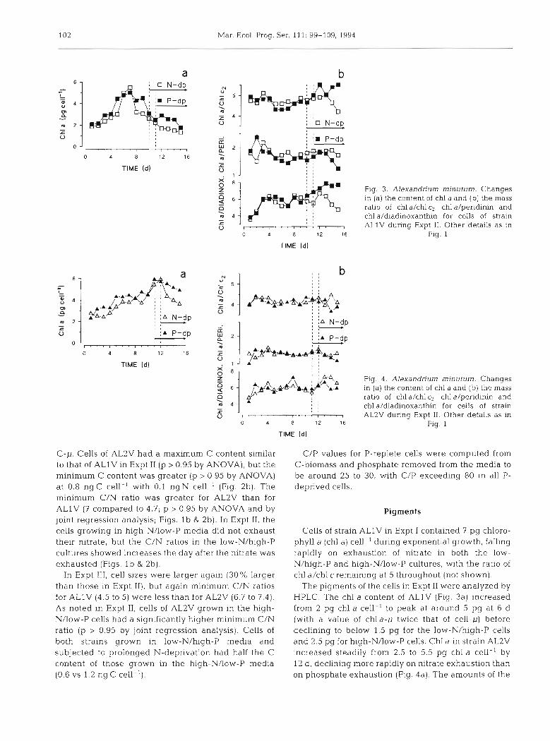
Fig. 3. Alexandrium minutum. Changes In (a) the content of chl a and (b) the mass
a 8 , m ratio of chl a/chl c2, chl a/peridinin and ! j I f l l , , , , chl a/diadinoxanthin for cells of strain - , , r a , 0 ALlV during Expt 11. Other details as in
o 12 16 Fig. 1
TlME (dl
8 , , 0 l
0 4 8 12 16
TlME (dl
Fig. 4 . Alexandriurn minutum. Changes In (a) the content of chl a and (b) the mass ratio of chl a/chl c2, chl a/peridinin and chl ddiadinoxanthin for cells of strain AL2V during Expt 11. Other details as in
Fig. 1
C-p. Cells of AL2V had a maximum C content similar to that of ALlV in Expt I1 (p > 0.95 by ANOVA), but the minimum C content was greater (p > 0.95 by ANOVA) at 0.8 ng C cell-' with 0.1 ng N cell-' (Fig. 2b). The minimum C/N ratio was greater for AL2V than for ALlV (7 compared to 4.7; p > 0.95 by ANOVA and by joint regression analysis; Figs. l b & 2b). In Expt 11, the cells growing in high-N/low-P media did not exhaust their nitrate, but the C/N ratios in the low-N/high-P cultures showed increases the day after the nitrate was exhausted (Figs. l b & 2b).
In Expt 111, cell sizes were larger again (30% larger than those in Expt 11), but again minimum C/N ratios for ALlV (4.5 to 5) were less than for AL2V (6.7 to 7.4). As noted in Expt 11, cells of AL2V grown in the high- N/low-P cells had a significantly higher minimum C/N ratio (p > 0.95 by joint regression analysis). Cells of both strains grown in low-N/high-P media and subjected to prolonged N-deprivation had half the C content of those grown in the high-N/low-P media (0.6 vs 1.2 n g C cell-').
C/P values for P-replete cells were computed from C-biomass and phosphate removed from the media to be around 25 to 30, with C/P exceeding 80 in all P- deprived cells.
Pigments
Cells of strain ALlV in Expt I contained 7 pg chloro- phyll a (chl a) cell-' during exponential growth, falling rapidly on exhaustion of nitrate in both the low- N/high-P and high-NAow-P cultures, with the ratio of chl a/chl c remaining at 5 throughout (not shown).
The pigments of the cells in Expt I1 were analyzed by HPLC. The chl a content of ALlV (Fig. 3a) increased from 2 pg chl a cell ' to peak at around 5 pg at 6 d (with a value of chla-,u twice that of cell-p) before declining to below 1.5 pg for the low-Nlhigh-P cells and 2.5 pg for high-NAow-P cells. Chl a in strain AL2V increased steadily from 2.5 to 5.5 pg chl a cell-' by 12 d, declining more rapidly on nitrate exhaustion than on phosphate exhaustion (Fig. 4a). The amounts of the
Flynn et al.: Changes in Alesandnum toxin content, biomass and pigments 103
-.
other pigments (chl c2, and the carotenoids peridinin and diadinoxanthin) covaried with that of chl a for both strains (Figs. 3b & 4b) although the relative proportions of chl alchl c2 and chl a/peridinin differed between the strains (p > 0.95 by ANOVA). Thus, the ratio of chl alchl c2 remained steady at 4.5 to 5 for ALlV and 4 to 4.5 for AL2V, while the ratio of chl a/peridinin remained around 1.7 for ALlV and 1.4 for AL2V.
Toxins
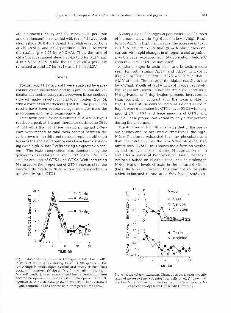
Toxins from ALlV in Expt I were analyzed by a pre- column oxidation method and by a postcolumn deriva- tization method. A comparison between these methods showed similar results for total toxin content (Fig. 5), with a correlation coefficient (r) of 0.91. The precolumn results have been calibrated against those from the postcolumn analysis of toxin standards.
Total toxin cell-' for both cultures of ALlV in Expt I reached a peak at 4 d and thereafter declined to 30 % of that value (Fig. 5). There was no significant differ- ence with respect to total toxin content between the cells grown in the different nutrient regimes, although towards the end a divergence may have been develop- ing (with high-N/low-P cells having a higher toxin con- tent). The toxin composition was dominated by the gonyautoxins GTX4 (60 %) and GTXl (20 to 30 %) with smaller amounts of GTX3 and GTX2. With increasing N-starvation the proportion of GTX4 increased (in the low-N/high-P cells to 78 %) with a pro rata decline in its isomeric form, GTX1.
o y ' , . , , i , , i , , , , , , 7
TIME Id)
Fig. 5. Alexandrium minutum. Changes in total toxin cell-' in cells of strain ALlV during Expt I. Cells grown in the low-N/high-P media (open symbol and heavy dashed line) became N-deprived (N-dp) at Day 6, and cells in the high- NAow-P media (closed symbols and heavy continuous line) became P-deprived (P-dp) at Day 6 and N-deprived at Day 9. Symbols denote data from post-column HPLC; heavy dashed
and continuous Lines denote data from precolumn HPLC
A comparison of changes in parameter-specific rates of increase, shown in Fig. 6 for the low-N/high-P cul- ture of ALlV in Expt I , shows that the increase in toxin cell-' in the pre-exponential growth phase was con- current with rapid changes in nitrogen-p and arginine- p as the cells recovered fl-om N-deprivation, before C content and cell division increased.
Similar changes in toxin cell-' and in toxin-p were seen for both strains ALlV and AL2V in Expt I1 (Fig. ?a, b). Toxin content in AL2V was 20 % of that in ALlV at most. The cause of the higher toxicity in the low-N/high-P cells of ALlV in Expt I1 (open symbols, Fig. ?a) is not known. In neither strain did short-term N-deprivation or P-deprivation promote increases in toxin content. In contrast with the toxin profile in Expt I, those of the cells for both ALlV and AL2V in Expt I1 were dominated by GTX4 (over 90 %) with only around 5 % GTXl and trace amounts of GTX3 and GTX2. These proportions varied by only a few percent during the experiment.
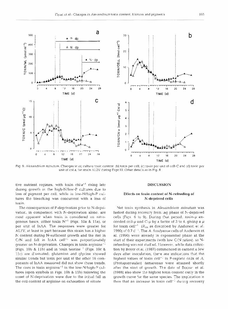
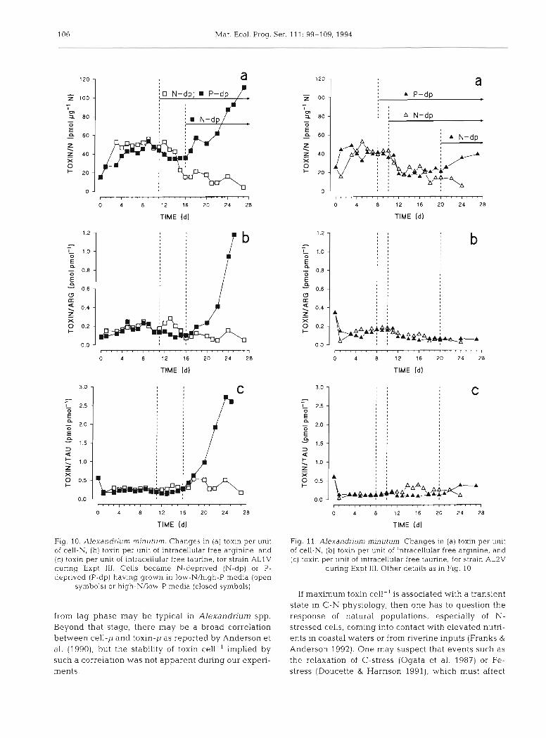
The duration of Expt 111 was twice that of the previ- ous studies and, as occurred during Expt I, the high- N/low-P cultures exhausted first the phosphate and then the nitrate, while the low-N/high-P exhausted nitrate only. Expt I11 thus shows the effects on synthe- sis and turnover of toxin during N-deprivation alone and after a period of P-deprivation. Again, net toxin synthesis halted on N-exhaustion, and on prolonged N-deprivation, levels of toxin in the culture declined (Figs. 8a & 9a). However, this was not so for cells which exhausted nitrate after they had already ex-
. ; 0 Cells
\ : v Carbon
r Nitrogen
TIME Id]
Fig. 6. Alexandrium minutum. Changes in parameter-specific rates of increase ('growth rates') for cells of ALlV grown in the low-N/high-P medium during Expt I. Cells became N-
deprived (N-dp) from Day 6. ARG: arginine
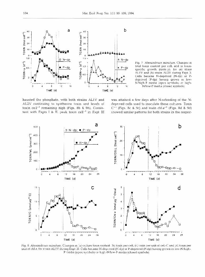
104 Mar. Ecol. Prog. Ser. 111: 99-109, 1994
Fig. 7 Alexandrium minutum. Changes in total toxin content per cell, and in toxin- specific growth (toxln-p), for (a) strain A L l V and (b) strain AL2V during Expt 11. Cells became N-deprived (N-dp) or P- deprived (P-dp) having grown in low- N/high-P media (open symbols) or high-
NAow-P me&a (closed symbols)
. . l , . . , . ' , . . . I
0 4 8 12 16
TlME Id)
0 4 8 12 16
TlME Id1
hausted the phosphate, with both strains ALlV and was attained a few days after N-refeedmg of the N- AL2V continuing to synthesize toxin and levels of deprived cells used to inoculate these cultures. Toxin toxin cell-' remaining high (Figs. 8b & 9b). Consis- C-' (Figs. 8c & 9c) and toxin chla-' (Figs. 8d & 9d) tent with Expts I & 11, peak toxin cell-' in Expt 111 showed similar patterns for both strains in the respec-
TlME id ) TlME (d l
l . ' . I r . . I . ~ . I r ' . I . . ' I " - I . " I
0 4 8 12 16 20 24 28
TlME [ d ) TlME Id]
Fig. 8. Alexandrium minutum. Changes in (a) culture toxin content. (b) toxin per cell, (c) toxin per unit of cell-C and (d) toxin per unit of chl a, for strain A L l V during Expt 111. Cells became N-deprived (N-dp) or P-deprived (P-dp) having grown in low-N/high-
P media (open symbols) or high-N/low-P media (closed symbols)
Flynn e t al.: Changes in Alexandrium toxin content, biomass and pigments 105
TIME ldl TIME (dj
1 1 ' 1 ~ 1 1 ' 1 ' " 1 ' ' ' 1 ' ' ' 1 ~ ~ ' ~ ' ' ~ 1
0 4 8 12 16 20 24 28
TlME Id) TlME (d)
Fig. 9. Alexandrium minutum. Changes In (a) culture toxin content, (b ) toxin per cell, (c) toxin per unit of cell-C and (d) toxin per unit of chl a, for strain AL2V during Expt 111. Other details as in Fig. 8
tive nutrient regimes, with toxin chla-' rising late during growth in the high-N/low-P cultures due to loss of pigment per cell, while in low-N/high-P cul- tures the bleaching was concurrent with a loss of toxin.
The consequences of P-deprivation prior to N-depri- vation, in comparison with N-deprivation alone, are most apparent when toxin is considered on nitro- genous bases, either toxin N-' (Figs. 10a & l l a ) , or per unit of InAA. The responses were greater for ALlV, at least in part because this strain has a higher N content during N-sufficient growth and the rise in C/N and fall in InAA cell-' was proportionately greater on N-deprivation. Changes in toxin arginine-' (Figs. lob & l l b ) and in toxin taurine-' (Figs. 10c &
l l c ) are illustrated; glutamine and glycine showed similar trends but toxin per unit of the other 16 conl- ponents of InAA measured did not show these trends. The rises in toxin arginine-' in the low-N/high-P cul- tures (open symbols in Figs. lob & l l b ) following the onset of N-deprivation were due to the initial fall in the cell content of arginine on exhaustion of nitrate.
DISCUSSION
Effects on toxin content of N-refeeding of N-deprived cells
Net toxin synthesis in Alexandrium minutum was fastest during recovery from lag phase of N-deprived cells (Figs. 6 to 9). During that period, toxin-p ex- ceeded cell-p and C-p by a factor of 3 to 4, giving a p for toxin cell-' (R,,, as described by Anderson et al. 1990) of 0.7 d-l. The A. fundyense cells of Anderson et al. (1990) were already in exponential phase at the start of their experiments (with low C/N ratios), so N- refeeding was not studied. However, while data collec- tion by Boyer et al. (1987) commenced in earnest a few days after inoculation, there are indications that the highest values of toxin cell-' in P-replete cells of A. (Protogonyaulax) tamarensis were attained shortly after the start of growth. The data of Boczar et al. (1988) also show the highest toxin content early in the growth curve for the same species. The implication is then that an increase in toxin cell-' during recovery
106 Mar. Ecol. Prog. Ser. 111: 99-109, 1994
l l l l l l l l l l l l l l l l l ' l ' I , . . I ' r , I
0 4 8 12 16 20 24 28
TlME (d]
TlME Id1
TlME (d)
Fig. 10. Alexandrjurn rninutum. Changes in (a) toxin per unit of cell-N, ( b ) toxin per unlt of intracellular free arginine, and (c) toxin per u n ~ t of intracellular free taurine, for strain ALlV during Expt 111. Cells became N-deprived (N-dp) or P- deprived (P-dp) havlng grown in low-N/high-P media (open
symbols) or high-NAow-P media (closed symbols)
from lag phase may be typical in Alexandnum spp. Beyond that stage, there may be a broad correlation between cell-p and toxin-p as reported by Anderson et al. (1990), but the stability of toxin cell-' implied by such a correlation was not apparent during our expen- ments.
l l l ~ r ~ ~ r I ~ ' ' r ' ' ' l ' " I " ' l ' l ' I
0 4 8 12 16 20 24 28
TlME (dl
TlME (dl
1 . ' I . . . r . ' ' I ' . . I ' ' n I ' ' ' I . . ' t
0 4 8 12 16 20 24 28
TlME (dl
Fig. 11. Alexandrium rninutum Changes in (a) toxin per unlt of cell-N, (b) toxin per unit of intracellular free arginine, and (c) toxin per unit of intracellular free taurine, for strain AL2V
during Expt 111. Other deta~ls as in Fig. 10
If maximum toxin cell-' is associated with a transient state in C-N physiology, then one has to question the response of natural populations, especially of N- stressed cells, coming into contact with elevated nutri- ents in coastal waters or from riverine inputs (Franks &
Anderson 1992). One may suspect that events such as the relaxation of C-stress (Ogata et al. 1987) or Fe- stress (Doucette & Harrison 1991), which must affect
Flynn et al.: Changes in Alexandriu m toxin content, biomass and pigments 107
amino acid synthesis, also have knock-on effects on the synthesis of nitrogenous toxins. The consequences of N-refeeding or P-refeeding of toxin-producing cells which are P-stressed need to be investigated.
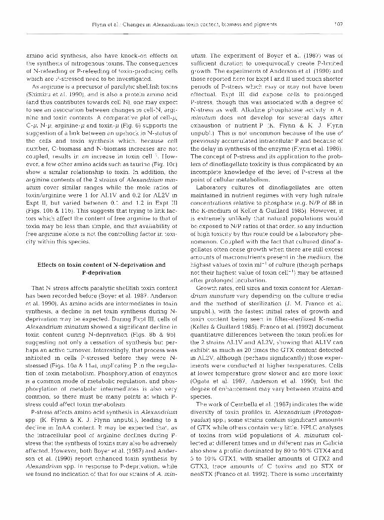
As arginine is a precursor of paralytic shellfish toxins (Shimizu et al. 1990), and is also a protein amino acid (and thus contributes towards cell-N), one may expect to see an association between changes in cell-N, argi- nine and toxin contents. A comparative plot of cell-p, C-p, N-p, arginine-p and toxin-p (Fig. 6) supports the suggestion of a link between an upshock in N-status of the cells and toxin synthesis which, because cell number, C-biomass and N-biomass increases are not coupled, results in a n increase in toxin cell-'. How- ever, a few other amino acids such as taurine (Fig. 10c) show a similar relationship to toxin. In addition, the arginine contents of the 2 strains of Alexandrium min- utum cover similar ranges while the mole ratios of toxidarginine were 1 for ALlV and 0.2 for AL2V in Expt 11, but varied between 0.1 and 1.2 in Expt 111 (Figs. l o b & l l b ) . This suggests that trying to link fac- tors which affect the content of free arginine to that of toxin may be less than simple, and that availability of free arginine alone is not the controlling factor in toxi- city within this species.
Effects on toxin content of N-deprivation and P-deprivation
That N-stress affects paralytic shellfish toxin content has been recorded before (Boyer e t al. 1987, Anderson et al. 1990). As amino acids are intermediates in toxin synthesis, a decline in net toxin synthesis during N- deprivation may be expected. During Expt 111, cells of Alexandrium minutum showed a significant decline in toxin content during N-deprivation (Figs. 8b & 9b), suggesting not only a cessation of synthesis but per- haps an active turnover. Interestingly, that process was inhibited in cells P-stressed before they were N- stressed (Figs. 10a & I l a ) , implicating P in the regula- tion of toxin metabolism. Phosphorylation of enzymes is a common mode of metabolic regulation, and phos- phorylation of metabolic intermediates is also very common, so there must be many points at which P- stress could affect toxin metabolism.
P-stress affects amino acid synthesis in Alexandrium spp. (K. Flynn & K. J. Flynn unpubl.), leading to a decline in InAA content. It may be expected that, as the intracellular pool of arginine declines during P- stress that the synthesis of toxins may also be adversely affected. However, both Boyer et al. (1987) and Ander- son et al. (1990) report enhanced toxin synthesis by Alexandrium spp. in response to P-deprivation, while we found no indication of that for our strains of A. min-
utum. The experiment of Boyer et al. (1987) was of sufficient duration to unequivocally create P-limited growth. The experiments of Anderson et al. (1990) and those reported here for Expt I and I1 used much shorter periods of P-stress which may or may not have been effectual. Expt 111 did expose cells to prolonged P-stress, though this was associated with a degree of N-stress as well. Alkaline phosphatase activity in A. minutum does not develop for several days after exhaustion of nutrient-P (K. Flynn & K. J. Flynn unpubl.). This is not uncommon because of the use of previously accumulated intracellular P and because of the delay in synthesis of the enzyme (Flynn et al. 1986). The concept of P-stress and its application to the prob- lem of dinoflagellate toxicity is thus complicated by a n incomplete knowledge of the level of P-stress at the point of cellular metabolism.
Laboratory cultures of dinoflagellates a re often maintained in nutrient regimes with very high nitrate concentrations relative to phosphate (e.g. N/P of 88 in the K-medium of Keller & Guillard 1985). However, it is extremely unlikely that natural populations would be exposed to N/P ratios of that order, so any induction of high toxicity by this route could be a laboratory phe- nomenon. Coupled with the fact that cultured dinofla- gellates often cease growth when there are still excess amounts of macronutrients present in the medium, the highest values of toxin ml-' of culture (though perhaps not their highest value of toxin cell-') may be attained after prolonged incubation.
Growth rates, cell sizes and toxin content for Alexan- drium minutum vary depending on the culture media and the method of sterilization (J. M. Franco et al. unpubl.), with the fastest initial rates of growth and toxin content being seen in filter-sterilized K-media (Keller & Guillard 1985). Franco et al. (1992) document quantitative differences between the toxin profiles for the 2 strains ALlV and AL2V, showing that ALlV can exhibit as much as 20 times the GTX content detected in AL2V, although (perhaps significantly) those exper- iments were conducted at higher temperatures. Cells at lower temperature grow slower and are more toxic (Ogata et al. 1987, Anderson et al. 1990), but the degree of enhancement may vary between strains and species.
The work of Cembella et al. (1987) indicates the wide diversity of toxin profiles in Alexandrium (Protogon- yaulax) spp.; some strains contain significant amounts of GTX while others contain very little. HPLC analyses of toxins from wdd populations of A, minutum col- lected at different times and in different rias in Galicia also show a profile dominated by 80 to 90 % GTX4 and 5 to 10% GTX1, with smaller amounts of GTX2 and GTX3, trace amounts of C toxins and no STX or neoSTX (Franco et al. 1992). There is some uncertainty
108 Mar. Ecol. Prog. Ser. 111: 99-109, 1994
over changes in toxin profiles during the growth of individual strains (Boyer et al. 1987, Boczar et al. 1988). We noted only minor changes (10 % or so) in the toxin profile during Expt I.
Differences between strains ALlV and AL2V
The differences between biomass, InAA content and pigmentation of the 2 strains of Alexandriurn rninutum do not appear to be profound. Cells of AL2V had a higher exponential-phase C/N ratio, though average cell sizes were similar, and had on average a higher InAA content, although this decreases in response to both N-deprivation and P-deprivation (Flynn & Flynn unpubl.). Thus, in Expt I1 and Expt I11 total InAA-N in ALlV decreased from 4 to 2 % of cell-N, and for AL2V, from 6 to 4 %, during nutrient deprivation. In all cases, cultures which depleted their nitrate showed a more rapid loss of InAA-N per cell than those which had exhausted phosphate.
There were significant, though low-magnitude, dif- ferences in the proportions of chl c2 and peridinin rela- tive to chl a in the 2 strains. It is of interest that the carotenoids, peridinin, diadinoxanthin and dinoxan- thin, covaried with chl a for both strains. While chl c2 and peridinin are light-harvesting pigments in dinofla- gellates (and so may be expected to covary with chl a), diadinoxanthin and dinoxanthin are suggested to have photo-protective functions (Prezelin & Boczar 1986). Although Demers et al. (1991) observed a rapid light- induced change in xanthophyll cycle pigments in Alexandnum excavatum, the epoxy-free diatoxanthin (formed by conversion of diadinoxanthin) was not detected in either strain of A. minutum. This could be due to the photon flux density (PFD) employed (200 pm01 m-2 S- ' ) being too low to induce this response; the ratio of chl a/diadinoxanthin was similar to that reported for A. excavatum growing at 50 pm01 m - 2 s-l (Demers et al. 1991). Net C-fixation rates, com-
puted from increases in algal C each day, peaked at around 0.3 ng C cell-' d- ' (0.025 ng C cell-' h- ' over the 12 h Light phase). These values are at the lower end of the range given by Glibert et al. (1988) for A. (Gonyaulax) tamarensis at a PFD of 200 pm01 m-' S- ' ,
although this may well reflect a difference in cell size. A complicating factor here is that flagellates, including dinoflagellates, often swim to form dense swarms in which the PFD available to the individual may be low- ered significantly. This may affect the C-N status of the cells.
Pigments could not be used as estimators of biomass or toxicity for Alexandrium minutum. Although the regression coefficients (r) for plots of cell number, bio- mass and toxicity against chl a for Expt I1 were all sig-
nificant, the coefficients of variation for the entire data sets for Expt 11, for cell/chl a (41 %), carbonlchl a (55 %), nitrogenlchl a (54 %) and toxinlchl a (93 %) were so large that predictive equations would be worthless. Even for individual cultures, the coefficients of varia- tion were between 20 % and 50 %. Temporal variations in toxin chl a- ' can be seen for Expt 111 in Figs. 8d & 9d.
Acknowledgements. This work was supported by research and capital equipment grants from the Natural Environment Research Council (UK) to K.J.F., funds from project AL192- 01 11-C02-01 (CICYT) to J.M.F and project 11.02 (IEO) to B.R. We thank Y. Pazos for assistance with Expt 11, and S. J. Wain- wright for advice on statistical matters.
LITERATURE CITED
Anderson, D. M, , Kullis, D. M., Sullivan, J. J. , Hall, S., Lee, C. (1990). Dynamics and physiology of saxitoxin production by the dmoflagellates Alexandnum spp. Mar. Biol. 104: 511-524
Blanco, J . J . , Marilio, L., Campos, M. J. (1985). The first toxic bloom of Gonyaulax tamarensis detected in Spain (1984). In: Anderson, D. M,, White, A. W., Baden, D. G. (eds.) Toxic dinoflagellates. Elsevier. New York. p. 79-84
Boczar, B. A., Beitler, M. K., Liston. J.. Sullivan, J. J., Cattolico, R. A. (1988). Paralytic shellfish toxins in Protogonyaulax tamarensis and Protogonyaulax catenella in axenic cul- ture. Plant Physiol. 88: 1285-1290
Boyer, G. L., Sullivan, J . J., Andersen. R. J., Harrison, P. J., Taylor, F. J. R. (1987). Effects of nutrient limitation on toxic production and composition in the marine dinoflagellate Protogonyaulax tamarensis. Mar. Biol. 96: 123- 128
Cembella, A. D. , Sullivan, J . J. , Boyer, G. L., Taylor, F. J . R., Andersen, R. J . (1987). Variahons in paralytic shellfish toxin composition within the Protogonyaulax tamaren- sislcatenella species complex; red tide dinoflagellates. Biochem. Syst. Ecol. 15: 171- 186
Delgado, M,, Estrada, M,, Camp, J., Fenandez. J. V., Sant- marti, M., Lleti, C. (1990). Development of a toxic Alexan- drium minutum Halim bloom in the harbour of Sant Carles d e la Rapita. Scientia mar. 54: 1-7
Demers, S., Roy, S., Gagnon, R., Vignoult, C. (1991). Rapid light-induced changes in cell fluorescence and in xantho- phyll-cycle pigments of Alexandrium excavatum (Dino- phyceae) and Thalassiosira pseudonana (Bacillario- phyceae); a photo-protection mechanism. Mar. Ecol. Prog. Ser. 76: 185- 194
Doucette, G. J . , Harrison, P J (1991). Aspects of iron and nitrogen nutrition in the red tide dinoflagellate Cymno- dinium sanguineum. I1 Effects of iron depletion and nitro- gen source on iron and nitrogen uptake. Mar. Biol. 110: 175-182
Erard-Le Denn, E. (1991). dexandrium minutum (Dinophy- cees]. In: Sournia, A.. et al. (eds.) Le phytoplancton nuisi- ble des cotes de France: de la biologie a la prevention. JFREMER, Brest, p. 83-90
Flynn, K. J., Flynn, K. (1992). Non-protein free amines in microalgae: consequences for the measurement of intra- cellular amino acids and of the glutamine/glutamate ratio. Mar. Ecol. Prog. Ser. 89: 73-79
Flynn, K. J. , Zapata, M,, Garrido, J L., Opik, H., Hipkin, C. R. (1993). Changes in carbon and nitrogen physiology during ammonium and nitrate nutntion and nltrogcn starvation in Isochrysis galbana. Eur. J Phycol. 28 47-52
Flynn et al.: Changes in Alexandrium t toxin content, biomass and pigments 109
Flynn, K. J . , Opik, H., Syrett, P. J . (1986) Localizat~on of the alkaline phosphatase and 5'-nucleotidase activities of the diatom Phaeodactylum tricornutum. J . gen. Microbiol. 132: 289-298
Franco, J . M. , Fernandez, P., Reguera, B (1992). The toxin profile of Alexandrium lusitanicum Balech from the Atlantic coast of the Iberian Peninsula. Comm. Meet. Int. Coun. Explor. Sea C.M.-ICES/L: 42
Franco, J. M., Fernandez-Vila, P. (1993). Separations of para- lytic shellfish toxins by reversed phase high performance liquid chromatography, with postcolumn reaction and flu- orometric detection. Chromatographia 35 613-620.
Franks, P. J . S., Anderson, D. M. (1992). Alongshore transport of a toxic phytoplankton bloom in a buoyancy current: Alexandrium Lamarense in the Gulf of Maine. Mar. Biol. 112: 153-164
Glibert, P. M., Kana, T. M., Anderson, D. M. (1988). Photosyn- thetic responses of Gonyaulax tamarensis during growth in a natural bloom and in batch culture. Mar. Ecol. Prog. Ser. 42: 303-309
Hallegraeff, G. M., Bolch, C. J., Blackburn, S. I. , Oshirna, Y. (1991). Species of the toxigenic dinoflagellate Alexan- drium in southeastern Australian waters. Botanica mar. 34: 575-587
Holligan, P. M., Williams, P. J . LeB., Purche, D., Harris, R. P. (1984). Photosynthesis, respiration and nitrogen supply of plankton populations in stratified, frontal and totally mixed shelf waters. Mar. Ecol. Prog. Ser. 17: 201 -213
Jeffrey, S. W., Humphrey, G. F. (1975). New spectrophoto- metric equations for determining chlorophylls a, b, c, and C:! in higher plants and natural phytoplankton. Biochem. Biophys. Pflanz. 167: 191 - 194
Jeffrey, S. W., Sielicki, M., Haxo, T. (1975). Chloroplast pig- ment patterns in dinoflagellates. J . Phycol. 11: 374-384
Keller, M. D., Guillard, R. R. L. (1985). Factors significant to marine dinoflagellate culture. In: Anderson, D. M., White, A. W., Baden, D. G. (eds.) Toxic dinoflagellates. Elsevier, New York, p . 113-116
Lawrence, J . F , Menard, C . , Charbonneau, C. F., Hall, S. (1991). A study of ten toxins associated with paralytic shellfish poison using prechromatographic oxidation and liquid chromatography with fluorescence detection. J . Ass. Off. Anal. Chem. 74: 404-409
This article was submitted to the editor
LeFevre, J . (1986). Aspects of the biology of frontal systems. Adv. mar. Biol. 23: 163 - 299
Margalef. R. (1978). Life-forms of phytoplankton as survival alternatives in an unstable environment. Oceanol. Acta 1: 493-509
Margalef, R., Estrada, M., Blasco, D. (1979). Functional mor- phology of organisms involved in red tides as adapted to decaying turbulence. In. Taylor, D. L., Seliger, H . H. (eds.) Toxic dinoflagellate blooms. Elsevier, Amsterdam, p. 89-94
Montresor, M., Marino, D., Zingone, A., Dafnis, G. (1990). Three Alexandrium species from coastal Tyrrhenian waters (Mediterranean Sea). In. Granell. E. , Sundstrom, B., Edler, L., Anderson, D. M. (eds.) Toxlc marine phyto- plankton. Elsevier, New York, p. 82-87
Ogata, T., Ishimaru, T., Kodama, M. (1987). Effect of water temperature and light intensity on growth rate and toxic- ity changes in Protogonyaulax tamarenas. Mar. Biol. 95: 217-220
Paasche, E., Bryceson, I., Tangen, K. (1984). lnterspecific vari- ation in dark nitrogen uptake by dinoflagellates. J. Phycol. 20: 394-401
Prezelin, B. B., Boczar, B. A. (1986). Molecular bases of cell absorption and fluorescence in phytoplankton: potential apphcations to studies in optical oceanography. In: Round, F., Chapman, G. (eds.) Progress in phycological research 4. Biopress Ltd. Bristol, p. 350-450
Reguera, B., Campos, M. J., Fraga, S. , Marino, J., Bravo, I. (1991). The monitoring of harmful algal blooms in Galicia (NW Spain). In: Fremy, J. (ed.) Colloque Internationale des Biotoxines Marines. Centre National d'Etudes Veteri- naires et Alimentaires, Paris, p. 217-223
Shirnizu, Y., Gupta, S., Pradad, A. V. K. (1990). Biosynthesis of dinoflagellate toxins. In: Graneli. E., Sundstrom, B., Edler, L., Anderson, D. M. (eds.) Toxic marine phytoplankton. Elsevier, New York, p. 62-71
Strickland. J . D. H., Parsons, T. R. (1972). A practical hand- book of seawater analysis. Bull. Fish. Res. Bd Can. 167: 1-311
Zapata, M. , Ayala, A. M., Franco, J M , Garrido, J . L. (1987). Separation of chlorophylls and their degradation products in marine phytoplankton by reversed-phase high per- formance liquid chromatography Chromatographia 23: 28-30
A4anuscript first received: August 4, 1993 Revised version accepted: April 8, 1994