Cellular organization of normal mouse liver: a histological, quantitative immunocytochemical, and...
23
Cellular Organization of Normal Mouse Liver: A Histological, Quantitative Immunocytochemical, and Fine Structural Analysis Janie L. Baratta 1 , Anthony Ngo 1 , Bryan Lopez 1 , Natasha Kasabwalla 1 , Kenneth J. Longmuir 2 , and Richard T. Robertson 1 1 Department of Anatomy & Neurobiology, School of Medicine, University of California, Irvine, Irvine CA, 92697 2 Department of Physiology & Biophysics, School of Medicine, University of California, Irvine, Irvine CA, 92697 Abstract The cellular organization of normal mouse liver was studied using light and electron microscopy and quantitative immunocytochemical techniques. The general histological organization of the mouse liver is similar to livers of other mammalian species, with a lobular organization based on the distributions of portal areas and central venules. The parenchymal hepatocytes were detected with immunocytochemical techniques to recognize albumin or biotin containing cells. The macrophage Kupffer cells were identified with F4-80 immunocytochemistry, Ito stellate cells were identified with GFAP immunocytochemistry, and endothelial cells were labeled with the CD-34 antibody. Kupffer cells were labeled with intravascularly administered fluorescently labeled latex microspheres of both large (0.5 μm) and small (0.03 μm) diameters, while endothelial cells were labeled only with small diameter microspheres. Neither hepatocytes nor Ito stellate cells were labeled by intravascularly administered latex microspheres. The principal fine structural features of hepatocytes and non- parenchymal cells of mouse liver are similar to those reported for rat. Counts of immunocytochemically labeled cells with stained nuclei indicated that hepatocytes constituted approximately 52% of all labeled cells, Kupffer cells about 18%, Ito cells about 8%, and endothelial cells about 22% of all labeled cells. Approximately 35% of the hepatocytes contained two nuclei; none of the Kupffer or Ito cells were double nucleated. The presence of canaliculi and a bile duct system appear similar to that reported for other mammalian species. The cellular organization of the mouse liver is quite similar to that of other mammalian species, confirming that the mouse presents a useful animal model for studies of liver structure and function. Keywords albumin; F4/80; GFAP; hepatocyte; immunocytochemistry Introduction The important roles performed by the liver, not only in the storage and release of nutrients but also in the neutralization and elimination of a variety of toxic substances, have prompted investigations of its cellular constituents and organization. Some of these studies have been carried out in human liver, but the importance of having an experimental model system has prompted several investigations of liver organization in laboratory mammals, primarily rats Address for correspondence: Richard T. Robertson, Ph.D., Department of Anatomy & Neurobiology, School of Medicine, University of California, Irvine, CA 92697-1280, Telephone: 949.824.6553, FAX: 929.824.1105, [email protected]. NIH Public Access Author Manuscript Histochem Cell Biol. Author manuscript; available in PMC 2009 October 14. Published in final edited form as: Histochem Cell Biol. 2009 June ; 131(6): 713–726. doi:10.1007/s00418-009-0577-1. NIH-PA Author Manuscript NIH-PA Author Manuscript NIH-PA Author Manuscript
-
Upload
independent -
Category
Documents
-
view
7 -
download
0
Transcript of Cellular organization of normal mouse liver: a histological, quantitative immunocytochemical, and...

Cellular Organization of Normal Mouse Liver: A Histological,Quantitative Immunocytochemical, and Fine Structural Analysis
Janie L. Baratta1, Anthony Ngo1, Bryan Lopez1, Natasha Kasabwalla1, Kenneth J.Longmuir2, and Richard T. Robertson11 Department of Anatomy & Neurobiology, School of Medicine, University of California, Irvine, IrvineCA, 926972 Department of Physiology & Biophysics, School of Medicine, University of California, Irvine, IrvineCA, 92697
AbstractThe cellular organization of normal mouse liver was studied using light and electron microscopy andquantitative immunocytochemical techniques. The general histological organization of the mouseliver is similar to livers of other mammalian species, with a lobular organization based on thedistributions of portal areas and central venules. The parenchymal hepatocytes were detected withimmunocytochemical techniques to recognize albumin or biotin containing cells. The macrophageKupffer cells were identified with F4-80 immunocytochemistry, Ito stellate cells were identified withGFAP immunocytochemistry, and endothelial cells were labeled with the CD-34 antibody. Kupffercells were labeled with intravascularly administered fluorescently labeled latex microspheres of bothlarge (0.5 μm) and small (0.03 μm) diameters, while endothelial cells were labeled only with smalldiameter microspheres. Neither hepatocytes nor Ito stellate cells were labeled by intravascularlyadministered latex microspheres. The principal fine structural features of hepatocytes and non-parenchymal cells of mouse liver are similar to those reported for rat. Counts ofimmunocytochemically labeled cells with stained nuclei indicated that hepatocytes constitutedapproximately 52% of all labeled cells, Kupffer cells about 18%, Ito cells about 8%, and endothelialcells about 22% of all labeled cells. Approximately 35% of the hepatocytes contained two nuclei;none of the Kupffer or Ito cells were double nucleated. The presence of canaliculi and a bile ductsystem appear similar to that reported for other mammalian species. The cellular organization of themouse liver is quite similar to that of other mammalian species, confirming that the mouse presentsa useful animal model for studies of liver structure and function.
Keywordsalbumin; F4/80; GFAP; hepatocyte; immunocytochemistry
IntroductionThe important roles performed by the liver, not only in the storage and release of nutrients butalso in the neutralization and elimination of a variety of toxic substances, have promptedinvestigations of its cellular constituents and organization. Some of these studies have beencarried out in human liver, but the importance of having an experimental model system hasprompted several investigations of liver organization in laboratory mammals, primarily rats
Address for correspondence: Richard T. Robertson, Ph.D., Department of Anatomy & Neurobiology, School of Medicine, University ofCalifornia, Irvine, CA 92697-1280, Telephone: 949.824.6553, FAX: 929.824.1105, [email protected].
NIH Public AccessAuthor ManuscriptHistochem Cell Biol. Author manuscript; available in PMC 2009 October 14.
Published in final edited form as:Histochem Cell Biol. 2009 June ; 131(6): 713–726. doi:10.1007/s00418-009-0577-1.
NIH
-PA Author Manuscript
NIH
-PA Author Manuscript
NIH
-PA Author Manuscript

(Blouin et al., 1977; Bouwens et al., 1992; Fahimi, 1982; Gard et al., 1985; Geerts, 2001;Kawada, 1997; Leo and Lieber, 1983; Marcos et al., 2004; Sigal et al., 1999; Sleyster andKnook, 1982; Wake et al, 1989; Widmann et al., 1971; Wisse, 1970; 1972; 1974; Yokota etal., 1981). In the species studied thus far, investigations have demonstrated that the liver iscomprised of several different populations of cells. These include the major parenchymal cells,the hepatocytes (Blouin et al., 1977; David, 1985; Jones and Mills, 1974; Loud, 1968; Yokotaet al., 1981), and a variety of non-parenchymal resident cells including a population ofmacrophages termed Kupffer cells (Bouwens et al., 1986; Fahimi, 1982; Naito et al., 1997;2004; Wake et al., Widmann et al., 1972; 1989; Wisse, 1974), fat storing cells termed Ito orstellate cells (Fahimi, 1982; Ito, 1973; Kawada, 1997; Sato et al., 2003; Senoo, 2004), andendothelial cells (Smedsrod et al., 1994; Stöhr et al., 1978; Widmann et al., 1972; Wisse,1972).
In recent years, the use of mice, and particularly genetically engineered mice, in researchlaboratories has increased markedly. Several studies have used mice in addressing particularquestions in liver structure and function (e.g., Bartök et al., 1983; Longmuir et al., 2007; Naitoet al., 1997; Robertson et al., 2008; Sigal et al., 1999; Stöhr et al., 1978; Yamada et al.,1990). Many of these studies assume similarities between the mouse and rat liver, but theputative similarities have not, to our knowledge, been demonstrated. Indeed, notable cases ofdifferences between rat and mouse liver have been reported (Stöhr et al., 1978). Because ofthe important role that will be played by mice in future studies of liver function, we believe itimperative to establish the baseline of normal cellular composition, to serve as a reference forthese future studies. The present paper reports the results of an empirical study of normal mouseliver, with emphasis on identifying the constituent populations of cells in the liver by usingimmunocytochemical markers, determining essential fine structural features of these cells, anddetermining the relative numbers of the different immunocytochemically identifiedpopulations of cells.
Materials and MethodsMaterials
Chemical supplies were purchased from Sigma Aldrich (St. Louis MO) unless specifiedotherwise.
AnimalsAll animal work was reviewed and approved by the University of California, Irvine,Institutional Animal Care and Use Committee (IACUC) prior to conducting the experiments.Adult (2 to 4 mo) BALB/c or ICR female mice, obtained from Charles River (WilmingtonCA), body weight approximately 20 to 25 g, were used for these experiments. Mice wereprovided standard laboratory mouse food ad libitum, and were housed in a satellite vivariumwith standard 12 hr light, 12 hr dark cycles.
Tissue PreparationFor studies of normal structure, 27 mice were deeply anesthetized with sodium pentobarbital(50mg/kg i.p.). Mice were perfused through the heart or through the portal vein with 10 mlsaline, using a pump at 5 ml/min, to clear the liver of blood, then followed with cold 4%paraformaldehyde in sodium phosphate buffer, pH 7.4 for approximately 15 minutes. Providingthe perfusion was successful and the liver cleared of blood, no differences were detected inmorphological features between the two routes of perfusion.
The liver lobes were removed, cut into 2–3 mm blocks, and fixed in 4% paraformaldehyde for1–18 hours before being placed in 30% sucrose for cryoprotection. Blocks of liver tissue were
Baratta et al. Page 2
Histochem Cell Biol. Author manuscript; available in PMC 2009 October 14.
NIH
-PA Author Manuscript
NIH
-PA Author Manuscript
NIH
-PA Author Manuscript

frozen in −20° C 2′methylbutane in preparation for sectioning with a cryostat. Frozen liversections were cut on a Reichert-Jung 1800 cryostat at 10–12 μm, mounted directly on superfrost slides (Fisher Scientific, Pittsburgh PA), and air dried for 10–30 min before processingfor immunocytochemistry, lectin binding, or hematoxylin and eosin (H&E) staining.
Latex Microsphere and Liposome InjectionsTwelve mice were lightly anesthetized with Ketamine-xylazine (mg/kg). Six of these micereceived injections into the tail vein of 25–100 μl of a saline solution containing Fluorospheres(fluorescently labeled latex microspheres; 2.5%; Molecular Probes –Invitrogen, Carlsbad CA).Fluorospheres with mean diameters of 0.5 μm or of 0.03 μm were used, injected eitherseparately or combined as a cocktail comprising equal volumes of the 2 stock suspensions.Fluorspheres of different sizes carried different fluorophores, either rhodamine or fluorescein.Following post-injection survival periods of 1 – 5h, animals were deeply anesthetized withsodium pentobarbital and perfused through the heart as described above.
The other six of these mice received injections of liposomes into the tail vein. Liposomes wereprepared as described previously (Longmuir et al., 2006; Robertson et al., 2008) and containeda liver targeting peptide sequence derived from the circumsporozoite protein of Plasmodiumberghei. The liposomes were made from a mPEG5000 conjugate of 22:1-PE, prepared byreacting 10 μmol di22:1-PE with 20 μmol of mPEG5000 carboxylic acid N-hydroxysuccinimidyl ester (#2M4M0H01, Nektar, Huntsville, AL) and 100 μmol oftriethylamine in dichloromethane. An addition of 1,2-Dipalmitoyl-sn-3-phosphatidylethanolamine, labeled with 1.4 nm gold particles obtained from Nanoprobes, Inc.,Yaphank NY, was used to enable localization of the liposome fragments using electronmicroscopy.
ImmunocytochemistrySerial sections of liver were cut on the cryostat and collected on Superfrost/Plus coated slides(Fisher Scientific, Pittsburgh PA) and processed for immunocytochemistry for albumin, biotin,F 4/80, glial fibrillary acidic protein (GFAP), or the endothelial cell marker CD-34. Slides withtissue sections were rinsed in Tris buffer three times and blocked for 1 hour in 3% normal goatserum (InVitrogen, Carlsbad CA) (for albumin, F4/80, CD-34) or 3% normal rabbit serum (forbiotin). Each primary antibody was tested parametrically, in dilutions of Tris buffer in blockingsolution, to determine the optimal antibody concentration to be used. The albumin antibody(Bethyl Labs; Montgomery TX) labeled with FITC was used at 1:500; the biotin antibody(Abcam; Cambridge MA) labeled with FITC was used at 1:500; the macrophage (Kupffer cell)antibody F/4/80 (Serotec, Raleigh NC) was used at 1:1000; the GFAP antibody (Dako;Carpinteria CA) was used at 1:3000; and the CD-34 antibody (Vector Labs) was used at 1:100.Sections were exposed to primary antibodies at room temperature and in the dark, overnight(16–18hr). The following day, slides were rinsed in Tris buffer three times. The sections forF4/80 then were incubated for 2 hours with either Alexa 488 or Alexa 546 goat anti-rat IgG at1:1000 (Invitrogen; Carlsbad CA), sections for GFAP were incubated with either Alexa 488or Alexa 546 goat anti-rabbit, at 1:1000 and sections for CD-34 were incubated with Alexa488 goat anti-mouse at 1:1000. Following incubation, slides were rinsed with Tris buffer andcoverslipped with Vectashield anti-fade fluorescent mounting medium with DAPI; DAPIserved as a fluorescent (ultraviolet – UV) stain for cell nuclei.
Lectin BindingCryotstat cut sections were mounted on slides and rinsed in Tris buffer. Tomato lectin (Vectorlabs; Burlingame CA) labeled with FITC was diluted (1:5000) in Tris buffer and sections wereincubated overnight in the dark. Slides were rinsed in Tris buffer and coverslipped withVectashield with DAPI.
Baratta et al. Page 3
Histochem Cell Biol. Author manuscript; available in PMC 2009 October 14.
NIH
-PA Author Manuscript
NIH
-PA Author Manuscript
NIH
-PA Author Manuscript

Hematoxylin and EosinSlide mounted 8–12 um cryostat sections were dehydrated through absolute alcohol andrehydrated to water. Slides were placed in hematoxylin stain for 4 minutes, rinsed in water,differentiated in 70% alcohol and stained in 0.01% eosin Y for 2 seconds, rinsed in 95% ethanol,dehydrated with absolute ethanol and cleared in xylenes for 15 minutes before coverslipping.
Electron MicroscopyLiver tissue from 9 animals was used for electron microscopic studies. Six of these animalshad received intravenous injections of liposomes, as described above. Following perfusionwith saline followed by 4% paraformaldeyde, tissue was post-fixed in 4% paraformaldehydeand 0.5% glutaraldehyde. Tissue blocks were cut on a Vibratome at 50 μm. Some sectionswere processed using a gold-enhancement kit (Goldenhance-EM; Nanoprobes Inc., Yaphank,NY) following the protocols described by the manufacturer. Other sections were processed forimmunocytochemistry using the F4-80 or GFAP antibodies. Sections were then placed in 1%osmium tetroxide for 30 min, embedded in epoxy, cut at 800 nm, and stained with uranyl acetateand lead citrate to enhance contrast. These thin sections were inspected on a Phillips EM-10electron microscope equipped with a Gatan digital camera.
Dissociated Cell StainingMice aged 30 days to adult were deeply anesthetized with sodium pentobarbital and perfusedthrough the portal vein with 25 ml cold 0.1M EDTA in calcium free DMEM followed by 25ml warm (37° C) 0.05% collagenase II in DMEM with calcium. Liver pieces were removed,minced and incubated with the collagenase medium at 37° C for 15 minutes. Tissue wascentrifuged at 1500 rpm for 5 minutes, the collagenase medium was removed, and the cells re-suspended in DMEM. The tissue was dissociated and strained through a 100um mesh toeliminate clumps of cells. The cells were then stained with trypan blue for 2 minutes beforephotographing and counting using a bright field microscope.
Image Collection and ProcessingSlides were examined using bright field illumination (H&E) or fluorescence illumination withrhodamine, fluorescein, or ultraviolet (DAPI) filter cubes. Digital images were captured usinga Nikon DS 5M digital camera and imported into Adobe Photoshop. When creatingphotographic plates for illustrations, brightness and contrast were adjusted for uniformitywithin a plate; no other alterations of images were done.
Quantitative AssessmentsRelative numbers of each of the different populations of cells were estimated by counting thenumber of nuclei of immunocytochemically labeled cells and correcting the raw counts byusing the method of Abercrombie (1946). Sections of 12 μm thickness, immunocytochemicallylabeled with a cell specific antibody and with DAPI nuclear stain, were examined. Digitalimages were collected using a 40× lens and imported into Photoshop. These images includedan area of 46,800 μm2 (260 μm X 180 μm). Cells that displayed clear immunocytochemicallabeling along with distinct DAPI positive nuclei were identified and their positions markedon an overlaid sheet in Photoshop. Diameters of DAPI stained nuclei were measured using theNikon DS-5M software for 2 point distances or from Photoshop images, using a reticule. Thenumber of labeled cells (defined as DAPI stained nucleus amid immunocytochemically labeledcytoplasm) in each section was adjusted by the formula presented by Abercrombie:
Baratta et al. Page 4
Histochem Cell Biol. Author manuscript; available in PMC 2009 October 14.
NIH
-PA Author Manuscript
NIH
-PA Author Manuscript
NIH
-PA Author Manuscript

In which P is the calculated average number of nuclei per region, A is the crude count of numberof nuclei of labeled cells per section, M is the tissue section thickness, and L is the averagediameter of nuclei.
Numbers of immunocytochemically and DAPI labeled cells were determined for morelocalized regions of the liver, using the liver acinus scheme of Rappaport et al. (1954). Threezones of approximate equivalent areas were identified in a liver acinus, including zone 1 closeto the portal area, zone 3, around the central venule, and a zone 2 situated between zones 1 and3. Photoshop images were analyzed by placing a graticule, measuring 7,500μm2 (50 μm ×150μm), over each of the zones and determining the numbers of labeled cells in each area.
ResultsHistological Organization of Normal Mouse Liver
Sections of mouse liver stained with a standard hematoxylin and eosin (H&E) procedure revealpatterns of cellular labeling similar to what has been reported for other mammalian species.Portal areas, containing elements of the hepatic triad, that is one or more small branches of theportal vein, a branch of the hepatic artery, and a small bile duct, along with lymphatic vesselsand a very small amount of connective tissue, could be identified (Fig. 1A; 1C). The presenceof portal areas along with central venules provided evidence of a lobular structure to the mouseliver. Liver cells are arranged in plates or cords, and are seen radiating from the regions ofcentral venules (Figs. 1A, 1B, 1D) and extending to the portal areas. The plates or cords ofcells are separated by sinusoidal capillaries (fig. 1G).
Tissue processed with H&E (or with the fluorescent DAPI stain, see below) reveals distinctlystained nuclei of varied shapes. Most common are cells having one, or sometimes two, largeround to slightly oval nuclei. Also detected in the H&E stained, or DAPI labeled, sections areother cells with smaller and darkly staining oval or oblong nuclei.
Immunocytochemical techniques were used to identify different populations of cells withinthe liver tissue. As shown in figure 1D, cells containing the large round nuclei were labeled byan antibody to albumin. Albumin immunoreactivity appeared as small puncta of intensefluorescence within the cytoplasm of virtually all of these cells (Fig. 1D), although the numbersof puncta as well as their apparent intensity displayed some variation between cells. Becauseof their immunoreactivity to albumin antibodies, these cells with large round nuclei weredeemed to be hepatocytes (Yokota et al., 1981).
Figure 1E shows immunoreactivity to the F4-80 antibody, which is known to labelmacrophages in other organs and labels some of the cells containing dense oblong nuclei inliver sections. The F4-80 positive cells were distributed throughout the liver lobules but werealways associated with a sinusoidal capillary. Within the liver lobules, F4-80 positive cellswere more frequently encountered in regions close to the portal area (region 1 of Rappaport etal., 1954) and were more sparse in regions closer to the central venule (region 3). Because theirmorphological features and F4-80 immunoreactivity are similar to macrophages described inother species, these F4-80 positive cells were deemed to be Kupffer cells (Austyn and Gordon,1981; von Kupffer, 1898).
Cells immunoreactive for glial fibrillary acidic protein (GFAP) also contained small oblongnuclei and also were distributed throughout the liver (Fig. 1F). The GFAP positive cells wereencountered less frequently than were the F4-80 positive cells. These cells were deemed to bethe stellate cells of Ito (Gard et al., 1985;Ito, 1973;Neubauer et al., 1996).
Baratta et al. Page 5
Histochem Cell Biol. Author manuscript; available in PMC 2009 October 14.
NIH
-PA Author Manuscript
NIH
-PA Author Manuscript
NIH
-PA Author Manuscript

Endothelial cells were identified by a combination of CD-34 immunoctochemistry and DAPIlabeling of nuclei. As shown in figures 2A and 2B, the CD-34 immunoreactivity appears as acell surface marker and clearly outlines sinusoidal capillaries in liver tissue. The CD-34immunoreactivity is associated with small and oval or oblong DAPI stained nuclei (arrows)and clearly are distinct from the large round nuclei that are characteristic of the parenchymalhepatocytes. Figure 2C–E presents a case in which a mouse received an intravascular injectionof small (0.03 μm) rhodamine labeled latex microspheres, and the tissue later sectioned andprocessed for CD-34 immunoreactivity. A comparison of figures 2C and 2D demonstrates thatthese smaller latex microspheres appear to label intensely the endothelial cells. Co-labeling ofendothelial cells by green Alexa 488 CD-34 immunoreactivity, red rhodamine labeled latexmicrospheres, and blue DAPI labeled nuclei is illustrated in the higher magnificationphotomicrograph of figure 2F.
Further Immunocytochemical Characterization of Liver CellsThe issue of whether the immunocytochemical markers resulted in overlapping populations ofcells was addressed. As was shown in figure 1, hepatocytes are labeled by immunoreactivityfor albumin. Figures 3A and 3B show two views of the same section, with rhodamine opticsto show immunoreactivity for albumin (Fig. 3A) and under fluorescein optics to showimmunoreactivity for biotin (Fig. 3B). Similar to the pattern for albumin, biotin labelingappeared punctate and also appeared to label most cells containing large round nuclei (Fig.3B). Figure 3C presents a merger of the images in 3A and 3B, along with an ultraviolet imageshowing the DAPI labeled blue nuclei, and demonstrates that virtually all cuboidal cells withlarge round nuclei are labeled with antibodies to albumin and also to biotin. These markers arefound within the same cells, although the intracellular distribution of green biotin puncta wasslightly different from the intracellular distribution of red albumin related puncta.
Albumin immunoreactivity was not found to co-express with immunoreactvity to F4-80, GFAPor CD-34.
Photomicrographs in Figure 3D-F demonstrate that F4-80 labeled cells are a separatepopulation from the GFAP labeled cells. The GFAP antibody labels a population of cells withrelatively small and oblong nuclei, and with slender and widely branching cellular processes(Fig. 3D). As noted above, these are the stellate cells identified by Ito (Fahimi, 1982; Gard etal., 1985; Geerts et al., 1990; Ito, 1973; Kawada, 1997; Neubauer et al., 1996). The F4-80antibody labels a population of cells (Bouwens et al., 1986; Naito et al., 1997; Widmann et al.,1972) that also displays small oblong nuclei, but these cells display widely branching butsomewhat broader dendritic processes (Fig. 3E). The photomicrographs of Figures 3D and 3Eare of the same section, processed for each antibody and using secondary antibodies withdifferent fluorescent labels. When the two images are combined (Fig. 3F) it can be seen thatthe two labels are associated with distinctly different cells.
The issue of the distinct populations of cells labeled by F4-80 or by GFAP was further exploredin animals that received intravascular injections of relatively large (0.5 μm) fluorescentlylabeled latex microspheres. These larger microspheres do not label endothelial cells, as do thesmaller (0.03 μm) microspheres as shown in figure 1, but do label macrophages throughoutthe body. In sections from the livers of these animals, the rhodamine (red) labeled microspherescould be seen contained within cell bodies. These sections were processed forimmunocytochemistry. Figure 3G demonstrates that virtually all cells containing redmicrospheres were labeled with the F4-80 antibody, although not every F4-80 positive cellcontained visible microspheres. In contrast, microspheres were not associated with GFAPlabeled cells (Fig. 3H).
Baratta et al. Page 6
Histochem Cell Biol. Author manuscript; available in PMC 2009 October 14.
NIH
-PA Author Manuscript
NIH
-PA Author Manuscript
NIH
-PA Author Manuscript

Double Nucleated CellsThe issue of determining whether these cells were double nucleated was addressed by studyingimmunocytochemically identified cell types along with DAPI staining of cell nuclei.
Figure 4A presents a merged image of a section taken using the fluorescein filter set to showtomato lectin binding, which labels the extracellular glycoprotein layer surrounding virtuallyall liver cells (McMillan et al., 1988), and the UV filter set to reveal DAPI labeling of nuclei.The white arrows indicate several figures that appear to be double nucleated cells. Figure 4Bshows a similar section, but this section was also processed for immunocytochemical labelingof albumin in red. The white arrows indicate structures, bordered by tomato lectin, containingalbumin, and also containing two DAPI stained nuclei. Analysis of 1869 albumin positive cellsreveals the presence of double nuclei detected in the plane of the section of 455 of these cells,or approximately 24%. Nuclei of albumin or biotin positive cells were round, with an averagediameter of 9.8 μm. In general, the nuclei of single nucleated cells appeared to have slightlygreater diameters than did the nuclei of double nucleated cells, although this was not astatistically significant difference.
In other cases (n = 4 animals), livers were prepared for dissociated cell suspensions and stainedwith Trypan blue. Figure 4C presents an example of several liver cells, some of which appearto contain only one nucleus (black arrows) while others contained two nuclei (white arrows).Counts of these preparations reveal that approximately 35% of the dissociated cells (259 of743) are double nucleated. Further, evidence of double labeled hepatocytes was also obtainedfrom electron microscopic studies of liver cells, as shown in figure 4D.
The possibility of other cell types containing double nuclei was examined using cell specificimmunocytochemistry. Figure 4E presents an example of a merged image showing F4-80positive cells with DAPI labeled nuclei. The nuclei of F4-80 positive cells typically were ovoidin shape, with a long axis that averaged 7.9 μm and a short axis of 4.5 μm. Of 355 F4-80 cellsexamined, there were no instances of cells with two nuclei. Similarly, figure 4F presents amerged image showing GFAP positive cells. Nuclei of GFAP positive cells also were ovoidor oblong in shape, with a long axis that averaged 6.3 μm and a short axis of 4.1 μm. Of the147 GFAP positive cells that were examined, none showed evidence of more than one nucleus.Studies of CD-34 labeled endothelial cells with DAPI stained nuclei revealed that nuclei ofthese cells also displayed oblong shapes, with a long axis that averaged 7.2 μm and a short axisof 3.9 μm. The CD-34 positive cells showed no clear evidence of double labeled cells. However,the borders between adjacent endothelial cells were often difficult to discern, and so it isdifficult to conclude that no double labeled endothelial cells exist.
Relative Numbers of Immunocytochemically Identified CellsNumbers of immunocytochemically identified cells were determined for sets of 12 μm thicksections, viewed with a 40× lens, in an area of 46,800 μm2 (260 μm × 180 μm), and the rawnumbers corrected by the method of Abercrombie (1946). As shown in figure 5, an average of33.4 cells with albumin positive cytoplasm and a DAPI labeled nucleus was calculated for thisfield region. The F4-80 positive cells comprised a smaller population, with an average of 12.6cells per field. The GFAP positive cells were the least frequent population, with an average of7.1 cells in each field. Endothelial cells, identified by a combination of DAPI labeled nucleiand the CD-34 antibody, were frequently encountered, with an average of 21.8 cells in eachfield. Wandering or temporarily ‘fixed’ leukocytes, including the ‘pit’ cells, were not includedin this analysis. Because the Abercrombie (1946) correction factor was employed, it is possibleto compare the numbers of cells labeled by each of these methods. Thus, the albumin positivecells constituted about 52% of all immunocytochemically labeled cells, the endothelial cells
Baratta et al. Page 7
Histochem Cell Biol. Author manuscript; available in PMC 2009 October 14.
NIH
-PA Author Manuscript
NIH
-PA Author Manuscript
NIH
-PA Author Manuscript
about 22% of labeled cells, F4-80 cells were abut 18% of all labeled cells, and the GFAPpositive cells constituted about 8% of all labeled cells.
The above analysis was a summary of numbers of positively labeled cells across all fields ofthe liver. Other investigators studying liver from other mammalian species have noted that thedistributions of some populations of cells vary between the 3 ‘zones’ that comprise the liveracinus (Rappaport et al., 1954). These include a region surrounding the portal area (see Fig.1A) termed zone 1, a region surrounding the central vein, termed zone 3, and a regionintermediate between the two, termed zone 2. Numbers of positive cells were counted in 7,500μm2 (50 μm × 150 μm) areas in each of these zones. Numbers of albumin positive cells wereremarkably consistent in different zones of the liver lobule. Numbers of F4-80 positive cellsdisplayed variation between different regions of the liver, with more cells observed in zone 1(4.5 per 7500 μm2) than in zone 2 (3.9/7500μm2) or zone 3 (3.7/7500μm2). Numbers of GFAPpositive cells were quite variable, with no consistent difference observed between the differentzones. Numbers of CD-34 positive endothelial cells did not vary between regions.
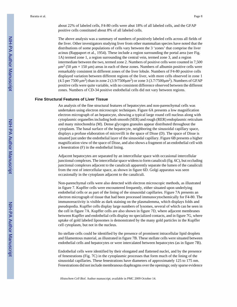
Fine Structural Features of Liver TissueAn analysis of the fine structural features of hepatocytes and non-parenchymal cells wasundertaken using electron microscopic techniques. Figure 6A presents a low magnificationelectron micrograph of an hepatocyte, showing a typical large round cell nucleus along withcytoplasmic organelles including both smooth (SER) and rough (RER) endoplasmic reticulumand many mitochondria (M). Dense glycogen granules appear distributed throughout thecytoplasm. The basal surface of the hepatocyte, neighboring the sinusoidal capillary space,displays a profuse elaboration of microvilli in the space of Disse (D). The space of Disse issituated just under the endothelial layer of the sinusoidal capillary. Figure 6B presents a highermagnification view of the space of Disse, and also shows a fragment of an endothelial cell witha fenestration (F) in the endothelial lining.
Adjacent hepatocytes are separated by an intercellular space with occasional intercellularjunctional complexes. The intercellular space widens to form canaliculi (fig. 6C), but occludingjunctional complexes adjacent to the canaliculi apparently separate the lumen of the canaliculifrom the rest of intercellular space, as shown in figure 6D. Golgi apparatus was seenoccasionally in the cytoplasm adjacent to the canaliculi.
Non-parenchymal cells were also detected with electron microscopic methods, as illustratedin figure 7. Kupffer cells were encountered frequently, either situated upon underlyingendothelial cells or as part of the lining of the sinusoidal capillaries. Figure 7A presents anelectron micrograph of tissue that had been processed immunocytochemically for F4-80. Theimmunoreactivity is visible as dark staining on the plasmalemma, which displays folds andpseudopodia. Kupffer cells display large numbers of lysomes, several of which can be seen inthe cell in figure 7A. Kupffer cells are also shown in figure 7D, where adjacent membranesbetween Kupffer and endothelial cells display no specialized contacts, and in figure 7G, whereuptake of gold labeled liposomes is demonstrated by the many gold particles in the Kupffercell cytoplasm, but not in the nucleus.
Ito stellate cells could be identified by the presence of prominent intracellular lipid dropletsand filamentous material, as illustrated in figure 7B. These stellate cells were situated betweenendothelial cells and hepatocytes or were intercalated between hepatocytes (as in figure 7B).
Endothelial cells were identified by their elongated and flattened nuclei, and by the presenceof fenestrations (Fig. 7C) in the cytoplasmic processes that form much of the lining of thesinusoidal capillaries. These fenestrations have diameters of approximately 125 to 175 nm.Fenestrations did not include membranous diaphragms over the openings; only sparse evidence
Baratta et al. Page 8
Histochem Cell Biol. Author manuscript; available in PMC 2009 October 14.
NIH
-PA Author Manuscript
NIH
-PA Author Manuscript
NIH
-PA Author Manuscript
of a very rudimentary basal lamina was seen associated with endothelial cells. At sites wherecell processes from apparent adjacent endothelial cells meet, specialized adhesive intercellularjunctional complexes are seen (Fig. 7E). Adhesive contacts are not seen between endothelialcells and hepatocytes or any other non-parenchymal cell. Finally, pit cells are occasionallyseen, as illustrated in figure 7F.
DiscussionOverview
The data presented here provide an overview of the histological and fine structural organizationof the mature mouse liver. It is clear that the fundamental features of the mouse liver areremarkably similar to the features of liver from other mammalian species. On the one hand,the clear similarities may detract from the noteworthiness of this manuscript; the mouse liverdisplays the same cellular and subcellular constituents as do the livers of other mammalianspecies. On the other hand, however, the data presented in this manuscript are vitally importantfor the interpretation and understanding of other results, using either wild type or geneticallyengineered mice, and provide a solid reference point against which other data can beinterpreted.
Although the very sparse connective tissue within the mouse liver makes recognition of itslobular structure (e.g., Rappaport et al., 1954) a challenge, the major features of the lobules,including the positions of portal areas and central venules, are recognizable (Fig. 1A; 1C). Thetypes of cells that comprise the mouse liver are similar to those that have been described inother mammalian species. The prominent cell type is the hepatocyte, characterized by thepresence of intracellular albumin (Bernuau et al., 1985; Yokota et al., 1981) or biotin. Alsofound are phagocytic Kupffer cells (von Kupffer, 1898), labeled with the F4-80 antibody(Naito et al, 1997), the numbers of which are approximately 35–40% of the number ofhepatocytes. Also studied were the Ito stellate cells (Fahimi, 1982; Gard et al., 1985; Ito,1973; Neubauer et al., 1996), whose numbers are about 8–10% of the number of hepatocytes.As with any organ, endothelial cells form much of the lining of the sinusoidal capillaries.Although the thin squamous endothelial cells do not contribute a great deal to the volume oftissue in the liver, the number of nuclei is approximately 22% of all liver nuclei andapproximately 40% of the number of hepatocytes. The relative proportions of these cells aresimilar to figures reported for other species (Bouwens et al., 1986; Geerts, 2001; Marcos et al.,2004; Naito et al., 1997; Sato et al., 2003). Further, the biliary system of the mouse liver appearssimilar to that of other mammalian species, with the presence of canaliculi between adjacenthepatocytes and small bile ductules that form part of portal areas. Thus, although the presentinvestigation did not reveal novel features of liver organization in the mouse, the value of thisstudy is that it establishes baseline data for the mouse and demonstrates the similarities of thehistological organization of the mouse liver and of other mammalian species. These dataunderscore the value of the mouse model for laboratory studies of liver structure and function.
Liver Cell TypesParenchymal cells in the liver are the hepatocytes, while non-parenchymal cells include theKupffer cells, Ito stellate cells, wandering leukocytes (including a population of lymphocytes,termed the ‘pit’ cells (Bouwens et al., 1992), and endothelial cells.
Different cell types in the liver were identified based upon their morphological features andby their immunoreactivity. Hepatocytes have long been recognized in H&E stained materialas round or cuboidal cells, and recent studies have demonstrated that they contain mRNA forexpressing the albumin gene and also are immunoreactive for albumin (Bernuau et al., 1985;Yokota et al., 1981). The present results from mouse are fully compatible with these previous
Baratta et al. Page 9
Histochem Cell Biol. Author manuscript; available in PMC 2009 October 14.
NIH
-PA Author Manuscript
NIH
-PA Author Manuscript
NIH
-PA Author Manuscript

reports. The present results indicate that albumin positive hepatocytes are encounteredapproximately three times more frequently than are Kupffer cells, and about six to seven timesmore frequently than Ito stellate cells.
Further, the present studies demonstrate that hepatocytes also contain biotin. Biotin appearsnot to be synthesized by hepatocytes but rather is taken up from food or produced by intestinalflora (Bowman and Russell, 2006), and is stored in the liver to support its role in metabolism.The presence of biotin within hepatocytes clearly is important for considerations of itsmetabolic role, but may also have importance in issues of immunocytochemical methodology.Immunocytochemical studies that use avidin-biotin binding as a step in localizing some antigenof interest may yield false positive results due to avidin binding to endogenous biotin ratherthan to biotinylated secondary antibodies (Ramos-Vara, 2005).
The fine structural features of mouse hepatocytes are similar to those reported for othermammalian species. Large round nuclei, and occasionally two nuclei, are prominent. Much ofthe cytoplasm is occupied by mitochodria, both smooth and rough endoplasmic reticulum, andscattered glycogen particles. The basolateral portion of the hepatocyte, which faces thesinusoidal capillaries, displays a rich microvillous elaboration of the plasmalemma; thismicrovillous border occupies much of the space of Disse. As in other mammalian species, theapical portion of hepatocytes is associated with canaliculi. Canaliculi are formed by a wideningof the intercellular space, although the canalicular lumens are set off from the intercellularspace by occluding junctions. Small profiles of Golgi apparatus were seen occasionally in theapical cytoplasm close to canaliculi, but we did not detect secretory vesicles associated withcanaliculi. The canaliculi are believed to form an anastomosing system, which lead eventuallythrough ducts of Hering into small bile ductules in the portal areas. These small bile ducts area consistent feature in the portal areas.
Monocyte derived macrophages are found in virtually every organ and tissue of the body, andcomprise the diffuse reticulo-endothelial system (Aschoff, 1924; Furth et al., 1972). In the liverthese macrophages are termed Kupffer cells (von Kupffer, 1898; Widmann et al., 1972).Although originally, these phagocytic cells were likely confused with the stellate cells thatwould later be identified by Ito (1973), later studies demonstrated that Kupffer cells can beidentified by their ability to phagocytose tracer substances, including carbon, India ink, or latexmicrospheres, and also by their immunoreactivity to the F4-80 antibody. The present studiesdemonstrated the presence of F4-80 positive Kupffer cells in mouse liver, and electronmicroscopic studies demonstrated this antibody labels a cell surface marker. Further, the resultsfrom studies of double labeling with fluorescently labeled latex microspheres and also fromimmunocytochemistry have demonstrated conclusively that the Kupffer cells are a populationof cells distinct from the Ito stellate (fat storing) cells.
The use of latex microspheres of different diameters was useful in demonstrating that Kupffercells could be labeled specifically with larger (0.5 μm) microspheres, while smallermicrospheres (0.03 μm) labeled both Kupffer cells and endothelial cells, as demonstratedpreviously (Wake et al., 1989).
Other investigators (Bouwens et al., 1986; 1992; Sleyster and Knook, 1982) have noted thatKupffer cells are not distributed homogenously in the liver and appear to show some variationin regard to their phagocytic activity. These authors (Sleyster and Knook, 1982; Bouwens etal., 1992; Bouwens et al., 1986) have reported that Kupffer cells are more frequentlyencountered and also are larger in regions around the portal areas than around the centralvenules. The present data corroborate this finding in the mouse, although the regionaldifferences in the mouse liver appear not as great as the regional differences reported for ratliver.
Baratta et al. Page 10
Histochem Cell Biol. Author manuscript; available in PMC 2009 October 14.
NIH
-PA Author Manuscript
NIH
-PA Author Manuscript
NIH
-PA Author Manuscript

Ito stellate cells are fat storing cells of the liver, and in the present studies these were identifiedby immunoreactivity to glial fibrillary acidic protein (GFAP) (Gard et al., 1985; Neubauer etal., 1996). Stellate cells are identifiable by their fine structural features of prominentintracellular lipid droplets and by cytoplasmic filamentous material. The intracellularfilamentous material likely forms the basis of their immunoreativity to GFAP and to desmin(Leo and Lieber, 1983; Yokoi et al., 1984; de Bleser et al, 1991). Quantitative estimates (Leoand Lieber, 1983; Yokoi et al., 1984; de Bleser et al, 1991) indicated that numbers of Ito stellatecells in rat liver were about 10–12% of hepatocytes, a fraction similar to the present results inmouse. Further, de Bleser et al. (1991) reported that stellate cells were found more frequentlyin peri-portal areas than in peri-central areas. The present study noted that stellate cells werenot distributed homogenously throughout the liver, but a consistent pattern between peri-portaland peri-central regions was not detected.
Endothelial cells are an important cell type in any organ, and certainly so in the liver. Liverendothelial cells are specialized, with the presence of fenestrations that appear aggregated intogroups that form ‘sieve plates’ (Wisse, 1972). The very sparse nature of a basal lamina beneaththe endothelial cells, along with the absence of diaphragmatic coverings of the fenestrations,allows for apparent relatively free movement of small molecules (less than 125 nm diameter)between the space of Disse and the capillary lumen. Wisse has demonstrated both small bristle-coated micropinocytotic vesicles and large smooth macropinocytotic vesicles in the endothelialcells. Small latex microspheres, diameter of 30 nm, are not detected in hepatocytes afterintravascular injection, although they do appear to label endothelial cells. This suggests thatthe latex microspheres are taken up by endothelial vesicles, but either do not reach the spaceof Disse or are not taken up by the hepatocyte microvillous border in the space of Disse.
Double Nucleated CellsThe present analysis indicated that a considerable portion of hepatocytes are double nucleated.The percentage gleaned from studies of tissue slices is likely to be an underestimate, however,because although two nuclei in the X or Y planes would be detected easily, if a second nucleuswas obscured in the Z plane or found within an adjacent section, it would be missed by thetechniques employed in this study. Another approach taken in the present studies was todissociate liver cells, and use the Trypan blue stain to detect double nucleated cells. Thisanalysis yielded higher numbers, of approximately 35% double nucleated cells. These resultsare in agreement with other recent studies (Wheatley, 1972; Gupta, 2000) that have reportedthat the population of hepatocytes includes many double nucleated cells. The functionalsignificance of double nucleated cells, however, is not clear.
No evidence was revealed to indicate that any other cell type within the liver included doublenucleated cells. Analysis of tissue sections immunolabeled with F4-80 for Kupffer cells or withGFAP for Ito stellate cells revealed no evidence of more than one DAPI stained nucleus.Kupffer and stellate cells probably were not detected in the dissociated cell studies, as differentsteps in the cell dissociation process need to be taken to include these cell populations(Smesdrod et tal., 1985; Riccalton-Banks et al., 2003). The thin squamous endothelial cellsdisplay extensive spread of cytoplasm and membranes, making the detection of two nucleiwithin one cell a very difficult endeavor. We are not aware of any examples of double nucleatedendothelial cells, and believe they are not likely to exist.
ConclusionGenetically engineered mice will play a very important role in future studies of liver function,and so it is vitally important to have baseline reference information on the cellular makeup ofnormal mouse liver. The present paper, using histological, quantitative immunocytochemical,and fine structural analyses, demonstrates that the cellular organization of the mouse liver is
Baratta et al. Page 11
Histochem Cell Biol. Author manuscript; available in PMC 2009 October 14.
NIH
-PA Author Manuscript
NIH
-PA Author Manuscript
NIH
-PA Author Manuscript
quite similar to that of other mammalian species, confirming that the mouse presents a usefulanimal model for studies of liver structure and function.
AcknowledgmentsSupported by NIH grant EB-003075
Reference ListAbercrombie M. Estimation of nuclear population from microtome sections. Anat Rec 1946;94:239–247.Aschoff L. Das Reticulo/endotheliale system. Ergebn Med Kinderheilk 1924;26:1–118.Austyn JM, Gordon S. F4/80, a monoclonal antibody directed specifically against the mouse macrophage.
Eur J Immunol 1981;11:805–815. [PubMed: 7308288]Bartök I, Töth J, Remenar E, Viragh S. Fine structure of perisinusoidal cells in developing human and
mouse liver. Acta Morphol Hung 1983;31:337–352. [PubMed: 6421096]Bernuau D, Poliard A, Tournier I, Sala-Trepat J, Feldmann G. All hepatocytes are involved in the
expression of the albumin gene in the normal adult rat: a demonstration by in situ hybridization andimmunoperoxidase techniques. Cell Biol Int Rep 1985;9:31–42. [PubMed: 2579741]
Blouin A, Bolender RP, Weibel ER. Distribution of organelles and membranes between hepatocytes andnonhepatocytes in the rat liver parenchyma. A stereological study. J Cell Biol 1977;72:441–455.[PubMed: 833203]
Bouwens L, Baekeland M, DeZanger R, Wisse E. Quantitation, tissue distribution and proliferationkinetics of Kupffer cells in normal liver. Hepatology 1986;6:718–722. [PubMed: 3733004]
Bouwens L, DeBleser P, Vanderkerken K, Geerts B, Wisse E. Liver cell heterogeneity: functions of non-parenchymal cells. Enzyme 1992;46:155–168. [PubMed: 1289080]
Bowman, BA.; Russell, RM. Present Knowledge in Nutrition. Vol. 9. Vol. 1. Washington DC: Int LifeSci Inst; 2006. Biotin.
David H. The hepatocyte. Development, differentiation, and ageing. Exp Pathol Suppl 1985;11:1–148.[PubMed: 2484913]
De Bleser P, Geerts A, Wisse E. Role of fat-storing cells in hepatic fibrogenesis. Retinoids as possibletherapeutic agents. Alcohol Alcohol Suppl 1991;1:3345–350.
Fahimi, HD. Sinusoidal endothelial cells and perisinusoidal fat-storing cells: structure and function. In:Arias, IM.; Popper, H.; Schachter, D.; Shafritz, DA., editors. The Liver: Biology and Pathobiology.Raven Press; New York: 1982. p. 495-506.
Furth, R.; von Cohn, ZA.; Hirsh, JG.; Humphry, JH.; Spector, WG.; Langevoort, HL. Bull WHO. Vol.46. 1972. The mononuclear phagocyte system: a new classification of macrophages, monocytes, andtheir precursors; p. 845-852.
Gard AL, White FP, Dutton G. Extra-neural glial fibrillary acidic protein (GFAP) immunoreactivity inperisinusoidal stellate cells of rat liver. J Neuroimmunol 1985;8:359–375. [PubMed: 3891783]
Geerts A. History, heterogeneity, developmental biology, and functions of quiescent hepatic stellate cells.Semin Liver Dis 2001;21:311–335. [PubMed: 11586463]
Geerts A, Bouwens L, Wisse E. Ultrastructure and function of hepatic fat-storing and pit cells. J ElectronMicrosc Tech 1990;14:247–256. [PubMed: 2187064]
Gupta S. Hepatic polyploidy and liver growth control. Sem Canc Biol 2000;10:161–171.Ito T. Recent advances in the study on the fine structure of the hepatic sinusoidal wall: a review. Gumma
Rep Med Sci 1973;6:119–163.Jones AL, Mills ES. Ultrastructural concepts of drug metabolism. I. The hepatocyte: structure and
function. Am J Drug Alcohol Abuse 1974;1:111–135. [PubMed: 4219713]Kawada N. The hepatic perisinusoidal stellate cell. Histol Histopathol 1997;12:1069–1080. [PubMed:
9302568]von Kupffer C. Über Sternzellen der Leber. Verhandl Anat Gesellsch 1898;12:8085.Leo M, Lieber C. Hepatic fibrosis after long-term administration of ethanol and moderate vitamin A
supplementation in the rat. Hepatology 1983;3:1–11. [PubMed: 6681608]
Baratta et al. Page 12
Histochem Cell Biol. Author manuscript; available in PMC 2009 October 14.
NIH
-PA Author Manuscript
NIH
-PA Author Manuscript
NIH
-PA Author Manuscript

Longmuir KJ, Robertson RT, Haynes SM, Baratta JL, Waring AJ. Effective targeting of liposomes toliver and hepatocytes in vivo by incorporation of a Plasmodium amino acid sequence. Pharm Res2006;23:759–769. [PubMed: 16550476]
Loud AV. A quantitative stereological description of the ultrastructure of normal rat liver parenchymalcells. J Cell Biol 1968;37:27–46. [PubMed: 5645844]
Marcos R, Monteiro RAF, Rocha E. Estimation of the number of stellate cells in a liver with the smoothfractionator. J Microsc 2004;215:174–182. [PubMed: 15315504]
McMillan PN, Hixson DC, Hevey KA, Naik S, Jauregui HO. Hepatocyte cell surface polarity asdemonstrated by lectin binding. J Histochem Cytochem 1988;36:1561–1571. [PubMed: 2848070]
Naito M, Hasegawa G, Ebe Y, Yamamoto T. Differentiation and function of Kupffer cells. Med ElectronMicrosc 2004;37:16–28. [PubMed: 15057601]
Naito M, Hasegawa G, Takahashi K. Development, differentiation, and maturation of Kupffer cells.Microsc Res Techn 1997;39:350–364.
Neubauer K, Knittel T, Aurisch S, Fellmer P, Ramadori G. Glial fibrillary acidic protein; a cell typespecific marker for Ito cells in vivo and in vitro. J Hepatol 1996;24:719–730. [PubMed: 8835748]
Ramos-Vara JA. Technical aspects of immunocytochemistry. Vet Pathol 2005;42:405–426. [PubMed:16006601]
Rappaport AM, Borrowy ZJ, Lougheed WM, Lotto WN. Subdivision of hexagonal liver lobules into astructural and functional unit; role in hepatic physiology and pathology. Anat Rec 1954;119:11–33.[PubMed: 13180999]
Riccalton-Banks L, Bhandari R, Fry J, Shakesheff KM. A simple method for the simultaneous isolationof stellate cells and hepatocytes from rat liver tissue. Molec Cell Biochem 2003;248:97–102.[PubMed: 12870660]
Robertson RT, Baratta JL, Haynes SM, Longmuir KJ. Liposomes incorporating a Plasmodium aminoacid sequence target heparan sulfate biding sites in liver. J Pharm Sci 2008;97:3257–3273. [PubMed:17932963]
Sato M, Suzuki S, Senoo H. Hepatic stellate cells: unique characteristics in cell biology and phenotype.Cell Struct Funct 2003;28:105–112. [PubMed: 12808230]
Senoo H. Structure and function of hepatic stellate cells. Med Electron Microsc 2004;37:3–15. [PubMed:15057600]
Sigal SH, Rajanshi P, Gorla GR, Saxena R, Sokhi RP, Gebhardt DF, Reid LM, Gupta S. Partialhepatectomy-induced polyploidy attenuates hepatocyte replication and activates cell aging events.Am J Physiol 1999;276:G1260–G1272. [PubMed: 10330018]
Sleyster EC, Knook DL. Relation between localization and function of rat liver Kupffer cells. Lab Invest1982;47:484–490. [PubMed: 6182391]
Smedsrod B, Pertoft H, Eggertsen G, Sundstrom C. Functional and morphological characterization ofcultures of Kupffer cells and liver endothelial cells prepared by means of density separation in Percol,and selective substrate adherence. Cell Tissue Res 1985;241:639–649. [PubMed: 2992796]
Smedsrod B, de Bleser PJ, Braet F, Lovisetti P, Vanderkerken K, Wisse E, Geerts A. Cell biology of liverendothelial and Kupffer cells. Gut 1994;35:1509–1516. [PubMed: 7828963]
Stöhr G, Deimann W, Fahimi HD. Peroxidase-positive endothelial cells in sinusoids of the mouse liver.J Histochem Cytochem 1978;26:409–411. [PubMed: 659841]
Wake K, Dicker K, Kirn A, Knkook DL, McCuskey RS, Bouwens L, Wisse E. Cell biology and kineticsof Kupffer cells in the liver. Int Rev Cytol 1989;118:173–229. [PubMed: 2691426]
Wheatley DN. Binucleation in mammalian liver. Exp Cell Res 1972;74:455–465. [PubMed: 4343021]Widmann JJ, Cotran RS, Fahmi HD. Mononuclear phagocytes (Kuffer cells) and endothelial cells.
Identification of two functional cell types in rat liver sinusoids by endogenous peroxidase activity. JCell Biol 1972;52:159–170. [PubMed: 4331297]
Wisse E. An electron microscopic study of the fenestrated endothelial lining of rat liver sinusoids. JUltrastruct Res 1970;31:125–150. [PubMed: 5442603]
Wisse E. An ultrastructural characterization of the endothelial cell in the rat liver sinusoid under normaland various experimental conditions, as a contribution to the distinction between endothelial andKupffer cells. J Ultrastruct Res 1972;38:528–562. [PubMed: 4335119]
Baratta et al. Page 13
Histochem Cell Biol. Author manuscript; available in PMC 2009 October 14.
NIH
-PA Author Manuscript
NIH
-PA Author Manuscript
NIH
-PA Author Manuscript

Wisse E. Observations on the fine structure and peroxidase cytochemistry of normal rat liver Kupffercells. J Ultrastruct Res 1974;46:393–426. [PubMed: 4363811]
Yamada M, Naito M, Takahashi K. Kupffer cell proliferation and glucan-induced granuloma formationin mice depleted of blood monocytes by strontium-89. J Leukoc Biol 1990;47:195–205. [PubMed:2307905]
Yokoi Y, Namihisa T, Kuroda H, Komatsu I, Miyazaki A, Wanatabe S, Usui K. Imunocytochemicaldetection of desmin in fat-storing cells (Ito cells). Hepatology 1984;4:709–714. [PubMed: 6204917]
Yokota S, Fahimi HD. Immunocytochemical localization of albumin in the secretory apparatus of ratliver parenchymal cells. Proc Natl Acad Sci USA 1981;78:4970–4974. [PubMed: 7029527]
Baratta et al. Page 14
Histochem Cell Biol. Author manuscript; available in PMC 2009 October 14.
NIH
-PA Author Manuscript
NIH
-PA Author Manuscript
NIH
-PA Author Manuscript

Fig. 1.Photomicrographs of 12 μm thick cryostat sections of mouse liver tissue. A: Bright field imageof an H&E stained section showing normal liver architecture, and components of basic liverlobules, with portal area and central venule. B: Another H&E stained section at highermagnification. C: Bright field image showing profiles in a portal area, including branch ofportal vein (pv), branch of hepatic artery (ha), and a small bile ductile (bd). D: Fluoresceinoptics showing Alexa 488 labeling of albumin immunoreactivity of hepatocytes; note the brightgreen packets of immunoreactivity in cytoplasm surrounding large round nuclei (white arrows)vacant of staining. E: Fluorescein optics showing Alexa 488 labeled F4-80 immunoreactivityof putative Kupffer cells. F: Alexa 488 labeled GFAP positive Ito stellate cells. G: Fluoresceinlabeled tomato lectin staining of cell borders including endothelial cells of sinusoidalcapillaries. cv: central venule; pa: portal area; pv: portal venule. Calibration bar in ‘A’ = 100μm; bar in ‘C’ = 25μm; bar in ‘G’ = 50 μm for ‘B’ and ‘D’ – ‘G’.
Baratta et al. Page 15
Histochem Cell Biol. Author manuscript; available in PMC 2009 October 14.
NIH
-PA Author Manuscript
NIH
-PA Author Manuscript
NIH
-PA Author Manuscript

Fig. 2.Fluorescence photomicrographs showing characteristics of endothelial cell labeling. A:Merged image showing Alexa 488 labeled CD-34 immunoreactivity (in green) and DAPIstained cell nuclei (in blue). B: Similar section as in ‘A’, but viewed at higher magnification.Note the many large round DAPI stained nuclei of hepatocytes. In addition, white arrowsindicate several smaller and ovoid DAPI stained nuclei associated with the CD-34 positiveendothelial cells. C: CD-34 immunoreactivity of endothelial cells of liver. D: Same section asin ‘C’ viewed under rhodamine optics, showing pattern of fluorescently labeled latex small(0.03 μm) microspheres. E: Same section as shown in ‘D’ and ‘E’, with merger of imagesviewed with fluorescein (green CD-34), rhodamine (red fluorescent latex microspheres) and
Baratta et al. Page 16
Histochem Cell Biol. Author manuscript; available in PMC 2009 October 14.
NIH
-PA Author Manuscript
NIH
-PA Author Manuscript
NIH
-PA Author Manuscript

ultraviolet (blue DAPI) optics. White arrow indicates the location of a putative endothelial cellnucleus, in ‘C’, ‘D’ and ‘E’. F: Merged image similar to ‘E’ but at higher magnification. Arrowsindicate two putative endothelial cell nuclei. Calibration bar in A = 100μm. Bar in E = 50μmfor C, D, and E. Bar in F = 50 μm for B and F.
Baratta et al. Page 17
Histochem Cell Biol. Author manuscript; available in PMC 2009 October 14.
NIH
-PA Author Manuscript
NIH
-PA Author Manuscript
NIH
-PA Author Manuscript

Fig. 3.Fluorescence photomicrographs showing cell specific labeling. A: Red Alexa 546 labeledantibody to albumin, demonstrating that virtually all cells containing round nuclei expressalbumin. B: Same section as ‘A’ viewed under fluorescein optics to show Alexa 488 labeledbiotin immunocytochemistry. C: Merged images from ‘A’ and ‘B’, demonstrating that virtuallyall cells containing round nuclei appear co-labeled with albumin and biotin. D: Sectionprocessed with Alexa 546 labeled antibody to GFAP; note the labeled cells with stellatemorphological features. E: Same section as shown in ‘D’, but viewed under fluorescein opticsand showing F4-80 labeled cells. F: Merged images from ‘D’ and ‘E’, demonstrating the twoimmunocytochemical procedures label distinctly separate populations of cells. G: Section from
Baratta et al. Page 18
Histochem Cell Biol. Author manuscript; available in PMC 2009 October 14.
NIH
-PA Author Manuscript
NIH
-PA Author Manuscript
NIH
-PA Author Manuscript

an animal that received intravenous injections of rhodamine labeled fluorescent latex larage(0.5 μm) microspheres, and then processed for F4-80 immunocytochemistry. Note redmicrospheres associated with green F4-80 labeled cells. H: Section from same animal as ‘G’,but processed for GFAP immunocytochemistry. Note red microspheres appear separate fromGFAP labeled cells. Calibration bar in F = 50μm for images A – F; calibration bar in H = 50μm for G and H.
Baratta et al. Page 19
Histochem Cell Biol. Author manuscript; available in PMC 2009 October 14.
NIH
-PA Author Manuscript
NIH
-PA Author Manuscript
NIH
-PA Author Manuscript

Fig. 4.Evidence for double nucleated cells in liver tissue. A. Merged image shows green fluoresceinlabeled tomato lectin labeling of cell borders and blue DAPI staining of nuclei in a liver tissuesection. White arrows indicate structures that appear to be cells with double nuclei. B: Mergedimage showing tissue labeled by a combination of tomato lectin binding (green) to reveal cellborders, and albumin immunocytochemistry (red) along with blue DAPI labeled nuclei. Whitearrows indicate several albumin positive cells within tomato lectin borders that display doublenuclei. C: Trypan blue stained dissociated liver cells. Note three cells with single nuclei (blackarrows) and two cells with double nuclei (white arrows). D: Electron micrograph of two nuclearprofiles within a liver cell. E: Merged images, showing cells double labeled with F4-80 (greenAlexa 488 label) and the blue DAPI labeled nuclei. F: Merged image showing cells doublelabeled with GFAP (green) and the blue DAPI labeled nuclei. White arrows in E and F indicatesingle nucleated cells. Calibration bar = 50 μm for ‘A’ and ‘B’, and for ‘E’ and ‘F’. Calibrationbar in ‘C’ = 25 μm; bar in ‘D’ = 5 μm.
Baratta et al. Page 20
Histochem Cell Biol. Author manuscript; available in PMC 2009 October 14.
NIH
-PA Author Manuscript
NIH
-PA Author Manuscript
NIH
-PA Author Manuscript

Fig. 5.Histograms summarizing results of counts of immunocytochemically identified cells withDAPI stained nuclei. The Y axis shows numbers of cells detected in a region with an area of48,000μm2, and thickness of 12 μm. Calculated numbers were adjusted using the method ofAbercrombie (1946). Data are mean ± s.e.m.
Baratta et al. Page 21
Histochem Cell Biol. Author manuscript; available in PMC 2009 October 14.
NIH
-PA Author Manuscript
NIH
-PA Author Manuscript
NIH
-PA Author Manuscript

Fig. 6.Electron micrographs of liver tissue. A: Relatively low magnification electron micrographshowing a portion of an hepatocyte. A large round nucleus of the hepatocyte (HN) is seen inthe lower part of the figure. Many profiles of rough endoplasmic reticulum (RER) can be seenthroughout the cytoplasm, often intermixed with mitochondria (M). The space of Disse (D)can be seen at the basal surface, just below processes of endothelial cells that border the lumen(Lu). B: Relatively high magnification electron micrograph shows a fenestration (F) in theendothelial cell (E) lining, over the space of Disse (D). C: Electron micrograph demonstratingthe inter-cellular space (arrows) and the presence of canaliculi between 2 adjacent hepatocytes.At this low magnification, intercellular occluding junctions are not visible. Nuclei ofhepatocytes are indicated (HN). D: Higher magnification electron micrograph illustrating acanaliculus, and adjacent occluding junctions (arrows).
Baratta et al. Page 22
Histochem Cell Biol. Author manuscript; available in PMC 2009 October 14.
NIH
-PA Author Manuscript
NIH
-PA Author Manuscript
NIH
-PA Author Manuscript

Fig. 7.Electron micrographs of non-parenchymal cells. A: Electron micrograph of a section processedimmunocytochemically for F4-80, showing cell surface immunoreactivity (black arrows) of aKupffer cell (K); the nucleus of the Kupffer cell (KN) is visible. White arrows indicate lysomeswithin the Kupffer cell. The Kupffer cell is situated in the sinusoidal lumen (Lu), above anhepatocyte (H). B: Micrograph showing an Ito stellate cell (I) situated between two hepatocytes(H) and an endothelial cell (E). The Ito cell is characterized by cytoplasmic lipid droplets (LD).C: High magnification electron micrograph showing the space of Disse (D) situated underfenestrated (F) endothelial processes (EP) above an hepatocyte (H) and below the capillarylumen (Lu). Also seen is a putative Ito cell process (IP). D: Close membrane association, butwithout specialized membrane attachments, between a Kupffer cell (K) and an endothelial cell(E). E: Adhering junctions (arrows) between processes of 2 endothelial cells (E), overlying thespace of Disse (D) and an hepatocyte (H). F: Pit cell with nucleus (PN) situated in a sinusoidallumen, adjacent to an endothelial cell (E). G: Micrograph showing gold labeled particlesassociated with engulfed liposomes in a Kupffer cell (Kupffer cell nucleus (KN) is indicated).
Baratta et al. Page 23
Histochem Cell Biol. Author manuscript; available in PMC 2009 October 14.
NIH
-PA Author Manuscript
NIH
-PA Author Manuscript
NIH
-PA Author Manuscript