Estrogen Stimulates Neuronal Nitric Oxide Synthase Protein Expression in Human Neutrophils
Upload
independentCategory
view
1download
0
Nitric oxide synthase localization in the rat neutrophils:immunocytochemical, molecular, and biochemical studies
R. Saini,* S. Patel,* R. Saluja,* A. A. Sahasrabuddhe,† M. P. Singh,‡ S. Habib,† V. K. Bajpai,§
and M. Dikshit*,1
*Cardiovascular Pharmacology Unit, §Electron Microscopy Unit, and †Division of Structural and Molecular Biology,Central Drug Research Institute, Lucknow, India; and ‡Industrial Toxicology Research Centre, Lucknow, India
Abstract: Nitric oxide (NO) modulates diversefunctions of polymorphonuclear neutrophils (PMNs),but localization of NO synthase (NOS) and identi-fication of its interacting proteins remain the leastdefined. The present study discerns subcellular dis-tribution of NOS and caveolin-1, a prominentNOS-interacting protein in rat PMNs. Localizationof NOS was explored by confocal and immunogoldelectron microscopy, and its activity was assessedby L-[3H] arginine and 4,5-diaminofluorescein di-acetate (DAF-2DA). Reverse transcriptase-poly-merase chain reaction using NOS primers andWestern blotting demonstrated the presence ofneuronal NOS (nNOS) and inducible NOS (iNOS)in PMNs. Immunocytochemical studies exhibiteddistribution of nNOS and iNOS in cytoplasm andnucleus, and L-[3H] citrulline formation and DAFfluorescence confirmed NOS activity in both frac-tions. NOS activity correlated positively with cal-modulin concentration in both of the fractions.nNOS and iNOS colocalized with caveolin-1, asevidenced by immunocytochemical and immuno-precipitation studies. The results thus provide firstevidence of nNOS and iNOS in the nuclear com-partment and suggest NOS interaction with caveo-lin-1 in rat PMNs. J. Leukoc. Biol. 79: 519–528;2006.
Key Words: polymorphonuclear leukocytes � nitric oxidesynthase � diaminofluorescein diacetate � caveolin-1 � nucleus �
subcellular localization
INTRODUCTION
Nitric oxide (NO) regulates several important functions ofpolymorphonuclear neutrophils (PMNs), including chemotaxis,adhesion, aggregation, apoptosis, and PMN-mediated bacterialkilling or tissue damage [1]. The presence of NO synthase(NOS) in the primary granules of PMNs has implied a pivotalcytotoxic role of NO in combating pathogens [2]. Several stud-ies from our laboratory have also demonstrated NO-mediatedmodulation of free radical generation from PMNs in variousphysiological and pathological conditions [3–8].
Although the presence of NO in the subcellular compart-ments can be of significance in modulating diverse targets, only
one study is available about the localization of NOS in intra-cellular locations by immunohistochemistry [2]. Inducible NOS(iNOS) and constitutive NOS activity and immunoreactivitywere associated with various subcellular compartments as wellas with the nucleus of endothelial cells [9], brown adipocytes[10], and astrocytes [11]. A recent report about the nuclearlocalization of biopterin (BH4) biosynthesizing enzymes,guanosine 5�-triphosphate cyclohydrolase I, 6-pyruvoyl-tetra-hydropterin synthase, and sepiapterin reductase is of consid-erable relevance to the availability of a redox-sensitive cofactorto support NO synthesis in the nuclear compartment [12].Although biochemical and molecular characteristics of NOSare well explored [5, 13, 14], there is no report available aboutNOS localization in rat PMNs. Although only neuronal NOS(nNOS) is constitutively expressed in rat PMNs, induction ofiNOS has been documented within 2 h after lipopolysaccharide(LPS) treatment [14]. Several splice variants of nNOS havebeen reported: nNOS�, a full-length protein has a PSD-95/discs large/ZO-1 homology (PDZ) domain, and nNOS� andnNOS� lack this domain [15]. Primers used by Greenberg et al.[14] did not verify the presence of PDZ domain in nNOS beingexpressed in rat PMNs.
Sequestration of NOS in intracellular compartments is aresult of various interacting proteins [16], which modulate itsenzymatic activity [17–19], spatial distribution [20], proximitywith regulators [21, 22], and intended targets [23]. Caveolin-1,a prominent NOS-interactive protein, negatively modulatesendothelial NOS (eNOS), nNOS, and iNOS by interfering withcalmodulin (CaM) binding [24–26]. The present study wasundertaken to investigate the hitherto unexplored subcellularlocalization of NOS isoforms and caveolin-1 in the rat-circu-lating PMNs.
MATERIALS AND METHODS
Reagents and antibodies
L-[3H] Arginine and goat anti-mouse and goat anti-rabbit protein A gold werefrom Amersham Pharmacia Biotech (Uppsala, Sweden). Uranyl acetate wasfrom Polaron Equipment Ltd. (Watford, UK). Monoclonal antibodies (mAb) to
1 Correspondence: Cardiovascular Pharmacology Unit, Central Drug Re-search Institute, Lucknow-226001, India. E-mail: [email protected]
Received June 15, 2005; revised September 23, 2005; accepted September29, 2005; doi: 10.1189/jlb.0605320.
0741-5400/06/0079-519 © Society for Leukocyte Biology Journal of Leukocyte Biology Volume 79, March 2006 519

nNOS (N-2280) and iNOS (N-9657) and all other chemicals were from Sigma-Aldrich Co. (St. Louis, MO).
Rabbit polyclonal antibodies against caveolin-1 (N-20: sc-894), iNOS(M-19: sc-650G), nNOS (K-20: sc-1025), and horseradish peroxidase goatanti-rabbit/donkey anti-goat were from Santa Cruz Biotechnology (CA). Goatanti-rabbit Oregon Green 488 (OG 488) and anti-mouse indocarbocyanin-5(Cy5) were from Molecular Probes (Eugene, OR). mAb to rat CD11b-fluores-cein isothiocyanate and CD45-phycoerythrin were from BD Biosciences (SanDiego, CA).
Animals
Male Sprague-Dawley rats (200–250 g) were obtained from the institute animalhouse. The animals were kept in a conventional housing facility and were usedin accordance with institutional ethical guidelines.
Neutrophil isolation
PMNs were isolated using the method of Yazdanbakhsh et al. [27] withmodifications. Buffy coat diluted with phosphate-buffered saline (PBS) con-taining tri sodium citrate 15 mM, 0.5% (w/v) bovine serum albumin (BSA), andglucose 10 mM was loaded over 1 ml Histopaque-1077 and centrifuged (1000g at 25�C) for 20 min. Original density of cells was restored by suspendingcells in rat plasma containing 5% (v/v) fetal calf serum for 30 min. PMNs wereseparated over Histopaque-1083 and -1119 gradients, washed with PBS,counted, and suspended in Hanks’ balanced salt solution [HBSS; composition(mM): NaCl 138, KCl 2.7, Na2HPO4 8.1, KH2PO4 1.5, glucose 10, pH7.2–7.4]. PMNs purity was �90% as assessed by CD11b/CD45; contaminatingcells in the PMN suspension were mostly lymphocytes.
Isolation of nuclei from PMNs and theirmorphological characterization
PMNs were fractionated according to Sanghavi et al. [28]. Briefly, cells(1�108cells/ml) were suspended in lysis buffer [composition (mM): HEPES10, NaCl 10, MgCl2 1.5, EDTA 1, dithiothreitol 5, 10% glycerol, and 0.15%Nonidet P-40 (NP-40), pH 7.4]. After brief vortexing and centrifugation (1700g at 4°C for 5 min), supernatant cytoplasmic fraction (CF) was collected.Nuclei fraction (NF) pellet was washed with lysis buffer (without NP-40) andfinally suspended in HBSS (equal to CF). Purity of NF was assessed by DNAcontent [29] and lactate dehydrogenase (LDH) activity [3]. Purity of NF and themorphological characterization of PMNs in buffy coat were identified undertransmission electron microscopy (TEM; FEI Philips Tecnai-12). Buffy coatand isolated nuclei were fixed overnight at 25°C with 2% (w/v) paraformalde-hyde and 1% (v/v) glutaraldehyde in PBS (pH 7.4). Fixed nuclei impregnatedin 4% buffered agarose were cut into smaller pieces using razor blades, washedthoroughly with PBS, post-fixed in buffered 1% osmium tetroxide for 3 h,dehydrated in ascending series of ethanol, and finally, embedded in Epon andAraldite plastic resin [30]. Ultrathin sections (80–100 nm) cut by Ultra Cutultramicrotome (Leica, Austria) were contrasted with uranyl acetate and leadcitrate and examined under TEM at 80 kV.
Assessment of NOS activity
4,5-Diaminofluorescein diacetate (DAF-2DA) fluorescence
Cells (1�07/ml) were washed with HBSS, preincubated with NOS inhibitor3-bromo 7-nitroindazole (7-NI; 1 mM), NOS substrate L-arginine (1 mM), orvehicle for 30 min at 37°C, and subsequently, incubated with DAF-2DA (10�M) for 5 min. DAF-loaded PMNs were dispensed on 0.01% (w/v) poly-L-lysine-coated coverslips, and NO production was visualized [31] using BioRadRadiance 2100 laser-scanning system equipped with a Nikon Eclipse TE 300confocal microscope, with a Plan Apo 60�/1.4 nicotinic acid oil immersionobjective. Excitation of Cy5, OG 488, and Hoechst 33258 was performed using637 nm red diode, 488 nm argon ion, and 405 nm blue diode lasers, andemitted light was detected through HQ660 longpass and HQ515/30 andHQ442/45 bandpass filters, respectively. Data were captured with BioRadLaserSharp 2000 Software 5.1, and photographs were processed using AdobePhotoshop software. Cells without DAF-2DA treatment or fixed cells (1 h with4% paraformaldehyde), incubated with DAF-2DA, were used as negativecontrols.
L-[3H] citrulline formation
Total NOS activity (in the presence of 1 mM calcium) and calcium-indepen-dent activity (in the absence of calcium and in the presence of 1 mM EGTA)were estimated [4] in PMNs (1�107cells) CF and NF, incubated in buffer[composition (mM): HEPES 25, NaCl 140, KCl 5.4, MgCl2 1, CaCl2 1, 7 �g/mlpepstatin A, 1.5 �g/ml soybean trypsin inhibitor, 5 �g/ml leupeptin, phenyl-methylsulfonyl fluoride (PMSF) 0.5 mM, and benzamidine 0.1 mM, pH 7.4]with cofactors [flavin adenine dinucleotide (FAD) 5 �M, flavin monouncleotide(FMN) 25 �M, reduced nicotinamide adenine dinucleotide phosphate(NADPH) 1 mM, BH4 10 �M, and 10 �g/ml CaM], and the L-[3H] arginine(0.16 �M) at 37°C. The reaction was terminated after 30 min by stop buffer[composition (mM): NaCl 118, KCl 4.7, KH2PO4 1.18, NaHCO3 1, EDTA 4,N-nitro-L-arginine methyl ester 1, pH 5.5] and was passed through a Dowex50W-X8 column (100–200 mesh, Na form). L-[3H] Citrulline was collectedin the unbound wash fraction, and bound L-[3H] arginine was eluted with 0.5M ammonium hydroxide to obtain citrulline formed from total arginine addedin each tube. To investigate the effect of CaM, NOS activity was estimated inCF and NF in the presence of calcium and 10 or 100 �g/ml CaM.
Flow cytometry
PMNs (2�106cells/ml) or nuclei were incubated for 30 min with L-arginine (1mM), 7-NI (1 mM), or vehicle at 37°C and subsequently, loaded with DAF-2DA (10 �M) for 30 min [7]. NO generation was measured by acquiring 10,000cells/nuclei from each sample and was analyzed by the Cell Quest program.
Assessment of NOS expressionReverse transcriptase-polymerase chain reaction (RT-PCR)
Total RNA from PMNs and brain samples was extracted using Trizol reagent(Invitrogen, Carlsbad, CA). RNA (5 �g) was reverse-transcribed with Super-Script II RT (Invitrogen) using oligo(dT) primers, as per the manufacturer’sinstructions. The cDNA was amplified in separate PCRs for NOS isoforms. ThePCR products were analyzed by electrophoresis on a 1.2% agarose gel andvisualized with ethidium bromide staining. The primers designed with aPrimer Select module of Lasergene (DNAStar Inc., Madison, WI) for nNOS(5�-AACGATCGGCCCTTGGTAGAC-3�; 5�-GGGCGGAGCTTTGTGCGAT-TTG-3�) amplified a 543-base pair (bp) fragment, iNOS (5�-GGACCACCTC-TATCAGGAA-3�; 5�-CCTCATGATAACGTTTCTGGC-3�) amplified a 314-bpfragment, and eNOS (5�-TGCTGCCCGAGATATCTTCAGT-3�; 5�-GGCTGC-CTTTTTCCAGTTGTTC-3�) amplified a 356-bp fragment. The amplificationreactions for 30 cycles were denaturation, 94°C, 30 s; annealing, 49°C, 60°C,and 65°C, 1 min for nNOS, iNOS, and eNOS, respectively; and extension,72°C, 1 min. RNA from normal and LPS-treated rat brain samples was used aspositive controls.
Western blotting (WB) and immunoprecipitation (IP)
PMNs lysate [composition of lysis buffer (mM): NaCl 100, Tris-HCl 10, EDTA1, 1 �g/ml aprotinin, 100 �g/ml PMSF, 20 �g/ml pepstatin, and sodiumorthovanadate 2 mM, pH 7.4], protein was loaded on 15% (for caveolin-1) or8% (for NOS) sodium dodecyl sulfate-polyacrylamide gel electrophoresis (SDS-PAGE) and transferred onto a polyvinylidene difluoride membrane. Blockingwas done overnight [PBS; composition (mM): NaCl 137, KCl 27, Na2HPO4 32,K2HPO4 15, KH2PO4 15, pH 7.4, containing 5% (w/v) BSA], and antibodyincubation was for 3 h with polyclonal caveolin-1 and NOS antibodies [1:1000diluted in PBS containing 0.1% (v/v) Tween-20 and 1% (w/v) BSA]. Thespecific bands were detected by enhanced chemiluminescence (Sigma-AldrichCo.), and blots were exposed to the Hyperfilms (Amersham Pharmacia Bio-tech).
The cells were lysed in radio IP assay buffer [PBS containing 0.1% SDS,0.5% sodium deoxycholate, 1% Triton X-100, and protease inhibitors (as inlysis buffer)] for 15 min. The supernatant was precleared with protein A/Gagarose (Santa Cruz Biotechnology) and incubated with 0.2 �g primary anti-body; after 2 h, 20 �l protein A/G agarose was added and incubated for 1 h.The beads were washed, resuspended, and subsequently analyzed by WB. Thesame concentration of normal immunoglobulin G (IgG) was used in controls forWB and IP.
Confocal microscopy
Subcellular distribution of nNOS, iNOS, and caveolin-1 was examined in fixedPMNs. The cells were fixed in 4% (w/v) paraformaldehyde in PBS (pH 7.4) for
520 Journal of Leukocyte Biology Volume 79, March 2006 http://www.jleukbio.org
30 min at 25°C and washed two times for 5 min each with PBS containing 0.5%(w/v) glycine. The washed cells were allowed to adhere on 0.01% (w/v)poly-L-lysine-coated coverslips, permeabilized with 0.2% (v/v) Triton X-100(5–10 min), and blocked with 10% goat serum in PBS for 30 min. Cells wereincubated overnight at 4°C with mAb against iNOS/nNOS and polyclonalantibodies against caveolin-1 at a dilution of 1:200 and were subsequentlystained at 4°C for 4 h with Cy5 and OG 488-tagged secondary antibodies(1:500), respectively. Nuclei were stained with Hoechst 33258 dye (3 �g/ml)at 25°C for 15 min. Control samples were processed separately by incubatingwith normal rabbit, mouse, and goat sera or by omitting primary antibodies.Coverslips were mounted in the fluorescence-mounting media (Oncogene, SanDiego, CA), and images were acquired as mentioned above. Red, green, andblue images were collected separately with excitations at 647 nm, 488 nm, and405 nm, respectively, to avoid bleed-through and merged for presentation(Adobe Photoshop software, San Jose, CA).
Immunogold electron microscopy
Buffy coat (obtained by centrifugation of blood at 2000 rpm for 20 min at 25°C)was fixed overnight with 2% (w/v) paraformaldehyde and 0.5% (v/v) glutaral-dehyde in PBS (pH 7.4) at 25°C. Small pieces of buffy coat were washed withPBS containing 0.5% (w/v) glycine to quench excess fixative, dehydrated in anascending series of ethanol, impregnated in London resin-White resin, andsubsequently polymerized at 60°C for 48 h. Ultrathin sections (80–100 nm)collected on nickel grids [32] were blocked in PBS containing 0.1% BSA (w/v),0.1% (v/v) teleost fish gelatin, and 0.05% (v/v) Tween-20 for 30 min andincubated with nNOS or iNOS mAb at 1:500 dilution in blocking buffer for 4 hat 4°C, and subsequently, the sections were incubated with 5/10/15 nmgold-coupled goat anti-mouse IgG (1:20) for 2 h at 37°C and examined underTEM at 80 kV.
In the dual-labeling experiments, ultrathin sections were incubated withpolyclonal caveolin-1 antibody (1:500) for 4 h at 4°C and subsequently, with10 nm gold-coupled goat anti-rabbit IgG (1:20) and then treated with 0.01%glutaraldehyde for 5 min to avoid leaching of gold particles. The excessglutaraldehyde was quenched with 0.5% (w/v) glycine, and sections wereincubated with nNOS or iNOS antibodies (1:500) as described above. Sectionswere washed with blocking buffer and treated with 15 nm gold-coupledanti-mouse IgG antibody (1:20) for 2 h at 37°C. The dual-labeled sections werecontrasted with uranyl acetate and examined under TEM. Control sampleswere processed separately by incubating with normal rabbit and mouse sera orby omitting primary immunoprobes.
Morphometric analysis was done in images captured at magnification11,000� from six grids of three independent experiments for nNOS or iNOS incytoplasm and nucleus [33]. Two gold particles positioned adjacent to eachother were also scored.
Statistical analysis
Results are mean � SEM of at least three to five independent experiments.Comparisons between two groups were performed by unpaired t-test withsignificance at P � 0.05.
RESULTS
NO generation, NOS activity, and NOS molecularcharacteristics evaluation in rat PMNs
NO formation assessment by DAF fluorescence
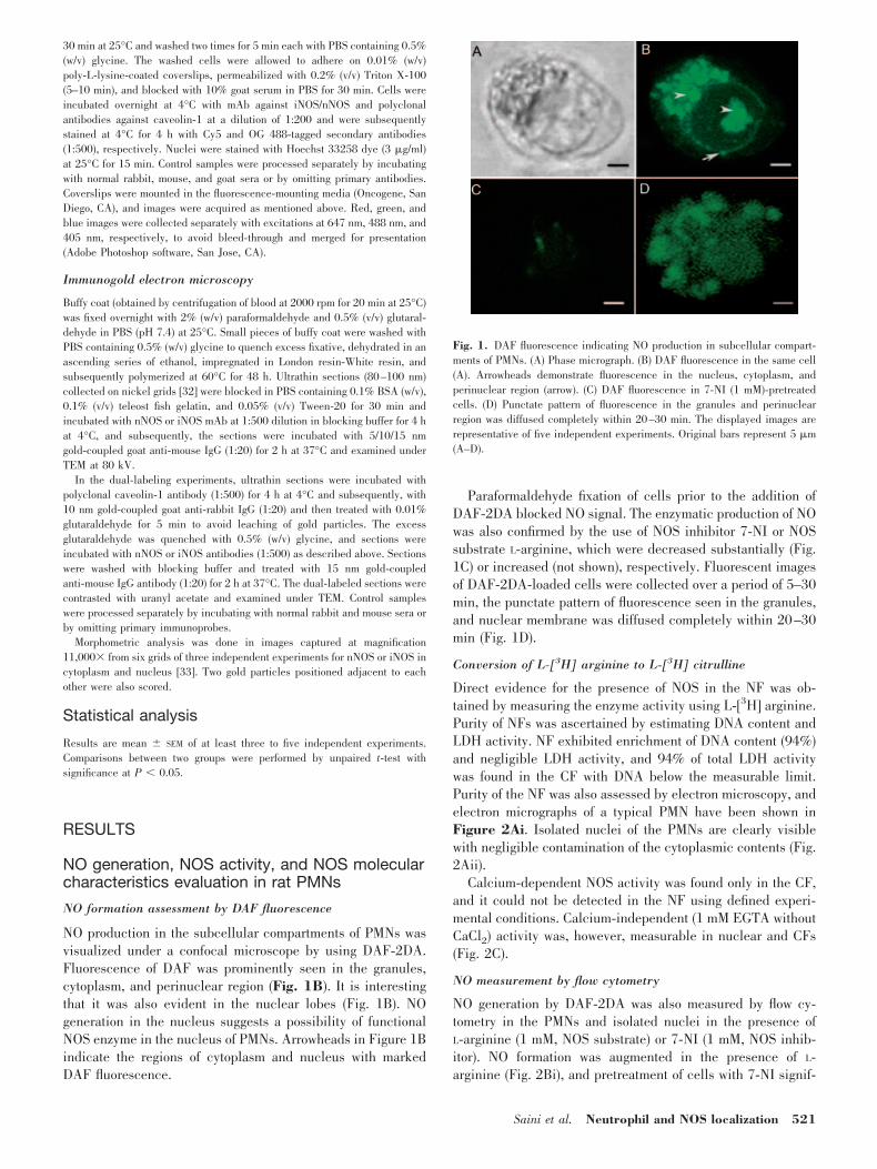
NO production in the subcellular compartments of PMNs wasvisualized under a confocal microscope by using DAF-2DA.Fluorescence of DAF was prominently seen in the granules,cytoplasm, and perinuclear region (Fig. 1B). It is interestingthat it was also evident in the nuclear lobes (Fig. 1B). NOgeneration in the nucleus suggests a possibility of functionalNOS enzyme in the nucleus of PMNs. Arrowheads in Figure 1Bindicate the regions of cytoplasm and nucleus with markedDAF fluorescence.
Paraformaldehyde fixation of cells prior to the addition ofDAF-2DA blocked NO signal. The enzymatic production of NOwas also confirmed by the use of NOS inhibitor 7-NI or NOSsubstrate L-arginine, which were decreased substantially (Fig.1C) or increased (not shown), respectively. Fluorescent imagesof DAF-2DA-loaded cells were collected over a period of 5–30min, the punctate pattern of fluorescence seen in the granules,and nuclear membrane was diffused completely within 20–30min (Fig. 1D).
Conversion of L-[3H] arginine to L-[3H] citrulline
Direct evidence for the presence of NOS in the NF was ob-tained by measuring the enzyme activity using L-[3H] arginine.Purity of NFs was ascertained by estimating DNA content andLDH activity. NF exhibited enrichment of DNA content (94%)and negligible LDH activity, and 94% of total LDH activitywas found in the CF with DNA below the measurable limit.Purity of the NF was also assessed by electron microscopy, andelectron micrographs of a typical PMN have been shown inFigure 2Ai. Isolated nuclei of the PMNs are clearly visiblewith negligible contamination of the cytoplasmic contents (Fig.2Aii).
Calcium-dependent NOS activity was found only in the CF,and it could not be detected in the NF using defined experi-mental conditions. Calcium-independent (1 mM EGTA withoutCaCl2) activity was, however, measurable in nuclear and CFs(Fig. 2C).
NO measurement by flow cytometry
NO generation by DAF-2DA was also measured by flow cy-tometry in the PMNs and isolated nuclei in the presence ofL-arginine (1 mM, NOS substrate) or 7-NI (1 mM, NOS inhib-itor). NO formation was augmented in the presence of L-arginine (Fig. 2Bi), and pretreatment of cells with 7-NI signif-
Fig. 1. DAF fluorescence indicating NO production in subcellular compart-ments of PMNs. (A) Phase micrograph. (B) DAF fluorescence in the same cell(A). Arrowheads demonstrate fluorescence in the nucleus, cytoplasm, andperinuclear region (arrow). (C) DAF fluorescence in 7-NI (1 mM)-pretreatedcells. (D) Punctate pattern of fluorescence in the granules and perinuclearregion was diffused completely within 20–30 min. The displayed images arerepresentative of five independent experiments. Original bars represent 5 �m(A–D).
Saini et al. Neutrophil and NOS localization 521
icantly reduced NO generation. Similar assay in the NF ofPMNs also demonstrated an increase in NO synthesis in thepresence of NOS substrate, L-arginine, and attenuation in thepresence of NOS inhibitor 7-NI (Fig. 2Bii). It confirmed NOS-dependent NO synthesis in the NF.
Molecular characterization of NOS
To identify NOS isoforms in the PMNs, RT-PCR experimentswere conducted by isolating RNA. The presence of nNOS andiNOS was evident (Fig. 3B), and signal for eNOS was notdetected in the rat PMNs (Fig. 3D). Absence of eNOS (Fig. 3D)and presence of nNOS in PMNs were confirmed by using ratbrain as a positive control, and for iNOS, LPS-treated brain wasused as a positive control. WB experiments further confirmedthe presence of these isoforms in the PMN lysate (Fig. 3, A and
C). Level of nNOS expression in PMNs was low in comparisonwith the cerebellum. Primers used for nNOS detection indi-cated the presence of the PDZ domain in NOS, which wasfurther substantiated by WB using N-terminal nNOS antibody(K-20: sc-1025).
NOS subcellular localization byimmunocytochemistry
Electron microscopy
Exploration of NOS localization by immunoelectron micros-copy revealed the presence of nNOS and iNOS in varioussubcellular compartments in the PMNs. Experiments con-ducted with mAb as well as polyclonal antibodies (data notshown) exhibited specific labeling and indicated the presence
Fig. 2. Purity assessment, NO generation, and NOS activity in CF andNF fractions of PMNs. (A) Electron micrographs of (i) neutrophil/(ii) NF.Original bars represent 1.4�m (i) and 1 �m (ii). N, Nucleus; PG, primarygranules. (B) Flow cytometry experiment demonstrating NO generationfrom (i) PMNs and (ii) isolated nuclei. NO generation in (i) PMNs: a,without DAF; b, with DAF; c, L-arginine (1 mM); and d, 7-NI (1 mM)treatment. (ii) Nuclei were gated by using 7-aminoactinomycin D staining:a, without DAF; b, loaded with DAF-2DA; c, L-arginine (1 mM); and, d7-NI (1 mM). Nuclei suspension also contained cofactors (FAD, FMN,NADPH, BH4, and CaM). The histograms are representative of fiveindependent experiments. (C) Ca2-dependent and -independent NOSactivities in the CF and NF. Values are mean � SEM of at least fiveexperiments. *, P � 0.01, in comparison with calcium-independentactivity.
Fig. 3. Molecular characteristics of NOS in ratPMNs. WB of (A) nNOS: lane 1, rat cerebellum (20�g); lane 2, PMNs (60 �g); (C) iNOS: lane 1, 60 �g;lane 2, 100 �g PMN lysate; and (G) caveolin-1:lane 1, PMN lysate (60 �g); lane 2, fibroblast L929(60 �g). (B) RT-PCR products of NOS isozymes inrat PMNs. Lane 1, Molecular weight marker; lane 2,nNOS (20 �l); lane 3, iNOS (10 �l). (D) RT-PCRproduct of eNOS. Lane 1, Molecular weight marker;lane 2, eNOS in rat brain; lane 3, eNOS in ratPMNs. (E) Lane 1, nNOS (20 �l); (F) iNOS (20 �l)in caveolin-1 IP from PMN lysate.
522 Journal of Leukocyte Biology Volume 79, March 2006 http://www.jleukbio.org
of nNOS as well as iNOS isoforms at various locations. Noappreciable binding was seen in the case of labeling only withsecondary antibody with variable sizes of gold particles (10 and15 nm). Evaluation with 5 nm gold particles was, however,obscure or difficult to differentiate from electron-dense gran-ules scattered in the cytoplasm and heterochromatin region ofnuclear lobes. Experiments were therefore conducted with 10and 15 nm gold particles; it was found that immunolabelingwith 15 nm gold particles was distinct and more convenient formorphometric analysis.
iNOS and nNOS staining was clearly seen in the nuclearmembrane, nucleus (Figs. 4, B and C, and 5D) and nuclearpore. iNOS/nNOS immunostaining was quite prominent alongthe plasma membrane (Figs. 4B and 5C) and in primarygranules (Figs. 4D and 5E). The higher magnification clearlyshowed gold particles corresponding to iNOS in the phagocyticcup (Fig. 4E), cytosol, and mitochondria (Fig. 4F). Compari-sons between nNOS and iNOS immunolabeling patterns re-vealed that nNOS was mostly localized in the cytoplasm, andnNOS immunoreactivity in the nucleus was significantly lessthan iNOS. Negligible background labeling with no apparentstaining in the primary granules, cytoplasm, and nucleus wasobserved in the controls (Figs. 4A and 5A).
NOS immunolabeling was quite evident as dimers (Figs. 4Band 5B), and in many fields, it was in monomeric form. Wecounted the gold particles to represent nNOS and iNOS dis-tribution in 15 and 16 representative cells, respectively. Totalnumber of gold particles to represent iNOS distribution inPMNs was 86 � 5 (n 16), which was significantly more thannNOS (39�2, n 15, P�0.001). Among the isoforms, distri-bution of nNOS was, however, different than iNOS. nNOS wasmuch more in the cytoplasmic compartments (almost 70%),and more than 50% iNOS was localized in the nuclear com-partment. Moreover, distribution of iNOS (4�1/cell) and nNOS
(5�2/cell) was similar in the azurophilic granules, and asso-ciation of nNOS (15�1/cell) with the plasma membrane wassignificantly higher than iNOS (8�2/cell, P�0.005).
Confocal microscopy
The presence of iNOS and nNOS in nuclear lobes of PMNs wasfurther assessed by immunofluorescent microscopy, which vi-sualized many cells to provide more information in the sameexperiment. iNOS and nNOS showed plasmalemmal, cytoplas-mic, as well as nuclear distribution. Three main sites could bevisualized clearly in these acquisitions. First, most of the cellsshowed diffused intracytoplasmic staining, although nNOSstaining intensity showed less variability, but iNOS stainingdiffered in intensity from cell to cell. Second, we found stainingin a discrete intracellular punctate pattern (Fig. 6, Aii andBii), consistent with a granular/vacuolar localization of NOS.Third, staining was clearly visible in the nuclear lobes but wasless than the cytoplasm (Fig. 6, Aii and Bii), which could be aresult of the less approachability of antibody in the nuclearlobes, as binding was done in permeabilized cells, unlikesections being used for electron microscopy. To determinewhether nNOS and iNOS were localized within the nucleus, westained nucleus as well as cells with NOS antibodies. Figure 6,Aii and Bii, provided convincing evidence of nNOS (red) andiNOS (red) in the nuclear lobes (as shown by arrowheads) ofPMNs. Preimmunized, serum-related immunoreactivity was notdetected in negative control samples (Fig. 6, Av and Bv).
IP and identification of NOS-interacting protein
As nNOS and iNOS interact with several proteins in variouscells, we explored the possibility of NOS interaction withcaveolin-1, an important NOS-interacting protein. We usedcaveolin-1 antibody for IP; following SDS-PAGE, the blotswere probed with nNOS or iNOS antibody. The presence ofboth NOS isoforms was seen (Fig. 3, E and F), thus suggestinginteraction of nNOS and iNOS with caveolin-1. The presence ofcaveolin-1 in PMN lysate was also evident by WB (Fig. 3G).
Fig. 4. Localization of NOS in PMNs by immunoelectron microscopy. Thepresence of iNOS (15 nm) in the nucleus, PG (azurophilic), plasma membrane,nuclear membrane, and cytosol is shown. (A) Immunoreactivity with preim-munized sera was not detected in the control samples. (B) iNOS staining asmonomers and dimers (arrows). (C) iNOS in nucleus, (D) in PG, (E) inphagocytic cup (5 nm gold particles), and (F) in mitochondria. Original brsrepresent 100 mm (A), 0.5 �m (B), 200 nm (C), 20 mm (D), 2 �m (E), and 40nm (F).
Fig. 5. Distribution of nNOS in the rat PMNs. nNOS is prominently observedin cytoplasm, plasma membrane, nucleus, and nuclear membrane, along theplasma membrane and in primary granules. nNOS staining in (A) negativecontrol treated only with gold-labeled, secondary antibody; (B) cytoplasm,plasma membrane, nucleus, and nuclear membrane; (C) along the plasmamembrane; (D) nucleus and nuclear membrane; and (E) primary granuels.Original bars represent 500 nm (A), 1 �m (B), and 200 nm (C–E).
Saini et al. Neutrophil and NOS localization 523
NOS and caveolin-1 colocalization studies
To investigate iNOS and nNOS colocalization with caveolin-1in the PMNs, we used immunoelectron microscopy as well asconfocal immunofluorescence microscopy. Colocalization ofiNOS and nNOS with caveolin-1 was performed by using 15 nmgold-conjugated secondary antibodies to immunolabel iNOS ornNOS and 10 nm gold-conjugated secondary antibodies tolabel caveolin-1, which was mostly found as oligomers (Fig. 7,A and B) as clusters of two to four gold particles correspondingto caveolin-1, which were occasionally present adjacent to theplasma membrane, nuclear membrane, nucleus, and cyto-plasm. iNOS (15 nm gold particle) colocalization with caveo-lin-1 (10 nm gold particles) is seen in the cytoplasm (Fig. 7D)and inside the nucleus (Fig. 7C). As nNOS immunostainingwas always less as compared with iNOS, areas of colocalizationof nNOS with caveolin-1 were also relatively less in the cyto-plasmic and nuclear compartments. Single labeling was alsodone simultaneously with double-labeling experiments forcaveolin-1 and NOS by using 10 and 15 nm, respectively, toascertain any artifactually cross-labeling.
To further corroborate these findings, iNOS/nNOS colocal-ization with caveolin-1 was also evaluated by confocal micros-copy. In these experiments, OG 488-labeled secondary anti-body was used, as density of NOS or caveolin-1 was not high inthese cells. No fluorescence was observed when the cells were
incubated alone with the secondary antibody. Caveolin-1 an-tibody binding was quite marked as was immunofluorescencelabeling of iNOS (Fig. 6Aii) or nNOS (Fig. 6Bii). NOS andcaveolin-1 exhibited subcellular distribution in cytoplasm aswell as in the nucleus. A punctate pattern of fluorescence wasseen in the plasmalemma and cytoplasm with relatively lessstaining of NOS and caveolin-1 in the nucleus (Fig. 6, A andB). Nuclear distribution of caveolin-1 was more in comparisonwith NOS isoforms (Fig. 6, Aiii and Biii). Areas of colocaliza-tion (Fig. 6, Aiv and Biv) of the two proteins are clearly evidentas yellow in the merged images.
CaM-mediated modulation of NOS activity
NOS activity in the PMN lysates NF and CF was estimated inthe absence and presence of CaM (10 and 100 �g/ml; Fig. 6C).With a tenfold increase in CaM concentration, NOS activitywas augmented significantly in the nuclear (117%) as well as inthe CFs (140%; Fig. 6C), indicating possible activity modula-tion of NOS by caveolin-1 and CaM in the PMNs.
DISCUSSION
Intracellular messengers, including pleiotrophic molecule NO,modulate diverse signaling cascades in the various subcellular
Fig. 6. Colocalization of NOS isoforms with caveo-lin-1 and CaM-mediated modulation of NOS activityin CF and NF of PMNs. (A) iNOS immunolabeling(i), nucleus (blue; ii), iNOS (red; iii), caveolin-1(green; iv), merge of ii and iii (v) negative controlincubated with preimmunized sera. (B) nNOS immu-nolabeling (i), nucleus (blue; ii), nNOS (red; iii),caveolin-1 (green; iv), merge of ii and iii (v) negativecontrol for caveolin-1 with preimmunized sera. Ar-rowheads indicate the nuclear regions with clearlyvisible iNOS or nNOS staining. Arrows in mergedimages indicate colocalization (yellow) of NOS withcaveolin-1 in nucleus. Original bars represent 5 �m(A, B). (C) Histogram representing NOS enzymeactivity in CF and NF of PMNs. Addition of CaM(100 �g/ml) significantly augmented NOS activity.Values are mean � SEM of three independent exper-iments. *, P � 0.05; **, P � 0.01, in comparisonwith the absence of Ca2 and CaM; and #, P � 0.05,in comparison with the presence of CaM (10 �g/ml).
524 Journal of Leukocyte Biology Volume 79, March 2006 http://www.jleukbio.org
compartments to regulate PMN functions such as adhesion,chemotaxis, phagocytosis, respiratory burst, and apoptosis.NOS inhibition augmented the number of neutrophils in themouse bone marrow and blood, suggesting a regulatory role ofNO, even in hematopoiesis [34]. Specific functions of NO inneurons, skeletal muscles, and endothelial cells have beenattributed to NOS localization in a particular compartment,despite that NO can easily reach to distant targets by diffusion[25, 35, 36]. Such compartmentalized distribution of NOS hasnot yet been explored in the PMNs. The present study aims toinvestigate intracellular NOS expression in rat-circulatingPMNs by using a combination of biochemical, molecular, con-focal, and immunoelectron microscopy techniques.
The N-nitrosation of DAF, a NO detection dye yielding ahighly green fluorescent triazole form (DAF-2T), offers not onlythe advantages of specificity, sensitivity, and a simple protocolfor the direct NO detection but also sufficient resolution tolocate NO in the subcellular compartments. The observationsrecorded within 5–10 min after DAF addition exhibited NOformation in the granules, perinuclear region, cytosol, andnuclear lobes (Fig. 1B), which was significantly reduced in thepresence of NOS inhibitor 7-NI (Fig. 1C). Fluorescent imagescollected after 20–30 min did not show the punctate pattern offluorescence in the granules and nuclear membrane, rather acompletely diffused pattern was seen (Fig. 1D). In view of theNO diffusibility [37], a part of fluorescence in the nucleuscould be a result of its dispersion from cytoplasmic compart-ments. Experiments with DAF were suggestive of NO formation
in the subcellular compartments and prompted us to undertakesystematic evaluation of NOS localization in the PMNs.
Prior to the NOS distribution studies, molecular character-istics of NOS were verified by RT-PCR using isoform-specificprimers. The presence of nNOS and iNOS mRNA in the PMNswas seen (Fig. 3B), and eNOS mRNA could not be detected(Fig. 3D). nNOS and iNOS expression was further confirmed byWB (Fig. 3, A and C). Only one report was found in literatureabout the characterization of NOS isoforms in rat PMNs [14].The level of nNOS expression in PMNs was low in comparisonwith the cerebellum, which is in agreement with our finding[14]. Constitutive expression of iNOS in the present study was,though, surprising, as iNOS induction was observed by Green-berg et al. [14] within 2 h after LPS treatment or bacterialinfections, with no adverse effect on the nNOS expression. Wehad also observed a significant increase in NOS activity inPMNs after LPS treatment in rats [5]. Ambient bacterial loadcould be responsible for the constitutive presence of iNOS inthe present study. Controversy in the expression of NOS iso-forms in rat brain has also been documented [38]. A systematicstudy conducted by Keilhoff et al. [38] demonstrated detect-able mRNA for iNOS and nNOS in rat brain during embryonicdevelopment on Day 10, and proteins were detected only atDay 15; others, however, failed to observe a constitutive pres-ence of iNOS in rat brain. Human PMNs, which exhibit similarfunctional responses to the addition of NO donors [1], however,constitutively express nNOS and iNOS [14, 39]. Constitutivepresence of iNOS has been documented in some cells and isnot considered to be the only inducible activity [40]. PMNs, animportant part of innate immunity, offer protection againstmicrobes due to generation of reactive oxygen and nitrogenspecies. Constitutive expression of iNOS in PMNs is to supportNO synthesis in large amounts, and macrophages express iNOSonly after exposure to LPS [40].
Based on WB and RT-PCR experiments (Fig. 3, A and B),nNOS in rat PMNs seems to be a full-length PDZ domain-containing protein [15]. PDZ domains are protein-interactiondomains, which play an important role in the targeting ofproteins to specific membrane compartments and their assem-bly into supramolecular complexes. PDZ domains, though,share a common, three-dimensional structure but generally,differ in their binding specificities. PDZ proteins mostly inter-act with target proteins without disrupting the overall structureand ligand function as a result of their ability to bind short,extreme C-terminal sequences [41–43]. PDZ domain interac-tion with the carboxyterminal tail is critical for PSD-95, Kchannels, and the N-methyl-D-aspartate receptor clustering,suggesting the physiological relevance of proper subcellularlocalization of these proteins [23]. PDZ domain of syntrophinmediates association of nNOS with the dystrophin complex inthe skeletal muscles [35]. Receptor tyrosine phosphatase-like protein, phosphofructokinase, calcium/CaM-dependentATPase, and carboxy-terminal-binding protein [41, 42, 44] arealso some important PDZ-interacting proteins. The last twoproteins have a strong presence in the nuclear compartmentand regulate transcription. As nNOS in PMNs also possess aPDZ domain, it is likely to interact with PDZ domain-interact-ing proteins in PMNs to regulate some important functions. The
Fig. 7. Colocalization of NOS with caveolin-1 in rat PMNs. (A) Caveolin-1 (10nm) localization in rat PMNs by immunoelectron microscopy. Arrows indicateclearly visible caveolin-1 immunolabeling. (B) Caveolin-1 immunolabeling(oligomers) prominently seen in the nucleus, nuclear membrane (NM), andplasma membrane (PM). (C) Colocalization of iNOS (15 nm) with caveolin-1(10 nm) in the nucleus and (D) cytoplasm. Areas of colocalization shown aremagnified in insets. Original bars represent 100 nm (A) and 100 mm (B–D).
Saini et al. Neutrophil and NOS localization 525

results obtained thus warrant further investigations to identifythe NOS-interacting proteins in PMNs.
Distribution of nNOS and iNOS proteins was subsequentlyexplored in the fixed PMNs by confocal and immunoelectronmicroscopy. Differences in the expression of nNOS and iNOSwith distinct overlapping in their distribution in various intra-cellular compartments [azurophilic granules (Figs. 4D and 5E),mitochondria (Fig. 4F), endoplasmic reticulum (not shown),and phagocytic vacuoles (Fig. 4E)] and their prominent pres-ence in nucleus were evidenced (Figs. 4–6). NOS colocaliza-tion was documented with myeloperoxidase within primarygranules [2], which was also apparent in the present study(Figs. 4D and 5E). The presence of NOS in various subcellularcompartments such as mitochondria [45], cardiac sarcoplasmicreticulum [46], insulin secretary granules [47], and mast cellgranules [48] has been documented. Moreover, the presence ofNOS isoforms in the nucleus of the cultured neonatal cardio-myocytes [46], pancreatic �-cells [47], brown adipocytes [10],mast cells [48], and astrocytes [11] has also been reported.NOS distribution in the subcellular compartments might be torestrict NO signaling to specific sites to perform explicit func-tions. To delineate the significance of nuclear NOS in PMNs,its role/trafficking in the myeloid cell lineage with particularemphasis on neutrophil maturation should be investigated.
Moreover, studies about NOS in the nuclear compartmentdemonstrated distinct patterns. In the cultured astrocytes,nNOS translocation to the nucleus on seventh day lasted foronly 10 h, which was speculated to down-regulate iNOS tran-scription [11]. Augmentation in the intracellular calcium levelduring mast cell activation further enhanced eNOS transloca-tion to the nucleus, but nNOS distribution remained unaltered[48]. All the studies conducted so far have remained specula-tive to define the role of nuclear NOS [10, 11, 46–48]. Besides,nuclear localization of CaM [49] and BH4 biosynthetic enzymes[12] are of significant relevance to the presence of NOS in thiscompartment. S-Nitrosylated glyceraldehyde 3-phosphate de-hydrogenase has been found to initiate apoptotic cell death inneurons, indicating that S-nitrosylation is a ubiquitous, post-translational modification to regulate protein functions andtheir translocation [50]. NO-mediated S-nitrosylation, ubiquiti-nation, oxidation, protein phosphorylation, activation of phos-phatases, or altered protein interactions might activate or in-hibit transcription by modulating transcription factors andsignaling cascade [51, 52]. Even initiation of the transcriptionprocess by RNA polymerase I and II in conjunction with actinand myosin I [53] seems to be modulated by cyclic guanosine3�,5�-monophosphate-dependent protein kinase by dephospho-rylating myosin light chain [54], which further signifies thepresence of intranuclear NOS. Studies are, however, requiredto define the exact role of NOS/NO in the nuclear compartment.
Morphometric analysis of the cells after immunoelectronmicroscopy revealed that PMNs have more iNOS in comparisonwith nNOS, which is in agreement with the biochemical as-sessment of NOS activity (Figs. 1 and 2C). iNOS immunore-activity was almost equally distributed in the nucleus andcytoplasm. Although 70% of the total nNOS resided in thecytoplasm (subcellular organelles excluding nucleus), only30% was localized in the nucleus. Calcium-independent ac-tivity, which is possibly a result of iNOS, was found in NF and
CF (Fig. 2C), exhibiting good correlation with the morphomet-ric analysis. Although the presence of nNOS was evident in thenucleus by immunolabeling (Figs. 5B and 6Bii), calcium-dependent NOS activity was detected only in the cytoplasmand could not be measured under the experimental conditionsused in the NF (Fig. 2C). Kinetic parameters for NO synthesis,such as oxygen affinity and dissociation constant to release NOfrom heme, are distinctly different for nNOS and iNOS [55].nNOS is likely to synthesize NO as well as nitrate, and iNOScan sustain NO formation even during hypoxic conditions. AsnNOS binding and calcium-dependent NOS activity are mostlyenriched in the cytoplasm, its metabolite nitrite and nitrate arelikely to be used more effectively in combination with myelo-peroxidase and hypochlorous acid to generate toxic nitrogenspecies, nitryl chloride, and nitrogen dioxide [5, 56, 57]. iNOS,which generate NO, even at low oxygen tension, might haveimportant implications in inflammatory conditions and cellsurvival. Although unprecedented, based on existing results, itcan be extrapolated that NOS might even perform functionsother than NO synthesis, such as protein activity modulation byphysical interactions, and their localization in the specificcellular compartment may not necessarily require enzymati-cally active NOS.
A tenfold increase in CaM concentration augmented NOSactivity substantially and significantly in both of the compart-ments (Fig. 6C). CaM, in high concentrations, displaces bind-ing of NOS with negative regulatory proteins, such as caveo-lin-1 [24]. Moreover, inhibitory influence of the autoinhibitoryloop at a FMN-binding site in nNOS is also removed by theexcess CaM [58–60]. Although the presence of caveolin-1 inhuman PMNs is controversial [61, 62], no such report isavailable about rat PMNs. The present study thus explored thepresence of caveolin-1 in the rat PMNs, which down-regulatesactivity of all three NOS isoforms [25, 26, 63, 64]. Caveolin-1antibody (Santa Cruz Biotechnolgoy, N-20: sc-894), used by usto demonstrate the presence of the caveolin-1 � isoform (full-length caveolin), has been used frequently by other investiga-tors [64, 65]. Caveolin-1 and its oligomers in rat PMNs wereseen by electron microscopy (Fig. 7, A and B) and subse-quently confirmed by WB and IP (Fig. 3, E–G). Caveolin-1 IPwas positive for nNOS (Fig. 3E) and iNOS (Fig. 3F) whenprobed with NOS antibodies, indicating their interaction in therat PMNs. Confocal and electron microscopy also confirmedcolocalization of nNOS and iNOS with caveolin-1 (Figs. 6, Aivand Biv, and 7, C and D). Caveolin-1, apart from keeping theenzyme inhibited in the resting cells, can also help in thetrafficking of NOS to different subcellular compartments [9, 66,67]. Use of caveolin-1 peptides might thus help to delineatemechanisms involved in CaM-induced activation of NOS andidentify the specific amino acids responsible for the observedinteraction.
The results obtained indicate a constitutive presence ofiNOS, nNOS, and caveolin-1 in the subcellular compartmentsof rat PMNs including nucleus. NOS seems to interact withcaveolin-1 in PMNs and remains inhibited. The present studydemonstrates for the first time functionally active NOS isoformsin the nuclear and intracellular compartments, adding a newapproach to ascertain the significance of NO in diverse func-tions of PMNs. As the initial product of NOS enzymatic catal-
526 Journal of Leukocyte Biology Volume 79, March 2006 http://www.jleukbio.org
ysis is a ferric heme-NO complex and not free NO [55],localization of NOS in subcellular compartments of rat PMNsmay serve a definitive role in the compartmentalized redoxsignaling.
ACKNOWLEDGMENTS
The study was supported by a financial grant to M. D. from theDepartment of Science and Technology, New Delhi, India, andan award of research fellowships to R. Saini, S. P., and R.Saluja from the Council of Scientific and Industrial Research,New Delhi, India. The authors gratefully acknowledge thecritical suggestions of Dr. Gopal Pande (Centre for Cellular andMolecular Biology, Hyderabad, India) and the technical helpprovided by Mrs. Abha Arya, Mrs. Madhuli Srivastava, and Mr.A. L. Vishwakarma.
REFERENCES
1. Sethi, S., Dikshit, M. (2000) Modulation of polymorphonuclear leukocytesfunction by nitric oxide. Thromb. Res. 100, 223–247.
2. Evans, T. J., Buttery, L. D. K., Carpenter, A., Springall, D. R., Polak,J. M., Cohen, J. (1996) Cytokine-treated human neutrophils contain in-ducible nitric oxide synthase that produces nitration of ingested bacteria.Proc. Natl. Acad. Sci. USA 93, 9553–9558.
3. Seth, P., Kumari, R., Dikshit, M., Srimal, R. C. (1994) Modulation of ratperipheral polymorphonuclear leukocyte response by nitric oxide andarginine. Blood 84, 2741–2748.
4. Sethi, S., Singh, M. P., Dikshit, M. (1999) Nitric oxide-mediated augmen-tation of polymorphonuclear free radical generation after hypoxia-reoxy-genation. Blood 93, 333–340.
5. Sethi, S., Sharma, P., Dikshit, M. (2001) Nitric oxide and oxygen derivedfree radical generation from control and lipopolysaccharide-treated ratpolymorphonuclear leukocyte. Nitric Oxide 5, 482–493.
6. Sharma, P., Barthwal, M. K., Dikshit, M. (2002) NO synthesis and itsregulation in the arachidonic-acid-stimulated rat polymorphonuclear leu-kocytes. Nitric Oxide 7, 119–126.
7. Sharma, P., Raghavan, S. A. V., Dikshit, M. (2003) Role of ascorbate inthe regulation of nitric oxide generation by polymorphonuclear leukocytes.Biochem. Biophys. Res. Commun. 309, 12–17.
8. Sharma, P., Raghavan, S. A. V., Saini, R., Dikshit, M. (2004) Functionalrole of ascorbic acid in the regulation of free radical generation andphagocytosis by polymorphonuclear leukocytes: a NO-mediated effect.J. Leukoc. Biol. 75, 1070–1078.
9. Feng, Y., Venema, V. J., Venema, R. C., Tsai, N., Caldwell, R. B. (1999)VEGF induces nuclear translocation of Flk-1/KDR, endothelial nitricoxide synthase, and caveolin-1 in vascular endothelial cells. Biochem.Biophys. Res. Commun. 256, 192–197.
10. Giordano, A., Tonello, C., Bulbarelli, A., Cozzi, V., Cinti, S., Carruba,M. O., Nisoli, E. (2002) Evidence for a functional nitric oxide synthasesystem in brown adipocyte nucleus. FEBS Lett. 514, 135–140.
11. Yuan, Z., Liu, B., Yuan, L., Zhang, Y., Dong, X., Lu, J. (2004) Evidenceof nuclear localization of neuronal nitric oxide synthase in culturedastrocytes of rats. Life Sci. 74, 3199–3209.
12. Elzaouk, L., Laufs, S., Heerklotz, D., Leimbacher, W., Blau, N., Resibois,A., Thony, B. (2004) Nuclear localization of tetrahydrobiopterin biosyn-thetic enzymes. Biochim. Biophys. Acta 1670, 56–68.
13. Yui, Y., Hattori, B., Kosuga, K., Eizawa, H., Hiki, K., Ohkawa, S.,Ohinishi, K., Terao, S., Kawai, C. (1991) Calmodulin-independent nitricoxide synthase from rat polymorphonuclear neutrophils. J. Biol. Chem.266, 3369–3371.
14. Greenberg, S. S., Ouyang, J., Zhao, X., Giles, T. D. (1998) Human and ratneutrophils constitutively express neural nitric oxide synthase mRNA.Nitric Oxide 2, 203–212.
15. Alderton, W. K., Cooper, C. E., Knowles, R. G. (2001) Nitric oxidesynthases: structure, function and inhibition. Biochem. J. 357, 593–615.
16. Kone, B. C., Kuncewicz, T., Zhang, W., Yu, Z. Y. (2003) Protein inter-actions with nitric oxide synthases: controlling the right time, the right
place, and the right amount of nitric oxide. Am. J. Physiol. Renal Physiol.285, F178–F190.
17. Ratovitski, E. A., Alam, M., Quick, R. A., McMillan, A., Bao, C., Koz-lovsky, C., Hand, T., Johnson, R., Mains, R., Eipper, B., Lowenstein, C. J.(1999a) Kalirin inhibition of inducible nitric-oxide synthase. J. Biol.Chem. 274, 993–999.
18. Ratovitski, E. A., Bao, C., Quick, R. A., McMillan, A., Kozlovsky, C.,Lowenstein, C. J. (1999b) An inducible nitric-oxide synthase (NOS)-associated protein inhibits NOS dimerization and activity. J. Biol. Chem.274, 30250–30257.
19. Dedio, J., Konig, P., Wohlfart, P., Schroeder, C., Kummer, W., Muller-Esterl, W. (2001) NOSIP, a novel modulator of endothelial nitric oxidesynthase activity. FASEB J. 15, 79–89.
20. Zimmermann, K., Opitz, N., Dedio, J., Renne, C., Muller-Ester, W., Oess,S. (2002) NOSTRIN: a protein modulating nitric oxide release and sub-cellular distribution of endothelial nitric oxide synthase. Proc. Natl. Acad.Sci. USA 99, 17167–17172.
21. McDonald, K. K., Zharikov, S., Block, E. R., Kilberg, M. S. (1997) Acaveolar complex between the cationic amino acid transporter 1 andendothelial nitric-oxide synthase may explain the “arginine paradox”.J. Biol. Chem. 272, 31213–31216.
22. Sun, J., Liao, J. K. (2002) Functional interaction of endothelial nitric oxidesynthase with a voltage-dependent anion channel. Proc. Natl. Acad. Sci.USA 99, 13108–13113.
23. Brenman, J. E., Chao, D. S., Gee, S. H., McGee, A. W., Craven, S. E.,Santillano, D. R., Wu, Z., Huang, F., Xia, H., Peters, M. F., Froehner,S. C., Bredt, D. S. (1996) Interaction of nitric oxide synthase with thepostsynaptic density protein PSD-95 and �1-syntrophin mediated by PDZdomains. Cell 84, 757–767.
24. Ju, H., Zou, R., Venema, V. J., Venema, R. C. (1997) Direct interactionof endothelial nitric oxide synthase and caveolin-1 inhibits synthaseactivity. J. Biol. Chem. 272, 18522–18525.
25. Sato, Y., Sagami, I., Shimizu, T. (2004) Identification of caveolin-1 inter-acting sites in neuronal nitric oxide synthase. J. Biol. Chem. 279,8827–8836.
26. Felley-Bosco, E., Bender, F., Quest, A. F. G. (2002) Caveolin-1-mediatedpost-transcriptional regulation of inducible nitric oxide synthase in humancolon carcinoma cells. Biol. Res. 35, 169–176.
27. Yazdanbakhsh, M., Eckmann, C. M., Roos, D. (1985) Characterization ofthe interaction of human eosinophils and neutrophils with opsonizedparticles. J. Immunol. 135, 1378–1384.
28. Sanghavi, D. M., Thelen, M., Thornberry, N. A., Casciola-Rosen, L.,Rosen, A. (1998) Caspase-mediated proteolysis during apoptosis: insightsfrom apoptotic neutrophils. FEBS Lett. 422, 179–184.
29. Burton, K. (1956) A study of the conditions and mechanisms of thediphenylamine reaction for the colorimetric estimation of deoxyribonu-cleic acid. Biochem. J. 62, 315–323.
30. Berryman, M. A., Rodewald, R. D. (1990) An enhanced method forpost-embedding immunocytochemical staining which preserves cell mem-branes. J. Histochem. Cytochem. 38, 159–170.
31. Qiu, W., Kass, D. A., Hu, Q., Ziegelstein, R. C. (2001) Determination ofshear stress-stimulated endothelial NO production assessed in real timeby 4,5-diaminofluorescein fluorescence. Biochem. Biophys. Res. Commun.286, 328–335.
32. Klinger, A., Reimann, F. M., Klinger, M. H. F., Stange, E. F. (1997)Clathrin-mediated endocytosis of high density lipoprotein3 in humanintestinal Caco-2 cells. A post-embedding immunocytochemical study.Biochim. Biophys. Acta 1345, 65–70.
33. Rizzo, V., McIntosh, D. P., Oh, P., Schnitzer, J. E. (1998) In situ flowactivates endothelial nitric oxide synthase in luminal caveolae of endo-thelium with rapid caveolin dissociation and calmodulin association.J. Biol. Chem. 273, 34724–34729.
34. Michurina, T., Krasnov, P., Balazs, A., Nakaya, N., Vasilieva, T., Kuzin,B., Khrushcov, N., Mulligan, R. C., Enklolpov, G. (2004) Nitric oxide is aregulator of hematopoietic stem cell activity. Mol. Ther. 10, 241–248.
35. Segalat, L., Grisoni, K., Archer, J., Vargas, C., Bertrand, A., Anderson,J. E. (2005) CAPON expression in skeletal muscle is regulated by posi-tion, repair, NOS activity, and dystrophy. Exp. Cell Res. 302, 170–179.
36. Reiner, M., Bloch, W., Addicks, K. (2001) Functional interaction ofcaveolin-1 and eNOS in myocardial capillary endothelium revealed byimmunoelectron microscopy. J. Histochem. Cytochem. 49, 1605–1609.
37. Crane, B. R., Tainer, J. A. (1997) The structure of nitric oxide synthaseoxygenase domain and inhibitor complexes. Science 278, 425–431.
38. Keilhoff, G., Seidel, B., Noack, H., Tischmeyer, W., Stanek, D., Wolf, G.(1996) Patterns of nitric oxide synthase at the messenger RNA and proteinlevels during early rat brain development. Neuroscience 75, 1193–1201.
Saini et al. Neutrophil and NOS localization 527

39. Cedergren, J., Follin, P., Forslund, T., Lindmark, M., Sundqvist, T.,Skogh, T. (2003) Inducible nitric oxide synthase (NOS II) is constitutive inhuman neutrophils. APMIS 111, 963–968.
40. Michel, T., Feron, O. (1997) Perspective series: nitric oxide and nitricoxide synthases. Nitric oxide synthases: which, where, how, and why?J. Clin. Invest. 100, 2146–2152.
41. Firestein, B. L., Bredt, D. S. (1999) Interaction of neuronal nitric oxidesynthase and phosphofructokinase-M. J. Biol. Chem. 274, 10545–10550.
42. Riefler, G. M., Firestein, B. L. (2001) Binding of neuronal nitric oxidesynthase (nNOS) to carboxy-terminal-binding protein (CtBP) changes thelocalization of CtBP from nucleus to the cytosol: a novel function fortargeting by the PDZ domain of nNOS. J. Biol. Chem. 276, 48262–48268.
43. Ort, T., Maksimova, E., Dirkx, R., Kachinsky, M. M., Berghs, S., Froehner,S. C., Solimena, M. (2000) The receptor tyrosine phosphatase-like proteinICA512 binds the PDZ domains of �2-syntrophin and nNOS in pancreatic�-cells. Eur. J. Cell Biol. 79, 621–630.
44. Schuh, K., Uldrijan, S., Telkamp, M., Rothlein, N., Neyses, L. (2001) Theplasma membrane calmodulin-dependent calcium pump: a major regulatorof nitric oxide synthase I. J. Cell Biol. 155, 201–205.
45. Gao, S., Chen, J., Brodsky, S. V., Huang, H., Adler, S., Lee, J. H.,Dhadwal, N., Cohen-Gould, L., Gross, S. S., Goligorsky, M. S. (2004)Docking of endothelial nitric oxide synthase (eNOS) to the mitochondrialouter membrane. J. Biol. Chem. 279, 15968–15974.
46. Xu, K. Y., Huso, D. L., Dawson, T. M., Bredt, D. S., Becker, L. C. (1999)Nitric oxide synthase in cardiac sarcoplasmic reticulum. Proc. Natl. Acad.Sci. USA 96, 657–662.
47. Lajoix, A. D., Reggio, H., Chardes, T., Peraldi-Roux, S., Tribillac, F.,Roye, M., Dietz, S., Broca, C., Manteghetti, M., Ribes, G., Wollheim,C. B., Gross, R. (2001) A neuronal isoform of nitric oxide synthaseexpressed in pancreatic �-cells controls insulin secretion. Diabetes 50,1311–1323.
48. Gilchrist, M., McCauley, S. D., Befus, A. D. (2004) Expression, localiza-tion, and regulation of NOS in human mast cell lines: effects on leukotri-ene production. Blood 104, 462–469.
49. Bachs, O., Agell, N., Carafoli, E. (1992) Calcium and calmodulin functionin the cell nucleus. Biochim. Biophys. Acta 1113, 259–270.
50. Hara, M. R., Agarwal, N., Kim, S. E., Cascio, M. B., Fujimuro, M., Ozeki,Y., Takahashi, M., Cheah, J. H., Tankou, S. K., Hester, L. D., Ferris, C. D.,Hayward, S. D., Snyder, H. S., Sawa, A. (2005) S-Nitrosylated GAPDHinitiates apoptotic cell death by nuclear translocation following Siah1binding. Nat. Cell Biol. 7, 663–674.
51. Turpaev, K., Bouton, C., Diet, A., Glatingny, A., Drapier, J. C. (2005)Analysis of differentially expressed genes in nitric oxide exposed humanmonocytic cells. Free Radic. Biol. Med. 38, 1392–1400.
52. Hess, D. T., Matsumoto, A., Kim, S., Marshall, H. E., Stamler, J. S. (2005)Protein S-nitrosylation: purview and parameters. Nat. Rev. Mol. Cell Biol.6, 150–166.
53. Philimonenko, V. V., Zhao, J., Iben, S., Dingova, H., Kysela, K., Kahle,M., Zentgraf, H., Hofmann, W. A., de Lanerolle, P., Hozak, P., Grummt,I. (2004) Nuclear actin and myosin I are required for RNA polymerase Itranscription. Nat. Cell Biol. 6, 1165–1172.
54. Surks, H. K., Mochizuki, N., Kasai, Y., Georgescu, S. P., Tang, K. M., Ito,M., Lincoln, T. M., Mendelsohn, M. E. (1999) Regulation of myosinphosphatase by a specific interaction with cGMP-dependent protein ki-nase I�. Science 286, 1583–1587.
55. Stuehr, D. J., Santolini, J., Wang, Z., Wei, C., Adak, S. (2004) Update onmechanism and catalytic regulation in the NO synthases. J. Biol. Chem.279, 36167–36170.
56. Van der Vliet, A., Eiserich, J. P., Halliwell, B., Cross, C. E. (1997)Formation of reactive nitrogen species during peroxidase-catalyzed oxi-dation of nitrite. J. Biol. Chem. 272, 7617–7625.
57. Eiserich, J. P., Hristova, M., Cross, C. E., Jones, A. D., Freeman, B. A.,Halliwell, B., Van Der Vliet, A. (1998) Formation of nitric oxide-derivedinflammatory oxidants by myeloperoxidase in neutrophils. Nature 391,393–397.
58. Su, Z., Blazing, M. A., Fan, D., George, S. E. (1995) The calmodulin-nitricoxide synthase interaction. Critical role of the calmodulin latch domain inenzyme activation. J. Biol. Chem. 270, 29117–29121.
59. Salerno, J. C., Harris, D., Irizarry, K., Patel, B., Morales, A. J., Smith,S. M. E., Martasek, P., Roman, L. J., Masters, B. S. E., Jones, C. L.,Weissman, B. A., Lane, P., Liu, Q., Gross, S. S. (1997) An autoinhibitorycontrol element defines calcium-regulated isoforms of nitric oxide syn-thase. J. Biol. Chem. 272, 29769–29777.
60. Jones, R. J., Smith, S. M. E., Gao, Y. T., DeMay, B. S., Mann, K. J.,Salerno, K. M., Salerno, J. C. (2004) The function of the small insertion inthe hinge subdomain in the control of constitutive mammalian nitric oxidesynthases. J. Biol. Chem. 279, 36876–36883.
61. Yan, S. R., Fumagalli, L., Berton, G. (1996) Activation of SRc familykinases in human neutrophils. Evidence that p58C-FGR and p53/56LYNredistributed to a Triton X-100-insoluble cytoskeletal fraction, also en-riched in caveolar protein caveolin, display an enhanced kinase activity.FEBS Lett. 380, 198–203.
62. Sengelov, H., Voldstedlund, M., Vinten, J., Borregaard, N. (1998) Humanneutrophils are devoid of the integral membrane protein caveolin. J. Leu-koc. Biol. 63, 563–566.
63. Venema, R. C. (2002) Post-translational mechanisms of endothelial nitricoxide synthase regulation by bradykinin. Int. Immunopharmacol. 2,1755–1762.
64. Bernatchez, P. N., Bauer, P. M., Yu, J., Prendergast, J. S., He, P., Sessa,W. C. (2005) Dissecting the molecular control of endothelial NO synthaseby caveolin-1 using cell-permeable peptides. Proc. Natl. Acad. Sci. USA102, 761–766.
65. Fujimoto, T., Kogo, H., Nomura, R., Une, T. (2000) Isoforms of caveolin-1and caveolar structure. J. Cell Sci. 113, 3509–3517.
66. Feron, O., Belhassen, L., Kobzik, L., Smith, T. W., Kelly, R. A., Michel,T. (1996) Endothelial nitric oxide synthase targeting to caveolae. Specificinteractions with caveolin isoforms in cardiac myocytes and endothelialcells. J. Biol. Chem. 271, 22810–22814.
67. Shaul, P. W., Smart, E. J., Robinson, L. J., German, Z., Yuhanna, I. S.,Ying, Y., Anderson, R. G. W., Michel, T. (1996) Acylation targets endo-thelial nitric oxide synthase to plasmalemmal caveolae. J. Biol. Chem.271, 6518–6522.
528 Journal of Leukocyte Biology Volume 79, March 2006 http://www.jleukbio.org
Copyright © 2022 FDOKUMEN