Secreted Frizzled-Related Protein-1 Inhibits RANKL-Dependent Osteoclast Formation

Cells of the Osteoclast Lineage as Mediators of theAnabolic Actions of Parathyroid Hormone in Bone
Amy J. Koh, Burak Demiralp, Kathleen G. Neiva, Joanna Hooten, Rahime M. Nohutcu, Hyunsuk Shim,Nabanita S. Datta, Russell S. Taichman, and Laurie K. McCauley
Department of Periodontics and Oral Medicine, School of Dentistry (A.J.K., J.H., N.S.D., R.S.T., L.K.M.), Oral HealthSciences Ph.D. Program, School of Dentistry (K.G.N.), and Department of Pathology, Medical School (L.K.M.), University ofMichigan, Ann Arbor, Michigan 48109; Winship Cancer Institute (H.S.), Emory University, Atlanta, Georgia 30322; andDepartment of Periodontology, Faculty of Dentistry (B.D., R.M.N.), Hacettepe University, Ankara 06100, Turkey
PTH is an anabolic agent used to treat osteoporosis, but itsmechanisms of action are unclear. This study elucidated tar-get cells and mechanisms for anabolic actions of PTH in miceduring bone growth. Mice with c-fos ablation are osteopetroticand lack an anabolic response to PTH. In this study, therewere no alterations in PTH-regulated osteoblast differentia-tion or proliferation in vitro in cells from c-fos �/� mice com-pared with �/�; hence, the impact of osteoclastic cells wasfurther investigated. A novel transplant model was used torescue the osteopetrotic defect of c-fos ablation. Vertebralbodies (vossicles) from c-fos �/� and �/� mice were implantedinto athymic hosts, and the c-fos �/� osteoclast defect wasrescued. PTH treatment to vossicle-bearing mice increased5-bromo-2�-deoxyuridine (BrdU) positivity in the bone mar-
row and increased bone area regardless of the vossicle geno-type. To inhibit recruitment of osteoclast precursors to wild-type vossicles, stromal derived factor-1 signaling was blocked,which blunted the PTH anabolic response. Treating mice withosteoprotegerin to inhibit osteoclast differentiation alsoblocked the anabolic action of PTH. In contrast, using c-srcmutant mice with a late osteoclast differentiation defect didnot hinder the anabolic action, suggesting key target cellsreside in the intermediately differentiated osteoclast popu-lation in the bone marrow. These results indicate that c-fos inosteoblasts is not critical for PTH action but that cells of theosteoclast lineage are intermediate targets for the anabolicaction of PTH. (Endocrinology 146: 4584–4596, 2005)
BONE IS A SPECIALIZED and dynamic tissue that un-dergoes continuous remodeling throughout life. Os-
teoblasts are the cells responsible for the synthesis and min-eralization of the bone matrix and hence have beenconsidered the primary targets for the actions of PTH in bone.PTH has both anabolic and catabolic actions in bone, butrecent interest has increased the focus on anabolic actions ofPTH due to its therapeutic potential. PTH, currently mar-keted as FORTEO in the United States for the treatment ofpostmenopausal osteoporosis, results in an increase in ver-tebral, femoral, and total-body bone mineral density (1). Al-though it has been reported that the anabolic actions of PTHare dependent on an intermittent regimen, the N-terminalpeptide of PTH (2), cAMP stimulation (3), inhibition of ap-optosis (4), and Runx2 (5), the complete picture of PTH actionis still unclear. Interestingly, recent clinical studies and ananimal study found that a bisphosphonate was capable ofblunting the anabolic response to PTH, suggesting that activebone remodeling may be critical for anabolic actions of PTH(6–8). These studies highlight the importance of understand-ing the mechanisms of PTH action to optimize therapeutic
regimens and combinatorial approaches to treat bonedisorders.
PTH often has differing effects in vitro compared with thein vivo setting, but one transcriptional mediator that is in-creased rapidly upon PTH administration in vivo and in vitrois c-fos (9, 10). The protooncogene c-fos is the cellular homo-logue of v-fos, which was identified originally in the Finkel,Biskis, Jinkins (FBJ-) and Finkel, Biskis, Reilly (FBR-) murinesarcoma viruses (11). The immediate early gene c-fos is amember of the activator protein-1 (AP-1) transcription factorcomplex. AP-1 is formed by Fos-related (c-Fos, FosB, Fra-1,and Fra-2) and Jun-related (c-Jun, JunB, and JunD) proteins.Inactivation of c-fos causes the bone remodeling disease os-teopetrosis, which is characterized by impaired osteoclasticbone resorption, resulting in a net increase in skeletal mass(12). Mice lacking c-fos have a profound osteoclast defectdemonstrating that c-fos is a critical transcriptional mediatorof osteoclast differentiation (12, 13). In contrast, when c-fos isoverexpressed in tissues, bone tumors develop that are typ-ically chondroblastic osteosarcomas, characterized by largeamounts of neoplastic bone with foci of cartilage (14). It isthought that the mechanisms regulating the number of os-teoblasts can be controlled, at least in part, by Fos and AP-1(15). The protooncogene c-fos is important in endochondralossification; however, studies with c-fos-deficient mice indi-cate that c-fos is not necessary for the differentiation of os-teoblasts (16, 17). Studies from our laboratory demonstratedthat mice with an ablation of the c-fos gene did not respondto PTH in an anabolic manner but instead displayed anantianabolic response to PTH vs. wild-type mice during a
First Published Online August 4, 2005Abbreviations: AP-1, Activator protein-1; BrdU, 5-bromo-2�-de-
oxyuridine; CXCR4, Cys-X-Cys chemokine receptor 4; DTT, dithiothre-itol; OPG, osteoprotegerin; RANK, receptor activator of nuclear factor�B; RANKL, RANK ligand; SDF-1, stromal derived factor-1; SSC, stan-dard saline citrate; TRAP, tartrate acid phosphatase.Endocrinology is published monthly by The Endocrine Society (http://www.endo-society.org), the foremost professional society serving theendocrine community.
0013-7227/05/$15.00/0 Endocrinology 146(11):4584–4596Printed in U.S.A. Copyright © 2005 by The Endocrine Society
doi: 10.1210/en.2005-0333
4584

period of endochondral bone growth (18). This work sub-stantiated a critical role for c-fos in the anabolic actions ofPTH. However, it is unclear whether this is a direct effect ofPTH on osteoblastic activity or an indirect effect associatedwith the lack of osteoclasts in these mutant mice. Elucidationof this selective modification of c-fos on the anabolic actionsof PTH will provide valuable insights into the actions of PTHand its therapeutic optimization, e.g. via combinatorial ap-proaches, for the treatment of skeletal deficiencies as in os-teoporosis or skeletal fractures. The goals of this study wereto examine the role of c-fos in PTH effects on osteoblastproliferation, differentiation, and gene expression and also toexamine the role of osteoclasts as indirect mediators of theanabolic actions of PTH in bone.
Materials and MethodsMurine models
All animals were maintained in accordance with institutional animalcare and use guidelines and experimental protocols approved by theInstitutional Animal Care and Use Committee of the University of Mich-igan. Mice (B6/129) heterozygous for the Fostm1Pa deletion mutationwere obtained from The Jackson Laboratory (Bar Harbor, ME). Neonatalmouse pups were typed at birth to distinguish homozygous, heterozy-gous, and wild-type mice. A PCR protocol using two sets of two primerswas used as described (17). Primers 022 (5�-CAA CGC CGA CTA CGAGGC GTC AT-3�) and 023 (5�-CAA GTG TGC ACG CGC TCA GACAA-3�) amplify a 299-bp endogenous band from Fos, primer 009 (5�-TAAAAC GCA CGG GTG TTG GGT-3�) and primer 024 (5�-CCC CTG CGAGTC ACA CCC CAG-3�) amplify a 190-bp band from the junctionfragment of neo and Fos. Standard PCR protocols were performed asdescribed (17). Mice (B6/129) heterozygous for the Srctm1Sor targetedmutation were also purchased from The Jackson Laboratory. A PCRprotocol based on the previously published characterization of thesemice was used (19). Primers E2 (5�-AGC AAC AAG AGC AAG CCCAAG GAC-3�) and E2–3 (5�-GTG ACG GTG TCC GAG GAG TTGAAG-3�) amplify a 200-bp wild-type band from c-src. Primers E2–3 and9603 (5�-TCA TAG CCG AAT AGC CTC TCC AC-3�) amplify a 350-bpmutant band. Phenotypes of homozygous mutants of both strains werealso confirmed by lack of tooth eruption. Breeding colonies were es-tablished using heterozygote mice to provide litter mate controls.Luciferase-expressing mice were kindly provided by Dr. Kurt Hanken-son (University of Michigan, Ann Arbor, MI) and have been described(20). These transgenic reporter mice have luciferase expression driven bythe nuclear factor �B-dependent portion of the HIV-1 long terminalrepeat and widely express luciferase upon injection of its luciferin sub-strate as we have described in another model system (21).
In vivo treatment protocols
Mice were administered hPTH(1–34) (Bachem, Torrence, CA) or ve-hicle (0.9% saline) by sc injection at 50 �g/kg�d or 80 �g/kg�d forindicated time periods. In selected experiments, osteoprotegerin (OPG)(R&D Systems, Minneapolis, MN) was administered at 4 mg/kg twicea week for the first 2 wk either alone or in combination with PTH. Forthe stromal derived factor-1 (SDF-1) studies, mice were administered amixture of a Cys-X-Cys chemokine receptor 4 (CXCR4) blocking peptidedesigned to inhibit SDF-1 binding to the CXCR4 (TN14003; NH2-RR-Nal-CY-Cit-K-DK-PYR-Cit-CR-amide, 1 �g/d) synthesized by theEmory Microchemical Facility (22) and antihuman/mouse SDF-1 IgGantibody [pre-B-cell growth-stimulating factor/SDF-1 monoclonalantibody (clone 79014), 25 �g/d, R&D Systems] administered ip dailyin 100 �l PBS. Controls were injected with a similar mixture of a controlpeptide and IgG antibody, where the control peptide was produced byrandomly scrambling the amino acid sequence of CXCR4 antagonistwhile maintaining the disulfide bond to maintain the U-type structureof the antagonist (NH2-KY-Nal-YR-DK-Cit-RCRRP-Cit-C-amide) (23).The peptide has been validated to inhibit cancer cell migration to SDF-1,inhibit SDF-1 binding to prostate cancer cells, and inhibit breast cancer
metastasis in mice (23, 24). In a previous study, we found that a 4-wkadministration of this peptide significantly reduced serum tartrate acidphosphatase (TRAP)5b levels (vehicle, 1.51 � 0.10 U/liter; anti-CXCR4peptide, 1.12 � 0.10 U/liter; P � 0.05) and osteoclastic activity in thebones of mice (24).
Isolation of primary calvarial cells and cell culture
Primary calvarial cells were isolated as previously described (25).Briefly, calvariae of mice (11–15 d old) were dissected, isolated fromperiosteum, and subjected to sequential digestions of 20, 40, and 90 minin collagenase A (2 mg/ml, Roche Molecular Biochemicals, Indianapolis,IN) with 0.25% trypsin (Invitrogen, San Diego, CA). Cells from the thirddigestion were washed, counted, and plated in �-MEM with 10% fetalbovine serum containing 100 U/ml penicillin and 100 mg/ml strepto-mycin, at different densities according to the assays. Medium waschanged every other day. Primary cultures were used without passagefor proliferation and mineralization assays, whereas cultures at the firstpassage were used for gene expression assays.
Cell enumeration and apoptosis
Primary osteoblasts isolated from calvariae were plated in 24-wellplates at a density of 5000 cells/cm2. Cells were treated with 0.1 �mPTH(1–34) or vehicle (HCl/BSA) every other day for 0–8 d. Cells wereenumerated using a hemocytometer at d 0, 4, 6, and 8 and cell viabilitydetermined by trypan blue dye exclusion.
The Cell Death Detection ELISA Plus Kit (Roche Molecular Biochemi-cals), a quantitative sandwich enzyme immunoassay, was used to mea-sure histone-complexed DNA fragments for apoptosis detection. Pri-mary osteoblastic cells were plated in 24-well plates at a density of 5000cells/cm2, and medium was changed every other day. The apoptosisELISA was performed at d 6 and 7. Etoposide (100 �m) for 36 h was usedas a positive control. Cell lysates were added to each well in a strepta-vidin-coated microplate and a mixture of antihistone-biotin and anti-DNA-peroxidase was added for 2 h. After incubation with 2.2�-azino-di[3-ethyl-benz-thiazolin-sulfonate] substrate, peroxidase was measured at405 nm against a 2.2�-azino-di[3-ethyl-benz-thiazolin-sulfonate] blank(reference wavelength at 490 nm). Cells were also enumerated using ahemocytometer at d 8, and cell viability was determined by trypan bluedye exclusion.
SDS-PAGE and Western analysis
Western analysis was performed as described (26). Briefly, cells werewashed twice with cold PBS, lysed for 30 min at 4 C after sonication withradioimmunoprecipitation assay buffer containing protease inhibitors(Sigma, St. Louis, MO), and cleared by centrifugation. An aliquot of eachlysate was removed for protein concentration determination. SDS-PAGEwas performed in 10–12% polyacrylamide gels. Each lane containedbetween 40–80 �g of cell (lysate) protein. Prestained molecular weightstandards were run in parallel lanes. After electrophoresis, the proteinswere transferred to polyvinylidene difluoride membrane (Bio-Rad Lab-oratories, Inc., Hercules, CA) in buffer containing 25 mm Tris-HCL, 192mm glycine, 20% vol/vol methanol, and 0.01% sodium dodecyl sulfate(pH 8.5). Residual protein binding sites on the membrane were blockedby incubation for 3 h to overnight in Tris-buffered saline with Tween 20buffer [20 mm Tris-HCL (pH 7.6), 137 mm NaCl, and 0.5% Tween 20]containing 5% nonfat dry milk. The membranes were incubated with theprimary antibody for 2 h to overnight. After washing with Tris-bufferedsaline with Tween 20, the secondary antibody (anti-IgG conjugated withhorseradish peroxidase) was added and incubated for 20–30 min. Fi-nally the proteins were visualized by autoradiography using an en-hanced chemiluminescence detection system (Pierce, Rockford, IL).
von Kossa staining and calcium content of cultures
Cells from c-fos genotypes were treated with mineralization media(ascorbic acid, 50 �g/ml and �-glycerophosphate, 10 mm) and hPTH(1–34) (0.1 �m) or vehicle (HCl/BSA) for 24 d. At the end of the cultureperiod, cells were fixed with 95% EtOH and stained with AgNO3 usingthe von Kossa method to detect phosphate deposits in bone nodules asdescribed (25). For calcium accumulation, cultures from c-fos genotypes
Koh et al. • Osteoclast Lineage Cells and PTH in Bone Endocrinology, November 2005, 146(11):4584–4596 4585

were treated with mineralization media and vehicle or hPTH(1–34) (0.1�m) every other day for 14 d. Calcium accumulation in cell layers wasdetermined after extraction with 15% trichloroacetic acid followed bycolorimetric determination via cresolphthalein complexone (Sigma).DNA was quantified using a fluorometer as described (27).
Northern blot analysis
Total RNA was isolated using the guanidinium isothiocyanatemethod and quantified by spectrophotometry as described (10). TotalRNA (10 �g) was electrophoresed on 1.2% agarose-formaldehyde gels.The RNA was transferred to nylon membranes (Duralon U.V., Strat-agene, La Jolla, CA) and UV cross-linked. The nylon membranes werehybridized with a cDNA probe encoding IL-6 or IGF-1. After hybrid-ization and washing, radioactive counts per minute were quantifiedusing an Instant Imager (Packard Instrument Co., San Diego, CA). Theblots were then exposed to Kodak X-OMAT film at �70 C for 24–72 hto obtain band images. Blots were also stripped and reprobed with acDNA probe for 18S rRNA to control for RNA loading.
Bone implants and histomorphometry
Lumbar vertebrae were sterile isolated from c-fos or c-c-src mutant,heterozygote, and wild-type mice 4 d after birth. Soft tissues weredissected, and vertebrae were sectioned into single vertebral bodies(vossicles) with a scalpel blade. Subsets of vossicles were microradio-graphed, fixed in formalin, and decalcified in 10% EDTA (pH 7.4) for 7 das baseline samples. Athymic (nude) mice (4–6 wk) (Harlan, Indianap-olis, IN) were used as transplant recipients. After anesthesia [ip injectionof a ketamine (90 mg/kg) and xylazine (5 mg/kg) cocktail], vossicles(four to six per mouse) were washed with Hanks’ balanced salt solutionand implanted into sc pouches. Briefly, 1-cm incisions were made alongthe backs of mice. Pouches were made on either side of the incision withblunt dissection and vossicles implanted. The next day, hPTH or vehicle,or in selected experiments OPG- or SDF-1-blocking peptides, were ad-ministered as described above and continued for 3 wk. Two hours beforethe mice were killed, 5-bromo-2�-deoxyuridine (BrdU) labeling reagent(Zymed, South San Francisco, CA) was administered ip at a dose of 10�l/g. At the time the mice were killed, vossicles were exposed to x-rayfilm (GREEN, Wolverine X-Ray, Dearborn, MI) at 4�, 32 kV, 15 sec ina FAXITRON x-ray machine (FAXITRON, Madison, WI) fixed in for-malin overnight, then demineralized in 10% EDTA (pH 7.4) for 7 d.Tissues were paraffin embedded, sectioned, stained for hematoxylin andeosin (H&E), and histomorphometry was performed.
Histological assays
Histomorphometric analysis was performed as previously described(18, 28). Briefly, paraffin-embedded vossicles and vertebrae were cut at5 �m, stained with H&E, and total bone area measured using acomputer-assisted bone histomorphometric analyzing system (Image-Pro Plus version 4.0, Media Cybernetics, Inc., Silver Spring, MD). TRAPstaining was performed using a leukocyte acid phosphatase assay fromSigma Diagnostics and data expressed as numbers of osteoclasts permillimeter of bone. BrdU staining was performed using the BrdU Stain-ing Kit from Zymed Laboratories per the manufacturer’s protocol.Briefly, vossicles were dewaxed, hydrated, and quenched with 3% H2O2for 10 min. Sections were trypsinized for 10 min, then denatured for 30min at room temperature. After 10 min of blocking, sections were in-cubated with biotinylated anti-BrdU for 1 h at room temperature, thenwith streptavidin-peroxidase for 10 min. Sections were stained brownwith diaminobenzidine and counterstained with hematoxylin. Afterdehydration and clearing, slides were mounted with Histomount andcoverslipped. Rabbit antiluciferase antibody (1:3200, Sigma) with En-Vision Rabbit Peroxidase Kit (Dako Corp., Capinteria, CA) was used forluciferase immunohistochemistry. Digoxin in situ hybridization wasperformed for SDF-1 detection in normal murine tibiae from micetreated with PTH (80 �g/kg) or vehicle for 3 wk. Paraffin sections weretreated with proteinase K, 10% formalin, and washed in standard salinecitrate (SSC). Hybridization was performed with digoxin-labeled SDF-1sense or antisense riboprobes at 55 C overnight. After incubation, theslides were washed in 4� SSC, Rnase A, 2� SSC-10 mm dithiothreitol(DTT), 1� SSC-10 mm DTT, and 0.5� SSC-10 mm DTT. The slides were
then washed, blocked in 2% blocking buffer for 1 h, 1:500 antibody, andcolor developed using nitro blue tetrazolium and 5-bromo-4-chloro-3-indolyl phosphate.
Statistical analysis
All in vitro experiments were performed at least three times withsimilar results and representative assay shown. In vivo experiments wereperformed with the indicated number of replicates per group. Statisticalanalysis was performed by ANOVA or Student’s t test using the Graph-Pad Instat statistical program with significance at P � 0.05.
ResultsProliferation assays
Adherence of cells to plates was similar in all genotypes asevidenced by similar cell counts at d 0 (�20 h after plating)among all c-fos genotypes (Fig. 1A). By d 4, there was a slightincrease in the number of cells from c-fos �/� mice, and cellsfrom all genotypes became confluent at d 5 or 6. The differ-ence between genotypes was greater at d 6 but was onlysignificant between c-fos �/� and c-fos �/� (P � 0.05). Afterconfluence, at d 8, the numbers of cells from c-fos �/� micewere higher than both c-fos �/� (P � 0.05) and �/� (P �0.01). There was no significant difference between c-fos �/�and �/� cell cultures at any time point. PTH(1–34) treat-ment did not alter the cell number of cultures from anygenotype at any time point (Fig. 1B). Similar to vehicle-treated cells, the PTH-treated c-fos �/� osteoblast cultureshad increased numbers of cells compared with c-fos �/�cultures at d 8 (P � 0.05). These data suggest that the c-fos�/� cells have a growth advantage after confluence but thatthere is no alteration in the response to PTH for anygenotype.
Apoptosis
To assess the role of apoptosis in the increased cell numberof cultures from c-fos mutant mice, an apoptosis ELISA wasperformed. Apoptosis was measured at d 6 and 7 because thecell enumeration assays revealed differences from d 6–8. Cellenumeration assays were performed at d 8 to validate thedifference in cell numbers among the genotypes and to de-termine whether etoposide altered the effect. There were nodifferences in apoptotic cell indices between the genotypes ateither d 6 or 7 (Fig. 1C). Etoposide treatment increased theapoptotic cell indices in both groups on both days (P � 0.05).At d 8, c-fos �/� cultures had higher numbers of viable cellsvs. c-fos �/� (P � 0.005), but both groups were reduced innumber with etoposide treatment to a similar extent. Thesedata suggest that the increase in cell number of the c-fosmutant mice was not due to a protection from apoptosis.
Cell cycle protein expression
To validate a proliferative advantage for the increase in cellnumbers in the c-fos mutant mice, analysis of cyclin D1 wasperformed. Cyclin D1 is a cell cycle progression factor at theG1 phase, and we have reported that it is reduced in responseto PTHrP treatment (29). The c-fos mutant mice had increasedlevels of cyclin D1 (Fig. 1, D and E). Treatment with PTHrPreduced cyclin D1 levels similarly in c-fos �/� and �/�cultures. This result suggests that the c-fos mutant mice have
4586 Endocrinology, November 2005, 146(11):4584–4596 Koh et al. • Osteoclast Lineage Cells and PTH in Bone
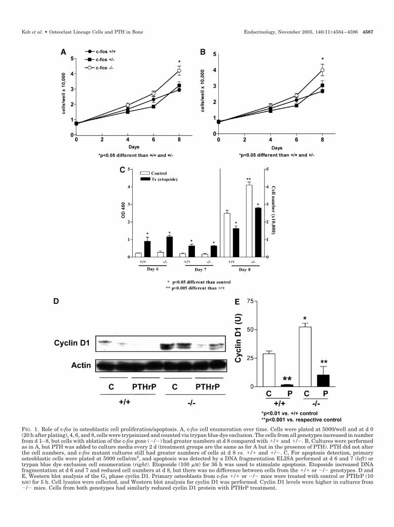
FIG. 1. Role of c-fos in osteoblastic cell proliferation/apoptosis. A, c-fos cell enumeration over time. Cells were plated at 5000/well and at d 0(20 h after plating), 4, 6, and 8, cells were trypsinized and counted via trypan blue dye exclusion. The cells from all genotypes increased in numberfrom d 1–8, but cells with ablation of the c-fos gene (�/�) had greater numbers at d 8 compared with �/� and �/�. B, Cultures were performedas in A, but PTH was added to culture media every 2 d (treatment groups are the same as for A but in the presence of PTH). PTH did not alterthe cell numbers, and c-fos mutant cultures still had greater numbers of cells at d 8 vs. �/� and �/�. C, For apoptosis detection, primaryosteoblastic cells were plated at 5000 cells/cm2, and apoptosis was detected by a DNA fragmentation ELISA performed at d 6 and 7 (left) ortrypan blue dye exclusion cell enumeration (right). Etoposide (100 �M) for 36 h was used to stimulate apoptosis. Etoposide increased DNAfragmentation at d 6 and 7 and reduced cell numbers at d 8, but there was no difference between cells from the �/� or �/� genotypes. D andE, Western blot analysis of the G1 phase cyclin D1. Primary osteoblasts from c-fos �/� or �/� mice were treated with control or PTHrP (10nM) for 5 h. Cell lysates were collected, and Western blot analysis for cyclin D1 was performed. Cyclin D1 levels were higher in cultures from�/� mice. Cells from both genotypes had similarly reduced cyclin D1 protein with PTHrP treatment.
Koh et al. • Osteoclast Lineage Cells and PTH in Bone Endocrinology, November 2005, 146(11):4584–4596 4587
a proliferative advantage through alteration of cell cycle pro-teins but that there is no alteration in their PTH-mediatedresponse.
Calcium incorporation and von Kossa staining
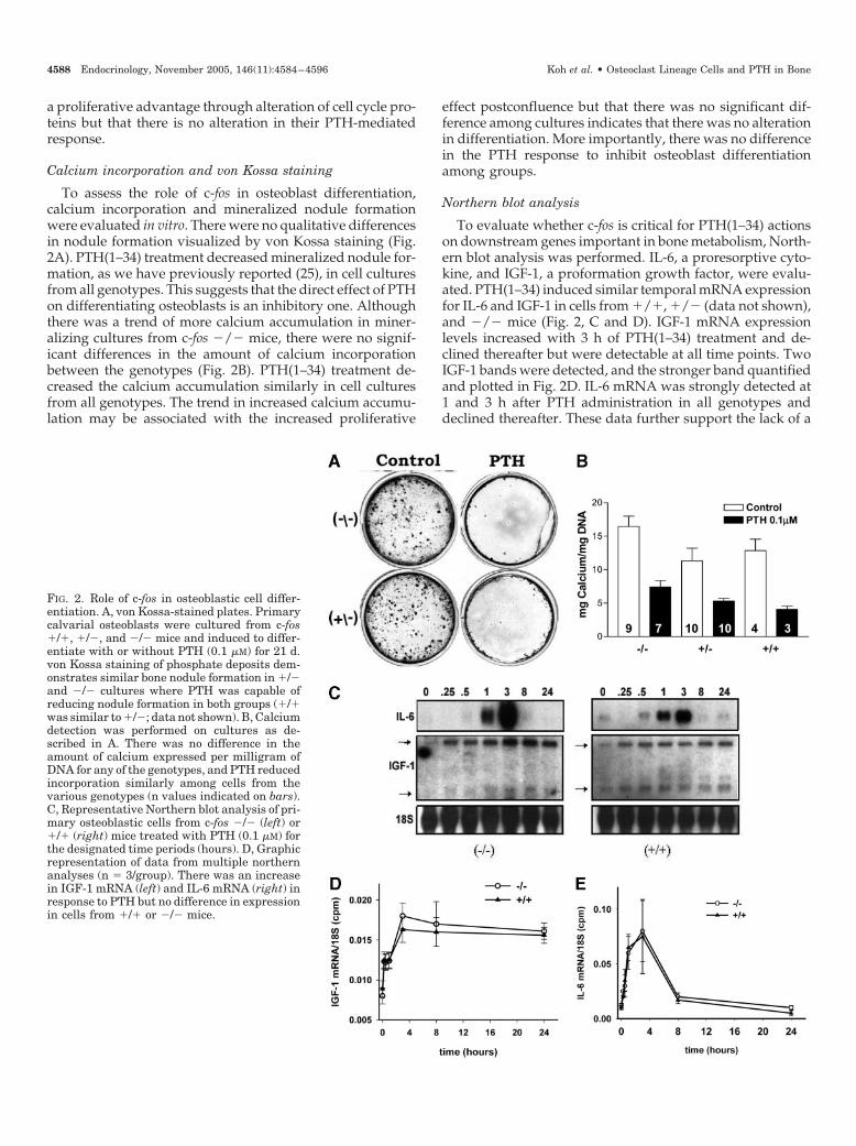
To assess the role of c-fos in osteoblast differentiation,calcium incorporation and mineralized nodule formationwere evaluated in vitro. There were no qualitative differencesin nodule formation visualized by von Kossa staining (Fig.2A). PTH(1–34) treatment decreased mineralized nodule for-mation, as we have previously reported (25), in cell culturesfrom all genotypes. This suggests that the direct effect of PTHon differentiating osteoblasts is an inhibitory one. Althoughthere was a trend of more calcium accumulation in miner-alizing cultures from c-fos �/� mice, there were no signif-icant differences in the amount of calcium incorporationbetween the genotypes (Fig. 2B). PTH(1–34) treatment de-creased the calcium accumulation similarly in cell culturesfrom all genotypes. The trend in increased calcium accumu-lation may be associated with the increased proliferative
effect postconfluence but that there was no significant dif-ference among cultures indicates that there was no alterationin differentiation. More importantly, there was no differencein the PTH response to inhibit osteoblast differentiationamong groups.
Northern blot analysis
To evaluate whether c-fos is critical for PTH(1–34) actionson downstream genes important in bone metabolism, North-ern blot analysis was performed. IL-6, a proresorptive cyto-kine, and IGF-1, a proformation growth factor, were evalu-ated. PTH(1–34) induced similar temporal mRNA expressionfor IL-6 and IGF-1 in cells from �/�, �/� (data not shown),and �/� mice (Fig. 2, C and D). IGF-1 mRNA expressionlevels increased with 3 h of PTH(1–34) treatment and de-clined thereafter but were detectable at all time points. TwoIGF-1 bands were detected, and the stronger band quantifiedand plotted in Fig. 2D. IL-6 mRNA was strongly detected at1 and 3 h after PTH administration in all genotypes anddeclined thereafter. These data further support the lack of a
FIG. 2. Role of c-fos in osteoblastic cell differ-entiation. A, von Kossa-stained plates. Primarycalvarial osteoblasts were cultured from c-fos�/�, �/�, and �/� mice and induced to differ-entiate with or without PTH (0.1 �M) for 21 d.von Kossa staining of phosphate deposits dem-onstrates similar bone nodule formation in �/�and �/� cultures where PTH was capable ofreducing nodule formation in both groups (�/�was similar to �/�; data not shown). B, Calciumdetection was performed on cultures as de-scribed in A. There was no difference in theamount of calcium expressed per milligram ofDNA for any of the genotypes, and PTH reducedincorporation similarly among cells from thevarious genotypes (n values indicated on bars).C, Representative Northern blot analysis of pri-mary osteoblastic cells from c-fos �/� (left) or�/� (right) mice treated with PTH (0.1 �M) forthe designated time periods (hours). D, Graphicrepresentation of data from multiple northernanalyses (n � 3/group). There was an increasein IGF-1 mRNA (left) and IL-6 mRNA (right) inresponse to PTH but no difference in expressionin cells from �/� or �/� mice.
4588 Endocrinology, November 2005, 146(11):4584–4596 Koh et al. • Osteoclast Lineage Cells and PTH in Bone

direct effect of PTH on c-fos mutant osteoblasts that differsfrom wild-type mice.
Vossicle model system
We have previously characterized an ectopic ossicle modelsystem that responds to PTH with an anabolic result that isgreater than endogenous bone (28, 30). The principle of thismodel system was used to separate out the mesenchymal andhematopoietic components of the bone and elucidate thetarget of the PTH-altered response in the c-fos �/� mutantmice. Figure 3A demonstrates the model system as describedin Materials and Methods. Vertebral bodies from 4-d-old c-fos�/� mice (Fig. 3B) at baseline (preimplantation) demon-strate early formation of trabecular bone and a hematopoieticmarrow cavity, whereas vertebral bodies from 4-d-old c-fos�/� mice have a minimal marrow cavity (Fig. 3C). How-ever, after implantation and PTH treatment, the c-fos �/�implants recruit a hematopoietic marrow and numerousTRAP� osteoclastic cells from the host tissues (Fig. 3D). Tovalidate the mesenchymal contribution of this model, weimplanted vertebral bodies from mice expressing luciferaseunder the HIV1 long terminal repeat promoter. In vivo im-
aging confirmed the expression of luciferase in the vossicleimplants (Fig. 3E). At the time the mice were killed, immu-nohistochemistry for luciferase was performed as described(21). Figure 3F demonstrates that osteoblastic cells lining thevossicle trabeculae in a PTH-treated (3 wk) mouse are lucif-erase positive and hence derived from the donor, not viarescue from the host.
Vossicles from c-fos �/� and �/� mice after 21 d ofimplantation appeared similar in size and organization (Fig.4A). Both genotypes displayed cortical and trabecular boneand a hematopoietic marrow. PTH treatment increased thebone area of implants from both genotypes to a similar extent(Fig. 4B). This substantiates that c-fos expression in the mes-enchymal compartment is dispensable for the anabolic ac-tions of PTH. TRAP staining was performed to determine thenumbers of osteoclasts present in vossicles (Fig. 4C). Al-though there was a trend for greater osteoclast numbers inthe c-fos �/� bones, there was no difference between groupsat this end point. Finally, BrdU staining demonstrated analtered pattern of positivity in vossicles treated with PTH(Fig. 4D). Vehicle-treated vossicles (c-fos �/� or �/�) dis-played BrdU positivity that tended to be restricted to the cells
FIG. 3. A, Vossicle implant experimental design.Vertebrae from neonatal (d 4) mice of various geno-types were dissected, trimmed of musculature, andimplanted into athymic hosts. PTH or vehicle wasadministered via sc injection for 3 wk before the micewere killed. B and C, Histologic view of vossicles atbaseline (vertebrae from neonatal d 4 mice dissectedand ready for implantation). Vertebra from a c-fos�/� (B) or �/� (C) mouse demonstrates developmentof marrow cavity in �/� vertebra is already estab-lished vs. the c-fos �/� osteopetrotic mutant. D, Rep-resentative c-fos �/� vossicle after implantation and21 d of PTH administration. TRAP staining revealsnumerous TRAP-positive cells and validates rescue ofthe osteopetrotic phenotype. E, Luciferase imaging ofa vossicle implant from a luciferase tagged mouseinto an athymic mouse and imaged with a CCD cam-era at 1 wk to demonstrate the localized luciferasepositivity at the implant site (three implants shown).F, Immunohistochemistry for luciferase from a lucif-erase-tagged vossicle from a mouse treated with PTHfor 3 wk. The positive luciferase staining in the os-teoblastic cells lining the bony trabeculae verifies thedonor origin of these cells. Magnification, �25 (B–D)or �200 (F).
Koh et al. • Osteoclast Lineage Cells and PTH in Bone Endocrinology, November 2005, 146(11):4584–4596 4589

FIG. 4. Vossicles from PTH-treated c-fos mutant and wild-type mice. A, Representative H&E-stained c-fos �/� and �/� vossicles from vehicle-and PTH (80 �g/kg�d; 21 d)-treated mice (�20). B, Graph of total bone area (Tt.Ar.) as a percentage of the overall size of vossicles from vehicle-and PTH-treated c-fos �/� and �/� mice (n � 12 or more/group). C, Numbers of osteoclasts per millimeter of bone from vossicles in B (n �8–9 per group; no significant difference between groups). D, Representative BrdU-stained sections from B demonstrating localized positivitylining the trabecular bone in vehicle-treated mice and a more widespread positivity in the PTH-treated mice from both genotypes (�160).
4590 Endocrinology, November 2005, 146(11):4584–4596 Koh et al. • Osteoclast Lineage Cells and PTH in Bone

lining the bony surfaces, whereas with PTH treatment therewas increased and a more widespread distribution of BrdUpositivity that extended throughout the bone marrow. Thissuggests that cells in the marrow compartment that is de-rived from the host are likely to be the important targets ofPTH action.
The finding of a rescued anabolic phenotype of PTH onc-fos �/� vossicles led us to explore the dependence of PTHon cells of the osteoclast lineage for the anabolic response.Mice with wild-type vossicle implants were treated withOPG, a decoy receptor for receptor activator of nuclear factor�B (RANK) ligand (RANKL), and an inhibitor of osteoclastdifferentiation. OPG was administered alone or in combi-nation with PTH (Fig. 5). OPG alone did not alter the bonearea of vossicles but did significantly block the PTH anabolicresponse. These data suggest that PTH requires osteoclasticactivity for its anabolic response and fits with the lack ofresponse in the intact c-fos osteopetrotic mouse. However,because OPG was administered early in the treatment pro-tocol, by the time the mice were killed, there was no differ-ence in the numbers of osteoclasts per millimeter of bone(data not shown). Interestingly, the c-fos mutant mice have anarrest in osteoclast differentiation fairly early in osteoclastdevelopment. The recruitment of hematopoietic cells to sitesof active resorption is the first step in osteoclastogenesis. Thishas been reported to be at least in part, due to the up-regulation of SDF-1 in stromal cells and its interaction withits receptor, CXCR4. We have previously shown that PTHup-regulates SDF-1 in the mesenchymal compartment of de-veloping ossicles so the impact of disrupting this system by
administering blocking agents to inhibit the CXCR4 wasdetermined (31). Figure 6A demonstrates that SDF-1 levelsare increased in tibiae of mice administered PTH in an an-abolic mode. Interestingly, blocking SDF-1 activity was suc-cessful in reducing the anabolic action of PTH in the vossiclemodel system of wild-type mice (Fig. 6, B and C).
To further identify the osteoclastic stage dependence forthis effect, we employed another osteopetrotic model, thec-src mutant mice. The c-src mutant mice have a defect in thesignaling that is necessary for osteoclast formation of a ruf-fled membrane and adherence to the bone substrate (32).Hence, they have osteoclastic cells present, but they do notfunction to resorb bone. Two studies were performed withthese mice. A regimen similar to what we published with thec-fos mutant mice was performed where PTH was adminis-tered to intact mutant mice from neonatal d 4–21 (18). Thesecond study used the vossicle model where vertebral bodiesfrom c-src mutant and wild-type mice were implanted intoathymic hosts and PTH administered over a 3-wk period.Interestingly, in both of these regimens, PTH displayed ananabolic response. Figure 7A demonstrates the microradio-graphic view of femurs, and Fig. 7B a histologic view ofvertebrae from vehicle- and PTH-treated c-src wild-type andmutant mice. PTH treatment resulted in increased radiopac-ity and increased trabecular bone in PTH-treated mice re-gardless of the c-src status. Figure 7, C and D, demonstratethe vossicle model where there is an increase in bone area inall genotypes of c-src mice. This suggests that the stage ofosteoclast differentiation is critical for anabolic actions ofPTH in this model system.
FIG. 5. Vossicles from mice treated with OPG with or with-out PTH. Vertebrae from wild-type mice were implantedinto athymic hosts and mice administered PTH (80 �g/kg�d;21 d) or OPG (4 mg/kg twice a week for the first 2 wk) or acombination of PTH and OPG. A, Representative H&E-stained vossicles (�25). B, Graph of total bone area (Tt.Ar.)of vossicles from indicated treatment groups (n � 8 control,n � 12 each for OPG, PTH, PTH, and OPG). PTH treatmentresulted in an anabolic response, and OPG significantlyblocked the anabolic response of PTH.
Koh et al. • Osteoclast Lineage Cells and PTH in Bone Endocrinology, November 2005, 146(11):4584–4596 4591

Discussion
The present study was performed to better understand theanabolic actions of PTH and delineate the specific role of c-fosas a transcriptional mediator of PTH actions in bone. Thiswork stemmed from our previous work where we found thatmice with ablation of the c-fos gene did not respond to PTHin an anabolic mode (18). The c-fos mutant mice respondedto PTH, but in an antianabolic mode where instead of anincrease in bone there was actually a reduction in bone areawith PTH treatment. These data suggested that c-fos geneexpression was critical for the anabolic actions of PTH andprompted the dissection of the specific cellular targets ofPTH that discriminated the anabolic and antianabolic effects.To do this, in vitro approaches and a novel in vivo model were
used. The in vitro studies indicate that the direct actions ofPTH on cells of the osteoblast lineage are to inhibit theirdifferentiation and mineralization. This is consistent with theantianabolic response that was found in vivo when c-fos wasablated. Our laboratory and others have previously pre-sented data that support an inhibition of mineralization byPTH (25, 33). More importantly, it became clear during thisinvestigation that c-fos may be an indirect target of PTHanabolic actions via its role in osteoclastogenesis and not viaa direct impact in the mesenchymal compartment.
Fos is important in growth, differentiation, transforma-tion, and regulation of specific gene expression, all of whichare key events in bone homeostasis (15). During embryo-genesis, c-fos is expressed in growth regions of fetal bones,
FIG. 6. Impact of blocking SDF-1 on the anabolicaction of PTH. A, in situ hybridization for SDF-1in tibiae from mice administered PTH (80 �g/kg)or vehicle (�100) for 3 wk. Positivity for SDF-1was detected in areas of bone formation in cellslining the trabecular surface with osteoblast-likeappearance. SDF-1 positivity was greater in micetreated with PTH. Vertebrae from wild-typemice were implanted into athymic hosts, andmice were treated with anti-CXCR4 blockingpeptide designed to inhibit SDF-1 binding toCXCR4 (1 �g/d) and antihuman/mouse SDF-1IgG antibody (25 �g/d) (group indicated as anti-SDF) with or without PTH (80 �g/kg�d) for 21 d.B, Representative H&E-stained vossicles (�25).C, Graph of total bone area (Tt.Ar.) of vossiclesfrom indicated treatment groups (n � 6 control,6 anti-SDF, 4 PTH, 8 PTH � anti-SDF). Data aremean � SEM. PTH treatment resulted in an an-abolic response, and inhibition of SDF-1 activitysignificantly blocked the anabolic response ofPTH.
4592 Endocrinology, November 2005, 146(11):4584–4596 Koh et al. • Osteoclast Lineage Cells and PTH in Bone

FIG. 7. Impact of c-src mutation on anabolicactions of PTH. A and B, Mice from heterozy-gous c-src �/� breedings were administeredPTH (50 �g/kg�d) or vehicle from d 4–21 post-natal age. Mice were killed, and microradiog-raphy revealed an anabolic PTH response infemurs (A) and an increase in vertebral tra-becular bone (B) from both c-src �/� and �/�mice. C, Vertebrae from 4-d-old c-src �/�,�/�, and �/� mice were implanted into athy-mic hosts and PTH (80 �g/kg�d) or vehicle ad-ministered for 21 d. Representative H&E staindemonstrates vossicles from PTH-treatedgroups regardless of genotype respond with anincrease in trabecular bone. Note the presenceof cartilage in the vehicle c-src mutant image,which is a common and random finding and isnot included in the bone area determination orosteoclast enumeration. D, Graph of total bonearea (Tt.Ar.) of vossicles from PTH-treated c-src �/�, �/�, and �/� mice (n � 12 and 11 forc-src �/� and �/� vehicle and PTH, respec-tively, and n � 7 and 8 for c-src �/� vehicleand PTH respectively). An anabolic responseto PTH was found regardless of the genotype.Magnification �2.5 (A), �30 (B), and �25 (C).
Koh et al. • Osteoclast Lineage Cells and PTH in Bone Endocrinology, November 2005, 146(11):4584–4596 4593

areas that have active osteoblast proliferation (34). Manystudies have suggested that c-fos is associated with and isimportant for cell proliferation (35). However, in the presentstudy, primary osteoblasts of mice with an ablation of c-foshad increased cell viability compared with their wild-typecounterparts. A previous study found c-fos-deficient fibro-blasts retain their ability to proliferate in vitro, indicatingthere was no difference in proliferation between genotypes,although they noted a slight increase in c-fos �/� cells at d4 (36). The cell types used were different from ours, but ourfindings were similar at that time point. The increased cellnumber at d 8 in the present study suggests an effect oncontact inhibition because the cells became confluent be-tween d 5 and 6. It has been proposed that c-fos may beimportant in apoptosis (37). However, in the present study,cells from both genotypes responded similarly to etoposidetreatment, which is a known apoptotic agent. Furthermore,cells from c-fos mutant mice had increased levels of cyclin D1,a cyclin responsible for G1 cell cycle progression. Because thecyclin D1 promoter contains an AP-1 site, and we have foundJunB to negatively regulate cyclin D1 (29), this suggests thatc-Fos may partner with JunB as cells become confluent torestrict their cell cycle progression and in the absence of Fosor JunB that cells may continue their proliferation. Impor-tantly, our studies found only this single difference betweenc-fos mutant and wild-type cells and did not find any dif-ferences in the response of the c-fos mutant cells to PTH. Thissuggests that the differences seen in the intact animal (lackof an anabolic response to PTH) are not associated with adirect effect of PTH on osteoblastic cells. Because c-fos is anessential mediator of osteoclastogenesis, the impact of cellsof the osteoclast lineage was evaluated.
Osteoclastogenesis is a complex process that includes re-cruitment of cells to the resorptive site, differentiation,multinucleation, and activation (38). Cross talk between os-teoclasts and osteoblasts has been discussed for many years,but most of the convincing evidence has centered on osteo-blast signaling to osteoclasts via the RANK/RANKL system.Osteoclast signaling to recruit osteoblastic cells to repair theresorptive lacunae has also been discussed, but much of thefocus has been on the osteoclast release of growth factorsstored in the bone, such as TGF�, that stimulate osteoblastdifferentiation or chemotaxis (39). The present study sup-ports a concept of cross talk between osteoclastic cells that arenot actively resorbing matrix but are multinucleated cells. Tosubstantiate this, four intervention strategies were investi-gated. Blocking SDF-1/CXCR4 was performed to inhibit re-cruitment of osteoclast progenitor cells. SDF-1 is an osteo-blast- or endothelial-derived chemokine that directsosteoclast precursors into bone marrow to appropriate sitesfor differentiation into active osteoclasts (40). Blocking SDF-1activity via the administration of blocking peptides and/orantibodies to the SDF-1 receptor, chemokine receptor-4(CXCR4), has been used as a method to block cancer metas-tasis and decrease osteoclastic activity (23, 24). The sameapproach was used here to block osteoclast recruitment dur-ing PTH administration. The anabolic response to PTH inmice was blocked when SDF-1 activity was inhibited. Thissuggests that cells recruited to the marrow in response toresorptive stimuli are necessary for the anabolic response to
PTH. The BrdU labeling studies revealed the widespreadpresence of proliferative cells in the bone marrow of PTH-treated mice and support key cellular targets in the marrow.Recent data of Calvi et al. (41) demonstrate a role for osteo-blasts in regulating the hematopoietic niche. They found thatPTH increased expression of the Notch ligand jagged 1 tosupport an increase in the number of hematopoietic cells.Findings of the present study further suggest that PTH maystimulate osteoblasts to recruit hematopoietic cells that thensignal back to osteoblasts to increase bone formation.
The SDF-1 blocking experiments do not restrict the cellulartargets to osteoclasts. Hence, OPG studies were performed toblock the differentiation of cells along the osteoclast lineage.OPG acts as a decoy receptor that blocks the interaction ofRANK and RANKL and hence inhibits osteoclast differen-tiation (32, 42). Administration of OPG in combination withPTH in the vossicle model was capable of blocking the an-abolic response. This further supports the necessity for os-teoclastogenesis as a key factor in the anabolic actions ofPTH. However, dendritic cells also express RANK, and OPGcould conceivably interact with RANK on the surface ofdendritic cells and alter PTH responses if a dendritic cell wasa relevant target. The expression of c-fos is essential for os-teoclastogenesis at the point which cells differentiate frommacrophages and fuse into multinucleated cells (12, 32). Micelacking c-fos do not have osteoclasts and are osteopetrotic butdisplay increased numbers of macrophages. Data in the in-tact c-fos mutants show that PTH is not anabolic (18), whereasin the vossicle transplants, which contain a host-derivedhematopoietic marrow and TRAP-positive multinucleatedosteoclastic cells and the anabolic PTH response is rescued,suggest that c-fos expression in the marrow is critical for theanabolic response. Furthermore, it suggests that macro-phages in abundance in the c-fos mutants cannot rescue thePTH response. Finally, data from the c-src mutant mice pro-vide evidence that the presence of active resorbing oste-oclasts may not be critical for the PTH response. The c-srcmutant mice lack functional osteoclasts due to a defect inactivation associated with cytoskeletal rearrangement andmotility (32). These mice present with numerous multinu-cleated TRAP-positive cells, but these cells fail to form ruffledborders and resorption lacunae (43, 44). The data in thepresent study where intact c-src mutant mice retained theability to mount an anabolic response to PTH suggest againstthe necessity for active bone resorption and subsequent re-lease of growth factors from the bone matrix to feedback toosteoblastic cells. Instead, these data implicate cells of theosteoclast lineage at the developmental stage of marrowlocalization and multinucleation before active bone resorp-tion. We hypothesize that these cells are producing a factor/factors that are signaling to osteoblasts to increase bone for-mation. Of note, the c-src mutant mice have been reported tohave an osteoblast phenotype as well as the osteoclast phe-notype that supports increased osteoblast activity (45, 46).Data from the vossicle model where osteoblasts are com-partmentalized from the hematopoietic cells suggest that thisalteration does not play into the effects of PTH because therewas no difference in bones from mutant or wild-type mice.
The data in the present study that implicate cells of theosteoclast lineage are particularly intriguing in light of the
4594 Endocrinology, November 2005, 146(11):4584–4596 Koh et al. • Osteoclast Lineage Cells and PTH in Bone

recent clinical studies where administration of an antiresorp-tive bisphosphonate in combination with PTH did not aug-ment the increase in bone volume. Bisphosphonates havebeen shown to inhibit osteoclast activity at four levels: 1)inhibition of osteoclast recruitment, 2) inhibition of osteoclastadhesion, 3) shortening of osteoclast lifespan, and 4) directinhibition of osteoclast activity (47). According to the hy-pothesis presented here, the alteration in recruitment wouldimpact the anabolic action of PTH, but the other actionsshould not compromise cells that may be key targets foranabolic actions of PTH. Furthermore, studies of combina-torial therapy where the antiresorptive calcitonin was ad-ministered in combination with PTH did not prove to aug-ment the anabolic action of PTH (48). In contrast, one studyreported that the combination of OPG with PTH did notcompromise the increase in bone strength that PTH mediated(49); however, this study was performed in rats and underan estrogen-depleted condition that could have modified theoutcome.
Other reports highlight cross talk mechanisms betweenosteoclasts and osteoblasts (50, 51). Two preliminary studiessuggest a juxtacrine interaction between osteoclasts and os-teoblasts (52, 53). To our knowledge, the present study is thefirst to provide data to support the role of osteoclastic cellsin the action of an anabolic agent although it has been pro-posed (54). Recent data support the concept of a stage-de-pendent impact of osteoclasts on the bone formation re-sponse. Pharmaceutical strategies were used to inhibitosteoclast acidification, and the data indicated that resorp-tion could be inhibited at this late stage of osteoclast differ-entiation without altering bone formation (55). Hence, thedata presented here and in conjunction with other studiesstrongly suggest that cells in the intermediately differenti-ated population of osteoclasts may be key targets for PTHanabolic action. Interestingly, that cells in the marrow weretargets of PTH action was proposed more than 10 years ago,but perhaps because of the focus then on catabolic actions ofPTH, little was done to substantiate this work in regards toanabolic actions (56). It is also important to recognize thatalthough the model system used here is not a model ofosteoporosis but may be more applicable to clinical scenariosin bone regeneration for local tissue engineering strategieswhere PTH has also been found to be anabolic (57, 58).
Previous work from our laboratory and others indicatethat cells of the osteoblastic lineage are critical for anabolicactions, but our in vitro data suggest that they are not thedirect targets of PTH action for this anabolic effect. This doesnot preclude their ability to, while directly inhibiting min-eralization, produce factor(s) that signal to cells in the mar-row that then direct other osteoblastic cells to form bone. Thismultilevel regulation may also address the vagaries of anintermittent PTH administration regimen. Data from ourossicle model system point to a role of cells of the osteoblastlineage that are relatively early in their differentiation pro-gram and are not lining or terminally differentiated cells (28).Collectively, these current and recent studies suggest thatPTH targets stromal cells to signal to cells of the hemato-poietic lineage to recruit to sites of remodeling. These cellsdifferentiate and multinucleate, and before adhering to thebone matrix and resorbing bone, they signal either via a
paracrine or juxtacrine manner to preosteoblastic cells todifferentiate and form bone. The dependence on an oste-oclastic cell could also be tested by analyzing the impact ofPTH on various other osteopetrotic mutant mouse modelssuch as the PU.1-deficient mice, the M-CSF mutant mice thatwould lack mature osteoclasts, and the �3 integrin-deficientmice that have osteoclasts but lack functional resorptive ac-tivity. The concept presented here has obvious clinical ap-plication in the consideration of combination antiresorptiveand anabolic agent therapies. The use of antiresorptives thattarget late stages of osteoclastic activity, such as antiintegrinsor proton pump inhibitors, might reduce bone resorptionwithout compromising the support of osteoblastic activity.
Acknowledgments
We thank Dr. Kurt Hankenson for providing the luciferase mice; ErinEalba, Ana Mattos, and Jason Wang for technical assistance; Daniel Hallfor technical support with bioluminescent optical imaging; Chris Stray-horn for histologic/tissue processing support; and T. John Martin forcritical reading of the manuscript.
Received March 21, 2005. Accepted July 26, 2005.Address all correspondence and requests for reprints to: Laurie K. McCau-
ley, D.D.S., Ph.D., Department of Periodontics and Oral Medicine, School ofDentistry, Room 3343, University of Michigan, 1011 North University Avenue,Ann Arbor, Michigan 48109-1078. E-mail: [email protected].
This work was supported by National Institutes of Health GrantsDK53904 (to L.K.M.) and DE13701 (to R.S.T.) and by a fellowship fromTUBITAK: The scientific and technological research council of Turkey(NATO-A2 to B.D.).
References
1. Neer RM, Arnaud CD, Zanchetta JR, Prince R, Gaich GA, Reginster JY,Hodsman AB, Eriksen EF, Ish-Shalom S, Genant H, Wang O, Mitlak BH 2001Effect of parathyroid hormone (1–34) on fractures and bone mineral densityin postmenopausal women with osteoporosis. New Engl J Med 10:1434–1441
2. Mohan S, Kutilek S, Zhang C, Shen HG, Kodama Y, Srivastava AK,Wergedal JE, Beamer WG, Baylink DJ 2000 Comparison of bone formationresponse to parathyroid hormone (1–34), (1–31), and (2–34) in mice. Bone27:471–478
3. Rixon RH, Whitfield JF, Gagnon L, Isaacs RJ, Maclean S, Chakravarthy B,Durkin JP, Neugebauer W, Ross V, Sung W, Willick GE 1994 Parathyroidhormone fragments may stimulate bone growth in ovariectomized rats byactivating adenylyl cyclase. J Bone Miner Res 9:1179–1189
4. Jilka RL, Weinstein RS, Bellido T, Roberson P, Parfitt AM, Manolagas SC1999 Increased bone formation by prevention of osteoblast apoptosis withparathyroid hormone. J Clin Invest 104:439–446
5. Bellido T, Ali AA, Plotkin LI, Fu Q, Gubrij I, Roberson PK, Weinstein RS,O’Brien CA, Manolagas SC, Jilka RL 2003 Proteasomal degradation of Runx2shortens parathyroid hormone-induced anti-apoptotic signaling in osteo-blasts. A putative explanation for why intermittent administration is neededfor bone anabolism. J Biol Chem 278:50259–50272
6. Black DM, Greenspan SL, Ensrud KE, Palermo L, McGowan JA, Lang TF,Garnero P, Bouxsein ML, Bilezikian JP, Rosen CJ 2003 The effects of para-thyroid hormone and alendronate alone or in combination in postmenopausalosteoporosis. New Engl J Med 25:1207–1215
7. Finkelstein JS, Hayes A, Hunzelman JL, Wyland JJ, Lee H, Neer RM 2003 Theeffects of parathyroid hormone, alendronate, or both in men with osteoporosis.New Engl J Med 25:1216–1226
8. Delmas PD, Vergnaud P, Arlot ME, Pastoureau P, Meunier PJ, Nilssen MH1995 The anabolic effect of human PTH (1–34) on bone formation is bluntedwhen bone resorption is inhibited by the bisphosphonate tiludronate–is acti-vated resorption a prerequisite for the in vivo effect of PTH on formation in aremodeling system? Bone 16:603–610
9. Clohisy JC, Scott DK, Brakenhoff KD, Quinn CO, Partridge NC 1992 Para-thyroid hormone induces c-fos and c-jun messenger RNA in rat osteoblasticcells. Mol Endocrinol 6:1834–1842
10. McCauley LK, Koh AJ, Beecher CA, Rosol TJ 1997 The proto-oncogene c-fosis transcriptionally regulated by PTH and PTHrP in a cAMP-dependent man-ner in osteoblastic cells. Endocrinology 138:5427–5433
11. Finkel MP, Biskis BO, Jinkins PB 1966 Virus induction of osteosarcomas inmice. Science 151:698–701
Koh et al. • Osteoclast Lineage Cells and PTH in Bone Endocrinology, November 2005, 146(11):4584–4596 4595

12. Grigoriadis AE, Wang ZQ, Cecchini MG, Hofstetter W, Felix R, Fleisch HA,Wagner EF 1994 c-Fos: a key regulator of osteoclast-macrophage lineage de-termination and bone remodeling. Science 266:443–448
13. Tolar J, Teitelbaum SL, Orchard PJ 2004 Osteopetrosis. New Engl J Med30:2839–2849
14. Wang ZQ, Grigoriadis AE, Mohle-Steinlein U, Wagner EF 1991 A novel targetcell for c-fos induced oncogenesis: development of chrondrogenic tumours inembryonic stem cell chimeras. EMBO J 10:2437–2450
15. Grigoriadis AE, Wang ZQ, Wagner EF 1995 Fos and bone cell development:lessons from a nuclear oncogene. Trends Genet 11:436–441
16. Wang ZQ, Ovitt C, Grigoriadis AE, Mohle-Steinlein U, Ruther U, WagnerEF1992 Bone and haematopoietic defects in mice lacking c-fos. Nature 360:741–745
17. Johnson RS, Spiegelman BM, Papaioannou V 1992 Pleiotropic effects of a nullmutation in the c-fos proto-oncogene. Cell 71:577–586
18. Demiralp B, Chen H, Koh-Paige AJ, Keller ET, McCauley LK 2002 Anaboliceffects of parathyroid hormone during endochondral bone growth are depen-dent on c-fos. Endocrinology 143:4038–4047
19. Soriano P, Montgomery C, Geske R, Bradley A 1991 Targeted disruption ofthe c-src proto-oncogene leads to osteopetrosis in mice. Cell 64:693–702
20. Yull FE, Wei H, Jansen ED, Everhart MB, Sadikot RT, Christman JW, Black-well T 2003 Bioluminescent detection of endotoxin effects on HIV-1 LTR-driven transcription in vivo. J Histochem Cytochem 51:741–749
21. Kalikin LM, Schneider A, Thakur MA, Fridman Y, Griffin LB, Dunn RL,Rosol TJ, Shah RB, Rehemtulla A, McCauley LK, Pienta KJ 2003 In vivovisualization of metastatic prostate cancer and quantitation of disease pro-gression in immunocompromised mice. Cancer Biol Ther 2:656–660
22. Tamamura H, Omagari A, Hiramatsu K, Kanamoto T, Xu YN, Kodama E,Matsuoka M, Hattori T, Yamamoto N, Nakashima H, Otaka A, Fujii N 2001Development specific CXCR4 inhibitors possessing high selectivity indexes aswell as complete stability in serum based on an anti-HIV peptide T140. BioorgMed Chem Lett 11:1897–1902
23. Liang Z, Wu T, Lou H, Yu X, Taichman RS, Lau SK, Nie S, Umbreit J, ShimH 2004 Inhibition of breast cancer metastasis by selective synthetic polypeptideagainst CXCR4. Cancer Research 64:4302–4308
24. Sun YX, Schneider A, Jung Y, Wang J, Dai J, Wang J, Cook K, Osman NI,Koh-Paige AJ, Shim H, Pienta KJ, Keller ET, McCauley LK, Taichman RS2005 Skeletal localization and neutralization of the SDF-1(CXCL12)/CXCR4axis blocks prostate cancer metastasis and growth in osseous sites in vivo.J Bone Miner Res 20:318–329
25. Koh AJ, Beecher CA, Rosol TJ, McCauley LK 1999 Cyclic AMP activation inosteoblastic cells: effects on PTH-1 receptors and osteoblastic differentiation invitro. Endocrinology 140:3154–3162
26. Chen C, Koh AJ, Datta NS, Zhang J, Keller ET, Xiao G, Franceschi RT,D’Silva N, McCauley LK 2004 Impact of the mitogen-activated protein kinasepathway on parathyroid hormone-related protein actions in osteoblasts. J BiolChem 279:29121–29129
27. McCauley LK, Koh AJ, Beecher CA, Cui Y, Decker JD, Francheschi RT 1995Effects of differentiation and transforming growth factor � on PTH/PTHrPreceptor mRNA levels in MC3T3–E1 cells. J Bone Miner Res 10:1243–1255
28. Pettway GJ, Schneider A, Koh AJ, Widjaja E, Morris MD, Meganck JA,Goldstein SA, McCauley LK 2005 Anabolic actions of PTH (1–34): use of anovel tissue engineering model to investigate temporal effects on bone. Bone36:959–970
29. Datta NS, Chen C, Berry JE, McCauley LK 2005 PTHrP signaling targets cyclinD1 and induces osteoblastic cell growth arrest. J Bone Miner Res 20:1051–1064
30. Schneider A, Taboas J, McCauley LK, Krebsbach PH 2003 Skeletal homeosta-sis in tissue-engineered bone. J Orthop Res 21:859–864
31. Jung Y, Wang J, Schneider A, Sun YX, Koh-Paige AJ, Osman NI, McCauleyLK, Taichman RS 2005 Regulation of SDF-1 (CXCL12) Production by osteo-blasts in the hematopoietic microenvironment and a possible mechanisms forstem cell homing. J Bone Miner Res 19:S389
32. Boyle WJ, Simonet WS, Lacey DL 2003 Osteoclast differentiation and acti-vation. Nature 423:337–342
33. Bellows CG, Ishida H, Aubin JE, Heersche JNM 1990 Parathyroid hormonereversibly suppresses the differentiation of osteoprogenitor cells into func-tional osteoblasts. Endocrinology 127:3111–3116
34. Dony C, Gruss P 1987 Proto-oncogene c-fos expression in growth regions offetal bone and mesodermal web tissue. Nature 328:711–714
35. Angel P, Karin M 1991 The role of Jun, Fos, and the AP-1 complex in cellproliferation and transformation. Biochim Biophys Acta 1072:129–157
36. Hu E, Mueller E, Olivero S, Papaioannou V, Johnson R, Spiegelman BM 1994Targeted disruption of the c-fos gene demonstrates c-fos-dependent and -in-dependent pathways for gene expression stimulated by growth factors oroncogenes. EMBO J 13:3094–3103
37. Smeyne RJ, Vendrell M, Hayward M, Baker S, Miao GG, Schilling K,Robertson LM, Curran T, Morgan JI 1993 Continuous c-fos expression pre-cedes programmed cell death in vivo. Nature 363:166–169
38. Teitelbaum SL 2000 Bone resorption by osteoclasts. Science 289:1504–150839. Centrella M, Horowitz MC, Wozney JM, McCarthy TL 1994 Transforming
growth factor-� gene family members and bone. Endocr Rev 15:27–3940. Yu X, Huang Y, Collin-Osdoby P, Osdoby P 2003 Stromal cell-derived factor-1
(SDF-1) recruits osteoclast precursors by inducing chemotaxis, matrix metal-loproteinase-9 (MMP-9) activity, and collagen transmigration. J Bone MinerRes 18:1404–1418
41. Calvi LM, Adams GB, Welbrecht KW, Weber JM, Olson DP, Knight MC,Martin RP, Schipani E, Divieti P, Bringhurst FR, Milner LA, KronenbergHM, Scadden DT 2003 Osteoblastic cells regulate the haematopoietic stem cellniche. Nature 425:841–846
42. Simonet WS, Lacey DL, Dunstan CR, Kelley M, Chang MS, Luthy R, NguyenHQ, Wooden S, Bennett L, Boone T, Shimamoto G, DeRose M, Elliott R,Colombero A, Tan HL, Trail G, Sullivan J, Davy E, Bucay N, Renshaw-GeggL, Hughes TM, Hill D, Pattison W, Campbell P, Sander S, Van G, TarpleyJ, Derby P, Lee R, Boyle WJ1997 Osteoprotegerin: a novel secreted proteininvolved in the regulation of bone density. Cell 89:159–161
43. Lowe C, Yondea T, Boyce BF, Chen H, Mundy GR, Soriano P 1993 Osteo-petrosis in Src-deficient mice is due to an autonomous defect of osteoclasts.Proc Natl Acad Sci USA 90:4485–4489
44. Boyce B, Yoneda T, Lowe C, Soriano P, Mundy GR 1992 Requirement of pp60c-src expression for osteoclasts to form ruffled borders and resorb bone in mice.J Clin Invest 90:1622–1627
45. Marzia M, Sims NA, Voit S, Migliaccio S, Taranta A, Bernardini S, Farag-giana T, Yoneda T, Mundy GR, Boyce BF, Baron R, Teti A 2000 Decreasedc-src expression enhances osteoblast differentiation and bone formation. J CellBiol 151:311–320
46. Zaidi SK, Sullivan AJ, Medina R, Ito Y, Van Wijnen AJ, Stein JL, Lian JB,Stein GS 2004 Tyrosine phosphorylation controls Runx2-mediated subnucleartargeting of YAP to repress transcription. EMBO J 23:790–799
47. Fleisch H, Reszka A, Rodan G, Rogers M 2002 Bisphosphonates: mechanismsof action. In: Bilezikian JP, Raisz LG, Rodan G, eds. Principles of bone biology.San Diego: Academic Press; 1361–1385
48. Hodsman AB, Fraher LJ, Watson PH, Ostbye T, Stitt LW, Adachi JD, TavesDH, Drost D 1997 A randomized controlled trial to compare the efficacy ofcyclical parathyroid hormone versus cyclical parathyroid hormone and se-quential calcitonin to improve bone mass in postmenopausal women withosteoporosis. J Clin Endocrinal Metab 82:620–628
49. Kostenuik PJ, Capparelli C, Morony S, Adamu S, Shimamoto G, Shen V,Lacey DL, Dunstan CR 2001 OPG and PTH-(1–34) have additive effects onbone density and mechanical strength in osteopenic ovariectomized rats. En-docrinology 142:4295–4304
50. Zhu J, Emerson SG 2004 A new bone to pick: osteoblasts and the haemato-poietic stem-cell niche. Bioessays 26:595–599
51. Taichman RS 2005 Blood and bone: two tissues whose fates are intertwinedto create the hematopoietic stem cell niche. Blood 105:2631–2639
52. Phan T, Han R, Davey T, Smut V, Zheng MH, Xu J 2004 Identification andfunctional characterization of a novel osteoclast-derived osteoblastic factor(ODOF). J Bone Miner Res 19:S37
53. Zhao C, Irie N, Shimoda K, Miyamoto T, Nishiwaki T, Ishikawa H, Suda T2004 Bidirectional signaling by Ephrinb2-EphB4 coordinates osteoclast andosteoblast functions to enhance bone formation. J Bone Miner Res 19:S14
54. Martin TJ 2004 Does bone resorption inhibition affect the anabolic responseto parathryoid hormone? Trends Endocrinol Metab 15:49–50
55. Karsdal MA, Henriksen K, Sorensen MG, Gram J, Schaller S, Dziegiel MH,Heegaard AM, Christophersen P, Martin TJ, Christiansen C, Bollerslev J2005 Acidification of the osteoclastic resorption compartment provides insightinto the coupling of bone formation to bone resorption. Am J Pathol 166:467–476
56. Rouleau MF, Mitchell J, Goltzman D1998 In vivo distribution of parathyroidhormone receptors in bone: evidence that a predominant osseous target cell isnot the mature osteoblast. Endocrinology 123:187–191
57. Andreassen TT, Willick GE, Morley P, Whitfield JF 2004 Treatment withparathyroid hormone hPTH(1–34), hPTH(1–31), and monocyclic hPTH(1–31)enhances fracture strength and callus amount after withdrawal fracturestrength and callus mechanical quality continue to increase. Calcif Tissue Int74:351–356
58. Chen H, Frankenberg L, Goldstein SA, McCauley LK 2003 The combinationof local and systemic PTH enhances fracture healing. Clin Orthop Relat Res416:291–302
Endocrinology is published monthly by The Endocrine Society (http://www.endo-society.org), the foremost professional society serving theendocrine community.
4596 Endocrinology, November 2005, 146(11):4584–4596 Koh et al. • Osteoclast Lineage Cells and PTH in Bone
Copyright © 2022 FDOKUMEN