Cell-Specific and Conditional Expression of Caffeoyl-Coenzyme A-3-O-Methyltransferase in Poplar
15
Cell-Specific and Conditional Expression of Caffeoyl- Coenzyme A-3-O-Methyltransferase in Poplar 1 Cuiying Chen, Hugo Meyermans, Bart Burggraeve, Riet M. De Rycke, Kentaro Inoue, Vera De Vleesschauwer, Marijke Steenackers, Marc C. Van Montagu, Gilbert J. Engler and Wout A. Boerjan* Vakgroep Moleculaire Genetica & Departement Plantengenetica, Vlaams Interuniversitair Instituut voor Biotechnologie (C.C., H.M., B.B., R.M.D.R., V.D.V., M.C.V.M., W.A.B.) and Laboratoire Associe ´ de l’Institut National de la Recherche Agronomique (France) (G.J.E.), Universiteit Gent, K.L. Ledeganckstraat 35, B–9000 Gent, Belgium; The Samuel Roberts Noble Foundation, P.O. Box 2180, 2510 Sam Noble Parkway, Ardmore, Oklahoma 73402 (K.I.); and Instituut voor Bosbouw en Wildbeheer, Gaverstraat 4, B–9500 Geraardsbergen, Belgium (M.S.) Caffeoyl coenzyme A-3-O-methyltransferase (CCoAOMT) plays an important role in lignin biosynthesis and is encoded by two genes in poplar (Populus trichocarpa). Here, we describe the expression pattern conferred by the two CCoAOMT promoters when fused to the gus-coding sequence in transgenic poplar (Populus tremula 3 Populus alba). Both genes were expressed similarly in xylem and differentially in phloem. In xylem, expression was preferentially observed in vessels and contact rays, whereas expression was barely detectable in storage rays and fibers, suggesting different routes to monolignol biosynthesis in the different xylem types. Furthermore, after wounding, fungal infection, and bending, the expression of both genes was induced concomitantly with de novo lignin deposition. Importantly, upon bending and leaning of the stem, the cell-specific expression pattern was lost, and both genes were expressed in all cell types of the xylem. CCoAOMT promoter activity correlated well with the presence of the CCoAOMT protein, as shown by immunolocalization. These expression data may explain, at least in part, the heterogeneity in lignin composition that is observed between cell types and upon different environmental conditions. Lignin is a major structural component of plant secondary cell walls and is, after cellulose, the most abundant organic polymer on earth. In vascular plants, lignin provides rigidity to the cell walls, con- fers impermeability to xylem vessels, and forms a physicochemical barrier against microbial attack (Monties, 1989; Northcote, 1989; Moerschbacher et al., 1990). Lignin is mainly derived from the dehydroge- native polymerization of three different hydroxycin- namyl alcohols, p-coumaryl, coniferyl, and sinapyl alcohol, which give rise to the p-hydroxyphenyl, guai- acyl (G), and syringyl (S) units of the lignin polymer, respectively. These units differ by the degree of me- thoxylation at the 3 and/or 5 positions of the aromatic ring (Fig. 1). Therefore, the hydroxylation and meth- ylation reactions are important in determining lignin composition. Although the overall biochemical route to lignin has been studied for many years, it is still largely unknown which reactions control the amount and composition of lignin (Boudet et al., 1995; Whetten and Sederoff, 1995; Dixon et al., 1996; Douglas, 1996; Baucher et al., 1998; Boudet, 1998; Whetten et al., 1998). The lignin polymer is complex and heteroge- nous with respect to the relative proportions of the three monolignol units and the different types of interunit linkages (for review, see Campbell and Sederoff, 1996). For example, lignin from gymno- sperms consists mainly of G units, whereas lignin from angiosperms is predominantly made up of both G and S units. Lignin from grasses incorporates also considerable amounts of p-hydroxyphenyl units. The content and composition of lignin do not only differ among taxa but also between different cell types of a single tissue. Even within a single cell wall, lignin structure and/or composition can show considerable variation (Joseleau and Ruel, 1997). Furthermore, lig- nin heterogeneity is influenced by environmental stress. This heterogeneity is probably caused by dif- ferences in the spatiotemporal expression of certain enzymes of the lignin biosynthetic pathway and by differences in their substrate specificities (Campbell and Sederoff, 1996). Both lignin content and composition are known to have an impact on several agro-industrial uses of plants. For instance, the amount of lignin and the S/G ratio are of critical importance in paper pulping and forage crop digestibility (for review, see Dixon et al., 1996; Baucher et al., 1998). A better understanding of the biosynthesis of lignin would provide opportu- 1 This work was supported by the Flemish Government (grant no. IBW/3/1998) and the European Union (grant nos. FAIR CT95– 0424 and INCO–DC IC18 –CT97– 0203). G.J.E. is a Research Engi- neer of the Institut National de la Recherche Agronomique (France). * Corresponding author; e-mail [email protected]; fax 32–9 –2645349. Plant Physiology, July 2000, Vol. 123, pp. 853–867, www.plantphysiol.org © 2000 American Society of Plant Physiologists 853 www.plant.org on June 28, 2015 - Published by www.plantphysiol.org Downloaded from Copyright © 2000 American Society of Plant Biologists. All rights reserved.
-
Upload
independent -
Category
Documents
-
view
0 -
download
0
Transcript of Cell-Specific and Conditional Expression of Caffeoyl-Coenzyme A-3-O-Methyltransferase in Poplar

Cell-Specific and Conditional Expression of Caffeoyl-Coenzyme A-3-O-Methyltransferase in Poplar1
Cuiying Chen, Hugo Meyermans, Bart Burggraeve, Riet M. De Rycke, Kentaro Inoue,Vera De Vleesschauwer, Marijke Steenackers, Marc C. Van Montagu, Gilbert J. Englerand Wout A. Boerjan*
Vakgroep Moleculaire Genetica & Departement Plantengenetica, Vlaams Interuniversitair Instituut voorBiotechnologie (C.C., H.M., B.B., R.M.D.R., V.D.V., M.C.V.M., W.A.B.) and Laboratoire Associe de l’InstitutNational de la Recherche Agronomique (France) (G.J.E.), Universiteit Gent, K.L. Ledeganckstraat 35, B–9000Gent, Belgium; The Samuel Roberts Noble Foundation, P.O. Box 2180, 2510 Sam Noble Parkway, Ardmore,Oklahoma 73402 (K.I.); and Instituut voor Bosbouw en Wildbeheer, Gaverstraat 4,B–9500 Geraardsbergen, Belgium (M.S.)
Caffeoyl coenzyme A-3-O-methyltransferase (CCoAOMT) plays an important role in lignin biosynthesis and is encoded bytwo genes in poplar (Populus trichocarpa). Here, we describe the expression pattern conferred by the two CCoAOMTpromoters when fused to the gus-coding sequence in transgenic poplar (Populus tremula 3 Populus alba). Both genes wereexpressed similarly in xylem and differentially in phloem. In xylem, expression was preferentially observed in vessels andcontact rays, whereas expression was barely detectable in storage rays and fibers, suggesting different routes to monolignolbiosynthesis in the different xylem types. Furthermore, after wounding, fungal infection, and bending, the expression of bothgenes was induced concomitantly with de novo lignin deposition. Importantly, upon bending and leaning of the stem, thecell-specific expression pattern was lost, and both genes were expressed in all cell types of the xylem. CCoAOMT promoteractivity correlated well with the presence of the CCoAOMT protein, as shown by immunolocalization. These expression datamay explain, at least in part, the heterogeneity in lignin composition that is observed between cell types and upon differentenvironmental conditions.
Lignin is a major structural component of plantsecondary cell walls and is, after cellulose, the mostabundant organic polymer on earth. In vascularplants, lignin provides rigidity to the cell walls, con-fers impermeability to xylem vessels, and forms aphysicochemical barrier against microbial attack(Monties, 1989; Northcote, 1989; Moerschbacher et al.,1990). Lignin is mainly derived from the dehydroge-native polymerization of three different hydroxycin-namyl alcohols, p-coumaryl, coniferyl, and sinapylalcohol, which give rise to the p-hydroxyphenyl, guai-acyl (G), and syringyl (S) units of the lignin polymer,respectively. These units differ by the degree of me-thoxylation at the 3 and/or 5 positions of the aromaticring (Fig. 1). Therefore, the hydroxylation and meth-ylation reactions are important in determining lignincomposition.
Although the overall biochemical route to ligninhas been studied for many years, it is still largelyunknown which reactions control the amount andcomposition of lignin (Boudet et al., 1995; Whetten
and Sederoff, 1995; Dixon et al., 1996; Douglas, 1996;Baucher et al., 1998; Boudet, 1998; Whetten et al.,1998). The lignin polymer is complex and heteroge-nous with respect to the relative proportions of thethree monolignol units and the different types ofinterunit linkages (for review, see Campbell andSederoff, 1996). For example, lignin from gymno-sperms consists mainly of G units, whereas ligninfrom angiosperms is predominantly made up of bothG and S units. Lignin from grasses incorporates alsoconsiderable amounts of p-hydroxyphenyl units. Thecontent and composition of lignin do not only differamong taxa but also between different cell types of asingle tissue. Even within a single cell wall, ligninstructure and/or composition can show considerablevariation (Joseleau and Ruel, 1997). Furthermore, lig-nin heterogeneity is influenced by environmentalstress. This heterogeneity is probably caused by dif-ferences in the spatiotemporal expression of certainenzymes of the lignin biosynthetic pathway and bydifferences in their substrate specificities (Campbelland Sederoff, 1996).
Both lignin content and composition are known tohave an impact on several agro-industrial uses ofplants. For instance, the amount of lignin and theS/G ratio are of critical importance in paper pulpingand forage crop digestibility (for review, see Dixon etal., 1996; Baucher et al., 1998). A better understandingof the biosynthesis of lignin would provide opportu-
1 This work was supported by the Flemish Government (grantno. IBW/3/1998) and the European Union (grant nos. FAIR CT95–0424 and INCO–DC IC18 –CT97– 0203). G.J.E. is a Research Engi-neer of the Institut National de la Recherche Agronomique(France).
* Corresponding author; e-mail [email protected]; fax32–9 –2645349.
Plant Physiology, July 2000, Vol. 123, pp. 853–867, www.plantphysiol.org © 2000 American Society of Plant Physiologists 853 www.plant.org on June 28, 2015 - Published by www.plantphysiol.orgDownloaded from Copyright © 2000 American Society of Plant Biologists. All rights reserved.

nities to develop strategies that allow a more eco-nomic use of raw materials in the agro-industry.Therefore, there is considerable interest in geneticengineering of lignin levels and/or composition toimprove digestibility of forage crops and pulpingproperties of trees (Baucher et al., 1998).
The biosynthesis pathway of the lignin precursorsproceeds through the common phenylpropanoidpathway starting from Phe and leading to the syn-thesis of cinnamoyl-coenzyme A (CoA) esters. Sub-sequently, the cinnamoyl-CoA esters are channeledinto the monolignol branch pathway to produce cin-namyl alcohols. Caffeic acid/5-hydroxyferulic acidO-methyl-transferase (COMT; EC 2.1.1.68) has beencharacterized in a number of species (Baucher et al.,1998; Whetten et al., 1998) and has long been consid-ered as the only methylating enzyme involved in lig-nification. However, it has been shown that theO-methylation of the lignin precursors can also occurat the level of the hydroxycinnamoyl-CoA esters (Ye etal., 1994; Inoue et al., 1998; Martz et al., 1998; Mengand Campbell, 1998; Li et al., 1999). A specificO-methyltransferase, caffeoyl-CoA-3-O-methyltrans-ferase (CCoAOMT; EC 2.1.1.104), which catalyzes themethylation of caffeoyl-CoA to feruloyl-CoA, was ini-tially characterized in cell suspensions of parsley andcarrot challenged with a fungal elicitor (Kneusel et al.,1989; Pakusch et al., 1989; Kuhnl et al., 1989) and hencewas believed to play a role in disease resistance. Later,based on the observation that CCoAOMT was mark-
edly induced during lignification of in vitro differen-tiating tracheary elements of zinnia, Ye et al. (1994)suggested that this gene could also have an importantfunction in lignification. Immunolocalization, tissueprinting studies, and promoter b-glucuronidase (GUS)fusion analyses in various plant species have shownthat expression of CCoAOMT is highly correlatedwith lignifying tissues (Ye and Varner, 1995; Ye, 1997;Inoue et al., 1998; Li et al., 1999). The possible involve-ment of CCoAOMT in lignification was further sup-ported by studies in which COMT had been down-regulated in transgenic tobacco and poplar (Populustremula 3 Populus alba). In these transgenic plants,thioacidolysis experiments revealed that the S to Gratio had decreased because of a drop in the numberof S units, whereas the number of G units had re-mained the same (Atanassova et al., 1995) or had evenincreased (Van Doorsselaere et al., 1995; Tsai et al.,1998; Lapierre et al., 1999). Because inhibition of theactivity of COMT did not reduce the production of Gunits, CCoAOMT was hypothesized to play a role inbypassing the COMT-mediated methylation of the lig-nin precursors at the cinnamoyl-CoA ester level (VanDoorsselaere et al., 1995; Lapierre et al., 1999). Finally,the involvement of CCoAOMT in lignification wasrecently demonstrated in transgenic tobacco, down-regulated for CCoAOMT (Zhong et al., 1998). Ligninanalysis of these plants showed an increased S to Gratio and a decreased lignin content because of anoverall decrease in both G and S units, indicating a
Figure 1. Phenylpropanoid and monolignol biosynthesis pathways. C3H, Coumarate 3-hydroxylase; CCoA3H, coumaroyl-CoA 3-hydroxylase; CCR, cinnamoyl-CoA reductase; F5H, ferulate 5-hydroxylase; CAld5H, coniferaldehyde 5-hydroxylase.
Chen et al.
854 Plant Physiol. Vol. 123, 2000 www.plant.org on June 28, 2015 - Published by www.plantphysiol.orgDownloaded from Copyright © 2000 American Society of Plant Biologists. All rights reserved.
role for CCoAOMT in the pathway toward G and Slignin.
We have recently isolated two genes encodingCCoAOMT from poplar (Chen et al., 1998, 1999). Here,we have focused on the spatiotemporal expressiondirected by the two poplar CCoAOMT promoters intransgenic poplar. Consistent with a predominant rolefor CCoAOMT in lignification, the expression drivenby both promoters was closely associated with ligni-fying tissues, both during normal development andupon biotic (fungal infection) and abiotic (woundingand bending) stress, which is known to influence lig-nin deposition. In non-stressed xylem tissue both pro-moters were shown to confer expression preferentiallyin vessel elements and in contact rays, whereas ex-pression was barely detectable in storage rays andfibers. However, upon bending of the stem, cell spec-ificity in the xylem was lost and the expression ofthe chimeric genes was observed in all cell types ofthe xylem. The CCoAOMT promoter activity corre-lated well with the presence of CCoAOMT protein,as shown by immunolocalization, indicating thatCCoAOMT production is transcriptionally regulated.This is the first report demonstrating differentialgene expression between different ray cell types andcell-specific alterations in CCoAOMT expressionupon mechanical stress in plants. Our data further-more underscore differential and conditional expres-sion of lignin biosynthesis genes as a molecularmechanism to explain lignin heterogeneity.
RESULTS
The Two CCoAOMT Promoters Confer Expression inLignifying Cells
We have previously demonstrated that CCoAOMTis encoded by two genes in poplar (Chen et al., 1999).To analyze the spatiotemporal expression patternconferred by the two CCoAOMT promoters, the2.0-kb and the 1.4-kb upstream regions of gPtC-CoAOMT1 and of gPtCCoAOMT2, respectively, werefused to the coding sequence of the gus gene. Poplarwas transformed with a T-DNA containing either thechimeric PCCoAOMT1-GUS or the PCCoAOMT2-GUS gene. For each construct, 20 independent pri-mary transformants were isolated. HistochemicalGUS assays were carried out on 3-month-oldgreenhouse-grown transgenic poplars (60 cm height,25 internodes). All transformants harboring the sameconstruct exhibited a similar qualitative pattern ofGUS localization. At the macroscopic level, GUSstaining revealed that the two chimeric genes wereexpressed in vascular tissue (leaves, petioles, stems,and roots). Seven representative transgenic lines foreach construct were chosen for further detailedanalyses.
In agreement with the results from Osakabe et al.(1996), in transversal sections of top internodes (inter-node 2–4), only the primary xylem but not the phloem
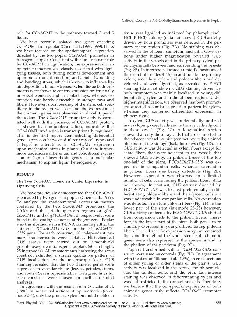
tissue was lignified as indicated by phloroglucinol-HCl (P-HCl) staining (data not shown). GUS activitydriven by both promoters was detected in the pri-mary xylem region (Fig. 2A). No staining was ob-served in the phloem, cambium, and pith. Observa-tions under higher magnification revealed GUSactivity in the vessels and in the primary xylem pa-renchyma cells between and surrounding the vessels(Fig. 2B). In internodes located at middle positions ofthe stem (internodes 8–15), in addition to the primaryxylem, secondary xylem and phloem fibers had de-veloped and were lignified, as revealed by P-HClstaining (data not shown). GUS staining driven byboth promoters was mainly localized in young dif-ferentiating xylem and in the phloem region. Underhigher magnification, we observed that both promot-ers directed a similar expression pattern in xylem,whereas they conferred differential expression inphloem tissue.
In xylem, GUS activity was preferentially localizedin developing vessel cells and in the ray cells adjacentto these vessels (Fig. 2C). A longitudinal sectionshows that only those ray cells that are connected tothe adjacent vessel by pits (contact ray cells) stainedblue but not the storage (isolation) rays (Fig. 2D). NoGUS activity was detected in xylem fibers except forsome fibers that were adjacent to the vessels thatshowed GUS activity. In phloem tissue of the topone-half of the plant, PCCoAOMT1-GUS was ex-pressed in companion cells, whereas expressionin phloem fibers was barely detectable (Fig. 2E).However, expression was observed in a limitednumber of cells surrounding the phloem fibers (datanot shown). In contrast, GUS activity directed byPCCoAOMT2-GUS was located preferentially in dif-ferentiating phloem fibers and the adjacent cells butwas undetectable in companion cells. No expressionwas detected in mature phloem fibers (Fig. 2F). In thelower part of the stem (internode 22–25) however,GUS activity conferred by PCCoAOMT1-GUS shiftedfrom companion cells to the phloem fibers. There-fore, in the lower part of the stem, both genes weresimilarly expressed in young differentiating phloemfibers. The cell-specific expression in xylem remainedthe same throughout the whole stem. Both chimericgenes were also expressed in the epidermis and inthe phellem of the periderm (Fig. 2G).
Poplars transformed with a PCaMV35S-GUS con-struct were used as controls (Fig. 2H). In agreementwith the data of Nilsson et al. (1996), in cross sectionsof either young or older stems of the plants, GUSactivity was localized in the cortex, the phloem tis-sue, the cambial zone, and the pith. Less-intensestaining was observed in differentiating xylem andwas not restricted to the contact ray cells. Therefore,we believe that the cell-specific expression of bothchimeric genes truly reflects CCoAOMT promoteractivity.
Caffeoyl-Coenzyme A-3-O-Methyltransferase Expression in Poplar
Plant Physiol. Vol. 123, 2000 855 www.plant.org on June 28, 2015 - Published by www.plantphysiol.orgDownloaded from Copyright © 2000 American Society of Plant Biologists. All rights reserved.

Figure 2. Histochemical analysis in transgenic poplar showing GUS activity or lignin deposition. A Thick transversal sectionof young stem of poplar transformed with PBINPOP1. B, Enlargement of A showing GUS activity in young xylem cells. C,Thin transversal section of the xylem in the middle part of the stem (PBINPOP1). D, Longitudinal section of the same stemas shown in C. E, Thin transversal section of the bark of the middle part of the stem (PBINPOP1). F, Thin section of the barkof the middle part of the stem (PBINPOP2). G, Thin transversal section of the bark showing GUS staining (PBINPOP1). H,
(Legend continues on facing page.)
Chen et al.
856 Plant Physiol. Vol. 123, 2000 www.plant.org on June 28, 2015 - Published by www.plantphysiol.orgDownloaded from Copyright © 2000 American Society of Plant Biologists. All rights reserved.
A similar temporal and cell-specific expression pat-tern was observed in leaves and petioles. In theleaves and petioles from the top part of the plant,expression of both chimeric genes was located in theprimary xylem where lignification takes place (Fig. 2,I, J, M, and N). In the leaves and petioles from themiddle part of the plant, both phloem fibers andxylem cells were highly lignified as indicated byP-HCl staining (data not shown). GUS staining re-vealed that both chimeric genes were highly ex-pressed in the xylem and PCCoAOMT2-GUS wasadditionally expressed in phloem fibers (Fig. 2, K, L,O, and P). In the leaves and petioles from the basalpart of the plant, staining in the vascular tissue wasless intense, presumably because xylem and phloemfibers were fully lignified at this stage. QuantitativeGUS analyses indicated a GUS activity gradient inthe leaves, with 2- to 3-fold higher levels in theyoungest leaves than in the oldest leaves (data notshown).
In addition to the expression in xylem, phloem, andperiderm, expression conferred by both chimericgenes was often detected in hair cells of young stems,petioles, and leaves. In agreement with the functionof CCoAOMT, these cells stained positively for phe-nolic compounds when treated with P-HCl (Fig. 2, Iand M). Additional expression was observed in themeristem of apical and axillary buds (data notshown). In root tissue, both chimeric genes wereexpressed in phloem fibers, vessels, and contact rays,as in the stem (data not shown).
Both CCoAOMT Promoters Are Responsive toBiotic and Abiotic Stress
In addition to the developmentally regulated dep-osition of lignin, de novo formation of lignin can beinduced at sites of wounding or pathogen attack. Atthese sites, lignin deposition plays a major role inplant defense by strengthening the cell wall to pre-vent the spread of the invading pathogen (Vance etal., 1980; Borg-Olivier and Monties, 1993; Hawkinsand Boudet, 1996). To investigate whether the expres-sion of the chimeric genes is associated with lignindeposition upon wounding, leaves and petioles of2-month-old in vitro as well as 3-month-oldgreenhouse-grown transgenic poplar were wounded(see “Materials and Methods”). Regardless the tissuetype (leaves or petioles), the developmental state(young or mature leaves), or the growth conditions(in vitro or greenhouse-grown), de novo lignin dep-
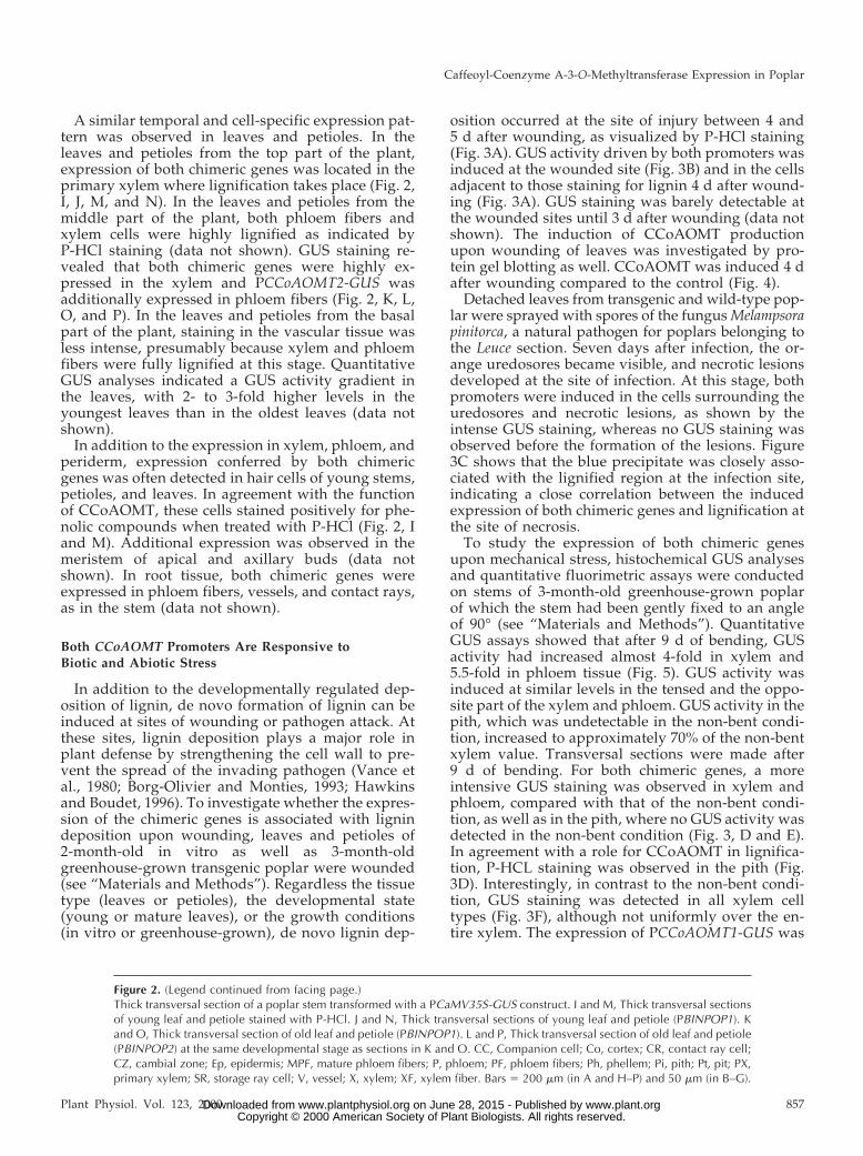
osition occurred at the site of injury between 4 and5 d after wounding, as visualized by P-HCl staining(Fig. 3A). GUS activity driven by both promoters wasinduced at the wounded site (Fig. 3B) and in the cellsadjacent to those staining for lignin 4 d after wound-ing (Fig. 3A). GUS staining was barely detectable atthe wounded sites until 3 d after wounding (data notshown). The induction of CCoAOMT productionupon wounding of leaves was investigated by pro-tein gel blotting as well. CCoAOMT was induced 4 dafter wounding compared to the control (Fig. 4).
Detached leaves from transgenic and wild-type pop-lar were sprayed with spores of the fungus Melampsorapinitorca, a natural pathogen for poplars belonging tothe Leuce section. Seven days after infection, the or-ange uredosores became visible, and necrotic lesionsdeveloped at the site of infection. At this stage, bothpromoters were induced in the cells surrounding theuredosores and necrotic lesions, as shown by theintense GUS staining, whereas no GUS staining wasobserved before the formation of the lesions. Figure3C shows that the blue precipitate was closely asso-ciated with the lignified region at the infection site,indicating a close correlation between the inducedexpression of both chimeric genes and lignification atthe site of necrosis.
To study the expression of both chimeric genesupon mechanical stress, histochemical GUS analysesand quantitative fluorimetric assays were conductedon stems of 3-month-old greenhouse-grown poplarof which the stem had been gently fixed to an angleof 90° (see “Materials and Methods”). QuantitativeGUS assays showed that after 9 d of bending, GUSactivity had increased almost 4-fold in xylem and5.5-fold in phloem tissue (Fig. 5). GUS activity wasinduced at similar levels in the tensed and the oppo-site part of the xylem and phloem. GUS activity in thepith, which was undetectable in the non-bent condi-tion, increased to approximately 70% of the non-bentxylem value. Transversal sections were made after9 d of bending. For both chimeric genes, a moreintensive GUS staining was observed in xylem andphloem, compared with that of the non-bent condi-tion, as well as in the pith, where no GUS activity wasdetected in the non-bent condition (Fig. 3, D and E).In agreement with a role for CCoAOMT in lignifica-tion, P-HCL staining was observed in the pith (Fig.3D). Interestingly, in contrast to the non-bent condi-tion, GUS staining was detected in all xylem celltypes (Fig. 3F), although not uniformly over the en-tire xylem. The expression of PCCoAOMT1-GUS was
Figure 2. (Legend continued from facing page.)Thick transversal section of a poplar stem transformed with a PCaMV35S-GUS construct. I and M, Thick transversal sectionsof young leaf and petiole stained with P-HCl. J and N, Thick transversal sections of young leaf and petiole (PBINPOP1). Kand O, Thick transversal section of old leaf and petiole (PBINPOP1). L and P, Thick transversal section of old leaf and petiole(PBINPOP2) at the same developmental stage as sections in K and O. CC, Companion cell; Co, cortex; CR, contact ray cell;CZ, cambial zone; Ep, epidermis; MPF, mature phloem fibers; P, phloem; PF, phloem fibers; Ph, phellem; Pi, pith; Pt, pit; PX,primary xylem; SR, storage ray cell; V, vessel; X, xylem; XF, xylem fiber. Bars 5 200 mm (in A and H–P) and 50 mm (in B–G).
Caffeoyl-Coenzyme A-3-O-Methyltransferase Expression in Poplar
Plant Physiol. Vol. 123, 2000 857 www.plant.org on June 28, 2015 - Published by www.plantphysiol.orgDownloaded from Copyright © 2000 American Society of Plant Biologists. All rights reserved.

also up-regulated in phloem fibers upon bending(Fig. 3F), whereas its expression was barely detect-able in this tissue in the non-bent condition (Fig. 2E).However, bending did not induce GUS activity inphloem companion cells in PCCoAOMT2-GUS-transformed poplars. Thus, bending stress resulted inthe loss of the cell-specific expression pattern in thevascular tissue and in a patchy expression of bothgenes in all lignifying cells of the xylem and phloem.
Immunolocalization of CCoAOMT in Poplar Stem
To verify whether the cell-specific and conditionalexpression driven by the CCoAOMT promoters coin-cided with the localization of the CCoAOMT protein,immunolocalization experiments were carried out onsimilar stem sections as those used for the GUS as-says with antiserum against alfalfa CCoAOMT (Ker-sey et al., 1999). With these antibodies, a single 28-kDprotein band was detected in the protein extractsfrom wild-type poplar stem by protein gel-blot anal-ysis, whereas no signal was detected in protein ex-tracts from transgenic poplars down-regulated forCCoAOMT (H. Meyermans and W. Boerjan, unpub-
lished results). This observation indicates that thealfalfa CCoAOMT antibodies can specifically recog-nize CCoAOMT in poplar. Poplar CCoAOMT waslocalized by immunogold complexes with silver en-hancement and examined by light microscopy. Ingeneral, labeling was present in differentiatingphloem and xylem. Virtually no labeling appeared inthe pith, cambium, and cortex. In sections throughthe top part of the stem, CCoAOMT was present inprimary xylem bundles (Fig. 6A), which is in agree-ment with the result from promoter-GUS assays (Fig.
Figure 4. Protein gel-blot analysis of poplar protein extracts. Crudeprotein extracts from poplar control leaf (CL) and wounded leaf (WL)separated by SDS-PAGE, immobilized on a nitrocellulose membrane,and incubated with anti-CCoAOMT antibodies. The protein molec-ular mass is shown at the left in kD.
Figure 3. CCoAOMT promoter activity in transgenic poplar upon biotic and abiotic stress conditions. A, Transversal sectionof a petiole stained for both lignin and GUS. B, GUS activity in a mechanically wounded leaf. C, M. pinitorca-infected leafstained for both lignin deposition and GUS activity. D, Double staining of a thick transversal section of a bent stem for ligninand GUS activity. E, Thick transversal section of a non-bent stem stained by P-HCl as control. F, Enlargement of a thintransversal section of the bent stem, transformed with PBINPOP1. CZ, Cambial zone; F, site of fungal infection; P, phloem;PF, phloem fibers; Pi, pith; W, wound; X, xylem. Bars 5 200 mm (in A, B, D, and E), 100 mm (in C), and 50 mm (in F).
Chen et al.
858 Plant Physiol. Vol. 123, 2000 www.plant.org on June 28, 2015 - Published by www.plantphysiol.orgDownloaded from Copyright © 2000 American Society of Plant Biologists. All rights reserved.

2B). In the middle and lower part of stem, CCoAOMTwas detected in differentiating xylem (Fig. 6B), in dif-ferentiating phloem fibers but not in mature ones (Fig.6C), as well as in the epidermis (Fig. 6D). Very faintlabeling was observed in the companion cells (datanot shown). In xylem tissue, as shown in Figure 6B,CCoAOMT was preferentially localized in contact raycells associated with vessels and in the vessels them-selves. This cell-specific localization of CCoAOMT insecondary xylem is similar to that of GUS directed byboth CCoAOMT promoters (Fig. 2, A–G). No signalcould be observed in control sections of the same stemtreated with the preimmune serum (Fig. 6H) nor in thesections of the transgenic poplar down-regulated forCCoAOMT (Fig. 6E).
To confirm that upon mechanical stress the cell-specific expression of CCoAOMT was altered, immu-nolocalization experiments were carried out on bentstems. As shown in Figure 6F, CCoAOMT was sig-nificantly induced in xylem tissue of stems that werebent for 9 d. In agreement with the induction ofCCoAOMT promoter activity upon bending, immu-nolabeling was detected in all cell types of the xylemtissue and most intensely in the rays cells. In additionto mechanical bending, poplars were leaned at anangle of 45°, and sections were made through thecurved part of the stem (see “Materials and Meth-ods”). Also under this condition, the expression ofCCoAOMT was significantly induced in all cell typesof the xylem (Fig. 6G) and revealed an expressionpattern similar to that after bending.
Subcellular Localization of CCoAOMT
It is still largely unclear where monolignol biosyn-thesis occurs at the subcellular level. Several studieshave suggested the existence of multienzyme com-
plexes, other studies have shown the association oflignin biosynthesis enzymes with cellular organelles(Smith et al., 1994; Nakashima et al., 1997; Samaj etal., 1998; Rasmussen and Dixon, 1999). To investigatewhether CCoAOMT is entirely cytosolic or fraction-ally associated with membranes, we have localizedCCoAOMT by electron microscopy on sections sim-ilar to those used for light microscopy and found asimilar cellular distribution of CCoAOMT. Along thedifferentiating xylem, immunogold particles ap-peared mostly in the contact rays and in the vessels,although in the vessels only a small layer of cyto-plasm remained prior to autolysis. Interestingly, atmore advanced stages of xylem development, afterautolysis of the vessels, no immunogold particlescould be detected anymore neither in mature vesselsnor in the contact rays adjacent to those mature ves-sels. Furthermore, labeling was observed in differen-tiating but not in mature phloem fibers, in the com-panion cells, in the epidermis, and in the phellem ofthe periderm, consistent with the promoter-GUS dataand the immunolocalizations carried out by lightmicroscopy. No signal was found in the cambium,pith, or cortex region (data not shown). A more de-tailed observation showed that the silver-enhancedparticles were randomly localized in the cytoplasmand were not located on the cell wall or associatedwith any organelles (Fig. 7A). When similar sectionswere incubated with preimmune serum, no specificbinding of gold particles was observed (Fig. 7B).
DISCUSSION
Here, we have shown that the two CCoAOMT pro-moters direct expression in lignifying cells and incells closely associated with lignifying cells, bothduring normal development and upon stress condi-tions known to influence lignin deposition (wound-ing, pathogen attack, and mechanical bending). Thesedata are consistent with a role for CCoAOMT in lig-nification. Furthermore, both chimeric CCoAOMTgenes were expressed in the epidermis (includinghair cells) of young stems, leaves, petioles, and in theshoot apex. The gPtCCoAOMT1 promoter addition-ally directed expression in phloem companion cells,which are generally accepted to be not lignified oronly poorly lignified. The deposition of phenoliccompounds in non-lignified cells or tissues mayserve important protective functions. In these cells,CCoAOMT may for example be involved in the bio-synthesis of lignans, which share the same monolig-nol precursors as lignin. Lignans are an abundantclass of phytochemicals in plants that play a role indefense, but little is known about their biosynthesisand their site of accumulation (Davin et al., 1992;Higuchi, 1997). The promoter of the eucalyptus cin-namyl alcohol dehydrogenase (CAD2) gene also con-ferred expression in poorly lignified cells, such as theperiderm of stems, cortical cells containing crystals,
Figure 5. Induction of GUS activity in transgenic poplar stems as aconsequence of mechanical bending. GUS activity was measured inxylem, phloem, and pith tissues. The data represent the average ofthree independent experiments. The SE is shown.
Caffeoyl-Coenzyme A-3-O-Methyltransferase Expression in Poplar
Plant Physiol. Vol. 123, 2000 859 www.plant.org on June 28, 2015 - Published by www.plantphysiol.orgDownloaded from Copyright © 2000 American Society of Plant Biologists. All rights reserved.

Figure 6. Immunolocalization of CCoAOMT in transversal sections of poplar stem by light microscopy. A, Young stem of3-month-old greenhouse-grown poplar. B, Xylem tissue of the middle part of a 3-month-old greenhouse-grown poplar stem.C and D, Bark tissue of old and young stem. E, Stem of transgenic poplar down-regulated for CCoAOMT. F, Bent stem. G,Leaned stem. H, Control section treated with preimmune serum. Co, Cortex; CR, contact ray cell; CZ, cambial zone; Ep,epidermis; F, phloem; MPF, mature phloem fibers; Pe, periderm; PF, phloem fibers; Ph, phellem; Pi, pith; PX, primary xylem;SR, storage ray cell; V, vessel; X, xylem; XF, xylem fiber. Bars 5 100 mm.
Chen et al.
860 Plant Physiol. Vol. 123, 2000 www.plant.org on June 28, 2015 - Published by www.plantphysiol.orgDownloaded from Copyright © 2000 American Society of Plant Biologists. All rights reserved.

companion cells, and cambial cells in transgenic pop-lar (Feuillet et al., 1995; Hawkins et al., 1997; Samaj etal., 1998). CAD2 catalyzes the conversion of cinna-maldehydes to cinnamyl alcohols (Fig. 1) and mayalso be involved in lignan synthesis in these cells(Hawkins et al., 1997). The expression data addition-ally show that both promoters are multifunctionalrather than diverged where each promoter hasadopted separate functions. This result is in contrast tothe 4-coumarate:CoA ligase (4CL) genes of poplar:Pt4CL1 is expressed in cells that participate in thedevelopmentally regulated lignification process(mainly in xylem), whereas the Pt4CL2 confers expres-sion in the epidermis of stem and leaf where it func-tions in the biosynthesis of phenylpropanoids otherthan lignin (Hu et al., 1998). Because immunolocaliza-tion of CCoAOMT showed the same tissue and cell-specific localization as revealed by GUS assays, we canconclude that the expression of CCoAOMT is mainlyregulated at the transcriptional level.
CCoAOMT Is Expressed in Vessels and ContactRay Cells
For several genes involved in lignin biosynthesis,the tissue-specific expression has been analyzed. Forexample, a bean Phe ammonia-lyase (PAL)-GUS fu-sion in tobacco and potato was expressed in all de-veloping xylem cells including ray cells (Bevan et al.,1989). Hauffe et al. (1991) reported that P4CL-GUSexpression was localized preferentially in differenti-ating xylem tissue and in xylem ray cells betweenhighly lignified vessels and fibers in transgenic to-bacco. The promoter of the CAD2 gene from eucalyp-tus conferred expression in phloem fibers, in the raysof differentiating xylem, and in the vascular cam-bium in transgenic poplar (Feuillet et al., 1995;Hawkins et al., 1997).
In contrast to the expression pattern conferred bythe PAL, 4CL, and CAD2 promoters, the promoter ofCCoAOMT was differentially expressed in differentray cell types. Under normal, non-stressed growthconditions, PCCoAOMT-GUS expression was prefer-entially observed in the contact ray cells that areconnected to vessels through pits and not in thestorage ray cells (isolation rays). These two types ofray cells have first been reported by Czaninski (1977),and they differ with respect to their contacts, throughpits, with vessels. In dicotyledonous plants, pits areformed in the secondary xylem between a paren-chyma cell and a conducting cell (vessel). Such pitsare considered to play an important role during xy-lem cell differentiation, providing channels throughwhich not only wall precursors and other metabolitesmay be transferred into the developing cell, but alsosignals that control their differentiation (for review,see Mezitt and Lucas, 1996). Our electron microscopystudies furthermore showed that at the stage thatvessels are still alive, as judged by the presence ofcytoplasm, CCoAOMT was present both in vesselsand in the adjacent contact rays. However, uponautolysis of the vessel, expression of CCoAOMT wascoincidentally switched off both in the vessels and inthe contact rays. The similarity in timing ofCCoAOMT expression in vessels and contact rayssuggests that the vessels and contact rays share acommon signal that triggers expression of theCCoAOMT genes in both cell types and that maybe transported through the connecting pits. This hy-pothesis is in agreement with recent data ofMurakami et al. (1999), who have shown by UVabsorption of cell walls that lignification of the cellwall of contact rays and vessels in Populus maximow-iczii occurs simultaneously and prior to that of thestorage rays and fibers.
The observation that the CCoAOMT promotersdrive expression in vessels and contact rays furtherraises the question whether the lignin composition ofthese cells differs from that of xylem fibers and stor-age rays. Higuchi (1997) has reported the predomi-nant presence of G units in the lignin of vessel cell
Figure 7. Subcellular localization of CCoAOMT in poplar stem byelectron microscopy. A, Contact ray cell from a stem section of themiddle part of a 3-month-old greenhouse-grown poplar. The labelingwith alfalfa CCoAOMT antibodies is concentrated in the cytosol. B,control section treated with preimmune serum. CR, Contact ray cell;Cyt, cytoplasm; SW, secondary wall. Arrows indicate gold particles.Bars 5 1 mm.
Caffeoyl-Coenzyme A-3-O-Methyltransferase Expression in Poplar
Plant Physiol. Vol. 123, 2000 861 www.plant.org on June 28, 2015 - Published by www.plantphysiol.orgDownloaded from Copyright © 2000 American Society of Plant Biologists. All rights reserved.

walls in angiosperm trees. The analysis of transgenicplants down-regulated for CCoAOMT and in vitroenzymatic assays support the idea that the differencein lignin composition is indeed due to differentialexpression of CCoAOMT. Zhong et al. (1998) haveshown that lignin from transgenic tobacco plantsdown-regulated for CCoAOMT, apart from having alower total Klason lignin content, is more depressedin G than in S units. Similar results were obtained intransgenic poplar, down-regulated for CCoAOMT(H. Meyermans, unpublished data).
Based on in vitro enzymatic assays, Osakabe et al.(1999) have recently proposed that the lignin precur-sors are predominantly synthesized via coniferalde-hyde and not via ferulic acid, 5-hydroxyferulic acid,and sinapic acid. These data are based on the obser-vation that coniferaldehyde inhibits the coniferalde-hyde 5-hydroxylase/ferulic acid 5-hydroxylase re-action from ferulate to 5-hydroxyferulate in vitro.The cell-specific expression of CCoAOMT suggeststhat in vessels and contact rays, G and S units maybe formed predominantly via caffeoyl-CoA andferuloyl-CoA, whereas in fibers and storage ray cellsG and S units may be formed preferentially via caf-feic acid, ferulic acid, and feruloyl-CoA. Based on thefact that COMT plays an important role in controllingS-unit biosynthesis (Atanassova et al., 1995; VanDoorsselaere et al., 1995; Tsai et al., 1998; Lapierreet al., 1999) by converting 5–hydroxyconiferaldehydeand 5–hydroxyconiferyl alcohol to sinapaldehyde andsinapyl alcohol, respectively (Humphreys et al., 1999;Osakabe et al., 1999), the conversion of caffeic acid toferulic acid may also be less efficient in cells that makeless S units. In vessels, where the S to G ratio is lowcompared with fibers (Saka and Goring, 1985),CCoAOMT may effectively bypass the COMT-mediated conversion of caffeic acid to ferulic acid.Together, the cell-specific expression of CCoAOMTand the different lignin composition in these cellssuggest that the metabolic flux through the phenyl-propanoid and lignin biosynthesis pathway is dif-ferent in different cell types.
CCoAOMT Promoters Are Responsive to Wounding andFungal Attack
The responses of plants to mechanical woundingand pathogen attack are often very similar and in-clude the rapid accumulation of phenolic com-pounds, the production of phytoalexins, the synthe-sis of hydrolytic enzymes, and the reinforcement ofcells with lignin and/or suberin at the site of injury(Blanchette and Biggs, 1992). In most of the casesexamined, the appearance of such defense substancesis the result of increased gene expression eitherwithin the affected tissue or throughout the plant.Most of the genes that code for enzymes of the ligninbiosynthesis pathway, such as PAL, 4CL, COMT, andcinnamate 4-hydroxylase (C4H), have been reported
to be induced by wounding, elicitors, and/or fungalinfection (Baucher et al., 1998). CCoAOMT activityhas been extensively studied in response to elicitorsin cell suspension culture (Kneusel et al., 1989; Kuhnlet al., 1989; Schmitt et al., 1991; Ni et al., 1996; Busamet al., 1997). Transient expression assays performedwith PCCoAOMT-GUS constructs in parsley proto-plasts showed a high expression level in response toelicitation (Grimmig and Matern, 1997). In tobacco,RNA gel-blot analyses showed an increased steady-state CCoAOMT mRNA level upon infection withtobacco mosaic virus or fungal elicitor (Martz et al.,1998). In our study the expression of both chimericCCoAOMT genes was strongly induced at the woundsite and at the infection site of leaves, concomitantlywith the deposition of lignin or lignin-like material.That GUS activity was observed in the cell layeradjacent to the lignifying cell layer suggests thatthese cells provide monolignols to their neighboringcells for lignification or the production of lignin-likematerial. These results show that the induced forma-tion of lignin or lignin-like material is closely corre-lated with the cellular localization of CCoAOMT geneexpression in response to both wounding and fungalattack in plants.
CCoAOMT Promoters Are Responsive toMechanical Stress
Mechanical stress caused by leaning stems resultsin compression wood in gymnosperms and tensionwood in angiosperms (Timell, 1986; Castera et al.,1994). Both compression and tension wood have beenshown to have an altered lignin content and compo-sition compared with normal non-stressed wood (Ti-mell, 1986; Rolando et al., 1992). In contrast to theintensive study of lignin biosynthesis enzymes, suchas PAL, 4CL, COMT, CAD, and cinnamoyl-CoA re-ductase in compression wood (Kutsuki and Higuchi,1981; Zhang and Chiang, 1997), little information isavailable on gene expression in tension wood. Here,we report on a gene that is induced upon bending ina hardwood species. The expression of both chimericCCoAOMT genes was up-regulated in all lignifyingtissues of the stem 9 d after mechanical bending.There was a pronounced increase in GUS activity inxylem and phloem and in the pith cells concomi-tantly with the deposition of lignin or lignin-likematerial in the pith. It is noteworthy that in responseto mechanical bending, the cell-specific expressionpattern directed by both CCoAOMT promoters in thestem was lost.
Mechanical stress may also be the reason whyPCCoAOMT1-GUS expression was observed inphloem fibers at the base of the stem and in the root,whereas no activity was observed in phloem fibers inthe top one-half of the stem. Immunolocalization ofCCoAOMT on sections of stems that were bent orleaned were in agreement with the promoter-GUS
Chen et al.
862 Plant Physiol. Vol. 123, 2000 www.plant.org on June 28, 2015 - Published by www.plantphysiol.orgDownloaded from Copyright © 2000 American Society of Plant Biologists. All rights reserved.

analyses and show that the induction of CCoAOMTexpression upon mechanical stress is regulated at thetranscriptional level. Considering the involvement ofCCoAOMT in lignification, our observations suggestthat the up-regulation of CCoAOMT in a non-cell-type-specific manner may alter lignin composition orcontent. However, Vander Mijnsbrugge et al. (2000)have recently reported that a poplar phenylcouma-ran benzylic ether reductase, which is the most abun-dant protein in poplar xylem and involved in thebiosynthesis of lignans (Gang et al., 1999), is up-regulated in xylem, phloem, and pith tissues ofpoplar stem in response to mechanical bending.Therefore, up-regulation of CCoAOMT in responseto bending may also be involved in the synthesis oflignans apart from lignin. The analysis of ligninamount and composition as well as soluble pheno-lics from transgenic poplar, down-regulated forCCoAOMT, may shed further light on the fate of theproduced monolignol intermediates upon mechani-cal stress.
CCoAOMT Is a Cytosolic Protein
The phenylpropanoid pathway is involved in thebiosynthesis of a wide variety of natural productsfrom plants. Little is known about the mechanism thecell uses to regulate the flux into the different endproducts of the pathway. Metabolic labeling experi-ments have suggested the existence of multienzymecomplexes that channel intermediates of phenylpro-panoid synthesis without their release into generalmetabolic pools (Stafford, 1981; Hrazdina, 1992; Ras-mussen and Dixon, 1999). Several of the enzymes ofthe phenylpropanoid and lignin-specific pathwayshave been studied for their subcellular distributionby electron microscopy. In French bean, PAL hasbeen localized in the cytosol whereas C4H was asso-ciated with the endoplasmic reticulum membraneand Golgi bodies (Smith et al., 1994). In poplar, CAD2has been localized in the cytosol, on endoplasmicreticulum membranes and on Golgi-derived vesicles(Samaj et al., 1998). In tracheary elements derivedfrom zinnia mesophyll cells, two different types ofPAL and CAD exist. For both enzymes, one isoformwas shown to be cytosolic and the other associated toGolgi-derived vesicles and secondary cell walls (Na-kashima et al., 1997). Recently, Rasmussen and Dixon(1999) have presented evidence for metabolic chan-neling, involving coupling of PAL and C4H, based onin vivo and in vitro labeling. These authors demon-strated that one form of PAL (PAL1) was associatedwith tobacco microsomes and was involved in chan-neling, and suggested that this isoform is in closephysical association with C4H on microsomal mem-branes. Our results show that CCoAOMT is randomlydistributed in the cytoplasm and is not directly in-volved in multienzyme complexes that are present atthe membrane.
In summary, our data show that the CCoAOMTpromoters are responsive to signals that control lig-nin deposition throughout plant development andadjust lignin quality according to environmental con-ditions. Taken together with the differentialCCoAOMT promoter activity in the different xylemcell types, our observations may explain, at least partof, the large heterogeneity in lignin amount and com-position that is observed between different cell typesand within individual cell walls (Joseleau and Ruel,1997; Baucher et al., 1998). Extensive promoter com-parisons and deletion analyses are necessary to iden-tify the cis elements that are involved in the re-sponses to the different signals that induceCCoAOMT promoter activity.
MATERIALS AND METHODS
Plasmid Constructions
The characterization of gPtCCoAOMT1 (accession no.AJ223620) and gPtCCoAOMT2 (accession no. AJ223621) hasbeen described previously (Chen et al., 1998, 1999). To fusethe 59-untranslated region sequences of both CCoAOMTgenes to the coding sequence of the GUS gene, an NcoI sitewas generated by PCR three codons downstream of theCCoAOMT start codon by using an oligonucleotide com-plementary to the 59 flanking vector DNA in combinationwith the 21-mer oligonucleotide 59-CTCTCCCATGGTGGCCATTAT-39 and 59-CTCTCCCATGGCGGCCATTAT-39 forgPtCCoAOMT1 and gPtCCoAOMT2, respectively (a singleline indicates the NcoI site and the reversed original startcodon is bold and underlined. By using these oligonucle-otides, the 1,994-bp and 1,363-bp promoter fragments ofgPtCCoAOMT1 and gPtCCoAOMT2 were generated byPCR, respectively. Subsequently, both PCR products weredigested with NcoI and SacI and cloned into the NcoI/SacIsite of pGUS1 (Peleman et al., 1989), yielding the plasmidsPGUSPOP1 and PGUSPOP2. Both chimeric PCCoAOMT1-GUS and PCCoAOMT2-GUS genes were subsequently iso-lated from PGUSPOP1 and PGUSPOP2, respectively, by anXbaI digest, and cloned into the XbaI site of the binaryvector PBIN19 (Bevan, 1984), resulting in the plasmidsPBINPOP1 and PBINPOP2, respectively.
Plant Material and Transformation
PBINPOP1, PBINPOP2, and PCaMV35-GUS (Nilsson etal., 1996) were mobilized to Agrobacterium tumefaciens strainC58C1Rif harboring the plasmid pMP90 by the freeze-thawmethod described by Zahm et al. (1984). Poplar (Populustremula 3 Populus alba Institut National de la RechercheAgronomique clone 717-1B4) was stably transformed withall three constructs according to Leple et al. (1992). Eachprimary transformant was derived from a different ex-plant. The plants were maintained and micropropagated invitro on one-half Murashige and Skoog medium at 24°Cwith a 16-h light/8-h dark cycle. Five-week-old plants weretransferred to soil and further grown in a greenhouse at21°C with the same light/dark cycle.
Caffeoyl-Coenzyme A-3-O-Methyltransferase Expression in Poplar
Plant Physiol. Vol. 123, 2000 863 www.plant.org on June 28, 2015 - Published by www.plantphysiol.orgDownloaded from Copyright © 2000 American Society of Plant Biologists. All rights reserved.

Histochemical GUS Assays
Histochemical staining for GUS activity was performedessentially as described by Jefferson et al. (1987) andHawkins et al. (1997). Small pieces of stems, roots, leaves,and petioles were excised and pretreated with 95% (v/v)acetone for 30 min at room temperature to prevent woundinduction and rinsed three times with 100 mm potassiumphosphate buffer (pH 7.0). GUS staining was carried out byincubating sections with 2 mm 5-bromo-4-chloro-3-indolyl-b-d-glucuronide, 0.1 mm potassium ferricyanide, and 0.1mm potassium ferrocyanide in 100 mm potassium phos-phate buffer (pH 7.0). Staining was allowed to proceed at37°C until blue stain developed in the samples (1–4 h).Subsequently, samples were fixed in 3% (v/v) glutaralde-hyde in 100 mm potassium phosphate buffer (pH 7.0) over-night at 4°C, washed in the same buffer, and embedded inHistoform according to the manufacturer’s protocol (His-toresin Embedding Kit, Heraeus Kulzer, Wehrheim, Ger-many). Thin sections (8 mm) were cut on a microtome(Reichert-Jung, Nussloch, Germany). For thick slices, smallpieces of stems, leaves, and petioles were pretreated with95% (v/v) acetone, embedded in 7% (w/v) agarose, andsectioned (75–150 mm) with a vibroslicer (Laborimpex,Brussels). GUS staining was carried out as described above.All the sections were mounted on microscope slides forphotography (Diaplan, Leitz, Wetzlar, Germany).
Quantitative GUS Assays
Quantitative GUS analyses were performed as describedby Jefferson et al. (1987). Protein concentrations were mea-sured with the method of Bradford (1976). GUS activity wasassayed by enzymatic conversion of 4-methylumbelliferyl-b-d-glucuronide to 4-methylumbelliferone which was quanti-fied with a fluorimeter (Labsystems Fluoreskan II, Helsinki).GUS activity was expressed in units per microgram ofprotein.
Lignin Staining
Lignin and/or phenolic compounds were revealed byP-HCl staining according to Speer (1987). Sections andsamples were incubated for 2 min in phloroglucinol solu-tion (1% in ethanol:water, 92:8 [v/v]) and then mounted in25% (v/v) HCl, prior to microscopic examination.
Wounding, Bending, Leaning, and Pathogen Infection
For the wounding experiments, petioles of 3-month-old,greenhouse-grown plants were wounded by making a 0.5-cm-long slit with a scalpel. Wounding of the leaves wasdone on plants that were grown axenically in glass jars for2 months. From each of six comparable plants, one-half ofa leaf was removed using a sterile scalpel, and a strip of leaftissue of approximately 2 mm bordering the cutting sitewas harvested after 0, 1, 2, 3, 4, and 5 d after wounding.The harvested samples were analyzed by histochemistryfor GUS activity and lignin deposition as well as by proteingel-blot analysis.
For the bending experiments, stems of 3-month-oldgreenhouse-grown wild-type and transgenic poplars weregently fixed at a 90° angle at internode 3 to 4 counted fromthe top of the stem for various periods. Non-bent stems ofthe same, vegetatively propagated transgenic line wereused as control. The bent part of the stem was collected forGUS assays and immunolocalization experiments. Forquantitative GUS assays, the xylem, phloem, and pith tis-sues from the bent stems were collected separately andimmediately frozen in liquid nitrogen. The harvested sam-ples were stored at 270°C until analysis.
Alternatively, 3-month-old greenhouse-grown poplarswere leaned at 45° for 9 d. The upper part of the stem(approximately 15–25 cm) grows vertically after leaning theplant. The samples for immunolocalization were takenfrom the curved part of the plant, i.e. between the obliqueand the vertical part of the stem.
Spores of the fungus Melampsora pinitorca were collectedfrom leaves of outside-grown poplar trees and determinedaccording to Pinon (1973). For fungal infection, detachedleaves were put upside down on wet filter paper in 9-cmPetri dishes. Subsequently, these leaves were sprayed for2 s with spores of the fungus, in a concentration of 200,000spores mL21, using a spray machine (Aku-SpruhpistoleW50, Wagner, Markdorf, Germany). The infected leaveswere subsequently floated upside down on water in 9-cmPetri dishes and incubated at 22°C in a greenhouse forvarious periods.
Protein Gel Blotting
Proteins were extracted with 100 mm Tris (tris-[hydroxymethyl]aminomethane)-HCl (pH 7.5), 2 mmEDTA, 20% (v/v) glycerol, and 1 mm dithiothreitol. Protein(5 mg) from leaves was separated by SDS-PAGE. The pro-tein gel was subsequently blotted on Hybond-C supermembrane according to the manufacturer’s instructions(Amersham, Aylesbury, UK). After incubation with anti-serum raised against alfalfa CCoAOMT (Kersey et al.,1999), filters were incubated with anti-rabbit IgG alkalinephosphatase conjugate (Roche Diagnostics, Brussels) at adilution of 1/2,500. The immunodetection was performedusing an alkaline phosphate p-toluidine salt (Duchefa,Haarlem, The Netherlands) and p-nitroblue tetrazoliumchloride (Duchefa) in the detection reaction.
Sample Preparation for Light and Electron Microscopy
Small pieces of tissue harvested from non-stressed stem,wounded stem, and stems that had been bent or leanedwere cut into pieces of approximately 1 mm3. These pieceswere immersed in fixation solution (2.5% [v/v] parafor-maldehyde and 0.3% [v/v] glutaraldehyde in 0.1 msodium-cacodylate buffer [21.4 g Na(CH3)2AsO2 z 3H2O2
in 1,000 mL of distilled water], pH 7.2) under vacuum for4 h at room temperature and then incubated for 14 h at 4°Cunder rotation. Following three washes for 2 h in 0.1 msodium-cacodylate buffer (pH 7.2) at 4°C, the samples weredehydrated through a graded ethanol series under rotation
Chen et al.
864 Plant Physiol. Vol. 123, 2000 www.plant.org on June 28, 2015 - Published by www.plantphysiol.orgDownloaded from Copyright © 2000 American Society of Plant Biologists. All rights reserved.
at 4°C as follows: 30% (v/v) ethanol for 2 h, 50% (v/v)ethanol for 2 h, 70% (v/v) ethanol overnight, 95% (v/v)ethanol for 2 h, and 95% (v/v) ethanol overnight. Thesamples were embedded by incubating them successivelyin a 1:1 (v/v) ratio of 95% (v/v) ethanol:LR White (LondonResin Co., Basingstoke, UK) at 4°C overnight and then forthree changes, at least 8 h each, in pure resin at 4°C. Thesamples were placed in gelatin capsules containing freshnitrogen-fluxed resin. Polymerization was performed byUV illumination at 4°C for 24 h followed by 24 h at 65°C toensure complete polymerization of the resin. These LRWhite-embedded samples were ready to be sectioned forimmunocytochemical localization of proteins using light orelectron microscopy.
Light Microscopy
LR White-embedded samples were cut in semithin sec-tions (1–3 mm) using a 2050 microtome (Reichert-Jung). Thesections were collected on Vectabond-coated glass slides.Rabbit polyclonal anti-CCoAOMT antibodies (Kersey et al.,1999) were used as primary antibody. The AuroProbe Oneand IntenSE reagents were used for immunogold silverstaining according to the manufacturer’s protocol (Amer-sham). Sections were examined with a light microscopeJenalumar (Zeiss, Jena, Germany).
Electron Microscopy
Ultrathin sections of gold interference color (60–90 nm)were made from the LR White-embedded samples using anultracut E (Reichert-Jung), and were collected on collodion-coated Cu grids. These sections were floated with thetissue-containing side downward for 5 min on a droplet ofblocking solution (0.1% [w/v] bovine serum albumin and0.05% [w/v] NaN3 in phosphate-buffered saline) to avoidnon-specific adsorption of the antibodies. Subsequently,the sections were incubated for 1 h with primary antiserumraised against alfalfa CCoAOMT (Kersey et al., 1999), di-luted 1:100 in blocking solution, followed by a 30-minincubation with a protein A-gold conjugate (15 nm; Amer-sham) diluted 1:50 in gold buffer (1% [w/v] bovine serumalbumin and 0.05% [w/v] NaN3 in phosphate-bufferedsaline). Sections were washed three times with gold buffer,three times with distilled water, and then allowed to air-dry. The grids were post-stained for 12 min with 2% (w/v)uranyl acetate, washed five times with distilled water, andthen left to air dry. Sections were examined using trans-mission electron microscopy (Elmiskop 101, Siemens,Karlsruhe, Germany).
ACKNOWLEDGMENTS
The authors thank Bjorn Sundberg for helpful discus-sions, Sabrina Neyrinck for technical assistance, Jan VanDoorsselaere and Jørgen Holst Christensen for criticalreading of the manuscript, Martine De Cock for help withpreparing it, and Rebecca Verbanck for the figures.
Received December 30, 1999; accepted March 11, 2000.
LITERATURE CITED
Atanassova R, Favet N, Martz F, Chabbert B, Tollier MT,Monties B, Fritig B, Legrand M (1995) Altered lignincomposition in transgenic tobacco expressingO-methyltransferase sequences in sense and antisenseorientation. Plant J 8: 465–477
Baucher M, Monties B, Van Montagu M, Boerjan W (1998)Biosynthesis and genetic engineering of lignin. Crit RevPlant Sci 17: 125–197
Bevan M (1984) Binary Agrobacterium vectors for planttransformation. Nucleic Acids Res 12: 8711–8721
Bevan M, Shufflebottom D, Edwards K, Jefferson R,Schuch W (1989) Tissue- and cell-specific activity of aphenylalanine ammonia-lyase promoter in transgenicplants. EMBO J 8: 1899–1906
Blanchette RA, Biggs AR (1992) Defense Mechanisms ofWoody Plants against Fungi (Springer Series in WoodScience). Springer-Verlag, Berlin
Borg-Olivier O, Monties B (1993) Lignin, suberin, phenolicacids and tyramine in the suberized, wound-inducedpotato periderm. Phytochemistry 32: 601–606
Boudet A-M (1998) A new view of lignification. TrendsPlant Sci 3: 67–71
Boudet AM, Lapierre C, Grima-Pettenati J (1995) Bio-chemistry and molecular biology of lignification. NewPhytol 129: 203–236
Bradford MM (1976) A rapid and sensitive method for thequantitation of microgram quantities of protein utilizingthe principle of protein-dye binding. Anal Biochem 72:248–254
Busam G, Junghanns KT, Kneusel RE, Kassemeyer H-H,Matern U (1997) Characterization and expression ofcaffeoyl-coenzyme A 3-O-methyltransferase proposedfor the induced resistance response of Vitis vinifera L.Plant Physiol 115: 1039–1048
Campbell MM, Sederoff RR (1996) Variation in lignincontent and composition: mechanisms of control andimplications for the genetic improvement of plants. PlantPhysiol 110: 3–13
Castera P, Nepveu G, Mahe F, Valentin G (1994) A studyon the growth stresses, tension wood distribution andother related wood defects in poplar (Populus eurameri-cana cv I214): end splits, specific gravity and pulp yield.Ann Sci For 51: 301–313
Chen C, Ardiles-Diaz W, Van Montagu M, Boerjan W(1999) A poplar gene for caffeoyl-coenzyme A 3-O-methyltransferase (accession no. AJ223620) (PGR99–085). Plant Physiol 120: 635
Chen C, Meyermans H, Van Doorsselaere J, Van MontaguM, Boerjan W (1998) A gene encoding caffeoyl coenzymeA 3-O-methyltransferase (CCoAOMT) from Populus tri-chocarpa (accession no. AJ223621) (PGR 98–104). PlantPhysiol 117: 719
Czaninski Y (1977) Vessel-associated cells. Int Assoc WoodAnat Bull 3: 51–55
Davin LB, Lewis NG, Umezawa T (1992) Phenylpropanoidmetabolism: biosynthesis of monolignols, lignans andneolignans, lignins and suberins. In HA Stafford, RKIbrahim, eds, Phenolic Metabolism in Plants, Recent Ad-
Caffeoyl-Coenzyme A-3-O-Methyltransferase Expression in Poplar
Plant Physiol. Vol. 123, 2000 865 www.plant.org on June 28, 2015 - Published by www.plantphysiol.orgDownloaded from Copyright © 2000 American Society of Plant Biologists. All rights reserved.
vances in Phytochemistry, Vol 26. Plenum Press, NewYork, pp 325–375
Dixon RA, Lamb CJ, Masoud S, Sewalt VJ, Paiva NL (1996)Metabolic engineering: prospects for crop improvementthrough the genetic manipulation of phenylpropanoidbiosynthesis and defense responses: a review. Gene 179:61–71
Douglas C (1996) Phenylpropanoid metabolism and ligninbiosynthesis: from weeds to trees. Trends Plant Sci 1:171–178
Feuillet C, Lauvergeat V, Deswarte C, Pilate G, Boudet A,Grima-Pettenati J (1995) Tissue- and cell-specific expres-sion of a cinnamyl alcohol dehydrogenase promoter intransgenic poplar plants. Plant Mol Biol 27: 651–667
Gang DR, Kasahara H, Xia Z-Q, Vander Mijnsbrugge K,Bauw G, Boerjan W, Van Montagu M, Davin LB, LewisNG (1999) Evolution of plant defense mechanisms: rela-tionships of phenylcoumaran benzylic ether reductases topinoresinol-lariciresinol and isoflavone reductases. J BiolChem 274: 7516–7527
Grimmig B, Matern U (1997) Structure of the parsleycaffeoyl-CoA O-methyltransferase gene, harbouring anovel elicitor responsive cis-acting element. Plant Mol Biol33: 323–341
Hauffe KD, Paszkowski U, Schulze-Lefert P, Hahlbrock K,Dangl JL, Douglas CJ (1991) A parsley 4CL-1 promoterfragment specifies complex expression patterns in trans-genic tobacco. Plant Cell 3: 435–443
Hawkins S, Boudet A (1996) Wound-induced lignin andsuberin deposition in a woody angiosperm (Eucalyptusgunnii Hook.): histochemistry of early changes in youngplants. Protoplasma 191: 96–104
Hawkins S, Samaj J, Lauvergeat V, Boudet A, Grima-Pettenati J (1997) Cinnamyl alcohol dehydrogenase: iden-tification of new sites of promoter activity in transgenicpoplar. Plant Physiol 113: 321–325
Higuchi T (1997) Biochemistry and Molecular Biology ofWood (Springer Series in Wood Science). Springer-Verlag,Berlin
Hrazdina G (1992) Compartmentation in aromatic metabo-lism. In HA Stafford, RK Ibrahim, eds, Phenolic Metabo-lism in Plants, Recent Advances in Phytochemistry, Vol26. Plenum Press, New York, pp 1–23
Hu W-J, Kawaoka A, Tsai C-J, Lung J, Osakabe K, Ebi-numa H, Chiang VL (1998) Compartmentalized expres-sion of two structurally and functionally distinct 4-cou-marate:CoA ligase genes in aspen (Populus tremuloides).Proc Natl Acad Sci USA 95: 5407–5412
Humphreys JM, Hemm MR, Chapple C (1999) New routesfor lignin biosynthesis defined by biochemical character-ization of recombinant ferulate 5-hydroxylase, a multi-functional cytochrome P450-dependent monooxygenase.Proc Natl Acad Sci USA 96: 10045–10050
Inoue K, Sewalt VJH, Ballance GM, Ni W, Sturzer C,Dixon RA (1998) Developmental expression and substratespecificities of alfalfa caffeic acid 3-O-methyltransferaseand caffeoyl coenzyme A 3-O-methyltransferase in rela-tion to lignification. Plant Physiol 117: 761–770
Jefferson RA, Kavanagh TA, Bevan MW (1987) GUS fu-sions: b-glucuronidase as a sensitive and versatile genefusion marker in higher plants. EMBO J 6: 3901–3907
Joseleau J-P, Ruel K (1997) Study of lignification by nonin-vasive techniques in growing maize internodes: an inves-tigation by Fourier transform infrared cross-polarization-magic angle spinning 13C-nuclear magnetic resonancespectroscopy and immunocytochemical transmission elec-tron microscopy. Plant Physiol 114: 1123–1133
Kersey R, Inoue K, Schubert KR, Dixon RA (1999) Immu-nolocalization of two lignin O-methyltransferases in stemsof alfalfa (Medicago sativa L.). Protoplasma 209: 46–57
Kneusel RE, Matern U, Nicolay K (1989) Formation oftrans-caffeoyl CoA from trans-4-coumaroyl CoA by Zn21-dependent enzymes in cultured plant cells and its activa-tion by an elicitor-induced pH shift. Arch Biochem Bio-phys 269: 455–462
Kuhnl T, Koch U, Heller W, Wellmann E (1989) Elicitorinduced S-adenosyl-l-methionine:caffeoyl-CoA 3-O-methyltransferase from carrot cell suspension cultures.Plant Sci 60: 21–25
Kutsuki H, Higuchi T (1981) Activities of some enzymes oflignin formation in reaction wood of Thuja orientalis, Meta-sequoia glyptostroboides and Robinia pseudoacacia. Planta152: 365–368
Lapierre C, Pollet B, Petit-Conil M, Toval G, Romero J,Pilate G, Leple J-C, Boerjan W, Ferret V, De Nadai V,Jouanin L (1999) Structural alterations of lignins in trans-genic poplars with depressed cinnamyl alcohol dehydro-genase or caffeic acid O-methyltransferase activity haveopposite impact on the efficiency of industrial kraft pulp-ing. Plant Physiol 119: 153–163
Leple JC, Brasileiro ACM, Michel MF, Delmotte F, JouaninL (1992) Transgenic poplars: expression of chimeric genesusing four different constructs. Plant Cell Rep 11: 137–141
Li L, Osakabe Y, Joshi CP, Chiang VL (1999) Secondaryxylem-specific expression of caffeoyl-coenzyme A 3-O-methyltransferase plays an important role in the methyl-ation pathway associated with lignin biosynthesis inloblolly pine. Plant Mol Biol 40: 555–565
Martz F, Maury S, Pincon G, Legrand M (1998) cDNAcloning, substrate specificity and expression study of to-bacco caffeoyl-CoA 3-O-methyltransferase, a lignin bio-synthetic enzyme. Plant Mol Biol 36: 427–437
Meng H, Campbell WH (1998) Substrate profiles and ex-pression of caffeoyl coenzyme A and caffeic acidO-methyltransferases in secondary xylem of aspen duringseasonal development. Plant Mol Biol 38: 513–520
Mezitt LA, Lucas WJ (1996) Plasmodesmal cell-to-cell trans-port of proteins and nucleic acids. Plant Mol Biol 32:251–273
Moerschbacher BM, Noll U, Gorrichon L, Reisener H-J(1990) Specific inhibition of lignification breaks hypersen-sitive resistance of wheat to stem rust. Plant Physiol 93:465–470
Monties B (1989) Lignins. In PM Dey, JB Harborne, eds,Methods in Plant Biochemistry: Plant Phenolics, Vol 1.Academic Press, New York, pp 113–157
Murakami Y, Funada R, Sano Y, Ohtani J (1999) The dif-ferentiation of contact cells and isolation cells in the xylem
Chen et al.
866 Plant Physiol. Vol. 123, 2000 www.plant.org on June 28, 2015 - Published by www.plantphysiol.orgDownloaded from Copyright © 2000 American Society of Plant Biologists. All rights reserved.

ray parenchyma of Populus maximowiczii. Ann Bot 84:429–435
Nakashima J, Awano T, Takabe K, Fujita M, Saiki H (1997)Immunocytochemical localization of phenylalanineammonia-lyase and cinnamyl alcohol dehydrogenase indifferentiating tracheary elements derived from Zinniamesophyll cells. Plant Cell Physiol 38: 113–123
Ni W, Sewalt VJH, Korth KL, Blount JW, Ballance GM,Dixon RA (1996) Stress response in alfalfa: XXI. Activa-tion of caffeic acid 3-O-methyltransferase and caffeoylcoenzyme A 3-O-methyltransferase genes does not con-tribute to changes in metabolite accumulation in elicitor-treated cell-suspension cultures. Plant Physiol 112:717–726
Nilsson O, Little CHA, Sandberg G, Olsson O (1996) Ex-pression of two heterologous promoters, Agrobacteriumrhizogenes rolC and cauliflower mosaic virus 35S, in thestem of transgenic hybrid aspen plants during the annualcycle of growth and dormancy. Plant Mol Biol 31: 887–895
Northcote DH (1989) Control of plant cell wall biosynthesis:an overview. In NG Lewis, MG Paice, eds, Plant Cell WallPolymers, ACS Symposium Series, Vol 399. AmericanChemical Society, Washington, DC, pp 1–15
Osakabe Y, Nanto K, Kitamura H, Kawai S, Kondo Y, FujiiT, Takabe K, Katayama Y, Morohoshi N (1996) Immuno-cytochemical localization of phenylalanine ammonia-lyase in tissues of Populus kitakamiensis. Planta 200: 13–19
Osakabe K, Tsao CC, Li L, Popko JL, Umezawa T, Carr-away DT, Smeltzer RH, Joshi CP, Chiang VL (1999)Coniferyl aldehyde 5-hydroxylation and methylation di-rect syringyl lignin biosynthesis in angiosperms. Proc NatlAcad Sci USA 96: 8955–8960
Pakusch A-E, Kneusel RE, Matern U (1989) S-Adenosyl-l-methionine:trans-caffeoyl-coenzyme A 3-O-methyltrans-ferase from elicitor-treated parsley cell suspension cul-tures. Arch Biochem Biophys 271: 488–494
Peleman J, Boerjan W, Engler G, Seurinck J, Botterman J,Alliotte T, Van Montagu M, Inze D (1989) Strong cellularpreference in the expression of a housekeeping gene ofArabidopsis thaliana encoding S-adenosylmethionine syn-thetase. Plant Cell 1: 81–93
Pinon J (1973) Les rouilles du peuplier en France: system-atique et repartition du stade uredien. Eur J For Pathol 3:221–228
Rasmussen S, Dixon RA (1999) Transgene-mediated andelicitor-induced perturbation of metabolic channeling atthe entry point into the phenylpropanoid pathway. PlantCell 11: 1537–1551
Rolando C, Monties B, Lapierre C (1992) Thioacidolysis. InSY Lin, CW Dence, eds, Methods in Lignin Chemistry.Springer-Verlag, Berlin, pp 334–349
Saka S, Goring DAI (1985) Localization of lignins in woodcell walls. In T Higuchi, ed, Biosynthesis and Biodegrada-tion of Wood Components. Academic Press, Orlando, FL,pp 51–62
Samaj J, Hawkins S, Lauvergeat V, Grima-Pettenati J,Boudet A (1998) Immunolocalization of cinnamyl alcoholdehydrogenase 2 (CAD 2) indicates a good correlationwith cell-specific activity of CAD 2 promoter in transgenicpoplar shoots. Planta 204: 437–443
Schmitt D, Pakusch A-E, Matern U (1991) Molecular clon-ing, induction, and taxonomic distribution of caffeoyl-CoA 3-O-methyltransferase, an enzyme involved in dis-ease resistance. J Biol Chem 266: 17416–17423
Smith CG, Rodgers MW, Zimmerlin A, Ferdinando D,Bolwell GP (1994) Tissue and subcellular immunolocal-isation of enzymes of lignin synthesis in differentiatingand wounded hypocotyl tissue of French beans (Phaseolusvulgaris L.). Planta 192: 155–164
Speer EO (1987) A method of retaining phloroglucinol proofof lignin. Stain Technol 62: 279–280
Stafford HA (1981) Phenylalanine ammonia-lyase. In EEConn, ed, Secondary Plant Products, The Biochemistry ofPlants, Vol 7. Academic Press, New York, pp 117–137
Timell TE (1986) Compression Wood in Gymnosperms.Springer-Verlag, Heidelberg
Tsai C-J, Popko JL, Mielke MR, Hu W-J, Podila GK,Chiang VL (1998) Suppression of O-methyltransferasegene by homologous sense transgene in quaking aspencauses red-brown wood phenotypes. Plant Physiol 117:101–112
Vance CP, Kirk TK, Sherwood RT (1980) Lignification as amechanism of disease resistance. Annu Rev Phytopathol18: 259–288
Vander Mijnsbrugge K, Beeckman H, De Rycke R, VanMontagu M, Engler G, Boerjan W (2000) Phenylcouma-ran benzylic ether reductase, a prominent poplar xylemprotein, is strongly associated with phenylpropanoid bio-synthesis in lignifying cells. Planta (in press)
Van Doorsselaere J, Baucher M, Chognot E, Chabbert B,Tollier M-T, Petit-Conil M, Leple J-C, Pilate G, Cornu D,Monties B, Van Montagu M, Inze D, Boerjan W, JouaninL (1995) A novel lignin in poplar trees with a reducedcaffeic acid/5-hydroxyferulic acid O-methyltransferaseactivity. Plant J 8: 855–864
Whetten R, Sederoff R (1995) Lignin biosynthesis. Plant Cell7: 1001–1013
Whetten RW, MacKay JJ, Sederoff RR (1998) Recent ad-vances in understanding lignin biosynthesis. Annu RevPlant Physiol Plant Mol Biol 49: 585–609
Ye Z-H (1997) Association of caffeoyl coenzyme A 3-O-methyltransferase expression with lignifying tissues inseveral dicot plants. Plant Physiol 115: 1341–1350
Ye Z-H, Kneusel RE, Matern U, Varner JE (1994) An alter-native methylation pathway in lignin biosynthesis in Zin-nia. Plant Cell 6: 1427–1439
Ye Z-H, Varner JE (1995) Differential expression of twoO-methyltransferases in lignin biosynthesis in Zinniaelegans. Plant Physiol 108: 459–467
Zahm P, Hohmeyer C, Geider K (1984) Site-specific mu-tagenesis of the Ti plasmid by transformation of Agrobac-terium tumefaciens with mutagenized T-DNA fragmentscloned in E. coli plasmids. Mol Gen Genet 194: 188–194
Zhang X-H, Chiang VL (1997) Molecular cloning of 4-cou-marate:coenzyme A ligase in loblolly pine and the roles ofthis enzyme in the biosynthesis of lignin in compressionwood. Plant Physiol 113: 65–74
Zhong R, Morrison WH III, Negrel J, Ye Z-H (1998) Dualmethylation pathways in lignin biosynthesis. Plant Cell10: 2033–2046
Caffeoyl-Coenzyme A-3-O-Methyltransferase Expression in Poplar
Plant Physiol. Vol. 123, 2000 867 www.plant.org on June 28, 2015 - Published by www.plantphysiol.orgDownloaded from Copyright © 2000 American Society of Plant Biologists. All rights reserved.