EXPANDED STATUS OF CHRISTIANITY COUNTRY PROFILE: GUATEMALA, 1980
Upload
independentCategory
view
2download
0
Neuron, Vol. 22, 623–633, March, 1999, Copyright 1999 by Cell Press
Caspase-8 Is Required for Cell Death Inducedby Expanded Polyglutamine Repeats
form aggregates or inclusions, as has been observed incultured cells overexpressing a truncated ataxin-3 (theproduct of the SCA3 gene) with an expanded polyglu-
Ivelisse Sanchez,* Chi-Jie Xu,* Peter Juo,*Akira Kakizaka,† John Blenis,*and Junying Yuan*‡
tamine tract (Ikeda et al., 1996), in transgenic mice ex-*Department of Cell Biologypressing a truncated huntingtin (the product of the HDHarvard Medical Schoolgene) (Bates and Davies, 1997), in Drosophila express-Boston, Massachusetts 02115ing a truncated ataxin-3 (Warrick et al., 1998), and in†The Fourth Departmentpostmortem brains of individuals with SCA3 or HD (DiFi-Osaka Biosciences Instituteglia et al., 1997; Paulson et al., 1997). Although it hasOsaka 565–0874been proposed that such abnormal inclusions partici-Japanpate in inappropriate protein–protein interactions thatlead to cell death, the nature of such interactions andthe mechanism by which cell death is induced remainSummaryunclear.
Mammalian apoptosis is regulated by evolutionarilyWe show here that caspase-8 is required for the deathconserved pathways whose critical components are ho-of primary rat neurons induced by an expanded poly-mologs of those that mediate programmed cell death inglutamine repeat (Q79). Expression of Q79 recruited andthe nematode Caenorhabditis elegans (Cryns and Yuan,activated caspase-8. Inhibition of caspase-8 blocked1998). The members of the mammalian Bcl-2 family ofpolyglutamine-induced cell death. Coexpression of Q79proteins are homologs of CED-9, which suppresses cellwith the caspase inhibitor CrmA, a dominant-negativedeath in C. elegans. Expression of Bcl-2 or Bcl-xL, twomutant of FADD (FADD DN), Bcl-2, or Bcl-xL, but notmajor anti-apoptotic members of the family, inhibitsan N-terminally tagged Bcl-xL, prevented the recruit-apoptosis induced by many different stimuli. Mamma-ment of caspase-8 and inhibited polyglutamine-inducedlian caspases are homologs of the product of the C.cell death. Furthermore, Western blot analysis re-elegans cell death gene ced-3 and play important rolesvealed the presence of activated caspase-8 in the in-in regulating apoptosis (Cryns and Yuan, 1998). A cyto-soluble fraction of affected brain regions from Hun-kine response–modifier gene (crmA) of cowpox virustington’s disease (HD) patients but not in those fromencodes a serpin that is a specific inhibitor of two mam-neurologically unremarkable controls, suggesting themalian caspases, caspase-1 and caspase-8 (Zhou et al.,relocation and activation of caspase-8 during the patho-1997). Two polyglutamine-containing proteins, hunting-genesis of HD. These results suggest an essential roletin and the DRPLA, are indeed cleaved by caspasesof caspase-8 in HD-related neural degenerative dis-during apoptosis, observations that have led to the hy-eases.pothesis that caspases may be responsible for cleavageof the mutant proteins containing expanded polygluta-Introductionmine tracts and the generation of toxic protein frag-ments in vivo (Goldberg et al., 1996; Miyashita et al.,Expansion of CAG trinucleotide repeats that encode1997; Wellington et al., 1998). However, a critical rolepolyglutamine tracts in otherwise unrelated proteins isfor caspases in the generation of such toxic proteinnow known to be the underlying cause of eight neurode-fragments, or a possible role for these enzymes aftergenerative diseases, including Huntington’s diseasethe initial cleavage event, remains to be demonstrated.
(HD) and spinocerebellar ataxia 3 (Huntington’s DiseaseThe Fas pathway of apoptosis plays an important role
Collaborative Research Group, 1993; Kawaguchi et al.,in the immune system, contributing to cytotoxic T lym-
1994). Such expansion of polyglutamine repeats ap- phocyte–mediated cytotoxicity and downregulation ofpears to constitute a toxic gain-of-function mutation immune responses. Activation of the Fas pathway bythat is selectively deleterious to the neurons affected in Fas ligand or agonistic antibodies induces oligomer-these diseases (Paulson and Fischbeck, 1996). Expres- ization of the Fas receptor, which results in exposuresion of cDNAs that encode truncated polypeptides con- of its intracellular protein–protein interaction domain,stituting mostly the expanded polyglutamine repeats, known as the death domain, and in the formation ofbut not of those that encode the corresponding full- death-inducing signaling complex (DISC), which trans-length proteins, has been shown to induce cell death duces the Fas death signal (Kischkel et al., 1995). Theby apoptosis (Ikeda et al., 1996; Davies et al., 1997). death domain of the Fas receptor thus interacts with andThese observations have led to the hypothesis that a recruits Fas/APO-1–associated death domain proteinprotein fragment derived from the full-length protein as- (FADD), an adapter protein that contains a death domainsociated with each of these diseases may adopt a con- in its C-terminal half and another protein–protein interac-formation that is toxic to neurons (Trottier et al., 1995) tion domain, termed the death effector domain, in itsand thereby result in neuronal degeneration (DiFiglia et N-terminal half (Chinnaiyan et al., 1995). FADD, in turn,al., 1997). Such truncated proteins have been shown to recruits caspase-8, which contains two death effector
domains in its N-terminal region and a caspase domainin its C-terminal region. Expression of a truncated FADD‡ To whom correspondence should be addressed (e-mail: jyuan@
hms.harvard.edu). (FADD DN) containing only the C-terminal death domain

Neuron624
has been shown to inhibit Fas-induced cell death (Chin- To determine whether caspases play a role in polyglu-tamine-induced cell death, we investigated the effect ofnaiyan et al., 1996).cotransfecting primary cultured neurons with expres-Given the importance of caspases in mammalian apo-sion constructs encoding Q79 and CrmA, a cowpox virusptosis, we have investigated the role of these enzymesserpin that is a specific inhibitor of caspases. Expressionin polyglutamine-induced cell death. We now show thatof CrmA inhibited Q79-induced cell death in all threecaspase-8 is recruited and activated by expanded poly-types of neurons (Figures 1C and 1D), suggesting thatglutamine repeats in cultured cells and primary neuronscaspases contribute to the death program activated byand that inhibition of caspase-8 recruitment and acti-polyglutamine repeats. The role of caspase activity invation blocks the cell death induced by these poly-Q79-induced neuronal death was also examined byglutamine repeats. Furthermore, we provide preliminarytransfecting neurons with the Q79 vector in the absenceevidence for the contribution of caspase-8 to the patho-or presence of 100 mM zVAD.fmk, a synthetic peptidegenesis of HD in vivo.inhibitor of caspases. The survival of the transfectedneurons was markedly increased by the presence ofResultszVAD.fmk (Figure 1C, top panel), again suggesting thatcaspase activity is required for polyglutamine-inducedInhibition of Polyglutamine Repeat–Inducedcell death.Cell Death by Bcl-2, CrmA, and FADD DN
Fas-induced apoptosis is one of the best understoodTo investigate the mechanism by which polyglutaminepathways of programmed cell death. We therefore in-inclusions induce cell death, we developed a transientvestigated whether the Fas apoptotic pathway plays atransfection system whereby primary neurons were en-role in neuronal cell death induced by polyglutaminegineered to express green fluorescent protein (GFP) andrepeats. We examined the effect of FADD DN, a domi-one of four different versions of ataxin-3. We first estab-nant-negative inhibitor of Fas-induced apoptosis, onlished cultures of primary cortical, striatal, and cerebel-polyglutamine repeat–induced neuronal death by co-lar neurons from embryonic day 17 (E17) rat embryostransfecting cultures of striatal, cerebellar, or corticalin serum-free medium as described by Brewer (1995).neurons with vectors encoding Q79 and FADD DN.We then transfected the cultured neurons with a GFPFADD DN markedly inhibited Q79-induced cell death,vector and expression constructs encoding truncatedas assessed 48 hr after transfection, suggesting thatataxin-3 that contained either 35 (Q35) or 79 (Q79) gluta-mediators of the Fas pathway may contribute to thismine residues (Igarashi et al., 1998). Expression of Q79,process (Figures 1C and 1D).but not of Q35, induced cell death in the cultured striatal
Thus, three different types of apoptosis inhibitors,and cerebellar neurons and, to a lesser extent, in theBcl-2, CrmA, and FADD DN, all inhibited polyglutaminecortical neurons (Figures 1A and 1C, bottom panel). Neu-repeat–induced neuronal cell death. Coexpression ofrons expressing Q79 show cell body shrinkage and chro-Bcl-2, CrmA, or FADD DN not only prevented the cellmosomal DNA condensation (Figure 1B). Given that bothdeath induced by Q79 but also preserved the extensiveQ79 and Q35 expression constructs encode only 21neurites of the cultured neurons (Figures 1B and 1D). In
amino acid residues (nine of which are the hemagglutinincontrast, although Bcl-2 and CrmA each inhibit neuronal
[HA] tag), in addition to the polyglutamine tract, its prod-cell death induced by trophic factor deprivation, they do
uct is essentially a polyglutamine peptide, and our re-not prevent the loss of neurites associated with trophic
sults should be relevant to HD and other polyglutamine factor removal (Allsopp et al., 1993; Gagliardini et al.,expansion diseases, including SCA3. 1994; see also Figure 1B). Our data thus indicate that
Because anti-apoptotic members of the Bcl-2 family Bcl-2, CrmA, and FADD DN may preserve neuronal func-are potent inhibitors of apoptosis in a variety of systems, tion in addition to preventing cell death in neurons ex-we examined whether expression of Bcl-2 or Bcl-xL in- pressing polyglutamine repeats.hibits cell death induced by Q79. Cerebellar neurons Although polyglutamine repeat–induced cell death inwere transfected with the GFP vector as well as with primary neuronal cultures most closely resembles theexpression constructs encoding Q79, Q79, and Bcl-2; neuronal degeneration apparent in individuals with poly-Q79 and Bcl-xL; or Q79 and Bcl-xL tagged at its N termi- glutamine expansion diseases, it is difficult to obtainnus with the Flag-epitope (N-Bcl-xL). Forty-eight hours large numbers of transfected neurons for biochemicalafter transfection, the percentage of cell death was de- analysis. Given that expression of polyglutamine repeatstermined by counting the numbers of dead (those has been shown to induce cell death in established cellrounded or shrunken without processes) or live (those lines, including COS-7 and 293T cells (Paulson et al.,with extensive processes) GFP-positive neurons. In 1997; Igarashi et al., 1998), we analyzed a similar trans-some experiments, uptakes of propidium iodide and fection system with cell lines. We cotransfected HeLatrypan blue were used to confirm the counting of dead cells with constructs encoding either 35 or 79 polyglu-cells, and staining with Hoechst dye was used to monitor tamine repeats of ataxin-3 and a GFP marker. Forty-apoptotic nuclear morphology. Expression of Bcl-2 or eight hours after transfection, the percentage of cellBcl-xL inhibited neuronal cell death induced by Q79 (Fig- death was determined by counting the numbers of roundures 1B and 1C). In contrast, N-Bcl-xL, which markedly (dead) versus flat (live) GFP-positive cells. Similar to theinhibits induction of apoptosis by the proapoptotic Bcl-2 results obtained with the primary neurons, Q79, but notfamily member Bid (Li et al., 1998), had no effect on Q35, induced apoptosis in HeLa cells (Figure 1E, leftcell death triggered by Q79 (Figures 1C and 1E). These panel). Furthermore, coexpression of CrmA, FADD DN,results suggest that a free N terminus is required for Bcl-2, or Bcl-xL, but not N-Bcl-xL, inhibited Q79-induced
HeLa cell death (Figure 1E, middle panel). Although asBcl-xL to inhibit polyglutamine repeat–induced cell death.

Caspase-8 and Polyglutamine-Induced Cell Death625
Figure 1. Induction of Cell Death by Q79 andIts Inhibition by CrmA, FADD DN, Bcl-2, andBcl-xL in Primary Neurons and HeLa Cells
(A) Induction of cell death in primary neuronsby expression of Q79 but not by that of Q35.Cultures of cerebellar and striatal neuronsfrom E17 rat embryos were transfected withboth a vector encoding a GFP marker andexpression constructs encoding the indi-cated ataxin-3 proteins. The percentage ofcell death was determined 48 hr after trans-fection (left panel); data are means from arepresentative experiment done in dupli-cates.(B) Nuclear morphology of striatal neuronsexpressing HA–Q79 and comparison of Bcl-2protection against Q79 and serum with-drawal–induced cell death. Striatal neuronswere transfected with HA–Q79/GFP or HA–Q79/Bcl-2/GFP in the presence of serum orBcl-2/GFP in the absence of serum andstained with Hoechst dye. The neuron ex-pressing HA–Q79 showed cell body shrink-age and nuclear condensation (arrow) Theneuron expressing both HA–Q79 and Bcl-2showed normal cell body and nuclear size,as well as neurites. The neuron expressingBcl-2 in the absence of serum showed normalcell body and nuclear size but without neu-rites.(C) Prevention of Q79-induced cell death inprimary neurons by coexpression of CrmA,FADD DN, Bcl-2, or Bcl-xL but not N-Bcl-xL.Primary cerebellar neurons (upper panel), orcerebellar, striatal, or cortical neurons (lowerpanel), were transfected with vectors encod-ing GFP and Q79 as well as with expressionconstructs encoding either CrmA, FADD DN,Bcl-2, Bcl-xL, or N-Bcl-xL in the presence orabsence of zVAD.fmk. Cell death was quanti-tated 48 hr after transfection. Data are meansfrom a representative experiment done in du-plicate. The experiments were repeated atleast twice with similar results.(D) Fluorescence photomicrographs of pri-mary cerebellar (left panels) or striatal (rightpanels) neurons 48 hr after transfection withboth the GFP vector and the indicated combi-nations of expression constructs encodingQ79, CrmA, and FADD DN.
(E) Induction of apoptosis in HeLa cells by Q79 and its inhibition by CrmA, FADD DN, Bcl-2, and Bcl-xL. HeLa cells were transfected with theGFP vector and either with expression constructs encoding Q79 or Q35 (left panel) or with the Q79 vector and constructs encoding CrmA,FADD DN, Bcl-2, Bcl-xL, or N-Bcl-xL as indicated (middle panel). The right panel is a comparison of the effect of N-terminal-tagged Bcl-xL incell death induced by Q79 and tBid. The percentage of cell death was determined 48 hr after transfection. Data are mean 6 SD from onerepresentive experiment out of four similar experiments.
previously shown (Li et al., 1998), N-terminal-tagged in this process. Because in vitro kinetic analyses re-vealed that CrmA inhibits caspase-1 and caspase-8Bcl-xL inhibited cell death induced by truncated Bid very
efficiently, it did not prevent Q79-induced cell death most potently (Zhou et al., 1997), with its effects oncaspases-3, 26, and 27 requiring concentrations sev-(Figure 1E, right panel). These data suggest that the
biochemistry of polyglutamine repeat–induced cell death eral orders of magnitude greater than those effectivefor caspases-1 and 28 (rendering caspase-3, 26, andin HeLa cells is similar to that in primary cultured neu-
rons, thus validating the use of HeLa cells as a model 27 physiologically irrelevant targets for CrmA), our dataimplicated caspase-1 or caspase-8 in polyglutamine re-system for further biochemical characterization of this
process. peat–induced cell death.To investigate further the role of caspase-1 in cell
death induced by polyglutamine inclusions, we trans-Requirement of Caspase-8 Activity for PolyglutamineRepeat–Induced Cell Death fected mouse embryonic fibroblasts that lack caspase-1
(caspase-12/2) with the Q79 vector. The extent of Q79-The inhibition of polyglutamine repeat–induced celldeath by CrmA and zVAD suggested a role for caspases induced cell death in caspase-12/2 cells did not differ

Neuron626
significantly from that observed in the correspondingwild-type (caspase-11/1) cells (Figure 2A, left panel), in-dicating that caspase-1 is not required for polygluta-mine-induced cell death. Similarly, the sensitivity of em-bryonic fibroblasts that lack caspase-2 or caspase-11to Q79-induced cell death was similar to that of wild-type cells (Figure 2A, left panel), suggesting that thesetwo caspases also do not play a critical role in thisprocess. Furthermore, MCF7 cells, which do not expresscaspase-3 (Janicke et al., 1998), are equally susceptibleto Q79-induced cell death (Figure 2A, middle panel),suggesting that caspase-3 is also not essential for poly-glutamine repeat–induced death.
A mutant Jurkat cell line specifically lacking caspase-8was generated by ethylmethane sulfonate–induced mu-tagenesis (Juo et al., 1998). We transfected the parentaland caspase-8 mutant Jurkat cells with the Q79 con-struct and determined the percentage of cell death onthe basis of propidium iodide uptake. Whereas the wild-type Jurkat cells were sensitive to Q79-induced celldeath, the caspase-8 mutant cells were resistant (Figure2A, right panel), suggesting caspase-8 is critical for poly-glutamine repeat–induced cell death. The role of cas-pase-8 in polyglutamine-induced cell death was furtherinvestigated in Jurkat cells transfected with a constructencoding a dominant-negative mutant of caspase-8(C360S), in which the cysteine residue from the activesite is replaced by serine, which inhibits apoptosis in-duced by Fas or tumor necrosis factor (Vincenz andDixit, 1997). Expression of caspase-8 C360S inhibitedQ79-induced cell death in the wild-type Jurkat cells butdid not further reduce the cell death in caspase-8 mutantJurkat cells (Figure 2A, right panel). These results indi-cate that inhibition of caspase-8 activity, either by a loss-of-function mutation or by expression of a dominant-negative mutant, results in inhibition of polyglutaminerepeat–induced cell death. In addition, our data suggestthat the dominant-negative caspase-8 mutant specifi-cally inhibits caspase-8, since caspase-8 C360S had noeffect in caspase-8 mutant cells.
To determine whether polyglutamine-induced deathFigure 2. Requirement of Caspase-8 for Polyglutamine Repeat–of primary neurons also requires caspase-8 activity, weInduced Cell Deathcotransfected cerebellar, striatal, and cortical neurons(A) Embryonic fibroblasts (EF cells) that lack caspases-1, 22, orwith the vectors encoding Q79 and the dominant-nega-211 or wild-type control cells (left panel), MCF7 cells (which lack
tive caspase-8 C360S mutant. Expression of caspase-8 caspase-3) (middle panel), and Jurkat cells (right panel) were trans-C360S inhibited Q79-induced death in all three types of fected with the vectors encoding GFP and Q79 (in the absence orneurons (Figure 2B). Together, these results indicate presence of a vector encoding the caspase-8 mutant C360S in the
case of Jurkat cells). The percentage of cell death was determinedthat caspase-8 is required for polyglutamine repeat–48 hr after transfection. Data are means from a representative exper-induced neuronal cell death.iment; p . 0.05 (pound sign), p , 0.05 (asterisk) versus correspond-ing control cells (Student’s t test). Cells lacking procaspase-8 orexpressing caspase-8 C360S but not cells mutant for caspase-1,
Caspase-8 Recruitment and Activation -2, -3, or -11 were resistant to expression of Q79.(B) Rat (E17) primary neurons were transfected with vectors encod-by Polyglutamine Repeatsing GFP and either Q79 or caspase-8 C360S (or both) as indicated,We next investigated the mechanism of caspase-8 acti-and 48 hr after transfection, the percentage of cell death was deter-vation by polyglutamine repeats. Given that oligomeriza-mined. Data are mean 6 SD from three experiments; p , 0.05
tion of caspase-8 is sufficient to induce its activation (asterisk) versus Q79 alone (Student’s t test). The dominant-negative(Yang et al., 1998), it was possible that polyglutamine mutant of caspase-8 C360S was able to block Q79-induced cellrepeats provide a surface for protein–protein interaction death in different populations of neurons.and recruit caspase-8 directly or indirectly. To determinewhether Q79 recruits caspase-8, we examined the effectof Q79 expression on the intracellular localization of a GFP–caspase-8 C360S vector (0.3 mg per well in a 6-wellGFP-tagged version of caspase-8 C360S in 293T cells. dish), the encoded protein was distributed evenly through-
out the cytoplasm (Figure 3A). In cells also transfectedIn cells transfected only with a low concentration of the

Caspase-8 and Polyglutamine-Induced Cell Death627
Figure 3. Recruitment and Activation of Cas-pase-8 by Polyglutamine Repeats
(A) 293T cells were transfected with a vectorencoding GFP-tagged caspase-8 C360S inthe absence (right panel) or presence (leftpanel) of a vector encoding HA-tagged Q79.The subcellular distribution of the caspase-8mutant was subsequently examined by GFPimmunofluorescence analysis.(B) HeLa cells were transfected (or not) witha Q79 vector in the absence or presence of100 mM zVAD. After continued incubation ofcells in the absence or presence of zVAD for48 hr, the distribution and activation of cas-pase-8 were examined by immunoblot analy-sis of soluble (s) and insoluble (p) subcellularfractions with a mAb to caspase-8 (a-casp-8). The positions of 55, 46, and 20 kDa immu-noreactive bands are indicated.(C) HeLa cells were transfected with the Q79expression vector and incubated in the ab-sence or presence of zVAD as in (B), afterwhich the percentage of cell death was deter-mined. Data are mean 6 SD of two experi-ments.
(D) Striatal neurons were transfected with a vector encoding GFP-tagged caspase-8 C360S in the absence or presence of HA-tagged Q79 inthe presence of 100 mM zVAD. The subcellular localization of polyglutamine inclusions and the caspase-8 mutant were subsequently detectedby immunostaining with a mAb to HA and GFP fluorescence, respectively. Caspase-8 C360S colocalized with polyglutamine inclusions.(E) Striatal neurons were transfected with a control vector or a vector encoding a C-terminal GFP-tagged Q79 in the presence of 100 mMZVAD, and the subcellular distributions of endogenous caspase-8 and Q79 were examined by immunostaining with a mAb to caspase-8 andGFP fluorescence, respectively. Endogenous caspase-8 was recruited to the Q79 inclusions present in the cell body and neurite (arrow).
with a construct encoding Q79 tagged with the hemag- polyglutamine inclusions in neurons, we transfectedcultured striatal neurons with vectors encoding GFP-glutinin-epitope, GFP-caspase-8 C360S formed aggre-
gates that closely resembled Q79 inclusions (Figure 3A), tagged caspase-8 C360S and HA-tagged Q79. Wechose to use caspase-8 C360S here, because it onlysuggesting that caspase-8 is recruited by polyglutamine
repeats. differs from wild-type caspase-8 by one amino acidchange at the active site that renders it inactive andTo determine whether endogenous caspase-8 is re-
cruited by polyglutamine repeats, we examined the dis- unable to induce cell death, unlike the wild-type cas-pase-8. GFP fluorescence revealed that the tagged cas-tribution of the endogenous protein in HeLa cells ex-
pressing Q79. Since overexpression of Q79 results in pase-8 mutant colocalized with the polyglutamine inclu-sions (Figure 3D), indicating that GFP–caspase-8 C360Sformation of insoluble inclusions, we reasoned that if
caspase-8 is recruited by such inclusions, it would likely was recruited by these inclusions.The possible recruitment of endogenous caspase-8exhibit a shift in subcellular distribution from the soluble
fraction to the insoluble fraction. We thus subjected the by polyglutamine inclusions in neurons was examinedby immunofluorescence analysis of primary striatal neu-soluble and insoluble fractions of control cells and of
cells expressing Q79 to Western blot analysis with a rons with the mAb to caspase-8. In control striatal neu-rons, caspase-8 immunoreactivity was evenly distributedmonoclonal antibody to caspase-8. In control cells, cas-
pase-8 was present mostly in the soluble fraction, throughout the cytoplasm; however, in neurons express-ing GFP-tagged Q79, caspase-8 appeared highly aggre-whereas in cells expressing Q79, it was recruited to
the insoluble fraction (Figure 3B). Caspase-8 was also gated at the sites of polyglutamine inclusions (Figure3E). The polyglutamine inclusions were detected aroundactivated in cells expressing Q79, but not in control
cells, as indicated by the appearance of 46 and 20 kDa or within nuclei as well as in neuronal processes (Figure3E), similar to the pattern observed in affected neuronsanti-caspase-8 immunoreactive bands that are the re-
ported molecular weights of activated caspase-8 sub- in brain tissue from individuals with HD (DiFiglia et al.,units (Martin et al., 1998), in addition to the 55 kDa 1997). These observations indicate that endogenousproenzyme. Activation of caspase-8, but not its recruit- caspase-8 is recruited by polyglutamine inclusions inment into the insoluble fraction in the Q79-expressing neurons and that such recruitment may result in thecells, was prevented by performing transfection and activation of caspase-8.subsequent incubation in the presence of zVAD (Figure3B). The caspase inhibitor also suppressed Q79-induceddeath in HeLa cells (Figure 3C). Thus, caspase-8 is acti- Inhibition of Polyglutamine-Mediated Caspase-8
Recruitment by Bcl-2, CrmA, or FADD DNvated in cells expressing Q79, and inhibition of caspase-8activation prevented polyglutamine repeat–induced cell The mechanisms by which Bcl-2, CrmA, and FADD DN
inhibit polyglutamine repeat–induced cell death weredeath.To investigate whether caspase-8 colocalizes with investigated by first examining the possibility that these
Neuron628
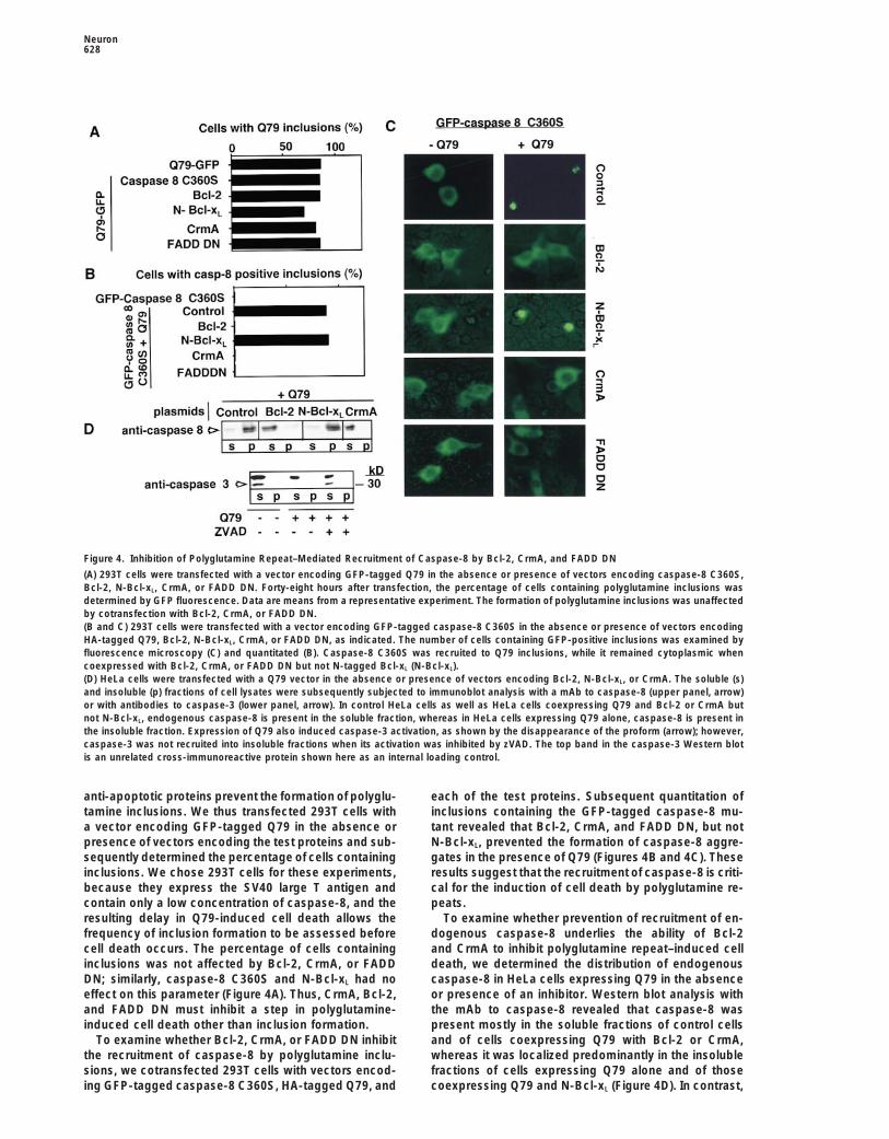
Figure 4. Inhibition of Polyglutamine Repeat–Mediated Recruitment of Caspase-8 by Bcl-2, CrmA, and FADD DN
(A) 293T cells were transfected with a vector encoding GFP-tagged Q79 in the absence or presence of vectors encoding caspase-8 C360S,Bcl-2, N-Bcl-xL, CrmA, or FADD DN. Forty-eight hours after transfection, the percentage of cells containing polyglutamine inclusions wasdetermined by GFP fluorescence. Data are means from a representative experiment. The formation of polyglutamine inclusions was unaffectedby cotransfection with Bcl-2, CrmA, or FADD DN.(B and C) 293T cells were transfected with a vector encoding GFP-tagged caspase-8 C360S in the absence or presence of vectors encodingHA-tagged Q79, Bcl-2, N-Bcl-xL, CrmA, or FADD DN, as indicated. The number of cells containing GFP-positive inclusions was examined byfluorescence microscopy (C) and quantitated (B). Caspase-8 C360S was recruited to Q79 inclusions, while it remained cytoplasmic whencoexpressed with Bcl-2, CrmA, or FADD DN but not N-tagged Bcl-xL (N-Bcl-xL).(D) HeLa cells were transfected with a Q79 vector in the absence or presence of vectors encoding Bcl-2, N-Bcl-xL, or CrmA. The soluble (s)and insoluble (p) fractions of cell lysates were subsequently subjected to immunoblot analysis with a mAb to caspase-8 (upper panel, arrow)or with antibodies to caspase-3 (lower panel, arrow). In control HeLa cells as well as HeLa cells coexpressing Q79 and Bcl-2 or CrmA butnot N-Bcl-xL, endogenous caspase-8 is present in the soluble fraction, whereas in HeLa cells expressing Q79 alone, caspase-8 is present inthe insoluble fraction. Expression of Q79 also induced caspase-3 activation, as shown by the disappearance of the proform (arrow); however,caspase-3 was not recruited into insoluble fractions when its activation was inhibited by zVAD. The top band in the caspase-3 Western blotis an unrelated cross-immunoreactive protein shown here as an internal loading control.
anti-apoptotic proteins prevent the formation of polyglu- each of the test proteins. Subsequent quantitation ofinclusions containing the GFP-tagged caspase-8 mu-tamine inclusions. We thus transfected 293T cells with
a vector encoding GFP-tagged Q79 in the absence or tant revealed that Bcl-2, CrmA, and FADD DN, but notN-Bcl-xL, prevented the formation of caspase-8 aggre-presence of vectors encoding the test proteins and sub-
sequently determined the percentage of cells containing gates in the presence of Q79 (Figures 4B and 4C). Theseresults suggest that the recruitment of caspase-8 is criti-inclusions. We chose 293T cells for these experiments,
because they express the SV40 large T antigen and cal for the induction of cell death by polyglutamine re-peats.contain only a low concentration of caspase-8, and the
resulting delay in Q79-induced cell death allows the To examine whether prevention of recruitment of en-dogenous caspase-8 underlies the ability of Bcl-2frequency of inclusion formation to be assessed before
cell death occurs. The percentage of cells containing and CrmA to inhibit polyglutamine repeat–induced celldeath, we determined the distribution of endogenousinclusions was not affected by Bcl-2, CrmA, or FADD
DN; similarly, caspase-8 C360S and N-Bcl-xL had no caspase-8 in HeLa cells expressing Q79 in the absenceor presence of an inhibitor. Western blot analysis witheffect on this parameter (Figure 4A). Thus, CrmA, Bcl-2,
and FADD DN must inhibit a step in polyglutamine- the mAb to caspase-8 revealed that caspase-8 waspresent mostly in the soluble fractions of control cellsinduced cell death other than inclusion formation.
To examine whether Bcl-2, CrmA, or FADD DN inhibit and of cells coexpressing Q79 with Bcl-2 or CrmA,whereas it was localized predominantly in the insolublethe recruitment of caspase-8 by polyglutamine inclu-
sions, we cotransfected 293T cells with vectors encod- fractions of cells expressing Q79 alone and of thosecoexpressing Q79 and N-Bcl-xL (Figure 4D). In contrast,ing GFP-tagged caspase-8 C360S, HA-tagged Q79, and

Caspase-8 and Polyglutamine-Induced Cell Death629
Figure 6. Recruitment and Activation of Caspase-8 in Caudate Tis-sue from Individuals with HD
The insoluble fractions of caudate tissue from four different HDpatients and five age-matched neurologically unremarkable individ-uals (control) were subjected to immunoblot analysis with a mAb tocaspase-8 (upper left panel). Anti-caspase-8 immunoreactive bandsof z46 kDa were detected in HD samples that were similar in molecu-lar weight to the activated caspase-8 in the insoluble fraction ofHeLa cells expressing Q79 (right panel). Antibodies to tubulin wereused to confirm that similar amounts of protein were loaded in eachlane (lower left panel).
vector and subsequently subjected soluble and insolu-Figure 5. Recruitment of FADD DN, but Not of Bcl-2, by Polygluta-
ble fractions of cell lysates to Western blot analysismine Repeatswith a mAb to AU1 (Figure 5A). Whereas FADD DN was(A) HeLa cells were transfected with vectors encoding either AU1-localized predominantly to the soluble fraction whentagged FADD DN or Bcl-2 in the absence or presence of a Q79expressed by itself, a substantial portion of the proteinvector, as indicated. Soluble (s) and insoluble (p) fractions of cell
lysates were subsequently subjected to immunoblot analysis with was present in the insoluble fraction of cells also ex-antibodies to AU1 or Bcl-2, as indicated. pressing Q79, suggesting that Q79 is able to recruit(B) HeLa cells expressing GFP and HA-Q79 (GFP/Q79), GFP-tagged FADD DN. A similar experiment revealed that Bcl-2 wasQ79 (Q79–GFP), GFP-tagged FADD DN (GFP–FADD DN), or GFP–
not recruited by Q79 (Figure 5A).FADD DN and Q79 (GFP–FADD DN/Q79) were visualized by fluores-The recruitment of FADD DN by Q79 was also appar-cence microscopy 24 hr after the transfection.
ent in 293T cells expressing GFP-tagged FADD DN and(C) HeLa cells expressing HA-tagged Q79 and AU1-tagged FADDDN (24 hr after the transfection) were immunostained with antibodies Q79. Consistent with previous observations (Perez andto HA and AU1 and examined by confocal microscopy; nuclear White, 1998), we showed that GFP-tagged FADD DN ismorphology was also revealed by staining with Hoechst dye. diffusely distributed within the cytoplasm and nucleus
of cells not expressing Q79 (Figure 5B). However, coex-pression of FADD DN with Q79 resulted in the formation
caspase-3 was largely restricted to the soluble fractions of FADD DN aggregates (Figure 5B). To determineof both control and Q79-expressing cells (Figure 4D). whether FADD DN colocalizes with Q79 inclusions, weThese results indicate that expression of Q79 recruits examined the localizations of HA-tagged Q79 and AU1-caspase-8 to the insoluble fraction and that inhibition tagged FADD DN in transfected HeLa cells by immuno-of Q79-induced cell death by Bcl-2, CrmA, or FADD DN cytochemistry and confocal microscopy. A substantialmay be related to the prevention of such caspase-8 portion of AU1-tagged FADD DN colocalized with therecruitment. polyglutamine inclusions. These results suggest that
polyglutamine inclusions are able to recruit FADD DNby interacting with its death domain and that such inter-Recruitment of FADD DN by Polyglutamine Repeats
Inhibition of polyglutamine repeat–induced cell death action may inhibit the recruitment of cell death–inducingproteins.by FADD DN (Figure 1) suggests that the Fas pathway
contributes to this process. To examine the possiblerole of extracellular signaling via the Fas pathway, we Detection of Caspase-8-Like Immunoreactivity
in the Insoluble Fraction of Affected Braininvestigated the effect of a neutralizing monoclonalantibody to Fas ligand (NOK-1, Pharmingen) on Q79- Regions from HD Patients
Finally, we investigated whether recruitment of caspase-8induced cell death. This antibody had no effect on Q79-induced HeLa cell death at a concentration of 1 mg/ml by polyglutamine inclusions could be detected in post-
mortem brain tissue from individuals with HD. We thus(data not shown), which is twice the concentration pre-viously shown to be sufficient to inhibit Fas ligand– determined the distribution of caspase-8 between solu-
ble and insoluble fractions of caudate nuclei. In four outinduced apoptosis (Kayagaki et al., 1995; Oyaizu et al.,1997). Thus, extracellular signaling via the Fas pathway of four caudate samples from HD patients, but in none
of the five samples from neurologically unremarkableis not involved in polyglutamine inclusion–induced celldeath. control individuals, we detected one or two bands of
z46 kDa in the insoluble fraction that reacted with theThe mechanism by which FADD DN inhibits Q79-induced cell death was further investigated by examin- mAb to caspase-8; these proteins were similar in size
to activated caspase-8 in the insoluble fraction of HeLaing the subcellular localization of FADD DN in HeLa and293T cells in the absence or presence of Q79 expression. cells expressing Q79 (Figure 6). In four out of four HD
cerebellum samples, which are unaffected in HD, weWe transfected HeLa cells with a vector encoding AU1-tagged FADD DN in the absence or presence of a Q79 did not detect similar caspase-8-like immunoreactivity

Neuron630
in the insoluble fraction (data not shown). In addition, accumulation of the corresponding truncated protein.The mechanism responsible for the generation of thethe amount of caspase-8-like immunoreactivity in HD
caudate samples was markedly greater than that in con- truncated protein in each disease also may underlie thetissue and cell-type specificity of neuronal degenera-trol caudate tissue, although no substantial difference
was apparent between the soluble fractions of HD and tion; that is, the specific conditions required for cleavageof the disease-associated protein may be present onlycontrol caudate samples (I. S. and J. Y., unpublished
data). These preliminary results suggest that caspase-8 in the neuronal populations affected in each disease.Although polyglutamine inclusions are detected only inor a related caspase-8-like protein is specifically re-
cruited to insoluble components of human caudate cells specific populations of vulnerable neurons in patientswith disease, other cell types are also sensitive to poly-and subsequently becomes activated.glutamine repeat–induced cell death when forced to ex-press a truncated protein containing such repeats. Asecond possible rate-limiting factor may be the availabil-Discussionity of appropriate caspases. We propose that the pres-ence of polyglutamine repeats in the nucleus may playThe mechanism of caspase activation has been the sub-
ject of intensive study. Activation of caspase-8 can be a role in the amplification of the apoptotic response,including the induction of caspase-8 or reduction ofachieved through FK506 binding protein– (FKBP-) medi-
ated dimerization of an FKBP–caspase-8 fusion protein caspase-8 degradation during decades of disease incu-bation period.(Muzio et al., 1998; Yang et al., 1998), These observa-
tions suggest that oligomerization of caspase-8 is suffi- Two inhibitors of caspase-8 recruitment, CrmA andFADD DN, appear to act by distinct mechanisms. Be-cient to induce its activation. In contrast, caspase-3,
which contains a much shorter prodomain, cannot be cause CrmA is a pseudosubstrate type of inhibitor ofcaspases, forming a tight complex with these enzymesactivated by a similar mechanism (Yang et al., 1998). To
date, Fas-induced apoptosis has been the only physio- (Komiyama et al., 1994), CrmA likely interacts directlywith caspase-8 to block its recruitment. The size of CrmAlogical process in which the recruitment and activation
of caspase-8 have been demonstrated. Polyglutamine may be critical here, since zVAD.fmk, which is also apseudosubstrate inhibitor, did not inhibit the recruitmentrepeats may promote a pathological mechanism of
protein–protein interaction that results in the recruitment of caspase-8. In fact, full-length caspase-8 has beenshown to interact with CrmA (Muzio et al., 1998). Inand activation of caspase-8. We have shown here that
caspase-8 is likely to be recruited and activated specifi- contrast, FADD DN may interact directly with polyglu-tamine repeats and thereby prevent the recruitment ofcally in the caudate nuclei of HD patients, suggesting
that the recruitment and activation of caspase-8 may caspase-8. FADD DN consists predominantly of a deathdomain that interacts homophilically with the death do-be responsible for neuronal degeneration in HD. The
mutant polyglutamine repeat–containing proteins have main of the Fas receptor. Although the mechanism bywhich caspase-8 is recruited to polyglutamine repeatsbeen shown to recruit a number of proteins, including
proteins involved in ubiquitination (Davies et al., 1997; remains unclear, we hypothesize that an adapter proteinsimilar to FADD, which contains a death domain and aGourfinkel-An et al., 1998) and molecular chaperone
HDJ-2 (HSDJ) (Cummings et al., 1998). Of all the known death effector domain, may act as a bridge between thepolyglutamine repeats and caspase-8. Our data thusproteins recruited by polyglutamine repeats, none is po-
tentially more dangerous to the cell than caspase-8, suggest that an alternative intracellular Fas pathwayplays a critical role in mediating neuronal degenerationwhich can trigger the activation of downstream compo-
nents of the apoptotic pathway and neuronal cell death. induced by polyglutamine inclusions and that inhibitionof the recruitment of caspase-8 may be able to preventAlthough our data appear to rule out the recruitment
of caspases, such as caspase-3, that contain a short or retard the progression of neuronal loss in polygluta-mine expansion diseases.prodomain, it remains to be examined whether other
caspases with a long prodomain are also recruited by We have shown that polyglutamine repeat–inducedcell death is inhibited by expression of Bcl-2 or Bcl-polyglutamine inclusions.
Apoptosis induced by oligomerization and activation xL but not N-Bcl-xL. Expression of Bcl-2 or Bcl-xL haspreviously been shown to inhibit apoptosis induced byof caspase-8 requires only hours (Muzio et al., 1998;
Yang et al., 1998), whereas neurodegenerative diseases a wide variety of stimuli. Two major mechanisms havebeen proposed to explain the actions of Bcl-2 and Bcl-such as HD develop over decades. Various rate-limiting
factors may contribute to this apparent discrepancy. xL, based on the observations that these proteins formion channels (Minn et al., 1997; Schendel et al., 1997)The first and most important rate-limiting factor is the
generation of a truncated protein consisting predomi- or interact directly with other mediators of apoptosis,such as CED-4 and Apaf-1 (Chinnaiyan et al., 1997; Pannantly of polyglutamine repeats. Expression of full-
length huntingtin or related proteins such as ataxin-3, et al., 1998). Although it is not clear whether these twomodes of action coexist, a free N terminus of Bcl-xL isregardless of the length of the polyglutamine repeat, is
not toxic to cells, whereas truncated versions of these required for the protein to protect cells from apoptosisonly in certain instances. We have recently shown thatproteins containing a sufficient number of polyglutamine
repeats are highly cytotoxic (Goldberg et al., 1996; Mi- Bid, a proapoptotic member of the Bcl-2 family, be-comes a potent inducer of apoptosis after cleavage byyashita et al., 1997; Wellington et al., 1998; the present
study). The decades required for these diseases to de- caspase-8 (Li et al., 1998). Whereas an N-terminallytagged form of Bcl-xL was highly efficient in inhibitingvelop may thus reflect the time required for sufficient

Caspase-8 and Polyglutamine-Induced Cell Death631
cell death induced by truncated Bid, it could not inhibit stress-related proteins, such as members of the Bcl-2polyglutamine repeat–induced cell death. A free N termi- and c-Jun N-terminal kinase (JNK) families, but notnus of Bcl-xL is required for its biochemical and func- enough to kill, since caspases are not activated; eventu-tional interaction with CED-4 and the CED-4-like domain ally, however, the stress response system may be over-in Apaf-1 (Chinnaiyan et al., 1997; Pan et al., 1998); our whelmed, and an apoptotic response, which could in-results suggest that a similar interaction may be required clude upregulation of caspases or reduction of caspasefor Bcl-xL to inhibit polyglutamine inclusion–induced cell turnover, may ensue. Such upregulation of pro- and anti-death. Since caspase-8 can interact with both Apaf-1 apoptotic proteins, such as bad, bax, and bcl-2, have(Hu et al., 1998) and Bcl-xL (Chinnaiyan et al., 1997), the been demonstrated in human tissues affected with othercompetition between caspase-8 binding to Apaf-1 and neurodegenerative diseases (Kitamura et al., 1998). ThisBcl-xL has been hypothesized to play a role in mediating scenario may be consistent with the apparent inductioncaspase-8 activation. We suggest that Bcl-xL may inhibit of caspase-8-like immunoreactivity in caudate tissuepolyglutamine repeat–induced cell death by competing from HD patients. The levels of caspase-8 in controlfor binding of caspase-8. caudate tissue are low compared with that in HeLa,
Studies with transgenic mice expressing exon 1 of Jurkat, and MCF7 cells (I. S. and J. Y., unpublishedthe huntingtin gene and mutant SCA1 as well as with data), whereas the amount of caspase-8 in the insolublebrain tissue from patients with SCA3 or HD have shown fraction of HD caudate tissue is markedly increased.that expanded polyglutamine repeats can form aggre- Such upregulation of caspase-8 may be required forgates within nuclei (Davies et al., 1997; DiFiglia et al., recruitment and activation of caspase-8 by cytoplasmic1997; Paulson et al., 1997). These observations have led polyglutamine repeats. After such cytoplasmic activa-to the hypothesis that the formation of such intranuclear tion, caspase-8 may enter the nucleus, together withinclusions is a critical step in neuronal degeneration. other downstream caspases, to complete the apoptoticRecently, however, two groups have provided evidence process.against a critical role of intranuclear inclusions in induc-ing neurodegeneration (Klement et al., 1998; Saudou et
Concluding Remarksal., 1998). Our study does not directly address the role ofWe have shown here that the recruitment of caspase-8inclusions, since induction of caspase-8 oligomerizationby polyglutamine repeats may contribute to the patho-by polyglutamine repeats may not require the formationgenesis of HD and related diseases. We suggest thatof microscopically visible inclusions. The system usedsuch recruitment may not be limited to HD-related neu-by Saudou et al. (1998) is similar to ours, except that theyrodegenerative disorders. Formation of intracellular ag-used a long version of huntingtin (amino acids 1–480 ofgregates occurs in several other diseases, as exempli-huntingtin) that may need to be further cleaved to acti-fied by the Lewy bodies associated with Parkinson’svate cell death, explaining a delay of up to 9 days beforedisease and the recent demonstration of aggregatesinducing cell death; in contrast, we used a constructof an amyotrophic lateral sclerosis–associated mutantexpressing a polyglutamine repeat only, and cell deathform of superoxide dismutase in transgenic mice (Bruijncan be induced directly by this peptide without furtheret al., 1998). Given that oligomerization may be a funda-cleavage.mental mechanism of caspase activation, other intracel-Although our data do not distinguish between thelular aggregates with properties similar to those of poly-roles of nuclear versus cytoplasmic repeats, we pro-glutamine inclusions also might be able to recruit andpose that caspase-8 recruitment by such repeats occursactivate caspases and induce cell death.predominantly within the cytoplasm for the following
reasons: first, caspase-8 is normally localized in thecytoplasm rather than in nuclei, and second, most Experimental Proceduresanti-apoptotic proteins, including Bcl-2 and Bcl-xL, nor-
Plasmid Constructionmally reside on the outer mitochondrial membraneA cDNA encoding the GFP-tagged C360S mutant of caspase-8 was(Merry and Korsmeyer, 1997). Thus, we suggest thatconstructed by digesting a caspase-8 C360S expression constructrecruitment and activation of caspase-8, as well as inhi- (kindly provided by V. Dixit) with KpnI and XhoI and cloning the
bition of caspase-8 by Bcl-2 and Bcl-xL, occur primarily released fragment into pEGFP-N2 (Clontech). The caspase-8 read-in the cytoplasm. In addition, although most caspase-8 ing frame (lacking the sequence encoding 45 amino acids of themolecules appeared to be associated with the Q79 inclu- small subunit at the C terminus) was thus placed in the reverse
orientation but within appropriate restriction sites. The resultingsions, we cannot rule out the possibility that the interac-plasmid was digested with BamHI and XhoI, and the released frag-tion of caspase-8 with the polyglutamine repeats occursment was cloned into pEGFP-N2 that had been digested with BglIIbefore aggregate formation; aggregation of the polyglu-and SalI. The resulting vector encodes caspase-8 C360S (lacking
tamine repeats may then simply serve to bring together 45 amino acids) fused at its C terminus with GFP. The vector encod-multiple caspase-8 molecules. In the case of the Fas ing FADD DN fused at its C terminus with GFP was generated byreceptor, the interaction of two caspase-8 molecules is digesting a FADD expression construct (kindly provided by V. Dixit)sufficient to induce their activation (Muzio et al., 1998; with SalI and BamHI and cloning the released fragment into pEGFP-
C1 (Clontech) that had been digested with the same two restrictionYang et al., 1998).enzymes. The vector encoding Q79 fused at its C terminus with GFPIf the cytoplasmic polyglutamine repeats play a pri-was prepared by digesting Q79-pIND with HindIII and ApaI andmary role in caspase recruitment and activation, whatcloning the released fragment into pEGFP-N2 that had been di-
then is the role of the intranuclear repeats? We propose gested with the same two enzymes. The Bcl-2 expression constructthat the presence of expanded polyglutamine repeats was RR/1 (Gagliardini et al., 1994). The construct encoding Bcl-xL
in the nucleus may be sufficient to induce a stress re- tagged at its N terminus with the Flag-epitope (N-Bcl-xL) was asdescribed by Li et al. (1998).sponse, which may initially result in the induction of

Neuron632
Cell Lines and Primary Neuronal Cultures for the Bcl-xL construct; Honglin Li and Roberto Sanchez-Olea forCells were seeded at a density of 2 3 104 per 35 mm well in Dulbec- helpful suggestions; Louise Bergeron for caspase-22/2 cells; Hongco’s modified Eagles medium supplemented with 10% fetal bovine Zhu for technical assistance; Ruchika Gupta for critical reading ofserum (FBS) (Hyclone) and were transfected by the standard calcium the manuscript; and Frederic Saudou and Michael E. Greenbergphosphate method. Striatal, cerebellar, and cortical neurons from for communicating unpublished results. The human tissue samplesE17 rat embryos (Taconic) were cultured in neurobasal medium were kindly provided by the Harvard Brain Tissue Resource Center,containing B27 supplement (Gibco) as described by Brewer (1995). which is supported in part by National Institutes of Health grantNeurons were plated at a density of 1 3 105 per well in 6-well plates. NS31862. This work was supported in part by a contract from theAfter 4 days in culture, neurons were transfected by the standard Hereditary Disease Foundation and an American Heart Associationcalcium phosphate method. One hour before transfection, the cul- Established Investigatorship (to J. Y.).ture medium was adjusted to 1% FBS, and half of the medium wasthen removed, to be added back after transfection. The neurons Received November 5, 1998; revised January 25, 1999.were exposed to the calcium phosphate and DNA precipitate for60 min, washed twice with neurobasal medium, and then cultured Referencesin the conditioned medium for 48 hr in the presence of amphidocolin(3 mg/ml) (Sigma). For the trophic factor deprivation experiments, Allsopp, T.E., Wyatt, S., Paterson, H.F., and Davies, A.M. (1993). Theneurons were transfected as mentioned above and cultured for 12 proto-oncogene bcl-2 can selectively rescue neurotrophic factor-hr before the media were exchanged for neurobasal culture media dependent neurons from apoptosis. Cell 73, 295–307.devoid of B27 supplement or serum. Bates, G.P., and Davies, S.W. (1997). Transgenic mouse models of
The percentage of cell death induced by Q79 was determined by neurodegenerative disease caused by CAG/polyglutamine expan-transfecting cells with 1 mg of the CMX–Q79 (HA-tagged) expression sions. Mol. Med. Today 3, 508–515.construct and 0.2 mg of pEGFP-N1 (Clontech) as a marker. In co-
Brewer, G.J. (1995). Serum-free B27/neurobasal medium supportstransfection experiments, constructs encoding FADD DN, CrmA,differentiated growth of neurons from the striatum, substantia nigra,Bcl-2, Bcl-xL, or N-Bcl-xL were used at a ratio of 5:1 with the CMX–septum, cerebral cortex, cerebellum, and dentate gyrus. J. Neurosci.Q79 expression construct. Caspase-8 C360S was used at a ratio ofRes. 42, 674–683.2:1 with the CMX–Q79 expression construct. Apoptotic cells were
identified by visual inspection of GFP-positive cells with a Nikon Bruijn, L.I., Houseweart, M.K., Kato, S., Anderson, K.L., Anderson,inverted fluorescence microscope. In early experiments, uptake of S.D., Ohama, E., Reaume, A.G., Scott, R.W., and Cleveland, D.W.trypan blue and propidium iodide was used to confirm the quantita- (1998). Aggregation and motor neuron toxicity of an ALS-linkedtion of cell death based on cell morphology, and staining with SOD1 mutant independent from wild-type SOD1. Science 281, 1851–Hoechst 33342 was used to examine nuclear morphology. Each 1854.experiment was performed at least in duplicate, with .100 cells Chinnaiyan, A.M., O’Rourke, K., Tewari, M., and Dixit, V.M. (1995).counted for each determination. FADD, a novel death domain–containing protein, interacts with the
death domain of Fas and initiates apoptosis. Cell 81, 505–512.Antibodies and Immunoblot Analysis
Chinnaiyan, A.M., Tepper, C.G., Seldin, M.F., O’Rourke, K., Kischkel,The mAb to caspase-3, -7 and -8 was generated by injecting ratsF.C., Hellbardt, S., Krammer, P.H., Peter, M.E., and Dixit, V.M. (1996).with appropriate recombinant caspases (Li et al., 1998). Anti-humanFADD/MORT1 is a common mediator of CD95 (Fas/APO-1) and tu-Bcl-2 antibody was purchased from Dako. For immunoblot analysis,mor necrosis factor receptor–induced apoptosis. J. Biol. Chem. 271,lysates of cells or of human tissue samples that had been grounded4961–4965.in liquid N2 were prepared in a solution containing 150 mM NaCl,Chinnaiyan, A.M., O’Rourke, K., Lane, B.R., and Dixit, V.M. (1997).1% NP-40, 0.1% SDS, and 50 mM Tris-HCl (pH 8.0), by vortexing andInteraction of CED-4 with CED-3 and CED-9: a molecular frameworkpassing through syringe needles. A soluble fraction was obtained byfor cell death. Science 275, 1122–1126.collecting the supernatant after centrifugation of the lysate in a
microfuge at 13,000 rpm for 15 min. The remaining insoluble pellets Cryns, V., and Yuan, J. (1998). Proteases to die for. Genes Dev. 12,were dissolved in SDS sample buffer (2% SDS, 100 mM dithiothrei- 1551–1570.tol, 60 mM Tris-HCl [pH 6.8], and 0.01% bromophenol blue) by Cummings, C.J., Mancini, M.A., Antalffy, B., DeFranco, D.B., Orr,boiling, vigorous mixing, and then adding urea to achieve a final H.T., and Zoghbi, H.Y. (1998). Chaperone suppression of aggrega-concentration of 2 M, before SDS–PAGE electrophoresis. tion and altered subcellular proteasome localization imply protein
misfolding in SCA1. Nat. Genet. 19, 148–154.Immunofluorescence and Confocal Microscopy
Davies, S.W., Turmaine, M., Cozens, B.A., DiFiglia, M., Sharp, A.H.,For immunostaining, HeLa cells and MCF7 cells were grown in Lab-Ross, C.A., Scherzinger, E., Wanker, E.E., Mangiarini, L., and Bates,tek slide culture chambers at a density of 2 3 103 per chamber andG.P. (1997). Formation of neuronal intranuclear inclusions underliestransfected by the calcium phosphate method. Twenty-four or forty-the neurological dysfunction in mice transgenic for the HD mutation.eight hours after transfection, the cells were fixed with 2% parafor-Cell 90, 537–548.maldehyde or, when quenching of GFP fluorescence was required,DiFiglia, M., Sapp, E., Chase, K.O., Davies, S.W., Bates, G.P., Vonsat-with ice-cold acetone for 10 min. Slides were washed with twotel, J.P., and Aronin, N. (1997). Aggregation of huntingtin in neuronalchanges of phosphate-buffered saline (PBS) and then incubated forintranuclear inclusions and dystrophic neurites in brain. Science20 min at room temperature in PBS containing 5% FBS and 0.1%277, 1990–1993.Triton X-100. Cells were then incubated for 2 hr with primary antibod-
ies diluted in PBS containing 1% FBS and 0.1% Triton X-100, and Gagliardini, V., Fernandez, P.A., Lee, R.K., Drexler, H.C., Rotello,subsequently for 30 min with secondary antibodies; they were R.J., Fishman, M.C., and Yuan, J. (1994). Prevention of vertebratewashed three times with PBS after each antibody incubation. After neuronal death by the crmA gene. Science 263, 826–828.incubation for 10 min with Hoechst 33342, the cells were mounted in Goldberg, Y.P., Nicholson, D.W., Rasper, D.M., Kalchman, M.A.,PBS containing 90% glycerol and phenylamine (10 mg/ml). Primary
Koide, H.B., Graham, R.K., Bromm, M., Kazemi-Esfarjani, P.,antibodies included mAb 11.1 to the HA-epitope and a mAb to the
Thornberry, N.A., Vaillancourt, J.P., and Hayden, M.R. (1996). Cleav-AU1-epitope (Babco). Slides were viewed with a Zeiss Axiophot
age of huntingtin by apopain, a proapoptotic cysteine protease, isinverted confocal microscope.
modulated by the polyglutamine tract. Nat. Genet. 13, 442–449.
Gourfinkel-An, I., Cancel, G., Duyckaerts, C., Faucheux, B., Hauw,AcknowledgmentsJ.J., Trottier, Y., Brice, A., Agid, Y., and Hirsch, E.C. (1998). Neuronaldistribution of intranuclear inclusions in Huntington’s disease withWe thank Ethan Signer for sparking our interest in this problem asadult onset. Neuroreport 9, 1823–1826.well as for support and encouragement during the experiments andHu, Y., Benedict, M.A., Wu, D., Inohara, N., and Nunez, G. (1998).helpful comments on the manuscript; Vishva Dixit for providing theBcl-XL interacts with Apaf-1 and inhibits Apaf-1-dependentcaspase-8 and FADD expression constructs and MCF7 cell line;
Suyue Wang for caspase-12/2 and caspase-112/2 cells; Honglin Li caspase-9 activation. Proc. Natl. Acad. Sci. USA 95, 4386–4391.
Caspase-8 and Polyglutamine-Induced Cell Death633
Huntington’s Disease Collaborative Research Group (1993). A novel Paulson, H.L., Perez, M.K., Trottier, Y., Trojanowski, J.Q., Subra-gene containing a trinucleotide repeat that is expanded and unstable mony, S.H., Das, S.S., Vig, P., Mandel, J.L., Fischbeck, K.H., andon Huntington’s disease chromosomes. Cell 72, 971–983. Pittman, R.N. (1997). Intranuclear inclusions of expanded polyglu-
tamine protein in spinocerebellar ataxia type 3. Neuron 19, 333–344.Igarashi, S., Koide, R., Shimohata, T., Yamada, M., Hayashi, Y.,Takano, H., Date, H., Oyake, M., Sato, T., et al. (1998). Suppression of Perez, D., and White, E. (1998). E1B 19K inhibits Fas-mediated apo-aggregate formation and apoptosis by transglutaminase inhibitors in ptosis through FADD-dependent sequestration of FLICE. J. Cell Biol.cells expressing truncated DRPLA protein with an expanded poly- 141, 1255–1266.glutamine stretch. Nat. Genet. 18, 111–117. Sandou, F., Finkbeiner, S., Devys, D., and Greenberg, M.E. (1998).Ikeda, H., Yamaguchi, M., Sugai, S., Aze, Y., Narumiya, S., and Huntingtin acts in the nucleus to induce apoptosis but death doesKakizuka, A. (1996). Expanded polyglutamine in the Machado- not correlate with the formation of intranuclear inclusions. Cell 95,Joseph disease protein induces cell death in vitro and in vivo. Nat. 55–66.Genet. 13, 196–202. Schendel, S.L., Xie, Z., Montal, M.O., Matsuyama, S., Montal, M.,Janicke, R.U., Ng, P., Sprengart, M.L., and Porter, A.G. (1998). Cas- and Reed, J.C. (1997). Channel formation by antiapoptotic proteinpase-3 is required for alpha-fodrin cleavage but dispensable for Bcl-2. Proc. Natl. Acad. Sci. USA 94, 5113–5118.cleavage of other death substrates in apoptosis. J. Biol. Chem. 273, Trottier, Y., Lutz, Y., Stevanin, G., Imbert, G., Devys, D., Cancel, G.,15540–15545. Saudou, F., Weber, C., David, G., Tora, L., et al. (1995). PolyglutamineJuo, P., Kuo, C.J., Yuan, J., and Blenis, J. (1998). Essential require- expansion as a pathological epitope in Huntington’s disease andment for caspase-8/FLICE in the initiation of the Fas-induced apo- four dominant cerebellar ataxias. Nature 378, 403–406.ptotic cascade. Curr. Biol. 8, 1001–1008. Vincenz, C., and Dixit, V.M. (1997). Fas-associated death domainKawaguchi, Y., Okamoto, T., Taniwaki, M., Aizawa, M., Inoue, M., protein interleukin-1beta-converting enzyme 2 (FLICE2), an ICE/Katayama, S., Kawakami, H., Nakamura, S., Nishimura, M., Akiguchi, Ced-3 homologue, is proximally involved in CD95- and p55-I., et al. (1994). CAG expansions in a novel gene for Machado-Joseph mediated death signaling. J. Biol. Chem. 272, 6578–6583.disease at chromosome 14q32.1. Nat. Genet. 8, 221–228.
Warrick, J.M., Paulson, H.L., Gray-Board, G.L., Bui, Q.T., Fischbeck,Kayagaki, N., Kawasaki, A., Ebata, T., Ohmoto, H., Ikeda, S., Inoue, K.H., Pittman, R.N., and Bonini, N.M. (1998). Expanded polygluta-S., Yoshino, K., Okumura, K., and Yagita, H. (1995). Metalloprotein- mine protein forms nuclear inclusions and causes neural degenera-ase-mediated release of human Fas ligand. J. Exp. Med. 182, 1777– tion in Drosophila. Cell 93, 939–949.1783.
Wellington, C.L., Ellerby, L.M., Hackam, A.S., Margolis, R.L., Trifiro,Kischkel, F.C., Hellbardt, S., Behrmann, I., Germer, M., Pawlita, M., M.A., Singaraja, R., McCutcheon, K., Salvesen, G.S., Propp, S.S.,Krammer, P.H., and Peter, M.E. (1995). Cytotoxicity-dependent Bromm, M., et al. (1998). Caspase cleavage of gene products associ-APO-1 (Fas/CD95)-associated proteins form a death-inducing sig- ated with triplet expansion disorders generates truncated fragmentsnaling complex (DISC) with the receptor. EMBO J. 14, 5579–5588. containing the polyglutamine tract. J. Biol. Chem. 273, 9158–9167.Kitamura, Y., Shimohama, S., Kamoshima, W., Ota, T., Matsuoka, Yang, X., Chang, H.Y., and Baltimore, D. (1998). AutoproteolyticY., Nomura, Y., Smith, M.A., Perry, G., Whitehouse, P.J., and Tani- activation of pro-caspases by oligomerization. Mol. Cell 1, 319–325.gushi, T. (1998). Alteration of proteins regulating apoptosis, Bcl-2,
Zhou, Q., Snipas, S., Orth, K., Muzio, M., Dixit, V.M., and Salvesen,Bcl-x, Bax, Bak, Bad, ICH-1, and CPP32, in Alzheimer’s disease.G.S. (1997). Target protease specificity of the viral serpin CrmA.Brain Res. 780, 260–269.Analysis of five caspases. J. Biol. Chem. 272, 7797–7800.
Klement, I.A., Skinner, P.J., Kaytor, M.D., Yi, H., Hersch, S.M., Clark,H.B., Zoghbi, H.Y., and Orr, H.T. (1998). Ataxin-1 nuclear localization A Note Added in Proofand aggregation: role in polyglutamine-induced disease in SCA1transgenic mice. Cell 95, 41–53. After our paper was accepted, Moulder et al. (1999) and Kim et al.Komiyama, T., Ray, C.A., Pickup, D.J., Howard, A.D., Thornberry, (1999) published their work on cell death induced by expandedN.A., Peterson, E.P., and Salvesen, G. (1994). Inhibition of interleu- polyglutamine repeats in primary rat cerebellar granular neuronskin-1 beta converting enzyme by the cowpox virus serpin CrmA. An and in striatal/neuroblastoma fusion cell line N18TG2, respectively.example of cross-class inhibition. J. Biol. Chem. 269, 19331–19337. Consistent with our results, both papers showed that the expressionLi, H., Zhu, H., Xu, C.-J., and Yuan, J. (1998). Cleavage of BID by of expanded poly-Q-induced activation of caspases and the inhibi-caspase-8 mediates the mitochondrial damage in the Fas pathway tion of caspases reduced or delayed neuronal toxicity. While theof apoptosis. Cell 94, 491–501. inhibition of caspases clearly has an effect on poly-Q-induced cell
death, Moulder et al. stressed that cerebellar granule neurons ex-Martin, D.A., Siegel, R.M., Zheng, L., and Lenardo, M.J. (1998). Mem-pressing poly-Q in the presence of Boc-aspartyl-(OMe)-fluorometh-brane oligomerization and cleavage activates the caspase-8 (FLICE/ylketone (BAF) still die with a delayed time course over 5 days. WhileMACHalpha) death signal. J. Biol. Chem. 273, 4345–4349.it is unclear whether BAF inhibits caspase-8 in the dose used inMerry, D.E., and Korsmeyer, S.J. (1997). Bcl-2 gene family in theMoulder’s experiment, we cannot exclude the existence of a cas-nervous system. Annu. Rev. Neurosci. 20, 245–267.pase-independent alternative pathway that is triggered after cas-
Minn, A.J., Velez, P., Schendel, S.L., Liang, H., Muchmore, S.W., pase inhibition. Such a pathway is proposed in cerebellar granularFesik, S.W., Fill, M., and Thompson, C.B. (1997). Bcl-x(L) forms an cells in the work done by the same group (Miller and Johnson,ion channel in synthetic lipid membranes. Nature 385, 353–357. 1996). We are currently exploring the molecular basis of a possibleMiyashita, T., Okamura-Oho, Y., Mito, Y., Nagafuchi, S., and Ya- caspase-independent pathway and testing whether such a mecha-mada, M. (1997). Dentatorubral pallidoluysian atrophy (DRPLA) pro- nism is induced following the inhibition of the recruitment and activa-tein is cleaved by caspase-3 during apoptosis. J. Biol. Chem. 272, tion of caspase-8.29238–29242. Kim, M., Lee, H.S., LaForet, G., McIntyre, C., Martin, E.J., Chang,Muzio, M., Stockwell, B.R., Stennicke, H.R., Salvesen, G.S., and P., Kim, T.W., Williams, M., Reddy, P.H., Tagle, D., et al. (1999).Dixit, V.M. (1998). An induced proximity model for caspase-8 activa- Mutant huntingtin expression in clonal striatal cells: dissociation oftion. J. Biol. Chem. 273, 2926–2930. inclusion formation and neuronal survival by caspase inhibition. J.
Neurosci. 19, 964–973.Oyaizu, N., Adachi, Y., Hashimoto, F., McCloskey, T.W., Hosaka, N.,Kayagaki, N., Yagita, H., and Pahwa, S. (1997). Monocytes express Miller, T.M., and Johnson, E.M., Jr. (1996). Metabolic and geneticFas ligand upon CD4 cross-linking and induce CD41 T cell apopto- analyses of apoptosis in potassium/serum-deprived rat cerebellarsis: a possible mechanism of bystander cell death in HIV infection. granule cells. J. Neurosci. 16, 7487–7495.J. Immunol. 158, 2456–2463. Moulder, K.L., Onodera, O., Burke, J.R., Strittmatter, W.J., and John-Pan, G., O’Rourke, K., and Dixit, V.M. (1998). Caspase-9, Bcl-XL, son, E.M., Jr. (1999). Generation of neuronal intranuclear inclusionsand Apaf-1 form a ternary complex. J. Biol. Chem. 273, 5841–5845. by polyglutamine-GFP: analysis of inclusion clearance and toxicity
as a function of polyglutamine length. J. Neurosci. 19, 705–715.Paulson, H.L., and Fischbeck, K.H. (1996). Trinucleotide repeats inneurogenetic disorders. Annu. Rev. Neurosci. 19, 79–107.
Copyright © 2022 FDOKUMEN