Carbon Monoxide Induces Cytoprotection in Rat Orthotopic Lung Transplantation via Anti-Inflammatory...
12
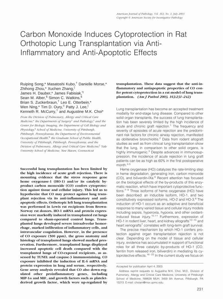
Carbon Monoxide Induces Cytoprotection in Rat Orthotopic Lung Transplantation via Anti- Inflammatory and Anti-Apoptotic Effects Ruiping Song,* Masatoshi Kubo, † Danielle Morse,* Zhihong Zhou,* Xuchen Zhang, James H. Dauber,* James Fabisiak, ¶ Sean M. Alber, § Simon C. Watkins, § Brian S. Zuckerbraun, † Leo E. Otterbein,* Wen Ning,* Tim D. Oury, ‡ Patty J. Lee, Kenneth R. McCurry, † and Augustine M.K. Choi* From the Division of Pulmonary, Allergy and Critical Care Medicine,* the Departments of Surgery † and Pathology, ‡ and the Center for Biologic Imaging, the Department of Cell Biology and Physiology, § School of Medicine, University of Pittsburgh, Pittsburgh, Pennsylvania; the Department of Environmental Occupational Health, ¶ the Graduate School of Public Health, University of Pittsburgh, Pittsburgh, Pennsylvania; and the Division of Pulmonary, Allergy and Critical Care Medicine, Yale University School of Medicine, New Haven, Connecticut Successful lung transplantation has been limited by the high incidence of acute graft rejection. There is mounting evidence that the stress response gene heme oxygenase-1 (HO-1) and/or its catalytic by- product carbon monoxide (CO) confers cytoprotec- tion against tissue and cellular injury. This led us to hypothesize that CO may protect against lung trans- plant rejection via its anti-inflammatory and anti- apoptotic effects. Orthotopic left lung transplantation was performed in Lewis rat recipients from Brown- Norway rat donors. HO-1 mRNA and protein expres- sion were markedly induced in transplanted rat lungs compared to sham-operated control lungs. Trans- planted lungs developed severe intraalveolar hemor- rhage , marked infiltration of inflammatory cells , and intravascular coagulation. However, in the presence of CO exposure (500 ppm), the gross anatomy and histology of transplanted lungs showed marked pres- ervation. Furthermore, transplanted lungs displayed increased apoptotic cell death compared with the transplanted lungs of CO-exposed recipients, as as- sessed by TUNEL and caspase-3 immunostaining. CO exposure inhibited the induction of IL-6 mRNA and protein expression in lung and serum, respectively. Gene array analysis revealed that CO also down-reg- ulated other proinflammatory genes, including MIP-1 and MIF, and growth factors such as platelet- derived growth factor, which were up-regulated by transplantation. These data suggest that the anti-in- flammatory and antiapoptotic properties of CO con- fer potent cytoprotection in a rat model of lung trans- plantation. (Am J Pathol 2003, 163:231–242) Lung transplantation has become an accepted treatment modality for end-stage lung disease. Compared to other solid organ transplants, the success of lung transplanta- tion has been severely limited by the high incidence of acute and chronic graft rejection. 1 The frequency and severity of episodes of acute rejection are the predomi- nant risk factors for chronic airway rejection, manifested as obliterative bronchiolitis. 2 Data from rodent allograft studies as well as from clinical lung transplantation show that the lung, in comparison to other solid organs, is highly immunogenic. 3 Despite advances in immunosup- pression, the incidence of acute rejection in lung graft patients can be as high as 60% in the first postoperative month. 4,5 Heme oxygenase (HO) catalyzes the rate-limiting step in heme degradation, generating iron, carbon monoxide (CO), and biliverdin-IXa. 6 Recent attention has focused on the biological effects of the by-products of this enzy- matic reaction, which have important cytoprotective func- tions. 7–9 Three isoforms of heme oxygenase (HO) have been described: an inducible isoform, HO-1, and two constitutively expressed isoforms, HO-2 and HO-3. 6 The induction of HO-1 occurs as an adaptive and beneficial response to many varied tissue and cellular injury models including sepsis, hyperoxia, hypoxia, and other oxidant- induced tissue injury. 10,11 Furthermore, expression of HO-1 in rodent liver, heart, and kidney allografts 12–15 and heart xenografts 7 correlates with long-term graft survival. The precise mechanism by which HO-1 confers pro- tection against organ transplantation rejection is not clear. Depending on the model of tissue and cellular injury, evidence has accumulated in support of functional roles for all three catalytic by-products of HO-1 (CO, ferritin from released iron, biliverdin) in mediating its cy- toprotective effects. 16 –22 In the current study we focus on Accepted for publication April 4, 2003. Address reprint requests to Augustine M.K. Choi, M.D., Division of Pulmonary, Allergy and Critical Care Medicine, University of Pittsburgh School of Medicine, NW628 MUH, 3459 5th Avenue, Pittsburgh, PA 15213. E-mail: [email protected]. American Journal of Pathology, Vol. 163, No. 1, July 2003 Copyright © American Society for Investigative Pathology 231
Transcript of Carbon Monoxide Induces Cytoprotection in Rat Orthotopic Lung Transplantation via Anti-Inflammatory...
Carbon Monoxide Induces Cytoprotection in RatOrthotopic Lung Transplantation via Anti-Inflammatory and Anti-Apoptotic Effects
Ruiping Song,* Masatoshi Kubo,† Danielle Morse,*Zhihong Zhou,* Xuchen Zhang,�
James H. Dauber,* James Fabisiak,¶
Sean M. Alber,§ Simon C. Watkins,§
Brian S. Zuckerbraun,† Leo E. Otterbein,*Wen Ning,* Tim D. Oury,‡ Patty J. Lee,�
Kenneth R. McCurry,† and Augustine M.K. Choi*From the Division of Pulmonary, Allergy and Critical Care
Medicine,* the Departments of Surgery † and Pathology,‡ and the
Center for Biologic Imaging, the Department of Cell Biology and
Physiology,§ School of Medicine, University of Pittsburgh,
Pittsburgh, Pennsylvania; the Department of Environmental
Occupational Health,¶ the Graduate School of Public Health,
University of Pittsburgh, Pittsburgh, Pennsylvania; and the
Division of Pulmonary, Allergy and Critical Care Medicine,� Yale
University School of Medicine, New Haven, Connecticut
Successful lung transplantation has been limited bythe high incidence of acute graft rejection. There ismounting evidence that the stress response geneheme oxygenase-1 (HO-1) and/or its catalytic by-product carbon monoxide (CO) confers cytoprotec-tion against tissue and cellular injury. This led us tohypothesize that CO may protect against lung trans-plant rejection via its anti-inflammatory and anti-apoptotic effects. Orthotopic left lung transplantationwas performed in Lewis rat recipients from Brown-Norway rat donors. HO-1 mRNA and protein expres-sion were markedly induced in transplanted rat lungscompared to sham-operated control lungs. Trans-planted lungs developed severe intraalveolar hemor-rhage, marked infiltration of inflammatory cells, andintravascular coagulation. However, in the presenceof CO exposure (500 ppm), the gross anatomy andhistology of transplanted lungs showed marked pres-ervation. Furthermore, transplanted lungs displayedincreased apoptotic cell death compared with thetransplanted lungs of CO-exposed recipients, as as-sessed by TUNEL and caspase-3 immunostaining. COexposure inhibited the induction of IL-6 mRNA andprotein expression in lung and serum, respectively.Gene array analysis revealed that CO also down-reg-ulated other proinflammatory genes, includingMIP-1� and MIF, and growth factors such as platelet-derived growth factor, which were up-regulated by
transplantation. These data suggest that the anti-in-flammatory and antiapoptotic properties of CO con-fer potent cytoprotection in a rat model of lung trans-plantation. (Am J Pathol 2003, 163:231–242)
Lung transplantation has become an accepted treatmentmodality for end-stage lung disease. Compared to othersolid organ transplants, the success of lung transplanta-tion has been severely limited by the high incidence ofacute and chronic graft rejection.1 The frequency andseverity of episodes of acute rejection are the predomi-nant risk factors for chronic airway rejection, manifestedas obliterative bronchiolitis.2 Data from rodent allograftstudies as well as from clinical lung transplantation showthat the lung, in comparison to other solid organs, ishighly immunogenic.3 Despite advances in immunosup-pression, the incidence of acute rejection in lung graftpatients can be as high as 60% in the first postoperativemonth.4,5
Heme oxygenase (HO) catalyzes the rate-limiting stepin heme degradation, generating iron, carbon monoxide(CO), and biliverdin-IXa.6 Recent attention has focusedon the biological effects of the by-products of this enzy-matic reaction, which have important cytoprotective func-tions.7–9 Three isoforms of heme oxygenase (HO) havebeen described: an inducible isoform, HO-1, and twoconstitutively expressed isoforms, HO-2 and HO-3.6 Theinduction of HO-1 occurs as an adaptive and beneficialresponse to many varied tissue and cellular injury modelsincluding sepsis, hyperoxia, hypoxia, and other oxidant-induced tissue injury.10,11 Furthermore, expression ofHO-1 in rodent liver, heart, and kidney allografts12–15 andheart xenografts7 correlates with long-term graft survival.
The precise mechanism by which HO-1 confers pro-tection against organ transplantation rejection is notclear. Depending on the model of tissue and cellularinjury, evidence has accumulated in support of functionalroles for all three catalytic by-products of HO-1 (CO,ferritin from released iron, biliverdin) in mediating its cy-toprotective effects.16–22 In the current study we focus on
Accepted for publication April 4, 2003.
Address reprint requests to Augustine M.K. Choi, M.D., Division ofPulmonary, Allergy and Critical Care Medicine, University of PittsburghSchool of Medicine, NW628 MUH, 3459 5th Avenue, Pittsburgh, PA15213. E-mail: [email protected].
American Journal of Pathology, Vol. 163, No. 1, July 2003
Copyright © American Society for Investigative Pathology
231

the functional role of CO in a rat model of orthotopic lungtransplantation. Given the anti-apoptotic and anti-inflam-matory properties of both HO-1 and CO, we hypothesizethat CO limits graft injury by maintaining cell viability andmodulating tissue inflammation. We evaluated the role ofCO in an acute model of rat lung transplantation, dem-onstrating that CO confers potent cytoprotection in thesetting of lung transplantation.
Materials and Methods
Human Pulmonary Fibroblast Cultures
Primary cell cultures of lung fibroblasts were isolatedfrom lung tissue obtained from endobronchial lung biop-sies of lung transplant recipients under a protocol ap-proved by the Institutional Review Board of the Universityof Pittsburgh. Fibroblasts were grown to confluence usingDulbecco’s modified Eagle’s medium (DMEM) supple-mented with 10% fetal bovine serum (FBS), both fromInvitrogen (Carlsbad, CA) prior to isolation of lysates forWestern blot analysis for HO-1 expression, as describedbelow.
Histological Grading of Human Lung BiopsySpecimen of Lung Transplant Recipients
Allograft rejection was graded according to the workingformulation of the International Society of Heart and LungTransplantation (ISHLT).23 Acute rejection was graded inA0 to A4 according to the presence and the extent ofperivascular and interstitial mononuclear cell infiltrates.All of the histological scoring was performed by a singleinvestigator in a blinded fashion.
Carbon Monoxide Exposure
For animal exposure, CO at a concentration of 1%(10,000 parts per million-ppm) in compressed air wasmixed with balanced air (21% oxygen) in a stainless steelmixing cylinder before delivery into the exposure cham-ber. A CO analyzer (Interscan Corporation, Chatsworth,CA) was used to measure CO levels in the chamber andthere were no fluctuations in the CO concentrations afterthe chamber had equilibrated. Recipient rats were ex-posed to 500 ppm CO after transplantation procedures.
Rat Lung Transplantation
Male Lewis (LEW) and Brown-Norway (BN) viral anti-body-free rats (250 to 300 g) were purchased from Har-lan (Madison, WI). All procedures involving animals wereapproved by the Institutional Animal Care and Use Com-mittee of the University of Pittsburgh.
Orthotopic left lung transplantation was performed inLEW recipient from BN donor by means of the cuff tech-nique as previously described.24 Donor rats were anes-thetized by intraperitoneal injection of sodium pentobar-bital and intubated through a tracheotomy. Animals were
mechanically ventilated with 100% oxygen, tidal volumeof 10 ml/kg and respiratory rate of 70 breaths/minute.After heparinization (1000 units/kg intravenously, a me-dian laparosternotomy was performed and the lungswere flushed through the main pulmonary artery with 20ml of cold saline delivered from a height of 25 cm. Theheart-lung block was removed with the lungs inflated atthe tidal volume. The left lungs were then divided andcuffs prepared. 14-gauge catheters were placed into thepulmonary artery and pulmonary vein and secured with8–0 polypropylene ligatures.
Recipient animals were premedicated by intramuscu-lar injection of ketamine chloride (40 mg/kg) and atropinesulfate (0.25 mg/kg), and intubated orotracheally. Theanimals were maintained under general anesthesia with amixture of isoflurane and oxygen. A left thoracotomy wasperformed through the fourth intercostal space. The hilumof the left lung was dissected, and the pulmonary artery,pulmonary vein, and the left main bronchus wereclamped with microvascular clamps. The pulmonary ar-tery and pulmonary vein were incised, and the cuffs of thedonor lung were inserted and fixed with 6–0 silk suture.The left main bronchus was anastomosed by a runningsuture of 9–0 nylon. After graft reperfusion, the recipi-ent’s native lung was removed, a temporary chest tubewas placed, which was removed after recovery fromanesthesia, and the thoracotomy was closed. After recov-ery from the anesthesia, animals were placed either inCO or air chamber.
Hematoxylin-Eosin Staining and Grading
Formalin-fixed, paraffin-embedded sections were usedfor conventional histology hematoxylin and eosin (H&E)staining. Allograft rejection was graded according to theworking formulation of the ISHLT.23 Acute rejection wasgraded in A0 to A4 according to the presence and theextent of perivascular and interstitial mononuclear cellinfiltrates. All of the histological scoring was performed bya single investigator in a blinded fashion.
Myeloperoxidase Activity
Myeloperoxidase (MPO) is a constituent enzyme foundprincipally in neutrophils that results in the formation ofhypochlorous acid. Its presence only in neutrophils al-lows it to be extracted from tissues and represents adirect correlation to neutrophil content.
Rat lungs from both transplant and transplant/COgroup were measured for MPO activity.25 50 mg of frozenlung samples were homogenized in phosphate buffercontaining 0.5% hexadecyltrimethylammonium bromide,sonicated for 15 seconds, and further disrupted by afreeze-thaw cycle in liquid nitrogen. The extract was cen-trifuged at 20,000 � g for 20 minutes at 4°C. The super-natants were assayed for MPO activity by measuring theabsorbance at 460 nm, using a 0.167 mg/dl solution ofo-dianisidine dihydrochloride in 0.0005% hydrogen per-oxide as the substrate. A similar amount from the samelung was weighed and desiccated in a 60°C oven to
232 Song et alAJP July 2003, Vol. 163, No. 1
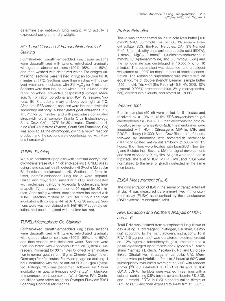
determine the wet-to-dry lung weight. MPO activity isexpressed per gram of dry weight.
HO-1 and Caspase-3 ImmunohistochemicalStaining
Formalin-fixed, paraffin-embedded lung tissue sectionswere deparaffinized with xylene, rehydrated graduallywith graded alcohol solutions (100%, 95%, and 80%),and then washed with deionized water. For antigen un-masking, sections were treated in trypsin solution for 10minutes at 37°C. Sections were then washed with deion-ized water and incubated with 3% H2O2 for 5 minutes.Sections were then incubated with a 1:300 dilution of therabbit polyclonal anti-active caspase-3 (Promega, Madi-son, WI) or rabbit polyclonal anti-HO-1 (Stressgen, Vic-toria, BC, Canada) primary antibody overnight at 4°C.After three PBS washes, sections were incubated with thesecondary antibody, a biotinylated goat anti-rabbit IgG,at 37°C for 30 minutes, and with peroxidase-conjugatedstrepavidin-biotin complex (Santa Cruz Biotechnology,Santa Cruz, CA) at 37°C for 30 minutes. Diaminobenzi-dine (DAB) substrate (Zymed, South San Francisco, CA)was applied as the chromogen, giving a brown reactionproduct, and the sections were counterstained with May-er’s hematoxylin.
TUNEL Staining
We also confirmed apoptosis with terminal deoxynucle-otidyl-transferase dUTP nick end-labeling (TUNEL) assayusing the in situ cell death detection kit (Roche MolecularBiochemicals, Indianapolis, IN). Sections of formalin-fixed, paraffin-embedded lung tissue were deparaf-finized and rehydrated, rinsed with PBS, and digestedwith proteinase K (Roche Molecular Biochemicals, Indi-anapolis, IN) at a concentration of 20 �g/ml for 20 min-utes. After being washed, sections were incubated withTUNEL reaction mixture at 37°C for 1 hour and thenincubated with converter-AP at 37°C for 30 minutes. Sec-tions were washed, stained with NBT/BCIP substrate so-lution, and counterstained with nuclear fast red.
TUNEL/Macrophage Co-Staining
Formalin-fixed, paraffin-embedded lung tissue sectionswere deparaffinized with xylene, rehydrated graduallywith graded alcohol solutions (100%, 95%, and 80%),and then washed with deionized water. Sections werethen incubated with Apoptosis Detection System (Fluo-rescein; Promega) for 30 minutes followed by an incuba-tion in normal goat serum (Sigma Chemie, Deisenhofen,Germany) for 40 minutes. For Macrophage co-staining, 1hour incubation with mouse anti-rat ED1 (2 �g/ml) (Sero-tec, Raleigh, NC) was performed, followed by 1 hourincubation in goat anti-mouse cy3 (2 �g/ml) (JacksonImmunoresearch Laboratories, West Grove, PA). Confo-cal slices were taken using an Olympus Fluoview BX61Scanning Confocal Microscope.
Protein Extraction
Tissue was homogenized on ice in cold lysis buffer [150mmol/L NaCl, 50 mmol/L Tris, pH 7.6, 1% sodium dode-cyl sulfate (SDS; Bio-Rad, Hercules, CA), 3% NonidetP-40, 5 mmol/L ethylenediaminetetraacetic acid (EDTA),1 mmol/L MgCl2, 2 mmol/L 1,3-dichloroisocoumarin, 2mmol/L 1,10-phenanthroline, and 0.5 mmol/L E-64] andthe homogenate was centrifuged at 10,000 � g for 10minutes. The supernatant was decanted, and an aliquotwas stored at �20°C for measurement of protein concen-tration. The remaining supernatant was mixed with anequal volume of double-strength Laemmli sample buffer[250 mmol/L Tris �HCl (Bio-Rad), pH 6.8, 4% SDS, 10%glycerol, 0.006% bromphenol blue, 2% �-mercaptoetha-nol], divided into aliquots, and stored at �80°C.
Western Blot
Protein samples (50 �l) were boiled for 5 minutes andresolved by a 10% to 12.5% SDS-polyacrylamide gelelectrophoresis (SDS-PAGE), then electroblotted onto ni-trocellulose membranes (Bio-Rad). The membranes wereincubated with HO-1, (Stressgen), MIP-1�, MIF, andPDGF antibody (1:1000; Santa Cruz Biotech) for 2 hours,followed by incubation with horseradish peroxidase(HRP)-conjugated anti-rabbit antibody (1:5000) for 1.5hours. The filters were treated with LumiGLO (New En-gland Biolabs Inc., Beverly, MA) for signal development,and then exposed to X-ray film. All gels were repeated intriplicate. The level of HO-1, MIP-1�, MIF, and PDGF werenormalized to the level of �-actin detected in the samemembrane.
ELISA Measurement of IL-6
The concentration of IL-6 in the serum of transplanted ratat day 4 was measured by enzyme-linked immunosor-bent assay (ELISA) as described by the manufacturer(R&D systems, Minneapolis, MN).
RNA Extraction and Northern Analysis of HO-1and IL-6
Total RNA was isolated from transplanted lung tissue atday 4 using TRIzol reagent (Invitrogen, Carlsbad, Califor-nia) according to the manufacturer’s instructions. TotalRNA (10 �g per lane) was denatured, electrophoresedon 1.2% agarose formaldehyde gels, transferred to apositively-charged nylon membrane (Hybond N�; Amer-sham Pharmacia Biotech, Piscataway, NJ) and UV cross-linked (Stratalinker; Stratagene, La Jolla, CA). Mem-branes were prehybridized for 1 to 2 hours at 60°C andsubsequently hybridized overnight at 60°C with randomprimer [32P]dCTP-labeled rat HO-1 cDNA and rat IL-6cDNA, cDNA. The blots were washed three times with asolution containing 0.5% bovine serum albumin, 5% SDS,and 1 mmol/L EDTA in 0.2X standard saline citrate at56°C to 60°C and then exposed to X-ray film at �80°C.
Carbon Monoxide in Lung Transplantation 233AJP July 2003, Vol. 163, No. 1

cDNA Expression Arrays Analysis
Total RNA (10 �g) from each group of 4 rat lungs wascombined with Atlas Rat 1.2 specific RNA primers, cDNAsynthesis (CDS) primer mix (Clontech, Palo Alto, CA). Thesolution was preheated to 94°C followed by annealing at70°C for 10 minutes and extension at 49°C for 35 minutes]50 mmol/L of Tris–HCl, pH 8.3, 75 mmol/L KCl, 3 mmol/LMgCl2, 5.6 mmol/L DTT, 0.6 mmol/L dNTPs, 100 �Ci[33P]dCTP (NEN, Boston, MA) and 200 units of Super-script II (Invitrogen)[. The reaction was stopped at 94°Cfor 5 minutes and then placed in an ice bath. Unincorpo-rated [33P]dCTP was removed on a Microspin SepharoseG-50 gel filtration column (Amersham Pharmacia Biotech,Piscataway, NJ). [33P]-cDNA hybridization to ClontechRat Atlas 1.2 membranes was done at 64°C in a micro-tube (Research Genetics, Huntsville, AL) containing 2 �gCot-1 DNA. For hybridization of the labeled [33P]-cDNAproduct, 250,000 Cerenkov cpm at 0 to 260 mKeV wereused. Phosphor screens were exposed for 36 hours; thearrays were wrapped and read in a Packard Cyclonephosphor imager (PerkinElmer Life Sciences, Boston,MA). The two array images were analyzed and comparedusing the Clontech AtlasImage 1.5 software. First, thephosphor image of each array was separately alignedand fine-tuned to the Atlas Image Grid Template. Afternormalizing the signal values of all of the genes on thearrays (global normalization), the two aligned arrays werecompared with each other using the control group as thereference to generate a color schematic diagram of thechanges in the treatment group and a tabular report ofthe data.
Statistical Analysis
Data are expressed as the mean � SE. Differences inmeasured variables between experimental and controlgroup were assessed using Student’s t-test. Statisticaldifference was accepted at P � 0.05.
Results
HO-1 Protein Levels are Increased in LungFibroblasts from Lung Transplant Patients
Lung fibroblasts were cultured from endobronchial lungbiopsies of lung transplant recipients. Correspondinglung biopsy specimens were graded for rejection gradeusing the criteria for human lung transplantation rejectiongrade at the University of Pittsburgh as described inMethods. Cultured fibroblasts were grown to confluencebefore analysis of cell lysates by Western blot for HO-1protein expression. As illustrated in Figure 1, we ob-served increasing levels of HO-1 protein expression withincreasing grades of acute rejection.
HO-1 mRNA and Protein Expression areInduced after Rat Lung Transplantation
Having demonstrated increased HO-1 protein in fibro-blasts cultured from human transplanted biopsy speci-mens, we examined HO-1 mRNA and protein levels in ratlung following transplantation. Total RNA was extractedfrom the transplanted lung at day 4 after rat orthotopiclung transplantation and subjected to Northern blot anal-yses for HO-1. Figure 2A demonstrates marked inductionof HO-1 mRNA in the transplanted lung (at 4 days aftertransplantation; n � 3) compared to sham control lungs(n � 3) as assessed by Northern blot analyses. Theincrease in HO-1 mRNA expression correlated with in-creased HO-1 protein by Western blot analysis in thetransplanted lungs as compared with sham-operatedlungs (Figure 2B). Immunohistochemical staining demon-strated markedly elevated HO-1 expression diffusely inalveolar and airway epithelial and vascular cells (brownstaining in the transplanted lung section; Figure 3, C andD) compared to the sham-operated control lung section(Figure 3, A and B).
Gross Anatomy of Transplanted Lungs
Given the known cytoprotective effects of CO, a byprod-uct of HO-1 activity, we examined whether exogenousCO could provide cytoprotection against lung injury in an
Figure 1. HO-1 protein level in lung fibroblasts from human transplantrecipients increased and correlated with acute rejection grade. Fibroblastscultured from lung biopsy specimens of transplanted patients were harvestedand cultured. Biopsy specimens were sent to pathology for acute rejectiongrading. Protein was extracted from cultured fibroblast and subjected toWestern blot hybridization with HO-1 antibody as described in Methods.Each lane represents pooled samples from a group of five patients with acuterejection grade I to III; control was from normal patient lung. Culturedfibroblast was used at passage 2 to 3. The same membranes were probedwith an antibody against �-actin to assure equal loading of the gel.
Figure 2. Rat lung transplantation increased HO-1 mRNA expression andprotein level. A: Total RNA from the transplanted lung (day 4) was extractedand subjected to Northern blot hybridization with a 32P-labeled HO-1 cDNAprobe as described in Methods. Each lane represents RNA extracted fromone rat (n � 3). Normalization for RNA loading is shown by labeling 18SrRNA of the same membrane. B: Protein from the transplanted lung (day 4)was extracted and subjected to Western blot hybridization with HO-1 anti-body as described in Methods. Each lane represents protein extracted fromone rat (n � 3). The same membranes were probed with an antibody against�-actin to assure equal loading of the gel.
234 Song et alAJP July 2003, Vol. 163, No. 1
acute model of orthotopic lung transplantation. Figure 4shows a representative lung tissue 6 days after orthotopiclung transplantation in rats (n � 6), compared to lungtissue from rats receiving CO (500 ppm, n � 6) over thistime period. In the absence of CO, the transplanted lungbecame markedly hyperemic and infarcted (Figure 4, Aand B), features which were notably absent in the trans-planted lung exposed to CO (Figure 4, C and D).
Histology and Grading of Transplanted Lungs
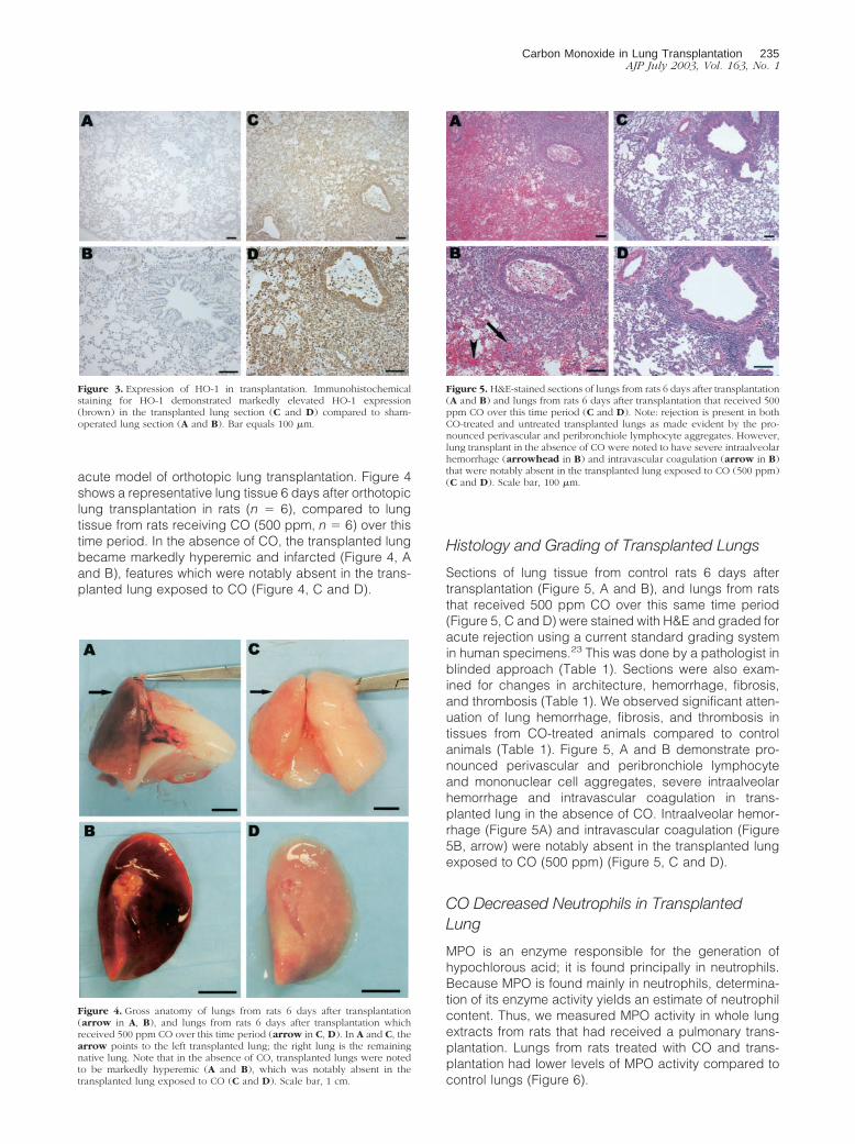
Sections of lung tissue from control rats 6 days aftertransplantation (Figure 5, A and B), and lungs from ratsthat received 500 ppm CO over this same time period(Figure 5, C and D) were stained with H&E and graded foracute rejection using a current standard grading systemin human specimens.23 This was done by a pathologist inblinded approach (Table 1). Sections were also exam-ined for changes in architecture, hemorrhage, fibrosis,and thrombosis (Table 1). We observed significant atten-uation of lung hemorrhage, fibrosis, and thrombosis intissues from CO-treated animals compared to controlanimals (Table 1). Figure 5, A and B demonstrate pro-nounced perivascular and peribronchiole lymphocyteand mononuclear cell aggregates, severe intraalveolarhemorrhage and intravascular coagulation in trans-planted lung in the absence of CO. Intraalveolar hemor-rhage (Figure 5A) and intravascular coagulation (Figure5B, arrow) were notably absent in the transplanted lungexposed to CO (500 ppm) (Figure 5, C and D).
CO Decreased Neutrophils in TransplantedLung
MPO is an enzyme responsible for the generation ofhypochlorous acid; it is found principally in neutrophils.Because MPO is found mainly in neutrophils, determina-tion of its enzyme activity yields an estimate of neutrophilcontent. Thus, we measured MPO activity in whole lungextracts from rats that had received a pulmonary trans-plantation. Lungs from rats treated with CO and trans-plantation had lower levels of MPO activity compared tocontrol lungs (Figure 6).
Figure 3. Expression of HO-1 in transplantation. Immunohistochemicalstaining for HO-1 demonstrated markedly elevated HO-1 expression(brown) in the transplanted lung section (C and D) compared to sham-operated lung section (A and B). Bar equals 100 �m.
Figure 4. Gross anatomy of lungs from rats 6 days after transplantation(arrow in A, B), and lungs from rats 6 days after transplantation whichreceived 500 ppm CO over this time period (arrow in C, D). In A and C, thearrow points to the left transplanted lung; the right lung is the remainingnative lung. Note that in the absence of CO, transplanted lungs were notedto be markedly hyperemic (A and B), which was notably absent in thetransplanted lung exposed to CO (C and D). Scale bar, 1 cm.
Figure 5. H&E-stained sections of lungs from rats 6 days after transplantation(A and B) and lungs from rats 6 days after transplantation that received 500ppm CO over this time period (C and D). Note: rejection is present in bothCO-treated and untreated transplanted lungs as made evident by the pro-nounced perivascular and peribronchiole lymphocyte aggregates. However,lung transplant in the absence of CO were noted to have severe intraalveolarhemorrhage (arrowhead in B) and intravascular coagulation (arrow in B)that were notably absent in the transplanted lung exposed to CO (500 ppm)(C and D). Scale bar, 100 �m.
Carbon Monoxide in Lung Transplantation 235AJP July 2003, Vol. 163, No. 1

TUNEL and Activated Caspase-3 Staining ofTransplanted Lung
Given that CO decreased tissue injury in transplantedlung, the effect of CO on cell death was determined usingTUNEL and caspase-3 staining. Staining for TUNEL dem-onstrated markedly elevated staining (blue; Figure 7, Aand B) in lungs from control rats after transplantationcompared to lungs from transplantation that received CO(500 ppm; Figure 7, C and D). Likewise, immunohisto-chemical staining revealed markedly-elevated activatedcaspase-3 expression (brown; Figure 8, A and B) controltransplanted lungs compared to transplanted lungs,which received 500 ppm CO (Figure 8, C and D). Thesedata suggest that the cytoprotective effect of CO in ratlung transplantation involves attenuation of cell death.Observation under high-power microscope indicated thatthe cell types that stained positive for TUNEL orcaspase-3 included macrophages, endothelial, and epi-thelial cells. CO treatment attenuated the death of allthese cell types.
We also performed TUNEL/macrophage co-staining todetermine the amount of macrophage apoptosis. Therewas markedly elevated TUNEL/macrophage staining (ar-rows point to double-positive) (Figure 9, C and D) in lungsfrom control rats after transplantation compared to lungs
from transplantation that received CO (500 ppm; Figure9, A and B).
CO Inhibited Transplantation-Induced IL-6Expression
Pulmonary and systemic IL-6 levels have been previouslyshown to correlate with levels of lung injury after trans-plantation.26–29 Total RNA from the transplanted lungwas extracted at day 4 after transplantation and sub-jected to Northern blot hybridization. IL-6 mRNA expres-sion was markedly induced in the transplanted lungscompared with sham-operated lungs. CO treatment (500ppm) significantly attenuated the increase of IL-6 mRNAexpression (Figure 10A). Serum IL-6 levels at 4 days aftertransplantation analyzed by ELISA was markedly in-creased in the transplanted rat compared with sham-operated rat. Rats treated with CO (500 ppm) displayeda significant decrease in serum IL-6 levels compared withrats receiving lung transplants in the absence of CO(Figure 10B).
Table 1. Histology of Rat Lung Transplantation with H&E Staining
Animal
GradingFibrosis/necrosis/
thrombosis Alveolar hemorrhage
RA CO RA CO RA CO
Day 6–1 4 2 � � 2 0Day 6–2 4 3 � � 2 0Day 6–3 4 4 � � 1 2Day 6–4 4 3 � � 1 0Day 6–5 4 2 � � 2 0Day 6–6 4 3 � � 2 0Average 4 2.8* � � 1.6 0.3*
*P � 0.05.RA, Room air control group; CO, Carbon monoxide (500 ppm) group; �, presence of; �, absence of.Alveolar hemorrhage: 0, none; 1, moderate; 2, severe.Day 6-1 to Day 6-6 denotes separate animal sacrificed 6 days after transplantation.
Figure 6. Myeloperoxidase activity was decreased in the CO-treated lungfrom transplantation. Rat lungs (n � 3) from both transplant and trans-plant/CO group were measured for MPO activity as described in Methods.MPO activity is expressed per gram of dry weight. CO treatment decreasedMPO level by 31%. (* P � 0.05 versus control).
Figure 7. Effect of CO on transplantation-induced TUNEL staining. Markedlyelevated TUNEL staining (A, arrow and blue staining in B) occurred in lungsfrom rats 6 days after transplantation compared to lungs (C and D) fromtransplantation which received 500 ppm CO over an equivalent time period.Scale bar, 100 �m.
236 Song et alAJP July 2003, Vol. 163, No. 1

Rat cDNA Array
To identify other inflammatory cytokines CO may modu-late, we performed cDNA array analysis in lung tissues 4days after transplantation in the absence or presence ofCO. Twenty-seven genes were up-regulated and 11genes were down-regulated (Table 2) in transplantedlung versus sham-operated control. Furthermore, 4 geneswere up-regulated and 16 genes were down-regulated(Table 3) in transplanted lung in the presence of CO (500
ppm) versus transplanted lung in the absence of CO.Summarizing the gene expression regulated by trans-plant and CO, there were 6 genes, including MIP-1�, MIF,and PDGF, that were up-regulated by transplant anddown-regulated by CO and 1 gene down-regulated bytransplant and up-regulated by CO (Table 4). We usedWestern blot hybridization with MIP-1�, MIF, and PDGFantibodies to confirm the identified gene product ofcDNA Array results (Figure 11).
HO-1 mRNA and Protein Expression areDecreased in the CO-Treated Rat
We have demonstrated an induction of HO-1 mRNA andprotein expression after rat lung transplantation. We alsoexamined HO-1 mRNA and protein levels in rat lungfollowing transplantation in the presence of CO (500ppm). Figure 12A demonstrates marked inhibition ofHO-1 mRNA in CO-treated transplanted lung (at 4 dayspost transplantation, n � 3) compared to transplant con-trol lungs (n � 3) as assessed by Northern blot analysis.The inhibition of HO-1 mRNA expression correlated withinhibition of HO-1 protein by Western blot analysis in thetransplanted lungs as compared with transplant controllungs (Figure 12B).
Discussion
HO-1 performs the rate-limiting step in the oxidative ca-tabolism of heme, and may represent a critical adaptivemechanism activated during cellular stress. The cytopro-tection provided by HO-1 may result from the eliminationof heme, a potential pro-oxidant, and/or the generation ofthree biologically active downstream mediators: biliver-din-IX�, CO, and ferritin from released iron.10 HO-1 hasbeen shown to confer cytoprotection in various in vitroand in vivo6,30 models of cellular and tissue injury includ-ing graft rejection in animal models of transplanta-tion.15,30,31 For example, HO-1 overexpression exerts
Figure 8. Effect of CO on transplantation-induced activated caspase-3 ex-pression. Immunohistochemical staining demonstrated markedly elevatedactivated caspase-3 expression (A, arrow and brown staining in B) in lungsfrom rats 4 days after transplantation compared to lungs (C and D) fromtransplantation which received 500 ppm CO over an equivalent time period.Scale bar, 100 �m.
Figure 9. TUNEL/macrophage co-staining. Sections were co-stained forTUNEL (green) and macrophage (red). A and B: Lung from rats 6 days aftertransplantation in the presence of CO (500 ppm). C and D: Lung fromtransplantation in the absence of CO. Arrows in B and D: Double-positivestaining. We demonstrated markedly elevated co-staining for TUNEL/macro-phage staining in lungs from rats after transplantation (C and D) comparedto lungs from transplantation that received CO (500 ppm; A and B). Magni-fications: A and C, �20; B and D, �60.
Figure 10. CO inhibited transplantation-induced IL-6 mRNA expression andserum IL-6. A: Total RNA from the transplanted lung was extracted andsubjected to Northern blot hybridization with a 32P-labeled IL-6 cDNA probeas described in Methods. Each lane represents pooled RNA extracted from 3rats (n � 3). Normalization for RNA loading is shown by labeling 18S rRNAof the same membrane. B: Serum was collected 4 days after transplantationand analyzed for IL-6 levels by ELISA. CO treatment decreased IL-6 level by51%. (* P � 0.05 versus control). Data represent the means � SE of samplesfrom three independent experiments.
Carbon Monoxide in Lung Transplantation 237AJP July 2003, Vol. 163, No. 1

beneficial effects in a number of transplantation models,including antigen-independent ischemia/reperfusion inju-ry,32 acute and chronic allograft rejection,15 and xeno-transplantation.7 Although the precise mechanism bywhich HO-1 confers cytoprotection in transplantationmodels is not clear, modulation by HO-1 of cell cycle
control,33 inflammation,34,35 and apoptosis9 may playcritical roles. HO-1 exerts profound direct and indirectinhibitory effects on the cascade of host inflammatoryresponses mediated by neutrophils, macrophages, andlymphocytes.10,35 These anti-inflammatory properties re-sult in cytoprotection in many experimental models of
Table 2. Rat cDNA Expression Array: Transplant vs. Control
Up Down Gene GenBank #
2.5 c-jun proto-oncogene; transcription factor AP1 X171631.7 Inhibitor of DNA binding 1 (ID1) D10862
2.0 Structure-specific recognition protein 1 (SSRP1); recombinationsignal sequence recognition protein; T160; CIIDBP
L08814
5.1 High mobility group protein 2 (HMG2) D844185.0 Proliferating cell nuclear antigen (PCNA); cyclin Y000472.9 Copper-zinc-containing superoxide dismutase1 (Cu-ZnSOD1) Y004041.8 Heat shock 60-kDa protein (HSP60); 60-kDa chaperonin (CPN60);
Gro EL homolog; mitochondrial matrix protein P1; p60lymphocyte protein
X54793
3.1 Heat shock 70-kDa protein (HSP70) Z271182.2 Signal transducer and activator of transcription 3 (STAT3) X918107.3 NF-�-B transcription factor p105 subunit (NFKBp105); NF-�-B
1P84; NF-�-B 1P98 (NF�B1); DNA-binding factor KBF1; EBP-1L26267
10.1 Protein kinase C �-I type (PKC-�I) � protein kinase C �-II type(PKC-�II)
M19007
X044402.1 Calcium/calmodulin-dependent protein kinase type IV (CAMKIV;
catalyticchain); CAMkinase-GRM63334
1.9 Transducin �-2 subunit; GTP-binding protein G(i)/G(s)/G(t) �subunit 2 (GNB2)
U34959
2.0 Calmodulin (CALM;CAM) X138172.2 Leukocyte common antigen-related tyrosine phosphatase (LAR) L11586
2.9 Tumor necrosis factor receptor 1 precursor (TNFR1) M631223.9 Angiotensin converting enzyme (ACE); somatic; dipeptidyl
carboxypeptidase I; kininase IIU03734
7.6 SR13 myelin protein; peripheral myelin protein 22 (PMP-22); CD25protein
M69139
Up Epidermal fatty acid-binding protein (E-FABP); cutaneous fattyacid-binding protein (C-FABP); DA11; FABP5
U13253
3.2 Nonspecific lipid-transfer protein precursor (NSL-TP); sterol carrierprotein 2 (SCP2); sterol carrier protein X (SCPX)
M34728
4.0 60S ribosomal protein L44 (RPL44); L36A M196352.1 Eukaryotic translation initiation factor 2 � subunit (EIF-2-�) J02646
2.5 60S ribosomal protein L21 M279052.0 60S ribosomal protein L19 (RPL19) J02650
1.8 40S ribosomal protein S11 K032502.7 40S ribosomal protein S17 (RPS17) K029338.5 Leukocyte common antigen precursor (LCA); CD45 antigen T200;
PTPRCM10072
4.4 Fibronectin receptor � subunit precursor; integrin � 1 U123095.7 Interleukin-4 receptor precursor (IL4R) X699032.0 Transforming growth factor � II receptor precursor (TGF � II
receptor; TGFBR2)L09653
3.3 Na�/K�ATPase � 1 subunit M286473.7 Small inducible cytokine A3 precursor (SCYA3); macrophage
inflammatory protein 1 � precursor (MIP1-�; MIP1A)U22414
4.0 PDGF-associated protein U417442.8 Macrophage migration inhibitory factor (MIF); glutathione-binding
13-kDa proteinU62326
2.3 Tissue inhibitor of metalloproteinase 2 precursor (TIMP2) L318843.3 Proteasome component C3 J028976.1 Proteasome subunit RC6-1 D30804
2.0 Metalloproteinase inhibitor 3 precursor; tissue inhibitor ofmetalloproteinase 3 (TIMP3)
U27201
2.6 Polyubiquitin D165542.6 Phospholipase A2 precursor; phosphatidylcholine 2-acylhydrolase;
PLA2G1BD00036
13.4 Hypoxanthine-guanine phosphoribosyltransferase (HPRT) M6398312.0 Glycerol dehyde3-phosphate dehydrogenase (GAPDH) M17701
2.4 Cytoplasmic �-actin (ACTB) V01217
238 Song et alAJP July 2003, Vol. 163, No. 1

graft injury, including ischemia/reperfusion, acute andchronic allograft, and xenograft rejection.7,32 Further-more, the HO-1 expression may simultaneously benefitboth local graft function and host systemic immune re-sponses.36 Thus, the HO-1 system may potentially serveas a novel therapeutic in organ transplantation.
Our laboratory has further tested the hypothesis thatthe cytoprotective effects of HO-1 may be mediated bythe by-products of HO catalysis of heme, namely biliver-din production, secondary of ferritin as a consequence ofheme-iron release, and the gaseous molecule CO. Wehave focused on one such catalytic by-product, the en-dogenously derived CO from HO. In the present study,we tested the hypothesis that a low concentration ofexogenous CO can confer cytoprotection against acuteallograft lung rejection via its anti-inflammatory and anti-apoptotic effects. We first had to demonstrate that HO-1
is up-regulated in human lung transplantation and ananimal model, to support the concept that HO-1 inductionoccurs as a stress response in lung transplantation. Wedemonstrated that the lung fibroblasts from transplantedpatients exhibit increasing levels of HO-1 protein expres-sion correlating with increasing grades of acute rejection(Figure 1). In a rat orthotopic lung transplantation model,we also demonstrated an induction of HO-1 mRNA andprotein expression after rat lung transplantation (Figure2). These data support the hypothesis that HO-1 is astress-inducible gene and support previous observationsthat HO-1 expression increases during lung transplanta-tion.30
We then examined the cytoprotective effects of CO ina clinically relevant lung transplant model. Six days afterrat lung orthotopic transplantation, the lungs were mark-edly hyperemic and infarcted (Figure 4, A and B). How-
Table 3. Rat cDNA Expression Array: Transplant/�CO vs. Transplant
Up Down Gene GenBank #
1.8 Platelet-derived growth factor B-chain (PDGFb); c-sis Z141171.7 Heat shock 70-kDa protein (HSP70) Z27118
1.7 Interleukin-6 receptor � chain; membrane glycoprotein gp130 M923402.6 Interferon regulatory factor 1 (IRF1) M34253
2.7 Angiotensin converting enzyme (ACE); somatic; dipeptidylcarboxypeptidase I; kininase II
U03734
2.8 Neuropilin 2 AF0162971.7 Set � isoform � Set � isoform; neural plasticity-related protein S68987
S689892.7 Epidermal fatty acid-binding protein (E-FABP); cutaneous fatty acid-
binding protein (C-FABP); DA11; FABP5U13253
1.9 Low-density lipoprotein receptor precursor (LDL receptor; LDLR) X137226.1 Arachidonate 12-lipoxygenase (12-LOX; ALOX12) L06040
3.2 �-arrestin 2 (ARRB2) M915902.0 Leukocyte common antigen precursor (LCA); CD45 antigen; T200;
PTPRCM10072
3.8 Fibronectin receptor � sub unit precursor; integrin � 1 U123092.0 Na,K-ATPase � 3 sub unit D844502.7 Small inducible cytokine A3 precursor (SCYA3); macrophage
inflammatory protein 1 � precursor (MIP1-�; MIP1A)U22414
2.4 PDGF-associated protein U417442.0 Macrophage migration inhibitory factor (MIF); glutathione-binding 13-
kDa proteinU62326
7.7 Plasminogen activator inhibitor 1 precursor (PAI1; PLANH1) M240673.7 Endothelin converting enzyme D29683
36.2 Tissue-type plasminogen activator (t-PA) M236974.0 Hypoxanthine-guanine phosphoribosyl transferase (HPRT) M639831.8 Glyceraldehyde3-phosphate dehydrogenase (GAPDH) M17701
Table 4. Genes Regulated by Transplant and CO
Regulated bytransplant (fold)
Regulated byCO (fold) Name of genes
13.7 22.7 Small inducible cytokine A3 precursor (SCYA3); macrophage inflammatoryprotein 1 � precursor (MIP1-�; MIP1A)
14.0 22.4 PDGF-associated protein13.8 22.0 Macrophage migration inhibitory factor (MIF); glutathione-binding 13-kDa
proteinup 22.7 Epidermal fatty acid-binding protein (E-FABP); cutaneous fatty acid-binding
protein (C-FABP); DA11; FABP518.5 22.0 Leukocyte common antigen precursor (LCA); CD45 antigen; T200; PTPRC14.4 23.8 Fibronectin receptor � subunit precursor; integrin � 123.9 12.7 Angiotensin converting enzyme (ACE); somatic;
dipeptidyl-carboxypeptidaseI; kininase II
1, up-regulation; 2, down-regulation.
Carbon Monoxide in Lung Transplantation 239AJP July 2003, Vol. 163, No. 1

ever, the transplanted lungs of rats receiving inhalationCO (500 ppm) were dramatically well-preserved (Figure4, C and D). Similar preservation was confirmed by his-tological analysis. Examination for changes in architec-ture, hemorrhage, fibrosis, and thrombosis showed thatlung transplants in the absence of CO displayed severeintraalveolar hemorrhage (Figure 5A) and intravascularcoagulation (Figure 5B, arrow), features that were notablyabsent in the transplanted lung exposed to CO (500 ppm;Figure 5, C and D). These data confirmed our hypothesisthat CO plays a major role in the protective effect of HO-1.
Studies show that ischemia/reperfusion (I/R) or anoxia/reoxygenation (A/R), processes that mark the acutephase of organ transplantation, induce apoptosis in thelungs and vascular cells, respectively.37,38 While apopto-sis may initiate early in the onset of ischemia, there isevidence that the process is amplified during reperfu-sion.39 It is well recognized that apoptosis occurs notonly in I/R of the heart, kidney, intestine, liver, and braininjury but also in organ transplantation.32 In addition,recent evidence with human lung transplantation impli-
cates apoptosis as an important contributor to graft injuryand severe organ dysfunction.39 The modulation of apo-ptosis may provide an important avenue of therapeuticintervention in lung injury, including I/R lung injury andlung transplantation. Therefore, we studied whether COconferred protection by modulating apoptosis. Immuno-histochemical staining of the transplanted lung sectionsdemonstrated marked elevations in TUNEL (Figure 7)and caspase-3 staining (Figure 8) in lungs after trans-plantation compared to lungs transplanted in the pres-ence of CO (500 ppm) over an equivalent time period.Both methods suggested that the protective effects of COwere mediated, in part, by inhibiting apoptosis.
Our lab has previously shown that CO confers anti-inflammatory10,35 effects. Therefore, we also evaluatedthe anti-inflammatory effect of CO in an orthotopic lungtransplantation model. Pulmonary and systemic IL-6 lev-els have been previously shown to correlate with levels ofinjury.26–29 We demonstrated, using Northern blot andELISA analysis, that IL-6 mRNA and protein expressionwere markedly induced in the transplanted lungs, butmarkedly inhibited by CO treatment (Figure 10). We alsoevaluated the effect of transplantation and CO treatmenton gene expression by cDNA gene array. ComparingRNA extracted from transplanted lung versus sham-op-erated control, 27 genes were up-regulated and 11genes were down-regulated (Table 2). Comparing RNAextracted from transplanted lung in the presence of 500ppm CO versus transplanted lung in the absence of CO,4 genes were up-regulated and 16 genes were down-regulated (Table 3). There were 6 genes (MIP1-�, PDGF-associated protein, MIF, Leukocyte common antigen pre-cursor (LCA), Fibronectin receptor-� subunit precursor;integrin �-1 epidermal fatty acid-binding protein (E-FABP); cutaneous fatty acid-binding protein (C-FABP,DA11, FABP5)) whose expressions were increased in thetransplanted lung, but attenuated in the presence of CO.The expression of 1 gene (angiotensin-converting en-zyme) decreased in the transplanted lung but increasedin the presence of CO (Table 3). We used Western blothybridization with MIP-1�, MIF, and PDGF antibodies toconfirm the cDNA array result (Figure 11). It is interestingto note that MIP-1�,40 MIF,41,42 and PDGF43,44 have allbeen implicated in the pathogenesis of human transplan-tation including lung transplantation and bronchiolitis ob-literans.40,43,44
It is well known that exposure to CO can be lethal athigh concentrations in the context of industrial or acci-dental exposure.45 Against this paradigm of CO toxicity,recent data have accumulated in the literature regardingthe possibility that CO at low concentrations can behaveas a regulatory molecule in cellular and biological pro-cesses.10,33,35 The concentration used for our studies isless than one-tenth of the CO concentration used in hu-mans during measurement of diffusion of lung for carbonmonoxide (DLCO) in pulmonary function testing (eg,3000 ppm CO used in DLCO tests). Furthermore,Stupfel46,47 demonstrated that long-term (2 years) ex-posure of rodents to low levels of CO (500 ppm) foundno significant alterations in physiological or biochemicalparameter.
Figure 11. CO decreased protein levels of multiple chemokines/cytokinesfollowing lung transplantation. Western blots were performed to confirmchanges in MIP-1�, MIF, and PDGF at the protein level, all of which dem-onstrated increase of mRNA expression in transplant group and decrease intransplant/CO group by cDNA array. Protein from the transplanted lung wasextracted and subjected to Western blot hybridization with MIP-1�, MIF, andPDGF antibodies as described in Methods. The same membranes wereprobed with an antibody against �-actin to assure equal loading of the gel.
Figure 12. CO decreased HO-1 mRNA expression and protein level. A: TotalRNA from the transplanted lung (day 4) was extracted and subjected toNorthern blot hybridization with a 32P-labeled HO-1 cDNA probe as de-scribed in Methods. Each lane represents RNA extracted from 1 rat (n � 3).Normalization for RNA loading is shown by labeling 18S rRNA of the samemembrane. B: Protein from the transplanted lung (day 4) was extracted andsubjected to Western blot hybridization with HO-1 antibody as described inMethods. Each lane represents protein extracted from 1 rat (n � 3). Thesame membranes were probed with an antibody against �-actin to assureequal loading of the gel.
240 Song et alAJP July 2003, Vol. 163, No. 1

In summary, these data demonstrate that CO confers apotent protective effect in the setting of lung transplanta-tion, which involves antiapoptotic and anti-inflammatorymechanisms. It is important to note that in our previousstudies using other models of lung injury,48–50 the cyto-protective effects of CO required pretreatment of CO. Inthis study, however, we observed cytoprotective effectsof CO without CO pretreatment of either recipient ordonor lungs, which makes it suitable for therapeutic usein the future. The data strongly suggest the formal pos-sibility that exogenous administration of low concentra-tions of CO may represent a viable novel therapeuticoption in lung transplantation. Studies are underway totest the efficacy of CO in larger animals (eg, pig) tosupport the application for human studies.
Acknowledgments
Supported by the National Institutes of Health (grants NIHHL55330, NIH HL60234, and NIH AI-42365 to A.M.K.C.).
References
1. Girgis RE, Tu I, Berry GJ, Reichenspurner H, Valentine VG, Conte JV,Ting A, Johnstone I, Miller J, Robbins RC, Reitz BA, Theodore J: Riskfactors for the development of obliterative bronchiolitis after lungtransplantation. J Heart Lung Transplant 1996, 15:1200–1208
2. Bando K, Paradis IL, Similo S, Konishi H, Komatsu K, Zullo TG,Yousem SA, Close JM, Zeevi A, Duquesnoy RJ, Manzetti J, KeenanRJ, Armitage JM, Hardesty RL, Griffith BP: Obliterative bronchiolitisafter lung and heart-lung transplantation: an analysis of risk factorsand management. J Thorac Cardiovasc Surg 1995, 110:4–13
3. Alwayn IP, Xu R, Adler WH, Kittur DS: Does high MHC class II geneexpression in normal lungs account for the strong immunogenicity oflung allografts? Transpl Int 1994, 7:43–46
4. Sibley RK, Berry GJ, Tazelaar HD, Kraemer MR, Theodore J, MarshallSE, Billingham ME, Starnes VA: The role of transbronchial biopsies inthe management of lung transplant recipients. J Heart Lung Trans-plant 1993, 12:308–324
5. Trulock EP: Management of lung transplant rejection. Chest 1993,103:1566–1576
6. Otterbein LE, Choi AM: Heme oxygenase: colors of defense againstcellular stress. Am J Physiol Lung Cell Mol Physiol 2000, 279:L1029–L1037
7. Sato K, Balla J, Otterbein L, Smith RN, Brouard S, Lin Y, Csizmadia E,Sevigny J, Robson SC, Vercellotti G, Choi AM, Bach FH, Soares MP:Carbon monoxide generated by heme oxygenase-1 suppresses therejection of mouse-to-rat cardiac transplants. J Immunol 2001, 166:4185–4194
8. Otterbein LE, Mantell LL, Choi AM: Carbon monoxide provides pro-tection against hyperoxic lung injury. Am J Physiol 1999, 276:L688–L694
9. Brouard S, Otterbein LE, Anrather J, Tobiasch E, Bach FH, Choi AM,Soares MP: Carbon monoxide generated by heme oxygenase 1suppresses endothelial cell apoptosis. J Exp Med 2000, 192:1015–1026
10. Morse D, Choi AM: Heme oxygenase-1: the “emerging molecule” hasarrived. Am J Respir Cell Mol Biol 2002, 27:8–16
11. Choi AM, Otterbein LE: Emerging role of carbon monoxide in physi-ologic and pathophysiologic states. Antioxid Redox Signal 2002,4:227–228
12. Coito AJ, Shaw GD, Li J, Ke B, Ma J, Busuttil RW, Kupiec-WeglinskiJW: Selectin-mediated interactions regulate cytokine networks andmacrophage heme oxygenase-1 induction in cardiac allograft recip-ients. Lab Invest 2002, 82:61–70
13. DeBruyne LA, Magee JC, Buelow R, Bromberg JS: Gene transfer ofimmunomodulatory peptides correlates with heme oxygenase-1 in-
duction and enhanced allograft survival. Transplantation 2000, 69:120–128
14. Tullius SG, Nieminen-Kelha M, Bachmann U, Reutzel-Selke A, JonasS, Pratschke J, Bechstein WO, Reinke P, Buelow R, Neuhaus P, VolkH: Induction of heme-oxygenase-1 prevents ischemia/reperfusion in-jury and improves long-term graft outcome in rat renal allografts.Transplant Proc 2001, 33:1286–1287
15. Avihingsanon Y, Ma N, Csizmadia E, Wang C, Pavlakis M, Giraldo M,Strom TB, Soares MP, Ferran C: Expression of protective genes inhuman renal allografts: a regulatory response to injury associatedwith graft rejection. Transplantation 2002, 73:1079–1085
16. Amersi F, Buelow R, Kato H, Ke B, Coito AJ, Shen XD, Zhao D, ZakyJ, Melinek J, Lassman CR, Kolls JK, Alam J, Ritter T, Volk HD, FarmerDG, Ghobrial RM, Busuttil RW, Kupiec-Weglinski JW: Upregulation ofheme oxygenase-1 protects genetically fat Zucker rat livers fromischemia/reperfusion injury. J Clin Invest 1999, 104:1631–1639
17. Coito AJ, Buelow R, Shen XD, Amersi F, Moore C, Volk HD, BusuttilRW, Kupiec-Weglinski JW: Heme oxygenase-1 gene transfer inhibitsinducible nitric oxide synthase expression and protects geneticallyfat Zucker rat livers from ischemia-reperfusion injury. Transplantation2002, 74:96–102
18. Guo X, Shin VY, Cho CH: Modulation of heme oxygenase in tissueinjury and its implication in protection against gastrointestinal dis-eases. Life Sci 2001, 69:3113–3119
19. Hegazy KA, Dunn MW, Sharma SC: Functional human heme oxygen-ase has a neuroprotective effect on adult rat ganglion cells afterpressure-induced ischemia. Neuroreport 2000, 11:1185–1189
20. Peng J, Lu R, Ye F, Deng HW, Li YJ: The heme oxygenase-1 pathwayis involved in calcitonin gene-related peptide-mediated delayed car-dioprotection induced by monophosphoryl lipid A in rats. Regul Pept2002, 103:1–7
21. Kato H, Amersi F, Buelow R, Melinek J, Coito AJ, Ke B, Busuttil RW,Kupiec-Weglinski JW: Heme oxygenase-1 overexpression protectsrat livers from ischemia/reperfusion injury with extended cold preser-vation. Am J Transplant 2001, 1:121–128
22. Zhou J, Zhu X, Zhang G, Ling T: Protective effect of hemoglobin-induced heme oxygenase-1 on injured lungs caused by limb isch-emia-reperfusion in rats. Chin J Traumatol 2002, 5:86–91
23. Yousem SA, Berry GJ, Cagle PT, Chamberlain D, Husain AN, HrubanRH, Marchevsky A, Ohori NP, Ritter J, Stewart S, Tazelaar HD: Revi-sion of the 1990 working formulation for the classification of pulmo-nary allograft rejection: lung rejection study group. J Heart LungTransplant 1996, 15:1–15
24. Mizuta T, Kawaguchi A, Nakahara K, Kawashima Y: Simplified ratlung transplantation using a cuff technique. Transplant Proc 1989,21:2601–2602
25. Klebanoff SJ, Waltersdorph AM, Rosen H: Antimicrobial activity ofmyeloperoxidase. Methods Enzymol 1984, 105:399–403
26. Lu KC, Jaramillo A, Lecha RL, Schuessler RB, Aloush A, Trulock EP,Mendeloff EN, Huddleston CB, Alexander Patterson G, MohanakumarT: Interleukin-6 and interferon-� gene polymorphisms in the develop-ment of bronchiolitis obliterans syndrome after lung transplantation.Transplantation 2002, 74:1297–1302
27. Scholma J, Slebos DJ, Boezen HM, van den Berg JW, van der Bij W,de Boer WJ, Koeter GH, Timens W, Kauffman HF, Postma DS: Eosin-ophilic granulocytes and interleukin-6 level in bronchoalveolar lavagefluid are associated with the development of obliterative bronchiolitisafter lung transplantation. Am J Respir Crit Care Med 2000, 162:2221–2225
28. Iacono A, Dauber J, Keenan R, Spichty K, Cai J, Grgurich W, BurckartG, Smaldone G, Pham S, Ohori NP, Yousem S, Williams P, Griffith B,Zeevi A: Interleukin-6 and interferon-� gene expression in lung trans-plant recipients with refractory acute cellular rejection: implicationsfor monitoring and inhibition by treatment with aerosolized cyclospor-ine. Transplantation 1997, 64:263–269
29. Yoshida Y, Iwaki YM, Pham S, Dauber JH, Yousem SA, Seevi A,Morita S, Griffith BP: Benefits of posttransplantation monitoring ofinterleukin 6 in lung transplantation. Ann Thorac Surg 1993, 55:89–93
30. Lu F, Zander DS, Visner GA: Increased expression of heme oxygen-ase-1 in human lung transplantation. J Heart Lung Transplant 2002,21:1120–1126
31. Katori M, Busuttil RW, Kupiec-Weglinski JW: Heme oxygenase-1 sys-tem in organ transplantation. Transplantation 2002, 74:905–912
32. Zhang X, Bedard EL, Potter R, Zhong R, Alam J, Choi AM, Lee PJ:
Carbon Monoxide in Lung Transplantation 241AJP July 2003, Vol. 163, No. 1

Mitogen-activated protein kinases regulate HO-1 gene transcriptionafter ischemia-reperfusion lung injury. Am J Physiol Lung Cell MolPhysiol 2002, 283:L815–L829
33. Song R, Mahidhara RS, Liu F, Ning W, Otterbein LE, Choi AM: Carbonmonoxide inhibits human airway smooth muscle cell proliferation viamitogen-activated protein kinase pathway. Am J Respir Cell Mol Biol2002, 27:603–610
34. Song R, Ning W, Liu F, Ameredes BT, Calhoun WJ, Otterbein LE, ChoiAMK: Regulation of IL-1�-induced GM-CSF production in humanairway smooth muscle cells by carbon monoxide. Am J Physiol LungCell Mol Physiol 2003, 284:L50–56
35. Otterbein LE, Bach FH, Alam J, Soares M, Tao Lu H, Wysk M, DavisRJ, Flavell RA, Choi AM: Carbon monoxide has anti-inflammatoryeffects involving the mitogen-activated protein kinase pathway. NatMed 2000, 6:422–428
36. Pileggi A, Molano RD, Berney T, Cattan P, Vizzardelli C, Oliver R,Fraker C, Ricordi C, Pastori RL, Bach FH, Inverardi L: Heme oxygen-ase-1 induction in islet cells results in protection from apoptosis andimproved in vivo function after transplantation. Diabetes 2001, 50:1983–1991
37. Yue TL, Wang C, Gu JL, Ma XL, Kumar S, Lee JC, Feuerstein GZ,Thomas H, Maleeff B, Ohlstein EH: Inhibition of extracellular signal-regulated kinase enhances ischemia/reoxygenation-induced apopto-sis in cultured cardiac myocytes and exaggerates reperfusion injuryin isolated perfused heart. Circ Res 2000, 86:692–699
38. Martinez-Mier G, Toledo-Pereyra LH, Bussell S, Gauvin J, VercruysseG, Arab A, Harkema JR, Jordan JA, Ward PA: Nitric oxide diminishesapoptosis and p53 gene expression after renal ischemia and reper-fusion injury. Transplantation 2000, 70:1431–1437
39. Ficher S, Mclean AA, Liu M, Cardella JA, Slutsky AS, Suga M, MoreiraJM, Keshavjee S: Dynamic changes in apoptotic and necrotic celldeath correlate with severity of ischemia-reperfusion injury in lungtransplantation. Am J Respir Crit Care Med 2000, 162:1932–1939
40. Farver CF, Raychaudhuri B, Malur A, Drazba J, Maurer J, Tubbs R,Mehta AC, Schilz R, Thomassen MJ: Increased alveolar macrophagenuclear factor-� B activation and macrophage inhibitory protein-1�
levels in lung transplant patients. Transplantation 2000, 70:1599–1603
41. Brown FG, Nikolic-Paterson DJ, Chadban SJ, Dowling J, Jose M,Metz CN, Bucala R, Atkins RC: Urine macrophage migration inhibitoryfactor concentrations as a diagnostic tool in human renal allograftrejection. Transplantation 2001, 71:1777–1783
42. Hou G, Valujskikh A, Bayer J, Stavitsky AB, Metz C, Heeger PS: Invivo blockade of macrophage migration inhibitory factor preventsskin graft destruction after indirect allorecognition. Transplantation2001, 72:1890–1897
43. Hertz MI, Henke CA, Nakhleh RE, Harmon KR, Marinelli WA, Fox JM,Kubo SH, Shumway SJ, Bolman RM, 3rd, Bitterman PB: Obliterativebronchiolitis after lung transplantation: a fibroproliferative disorderassociated with platelet-derived growth factor. Proc Natl Acad SciUSA 1992, 89:10385–10389
44. Aris RM, Walsh S, Chalermskulrat W, Hathwar V, Neuringer IP: Growthfactor upregulation during obliterative bronchiolitis in the mousemodel. Am J Respir Crit Care Med 2002, 166:417–422
45. Nicholls JC: Carbon monoxide: the elusive environmental toxicant.Med Hypotheses 2001, 57:591–592
46. Stupfel M, Bouley G: Physiological and biochemical effects on ratsand mice exposed to small concentrations of carbon monoxide forlong periods. Ann NY Acad Sci 1970, 174:342–368
47. Demaria Pesce VH, Stupfel M, Gourlet V, Lemercerre C: Age andsurvival of an acute carbon monoxide intoxication: an animal model.Sci Total Environ 1987, 65:41–51
48. Otterbein LE, Chin BY, Mantell LL, Stansberry L, Horowitz S, Choi AM:Pulmonary apoptosis in aged and oxygen-tolerant rats exposed tohyperoxia. Am J Physiol 1998, 275:L14–L20
49. Chapman JT, Otterbein LE, Elias JA, Choi AM: Carbon monoxideattenuates aeroallergen-induced inflammation in mice. Am J PhysiolLung Cell Mol Physiol 2001, 281:L209–L216
50. Otterbein LE, Haga M, Zuckerbaun BS, Liu F, Song R, Usheva A,Stachulak C, Bodyak N, Smith RN, Csizmadia E, Tyagi S, Akamatsu Y,Flavell RJ, Billiar TR, Tzeng E, Bach F, Choi AMK, Soares MP: Carbonmonoxide suppresses arteriosclerotic lesions associated with chronicgraft rejection and with balloon injury. Nat Med 2003, 9:183–190
242 Song et alAJP July 2003, Vol. 163, No. 1