Selective anti-tumor activity of the novel fluoropyrimidine polymer F10 towards G48a orthotopic GBM...
8
LABORATORY INVESTIGATION Selective anti-tumor activity of the novel fluoropyrimidine polymer F10 towards G48a orthotopic GBM tumors William H. Gmeiner • Carla Lema-Tome • Denise Gibo • Jamie Jennings-Gee • Carol Milligan • Waldemar Debinski Received: 2 July 2013 / Accepted: 28 November 2013 Ó The Author(s) 2013. This article is published with open access at Springerlink.com Abstract F10 is a novel anti-tumor agent with minimal systemic toxicity in vivo and which displays strong cyto- toxicity towards glioblastoma (GBM) cells in vitro. Here we investigate the cytotoxicity of F10 towards GBM cells and evaluate the anti-tumor activity of locally-administered F10 towards an orthotopic xenograft model of GBM. The effects of F10 on thymidylate synthase (TS) inhibition and Topoi- somerase 1 (Top1) cleavage complex formation were eval- uated using TS activity assays and in vivo complex of enzyme bioassays. Cytotoxicity of F10 towards normal brain was evaluated using cortices from embryonic (day 18) mice. F10 displays minimal penetrance of the blood–brain barrier and was delivered by intra-cerebral (i.c.) administration and prospective anti-tumor response towards luciferase-express- ing G48a human GBM tumors in nude mice was evaluated using IVIS imaging. Histological examination of tumor and normal brain tissue was used to assess the selectivity of anti- tumor activity. F10 is cytotoxic towards G48a, SNB-19, and U-251 MG GBM cells through dual targeting of TS and Top1. F10 is not toxic to murine primary neuronal cultures. F10 is well-tolerated upon i.c. administration and induces significant regression of G48a tumors that is dose-dependent. Histological analysis from F10-treated mice revealed tumors were essentially completely eradicated in F10-treated mice while vehicle-treated mice displayed substantial infiltration into normal tissue. F10 displays strong efficacy for GBM treatment with minimal toxicity upon i.c. administration establishing F10 as a promising drug-candidate for treating GBM in human patients. Keywords Glioblastoma Thymidylate synthase Topoisomerase 1 Intra-cerebral drug administration EphA2 Introduction Glioblastoma (GBM) is the most common malignant brain tumor and one of the deadliest human malignancies [1, 2]. Optimal therapy results in survival times of *15 months for newly diagnosed cancer and 5–7 months for recurrent disease [3]. New therapeutic modalities are urgently nee- ded. We recently demonstrated that the novel fluoropyr- imidine (FP) anti-tumor agent F10 (Fig. 1a) displayed strong anti-leukemic activity towards genetically-engi- neered syngeneic murine models [4] of acute myeloid [5] and acute lymphoid leukemia [6] that replicate the poor response of human patients to chemotherapy. F10 is a polymer of 5-fluoro-2 0 -deoxyuridine-5 0 -O-monophosphate (FdUMP), the thymidylate synthase (TS) inhibitory metabolite of 5-fluorouracil (5-FU). Anti-leukemic activity with F10 is achieved with markedly reduced systemic Electronic supplementary material The online version of this article (doi:10.1007/s11060-013-1321-1) contains supplementary material, which is available to authorized users. W. H. Gmeiner (&) J. Jennings-Gee W. Debinski Department of Cancer Biology, Wake Forest School of Medicine, Winston-Salem, NC 27157, USA e-mail: [email protected] W. H. Gmeiner C. Lema-Tome D. Gibo W. Debinski Brain Tumor Center of Excellence, Wake Forest School of Medicine, Winston-Salem, NC 27157, USA C. Lema-Tome D. Gibo W. Debinski Department of Neurosurgery, Wake Forest School of Medicine, Winston-Salem, NC 27157, USA C. Milligan Department of Neurobiology and Anatomy, Wake Forest School of Medicine, Winston-Salem, NC 27157, USA 123 J Neurooncol DOI 10.1007/s11060-013-1321-1
-
Upload
independent -
Category
Documents
-
view
6 -
download
0
Transcript of Selective anti-tumor activity of the novel fluoropyrimidine polymer F10 towards G48a orthotopic GBM...

LABORATORY INVESTIGATION
Selective anti-tumor activity of the novel fluoropyrimidinepolymer F10 towards G48a orthotopic GBM tumors
William H. Gmeiner • Carla Lema-Tome •
Denise Gibo • Jamie Jennings-Gee •
Carol Milligan • Waldemar Debinski
Received: 2 July 2013 / Accepted: 28 November 2013
� The Author(s) 2013. This article is published with open access at Springerlink.com
Abstract F10 is a novel anti-tumor agent with minimal
systemic toxicity in vivo and which displays strong cyto-
toxicity towards glioblastoma (GBM) cells in vitro. Here we
investigate the cytotoxicity of F10 towards GBM cells and
evaluate the anti-tumor activity of locally-administered F10
towards an orthotopic xenograft model of GBM. The effects
of F10 on thymidylate synthase (TS) inhibition and Topoi-
somerase 1 (Top1) cleavage complex formation were eval-
uated using TS activity assays and in vivo complex of
enzyme bioassays. Cytotoxicity of F10 towards normal brain
was evaluated using cortices from embryonic (day 18) mice.
F10 displays minimal penetrance of the blood–brain barrier
and was delivered by intra-cerebral (i.c.) administration and
prospective anti-tumor response towards luciferase-express-
ing G48a human GBM tumors in nude mice was evaluated
using IVIS imaging. Histological examination of tumor and
normal brain tissue was used to assess the selectivity of anti-
tumor activity. F10 is cytotoxic towards G48a, SNB-19, and
U-251 MG GBM cells through dual targeting of TS and
Top1. F10 is not toxic to murine primary neuronal cultures.
F10 is well-tolerated upon i.c. administration and induces
significant regression of G48a tumors that is dose-dependent.
Histological analysis from F10-treated mice revealed tumors
were essentially completely eradicated in F10-treated mice
while vehicle-treated mice displayed substantial infiltration
into normal tissue. F10 displays strong efficacy for GBM
treatment with minimal toxicity upon i.c. administration
establishing F10 as a promising drug-candidate for treating
GBM in human patients.
Keywords Glioblastoma � Thymidylate synthase �Topoisomerase 1 � Intra-cerebral drug administration �EphA2
Introduction
Glioblastoma (GBM) is the most common malignant brain
tumor and one of the deadliest human malignancies [1, 2].
Optimal therapy results in survival times of *15 months
for newly diagnosed cancer and 5–7 months for recurrent
disease [3]. New therapeutic modalities are urgently nee-
ded. We recently demonstrated that the novel fluoropyr-
imidine (FP) anti-tumor agent F10 (Fig. 1a) displayed
strong anti-leukemic activity towards genetically-engi-
neered syngeneic murine models [4] of acute myeloid [5]
and acute lymphoid leukemia [6] that replicate the poor
response of human patients to chemotherapy. F10 is a
polymer of 5-fluoro-20-deoxyuridine-50-O-monophosphate
(FdUMP), the thymidylate synthase (TS) inhibitory
metabolite of 5-fluorouracil (5-FU). Anti-leukemic activity
with F10 is achieved with markedly reduced systemic
Electronic supplementary material The online version of thisarticle (doi:10.1007/s11060-013-1321-1) contains supplementarymaterial, which is available to authorized users.
W. H. Gmeiner (&) � J. Jennings-Gee � W. Debinski
Department of Cancer Biology, Wake Forest School of
Medicine, Winston-Salem, NC 27157, USA
e-mail: [email protected]
W. H. Gmeiner � C. Lema-Tome � D. Gibo � W. Debinski
Brain Tumor Center of Excellence, Wake Forest School of
Medicine, Winston-Salem, NC 27157, USA
C. Lema-Tome � D. Gibo � W. Debinski
Department of Neurosurgery, Wake Forest School of Medicine,
Winston-Salem, NC 27157, USA
C. Milligan
Department of Neurobiology and Anatomy, Wake Forest School
of Medicine, Winston-Salem, NC 27157, USA
123
J Neurooncol
DOI 10.1007/s11060-013-1321-1

toxicities relative to the current treatment [7]. As F10 also
displays strong cytotoxicity towards the central nervous
system (CNS) malignancies included in the NCI 60 cell
line panel [8], we sought to determine to what extent F10
would be effective for treating human GBM in an ortho-
topic setting and to what extent F10 cytotoxicity would
differentiate between normal brain and malignant tissue.
The present studies address to what extent the high sen-
sitivity of GBM cell lines to F10 in vitro can be harnessed to
achieve strong anti-tumor activity towards a realistic animal
model of GBM in vivo. We utilized an orthotopic xenograft
model of GBM in which luc-transfected G48a GBM cells
were injected directly into the brains of immunocompro-
mised mice. The G48a cell line was established by our (WD)
laboratory from cells isolated from a patient with multi-foci
GBM [9, 10]. Intra-cerebral (i.c.) injection of G48a cells
results in formation of a highly invasive and highly prolif-
erative malignancy that replicates the characteristic features
of the human disease. F10 does not readily penetrate the
blood–brain barrier (BBB); however we report that i.c.
administration of F10 results in dramatic regression of G48a
tumors. The results demonstrate that the high sensitivity of
F10 towards CNS malignancies in the NCI 60 cell line
screen may be successfully translated into an efficacious
in vivo treatment.
Materials and methods
Ethical statement
All animal experiments were performed in accordance with
protocols approved by the Wake Forest School of Medicine
Animal care and Use Committee in accordance with
National Institutes of Health guidelines.
Cell culture and ICE bioassay
G48a, U-251 and SNB-19 cells were grown in RPMI 1640
supplemented with 10 % FBS and glucose (2 g/L). In vivo
complex of enzyme (ICE) bioassays were completed using
methods previously described [11]. Cell samples were
counted and equalized for cell number. Primary antibody
(mouse anti-human DNA Top I, BD Pharmingen) was added
at 1:500 dilution. Secondary antibody (Cell Signaling) was
used at 1:1,000. ECL Lightning-Plus (PerkinElmer) was
Fig. 1 F10 is cytotoxic to GBM
cells through induction of
thymineless death. a Chemical
structure of F10; b Metabolic
conversion of F10 produces
FdUMP and initiates
Topoisomerase 1-mediated
DNA double strand breaks;
c Cytotoxicity of F10 to G48
cells; d TS inhibitory activity of
F10 towards SNB-19 (top),
G48a (middle), and U-251
(bottom) cells at 10-6 (asterisk)
and 10-5 M (double asterisk)
and for raltitrexed at 10-6 M.
e Western blots (for three
independent samples)
evaluating TS expression in
G48a, U-251, and SNB-19 cells
relative to HL60 cells that are
sensitive to F10 at nM
concentrations. Overall
sensitivity to F10 inversely
correlates with TS expression
J Neurooncol
123

used for detection of the Topoisomerase 1 cleavage complex
(Top1CC).
TS catalytic activity assays
GBM cells were plated at a density of 1.5 9 106 cells in
100 mm2 plates. Cells were grown overnight in RPMI 1640
medium with 10 % FBS. Cells were treated with 5-FU,
F10, or raltitrexed at the indicated concentrations and
incubated for 0–48 h, harvested, and lysed by freeze-frac-
turing. Following centrifugation of cell lysates, superna-
tants were assayed for protein content and TS catalytic
activity as previously described [12]. Thymidine (Thy)-
rescue was accomplished by adding Thy at 80 lM as the
20 lM dose previously described [13] was not effective.
Caspase activity and viability assays
Caspase 3/7, 8, and 9 activity and cell viability assays were
performed using Promega Caspase-Glo and Cell-Titer Glo
Luminescent Assay reagents. Briefly, 2 9 105 cells were
plated in 24 well plates in triplicate and drug treatments
started 20–24 h after plating. Cells were re-suspended
before 50–100 lL aliquots were taken at the indicated
times and mixed with an equal volume of assay kit reagent
in a 96-well white plate. The plates were then incubated at
RT for 30–60 min and visualized using a Tecan Genios
luminescence plate reader. Apoptosis data was normalized
for cell number using viability data [14].
Intracranial GBM model
G48a-luc cells were suspended into Hanks Balanced Salt
Solution to a density of 105 cells/lL. Nude (nu/nu) mice (7-
week old) were obtained from NCI. Mice were anesthetized
using ketamine/xylazine and placed on a small-animal head-
holding frame. A scalp incision was made to determine the
drill-hole location, 1 mm right and 1 mm anterior to lambda.
A 27-gauge needle attached to a 10 lL sterile Hamilton
syringe (Hamilton, Reno, NV) was stereotactically inserted
3 mm below the dural surface and cells were injected into the
deep white matter of the posterior thalamus. Cells were
injected in a total of 5 lL over a 5 min period. The syringe
was removed 2 min after injection was complete. After
removal of the syringe, the hole was covered with cranio-
plastic liquid. Animals were sutured, allowed to recover on a
heating pad, and returned to their cages. Animal weight and
behavior was monitored every 3 days.
Animal groups and treatments
Starting on day 21 after tumor cell implantation, mice were
treated by i.c. infusion (0.5 lL/h) of F10 at three
concentrations (80, 120 and 160 mg/kg) for a duration of
7 days. Drug was prepared in phosphate buffered saline.
Osmotic pumps (Alzet model 1007D, Alzet, Cupertino,
CA) were primed in sterile saline overnight at 37 �C
according to manufacturer’s specification. The pumps were
then coupled to brain infusion kits (Alzet model 3). Ani-
mals were anesthetized using ketamine/xylazine and placed
on a small-animal head holding frame, and the location of
initial injection was retraced. The cannula was then inser-
ted to a depth of 3 mm and secured with cranio-plastic
adhesive and the pump was placed subcutaneously between
the shoulder blades. Animals were sutured, allowed to
recover and evaluated for any motor deficits resulting from
the surgical procedure. Each treatment group had five
animals.
IVIS imaging
Tumor growth was monitored by evaluating biolumines-
cence [15] using the IVIS Lumina II imaging system
(Xenogen Corporation, Alameda, CA). Animals received
an intraperitoneal (i.p.) injection of D-luciferin (150 mg/kg,
stock solution 15 mg/mL in sterile PBS, Goldbio, St.
Louis, MO). After 10 min, animals were anesthetized with
isofluorane until non-responsive, and then placed in the
imaging chamber. Three bioluminescent imaging [15]
acquisitions were collected at different exposure times (10,
60, 300 s), at the end of the study measurements at a
exposure time of 10 s were chosen for the analysis, as the
other exposures resulted in overexposure of some images
that would have affected the accuracy of the results. Mice
were allowed to recover and returned to their cages. Data
were analyzed based on total photon flux emission (pho-
tons) in the region of interest over the intracranial space
using Living Image software (Xenogen Corp.). The aver-
age of the change in total photon per treatment group at the
end of the study was compared using a t-test comparing
each treatment group against vehicle (Prism, GraphPad
Software, La Jolla, CA).
Tissue processing and immunohistochemistry
Animals were deeply anesthetized with an i.p injection of
ketamine/xylazine and fixed by transcardial perfusion
through the heart with PBS followed by 4 % paraformal-
dehyde in PBS (PFA). Brains were removed and fixed
overnight in 4 % PFA. Brains were then cryoprotected in
30 % sucrose-PBS at 4 �C for 2–3 days. Brains were
embedded in tissue-freezing medium (Triangle Biomedical
Sciences, Durham, NC) and sections were cut to a thick-
ness of 10 lm, thawed onto SuperFrost Plus slides (Fisher,
Pittsburgh, PA) and kept at -20 �C until further processing
as described in supplementary methods.
J Neurooncol
123

Cortical neuronal cultures
Cortical neuronal cultures were prepared using previously
described protocols [16] and described in Supplementary
Methods. Results are expressed as percent control
(mean ± SD) where control represents cultures without the
addition of 5-FU or F10 (n = 4 individual experiments
with at least two wells per condition). Statistical signifi-
cance was determined with a one-way ANOVA followed
by the Bonferroni post-hoc test.
Results
F10 is cytotoxic towards GBM cells
We evaluated the cytotoxic mechanism of F10 towards
G48a, SNB-19, and U-251 cells to gain insight into the
processes by which this novel FP polymer may be effective
for GBM treatment. Although not exceptionally sensitive
to F10, the G48a cell line (IC50 * 1 9 10-6 M; Fig. 1c)
was selected for further study in vivo because G48a cells
form a highly invasive and rapidly growing tumor upon
injection into the brains of immunocompromised mice [9].
This orthotopic GBM model replicates several of the
challenging features associated with treatment of the
human disease in that it is highly infiltrative and rapidly
growing.
F10 targets TS and Top1 in GBM cells
We evaluated TS activity and the ability of exogenous
thymidine (Thy) to rescue the cytotoxic effects of F10 to
determine to what extent F10-induced thymineless death
towards GBM cells (Fig. 1). F10 inhibited TS completely
in G48a, U-251, and SNB-19 GBM cells (Fig. 1d),
although higher F10 concentrations were required for G48a
and U-251 cells than in previous studies [5, 12]. The anti-
folate Raltitrexed [17] also effectively inhibited TS in these
GBM cells (Fig. 1d). Western blots revealed a strong
inverse correlation between TS expression and F10 sensi-
tivity (Fig. 1e). Thymidine (Thy)-rescue experiments [13]
demonstrated exogenous Thy reversed F10 cytotoxicity
during the first 18 h of treatment (supplementary Fig. 1a)
but was not effective at later time points (supplementary
Fig. 1b, c). Top1 cleavage complexes (Top1CC) were also
detected in GBM cells following F10 treatment (supple-
mentary Fig. 1d, e) and exogenous Thy was able to reverse
Top1CC formation only if provided prior to DNA repli-
cation. The results are consistent with F10 cytotoxicity
arising from the dual targeting of Top1 and TS in GBM
cells.
F10 displays minimal toxicity towards primary cortical
neurons
Strong differential cytotoxicity towards malignant relative
to non-malignant cells is critical for effective cancer
treatment. To gain insight into the sensitivity of normal
brain tissue to F10, we evaluated the cytotoxicity of F10
towards primary cortical neuronal cultures from mice. We
also evaluated 5-FU that has been shown to be toxic to
normal neuronal cells [18]. The results are shown in Fig. 2.
F10 treatment at concentrations as high as 1 lM resulted in
no significant reduction in viability for primary neurons
while 5-FU treatment (1 lM) resulted in *50 % decreased
Fig. 2 F10 is not toxic to primary neuronal cultures and does not
damage normal brain upon i.c. administration. a Viability of primary
neuronal cells grown in tissue culture following treatment with F10 or
5-FU at the indicated doses. 5-FU, but not F10, significantly decreased
neuronal survival at 1 lM relative to control (p \ 0.05). ANOVA
followed by Newman–Keuls multiple comparison test: control versus
1 lM 5-FU; p B 0.05; 1 lm 5-FU versus 1 lM F10; p B 0.05. b H&E
stained section from the brain of mice treated with F10 at 120 mg/kg
J Neurooncol
123

viability. The 1 lM dose for F10 exceeded the GI50 value
for all CNS malignancies included in the NCI 60 cell line
panel. Thus, F10 displays a substantial therapeutic window
with preferential cytotoxicity towards malignant cells.
F10 is well-tolerated upon i.c. administration
and efficacious for GBM
F10 does not penetrate the BBB in healthy mice (data not
shown), thus intra-cranial (i.c.) administration of F10
results in high local concentrations that may be therapeu-
tically beneficial. Dose-finding studies in nude mice dem-
onstrate that F10 administered i.c. using an Alzet osmotic
mini-pump at doses up to 200 mg/kg administered over
7 days are well-tolerated and do not cause damage to
normal brain (Fig. 2b). The total dose administered in these
studies is considerably less than used in recent leukemia
studies evaluating systemic treatment (200 mg/kg/
dose 9 4 doses over 7 days) [5], consistent with F10 being
retained within the BBB upon i.c. administration. At a dose
of 200 mg/kg over 7 days mice displayed mild light-sen-
sitivity and were somewhat lethargic. No serious morbid-
ities were observed with F10 treatment.
F10 efficacy was evaluated using G48a orthotopic xe-
nografts in nude mice. F10 was administered at 80, 120,
and 160 mg/kg over 7 days and anti-tumor activity was
evaluated by IVIS imaging (Fig. 3a). The results for the 80
and 120 mg/kg treatment are summarized in Fig. 3b. Based
on the luminescence signal, tumors grew rapidly in mice
receiving vehicle-only with mean tumor volumes increas-
ing *600% over the course of the study. In contrast, i.c.
administered F10 resulted in significant tumor regression
(Fig. 3b). Mean tumor luminescence (measured by %-
change in photon emission) for mice treated with 120 mg/kg
F10 were significantly decreased relative to control
(p \ 0.01). All mice receiving F10 treatment displayed
tumor regression. Interestingly, mice treated with 160 mg/kg
F10 did not display luminescent signal decreased to the
extent observed for the other treatment groups due to drug-
related effects on luminescence imaging. Inspection of tis-
sues, however, indicated a strong dose-dependent anti-tumor
response for all F10 doses.
F10 is selectively cytotoxic towards GBM cells in vivo
Histological examination of brain tissue revealed that F10
treatment caused selective death of GBM cells with no
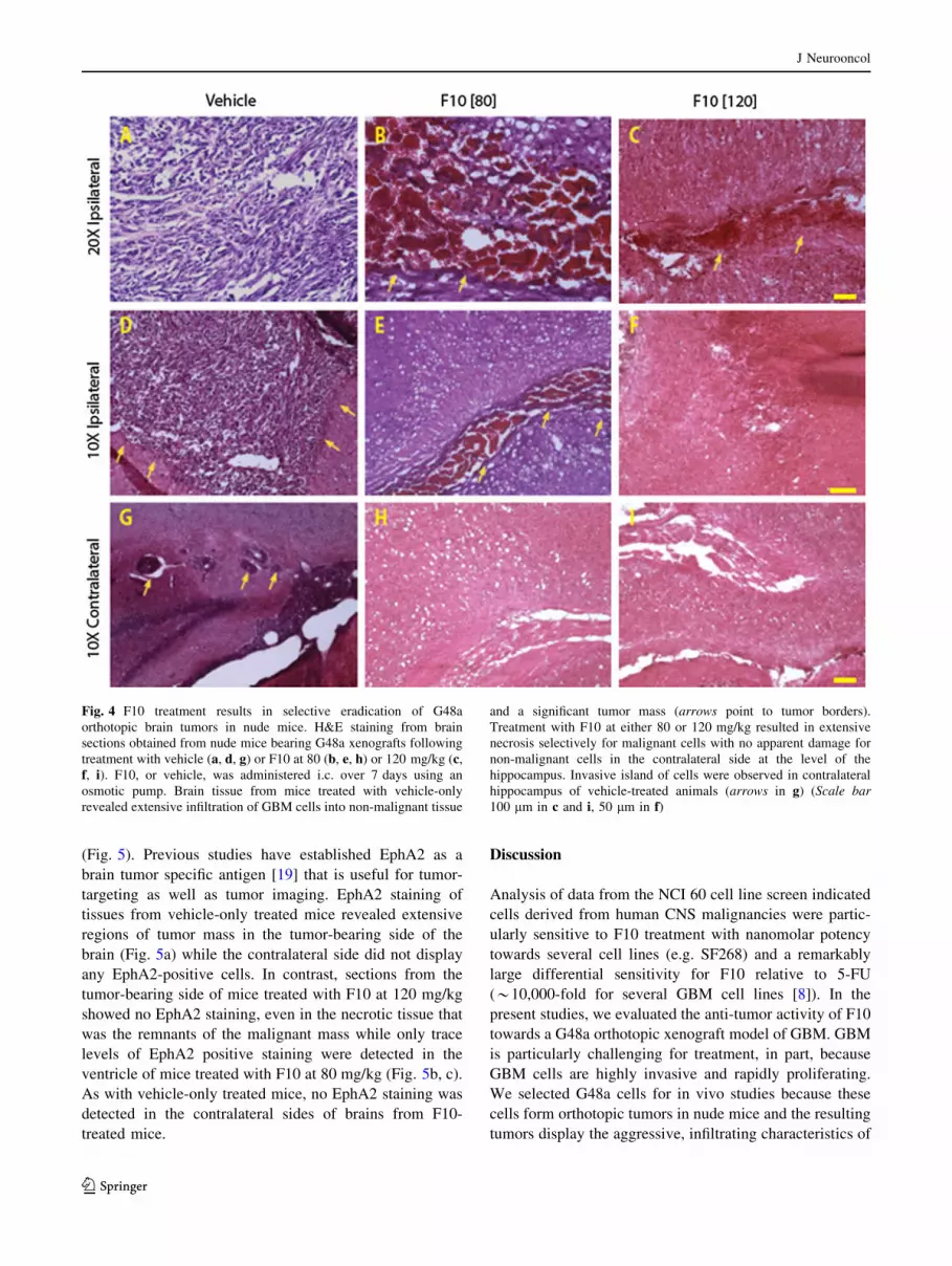
apparent damage to normal brain tissue (Fig. 4a, d). G48a
cells produced highly infiltrative and rapidly growing
tumors in the brains of immunocompromised mice with
tumors localized to the hemisphere where cells were
injected. H&E staining of brain sections from vehicle- and
F10-treated mice demonstrated that F10 treatment resulted
in extensive areas of necrosis selectively within tumor
tissue and only in the tumor-bearing side of the brain
(Fig. 4a–f). The extent of F10-induced necrosis was dose-
dependent with the 120 mg/kg dose (Fig. 4c, f) inducing
greater tumor cell necrosis than the 80 mg/kg dose
(Fig. 4b, e). There was no observable necrosis in the con-
tralateral side of brains from F10-treated mice (Fig. 4g–i).
To further validate selective death of malignant cells
in vivo we performed EphA2-staining of brain sections
Fig. 3 F10 treatment results in significant and dose-responsive
regression of G48a orthotopic xenografts. a Luciferase signal from
F10- and vehicle-treated nude mice at 80 and 120 mg kg doses.
Treatment with F10 results in marked decrease in luciferase-signal for
treated mice. b Mean tumor luminescence calculated from the
luciferase images. F10 treatment results in regression of G48a
xenografts that is highly significant (p \ 0.01) relative to vehicle for
the 120 mg/kg treatment
J Neurooncol
123
(Fig. 5). Previous studies have established EphA2 as a
brain tumor specific antigen [19] that is useful for tumor-
targeting as well as tumor imaging. EphA2 staining of
tissues from vehicle-only treated mice revealed extensive
regions of tumor mass in the tumor-bearing side of the
brain (Fig. 5a) while the contralateral side did not display
any EphA2-positive cells. In contrast, sections from the
tumor-bearing side of mice treated with F10 at 120 mg/kg
showed no EphA2 staining, even in the necrotic tissue that
was the remnants of the malignant mass while only trace
levels of EphA2 positive staining were detected in the
ventricle of mice treated with F10 at 80 mg/kg (Fig. 5b, c).
As with vehicle-only treated mice, no EphA2 staining was
detected in the contralateral sides of brains from F10-
treated mice.
Discussion
Analysis of data from the NCI 60 cell line screen indicated
cells derived from human CNS malignancies were partic-
ularly sensitive to F10 treatment with nanomolar potency
towards several cell lines (e.g. SF268) and a remarkably
large differential sensitivity for F10 relative to 5-FU
(*10,000-fold for several GBM cell lines [8]). In the
present studies, we evaluated the anti-tumor activity of F10
towards a G48a orthotopic xenograft model of GBM. GBM
is particularly challenging for treatment, in part, because
GBM cells are highly invasive and rapidly proliferating.
We selected G48a cells for in vivo studies because these
cells form orthotopic tumors in nude mice and the resulting
tumors display the aggressive, infiltrating characteristics of
Fig. 4 F10 treatment results in selective eradication of G48a
orthotopic brain tumors in nude mice. H&E staining from brain
sections obtained from nude mice bearing G48a xenografts following
treatment with vehicle (a, d, g) or F10 at 80 (b, e, h) or 120 mg/kg (c,
f, i). F10, or vehicle, was administered i.c. over 7 days using an
osmotic pump. Brain tissue from mice treated with vehicle-only
revealed extensive infiltration of GBM cells into non-malignant tissue
and a significant tumor mass (arrows point to tumor borders).
Treatment with F10 at either 80 or 120 mg/kg resulted in extensive
necrosis selectively for malignant cells with no apparent damage for
non-malignant cells in the contralateral side at the level of the
hippocampus. Invasive island of cells were observed in contralateral
hippocampus of vehicle-treated animals (arrows in g) (Scale bar
100 lm in c and i, 50 lm in f)
J Neurooncol
123

the human disease. Our results demonstrate that F10
administered i.c. is not only highly effective at reducing the
growth rate of G48a xenografts in vivo, but that F10
actually induces significant tumor reduction (Fig. 3).
Tumor reduction was achieved in a dose-dependent manner
with histological analysis indicating extensive necrosis
(Fig. 4) and essentially complete tumor eradication based
on elimination of EphA2 staining (Fig. 5). Importantly, this
dramatic anti-tumor effect was achieved with no apparent
damage to normal brain tissue (Fig. 4) demonstrating the
potential for F10 to be administered via convection
enhanced delivery (CED) in patients. CED [20, 21] is a
minimally invasive technique of delivering drugs directly
to brain tumors. A Phase 3 trial of IL-13 toxic conjugate
showed equivalency to standard of care demonstrating the
feasibility of CED for effective treatment [22].
One of the more intriguing findings from the present
work is that the strong anti-tumor activity for F10 occurs
with no apparent damage to normal brain tissue, including
brain tissue proximal to the tumor mass. These in vivo
results are consistent with the lack of toxicity for F10
towards primary neuronal cultures (Fig. 2) and contrast
sharply with the cytotoxicity of F10 towards GBM cells
(Fig. 1). Our previous studies demonstrated that F10
selectively targets replicating cells with the lethal lesions
being trapped Top1CC [11]. As mature neuronal cells have
low proliferative capacity, the lack of toxicity for F10
towards these cells is not unexpected. In contrast, the con-
ventional FP 5-FU is considerably more cytotoxic towards
primary neuronal cultures than F10 (Fig. 2), likely as a
consequence of RNA-mediated effects or metabolites [23]
such as alpha-fluoro-beta-alanine (FBAL) (Fig. 1b) that are
highly neurotoxic [24]. Nonetheless, establishing that the
preferential cytotoxicity of F10 towards malignant cells
in vitro can be achieved in vivo is a significant accom-
plishment that establishes the feasibility of using F10 for
treatment of GBM in humans, particularly with local
administration. To date, conventional FPs have displayed
limited utility for GBM treatment in humans [25]. Thus, the
promising activity towards GBM observed in the present
studies with F10 indicates this novel polymeric FP may be
fundamentally different from other FPs in this regard.
Acknowledgments We thank Jane Stupe for performing the pri-
mary neuronal and astrocyte cultures. National Institutes of Health
(P30 CA012197); WFSM Brain Tumor Center of Excellence and the
Mr. and Mrs. A. Tab Williams Jr. and Family Neuroscience Research
and Program Development Endowment (CM).
Conflict of interest Wake Forest University Office of Technology
Asset Management plans has filed a provisional patent application
(WG & WD).
Fig. 5 F10 treatment results in selective eradication of EphA2-
stained G48a cells. EphA2 staining from brain sections obtained from
nude mice bearing G48a xenografts following treatment with vehicle
only (a, d) or F10 at 80 (b, e) or 120 mg/kg (c, f). Strong EphA2
staining is observed for tumor tissue in vehicle-only treated mice with
no EphA2 staining in adjacent non-malignant tissue except in isolated
invasive cells (arrows indicate tumor borders and point to invasive
cells away from tumor core). EphA2 staining is greatly diminished in
region of the residual tumor (indicated by arrows) from mice treated
with F10 at 80 mg/kg and is absent in mice treated with F10 at 120
mg/kg (Scale bar 50 lm)
J Neurooncol
123

Open Access This article is distributed under the terms of the
Creative Commons Attribution License which permits any use, dis-
tribution, and reproduction in any medium, provided the original
author(s) and the source are credited.
References
1. Krex D, Klink B, Hartmann C, von Deimling A, Pietsch T, Simon
M, Sabel M, Steinbach JP, Heese O, Reifenberger G, Weller M,
Schackert G (2007) Long-term survival with glioblastoma mul-
tiforme. Brain 130(Pt 10):2596–2606
2. Hulleman E, Helin K (2005) Molecular mechanisms in glioma-
genesis. Adv Cancer Res 94:1–27
3. Henriksson R, Asklund T, Poulsen HS (2011) Impact of therapy
on quality of life, neurocognitive function and their correlates in
glioblastoma multiforme: a review. J Neurooncol 104(3):639–646
4. Zuber J, Radtke I, Pardee TS, Zhao Z, Rappaport AR, Luo W,
McCurrach ME, Yang MM, Dolan ME, Kogan SC, Downing JR,
Lowe SW (2009) Mouse models of human AML accurately
predict chemotherapy response. Genes Dev 23(7):877–889
5. Pardee TS, Gomes E, Jennings-Gee J, Caudell D, Gmeiner WH
(2012) Unique dual targeting of thymidylate synthase and topo-
isomerase 1 by FdUMP[10] results in high efficacy against AML
and low toxicity. Blood 119:3561–3570
6. Pardee T, Jennings-Gee J, Stadelman K, Caudell D, Gmeiner WH
(2013) The poison oligonucleotide F10 is efficiently taken up by,
and highly effective against, acute lymphoblastic leukemia while
sparing normal hematopoietic cells. Blood 122:2674
7. Feldman EJ (2011) Too much ara-C? Not enough daunorubicin?
Blood 117(8):2299–2300
8. Gmeiner WH, Reinhold WC, Pommier Y (2010) Genome-wide
mRNA and microRNA profiling of the NCI 60 cell-line screen
and comparison of FdUMP[10] with fluorouracil, floxuridine, and
topoisomerase 1 poisons. Mol Cancer Ther 9(12):3105–3114
9. Debinski W, Gibo DM (2005) Fos-related antigen 1 modulates
malignant features of glioma cells. Mol Cancer Res 3(4):237–249
10. Wykosky J, Gibo DM, Debinski W (2007) A novel, potent, and
specific ephrinA1-based cytotoxin against EphA2 receptor
expressing tumor cells. Mol Cancer Ther 6(12 Pt 1):3208–3218
11. Liao ZY, Sordet O, Zhang HL, Kohlhagen G, Antony S, Gmeiner
WH, Pommier Y (2005) A novel polypyrimidine antitumor agent
FdUMP[10] induces thymineless death with topoisomerase
I-DNA complexes. Cancer Res 65(11):4844–4851
12. Gmeiner WH, Trump E, Wei C (2004) Enhanced DNA-directed
effects of FdUMP[10] compared to 5FU. Nucleosides Nucleo-
tides Nucleic Acids 23(1–2):401–410
13. Wang W, McLeod HL, Cassidy J, Collie-Duguid ES (2007)
Mechanisms of acquired chemoresistance to 5-fluorouracil and
tomudex: thymidylate synthase dependent and independent net-
works. Cancer Chemother Pharmacol 59(6):839–845
14. Sharpe R, Pearson A, Herrera-Abreu MT, Johnson D, Mackay A,
Welti JC, Natrajan R, Reynolds AR, Reis-Filho JS, Ashworth A,
Turner NC (2011) FGFR signaling promotes the growth of triple-
negative and basal-like breast cancer cell lines both in vitro and
in vivo. Clin Cancer Res 17(16):5275–5286
15. Mroz P, Bhaumik J, Dogutan DK, Aly Z, Kamal Z, Khalid L, Kee
HL, Bocian DF, Holten D, Lindsey JS, Hamblin MR (2009)
Imidazole metalloporphyrins as photosensitizers for photody-
namic therapy: role of molecular charge, central metal and
hydroxyl radical production. Cancer Lett 282(1):63–76
16. Hockfield S, Carlson S, Carol Evans P, Pintar LJ, Silberstein L
(1993) Primary cultures from the central nervous system.
Molecular probes of the nervous system. Cold Spring Harbor
Laboratory Press, Plainview, pp 34–55
17. Graham-Cole CL, Thomas HD, Taylor GA, Newell DR, Melton
RG, Hesp R, Boddy AV (2007) An evaluation of thymidine
phosphorylase as a means of preventing thymidine rescue from
the thymidylate synthase inhibitor raltitrexed. Cancer Chemother
Pharmacol 59(2):197–206
18. Han R, Yang YM, Dietrich J, Luebke A, Mayer-Proschel M,
Noble M (2008) Systemic 5-fluorouracil treatment causes a
syndrome of delayed myelin destruction in the central nervous
system. J Biol 7(4):12
19. Wykosky J, Debinski W (2008) The EphA2 receptor and ephri-
nA1 ligand in solid tumors: function and therapeutic targeting.
Mol Cancer Res 6(12):1795–1806
20. Debinski W, Tatter S (2009) Convection-enhanced delivery for
the treatment of brain tumors. Expert Rev Neurother 9(10):
1519–1527
21. Debinski W, Tatter S (2010) Convection-enhanced delivery to
achieve widespread distribution of viral vectors: predicting clin-
ical implementation. Curr Opin Mol Ther 12(6):647–653
22. Mueller S, Poller MY, Lee B, Kunwer S, Pedain C, Wembacher-
Schroder E, Mittermeyer S, Westphal M, Sampson JH, Vogel-
baum MA, Croteau D, Chang SM (2011) Effects of imaging and
catheter characteristics on clinical outcomes for patients in the
PRECISE study. J Neurooncol 101(2):267–277
23. Pritchard DM, Watson AJ, Potten CS, Jackman AL, Hickman JA
(1997) Inhibition by uridine but not thymidine of p53-dependent
intestinal apoptosis initiated by 5-fluorouracil: evidence for the
involvement of RNA perturbation. Proc Natl Acad Sci USA
94(5):1795–1799
24. Yamashita K, Yada H, Ariyoshi T (2004) Neurotoxic effects of
alpha-fluoro-beta-alanine (FBAL) and fluoroacetic acid (FA) on
dogs. J Toxicol Sci 29(2):155–166
25. Miller CR, Williams CR, Buchsbaum DJ, Gillespie GY (2002)
Intratumoral 5-fluorouracil produced by cytosine deaminase/5-
fluorocytosine gene therapy is effective for experimental human
glioblastomas. Cancer Res 62(3):773–780
J Neurooncol
123