Morphology-Based Automatic Seizure Detector for Intracerebral EEG Recordings
Brain and blood microRNA expression profilingof ischemic stroke, intracerebral hemorrhage, andkainate seizures
Da-Zhi Liu, Yingfang Tian, Bradley P Ander, Huichun Xu, Boryana S Stamova,Xinhua Zhan, Renee J Turner, Glen Jickling and Frank R Sharp
Department of Neurology and the M.I.N.D. Institute, University of California at Davis Medical Center,Sacramento, California, USA
MicroRNAs (miRNAs) regulate gene expression and have a critical role in many biologic andpathologic processes. We hypothesized that miRNA expression profiles in injured brain(hippocampus) would show common as well as unique profiles when compared with those ofblood. Adult, untouched, control rats were compared with rats with sham surgeries, ischemicstrokes, brain hemorrhage (lysed blood, fresh blood, or thrombin), and kainate-induced seizures.Brain and whole-blood miRNA expression profiles were assessed 24h later using TaqMan rodentmiRNA arrays. MicroRNA response profiles were different for each condition. Many miRNAschanged more than 1.5-fold in brain and blood after each experimental manipulation, and severalmiRNAs were upregulated or downregulated in both brain and blood after a given injury. A fewmiRNAs (e.g., miR-298, miR-155, and miR-362-3p) were upregulated or downregulated more thantwofold in both brain and blood after several different injuries. The results show the possible use ofblood miRNAs as biomarkers for brain injury; that selected blood miRNAs may correlate with miRNAchanges in the brain; and that many of the mRNAs, previously shown to be regulated in brain andblood after brain injury, are likely accounted for by changes in miRNA expression.Journal of Cerebral Blood Flow & Metabolism (2010) 30, 92–101; doi:10.1038/jcbfm.2009.186; published online2 September 2009
Keywords: blood; brain; epilepsy; hemorrhage; ischemia; microRNA (miRNA)
Introduction
RNA expression studies offer unique opportunitiesfor diagnosis and prognosis, as well as for under-standing the pathogenesis of many medical andneurologic diseases (Sharp et al, 2006, 2007). Ourprevious studies of mRNA expression profiles haveshown that a large number of genes are induced orsuppressed in brain and blood after cerebral ische-mia, intracerebral hemorrhage, and kainate-inducedseizures (Lu et al, 2006; Tang et al, 2001, 2002).However, the specific factors that lead to theregulation of large numbers of genes (mRNAs) ineach condition remain unclear.
The recent discovery of microRNAs (miRNAs) askey regulators of gene function has introduced a new
level and mechanism of gene regulation (Bartel,2004; Chen and Rajewsky, 2007; Filip, 2007).Although there are only hundreds of miRNAs, eachof them can potentially regulate hundreds of targetgenes (Guarnieri and DiLeone, 2008), with theprediction that more than one-third of all humangenes may be regulated by miRNAs (Esquela-Kerscher and Slack, 2006). Functionally, miRNAs areimplicated in fundamental cellular processes includ-ing cell-cycle regulation (Matsubara et al, 2007),hematopoietic differentiation (Hatfield and Ruohola-Baker, 2008), immune responses (Bi et al, 2009), andcellular metabolism (Gauthier and Wollheim, 2006).
MicroRNA expression has been examined invarious tumors (Guarnieri and DiLeone, 2008; Luet al, 2005; Nicoloso and Calin, 2008), in Alzheimer’sdisease (Hebert et al, 2008), Parkinson’s disease (Kimet al, 2007), Down’s syndrome (Kuhn et al, 2008),schizophrenia (Beveridge et al, 2008), and stroke(Dharap et al, 2009; Jeyaseelan et al, 2008). ThesemiRNA expression profiles may be as diagnosticallyuseful as mRNA expression profiles (Guarnieri andDiLeone, 2008; Lu et al, 2005). Therefore, wepostulated that miRNAs alone could be adequate
Received 29 May 2009; revised 7 July 2009; accepted 10 August2009; published online 2 September 2009
Correspondence: Dr D-Z Liu, Department of Neurology and theM.I.N.D. Institute, University of California at Davis MedicalCenter, 2805 50th Street, Sacramento, CA 95817, USA.E-mail: [email protected] study was supported by NIH Grant NS054652 (FRS).
Journal of Cerebral Blood Flow & Metabolism (2010) 30, 92–101& 2010 ISCBFM All rights reserved 0271-678X/10 $32.00
www.jcbfm.com

biomarkers and that miRNA expression profilescould explain, at least in part, the mRNA regulationin brain and blood after different neurologic dis-eases. To begin to address this idea, adult untouchedcontrol rats were compared with rats with shamsurgeries, ischemic strokes, intracerebral hemorrhage(lysed blood, fresh blood, thrombin), and kainate-induced seizures. Total RNA (plus miRNA) wasisolated 24h later from brain and blood, and miRNAexpression profiles were assessed using TaqManRodent miRNA Arrays. The results show uniquemiRNA profiles for brain, blood, and for each experi-mental condition, with some miRNAs being regulatedin both brain and blood for a given injury. Moreover,many of the mRNA targets of regulated miRNAs havepreviously been reported to be regulated in brain andblood after stroke, hemorrhage, and seizures.
Materials and methods
Experiments were carried out in accordance with NationalInstitutes of Health guidelines and were approved by theInstitutional Animal Care and Use Committee (University ofCalifornia at Davis). Male Sprague–Dawley rats (n=48 total),weighing 300 to 320g, were used in this study. The animalswere allowed free access to food and water and weremaintained on a 12-h light/dark cycle. Rats were anesthe-tized with isoflurane (Minrad, New York, NY, USA) andtheir body temperature was maintained at 37.01C±0.21C.They were allowed to fully recover after a procedure in anincubator maintained at 371C, and were then returned totheir home cages with free access to food and water.
Brain Ischemia
Rats (n=6) were anesthetized with isoflurane, and the neckskin and muscle were incised to isolate the left commoncarotid artery. A 3-0 monofilament nylon suture wasthreaded through the external carotid artery stump into theinternal carotid artery and up to the stem of the middlecerebral artery (MCA). The suture was anchored in place toproduce a permanent MCA occlusion. This ‘suture’-inducedMCA occlusion produces reliable infarction in the MCAdistribution (Liu et al, 2005). Only animals with ipsilateralhemispheric infarction were selected for this study.
Brain Hemorrhage
Rats were anesthetized and placed in a stereotaxic frame(Kopf Instruments, Tujunga, CA, USA). They receivedintraventricular (!0.9mm posterior, 1.4 lateral to bregma,and 4.6mm deep) injections (intracerebroventricular) of50 mL lysed blood (n=6), 50 mL fresh autologous tailvein blood (n=6), or thrombin (n=6) (20U per animal in50 mL of saline) (Paxinos and Watson, 1998). All threemodels have been used to study various aspects of thepathophysiology of intracerebral hemorrhage (Liu et al,2008; Tang et al, 2001, 2002).
Kainate Seizures
Rats (n=6) were injected subcutaneously with 10mg/kg ofkainic acid (Sigma, St Louis, MO, USA) dissolved in saline.This kainic acid model produces status epilepticus thatinjures neurons in the cortex and other brain regions (Zhanget al, 1996, 1997). Animals that had severe, prolongedgeneralized seizures were selected for this study.
Sham Surgeries
For sham ischemia, rats (n=6) were anesthetized, the sutureinserted into the internal carotid artery, and then removedwithout being advanced to the stem of the MCA. For shambrain hemorrhage, rats (n=6) were anesthetized and 50mL ofsaline was injected into the left cerebral ventricle.
Untouched Controls
Rats (n=6) that had not been handled in any way wereused as controls. All experimental and control animalswere housed in the same room for 1 week before the studyand then during the study.
Total RNA (Plus microRNA) Isolation from Rat Brainand Blood Samples
At 24h (1 day) after each operation or assignment as anuntouched control, all subjects were anesthetized withisoflurane. Blood (0.5mL) was drawn from the tail veininto EDTA tubes (Becton Dickinson and Company,Franklin Lakes, NJ, USA), immediately mixed with 1.3mLRNA (Ambion, Austin, TX, USA), and stored at !801C.Animals were then killed, brain was removed, and thehippocampus was dissected as rapidly as possible, frozen,and stored at !801C. For ischemia and hemorrhage rats, thehippocampus adjacent to the hemispheric infarction and thehippocampus ipsilateral to ventricular injections were dis-sected, respectively. Hippocampus was chosen because itshould sustain some cell injury or cell death in most of themodels without including any areas of infarction.Total RNA (plus miRNA) was isolated from hippocam-
pus using a miRNeasy Mini Kit (Qiagen, Valencia, CA,USA) and a Qiacube (Qiagen). Total RNAwas isolated fromblood using the Mouse RiboPure-Blood RNA Isolation Kit(Ambion). The concentration and integrity of total RNAwas determined using a NanoDrop-1000 spectrophot-ometer (Thermo Scientific, Wilmington, DE, USA) andAgilent 2100 Bioanalyzer (Agilent Technologies, SantaClara, CA, USA). RNA samples were stored at !801Cbefore TaqMan miRNA array studies.
TaqMan miRNA Arrays
TaqMan MicroRNA Arrays (384-Well Fluidic Cards) (Ap-plied Biosystems, Foster City, CA) were used to profile 381mature miRNAs. U6 was used as the endogenous control inquadruplicate and one assay not related to rat wasincluded as a negative control. Total RNA (800ng each
MicroRNA expression after neurologic diseasesD-Z Liu et al
93
Journal of Cerebral Blood Flow & Metabolism (2010) 30, 92–101
sample) was converted into cDNA using Megaplex RTPrimers (Applied Biosystems) and TaqMan miRNA RT Kits(Applied Biosystems). cDNAs were mixed with TaqManUniversal PCR Master Mix (Applied Biosystems) and thenloaded into TaqMan rodent miRNA fluidic cards (AppliedBiosystems). The cards were run using an Applied Bio-systems 7900HT real-time PCR instrument equipped with aheating block for the fluidic card (Applied Biosystems).
Statistical Analysis
As cycle threshold (CT) scores > 35 are considered to benonspecific (undetectable) (Schmittgen et al, 2008), miR-NAs in which 95% of individual observations had a raw CT
score > 35 were excluded from the final data analysis.Using these filtering criteria, 97 miRNAs were removedfrom the brain analysis, and 153 miRNAs were removedfrom blood analyses. Partek Genomics Suite (Partek,St Louis, MI, USA) was used to carry out statisticalanalyses. Fold change filters were applied to select themiRNAs that were regulated more than 1.5- or 2-fold.A t-test with or without FDR (false discovery rate of5%=no more than 5% false positives) correction formultiple comparisons was used to examine the signifi-cance of miRNAs regulated in each experimental condition
compared with that of the untouched control group.MicroRNAs that changed Z1.5-fold and had a P<0.05were subjected to hierarchical clustering using Euclideandistance on the basis of their relative mean expression.
Biologic Functional Analyses of MicroRNAs Regulatedin Both Brain and Blood
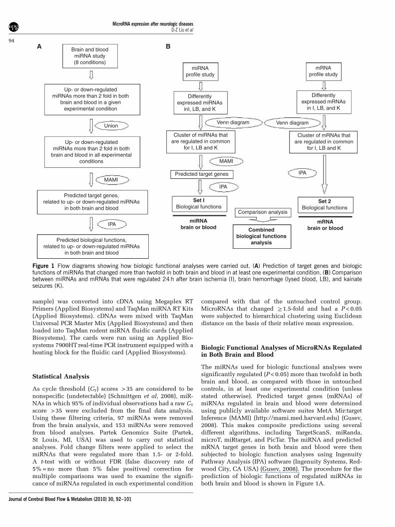
The miRNAs used for biologic functional analyses weresignificantly regulated (P<0.05) more than twofold in bothbrain and blood, as compared with those in untouchedcontrols, in at least one experimental condition (unlessstated otherwise). Predicted target genes (mRNAs) ofmiRNAs regulated in brain and blood were determinedusing publicly available software suites MetA Mir:targetInference (MAMI) (http://mami.med.harvard.edu) (Gusev,2008). This makes composite predictions using severaldifferent algorithms, including TargetScanS, miRanda,microT, miRtarget, and PicTar. The miRNA and predictedmRNA target genes in both brain and blood were thensubjected to biologic function analyses using IngenuityPathway Analysis (IPA) software (Ingenuity Systems, Red-wood City, CA USA) (Gusev, 2008). The procedure for theprediction of biologic functions of regulated miRNAs inboth brain and blood is shown in Figure 1A.
Brain and bloodmiRNA study(8 conditions)
Up- or down-regulatedmiRNAs more than 2 fold in both
brain and blood in a givenexperimental condition
Up- or down-regulatedmiRNAs more than 2 fold in both
brain and blood in all experimentalconditions
Union
MAMI
IPAmiRNA
brain or blood Combinedbiological functions
analysis
mRNAbrain or blood
Set 2Biological functions
Comparison analysis
Set IBiological functions
IPA
IPAPredicted target genes
MAMI
Venn diagram Venn diagram
Differentlyexpressed miRNAs
inI, LB, and K
miRNAprofile study
mRNAprofile study
Differentlyexpressed mRNAs
in I, LB, and K
Cluster of mRNAs thatare regulated in common
for I, LB and K
Cluster of miRNAs thatare regulated in common
for I, LB and K
Predicted target genes,related to up- or down-regulated miRNAs
in both brain and blood
Predicted biological functions,related to up- or down-regulated miRNAs
in both brain and blood
Figure 1 Flow diagrams showing how biologic functional analyses were carried out. (A) Prediction of target genes and biologicfunctions of miRNAs that changed more than twofold in both brain and blood in at least one experimental condition. (B) Comparisonbetween miRNAs and mRNAs that were regulated 24h after brain ischemia (I), brain hemorrhage (lysed blood, LB), and kainateseizures (K).
MicroRNA expression after neurologic diseasesD-Z Liu et al
94
Journal of Cerebral Blood Flow & Metabolism (2010) 30, 92–101

Comparison of Biologic Functions of MicroRNAs andmRNAs Regulated in Brain and Blood
For miRNA–mRNA biologic function analyses, miRNAsregulated more than twofold in either brain or blood afterbrain ischemia, brain hemorrhage (lysed blood), andkainate-induced seizures were entered into MAMI toobtain the predicted target genes (Figure 1B, left panel).The exported target genes were then entered into IPA toobtain a set of biologic functions (Figure 1B, left panel).Similarly, regulated mRNAs, obtained from our previouslypublished studies (Lu et al, 2006; Tang et al, 2001, 2002),were input into IPA to yield another set of biologicfunctions (Figure 1B, right panel). We then compared thetwo sets of data to derive the functional pathways that werecommon to the miRNAs and mRNAs regulated for each ofthe injuries in both brain and blood (Figure 1B).
Results
Endogenous Control MicroRNAs in Brain and Blood
Five potential endogenous control miRNAs (U6,U87, Y1, snoRNA135, and snoRNA202) were used.In the brain, U6, U87, Y1, and snoRNA135 werestable (regulated <1.5-fold) in each of the sevenexperimental conditions, whereas snoRNA202 de-creased more than 1.5-fold in one or more experi-mental conditions (Supplementary Table 1). Inblood, U6, U87, snoRNA135, and snoRNA202 werestable (regulated <1.5-fold) in each of the sevenexperimental conditions, whereas Y1 was upregu-lated more than 1.5-fold in one or more experimentalconditions (Supplementary Table 1). Thus, U6, U87,and snoRNA134 were the most stable in both brainand blood across experimental conditions. U6 wasused as the endogenous control for all of the datareported in this study because it was the most stablyexpressed miRNA across all subjects in the controland experimental groups.
Brain and Blood MicroRNA Response
Many miRNAs changed more than 1.5-fold in brainand blood after each experimental condition as com-pared with those in untouched controls (Table 1;Supplementary Table 1). A number of miRNAs thatchanged 1.5-fold on an average were significantlychanged (P<0.05), but relatively few of these miRNAspassed the false discovery rate multiple comparisoncorrection threshold (FDR <5%) (Table 1; Supple-mentary Table 1). Several miRNAs were upregulatedor downregulated in both brain and blood after a giveninjury (Table 1; Supplementary Table 1). A fewmiRNAs, such as miR-298, miR-155, and miR-362-3p, were upregulated or downregulated in both brainand blood after several different injuries (Table 1;Supplementary Table 1).
Unique MicroRNA Expression Profiles in Brain andBlood
As there may not be specifically regulated miRNAsfor a given disease, we looked for unique patterns ofmiRNA expression for each of the experimentalconditions using cluster analysis. This showed thateach experimental condition had a unique miRNAexpression pattern in both brain (Figure 2, left panel)and blood (Figure 2, right panel). For the eightexpression profiles shown, no profile was identical.The different miRNA expression patterns, includingsham controls versus untouched controls, emphasizethe uniqueness of the different brain and bloodmiRNA responses for each condition.
Function Analysis of MicroRNAs Regulated in BothBrain and Blood
A biologic function analysis was carried out on themiRNAs regulated more than twofold in both brainand blood in at least one experimental condition.Five miRNAs (miR-298, miR-200b, miR-205, miR-107, and miR-423-5p) were upregulated in both brainand blood, and four miRNAs (miR-155, miR-362-3p,miR-10b, and miR-188-5p) were downregulated morethan twofold in both brain and blood in at least oneexperimental condition (Table 1, marked with #). Thetop 10 ranked biologic functions associated withcommonly upregulated miRNAs include cell cycle,cancer, gene expression, cellular growth and prolif-eration, cellular development, posttranslationalmodification, reproductive system disease, connec-tive tissue development and function, cardiovascularsystem development and function, and cell death(Figure 3A, Supplementary Figure 1A). The top 10ranked biologic functions associated with commonlydownregulated miRNAs included gene expression,cell death, hematological development and function,organismal development, organ development, cardi-ovascular system development and function, cellcycle, cellular development, connective tissue devel-opment and function, and hematopoiesis (Figure 3B,Supplementary Figure 1B).
MicroRNAs Regulated by Multiple Conditions in eitherBrain or Blood
Many upregulated miRNAs were modulated in twoor more of the groups in either brain or blood. Inbrain, 24 miRNAs were upregulated more thantwofold by brain ischemia, 17 by brain hemorrhage(lysed blood), and 13 by kainate seizures (Figure 4A,left panel). In addition, 13 miRNAs were down-regulated more than twofold by brain ischemia, 12 bybrain hemorrhage (lysed blood), and 18 by kainateseizures (Figure 4A, right panel). A Venn diagram ofthese results showed that one miRNA (miR-542-3p)was upregulated (Figure 4A, left panel) and fourmiRNAs (miR-155, miR-362-3p, miR-122, and
MicroRNA expression after neurologic diseasesD-Z Liu et al
95
Journal of Cerebral Blood Flow & Metabolism (2010) 30, 92–101

Table 1 Number of miRNAs that changed in brain and blood 24 h after each experimental condition as compared to untouched controls (C)
Comparison Up- ordownregulated
Sample No. ofmiRNA detectable
miRNAs regulated in both brain andblood (fold change >1.5)
No. of regulated miRNA
Fold change >1.5 Fold change >1.5and P<0.05
Fold change >1.5,P <0.05 and FDR <0.05
SI versus C Up Brain 198 miR-298, miR-503, miR-672 43 5 0Blood 49 19 3 0
Down Brain 85 miR-155,a miR-362-3p 26 1 0Blood 103 44 1 0
I versus C Up Brain 168 miR-10a, miR-182, miR-200b, miR-298 43 6 1Blood 33 16 3 1
Down Brain 115 miR-155,a miR-362-3p,a miR-223, miR-210 22 5 2Blood 119 92 38 19
SH versus C Up Brain 143 miR-148b, miR-503 36 3 0Blood 76 19 3 0
Down Brain 140 miR-10b,a miR-203, miR-362-3p 27 8 4Blood 76 36 3 2
B versus C Up Brain 163 miR-298,a miR-200b,a miR-205,a miR-345,miR-423-5p
34 6 0
Blood 104 39 4 1Down Brain 120 None 31 7 2
Blood 48 20 1 0
LB versus C Up Brain 150 miR-298,a miR-423-5p,a miR-10a, miR-345-5p,miR-674
34 6 0
Blood 87 30 6 1Down Brain 133 miR-155, miR-20b-3p, miR-200a 28 6 1
Blood 65 33 6 0
T versus C Up Brain 234 miR-107,a miR-200b, miR-331-5p, miR-672 69 12 0Blood 29 17 2 0
Down Brain 49 miR-155,a miR-188-5pa 19 3 3Blood 123 63 10 0
K versus C Up Brain 104 miR-298a 21 1 0Blood 47 15 2 0
Down Brain 179 miR-155,a miR-29c, miR-34b-3p, miR-98,miR-122, miR-203, miR-450a
39 9 4
Blood 105 43 1 0
miRNA, microRNA; RT-PCR, real-time PCR.Experimental conditions included untouched control (C), sham ischemia (SI), ischemia (I), sham hemorrhage (SH), fresh blood (B), lysed blood (LB), thrombin (T) and kainate seizures (K).The columns indicate the numbers of miRNAs that were detectable by RT-PCR; had a fold change >1.5; a fold change >1.5 and P<0.05; a fold change >1.5, P<0.05 and FDR <0.05.aRepresents miRNAs regulated in both brain and blood and had a fold change >2.
MicroRNA
expressionafterneurologic
diseasesD-Z
Liuetal
96
JournalofCerebralBloodFlow
&Metabolism
(2010)30,92–101

Figure 2 Hierarchical clustering of miRNAs that changed more than 1.5-fold shows the unique miRNA expression profiles in brain(left panel) and blood (right panel) 24 h after each experimental condition. Red, upregulation; yellow, little change; and blue,downregulation. This experiment was performed on eight groups of rats including untouched controls (C), sham brain ischemia (SI),brain ischemia (I), sham brain hemorrhage (SH), fresh blood-induced brain hemorrhage (B), lysed blood-induced brain hemorrhage(LB), thrombin injections (T), and kainate seizures (K).
Biological functions ofup-regulated micorRNAs in
both brain and blood
Biological functions ofdown-regulated micorRNAs in
both brain and blood
Analysis: miRNAs up-regulated (Fold change>2) in both brain and blood in at leastone experimental condition
Analysis: miRNAs down-regulated (Fold change>2) in both brain and blood in at leastone experimental condition
cell cycle gene expression
cell death
organismal development
organ development
cell cycle
hematopoiesis
cellular development
connective tissue developmentand function
cardiovascular systemdevelopment and function
hematological development andfunction
Threshold-log(p-value)
0.0 0.5 1.0 1.5 2.0 2.5 3.0 3.5 4.0 4.5 5.0 5.5 6.0 6.5 7.0
Threshold-log(p-value)
0.0 0.5 1.0 1.5 2.0 2.5 3.0 3.5 4.0 4.5 5.0 5.5 6.0 6.5
cancer
gene expression
cellular growth and proliferation
cellular development
post-translational modification
reproductive system disease
cardiovascular systemdevelopment and functioncell death
connective tissue developmentand function
Figure 3 Top 10 ranked biologic functions of miRNAs (fold change >2) regulated in both brain and blood in at least oneexperimental condition. (A) Biologic functions of miRNAs upregulated in both brain and blood. (B) Biologic functions of miRNAsdownregulated in both brain and blood.
MicroRNA expression after neurologic diseasesD-Z Liu et al
97
Journal of Cerebral Blood Flow & Metabolism (2010) 30, 92–101

miR-450a-5p) were downregulated in all three con-ditions (Figure 4A, right panel). In blood, 10 miRNAswere upregulated more than twofold by brainischemia, 21 by brain hemorrhage (lysed blood),and 21 by kainate seizures (Figure 4B, left panel). Inaddition, 65 miRNAs were downregulated more thantwofold by brain ischemia, 20 by brain hemorrhage(lysed blood), and 10 by kainate seizures (Figure 4B,right panel). The Venn diagram of these res-ults showed that five miRNAs (miR-96, miR-152,miR-298, miR-333, and miR-505) were upregulated(Figure 4B, left panel) and seven miRNAs (miR-125a-5p, miR-130b, miR-142-3p, miR-330, miR-342-5p,miR-685, and miR-347) were downregulated in allthree conditions (Figure 4B, right panel).
Analyses of MicroRNA Target Genes (mRNAs) andPreviously Reported Regulated mRNAs in Both Brainand Blood After Ischemia, Hemorrhage, and Seizures
We previously reported unique mRNA expressionprofiles in both brain and blood after brain ischemia,brain hemorrhage (lysed blood), and kainate-inducedseizures (Tang et al, 2001, 2002). To make the prev-iously reported data comparable with the data in thisstudy, it was re-normalized and re-analyzed usingPartek software (included in Supplementary Tables 2Aand B). Using the work flow in Figure 1B, the biologicfunctions of the mRNAs regulated in our previousstudies were compared with the biologic functions ofthe miRNAs regulated in this study, for both brain andblood. Figures 5A and 5B show that almost all of thepredicted biologic functions for regulated miRNAsand mRNAs overlapped considerably. The top 10ranked functions in brain for regulated miRNAs andmRNA are cell death, organismal development, can-cer, cell cycle, cell morphology, hematological disease,hematological system development function, hemato-poiesis, immunologic disease, and gene expression(Figure 5A, Supplementary Figure 2A). The top 10ranked functions in blood for regulated miRNAs andmRNA are cell cycle, cell death, cancer, cell morphol-ogy, organismal development, cellular movement,cellular growth and proliferation, cellular develop-ment, development disorder, and gene expression(Figure 5B, Supplementary Figure 2B). Furthermore,the top five ranked functions that overlapped forboth brain and blood included cell cycle, cell death,cancer, cell morphology, and organismal development(Figures 5A and 5B).
Discussion
Amajor finding of this study is that different patternsof miRNA expression occur in brain and blood 24hafter brain ischemia, brain hemorrhage, kainateseizures, and even sham surgeries, compared withuntouched control animals. A number of miRNAswere significantly regulated (P<0.05) more than
1.5-fold in brain and blood after each brain injury.Several miRNAs were upregulated or downregulatedin both brain and blood after a given injury; and afew miRNAs, including miR-298, mir-155, mir-362-3p, etc., were upregulated or downregulated in bothbrain and blood after several different injuries. Theseresults show the possible use of blood miRNAs asbiomarkers for selected brain miRNAs after one ormore specific types of brain injuries. They alsoprovide a partial mechanism for specific miRNA-induced mRNA expression profiles in brain andblood after different types of brain injury.
MicroRNA expression levels can be studied usingseveral different methods: microarray analysis, real-time PCR (RT-PCR), northern blots, in situ hybridi-zation, and solution hybridization. Of these techni-ques, TaqMan miRNA assays are specific for maturemiRNAs and discriminate among related miRNAsthat differ by as little as one nucleotide (Chen et al,2005). Furthermore, RT-PCR assays are not affectedby genomic DNA contamination (Chen et al, 2005).Therefore, TaqMan RT-PCR miRNA assays are a
Figure 4 Venn diagrams showing the numbers of miRNAs thatwere regulated more than twofold in brain and blood 24 h afterbrain ischemia (I), brain hemorrhage (LB, lysed blood), andkainate seizures (K) compared with untouched controls (C). (A)MicroRNAs upregulated (left panel) and downregulated (rightpanel) in the brain. (B) MicroRNAs upregulated (left panel) anddownregulated (right panel) in blood.
MicroRNA expression after neurologic diseasesD-Z Liu et al
98
Journal of Cerebral Blood Flow & Metabolism (2010) 30, 92–101

sensitive and accurate method for assessing miRNAexpression.
A remarkable finding in this study is that miR-298was the only miRNA that was upregulated in bothbrain and blood after almost all experimental condi-tions. Thus, this is consistent with the finding thatmiR-298 was also upregulated in the brain aftertransient MCA occlusions in another recent study(Jeyaseelan et al, 2008). Although miR-298 has manyfunctions, it was recently shown to recognizespecific binding sites in the 30-UTR of b-amyloidprecursor protein-converting enzyme-1 (BACE1)mRNA and to regulate BACE1 protein expression incultured neuronal cells (Boissonneault et al, 2009).This particular miRNA also emphasizes the fact thatmiR-298 likely regulates hundreds of other mRNAsand that the dozens of miRNAs shown to beregulated in brain and blood in this study regulatehundreds of target mRNAs in brain and blood,many of which were shown by us to be regulatedin our previous studies (Lu et al, 2006; Tang et al,2001, 2002).
Recent studies indicate that some miRNAs that areselectively and/or highly expressed in immune
cells—including the miR-17-92 cluster, miR-150,miR-155, miR-181, and miR-223—have a ‘permis-sive’ function in the maturation, proliferation, anddifferentiation of myeloid and lymphoid cells (Tsit-siou and Lindsay, 2009). In addition, miR-155negatively regulates the immune response by repres-sing Src homology-2 domain-containing inositol5-phosphatase 1 (SHIP1) through direct 30-UTRinteractions (O’Connell et al, 2009). In this study,miR-155 was downregulated in both brain and bloodafter almost all experimental conditions. This isconsistent with our previous genomic studies thatthe expression of Lyn, one of the Src family ofkinases, was upregulated 21-fold after intracerebralhemorrhage (Lu et al, 2006; Tang et al, 2002).
Many of the miRNAs upregulated or downregu-lated by each experimental condition were modu-lated in two or more of the groups. The miRNAsregulated in common by the various conditionsmight provide an index of common mechanisms ofinjury. Therefore, we used the commonly regulatedmiRNAs for predicting biologic functions. Theresults show that the five top-ranked functionspredicted by the commonly regulated miRNAs and
BrainComparison analysis: Comparison analysis:
Blood
Predicted target genes of regulated miRNAs that are common for I, LB and K.
mRNAs that are regulated in common forI, LB, and K.
Predicted target genes of regulated miRNAs that are common for I, LB and K.
mRNAs that are regulated in common for I, LB, and K.
cell death
cell death
cancer
cell morphology
organismal development
cellular movement
cellular growth and proliferation
cellular development
development disorder
gene expression
cell cycle
organismal development
cancer
cell cycle
cell morphology
hematological disease
hematological systemdevelopment function
hematopoiesis
immunological disease
gene expression
Threshold-log(p-value)
0 1 2 3 4 5 6 7 8 9 10 11 12 13 14 15 16
Threshold
0 1 2 3 4 5 6 7 8 9 10-log(p-value)
Figure 5 Top 10 ranked biologic functions of miRNAs and the mRNAs that were regulated in brain and blood after all three maintypes of brain injury: brain ischemia (I), brain hemorrhage (LB, lysed blood), and kainate-induced seizures (K). The log of the P-valueis shown for each biologic function for both regulated miRNAs (dark blue) and mRNAs (light blue). Regulated miRNAs were derivedfrom this study, and regulated mRNAs were derived from our previous studies (Lu et al, 2006; Tang et al, 2001, 2002). (A)Comparison of the biologic functions of regulated miRNAs with those of regulated mRNAs in brain. (B) Comparison of the biologicfunctions of regulated miRNAs with those of regulated mRNAs in blood.
MicroRNA expression after neurologic diseasesD-Z Liu et al
99
Journal of Cerebral Blood Flow & Metabolism (2010) 30, 92–101

mRNAs in both brain and blood included cell cycle,cell death, cancer, cell morphology, and organismaldevelopment. This suggests that there are somefunctional responses of miRNAs and mRNAs thatare similar in brain and blood after many types ofacute brain injury. Future studies are required todetermine exactly which factors regulate a similarcross talk in both brain and blood between themiRNAs and mRNAs in each. Such factors couldinclude similar brain and blood stress responses toinjury, or similar brain and blood immune responsesto injury.
The ‘cell cycle’ was among the top-ranked func-tions for miRNA regulated in both brain and blood,and for mRNAs and miRNAs that changed in brainand blood 1 day after injury. This suggests that the‘cell-cycle’ pathway might be relevant to suchinjuries. The miRNAs induced in blood related tothe ‘cell cycle’ may relate to the proliferation of whiteblood cells and the blood inflammatory response toacute brain injury. The miRNAs activated in brain in‘cell-cycle’ pathways could relate to glial prolifera-tion, and could potentially relate to the possibilitythat cell cycle re-entry leads to postmitotic neuronaldeath. Cell cycle re-entry in neurons has beenconfirmed in many brain diseases, including stroke,brain hemorrhage, traumatic brain injury (TBI), andepilepsy (Di Giovanni et al, 2005; Imai et al, 2002;Nagy and Esiri, 1998; O’Hare et al, 2002). Inhibitionof cyclin-dependent kinases increases neuronalsurvival and improves behavioral outcomes inanimal models of these diseases (Di Giovanni et al,2005; Hilton et al, 2008; Park et al, 2000; Verdaguer etal, 2003; Verdaguer et al, 2004). Thus, the miRNAsinvolved in the cell cycle in brain could relate to thebirth of new cells including glia, and/or relate toneuronal re-entry into the cell cycle and death ofthese cells.
Although only a limited number of disease condi-tions were studied, data show that brain and bloodmiRNA responses were unique for each. Oneexample of this difference was the unique brainand blood miRNA response of sham-operated controlanimals to that of untouched control animals.Although there are miRNAs specifically regulatedby each of the conditions, we propose that it is thecomposite brain and blood miRNA response of all ofthe regulated miRNAs that may provide miRNAfingerprints that can be distinguished for manydifferent disease-related states.
Finally, several experimental limitations need tobe mentioned. Although the functions of regulatedmiRNAs described in this study significantly overlapthe functions of mRNAs reported in our previousstudies, actual mRNA levels were not measured inthis study. This must be carried out in future studies.In addition, for those mRNAs that are presumedtargets of specific miRNAs, these target mRNAs mustalso be measured in future studies to show theiractual regulation. Second, animals were not perfusedat the conclusion of an experiment because of
concern that this would affect miRNA expression.Thus, the miRNAs that are reported as beingregulated in both brain and blood would representthe maximum number that was regulated, as somecould represent contamination of brain by blood.This is likely a minor problem because of the smallvolume of blood compared with brain, the fewshared miRNAs in brain and blood, and the compar-ison of hemorrhage and control showed no sharedmiRNAs.
Conflict of interest
The authors declare no conflict of interest.
References
Bartel DP (2004) MicroRNAs: genomics, biogenesis, me-chanism, and function. Cell 116:281–97
Beveridge NJ, Tooney PA, Carroll AP, Gardiner E, BowdenN, Scott RJ, Tran N, Dedova I, Cairns MJ (2008)Dysregulation of miRNA 181b in the temporal cortexin schizophrenia. Hum Mol Genet 17:1156–68
Bi Y, Liu G, Yang R (2009) MicroRNAs: novel regulatorsduring the immune response. J Cell Physiol 218:467–72
Boissonneault V, Plante I, Rivest S, Provost P (2009)MicroRNA-298 and microRNA-328 regulate expressionof mouse beta-amyloid precursor protein-convertingenzyme 1. J Biol Chem 284:1971–81
Chen C, Ridzon DA, Broomer AJ, Zhou Z, Lee DH, NguyenJT, Barbisin M, Xu NL, Mahuvakar VR, Andersen MR,Lao KQ, Livak KJ, Guegler KJ (2005) Real-time quanti-fication of microRNAs by stem-loop RT-PCR. NucleicAcids Res 33:e179
Chen K, Rajewsky N (2007) The evolution of generegulation by transcription factors and microRNAs.Nat Rev Genet 8:93–103
Dharap A, Bowen K, Place R, Li LC, Vemuganti R (2009)Transient focal ischemia induces extensive temporalchanges in rat cerebral microRNAome. J Cereb BloodFlow Metab 29:675–87
Di Giovanni S, Movsesyan V, Ahmed F, Cernak I, SchinelliS, Stoica B, Faden AI (2005) Cell cycle inhibitionprovides neuroprotection and reduces glial proliferationand scar formation after traumatic brain injury. ProcNatl Acad Sci USA 102:8333–8
Esquela-Kerscher A, Slack FJ (2006) Oncomirs—micro-RNAs with a role in cancer. Nat Rev Cancer 6:259–69
Filip A (2007) [MiRNA—new mechanisms of gene expres-sion control]. Postepy Biochem 53:413–9
Gauthier BR, Wollheim CB (2006) MicroRNAs: ‘ribo-regulators’ of glucose homeostasis. Nat Med 12:36–8
Guarnieri DJ, DiLeone RJ (2008) MicroRNAs: a new class ofgene regulators. Ann Med 40:197–208
Gusev Y (2008) Computational methods for analysis ofcellular functions and pathways collectively targeted bydifferentially expressed microRNA. Methods 44:61–72
Hatfield S, Ruohola-Baker H (2008) microRNA and stemcell function. Cell Tissue Res 331:57–66
Hebert SS, Horre K, Nicolai L, Papadopoulou AS, Mande-makers W, Silahtaroglu AN, Kauppinen S, Delacourte A,De Strooper B (2008) Loss of microRNA cluster miR-29a/b-1 in sporadic Alzheimer’s disease correlates with
MicroRNA expression after neurologic diseasesD-Z Liu et al
100
Journal of Cerebral Blood Flow & Metabolism (2010) 30, 92–101

increased BACE1/beta-secretase expression. Proc NatlAcad Sci USA 105:6415–20
Hilton GD, Stoica BA, Byrnes KR, Faden AI (2008)Roscovitine reduces neuronal loss, glial activation,and neurologic deficits after brain trauma. J Cereb BloodFlow Metab 28:1845–59
Imai H, Harland J, McCulloch J, Graham DI, Brown SM,Macrae IM (2002) Specific expression of the cellcycle regulation proteins, GADD34 and PCNA, in theperi-infarct zone after focal cerebral ischaemia in the rat.Eur J Neurosci 15:1929–36
Jeyaseelan K, Lim KY, Armugam A (2008) MicroRNAexpression in the blood and brain of rats subjected totransient focal ischemia by middle cerebral arteryocclusion. Stroke 39:959–66
Kim J, Inoue K, Ishii J, Vanti WB, Voronov SV, Murchison E,Hannon G, Abeliovich A (2007) A microRNA feedbackcircuit in midbrain dopamine neurons. Science 317:1220–4
Kuhn DE, Nuovo GJ, MartinMM,Malana GE, Pleister AP, JiangJ, Schmittgen TD, Terry AV, Jr, Gardiner K, Head E, FeldmanDS, Elton TS (2008) Human chromosome 21-derivedmiRNAs are overexpressed in Down syndrome brains andhearts. Biochem Biophys Res Commun 370:473–7
Liu DZ, Xie KQ, Ji XQ, Ye Y, Jiang CL, Zhu XZ (2005)Neuroprotective effect of paeoniflorin on cerebralischemic rat by activating adenosine A1 receptor in amanner different from its classical agonists. Br JPharmacol 146:604–11
Liu DZ, Cheng XY, Ander BP, Xu H, Davis RR, Gregg JP,Sharp FR (2008) Src kinase inhibition decreasesthrombin-induced injury and cell cycle re-entry instriatal neurons. Neurobiol Dis 30:201–11
Lu A, Tang Y, Ran R, Ardizzone TL, Wagner KR, Sharp FR(2006) Brain genomics of intracerebral hemorrhage.J Cereb Blood Flow Metab 26:230–52
Lu J, Getz G, Miska EA, Alvarez-Saavedra E, Lamb J, PeckD, Sweet-Cordero A, Ebert BL, Mak RH, Ferrando AA,Downing JR, Jacks T, Horvitz HR, Golub TR (2005)MicroRNA expression profiles classify human cancers.Nature 435:834–8
Matsubara H, Takeuchi T, Nishikawa E, Yanagisawa K,Hayashita Y, Ebi H, Yamada H, Suzuki M, Nagino M,Nimura Y, Osada H, Takahashi T (2007) Apop-tosis induction by antisense oligonucleotides againstmiR-17-5p and miR-20a in lung cancers overexpressingmiR-17-92. Oncogene 26:6099–105
Nagy Z, Esiri MM (1998) Neuronal cyclin expression in thehippocampus in temporal lobe epilepsy. Exp Neurol150:240–7
Nicoloso MS, Calin GA (2008) MicroRNA involvement inbrain tumors: from bench to bedside. Brain Pathol 18:122–9
O’Connell RM, Chaudhuri AA, Rao DS, Baltimore D (2009)Inositol phosphatase SHIP1 is a primary target ofmiR-155. Proc Natl Acad Sci USA 106:7113–8
O’Hare M, Wang F, Park DS (2002) Cyclin-dependentkinases as potential targets to improve stroke outcome.Pharmacol Ther 93:135–43
Park DS, Obeidat A, Giovanni A, Greene LA (2000) Cellcycle regulators in neuronal death evoked by excitotoxicstress: implications for neurodegeneration and itstreatment. Neurobiol Aging 21:771–81
Paxinos G, Watson C (1998) The rat brain in stereotaticcoordinates. 4th edn. London: Academic Press
Schmittgen TD, Lee EJ, Jiang J, Sarkar A, Yang L, Elton TS,Chen C (2008) Real-time PCR quantification of precursorand mature microRNA. Methods 44:31–8
Sharp FR, Xu H, Lit L, Walker W, Apperson M, Gilbert DL,Glauser TA, Wong B, Hershey A, Liu DZ, Pinter J, ZhanX, Liu X, Ran R (2006) The future of genomic profiling ofneurological diseases using blood. Arch Neurol63:1529–36
Sharp FR, Xu H, Lit L, Walker W, Pinter J, Apperson M,Verro P (2007) Genomic profiles of stroke in blood.Stroke 38:691–3
Tang Y, Lu A, Aronow BJ, Sharp FR (2001) Blood genomicresponses differ after stroke, seizures, hypoglycemia,and hypoxia: blood genomic fingerprints of disease.Ann Neurol 50:699–707
Tang Y, Lu A, Aronow BJ, Wagner KR, Sharp FR (2002)Genomic responses of the brain to ischemic stroke,intracerebral haemorrhage, kainate seizures, hypogly-cemia, and hypoxia. Eur J Neurosci 15:1937–52
Tsitsiou E, Lindsay MA (2009) microRNAs and theimmune response. Curr Opin Pharmacol 9:514–20
Verdaguer E, Jorda EG, Canudas AM, Jimenez A, SuredaFX, Rimbau V, Pubill D, Escubedo E, Camarasa J, PallasM, Camins A (2003) 3-Amino thioacridone, a selectivecyclin-dependent kinase 4 inhibitor, attenuates kainicacid-induced apoptosis in neurons. Neuroscience120:599–603
Verdaguer E, Jimenez A, Canudas AM, Jorda EG, SuredaFX, Pallas M, Camins A (2004) Inhibition of cell cyclepathway by flavopiridol promotes survival of cerebellargranule cells after an excitotoxic treatment. J PharmacolExp Ther 308:609–16
Zhang X, Boulton AA, Yu PH (1996) Expression of heatshock protein-70 and limbic seizure-induced neuronaldeath in the rat brain. Eur J Neurosci 8:1432–40
Zhang X, Gelowitz DL, Lai CT, Boulton AA, Yu PH (1997)Gradation of kainic acid-induced rat limbic seizuresand expression of hippocampal heat shock protein-70.Eur J Neurosci 9:760–9
Supplementary Information accompanies the paper on the Journal of Cerebral Blood Flow & Metabolism website (http://www.nature.com/jcbfm)
MicroRNA expression after neurologic diseasesD-Z Liu et al
101
Journal of Cerebral Blood Flow & Metabolism (2010) 30, 92–101
Copyright © 2022 FDOKUMEN