Angiogenesis inhibitor DC101 delays growth of intracerebral glioblastoma but induces morbidity when...
7
Angiogenesis inhibitor DC101 delays growth of intracerebral glioblastoma but induces morbidity when combined with irradiation Joost J.C. Verhoeff a , Lukas J.A. Stalpers a , Cornelis J.F. Van Noorden b , Dirk Troost c , Marja D. Ramkema c , Chris van Bree a , Ji-Ying Song d , Mila Donker e , Martha Chekenya f , W. Peter Vandertop e , Dick J. Richel a , Wouter R. van Furth e, * a Laboratory of Experimental Oncology and Radiobiology, Center for Experimental and Molecular Medicine, Academic Medical Center, University of Amsterdam, Meibergdreef 9, 1105 AZ Amsterdam, The Netherlands b Department of Cell Biology and Histology, Academic Medical Center, University of Amsterdam, Meibergdreef 15, 1105 AZ Amsterdam, The Netherlands c Department of Neuropathology, Academic Medical Center, University of Amsterdam, Meibergdreef 9, 1105 AZ Amsterdam, The Netherlands d Experimental Animal Pathology, The Netherlands Cancer Institute, Plesmanlaan 121, 1066 CX Amsterdam, The Netherlands e Department of Neurosurgery, Academic Medical Center, University of Amsterdam, Meibergdreef 9, 1105 AZ Amsterdam, The Netherlands f Department of Biomedicine, University of Bergen, Jonas Lies vei 91, N-5009 Bergen, Norway article info Article history: Received 3 March 2009 Received in revised form 28 April 2009 Accepted 29 April 2009 Keywords: Intracranial Glioma Radiotherapy Angiogenesis inhibition Nude mouse abstract The combination of irradiation with angiogenic inhibition is increasingly being investi- gated for treatment of glioblastoma multiforme (GBM). We investigated whether vascular endothelial growth factor receptor-2 (VEGFR-2) inhibitor DC101 affects morbidity and tumor growth in irradiated and non-irradiated intracerebral GBM-bearing mice, controlled with sham treatments. End-points were toxicity, morbidity and histology. Irradiation either or not combined, reduced tumor size strongly, whereas DC101 mono-treatment reduced tumor size by 64%. Irradiation delayed morbidity from 5.8 weeks in sham-treated mice to 10.3 weeks. Morbidity after combined treatment occurred after 5.9 weeks. Treat- ment with angiogenesis inhibitor DC101 delays tumor growth but it induces morbidity, by itself or combined with irradiation. Ó 2009 Elsevier Ireland Ltd. All rights reserved. 1. Introduction Glioblastoma multiforme (GBM) accounts for approxi- mately one third of all intracranial tumors and is one of the most aggressive brain tumors. Conventional treatment of GBM consists of surgical resection followed by radio- therapy and chemotherapy. Despite this intense treatment, tumors invariably recur, usually arising within 2 cm of the original resection margin [1]. The mechanisms of the high radioresistance of GBM are largely unknown, but the vascular system is considered to play a key role in radiation response, tumor growth and invasion [2,3]. Vascular endothelial growth factor (VEGF) is the best-characterized and likely the most potent angio- genic factor. VEGF increases vascular permeability and stimulates vessel formation by recruiting progenitor endo- thelial cells [4,5]. Systemic therapy directed against the VEGF pathway improves the response to focal irradiation of a subcutaneous GBM tumor in a mouse model [6]. How- ever, location of the tumor, subcutaneous in the flank ver- sus intracranial, affects irradiation effects on gene expression in the tumor [7]. The microenvironment and tu- mor location have impact on tumor growth, tumoral vessel formation, metastasis and therapy [8–11]. Endothelial cells in GBM tumors also show different gene expression pat- terns depending on the location of the tumors in the body [12]. Therefore, an orthotopic GBM tumor model is consid- ered to be clinically more relevant. 0304-3835/$ - see front matter Ó 2009 Elsevier Ireland Ltd. All rights reserved. doi:10.1016/j.canlet.2009.04.038 * Corresponding author. Tel.: +31 205669111; fax: +31 206091278. E-mail address: [email protected] (W.R. van Furth). Cancer Letters 285 (2009) 39–45 Contents lists available at ScienceDirect Cancer Letters journal homepage: www.elsevier.com/locate/canlet
-
Upload
independent -
Category
Documents
-
view
5 -
download
0
Transcript of Angiogenesis inhibitor DC101 delays growth of intracerebral glioblastoma but induces morbidity when...
Cancer Letters 285 (2009) 39–45
Contents lists available at ScienceDirect
Cancer Letters
journal homepage: www.elsevier .com/locate /canlet
Angiogenesis inhibitor DC101 delays growth of intracerebralglioblastoma but induces morbidity when combined with irradiation
Joost J.C. Verhoeff a, Lukas J.A. Stalpers a, Cornelis J.F. Van Noorden b, Dirk Troost c,Marja D. Ramkema c, Chris van Bree a, Ji-Ying Song d, Mila Donker e, Martha Chekenya f,W. Peter Vandertop e, Dick J. Richel a, Wouter R. van Furth e,*
a Laboratory of Experimental Oncology and Radiobiology, Center for Experimental and Molecular Medicine, Academic Medical Center, University of Amsterdam,Meibergdreef 9, 1105 AZ Amsterdam, The Netherlandsb Department of Cell Biology and Histology, Academic Medical Center, University of Amsterdam, Meibergdreef 15, 1105 AZ Amsterdam, The Netherlandsc Department of Neuropathology, Academic Medical Center, University of Amsterdam, Meibergdreef 9, 1105 AZ Amsterdam, The Netherlandsd Experimental Animal Pathology, The Netherlands Cancer Institute, Plesmanlaan 121, 1066 CX Amsterdam, The Netherlandse Department of Neurosurgery, Academic Medical Center, University of Amsterdam, Meibergdreef 9, 1105 AZ Amsterdam, The Netherlandsf Department of Biomedicine, University of Bergen, Jonas Lies vei 91, N-5009 Bergen, Norway
a r t i c l e i n f o a b s t r a c t
Article history:Received 3 March 2009Received in revised form 28 April 2009Accepted 29 April 2009
Keywords:IntracranialGliomaRadiotherapyAngiogenesis inhibitionNude mouse
0304-3835/$ - see front matter � 2009 Elsevier Ireldoi:10.1016/j.canlet.2009.04.038
* Corresponding author. Tel.: +31 205669111; faxE-mail address: [email protected] (W.R. v
The combination of irradiation with angiogenic inhibition is increasingly being investi-gated for treatment of glioblastoma multiforme (GBM). We investigated whether vascularendothelial growth factor receptor-2 (VEGFR-2) inhibitor DC101 affects morbidity andtumor growth in irradiated and non-irradiated intracerebral GBM-bearing mice, controlledwith sham treatments. End-points were toxicity, morbidity and histology. Irradiationeither or not combined, reduced tumor size strongly, whereas DC101 mono-treatmentreduced tumor size by 64%. Irradiation delayed morbidity from 5.8 weeks in sham-treatedmice to 10.3 weeks. Morbidity after combined treatment occurred after 5.9 weeks. Treat-ment with angiogenesis inhibitor DC101 delays tumor growth but it induces morbidity,by itself or combined with irradiation.
� 2009 Elsevier Ireland Ltd. All rights reserved.
1. Introduction
Glioblastoma multiforme (GBM) accounts for approxi-mately one third of all intracranial tumors and is one ofthe most aggressive brain tumors. Conventional treatmentof GBM consists of surgical resection followed by radio-therapy and chemotherapy. Despite this intense treatment,tumors invariably recur, usually arising within 2 cm of theoriginal resection margin [1].
The mechanisms of the high radioresistance of GBM arelargely unknown, but the vascular system is considered toplay a key role in radiation response, tumor growth and
and Ltd. All rights reserved.
: +31 206091278.an Furth).
invasion [2,3]. Vascular endothelial growth factor (VEGF)is the best-characterized and likely the most potent angio-genic factor. VEGF increases vascular permeability andstimulates vessel formation by recruiting progenitor endo-thelial cells [4,5]. Systemic therapy directed against theVEGF pathway improves the response to focal irradiationof a subcutaneous GBM tumor in a mouse model [6]. How-ever, location of the tumor, subcutaneous in the flank ver-sus intracranial, affects irradiation effects on geneexpression in the tumor [7]. The microenvironment and tu-mor location have impact on tumor growth, tumoral vesselformation, metastasis and therapy [8–11]. Endothelial cellsin GBM tumors also show different gene expression pat-terns depending on the location of the tumors in the body[12]. Therefore, an orthotopic GBM tumor model is consid-ered to be clinically more relevant.
40 J.J.C. Verhoeff et al. / Cancer Letters 285 (2009) 39–45
Since angiogenesis inhibitors are thought to normalizetumor vessels resulting in improved blood flow and oxy-genation [13], the combination of angiogenesis inhibitionwith irradiation might potentiate the cell kill and sensitizeeffects of irradiation [14]. First clinical trials show a safeand feasible use of combined irradiation and angiogenicinhibition, although toxicities (e.g. intratumoral hemor-rhage, wound dehiscence, and bowel perforation) and pat-terns of relapse (e.g. satellite formation) need to bemonitored closely [15,16]. High-dose irradiation of themouse brain is restricted by acute irradiation toxicity tothe esophagus and trachea [17]. Therefore, single fractionor low-dose short-term treatments are usually given[6,18–22]. To circumvent toxicity we developed an intra-cranial irradiation model using superficially-implantedlow-active iodine-125 brachytherapy seeds, that allowsclinically relevant radiotherapy of the murine brain, with-out side effects [17]. First a guide-screw with a central ca-nal was implanted intracranially, 3 weeks later humanglioma cells were administered through the canal of theguide-screw and 1 week later tightly fitting radioactive orsham seeds were inserted. In the present study, we inves-tigated the effects of treatment with the vascular endothe-lial growth factor receptor-2 (VEGFR-2) blocker DC101, byitself or in combination with irradiation, on tumor growthand morbidity in this orthotopic GBM mouse model withintracranial irradiation. Morbidity was defined as at least20% weight loss and/or signs of neurological pathology.
2. Materials and methods
2.1. Cell line
The human derived GBM cell line U251-NG2 was se-lected for the present study. It is a transfected U251 cellline which overexpresses glial precursor proteoglycanNG2, showing an invasive, highly-vascular growth patternin the rat brain [23,24]. We selected this cell line for thepresent study because U251-NG2 tumors reflect as closelyas possible clinically relevant GBM growth and treatmentresponse. The cell line is not overly radiosensitive and doesnot depend too strongly on the VEGF pathway [25]. Thehigh vascularization of U251-NG2 tumors is consideredto be the result of diminished inhibition of angiogenesis[23]. In addition, it has the typical mutational status ofmany GBMs (p53 and PTEN) [26]. VEGF expression levelsare not different from other GBM cell lines that are notgenetically modified. The U251-NG2 cell line was xeno-transplanted into the nude mouse brain. Cells were cul-tured and transplanted as described previously [17].
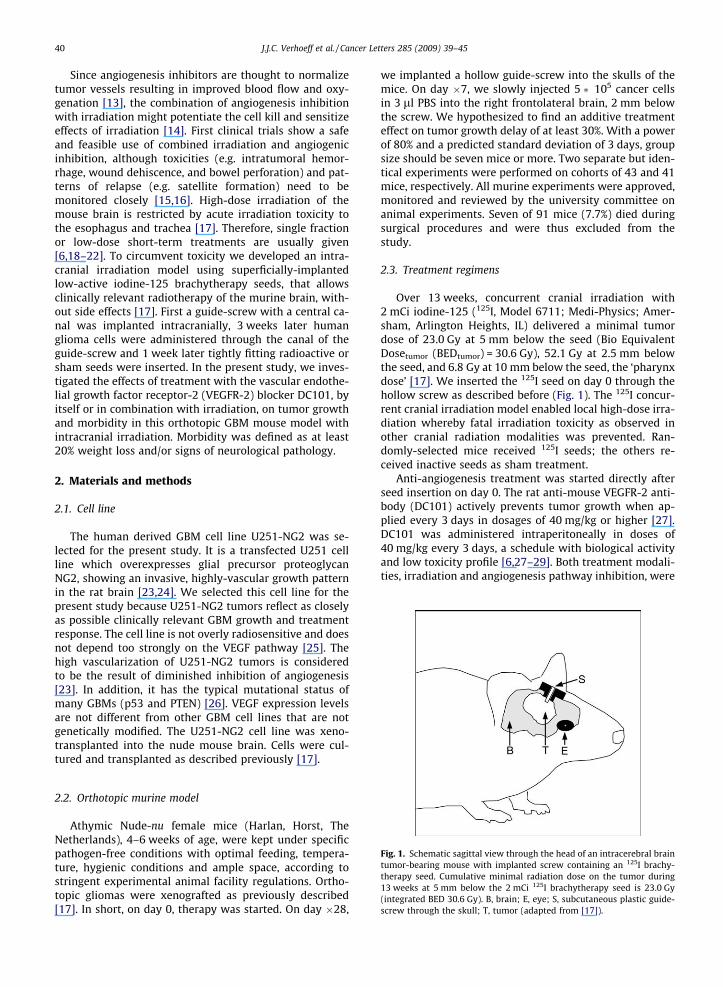
Fig. 1. Schematic sagittal view through the head of an intracerebral braintumor-bearing mouse with implanted screw containing an 125I brachy-therapy seed. Cumulative minimal radiation dose on the tumor during13 weeks at 5 mm below the 2 mCi 125I brachytherapy seed is 23.0 Gy(integrated BED 30.6 Gy). B, brain; E, eye; S, subcutaneous plastic guide-screw through the skull; T, tumor (adapted from [17]).
2.2. Orthotopic murine model
Athymic Nude-nu female mice (Harlan, Horst, TheNetherlands), 4–6 weeks of age, were kept under specificpathogen-free conditions with optimal feeding, tempera-ture, hygienic conditions and ample space, according tostringent experimental animal facility regulations. Ortho-topic gliomas were xenografted as previously described[17]. In short, on day 0, therapy was started. On day �28,
we implanted a hollow guide-screw into the skulls of themice. On day �7, we slowly injected 5 � 105 cancer cellsin 3 ll PBS into the right frontolateral brain, 2 mm belowthe screw. We hypothesized to find an additive treatmenteffect on tumor growth delay of at least 30%. With a powerof 80% and a predicted standard deviation of 3 days, groupsize should be seven mice or more. Two separate but iden-tical experiments were performed on cohorts of 43 and 41mice, respectively. All murine experiments were approved,monitored and reviewed by the university committee onanimal experiments. Seven of 91 mice (7.7%) died duringsurgical procedures and were thus excluded from thestudy.
2.3. Treatment regimens
Over 13 weeks, concurrent cranial irradiation with2 mCi iodine-125 (125I, Model 6711; Medi-Physics; Amer-sham, Arlington Heights, IL) delivered a minimal tumordose of 23.0 Gy at 5 mm below the seed (Bio EquivalentDosetumor (BEDtumor) = 30.6 Gy), 52.1 Gy at 2.5 mm belowthe seed, and 6.8 Gy at 10 mm below the seed, the ‘pharynxdose’ [17]. We inserted the 125I seed on day 0 through thehollow screw as described before (Fig. 1). The 125I concur-rent cranial irradiation model enabled local high-dose irra-diation whereby fatal irradiation toxicity as observed inother cranial radiation modalities was prevented. Ran-domly-selected mice received 125I seeds; the others re-ceived inactive seeds as sham treatment.
Anti-angiogenesis treatment was started directly afterseed insertion on day 0. The rat anti-mouse VEGFR-2 anti-body (DC101) actively prevents tumor growth when ap-plied every 3 days in dosages of 40 mg/kg or higher [27].DC101 was administered intraperitoneally in doses of40 mg/kg every 3 days, a schedule with biological activityand low toxicity profile [6,27–29]. Both treatment modali-ties, irradiation and angiogenesis pathway inhibition, were
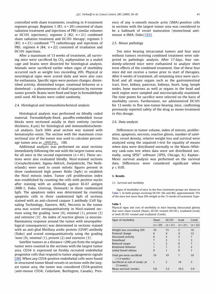
Table 1Physical signs and cues of morbidity in mice bearing intracranial gliomathat were sham treated (Sham), DC101 treated (DC101), irradiated (Irrad)or both DC101 treated and irradiated (Comb).
Signs of morbidity Sham DC101 Irrad Comb
(n = 20) (n = 21) (n = 21) (n = 22)
Weight loss exceeding 20% 10 15 5 16Postural change 6 4 4 12Decreased activity 6 5 5 7Domehead 7 3 1 3Reduced turgor 1 2 0 7Rotational behavior 1 1 3 2Lethal bowel edema 0 1 0 1
Total pre-term sacrificed(<13 weeks)
19 19 7 19
Sacrificed at end of study (at13 weeks)
1 2 14 3
Mean survival (weeks) 5.8 5.2 10.3 5.9
J.J.C. Verhoeff et al. / Cancer Letters 285 (2009) 39–45 41
controlled with sham treatments, resulting in 4 treatmentregimen groups. Regimen 1 (R1; n = 20) consisted of shamradiation treatment and injections of PBS (similar volumesas DC101 injections); regimen 2 (R2; n = 21) combinedsham radiation treatment and DC101 therapy; regimen 3(R3; n = 21) combined 125I radiotherapy and injections ofPBS; regimen 4 (R4; n = 22) consisted of irradiation andDC101 injections.
After a maximum of 13 weeks of treatment, all surviv-ing mice were sacrificed by CO2 asphyxiation in a sealedcage and brains were dissected for histological analysis.Animals were sacrificed earlier when signs of morbidityoccurred such as weight loss exceeding 20%. Physical orneurological signs were scored daily and were also cuesfor euthanasia. Specific signs were posture changes, dimin-ished activity, diminished turgor, rotational behavior or adomehead – a phenomenon of skull expansion by extremetumor growth. Brains were fixed and kept in formaldehydeuntil used. All brains were coded randomly.
2.4. Histological and immunohistochemical analysis
Histological analysis was performed on blindly codedmaterial. Formaldehyde-fixed, paraffin-embedded tissueblocks were sectioned axially in their entirety (sectionthickness, 8 lm) for histological and immunohistochemi-cal analysis. Each 20th axial section was stained withhematoxylin–eosin. The section with the maximum crosssectional size of the tumor was used to calculate percent-age tumor area as tumor area
total brain area � 100.Additional analysis was performed on axial sections
immediately following the one with the largest tumor area,for all mice in the second experiment (n = 41). These sec-tions were also evaluated blindly. Nissl-stained sections(Cresylechtviolet; Sigma–Aldrich, Zwijndrecht, The Neth-erlands) were used to count mitotic figures present inthree randomized high power fields (hpfs) to establishthe Nissl mitosis index. Tumor cell proliferation indexwas established by counting the cells with positive nucleiafter staining with an antibody against KI-67 antigen(MIB-1; Dako, Glostrup, Denmark) in three randomizedhpfs. The apoptosis index was determined by countingapoptotic cells in three randomized hpfs of sectionsstained with an anti-cleaved caspase 3 antibody (Cell Sig-naling Technology, Danvers, MA). Necrosis in the tumorarea was scored semiquantitatively in Nissl-stained sec-tions using the grading ‘none (0), minimal (1), present (2)and extensive (3)’. An index of reactive gliosis (a neuroin-flammatory response around the tumor with neuropatho-logical consequences) was determined in sections stainedwith an anti-glial fibrillary acidic protein (GFAP) antibody(Dako) and scored semiquantitatively using the grading‘none (0), minimal (1), present (2) and extensive (3)’.
Satellite tumors at a distance >200 lm from the originaltumor were counted in the sections with the largest tumorarea. CD34 is expressed on freshly recruited endothelialprogenitor cells that respond to tumor angiogenesis signals[30]. When any CD34-positive endothelial cells were foundin structured tumor blood vessels in sections with the larg-est tumor area, the tumor was considered CD34-positive(anti-mouse CD34; Cedarlane, Burlington, Canada). Pres-
ence of any a-smooth muscle actin (SMA)-positive cellsin sections with the largest tumor area was considered tobe a hallmark of vessel maturation (monoclonal anti-mouse a-SMA; Dako) [31].
2.5. Mouse pathology
Ten mice bearing intracranial tumors and four micewithout tumors receiving combined treatment were sub-jected to pathologic analysis. After 17 days, four ran-domly-selected mice were euthanized to analyze shortterm effects of the combined treatment. One of these fourmice did not receive a tumor prior to start of therapies.After 6 weeks of treatment, all remaining mice were sacri-ficed and all major organs such as the gastrointestinaltract, liver, kidney, pancreas, kidneys, heart, lung, lymphnodes, bone marrows as well as organs in the head andneck region were sampled and microscopically examined.The time points for sacrifice were selected on the basis ofmorbidity curves. Furthermore, we administered DC101for 13 weeks to five non-tumor-bearing mice, confirmingpreviously reported safety of the drug as mono-treatmentin this dosage.
2.6. Data analysis
Differences in tumor volume, index of mitosis, prolifer-ation, apoptosis, necrosis, reactive gliosis, number of satel-lites, vessel density, relative weight loss and survival wereanalyzed using the unpaired t-test for equality of meanswhen data were distributed normally or the Mann–Whit-ney rank-sum test when data were not distributed nor-mally, using SPSS� software (SPSS, Chicago, IL). Kaplan–Meier survival analysis was performed on the survivaldata. Differences were considered significant whenp 6 0.05.
3. Results
3.1. Survival and morbidity
Signs of morbidity of mice in the four treatment groups are shown inTable 1. In both groups receiving DC101 (R2 and R4), approximately 75%of the mice lost more than 20% weight in the 13 weeks of treatment. Espe-
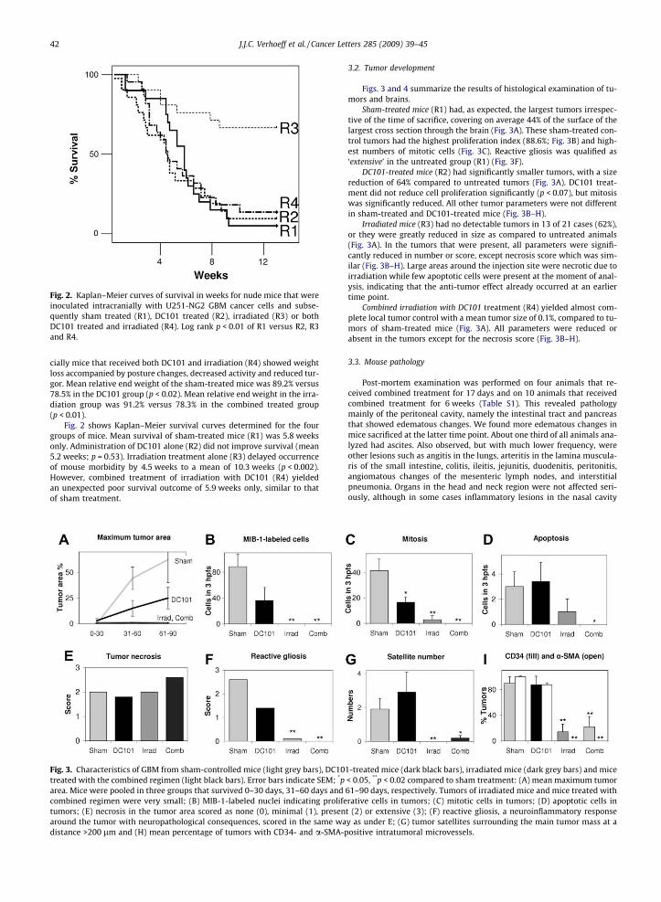
Fig. 2. Kaplan–Meier curves of survival in weeks for nude mice that wereinoculated intracranially with U251-NG2 GBM cancer cells and subse-quently sham treated (R1), DC101 treated (R2), irradiated (R3) or bothDC101 treated and irradiated (R4). Log rank p < 0.01 of R1 versus R2, R3and R4.
42 J.J.C. Verhoeff et al. / Cancer Letters 285 (2009) 39–45
cially mice that received both DC101 and irradiation (R4) showed weightloss accompanied by posture changes, decreased activity and reduced tur-gor. Mean relative end weight of the sham-treated mice was 89.2% versus78.5% in the DC101 group (p < 0.02). Mean relative end weight in the irra-diation group was 91.2% versus 78.3% in the combined treated group(p < 0.01).
Fig. 2 shows Kaplan–Meier survival curves determined for the fourgroups of mice. Mean survival of sham-treated mice (R1) was 5.8 weeksonly. Administration of DC101 alone (R2) did not improve survival (mean5.2 weeks; p = 0.53). Irradiation treatment alone (R3) delayed occurrenceof mouse morbidity by 4.5 weeks to a mean of 10.3 weeks (p < 0.002).However, combined treatment of irradiation with DC101 (R4) yieldedan unexpected poor survival outcome of 5.9 weeks only, similar to thatof sham treatment.
Fig. 3. Characteristics of GBM from sham-controlled mice (light grey bars), DC10treated with the combined regimen (light black bars). Error bars indicate SEM; *parea. Mice were pooled in three groups that survived 0–30 days, 31–60 days andcombined regimen were very small; (B) MIB-1-labeled nuclei indicating prolifetumors; (E) necrosis in the tumor area scored as none (0), minimal (1), presenaround the tumor with neuropathological consequences, scored in the same wadistance >200 lm and (H) mean percentage of tumors with CD34- and a-SMA-p
3.2. Tumor development
Figs. 3 and 4 summarize the results of histological examination of tu-mors and brains.
Sham-treated mice (R1) had, as expected, the largest tumors irrespec-tive of the time of sacrifice, covering on average 44% of the surface of thelargest cross section through the brain (Fig. 3A). These sham-treated con-trol tumors had the highest proliferation index (88.6%; Fig. 3B) and high-est numbers of mitotic cells (Fig. 3C). Reactive gliosis was qualified as‘extensive’ in the untreated group (R1) (Fig. 3F).
DC101-treated mice (R2) had significantly smaller tumors, with a sizereduction of 64% compared to untreated tumors (Fig. 3A). DC101 treat-ment did not reduce cell proliferation significantly (p < 0.07), but mitosiswas significantly reduced. All other tumor parameters were not differentin sham-treated and DC101-treated mice (Fig. 3B–H).
Irradiated mice (R3) had no detectable tumors in 13 of 21 cases (62%),or they were greatly reduced in size as compared to untreated animals(Fig. 3A). In the tumors that were present, all parameters were signifi-cantly reduced in number or score, except necrosis score which was sim-ilar (Fig. 3B–H). Large areas around the injection site were necrotic due toirradiation while few apoptotic cells were present at the moment of anal-ysis, indicating that the anti-tumor effect already occurred at an earliertime point.
Combined irradiation with DC101 treatment (R4) yielded almost com-plete local tumor control with a mean tumor size of 0.1%, compared to tu-mors of sham-treated mice (Fig. 3A). All parameters were reduced orabsent in the tumors except for the necrosis score (Fig. 3B–H).
3.3. Mouse pathology
Post-mortem examination was performed on four animals that re-ceived combined treatment for 17 days and on 10 animals that receivedcombined treatment for 6 weeks (Table S1). This revealed pathologymainly of the peritoneal cavity, namely the intestinal tract and pancreasthat showed edematous changes. We found more edematous changes inmice sacrificed at the latter time point. About one third of all animals ana-lyzed had ascites. Also observed, but with much lower frequency, wereother lesions such as angitis in the lungs, arteritis in the lamina muscula-ris of the small intestine, colitis, ileitis, jejunitis, duodenitis, peritonitis,angiomatous changes of the mesenteric lymph nodes, and interstitialpneumonia. Organs in the head and neck region were not affected seri-ously, although in some cases inflammatory lesions in the nasal cavity
1-treated mice (dark black bars), irradiated mice (dark grey bars) and mice< 0.05, **p < 0.02 compared to sham treatment: (A) mean maximum tumor61–90 days, respectively. Tumors of irradiated mice and mice treated withrative cells in tumors; (C) mitotic cells in tumors; (D) apoptotic cells int (2) or extensive (3); (F) reactive gliosis, a neuroinflammatory responsey as under E; (G) tumor satellites surrounding the main tumor mass at aositive intratumoral microvessels.
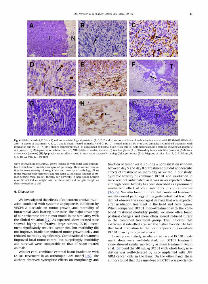
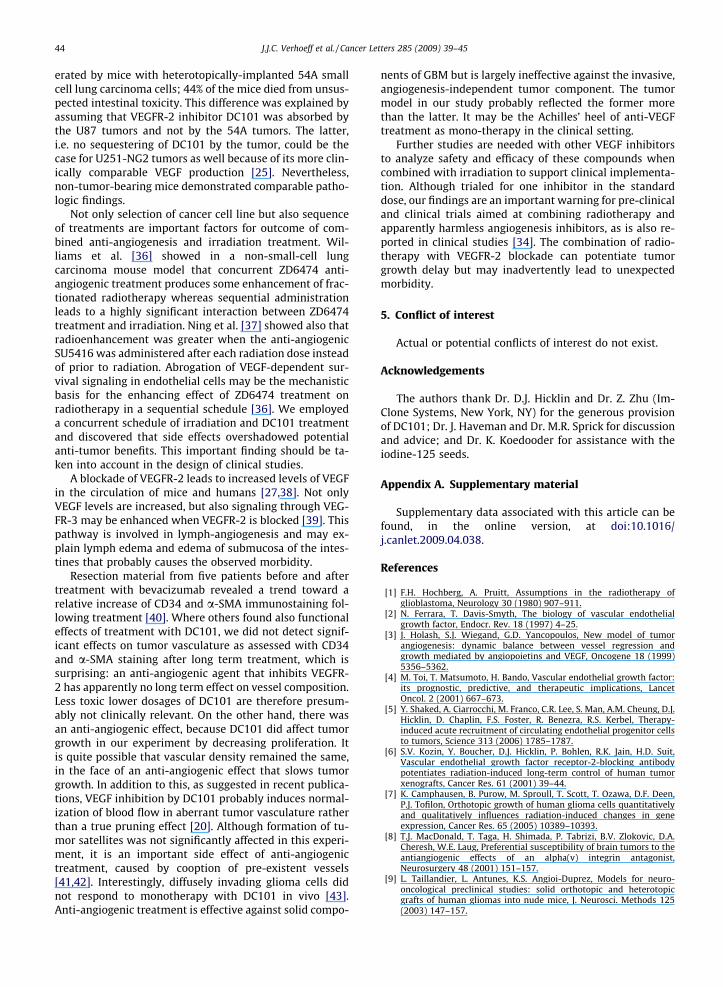
Fig. 4. H&E-stained (A, F, G and I) and immunohistologically-stained (B, C, D, E and H) sections of brain of nude mice inoculated with U251-NG2 GBM cellsafter 13 weeks of treatment. A, B, C, D and E: sham-treated animals; F and G: DC101 treated animals; H: Irradiated animals; I: Combined treatment withirradiation and DC101. (A) H&E-stained large tumor load (T) surrounded by normal brain tissue (N). (B) Anti-active caspase 3 staining showing an apoptoticcell (arrow). (C) SMA-positive vessels (arrows). (D) MIB-1-labeled nuclei (arrows), (E) Reactive gliosis (#). (F) Invading tumor satellites (arrows). (G) Mitoticcancer cells (arrows). (H) Apoptotic cancer cells (arrows) in anti-active caspase 3 staining. (I) Largest tumor (T) in R4 group of mice. Bars, A, D, F: 2.0 mm, B,C, G, H: 0.2 mm, E, I: 0.5 mm.
J.J.C. Verhoeff et al. / Cancer Letters 285 (2009) 39–45 43
were observed. In one animal, severe lesions of lymphoma were encoun-tered, which were probably background pathology. There was no correla-tion between severity of weight loss and severity of pathology. Non-tumor-bearing mice demonstrated the same pathological findings as tu-mor-bearing mice. DC101 therapy for 13 weeks in non-tumor-bearingmice did not induce weight loss, but these mice did not gain weight assham-treated mice did.
4. Discussion
We investigated the effects of concurrent cranial irradi-ation combined with systemic angiogenesis inhibition byVEGFR-2 blockade on tumor growth and morbidity ofintracranial GBM-bearing nude mice. The major advantageof our orthotopic brain tumor model is the similarity withthe clinical situation [17]. As expected, sham-treated miceshowed highly proliferative, large tumors. DC101 treat-ment significantly reduced tumor size, but morbidity didnot improve. Irradiation induced tumor growth delay andreduced morbidity significantly. Combinational treatmentimproved local tumor control but, surprisingly, morbidityand survival were comparable to that of sham-treatedanimals.
Winkler et al. combined external beam irradiation withDC101 treatment in an orthotopic GBM model [20]. Theauthors observed synergistic effects on morphology and
function of tumor vessels during a normalization window,between day 5 and day 8 of treatment but did not describeeffects of treatment on morbidity as we did in our study.Systemic toxicity of combined DC101 and irradiation inmice was not anticipated, as it was never reported before,although bowel toxicity has been described as a prominentinadvertent effect of VEGF inhibitors in clinical studies[32–35]. We also found in mice that combined treatmentmainly caused pathology of the gastrointestinal tract. Wedid not observe the esophageal damage that was expectedafter irradiation treatment in the head and neck region.When comparing DC101 mono-treatment with the com-bined treatment morbidity profile, we more often foundpostural changes and more often scored reduced turgorin the combined treatment group. This indicates thatextracranial side effects caused the higher toxicity. The factthat local irradiation to the brain appears to exacerbateDC101 toxicity is of great concern.
In our present study, irradiation alone and DC101 treat-ment alone were well-tolerated, but DC101 treatmentalone showed similar morbidity as sham treatment. Kozinet al. [6] found that 40 mg/kg DC101 with whole body irra-diation was well-tolerated by mice implanted with U87GBM cancer cells in the flank. On the other hand, theseauthors found that the same dose of DC101 was poorly tol-
44 J.J.C. Verhoeff et al. / Cancer Letters 285 (2009) 39–45
erated by mice with heterotopically-implanted 54A smallcell lung carcinoma cells; 44% of the mice died from unsus-pected intestinal toxicity. This difference was explained byassuming that VEGFR-2 inhibitor DC101 was absorbed bythe U87 tumors and not by the 54A tumors. The latter,i.e. no sequestering of DC101 by the tumor, could be thecase for U251-NG2 tumors as well because of its more clin-ically comparable VEGF production [25]. Nevertheless,non-tumor-bearing mice demonstrated comparable patho-logic findings.
Not only selection of cancer cell line but also sequenceof treatments are important factors for outcome of com-bined anti-angiogenesis and irradiation treatment. Wil-liams et al. [36] showed in a non-small-cell lungcarcinoma mouse model that concurrent ZD6474 anti-angiogenic treatment produces some enhancement of frac-tionated radiotherapy whereas sequential administrationleads to a highly significant interaction between ZD6474treatment and irradiation. Ning et al. [37] showed also thatradioenhancement was greater when the anti-angiogenicSU5416 was administered after each radiation dose insteadof prior to radiation. Abrogation of VEGF-dependent sur-vival signaling in endothelial cells may be the mechanisticbasis for the enhancing effect of ZD6474 treatment onradiotherapy in a sequential schedule [36]. We employeda concurrent schedule of irradiation and DC101 treatmentand discovered that side effects overshadowed potentialanti-tumor benefits. This important finding should be ta-ken into account in the design of clinical studies.
A blockade of VEGFR-2 leads to increased levels of VEGFin the circulation of mice and humans [27,38]. Not onlyVEGF levels are increased, but also signaling through VEG-FR-3 may be enhanced when VEGFR-2 is blocked [39]. Thispathway is involved in lymph-angiogenesis and may ex-plain lymph edema and edema of submucosa of the intes-tines that probably causes the observed morbidity.
Resection material from five patients before and aftertreatment with bevacizumab revealed a trend toward arelative increase of CD34 and a-SMA immunostaining fol-lowing treatment [40]. Where others found also functionaleffects of treatment with DC101, we did not detect signif-icant effects on tumor vasculature as assessed with CD34and a-SMA staining after long term treatment, which issurprising: an anti-angiogenic agent that inhibits VEGFR-2 has apparently no long term effect on vessel composition.Less toxic lower dosages of DC101 are therefore presum-ably not clinically relevant. On the other hand, there wasan anti-angiogenic effect, because DC101 did affect tumorgrowth in our experiment by decreasing proliferation. Itis quite possible that vascular density remained the same,in the face of an anti-angiogenic effect that slows tumorgrowth. In addition to this, as suggested in recent publica-tions, VEGF inhibition by DC101 probably induces normal-ization of blood flow in aberrant tumor vasculature ratherthan a true pruning effect [20]. Although formation of tu-mor satellites was not significantly affected in this experi-ment, it is an important side effect of anti-angiogenictreatment, caused by cooption of pre-existent vessels[41,42]. Interestingly, diffusely invading glioma cells didnot respond to monotherapy with DC101 in vivo [43].Anti-angiogenic treatment is effective against solid compo-
nents of GBM but is largely ineffective against the invasive,angiogenesis-independent tumor component. The tumormodel in our study probably reflected the former morethan the latter. It may be the Achilles’ heel of anti-VEGFtreatment as mono-therapy in the clinical setting.
Further studies are needed with other VEGF inhibitorsto analyze safety and efficacy of these compounds whencombined with irradiation to support clinical implementa-tion. Although trialed for one inhibitor in the standarddose, our findings are an important warning for pre-clinicaland clinical trials aimed at combining radiotherapy andapparently harmless angiogenesis inhibitors, as is also re-ported in clinical studies [34]. The combination of radio-therapy with VEGFR-2 blockade can potentiate tumorgrowth delay but may inadvertently lead to unexpectedmorbidity.
5. Conflict of interest
Actual or potential conflicts of interest do not exist.
Acknowledgements
The authors thank Dr. D.J. Hicklin and Dr. Z. Zhu (Im-Clone Systems, New York, NY) for the generous provisionof DC101; Dr. J. Haveman and Dr. M.R. Sprick for discussionand advice; and Dr. K. Koedooder for assistance with theiodine-125 seeds.
Appendix A. Supplementary material
Supplementary data associated with this article can befound, in the online version, at doi:10.1016/j.canlet.2009.04.038.
References
[1] F.H. Hochberg, A. Pruitt, Assumptions in the radiotherapy ofglioblastoma, Neurology 30 (1980) 907–911.
[2] N. Ferrara, T. Davis-Smyth, The biology of vascular endothelialgrowth factor, Endocr. Rev. 18 (1997) 4–25.
[3] J. Holash, S.J. Wiegand, G.D. Yancopoulos, New model of tumorangiogenesis: dynamic balance between vessel regression andgrowth mediated by angiopoietins and VEGF, Oncogene 18 (1999)5356–5362.
[4] M. Toi, T. Matsumoto, H. Bando, Vascular endothelial growth factor:its prognostic, predictive, and therapeutic implications, LancetOncol. 2 (2001) 667–673.
[5] Y. Shaked, A. Ciarrocchi, M. Franco, C.R. Lee, S. Man, A.M. Cheung, D.J.Hicklin, D. Chaplin, F.S. Foster, R. Benezra, R.S. Kerbel, Therapy-induced acute recruitment of circulating endothelial progenitor cellsto tumors, Science 313 (2006) 1785–1787.
[6] S.V. Kozin, Y. Boucher, D.J. Hicklin, P. Bohlen, R.K. Jain, H.D. Suit,Vascular endothelial growth factor receptor-2-blocking antibodypotentiates radiation-induced long-term control of human tumorxenografts, Cancer Res. 61 (2001) 39–44.
[7] K. Camphausen, B. Purow, M. Sproull, T. Scott, T. Ozawa, D.F. Deen,P.J. Tofilon, Orthotopic growth of human glioma cells quantitativelyand qualitatively influences radiation-induced changes in geneexpression, Cancer Res. 65 (2005) 10389–10393.
[8] T.J. MacDonald, T. Taga, H. Shimada, P. Tabrizi, B.V. Zlokovic, D.A.Cheresh, W.E. Laug, Preferential susceptibility of brain tumors to theantiangiogenic effects of an alpha(v) integrin antagonist,Neurosurgery 48 (2001) 151–157.
[9] L. Taillandier, L. Antunes, K.S. Angioi-Duprez, Models for neuro-oncological preclinical studies: solid orthotopic and heterotopicgrafts of human gliomas into nude mice, J. Neurosci. Methods 125(2003) 147–157.
J.J.C. Verhoeff et al. / Cancer Letters 285 (2009) 39–45 45
[10] B. Blouw, H. Song, T. Tihan, J. Bosze, N. Ferrara, H.P. Gerber, R.S.Johnson, G. Bergers, The hypoxic response of tumors is dependent ontheir microenvironment, Cancer Cell 4 (2003) 133–146.
[11] E.I. Fomchenko, E.C. Holland, Mouse models of brain tumors andtheir applications in preclinical trials, Clin. Cancer Res. 12 (2006)5288–5297.
[12] C. Charalambous, F.M. Hofman, T.C. Chen, Functional and phenotypicdifferences between glioblastoma multiforme-derived and normalhuman brain endothelial cells, J. Neurosurg. 102 (2005) 699–705.
[13] R.K. Jain, Normalization of tumor vasculature: an emerging conceptin antiangiogenic therapy, Science 307 (2005) 58–62.
[14] C. Nieder, N. Wiedenmann, N. Andratschke, M. Molls, Current statusof angiogenesis inhibitors combined with radiation therapy, CancerTreat. Rev. 32 (2006) 348–364.
[15] P.H. Gutin, F.M. Iwamoto, K. Beal, N.A. Mohile, S. Karimi, B.L. Hou, S.Lymberis, Y. Yamada, J. Chang, L.E. Abrey, Safety and efficacy ofbevacizumab with hypofractionated stereotactic irradiation forrecurrent malignant gliomas, Int. J. Radiat. Oncol Biol. Phys. (2009).
[16] A. Narayana, P. Kelly, J. Golfinos, E. Parker, G. Johnson, E. Knopp, D.Zagzag, I. Fischer, S. Raza, P. Medabalmi, P. Eagan, M.L. Gruber,Antiangiogenic therapy using bevacizumab in recurrent high-gradeglioma: impact on local control and patient survival, J. Neurosurg.110 (2009) 173–180.
[17] J.J. Verhoeff, L.J. Stalpers, A.W. Coumou, K. Koedooder, C. Lavini, C.J.Van Noorden, J. Haveman, W.P. Vandertop, W.R. van Furth,Experimental iodine-125 seed irradiation of intracerebral braintumors in nude mice, Radiat. Oncol. 2 (2007) 38.
[18] E.L. Lund, L. Bastholm, P.E. Kristjansen, Therapeutic synergy of TNP-470 and ionizing radiation: effects on tumor growth, vesselmorphology, and angiogenesis in human glioblastoma multiformexenografts, Clin. Cancer Res. 6 (2000) 971–978.
[19] C. Hess, V. Vuong, I. Hegyi, O. Riesterer, J. Wood, D. Fabbro, C.Glanzmann, S. Bodis, M. Pruschy, Effect of VEGF receptor inhibitorPTK787/ZK222584 [correction of ZK222548] combined with ionizingradiation on endothelial cells and tumour growth, Br. J. Cancer 85(2001) 2010–2016.
[20] F. Winkler, S.V. Kozin, R.T. Tong, S.S. Chae, M.F. Booth, I. Garkavtsev, L.Xu, D.J. Hicklin, D. Fukumura, E. di Tomaso, L.L. Munn, R.K. Jain,Kinetics of vascular normalization by VEGFR2 blockade governs braintumor response to radiation: role of oxygenation, angiopoietin-1, andmatrix metalloproteinases, Cancer Cell 6 (2004) 553–563.
[21] J.N. Sarkaria, B.L. Carlson, M.A. Schroeder, P. Grogan, P.D. Brown, C.Giannini, K.V. Ballman, G.J. Kitange, A. Guha, A. Pandita, C.D. James,Use of an orthotopic xenograft model for assessing the effect ofepidermal growth factor receptor amplification on glioblastomaradiation response, Clin. Cancer Res. 12 (2006) 2264–2271.
[22] G. Tabatabai, B. Frank, A. Wick, D. Lemke, G. von Kurthy, U.Obermuller, S. Heckl, G. Christ, M. Weller, W. Wick, Synergisticantiglioma activity of radiotherapy and enzastaurin, Ann. Neurol. 61(2007) 153–161.
[23] M. Chekenya, M. Hjelstuen, P.O. Enger, F. Thorsen, A.L. Jacob, B.Probst, O. Haraldseth, G. Pilkington, A. Butt, J.M. Levine, R. Bjerkvig,NG2 proteoglycan promotes angiogenesis-dependent tumor growthin CNS by sequestering angiostatin, FASEB J. 16 (2002) 586–588.
[24] C. Brekke, A. Lundervold, P.O. Enger, C. Brekken, E. Stalsett, T.B.Pedersen, O. Haraldseth, P.G. Kruger, R. Bjerkvig, M. Chekenya, NG2expression regulates vascular morphology and function in humanbrain tumours, Neuroimage 29 (2006) 965–976.
[25] K.E. Hovinga, L.J. Stalpers, C. van Bree, M. Donker, J.J. Verhoeff, H.M.Rodermond, D.A. Bosch, W.R. van Furth, Radiation-enhancedvascular endothelial growth factor (VEGF) secretion inglioblastoma multiforme cell lines – a clue to radioresistance?, JNeurooncol. 74 (2005) 99–103.
[26] M. Chekenya, C. Krakstad, A. Svendsen, I.A. Netland, V. Staalesen, B.B.Tysnes, F. Selheim, J. Wang, P.O. Sakariassen, T. Sandal, P.E. Lonning,T. Flatmark, P.O. Enger, R. Bjerkvig, M. Sioud, W.B. Stallcup, Theprogenitor cell marker NG2/MPG promotes chemoresistance byactivation of integrin-dependent PI3K/Akt signaling, Oncogene 27(2008) 5182–5194.
[27] G. Bocci, S. Man, S.K. Green, G. Francia, J.M. Ebos, J.M. du Manoir, A.Weinerman, U. Emmenegger, L. Ma, P. Thorpe, A. Davidoff, J. Huber,D.J. Hicklin, R.S. Kerbel, Increased plasma vascular endothelialgrowth factor (VEGF) as a surrogate marker for optimaltherapeutic dosing of VEGF receptor-2 monoclonal antibodies,Cancer Res. 64 (2004) 6616–6625.
[28] P. Kunkel, U. Ulbricht, P. Bohlen, M.A. Brockmann, R. Fillbrandt, D.Stavrou, M. Westphal, K. Lamszus, Inhibition of glioma angiogenesisand growth in vivo by systemic treatment with a monoclonalantibody against vascular endothelial growth factor receptor-2,Cancer Res. 61 (2001) 6624–6628.
[29] K. Lamszus, M.A. Brockmann, C. Eckerich, P. Bohlen, C. May, U.Mangold, R. Fillbrandt, M. Westphal, Inhibition of glioblastomaangiogenesis and invasion by combined treatments directed againstvascular endothelial growth factor receptor-2, epidermal growthfactor receptor, and vascular endothelial-cadherin, Clin. Cancer Res.11 (2005) 4934–4940.
[30] Y.C. Yung, S. Cheshier, J.G. Santarelli, Z. Huang, A. Wagers, I.Weissman, V. Tse, Incorporation of naive bone marrow derivedcells into the vascular architecture of brain tumor, Microcirculation11 (2004) 699–708.
[31] R.K. Jain, Molecular regulation of vessel maturation, Nat. Med. 9(2003) 685–693.
[32] F.A. Eskens, J. Verweij, The clinical toxicity profile of vascularendothelial growth factor (VEGF) and vascular endothelial growthfactor receptor (VEGFR) targeting angiogenesis inhibitors; a review,Eur. J. Cancer 42 (2006) 3127–3139.
[33] T. Kamba, D.M. McDonald, Mechanisms of adverse effects of anti-VEGF therapy for cancer, Br. J. Cancer 96 (2007) 1788–1795.
[34] N.A. Peters, D.J. Richel, J.J. Verhoeff, L.J. Stalpers, Bowel perforationafter radiotherapy in a patient receiving sorafenib, J. Clin. Oncol. 26(2008) 2405–2406.
[35] J. Dietrich, A.D. Norden, P.Y. Wen, Emerging antiangiogenictreatments for gliomas – efficacy and safety issues, Curr. Opin.Neurol. 21 (2008) 736–744.
[36] K.J. Williams, B.A. Telfer, S. Brave, J. Kendrew, L. Whittaker, I.J.Stratford, S.R. Wedge, ZD6474, a potent inhibitor of vascularendothelial growth factor signaling, combined with radiotherapy:schedule-dependent enhancement of antitumor activity, Clin.Cancer Res. 10 (2004) 8587–8593.
[37] S. Ning, D. Laird, J.M. Cherrington, S.J. Knox, The antiangiogenicagents SU5416 and SU6668 increase the antitumor effects offractionated irradiation, Radiat. Res. 157 (2002) 45–51.
[38] M.L. Veronese, A. Mosenkis, K.T. Flaherty, M. Gallagher, J.P.Stevenson, R.R. Townsend, P.J. O’Dwyer, Mechanisms ofhypertension associated with BAY 43–9006, J. Clin. Oncol. 24(2006) 1363–1369.
[39] J. Goldman, J.M. Rutkowski, J.D. Shields, M.C. Pasquier, Y. Cui, H.G.Schmokel, S. Willey, D.J. Hicklin, B. Pytowski, M.A. Swartz,Cooperative and redundant roles of VEGFR-2 and VEGFR-3signaling in adult lymphangiogenesis, FASEB J. 21 (2007) 1003–1012.
[40] I. Fischer, C.H. Cunliffe, R.J. Bollo, S. Raza, D. Monoky, L. Chiriboga,E.C. Parker, J.G. Golfinos, P.J. Kelly, E.A. Knopp, M.L. Gruber, D.Zagzag, A. Narayana, High-grade glioma before and after treatmentwith radiation and avastin: initial observations, Neuro Oncol. 10(2008) 700–708.
[41] J.L. Rubenstein, J. Kim, T. Ozawa, M. Zhang, M. Westphal, D.F. Deen,M.A. Shuman, Anti-VEGF antibody treatment of glioblastomaprolongs survival but results in increased vascular cooption,Neoplasia 2 (2000) 306–314.
[42] K. Lamszus, P. Kunkel, M. Westphal, Invasion as limitation to anti-angiogenic glioma therapy, Acta Neurochir. (Suppl. 88) (2003) 169–177.
[43] T. Martens, Y. Laabs, H.S. Gunther, D. Kemming, Z. Zhu, L. Witte, C.Hagel, M. Westphal, K. Lamszus, Inhibition of glioblastoma growthin a highly invasive nude mouse model can be achieved by targetingepidermal growth factor receptor but not vascular endothelialgrowth factor receptor-2, Clin. Cancer Res. 14 (2008) 5447–5458.