Biotechnology of water and salinity stress tolerance
13
This article appeared in a journal published by Elsevier. The attached copy is furnished to the author for internal non-commercial research and education use, including for instruction at the authors institution and sharing with colleagues. Other uses, including reproduction and distribution, or selling or licensing copies, or posting to personal, institutional or third party websites are prohibited. In most cases authors are permitted to post their version of the article (e.g. in Word or Tex form) to their personal website or institutional repository. Authors requiring further information regarding Elsevier’s archiving and manuscript policies are encouraged to visit: http://www.elsevier.com/copyright
-
Upload
marianosamaniego -
Category
Documents
-
view
2 -
download
0
Transcript of Biotechnology of water and salinity stress tolerance

This article appeared in a journal published by Elsevier. The attachedcopy is furnished to the author for internal non-commercial researchand education use, including for instruction at the authors institution
and sharing with colleagues.
Other uses, including reproduction and distribution, or selling orlicensing copies, or posting to personal, institutional or third party
websites are prohibited.
In most cases authors are permitted to post their version of thearticle (e.g. in Word or Tex form) to their personal website orinstitutional repository. Authors requiring further information
regarding Elsevier’s archiving and manuscript policies areencouraged to visit:
http://www.elsevier.com/copyright

Author's personal copy
Available online at www.sciencedirect.com
Biotechnology of water and salinity stress toleranceJose M Pardo
Drought and salinity are among the environmental factors that
constrain agricultural productivity most dramatically. Classical
breeding programs aiming to improve stress tolerance have
been hampered by the multigenic nature of the trait and the
seemingly scarce natural genetic variability in crop plants.
Novel genetic determinants governing the function of stomata
and improving the performance of plants under water shortage
have been identified and show promise of application in crops.
Moreover, receptors of the stress hormone abscisic acid have
been characterized and their interplay with key regulatory
components is being understood. A critical factor of salinity
tolerance in plants is the ability to exclude Na+ from the shoot,
and the modification of specific Na+ transport processes has
yielded enhanced salinity tolerance.
Address
Instituto de Recursos Naturales y Agrobiologia (IRNASE), Consejo
Superior de Investigaciones Cientificas, Reina Mercedes, 10. Sevilla –
41012, Spain
Corresponding author: Pardo, Jose M ([email protected])
Current Opinion in Biotechnology 2010, 21:185–196
This review comes from a themed issue on
Plant biotechnology
Edited by Antonio Molina and Jim Haseloff
Available online 26th February 2010
0958-1669/$ – see front matter
# 2010 Elsevier Ltd. All rights reserved.
DOI 10.1016/j.copbio.2010.02.005
IntroductionEnvironmental factors are the primary cause of crop
failure, causing average yield losses of more than 60%
for major crops worldwide [1]. The abiotic stresses that
plants encounter most frequently and that adversely
affect growth are drought, salinity, flooding, and low or
high temperature. Environmental clues trigger physio-
logical and molecular responses enabling the plant to
prevent or minimize exposure to stressful conditions, or
to acclimate and overcome the unavoidable hurdle [2].
Among the adaptations aimed to drought-avoidance are
growing deeper roots in pursuit of receding groundwater
or to entirely escape dry periods by completing the life
cycle while water is available. On the other hand, mol-
ecular responses that are associated with the acclimatiz-
ation to water and salinity stress are multigenic and often
interrelated, thus hindering the dissection of signaling
processes and the identification of major determinants of
stress tolerance. Transcriptomic, proteomic, and molecu-
lar genetics approaches have identified many stress-
related genes, which are generally classified into two
major groups [2]. One group is involved in signaling
cascades, transcriptional control, and the degradation of
transcripts or proteins, whereas members of the other
group function in membrane protection, osmoprotection,
as antioxidants and as reactive oxygen species (ROS)
scavengers. Simultaneously, metabolic pathways are
adjusted to regain homeostasis in a changing environ-
ment.
The word drought is often used as a way of expressing
water deficit imposed by withholding irrigation, but
drought is in fact an ecophysiological term that describes
a prolonged period of abnormally low rainfall. Hence, I
will rather use the more precise terms water deficit, that is
when the quantity of water available to the plant is
insufficient to meet basic needs, and desiccation when
the relative water content (RWC) in plant tissues falls
drastically. Values of RWC around 85–95% are found in
well-hydrated tissues. A RWC lower than the critical
mark of 50% typically results in plant death, with the
notable exceptions of mature orthodox seeds and xero-
phytic plant species [1]. Water deficit can be induced by
many environmental conditions and it is often associated
with concurrent stresses, like salinity and high or low
temperatures. Indeed, salinity stress and water deficit are
intimately related. Salts dissolved in the soil solution
reduce the water potential (i.e. diminish water availability
to the plant) and water uptake by roots is thermodyna-
mically hampered. Within minutes of being exposed to
high salt concentrations plant growth is impaired, a
response due to hyperosmolarity of the soil solution
[3�]. Mild osmotic stress leads rapidly to growth inhibition
of shoots, whereas roots may continue to elongate, seek-
ing water from deeper soil layers. In the short term,
osmotic adjustment is the primary issue. The subsequent
accumulation of salts in the cells and the apoplast become
critical mainly in plants exposed to long-term salinity
(days or weeks) and results in necrosis of the tissues,
particularly in old leaves that cannot dilute the incoming
salts by cell expansion. Ion-specific stress maladies are
superimposed on those caused by dehydration. Molecular
responses to water and salt stress are largely identical
except for the ionic component [2,3�]. For most species,
Na+ appears to reach a toxic concentration before other
ions do, and thus most studies have concentrated on the
control of Na+ homeostasis [4]. Maintenance of appro-
priate intracellular K+/Na+ balance is critical for metabolic
function as Na+ cytotoxicity is largely due to competition
with K+ for binding sites in enzymes essential for cellular
www.sciencedirect.com Current Opinion in Biotechnology 2010, 21:185–196

Author's personal copy
functions [5]. Another consequence is the production of
ROS, which then in turn affects cellular structures and
metabolism negatively [2].
Although considerable progress was made to increase and
secure crop yield through conventional breeding, the goal
of improving the resistance of crops to abiotic stresses has
seen limited success because of the complex, multigenic
nature of the traits and the narrow genetic variation in the
gene pools of major crops. Numerous genes and proteins
have been shown to affect the tolerance to environmental
stress in an array of plant species, which together compose
a complex puzzle with a myriad of individual elements
and crisscrossing signal transduction pathways that are not
easily integrated into a holistic model. The use of hardy
wild relatives of crop plants as a source of genetic deter-
minants of stress tolerance is time-consuming and labor-
intensive. Further, undesirable genes are often trans-
ferred in combination with desirable ones. Classical
breeding approaches have revealed that stress tolerance
traits are dispersed in various quantitative trait loci
(QTLs), which make genetic selection of these traits
difficult, albeit a few success stories regarding QTLs
related to salinity will be described herein. The use of
molecular markers and the development of powerful
bioinformatics tools have led to the identification of genes
with major contributions to Na+ and K+ homeostasis using
the QTL approach.
This review focuses on recent advances following mol-
ecular genetics approaches in model plants. I will present
our current understanding of Na+ transport in plants,
arguably the most relevant ion in salinity tolerance and
which is one of the most extensively studied processes
regarding salt stress. Recent developments in abscisic
acid (ABA) perception, improving resistance to water
deficit by means of controlling the function of stomata,
and achieving the survival of higher plants to severe
desiccation will also be highlighted and discussed.
Water stress and desiccationStomatal pores regulate gas exchange for photosynthesis
and the loss of water by transpiration. Plants fine-tune the
opening and closing of their stomata to optimize the
conflicting needs of gas exchange while limiting water
loss through evapo-transpiration. The engineering of
stomatal closure as a means to reduce water loss is an
attractive approach to improve the performance of plants
under water limitation, thereby meeting the pressing
need of developing crops with higher water use efficiency
(WUE, or biomass production per unit of water used). In
principle, the trade-off of diminished gas exchange, with
the predictable decline of photosynthetic activity, should
be taken into account when designing plants with
reduced water loss through enhanced stomata closure.
For instance, the ABA-hypersensitive era1 and cbp20mutants of Arabidopsis, which retain more water when
subjected to limited water supply because of restricted
stomatal exchange, could not compete favorably with
wild-type plants in mixed populations growing in the
same pots [6�]. Roots of neighboring wild-type plants
competed for water in the soil around the mutants, whose
water potential declined following that of their wild-type
neighbors. Thus, reduced transpiration may be necessary
but not sufficient to keep plants hydrated in a progress-
ively drying environment [6�]. However, there is sub-
stantial natural genetic variation of WUE among species
and genotypes within a species, and the underlying
mechanisms that regulate transpiration are just beginning
to be understood [7�]. Transpiration rates and net carbon
dioxide (CO2) assimilation show different slopes in
response to changes in stomatal conductance, and small
reductions in stomatal transpiration that minimize the
negative impact on CO2 assimilation translate into
improved WUE. Current evidence suggests that plants
have physiological mechanisms for improving WUE that
are under genetic control [7�]. Moreover, transpiration
occurs not only through stomata, but also across the
cuticle and the boundary layer. Because of the differential
diffusion properties of water and CO2 through these
pathways, it is feasible that WUE could be improved
by decreasing transpiration without a concomitant
reduction in CO2 uptake [7�].
Many signaling components have been identified that are
involved in the control of stomatal aperture, including
second messengers, protein kinases and phosphatases,
phospholipases, and constituents of the machinery con-
trolling RNA metabolism [8]. Recently, several transcrip-
tion factors have been implicated in the regulation of
stomatal movements in Arabidopsis, adding an additional
level of regulation to the signaling network that controls
stomatal aperture. The transcription factors AtMYB60
and AtMYB61 are expressed in guard cells, and they play
opposite roles in the response of stomata to environmen-
tal signals [9,10]. The expression of AtMYB60 is nega-
tively modulated during water stress whereas light, which
promotes stomatal aperture, increases AtMYB60 gene
expression. A null myb60 mutation reduced stomatal
opening and wilting under water stress conditions. How-
ever, stomatal closure induced by ABA or dark was
unaffected by the myb60 mutation. In contrast to
AtMYB60, AtMYB61 is expressed only in the dark, under
conditions when stomata are usually closed. Stomata in
the myb61 mutant were more open than in the wild-type
plant. Neither ABA nor water stress affected AtMYB61
expression, suggesting that this transcription factor does
not mediate ABA-induced reductions in stomatal aper-
ture. Hence, these MYB transcription factors are primar-
ily involved in light-induced opening and dark-induced
closure. Ectopic overexpression of another MYB protein
of Arabidopsis, MYB44, which is normally expressed in
the vasculature and in guard cells, downregulated an array
of 2C-type protein phosphatases that are well-known
186 Plant biotechnology
Current Opinion in Biotechnology 2010, 21:185–196 www.sciencedirect.com

Author's personal copy
negative regulators of ABA signaling (see below) [11].
Consequently, transgenic plants expressing MYB44 had a
faster ABA-induced stomatal closure response than wild-
type plants. The MYB44 gene was itself upregulated by
dehydration, low temperature, and salinity, thereby
enhancing ABA signal relay.
A mutant with improved resistance to water shortage and
to oxidative stress, enhanced drought tolerance 1, was iso-
lated in a gain-of-function mutant screen in Arabidopsis[12]. The edt1 plants showed an array of stress-related
traits, including a more extensive root system with deeper
primary roots and more lateral roots than the wild-type
plants, and higher levels of ABA, proline, and superoxide
dismutase. The more developed root system of the
mutant improved accessibility to water. In addition, the
edt1 plants showed 30% reduction in stomatal density per
area unit, which was presumably a consequence of larger
and fewer cells in the epidermis of the mutant. Surpris-
ingly, although the rate of transpiration was lower in the
mutant, the rate of photosynthesis was higher compared
to the wild type. Consequently, the WUE was greater in
the mutant than in the wild-type plant. The increased
photosynthesis rate of the mutant is unexpected, as
reduced stomatal density is believed to decrease CO2
exchange. The phenotypes of edt1 were brought about by
the T-DNA activation tagging of the gene HDG11 encod-
ing a putative homeodomain-START transcription factor.
Overexpression of HDG11 in transgenic tobacco recapi-
tulated water stress tolerance associated with modified
root architecture and reduced leaf stomatal density. Tar-
gets that HDG11 may directly or indirectly regulate
included effectors of ABA synthesis and signal relay
(NCED3, LOS5/ABA3, CIPK3, and ABI3), the proline
biosynthetic enzyme P5CS, Ca2+ and K+ transporters
(CAX3 and KAT1), and the transcriptional regulators
ERECTA, which is known to affect stomatal density,
RGAL, a RGA-like DELLA protein that is a negative
regulator of GA signaling, and IAA28, which negatively
regulates lateral root formation [12]. The HDG11 gene did
not respond itself to stress treatments and the knockout
mutant had no discernible phenotype.
The HARDY (HRD) gene encoding an AP2/ERF-like
transcription factor was also identified as a gain-of-func-
tion mutation in Arabidopsis. The hardy mutant had
increased mesophyll cell layers, produced a denser root
network, and showed enhanced tolerance to water deficit
and salinity [13]. The mechanism through which HARDY
regulates these processes is not known, albeit higher
efficiency could be related to the increased number of
photosynthetic mesophyll cells in thicker leaves. Over-
expression of HRD in rice reduced transpiration while
increasing WUE [13]. Adaptation to water deficit is often
associated not only with stomatal control of water use but
also with rooting depth. The HDG11 and HARDY
proteins link both phenomena molecularly, suggesting
that these proteins may have evolved as master switches
directing the evolution of plant species better suited to
survive drought.
The Arabidopsis abo1 mutant was isolated in a genetic
screen for mutants with altered water stress responses.
This drought-resistant mutant shows hypersensitive
seedling growth and enhanced stomatal closing in
response to ABA [14]. The abo1 mutation mapped in
gene ELO2, encoding the largest subunit of Elongator, a
multifunctional complex with roles in transcription
elongation, secretion, and tRNA modification. Alike
the edt1 mutant, abo1 had a reduced density of functional
stomata compared to wild-type plants. Some pairs of
guard cells did not form normal stomata or formed sto-
mata with very small pores, albeit the total number of
guard cells that formed stomata with or without pores in
the abo1 plant was almost the same as that in the wild-
type genotype. Thus, the abo1 mutation appears to affect
only the development of guard cells and their adjacent
pavement cells, not the division and differentiation of
their precursor cells. Moreover, abo1 affected the stomatal
sensitivity to ABA, unlike other stomatal developmental
mutants that do not show defects in ABA sensitivity.
Additional transcription factors affecting water relations
are NF-YA5 and NF-YB1. The Nuclear Factor Y (NF-Y)
complex is composed of three subunits: NF-YA, NF-YB,
and NF-YC. Initially, a heterodimer is formed in the
cytoplasm between subunits NF-YB and NF-YC. This
dimer then translocates to the nucleus, where the third
subunit NF-YA, which provides the DNA sequence-
specific interaction, is recruited to generate the mature,
heterotrimeric NF-Y transcription factor [15]. Mature
NF-Y binds promoters with the core pentamer nucleotide
sequence CCAAT, and this can result in either positive or
negative transcriptional regulation. In animals and yeast,
each subunit of NF-Y is encoded by a single gene,
whereas the Arabidopsis genome encodes 10 NF-YAs,
13 NF-YBs, and 13 NF-YCs [16]. Transgenic Arabidopsisoverexpressing NF-YB1 did not wilt as much as the wild-
type plants and maintained higher photosynthetic rates
under water deprivation. Its ortholog gene in maize,
ZmNF-YB2, also led to enhanced drought resistance in
this crop [17��]. Simulated drought conditions reduced
maize yield by more than 50% in control lines, while the
best-performing transgenic maize line produced up to
50% more than control plants. A number of stress-related
parameters, including chlorophyll content, stomatal con-
ductance, leaf temperature, reduced wilting, and main-
tenance of photosynthesis, were all improved in the
transgenics. Overexpression of another NF-Y subunit,
NF-YA5, also reduced drought susceptibility and stoma-
tal aperture in Arabidopsis, while nfya5 mutants had the
opposite phenotype [18]. Messenger RNA of gene NF-YA5 was upregulated by water stress in an ABA-depend-
ent manner. This transcript is a target for miRNA169,
Tolerance to Water Deficit and Salinity Pardo 187
www.sciencedirect.com Current Opinion in Biotechnology 2010, 21:185–196
Author's personal copy
which is itself downregulated by ABA. Similar to nfya5loss-of-function, overexpression of miRNA169 rendered
plants more susceptible to water stress. However, pro-
moter:b-glucuronidase (GUS) analysis suggested that
part of NF-YA5 induction occurred at the transcriptional
level. Thus, NF-YA5 is regulated both transcriptionally
and post-transcriptionally, with the downregulation of
miR169 by water stress contributing to greater expression
of NF-YA5 in these conditions. Microarray analysis impli-
cated NF-YA5 in the expression of a surprisingly low
number of stress-responsive genes. In turn, the regulon
under the control of subunit NF-YB1 did not significantly
overlap with that of CBF4, a drought-induced transcrip-
tion factor, or with the ABA-response regulon [17��]. The
candidate target genes of NF-YB1 do not have obvious
associations with stress tolerance, and some of them
appear to be related to polysaccharide metabolism.
The lack of substantial overlap between the target genes
of NF-YA5 and NF-YB1 indicates that the two transcrip-
tion factors may be governing separate gene regulons.
There might be a multiplicity of NF-Y dependent reg-
ulons as a result of combinatorial formation of trimeric
NF-Y complexes. We can reasonably expect that NF-YC
subunit(s) will eventually be discovered to complete
defined trimeric NF-Y complex. Together, these data
suggest that NF-Y proteins are components of a pre-
viously unrecognized transcription-regulated response
pathways(s) to water stress, although the precise mech-
anism by which NF-Y proteins improve the performance
of plants under water shortages remains unknown.
When strategies to reduce water loss fail, the drying plant
is left with the last resource of withstanding desiccation.
In the plant kingdom, only resurrection plants and ortho-
dox seeds are able to withstand drastic water loses (water
potentials F � �100 MPa) and survive desiccation.
Recently, Jordano’s lab reported that young seedlings
of transgenic tobacco ectopically expressing the sun-
flower, seed-specific transcription factor HaHSFA9 (heat
stress factor A9) could withstand severe dehydration
[19�]. Fast-drying procedures were used achieving losses
of �98% of total water content (to F � �40 MPa). True
leaves efficiently survived dehydration but most roots did
not recover, which limited survival of the whole seed-
lings. These results represent a first step toward transfer-
ring tolerance to severe dehydration from seeds to
vegetative tissues of homohydric (nonresurrection)
plants. A crucial difference from previous studies might
be the use of a transcription factor, HaHSFA9, which is
specifically involved in the activation of a genetic program
expressed only in seeds. Gain-of-function in transgenic
tobacco has shown that the genetic program activated by
HaHSFA9 contributes to seed longevity and to embryo
desiccation tolerance [19�]. The HaHSFA9 program does
not include late embryogenesis abundant (LEA) genes
among target genes, but instead a subset of cytosolic small
HSP of class I (sHSP CI), which differ from similar genes
activated by heat or drought in vegetative tissues [19�,20].
However, there could be a similar specificity for LEAgenes in connection with desiccation tolerance. The
accumulation in vegetative tissues of a resurrection plant
of LEA mRNAs that might correspond to seed-specific
genes in Arabidopsis has also been reported [21]. These
observations suggest that genetic programs involved in
desiccation tolerance are embryo-specific in most plants,
except in resurrection plants. Such programs can be
reinduced in vegetative tissues by osmotic stress and
ABA, but only early during seed germination, and
then are shutdown in nonresurrection plants. During
germination, embryonic transcription factors such as
ABI5 and ABI3 are progressively diluted, degraded
and/or repressed [22]. In Arabidopsis, AtHSFA9 (the
putative HaHSFA9 ortholog gene) controls a similar
genetic program and is a direct target of ABI3 [23].
Additional transcription factors may become limiting after
germination. Moreover, the transcription factor(s) specifi-
cally involved in the activation of the subset of seed-
specific LEA genes is (are) unknown. If they were ident-
ified, their ectopic overexpression might also lead to
improved vegetative tolerance to severe dehydration.
ABA perception and responseABA is an important phytohormone regulating seed dor-
mancy, germination, seedling growth, and plant transpira-
tion. Multiple sites of ABA perception, inside and outside
of the cell surface, have been biochemically detected [8].
In the last few years, several candidate receptors (FCA,
CHLH, and GCR2) have been proposed based on their
purported capacity to bind ABA in vitro. However, the
report on the FCA receptor has been retracted and data on
GCR2 and CHLH have been questioned based on their
localization, lack of specificity for ABA stereoisomers, and
inability to genetically interact with previously defined,
key components of the ABA signaling pathway [24,25].
The latest addition to this list are GTG1 and GTG2, two
membrane proteins with homology to G-protein-coupled
receptors (GPCRs) that interact with the sole ArabidopsisG protein a subunit GPA1, although they also have
intrinsic GTP-binding and GTPase activity [26]. Evi-
dence was provided that GTGs specifically bind the
biologically active (+)-ABA stereoisomer with apparent
dissociation constant of ca. 20 nM. However, the stoichi-
ometry of binding was very low — only 1% of the GTG
present in the in vitro assay actually bound ABA. Mutants
lacking GTGs had reduced ABA-induced gene expres-
sion and displayed low sensitivity to ABA in seed germi-
nation, early seedling growth, and ABA-induced stomatal
closure assays. Together, these are credible hallmarks
substantiating the involvement of GTGs in ABA percep-
tion at the plasma membrane. On the basis of biochemical
and genetic evidence these authors also suggested that
GPA1 would negatively regulate ABA signaling by pro-
moting the GTP-bound conformation of GTG, which
exhibits weaker ABA binding.
188 Plant biotechnology
Current Opinion in Biotechnology 2010, 21:185–196 www.sciencedirect.com

Author's personal copy
A recent and exciting development in the field has been
the discovery of novel (and likely bona fide) ABA receptor
proteins. The protein phosphatases 2C (PP2C) ABI1 and
ABI2 (ABA-Insensitive) have long been known as nega-
tive regulators of ABA signaling (the abi1-1 and abi2-1alleles isolated initially were single dominant mutations
that rendered the mutant plants highly insensitive to
exogenous ABA). In a yeast two-hybrid screen for plant
proteins that interact with ABI2, Ma et al. [27] identified
members of the RCAR protein family (regulatory com-
ponent of ABA receptor). RCAR1 and related proteins
bind ABA and block the phosphatase activity of PP2Cs in
an ABA-dependent manner (Figure 1). Furthermore, the
ABA affinity of the RCAR1–ABI2 protein complex was
much higher than that of RCAR1 alone, which is con-
sistent with a heterodimeric receptor complex or the
generation of a highly stable ternary complex that effec-
tively titrates ABA from the medium, as suggested from
the analysis of ABA binding by PYL5 (similar to RCAR1,
see below) in the presence of the PP2C family member
HAB1 [28]. The dominant mutation abi1-1 abolished the
interaction with RCAR1 and conferred insensitivity to
ABA. Transgenic plants overexpressing RCAR1 were
hypersensitive to ABA, while reducing the expression
of RCAR1 by RNA interference counteracted the ABA
response. RCAR1 belongs to a protein family with 14
members. Other RCARs also mediated ABA-dependent
regulation of ABI1 and ABI2, consistent with a combi-
natorial assembly of receptor complexes. Simultaneously,
in a chemical genetic screen using pyrabactin, a ABA
agonist that inhibits seed germination, Park et al. [29��]isolated several pyr1 (pyrabactin resistance 1) allelic
mutants. PYR1 belongs to the same protein family than
RCARs. These authors showed that pyrabactin and ABA
promoted the interaction of PYR1 with PP2Cs and the
enzymatic inhibition of the protein phosphatases.
Mutants of PYR1 with reduced pyrabactin binding also
reduced ABA-induced PYR1–PP2C interactions, as did
the dominant abi1-1 and abi2-1 mutations causing ABA
insensitivity. Triple and quadruple pyr1 and pyr1-like
( pyl) mutants were insensitive to ABA. PYR/PYL/RCAR
proteins specifically interact with members of group A of
the PP2Cs (ABI1, ABI2, HAB1, and PP2CA/AHG3) that
negatively regulate ABA responses [28]. The redundant
PYR/PYL/RCAR and PP2C gene families, together with
the combinatorial nature of their interaction explain why
these ABA receptors have eluded classical genetic screens
for so long.
Proteins PYR/PYL/RCAR contain an START domain
comprising a conserved hydrophobic ligand-binding
pocket that can bind ABA. The crystal structure of
PYR/PYL proteins bound to ABA has been resolved
[30–32] and shows that PYR/PYL consists of a dimer in
which one of the subunits is bound to ABA. In the ligand-
bound subunit, the loops surrounding the entry to the
binding cavity fold over the ABA molecule as a gate and
latch, enclosing it inside (Figure 1). The same loops in the
unbound form adopt an open conformation. Moreover,
the ABA-induced conformational change involving the
loops surrounding the entry to the ABA binding cavity
creates a surface that enables the receptor to dock into the
PP2C active site and inhibit the phosphatase by locking
the entry of substrate proteins [32].
In addition to PYR/PYL/RCAR, PP2Cs are known to
physically interact with SnRK2s, a group of ABA-acti-
vated protein kinases that are positive regulators of ABA
signaling [33–35]. PP2Cs efficiently inhibited SnRK2s via
dephosphorylation of Ser residues in the activation loop
[36,37]. SnRK2 kinases phosphorylate and activate the
transcription factors of the ABFs/AREBs family relying
ABA signals [38]. The ABFs bind to ABA-responsive
promoter elements (ABREs) to induce ABA-dependent
gene expression [39]. These signaling intermediaries
Tolerance to Water Deficit and Salinity Pardo 189
Figure 1
Current model of ABA receptor signaling in Arabidopsis. In the absence
of ABA (diamond A), the protein phosphatase PP2C binds to and
counteracts the phosphorylation of the activation loop in the protein
kinase SnRK2, keeping it inactive. This interaction is constant in the
absence of ABA. The locking of ABA into the receptor protein PYR/PYL/
RCAR (AR) exposes the gating loop of the receptor protein and creates a
binding surface for the active site of PP2C, thereby relieving SnRK2 from
inhibition. Next, the activated SnRK2 phosphorylates downstream
targets, including ABA-responsive transcription factors (ABF) to start
transcriptional responses.
www.sciencedirect.com Current Opinion in Biotechnology 2010, 21:185–196

Author's personal copy
have now been shown to act in concert with the PYR/
PYL/RCAR receptors [40]. Coexpression of PYR1, ABI1,
SnRK2.6/OST1, and the transcription factor ABF2/
AREB1 into Arabidopsis protoplasts resulted in ABA-
responsive gene expression of the reporter construct
RD29B::LUC. The dominant abi1-1 mutation disrupted
the interaction between ABI1 and PYR1 and precluded
ABA-dependent expression of RD29B::LUC. All of the
tested PYR/PYLs were operative in this test, an indication
that every PYR/PYLs is likely to function as ABA re-
ceptor. ABA-dependent phosphorylation of ABF2 could
be recapitulated in the test tube with purified proteins,
demonstrating that these components are sufficient for
ABA signal output. In the presence of ABA, PYR/PYL
impaired the interaction of several PP2Cs with SnRK2.6,
relieving the kinase from inhibition and leading to phos-
phorylated ABF2. Without ABA, PYR1 could not reverse
the inhibitory effect of ABI1 on SnRK2.6. These results
have been compiled into a model (Figure 1) that posits
that SnRK2 kinases are kept inactive by the PP2Cs
through physical interaction and dephosphorylation. In
the presence of ABA, the PYR/PYL receptor proteins
bind and sequester PP2C phosphatases, thereby relieving
the SnRK2 kinases, which in turn phosphorylate ABA-
responsive transcription factors. The activity or expres-
sion of the signal intermediaries in this surprisingly com-
pact core regulatory module may be modified by other
elements previously identified as being involved in ABA
responses.
SalinityThe excess of salts in the soil solution poses an additional
challenge to the plant besides the impediment of water
uptake. Na+ and other ions taken up by roots are trans-
ported to shoots in the transpiration stream, where they
accumulate over time [3�]. Elevated concentrations of
salts are built in the apoplast, and eventually inside the
cell, as water evaporates. The accumulation of ions in
plant tissues results in progressive damage. These ionic
specific stress effects are superimposed on those caused
by hyperosmolarity [3�]. Sodium has a strong inhibitory
effect on K+ uptake by cells, presumably by interfering
with transporters in the root plasma membrane such as
K+-selective ion channels and the HAK/KUP transporters
that mediate high-affinity and low-affinity K+ transport at
the plasmalemma and tonoplast [41,42]. On the other
hand, members of the HKT gene family are Na+-specific
transporters (although they were initially described as
high-affinity K+ transporters and hence their name) that
mediate either preferential Na+ transport or Na+–K+
symport, partly depending on whether the specific trans-
porter has a highly conserved serine (subfamily 1) or
glycine (subfamily 2) residue in the first pore loop of
the protein, and on the extracellular Na+/K+ ratio
[43��,44]. Generally, HKT members of subfamily 1 have
a relatively higher Na+-to-K+ selectivity than subfamily 2
HKT transporters. Electrophysiological evidence
suggests that weakly voltage-dependent nonselective
cation channels (NSCCs) may constitute a major pathway
for passive Na+ entry into the roots at high soil NaCl
concentrations, but their molecular identity remains elu-
sive [4,45]. Several members of the CNGCs (cyclic-
nucleotide-gated channels) family of Arabidopsis(AtCNGC1, AtCNGC3, and AtCNGC4) are permeable
to K+ and Na+ [45]. However, mutations that cause a
major reduction in Na+ uptake by roots have not yet been
found in any plant species, with the limited exceptions of
HKTs of subfamily 2 in cereals (see below). Thus, the
underlying Na+ transporters or regulators remain to be
unequivocally identified.
In Arabidopsis, loss-of-function of the only one HKT1;1gene encoding a Na+-selective transporter caused the
accumulation of Na+ in leaves but reduced Na+ concen-
trations in roots, with little effect on the net uptake of Na+
by the plant [46–48]. AtHKT1;1 is preferentially
expressed in the vasculature, where it is thought to
regulate the Na+ distribution between roots and shoots
[43��,48]. Two complementary functions for AtHKT1;1
have been proposed (Figure 2). The phloem recirculation
model posits that Na+ is loaded into shoot phloem cells by
AtHKT1;1 and then transferred to roots via the downward
stream of phloem, preventing Na+ overaccumulation in
shoots [49]. However, there seems to be little (10% or
less) retranslocation of Na+ from leaves via the phloem
relative to the amount imported in the transpiration
stream via the xylem [4,50,51]. On the other hand,
AtHKT1;1 is generally accepted to mediate the retrieval
of Na+ from the xylem sap, thereby restricting the amount
of Na+ reaching the photosynthetic tissues [48,51]. These
two Na+ transport processes could be functionally linked
to achieve basipetal translocation of Na+ because ions that
were unloaded by xylem parenchyma cells might be
transported into the phloem via symplastic diffusion
(Figure 2). Engineered expression of AtHKT1;1 in the
root pericycle of Arabidopsis enhanced inward Na+-trans-
port in the targeted cells, reduced root-to-shoot transfer of
Na+ and improved salt tolerance [52��]. Notwithstanding
these results, ion profiling of shoot tissue from 12 differ-
ent Arabidopsis accessions revealed two coastal salt-toler-
ant ecotypes that accumulated higher shoot levels of Na+
than other accessions [53]. Reciprocal grafting exper-
iments suggested that reduced AtHKT1;1 expression in
the roots of the salt-tolerant accessions was responsible for
elevated shoot Na+. However, it remains unclear whether
the reduced activity of AtHKT1;1 was the sole basis for
enhanced tolerance or there were other processes at play
that could also contribute to salt tolerance linked to
enhanced Na+ accumulation such as improved capacity
for Na+ sequestration in vacuoles [54].
Similar studies in cereals have shown that natural vari-
ation in the activity or expression of HKT transporters
may be a genetic resource for enhanced NaCl tolerance.
190 Plant biotechnology
Current Opinion in Biotechnology 2010, 21:185–196 www.sciencedirect.com
Author's personal copy
QTLs analyses showed that greater shoot K+ content of
the relatively salt-tolerant rice cultivar Nona Bokra cose-
gregated with the presence of an allelic variant of SKC1(shoot K+ content) with greater activity relative to that of
the salt-sensitive Koshihiraki variety [55]. SKC1
(renamed OsHKT1;5) is a plasma membrane K+-inde-
pendent, Na+-selective transporter that is preferentially
expressed in the parenchyma cells surrounding xylem
vessels. The greater Na+ concentration in the xylem sap
and leaves of the salt-sensitive variety would be a con-
sequence of a weaker SKC1 allele and reduced Na+
reabsorption from the xylem. Quantitative genetic
analyses in wheat have also led to the identification of
two loci, Nax1 and Nax2, that reduced Na+ accumulation
in the leaf blade by excluding Na+ from the xylem by two
different mechanisms [50]. The process controlled by
Nax2 was confined to the roots and had the effect of
reducing the transport of Na+ from root to shoot, pre-
sumably by improved discrimination between Na+ and K+
at the point of xylem loading. The Nax1 locus enhanced
the retention of Na+ in the leaf sheath, thus restricting
further passage to the leaf blade [50]. High-resolution
mapping and sequencing analyses of known Na+ trans-
porter genes have suggested that the Nax1 and Nax2 loci
are attributable to polymorphisms in wheat HKT genes
encoding proteins of the subfamily 1 with preferred Na+
transport [56,57]. These results strongly indicate that Na+
exclusion from the transpiration stream may be an import-
ant mechanism in the salt tolerance of cereals, alike many
other plant species [4]. It should be pointed out however
that most studies concerning QTL analysis for salt tol-
erance are based in Na+ and/or K+ content in tissues or
organs, and not directly in salt tolerance. Often, higher
K+/Na+ ratios are regarded as determinants of salt toler-
ance itself without considering any other agronomical or
physiological traits. In fact, the SKC QTL of rice did not
Tolerance to Water Deficit and Salinity Pardo 191
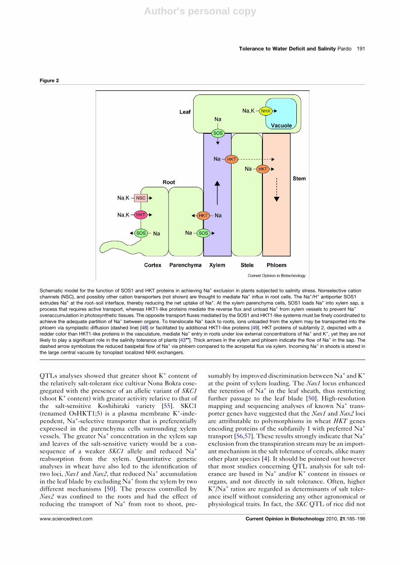
Figure 2
Schematic model for the function of SOS1 and HKT proteins in achieving Na+ exclusion in plants subjected to salinity stress. Nonselective cation
channels (NSC), and possibly other cation transporters (not shown) are thought to mediate Na+ influx in root cells. The Na+/H+ antiporter SOS1
extrudes Na+ at the root–soil interface, thereby reducing the net uptake of Na+. At the xylem parenchyma cells, SOS1 loads Na+ into xylem sap, a
process that requires active transport, whereas HKT1-like proteins mediate the reverse flux and unload Na+ from xylem vessels to prevent Na+
overaccumulation in photosynthetic tissues. The opposite transport fluxes mediated by the SOS1 and HKT1-like systems must be finely coordinated to
achieve the adequate partition of Na+ between organs. To translocate Na+ back to roots, ions unloaded from the xylem may be transported into the
phloem via symplastic diffusion (dashed line) [48] or facilitated by additional HKT1-like proteins [49]. HKT proteins of subfamily 2, depicted with a
redder color than HKT1-like proteins in the vasculature, mediate Na+ entry in roots under low external concentrations of Na+ and K+, yet they are not
likely to play a significant role in the salinity tolerance of plants [43��]. Thick arrows in the xylem and phloem indicate the flow of Na+ in the sap. The
dashed arrow symbolizes the reduced basipetal flow of Na+ via phloem compared to the acropetal flux via xylem. Incoming Na+ in shoots is stored in
the large central vacuole by tonoplast localized NHX exchangers.
www.sciencedirect.com Current Opinion in Biotechnology 2010, 21:185–196

Author's personal copy
show a significant correlation coefficient with survival to
salt stress [58]. A clear difference should be made be-
tween QTLs responsible of ionic balance and QTLs for
salt tolerance.
Members of subfamily 2 of HKT transporters mediate
Na+ uptake by roots, particularly when extracellular K+ is
limiting [59,60]. The transcript level of several subfamily
2 HKT genes has been shown to increase by K+-starvation
in cereals and downregulated by salinity (reviewed in
[43��]). At low-K+ availability, moderate levels of Na+
actually promote plant growth by replacing K+ for osmotic
adjustment [42]. Thus, subfamily 2 HKTs may be
involved in providing ‘nutritional’ Na+ under K+
starvation rather than in salinity stress [43��,60].
Comparisons of unidirectional Na+ fluxes and rates of net
accumulation of Na+ in roots indicate that 70–95% of the
Na+ fluxed into the root symplast is extruded back to the
apoplast, and that small differences in Na+ exclusion
capacity lead to major changes in the net accumulation
of Na+ [4,50]. In Arabidopsis, the plasma membrane Na+/H+
exchanger SOS1 facilitates Na+ homeostasis by extruding
the ion from root epidermal cells at the root–soil interface
[61,62] (Figure 2). SOS1 is preferentially expressed in
xylem parenchyma cells and analyses of the Na+ root/shoot
partitioning in roots of sos1 plants under different salt
regimes indicate that SOS1 participates in the redistribu-
tion of Na+ between the root and shoot, likely working in
concert with AtHKT1;1 at the plasma membrane of xylem
parenchyma cells [48,61,63] (Figure 2). At the parench-
yma–xylem interface, the efflux of Na+ from the parench-
yma cells and loading of the xylem sap must be active (i.e.
energetically costly) owing to the plasma membrane poten-
tial, negative inside, and would require a Na+/H+ exchan-
ger such as SOS1 [61]. Additional evidence of the
involvement of SOS1 in long-distance Na+ transport has
been produced recently in the halophytic Arabidopsis-relative Thellungiella salsuginea (a.k.a. T. halophila) and in
tomato [64,65]. Lower net Na+ flux was observed in the
xylem sap of tomato plants with suppressed SOS1 activity
[65]. Downregulation of ThSOS1 in Thellungiella increased
Na+ accumulation in the root tip and in the stele. Maximal
Na+ accumulation, concomitant with a decrease in the K+
content, was found in the root xylem parenchyma. These
cells presented a Na+–K+ ratio more than 12 times higher
than equivalent cells in wild-type plants.
Reduced or abolished activity of SOS1 interferes with K+
nutrition and long-distance transport ([65] and references
therein). Mutations in rice and Arabidopsis HKT Na+
transporters also reduce K+ accumulation in shoots during
salt exposure [48,55]. Since greater Na+ levels in the xylem
sap of athkt1;1 mutants were accompanied by reduced
xylem sap K+ levels, the coupling of Na+ unloading by
the xylem-localized HKT transporters with K+ loading into
xylem vessels via depolarization-activated K+ channels has
been hypothesized [43��]. HKT-mediated Na+ uptake
from xylem vessels would induce membrane depolariz-
ation of xylem parenchyma cells, which in turn could
activate depolarization-activated outward-rectifying K+
channels. Reduced contents of K+ in Na+-loaded xylem
parenchyma cells in SOS1-suppressed Thellungiella plants
are coherent with this model [64]. On the other hand,
cytosolic Na+ has been shown to inhibit the K+ channel
AKT1 involved in K+ uptake by roots [41]. AKT1 is
apparently a target of salt stress in sos1 plants, resulting
in poor growth because of impaired K+ uptake. Mutant
analyses showed that akt1 seedlings were salt sensitive
during early seedling development, but skor seedlings were
normal. The SKOR channel mediates K+ release into the
xylem vessels from xylem parenchyma cells. Thus, the
effect of Na+ on K+ transport is probably more important at
the uptake stage than at the xylem loading stage.
The activity of the SOS1 exchanger is regulated through
protein phosphorylation by the SOS2–SOS3 kinase com-
plex in Arabidopsis [62,66]. SOS2/CIPK24 is a serine/
threonine protein kinase of the SnRK3/CIPK family.
SOS3/CBL4 is a myristoylated, membrane bound Ca2+
sensor belonging to the recoverin-like family of SCaBPs/
CBLs. Upon Ca2+ binding, SOS3 binds to and enhances
the protein kinase activity of SOS2 [67]. Besides activat-
ing SOS2, SOS3 was shown to recruit SOS2 to the plasma
membrane to facilitate interaction with SOS1 [66]. SOS2
also interacts with SCaBP8/CBL10 to form an alternative
protein kinase complex that regulates SOS1 at the plasma
membrane [68]. SOS2 has recently been shown to phos-
phorylate SCaBP8/CBL10 at its C-terminus [69], thus
adding a new layer of regulation to CBL proteins besides
Ca2+ binding and fatty acyl modifications [70]. This
phosphorylation was induced by salt stress, occurred at
the membrane, stabilized the SCaBP8–SOS2 interaction,
and enhanced plasma membrane Na+/H+ exchange
activity [69]. Surprisingly, interaction of SOS2/CIPK24
with SCaBP8/CBL10 may also result in localization of the
kinase complex at the vacuolar membrane where it med-
iates salt tolerance by regulating the accumulation of Na+
in shoot tissues by an as yet undefined mechanism that
may involve regulation of the Na+/H+ exchange at the
tonoplast [71,72]. Regulation of the tonoplast V-ATPase
by SOS2 in the absence of CBL proteins has also been
reported [73]. Presumably, the post-translational modifi-
cations of SCaBP8/CBL10 or the interaction of combi-
natorial protein kinase complexes with specific targets in
different cellular membranes may ultimately define the
localization of the protein kinase in vivo.
The identification of additional SOS2-interacting proteins
indicates a connection between SOS2 and ROS signaling
[74�]. SOS2 physically interacts with the H2O2 signaling
protein nucleoside diphosphate kinase 2 (NDPK2) and
with catalases 2 and 3. A sos2 ndpk2 double mutant did not
accumulate H2O2 in response to salt stress, suggesting that
192 Plant biotechnology
Current Opinion in Biotechnology 2010, 21:185–196 www.sciencedirect.com

Author's personal copy
it is altered signaling rather than H2O2 toxicity alone that is
responsible for the increased salt sensitivity of the sos2ndpk2 double mutant relative to single mutants. The effect
of NDPK2 on H2O2 signaling and stress sensitivity may be
mediated at least in part by the interaction with and
stimulation of two H2O2-responsive mitogen-activated
protein kinases, AtMPK3 and AtMPK6. The interaction
of SOS2 with NDPK2 occurred at the FISL motif, the
same protein domain required for SOS2 interaction with
SOS3 and adjacent to the interaction domain with the
protein phosphatase ABI2. Interaction with SOS2 inhib-
ited NDPK2 histidine autophosphorylation, indicating
modulation of NDPK2 activity and, in turn, of its down-
stream targets MPK3 and MPK6. The link between the
SOS pathway for Na+ stress and ROS signaling is further
substantiated by the interaction of the C-terminal cyto-
plasmic tail of SOS1 with RCD1 (radical-induced cell
death 1), a regulator of oxidative-stress responses [75],
and by the isolation of enh1 mutant as an enhancer of
the salt sensitivity of the sos3 mutant [76]. The ENH1gene encodes a chloroplast-localized rubredoxin-like
protein that shows greatest sequence similarity to rubre-
doxin proteins that play a role in superoxide detoxification.
The enh1 mutation causes enhanced accumulation of ROS,
particularly under salt stress. Since sos2 but not sos3mutants show increased sensitivity to oxidative stress
and the enh1 mutation does not enhance sos2 phenotypes,
it appears that ENH1 also functions in the SOS2-depend-
ent ROS signaling. The ion transporter SOS1 affects ENH1expression under salt stress by a process that may involve
RCD1.
Regulatory processes involving CIPK–CBL modules have
also been described for the control of K+ channels facil-
itating K+ uptake. A genetic screen for inability to grow in
low external K+ demonstrated that the protein kinase
CIPK23 is required for K+ uptake by roots under low-K+
conditions. CIPK23 bound to one of the two alternative
subunits CBL1 and CBL9 directly phosphorylates and
activates the plasma membrane K+ channel AKT1
[77��]. The interaction between CIPK23 and AKT1
involved the kinase domain of the CIPK and the ankyrin
repeat domain of the channel [78�]. CIPK23 was also
identified in a genetic screen for enhanced drought toler-
ance. In the cipk23 mutant, reduced transpirational water
loss from leaves coincided with enhanced ABA sensitivity
of guard cells during opening and closing, without notice-
able alterations in ABA content in the plant [79]. The cbl1cbl9 double mutant, but not the cbl1 or cbl9 single mutants,
exhibited altered phenotypes for stomatal responses and
low-potassium sensitivity. Thus, plasma membrane-loca-
lized CBL1–CIPK23 and CBL9–CIPK23 complexes sim-
ultaneously regulate K+ transport processes in roots and in
stomatal guard cells. For channel inactivation, the 2C-type
protein phosphatase AIP1 (for AKT1-interacting PP2C 1)
physically interacts and inhibits AKT1, but channel
protein dephosphorylation was not shown. Interestingly,
Luan’s lab has recently reported that PP2CA, another
member of the PP2C phosphatase family, inhibits the
activity of the guard cell anion channel SLAC1 by two
mechanisms: first, by dephosphorylating the SLAC1 chan-
nel protein and second, by physical interaction with the
SLAC1-activating protein kinase OST1, leading to inhi-
bition of the kinase independently of phosphatase activity
[80]. OST1 (SnRK2.6) is a member of the ArabidopsisSnRK2-type protein kinase family that includes other
members that function in the ABA response. OST1 was
shown to phosphorylate and activate SLAC1 but whether
PP2CA dephosphorylated the amino acid residue(s) phos-
phorylated by OST1 was not formally demonstrated [80].
Further work should elucidate the details of this regulatory
mechanism. These results demonstrate that two opposing
regulatory pathways involving SnKR2/3 kinases and 2C-
type phosphatases function in guard cells. The CIPK23–AIP1 module controls cell expansion and stomatal aperture
by the likely regulation of inward K+-channels that remain
to be identified (K+-channel proteins AKT1–3 and KAT1
are expressed in guard cells), whereas the OST1–PP2CA
module mediates cell shrinkage and stomatal closure by
regulating ion efflux via the anion channel SLAC1. A
similar CIPK–PP2C switch may operate in Na+ efflux
processes since SOS2 interacts with the 2C-type protein
phosphatase ABI2 [81]. In this case also, protein–protein
interactions may be as important as phosphorylation–dephosphorylation events for signal output. Structural data
and protein competition assays show that the binding of
ABI2 and SOS3 to SOS2 is mutually exclusive and that
SOS2 may be inhibited by the binding of ABI2 [67].
Presumably, Ca2+-bound SOS3 displaces ABI2 and acti-
vates SOS2 upon salt-induced Ca2+ signals. Together,
these findings provide evidence that the SnRK2 and
SnRK3/CIPK protein kinases together with 2C-type
protein phosphatases form a network of molecular switches
that regulate ion transporters in plants that are relevant to
salinity and water stress tolerance.
Concluding remarksMany factors related to the plant response to water and
salinity stress have been identified and many of these
factors have already been shown effective for engineering
stress tolerance in model plants. The manipulation of
signaling factors has produced the most impressive results
arguably because they control a broad range of down-
stream events, which results in superior tolerance. They
are potential candidates for multiple stress tolerance
owing to the interlaced nature of stress signaling net-
works. The structural details of the ABA–receptor inter-
actions that are being elucidated may pave the way for the
development of agonist molecules or the molecular tai-
loring of proteins (e.g. modulation of the activity of key
proteins guided by structural knowledge of these target
proteins) that could be useful for crop plants to cope with
water shortage. Controlled stress-dependent activation or
deactivation of signal components might function as a
Tolerance to Water Deficit and Salinity Pardo 193
www.sciencedirect.com Current Opinion in Biotechnology 2010, 21:185–196

Author's personal copy
molecular switch for the biotechnological manipulation of
stress responses. Effective expression systems, including
cell type-specific and stress-inducible promoters will be
required to fine-tune the plant response to stress accord-
ing to the time and circumstances for the onset of the
environmental insult. Commonly used constitutive pro-
moters are not always effective or can have negative
effects on plant growth or development. The importance
of cell type-specific processes is best exemplified by
AtHKT1;1. Constitutive expression of AtHKT1;1 causes
increased shoot accumulation of Na+ and reduced salinity
tolerance, whereas specific transgene expression in the
stele of roots has the opposite effect, that is Na+ exclusion
from the shoot and enhanced salinity tolerance [52��].
Admittedly, the vast majority of stress genes have been
tested using model plants in highly controlled experimen-
tal settings. Engineering crop plants with improved stress
tolerance is still in its early stages. The number of
transgenic crop plants that have undergone field trials
is undoubtedly small, but some have produced encoura-
ging results [17��,82]. Engineered mechanisms of stress
tolerance might promote survival during periods of
intense or prolonged stress or maintain crop’s productivity
under conditions of moderate environmental stress. Most
of the basic research has been focused on early develop-
mental stages. In many cases, however, the reproductive
parts of crop plants are harvested and future progress in
producing stress tolerant crops relies on research efforts to
improve reproductive success.
Last, plants respond to specific clues of water deficit and
salinity stress within seconds, yet the mechanisms by
which plants perceive the presence of salts or sense turgor
remain obscure. Filling this gap in our knowledge is a
pressing need given the importance of these initial steps
of plant responses to changes in the environment.
AcknowledgementsThe author is indebted to Juan Jordano, Francisco J Quintero, Irene Villalta,and Pedro L Rodriguez for helpful comments on the manuscript. Heapologizes to the authors whose original contributions could not be citedherein because of space constrains. This work was supported by grantsBIO2009-08641 and CSD2007-00057 from MICINN, and by grant P06-AGR-01482 from Junta de Andalucia.
References and recommended readingPapers of particular interest, published within the period of review,have been highlighted as:
� of special interest
�� of outstanding interest
1. Bray EA, Bailey-Serres J, Weretilnyk E: Responses to abioticstresses. In Biochemistry and Molecular Biology of Plants. Editedby Gruissem W, Buchannan B, Jones R. American Society of PlantPhysiologists; 2000:1158-1203.
2. Bartels D, Sunkar R: Drought and salt tolerance in plants. CritRev Plant Sci 2005, 24:23-58.
3.�
Munns R, Tester M: Mechanisms of salinity tolerance. Annu RevPlant Biol 2008, 59:651-681.
This is an up-to-date review that deals with the physiological andmolecular mechanisms of tolerance to osmotic and ionic componentsof salinity stress at the cellular, organ, and whole-plant levels.
4. Tester M, Davenport R: Na+ tolerance and Na+ transport inhigher plants. Ann Bot 2003, 91:503-527.
5. Serrano R: Salt tolerance in plants and microorganisms:toxicity targets and defense responses. Int Rev Cytol: A SurvCell Biol 1996, 165:1-52.
6.�
Bacso R, Janda T, Galiba G, Papp I: Restricted transpirationmay not result in improved drought tolerance in a competitiveenvironment for water. Plant Sci 2008, 174:200-204.
This study is of interest because it shows the possible shortcomings ofplants that have been modified to restrict water and gas exchange. Thetolerance phenotype disappeared when the root systems of neighboringwild-type Arabidopsis plants competed for water in the soil arounddrought tolerant mutants.
7.�
Yoo CY, Pence HE, Hasegawa PM, Mickelbart MV: Regulation oftranspiration to improve crop water use. Crit Rev Plant Sci 2009,28:410-431.
However, and in contrast to Ref. [6�], this excellent review shows that theexisting gene pools exhibit allelic variation for WUE through mechanismsthat regulate transpiration. The authors argue that it is feasible that WUEcould be improved by decreasing transpiration without a concomitantreduction in CO2 uptake. Further, the genetic determinants that regulatetranspiration and WUE in the context of agricultural application arereviewed.
8. Sirichandra C, Wasilewska A, Vlad F, Valon C, Leung J: The guardcell as a single-cell model towards understanding droughttolerance and abscisic acid action. J Exp Bot 2009 doi: 10.1093/jxb/ern340.
9. Cominelli E, Galbiati M, Vavasseur A, Conti L, Sala T, Vuylsteke M,Leonhardt N, Dellaporta SL, Tonelli C: A guard-cell-specific MYBtranscription factor regulates stomatal movements and plantdrought tolerance. Curr Biol 2005, 15:1196-1200.
10. Liang YK, Dubos C, Dodd IC, Holroyd GH, Hetherington AM,Campbell MM: AtMYB61, an R2R3–MYB transcription factorcontrolling stomatal aperture in Arabidopsis thaliana. Curr Biol2005, 15:1201-1206.
11. Jung C, Seo JS, Han SW, Koo YJ, Kim CH, Song SI, Nahm BH,Choi YD, Cheong J-J: Overexpression of AtMYB44 enhancesstomatal closure to confer abiotic stress tolerance intransgenic Arabidopsis. Plant Physiol 2008, 146:623-635.
12. Yu H, Chen X, Hong YY, Wang Y, Xu P, Ke SD, Liu HY, Zhu JK,Oliver DJ, Xiang CB: Activated expression of an ArabidopsisHD-START protein confers drought tolerance with improvedroot system and reduced stomatal density. Plant Cell 2008,20:1134-1151.
13. Karaba A, Dixit S, Greco R, Aharoni A, Trijatmiko KR, Marsch-Martinez N, Krishnan A, Nataraja KN, Udayakumar M, Pereira A:Improvement of water use efficiency in rice by expression ofHARDY, an Arabidopsis drought and salt tolerance gene. ProcNatl Acad Sci 2007, 104:15270-15275.
14. Chen ZZ, Zhang HR, Jablonowski D, Zhou XF, Ren XZ, Hong XH,Schaffrath R, Zhu JK, Gong ZH: Mutations in ABO1/ELO2, asubunit of holo-elongator, increase abscisic acid sensitivityand drought tolerance in Arabidopsis thaliana. Mol Cell Biol2006, 26:6902-6912.
15. Kahle J, Baake M, Doenecke D, Albig W: Subunits of theheterotrimeric transcription factor NF-Y are imported into thenucleus by distinct pathways involving importin {beta} andimportin 13. Mol Cell Biol 2005, 25:5339-5354.
16. Gusmaroli G, Tonelli C, Mantovani R: Regulation of novelmembers of the Arabidopsis thaliana CCAAT-binding nuclearfactor Y subunits. Gene 2002, 283:41-48.
17.��
Nelson DE, Repetti PP, Adams TR, Creelman RA, Wu J,Warner DC, Anstrom DC, Bensen RJ, Castiglioni PP,Donnarummo MG et al.: Plant nuclear factor Y (NF-Y) B subunitsconfer drought tolerance and lead to improved corn yields onwater-limited acres. Proc Natl Acad Sci 2007, 104:16450-16455.
This is a comprehensive work in which basic science is put to the test inthe field. Transgenic maize showed superior yield under realistic water-limited environments. The application of this technology has the potential
194 Plant biotechnology
Current Opinion in Biotechnology 2010, 21:185–196 www.sciencedirect.com

Author's personal copy
to significantly impact maize production systems that experiencedrought.
18. Li W-X, Oono Y, Zhu J, He X-J, Wu J-M, Iida K, Lu X-Y, Cui X, Jin H,Zhu J-K: The Arabidopsis NFYA5 transcription factor isregulated transcriptionally and posttranscriptionally topromote drought resistance. Plant Cell 2008, 20:2238-2251.
19.�
Prieto-Dapena P, Castano R, Almoguera C, Jordano J: Theectopic overexpression of a seed-specific transcriptionfactor, HaHSFA9, confers tolerance to severe dehydration invegetative organs. Plant J 2008, 54:1004-1014.
This paper shows that heat-shock transcription factors convey toleranceto extreme dessication at early stages after germination without a nega-tive impact on plant growth. It further speculates that the expressionpattern of seed-specific HSF genes could have changed during evolutionto bring about the tolerance to vegetative desiccation in resurrectionplants.
20. Almoguera C, Prieto-Dapena P, Diaz-Martin J, Espinosa JM,Carranco R, Jordano J: The HaDREB2 transcription factorenhances basal thermotolerance and longevity of seedsthrough functional interaction with HaHSFA9. BMC Plant Biol2009, 9:75.
21. Illing N, Denby KJ, Collett H, Shen A, Farrant JM: The signature ofseeds in resurrection plants: a molecular and physiologicalcomparison of desiccation tolerance in seeds and vegetativetissues. Integr Comp Biol 2005, 45:771-787.
22. Perruc E, Kinoshita N, Lopez-Molina L: The role of chromatin-remodeling factor PKL in balancing osmotic stress responsesduring Arabidopsis seed germination. Plant J 2007, 52:927-936.
23. Kotak S, Vierling E, Baumlein H, Koskull-Doring P: A noveltranscriptional cascade regulating expression of heat stressproteins during seed development of Arabidopsis. Plant Cell2007, 19:182-195.
24. McCourt P, Creelman R: The ABA receptors — we report youdecide. Curr Opin Plant Biol 2008, 11:474-478.
25. Pennisi E: Stressed out over a stress hormone. Science 2009,324:1012.
26. Pandey S, Nelson DC, Assmann SM: Two novel GPCR-type Gproteins are abscisic acid receptors in Arabidopsis. Cell 2009,136:136-148.
27. Ma Y, Szostkiewicz I, Korte A, Moes D, Yang Y, Christmann A,Grill E: Regulators of PP2C phosphatase activity function asabscisic acid sensors. Science 2009, 324:1064-1068.
28. Santiago J, Rodrigues A, Saez A, Rubio S, Antoni R, Dupeux F,Park SY, Marquez JA, Cutler SR, Rodriguez PL: Modulation ofdrought resistance by the abscisic acid–receptor PYL5through inhibition of clade A PP2Cs. Plant J 2009, 60:575-588.
29.��
Park S-Y, Fung P, Nishimura N, Jensen DR, Fujii H, Zhao Y,Lumba S, Santiago J, Rodrigues A, Chow T-fF et al.: Abscisic acidinhibits type 2C protein phosphatases via the PYR/PYL familyof START proteins. Science 2009, 324:1068-1071.
This work and a series of related reports described in the main text andreferenced below, are landmark papers that show very convincingly thatPYR/PYL/RCAR proteins are ABA receptors functionally linked to well-known ABA signaling intermediaries and ABA responses.
30. Santiago J, Dupeux F, Round A, Antoni R, Park SY, Jamin M,Cutler SR, Rodriguez PL: The abscisic acid receptor PYR1 incomplex with abscisic acid. Nature 2009, 464:665-668.
31. Melcher K, Ng L-M, Zhou XE, Soon F-F, Xu Y, Suino-Powell KM,Park S-Y, Weiner JJ, Fujii H, Chinnusamy V et al.: A gate-latch-lock mechanism for hormone signalling by abscisic acidreceptors. Nature 2009, 462:602-608.
32. Yin P, Fan H, Hao Q, Yuan X, Wu D, Pang Y, Yan C, Li W, Wang J,Yan N: Structural insights into the mechanism of abscisicacid signaling by PYL proteins. Nat Struct Mol Biol 2009,16:1230-1236.
33. Mustilli AC, Merlot S, Vavasseur A, Fenzi F, Giraudat J:Arabidopsis OST1 protein kinase mediates the regulation ofstomatal aperture by abscisic acid and acts upstream ofreactive oxygen species production. Plant Cell 2002,14:3089-3099.
34. Yoshida R, Umezawa T, Mizoguchi T, Takahashi S, Takahashi F,Shinozaki K: The regulatory domain of SRK2E/OST1/SnRK2. 6interacts with ABI1 and integrates abscisic acid (ABA) andosmotic stress signals controlling stomatal closure inArabidopsis. J Biol Chem 2006, 281:5310-5318.
35. Fujii H, Zhu JK: Arabidopsis mutant deficient in 3 abscisic acid-activated protein kinases reveals critical roles in growth,reproduction, and stress. Proc Natl Acad Sci 2009,106:8380-8385.
36. Umezawa T, Sugiyama N, Mizoguchi M, Hayashi S, Myouga F,Yamaguchi-Shinozaki K, Ishihama Y, Hirayama T, Shinozaki K:Type 2C protein phosphatases directly regulate abscisic acid-activated protein kinases in Arabidopsis. Proc Natl Acad Sci2009, 106:17588-17593.
37. Vlad F, Rubio S, Rodrigues A, Sirichandra C, Belin C, Robert N,Leung J, Rodriguez PL, Lauriere C, Merlot S: Proteinphosphatases 2C regulate the activation of the Snf1-relatedkinase OST1 by abscisic acid in Arabidopsis. Plant Cell 2009doi: 10.1105/tpc.109.069179.
38. Furihata T, Maruyama K, Fujita Y, Umezawa T, Yoshida R,Shinozaki K, Yamaguchi-Shinozaki K: Abscisic acid-dependentmultisite phosphorylation regulates the activity of atranscription activator AREB1. Proc Natl Acad Sci 2006,103:1988-1993.
39. Shinozaki K, Yamaguchi-Shinozaki K: Gene networks involvedin drought stress response and tolerance. J Exp Bot 2007,58:221-227.
40. Fujii H, Chinnusamy V, Rodrigues A, Rubio S, Antoni R, Park SY,Cutler SR, Sheen J, Rodriguez PL, Zhu JK: In vitro reconstitutionof an abscisic acid signalling pathway. Nature 2009,462:660-664.
41. Qi Z, Spalding EP: Protection of plasma membrane K+ transportby the salt overly sensitive1 Na+–H+ antiporter during salinitystress. Plant Physiol 2004, 136:2548-2555.
42. Rodriguez-Navarro A, Rubio F: High-affinity potassium andsodium transport systems in plants. J Exp Bot 2006,57:1149-1160.
43.��
Horie T, Hauser F, Schroeder JI: HKT transporter-mediatedsalinity resistance mechanisms in Arabidopsis and monocotcrop plants. Trends Plant Sci 2009, 14:660-668.
This is an excellent up-to-date review on the biochemistry and iontransport selectivity of HKT transporters, together with their physiologicalroles on the salinity tolerance of plants.
44. Yao X, Horie T, Xue S, Leung H-Y, Katsuhara M, Brodsky DE,Wu Y, Schroeder JI: Differential sodium and potassiumtransport selectivities of the rice OsHKT2;1 andOsHKT2;2 transporters in plant cells. Plant Physiol 2010,152:341-355.
45. Demidchik V, Maathuis FJ: Physiological roles of nonselectivecation channels in plants: from salt stress to signalling anddevelopment. New Phytol 2007, 175:387-404.
46. Rus A, Lee BH, Munoz-Mayor A, Sharkhuu A, Miura K, Zhu JK,Bressan RA, Hasegawa PM: AtHKT1 facilitates Na+
homeostasis and K+ nutrition in planta. Plant Physiol 2004,136:2500-2511.
47. Essah PA, Davenport R, Tester M: Sodium influx andaccumulation in Arabidopsis. Plant Physiol 2003, 133:307-318.
48. Sunarpi, Horie T, Motoda J, Kubo M, Yang H, Yoda K, Horie R,Chan WY, Leung HY, Hattori K et al.: Enhanced salt tolerancemediated by AtHKT1 transporter-induced Na+ unloadingfrom xylem vessels to xylem parenchyma cells. Plant J 2005,44:928-938.
49. Berthomieu P, Conejero G, Nublat A, Brackenbury WJ, Lambert C,Savio C, Uozumi N, Oiki S, Yamada K, Cellier F: Functionalanalysis of AtHKT1 in Arabidopsis shows that Na+
recirculation by the phloem is crucial for salt tolerance. EMBOJ 2003, 22:2004-2014.
50. James RA, Davenport RJ, Munns R: Physiologicalcharacterization of two genes for Na+ exclusion in durumwheat, Nax1 and Nax2. Plant Physiol 2006, 142:1537-1547.
Tolerance to Water Deficit and Salinity Pardo 195
www.sciencedirect.com Current Opinion in Biotechnology 2010, 21:185–196

Author's personal copy
51. Davenport RJ, Munoz-Mayor A, Jha D, Essah PA, Rus A, Tester M:The Na+ transporter AtHKT1;1 controls retrieval of Na+ fromthe xylem in Arabidopsis. Plant Cell Environ 2007, 30:497-507.
52.��
Moller IS, Gilliham M, Jha D, Mayo GM, Roy SJ, Coates JC,Haseloff J, Tester M: Shoot Na+ exclusion and increased salinitytolerance engineered by cell type-specific alteration of Na+
transport in Arabidopsis. Plant Cell 2009, 21:2163-2178.Excellent work in which HKT1;1 expression was targeted to specific celltypes in the mature root of Arabidopsis to increase the retrieval of Na+
from the transpiration stream.
53. Rus A, Baxter I, Muthukumar B, Gustin J, Lahner B, Yakubova E,Salt DE: Natural variants of AtHKT1 enhance Na+ accumulationin two wild populations of Arabidopsis. PLoS Genet 2006, 2:e210.
54. Apse MP, Blumwald E: Na+ transport in plants. FEBS Lett 2007,581:2247-2254.
55. Ren Z-H, Gao J-P, Li L-G, Cai X-L, Huang W, Chao D-Y, Zhu M-Z,Wang Z-Y, Luan S, Lin H-X: A rice quantitative trait locus for salttolerance encodes a sodium transporter. Nat Genet 2005,37:1141-1146.
56. Huang SB, Spielmeyer W, Lagudah ES, James RA, Platten JD,Dennis ES, Munns R: A sodium transporter (HKT7) is acandidate for Nax1, a gene for salt tolerance in durum wheat.Plant Physiol 2006, 142:1718-1727.
57. Byrt CS, Platten JD, Spielmeyer W, James RA, Lagudah ES,Dennis ES, Tester M, Munns R: HKT1;5-like cation transporterslinked to Na+ exclusion loci in wheat, Nax2 and Kna1. PlantPhysiol 2007, 143:1918-1928.
58. Lin HX, Zhu MZ, Yano M, Gao JP, Liang ZW, Su WA, Hu XH,Ren ZH, Chao DY: QTLs for Na+ and K+ uptake of the shootsand roots controlling rice salt tolerance. Theor Appl Genet2004, 108:253-260.
59. Laurie S, Feeney KA, Maathuis FJM, Heard PJ, Brown SJ,Leigh RA: A role for HKT1 in sodium uptake by wheat roots.Plant J 2002, 32:139-149.
60. Horie T, Costa A, Kim TH, Han MJ, Horie R, Leung HY, Miyao A,Hirochika H, An G, Schroeder JI: Rice OsHKT2;1 transportermediates large Na+ influx component into K+-starved roots forgrowth. EMBO J 2007, 26:3003-3014.
61. Shi HZ, Quintero FJ, Pardo JM, Zhu JK: The putative plasmamembrane Na+/H+ antiporter SOS1 controls long-distanceNa+ transport in plants. Plant Cell 2002, 14:465-477.
62. Qiu QS, Guo Y, Dietrich MA, Schumaker KS, Zhu JK: Regulationof SOS1, a plasma membrane Na+/H+ exchanger inArabidopsis thaliana, by SOS2 and SOS3. Proc Natl Acad SciU S A 2002, 99:8436-8441.
63. Pardo JM, Cubero B, Leidi EO, Quintero FJ: Alkali cationexchangers: roles in cellular homeostasis and stresstolerance. J Exp Bot 2006, 57:1181-1199.
64. Oh D-H, Leidi E, Zhang Q, Hwang S-M, Li Y, Quintero FJ, Jiang X,D’Urzo MP, Lee SY, Zhao Y et al.: Loss of halophytism byinterference with SOS1 expression. Plant Physiol 2009,151:210-222.
65. Olias R, Eljakaoui Z, Li J, Alvarez De Morales P, Marin-Manzano MC, Pardo JM, Belver A: The plasma membrane Na+/H+ antiporter SOS1 is essential for salt tolerance in tomato andaffects the partitioning of Na+ between plant organs. Plant, CellEnviron 2009, 32:904-916.
66. Quintero FJ, Ohta M, Shi HZ, Zhu JK, Pardo JM: Reconstitution inyeast of the Arabidopsis SOS signaling pathway for Na+
homeostasis. Proc Natl Acad Sci U S A 2002, 99:9061-9066.
67. Sanchez-Barrena MJ, Fujii H, Angulo I, Martinez-Ripoll M, Zhu JK,Albert A: The structure of the C-terminal domain of the proteinkinase AtSOS2 bound to the calcium sensor AtSOS3. Mol Cell2007, 26:427-435.
68. Quan RD, Lin HX, Mendoza I, Zhang YG, Cao WH, Yang YQ,Shang M, Chen SY, Pardo JM, Guo Y: SCABP8/CBL10, aputative calcium sensor, interacts with the protein kinaseSOS2 to protect Arabidopsis shoots from salt stress. Plant Cell2007, 19:1415-1431.
69. Lin H, Yang Y, Quan R, Mendoza I, Wu Y, Du W, Zhao S,Schumaker KS, Pardo JM, Guo Y: Phosphorylation of SOS3-LIKE CALCIUM BINDING PROTEIN8 by SOS2 protein kinasestabilizes their protein complex and regulates salt tolerance inArabidopsis. Plant Cell 2009, 21:1607-1619.
70. Batistic O, Sorek N, Schultke S, Yalovsky S, Kudla J: Dual fattyacyl modification determines the localization and plasmamembrane targeting of CBL/CIPK Ca2+ signaling complexes inArabidopsis. Plant Cell 2008, 20:1346-1362.
71. Kim B-G, Waadt R, Cheong YH, Pandey GK, Dominguez-Solis JR,Schultke S, Lee SC, Kudla J, Luan S: The calcium sensor CBL10mediates salt tolerance by regulating ion homeostasis inArabidopsis. Plant J 2007, 52:473-484.
72. Qiu QS, Guo Y, Quintero FJ, Pardo JM, Schumaker KS, Zhu JK:Regulation of vacuolar Na+/H+ exchange in Arabidopsisthaliana by the salt-overly-sensitive (SOS) pathway. J BiolChem 2004, 279:207-215.
73. Batelli G, Verslues PE, Agius F, Qiu Q, Fujii H, Pan S,Schumaker KS, Grillo S, Zhu JK: SOS2 promotes salt tolerancein part by interacting with the vacuolar H+-ATPase andupregulating its transport activity. Mol Cell Biol 2007,27:7781-7790.
74.�
Verslues PE, Batelli G, Grillo S, Agius F, Kim YS, Zhu J, Agarwal M,Katiyar-Agarwal S, Zhu JK: Interaction of SOS2 with nucleosidediphosphate kinase 2 and catalases reveals a point ofconnection between salt stress and H2O2 signaling inArabidopsis thaliana. Mol Cell Biol 2007, 27:7771-7780.
This paper demonstrates the interaction of the protein kinase SOS2, a keyregulatory intermediary of Na+ homeostasis, with both NDPK2 and CATs,and thereby reveals a point of cross-talk between salt stress responseand ROS signaling.
75. Katiyar-Agarwal S, Zhu J, Kim K, Agarwal M, Fu X, Huang A,Zhu JK: The plasma membrane Na+/H+ antiporter SOS1interacts with RCD1 and functions in oxidative stresstolerance in Arabidopsis. Proc Natl Acad Sci U S A 2006,103:18816-18821.
76. Zhu J, Fu X, Koo YD, Zhu JK, Jenney FE Jr, Adams MWW, Zhu Y,Shi H, Yun DJ, Hasegawa PM, Bressan RA: An enhancer mutantof Arabidopsis salt overly sensitive 3 mediates both ionhomeostasis and the oxidative stress response. Mol Cell Biol2007, 27:5214-5224.
77.��
Xu J, Li HD, Chen LQ, Wang Y, Liu LL, He L, Wu WH: A proteinkinase, interacting with two calcineurin B-like proteins,regulates K+ transporter AKT1 in Arabidopsis. Cell 2006,125:1347-1360.
This is a seminal study showing that plants cope with limited soilpotassium by coupling potassium channel activation to a cellular calciumsignaling network.
78.�
Lee SC, Lan W-Z, Kim B-G, Li L, Cheong YH, Pandey GK, Lu G,Buchanan BB, Luan S: A protein phosphorylation/dephosphorylation network regulates a plant potassiumchannel. Proc Natl Acad Sci 2007, 104:15959-15964.
This paper complements the above report by showing that PP2Cscounteract the activation of the AKT1 channel by phosphorylation.
79. Cheong YH, Pandey GK, Grant JJ, Batistic O, Li L, Kim B-G, Lee S-C, Kudla J, Luan S: Two calcineurin B-like calcium sensors,interacting with protein kinase CIPK23, regulate leaftranspiration and root potassium uptake in Arabidopsis. PlantJ 2007, 52:223-239.
80. Lee SC, Lan W, Buchanan BB, Luan S: A protein kinase-phosphatase pair interacts with an ion channel to regulateABA signaling in plant guard cells. Proc Natl Acad Sci 2009,106:21419-21424.
81. Ohta M, Guo Y, Halfter U, Zhu JK: A novel domain in the proteinkinase SOS2 mediates interaction with the proteinphosphatase 2C ABI2. Proc Natl Acad Sci U S A 2003,100:11771-11776.
82. Xue ZY, Zhi DY, Xue GP, Zhang H, Zhao YX, Xia GM: Enhancedsalt tolerance of transgenic wheat (Tritivum aestivum L.)expressing a vacuolar Na+/H+ antiporter gene with improvedgrain yields in saline soils in the field and a reduced level of leafNa+. Plant Sci 2004, 167:849-859.
196 Plant biotechnology
Current Opinion in Biotechnology 2010, 21:185–196 www.sciencedirect.com