Biochemical, genetic and physiological characterization of venom components from two species of...
10
Biochemical, genetic and physiological characterization of venom components from two species of scorpions: Centruroides exilicauda Wood and Centruroides sculpturatus Ewing Norma A. Valdez-Cruz a , Sonia Dávila b , Alexei Licea c , Miguel Corona a , Fernando Z. Zamudio a , Jesús García-Valdes a , Leslie Boyer d , Lourival D. Possani a, * a Department of Molecular Medicine and Bioprocesses, Institute of Biotechnology, National Autonomous University of Mexico, Avenida Universidad 2001, Apartado Postal 510-3, Cuernavaca 62210, Mexico b Department of Genetic Evolution, Institute of Ecology, National Autonomous University of Mexico, Ciudad Universitaria 04510, Mexico DF, Mexico c Department of Aquaculture, Marine Biotechnology, Centro de Investigación Científica y de Educación de Ensenada, Km 107, Carretera Tijuana-Ensenada, Ensenada BC, Mexico d Arizona Poison and Drug Information Center, University of Arizona Health Sciences Center 1501 N. Campbell avenue, Tucson, AZ 85724, USA Received 4 November 2003; accepted 14 May 2004 Available online 08 June 2004 Abstract Current literature concerning the taxonomic names of two possibly distinct species of scorpions from the genus Centruroides (sculpturatus and/or exilicauda) is controversial. This communication reports the results of biochemical, genetic and electrophysiological experiments conducted with C. exilicauda Wood of Baja California (Mexico) and C. sculpturatus Ewing of Arizona (USA). The chromatographic profile fractionation of the soluble venom from both species of scorpions is different. The N-terminal amino acid sequence for nine toxins of C. exilicauda was determined and compared with those from C. sculpturatus. Lethality tests conducted in mice support the idea that C. exilicauda venom should be expected to be medically less important than C. sculpturatus. Thirteen genes from the venomous glands of the scorpion C. exilicauda were obtained and compared with previously published sequences from genes of the species C. sculpturatus. Genes coding for cytochrome oxidase I and II of both species were also sequenced. A phylogenetic tree was generated with this information showing important differences between them. Additionally, the results of electrophysiological assays conducted with the venom from both species on the Ca 2+ -dependent K + -channels, showed significant differences. These results strongly support the conclusion that C. exilicauda and C. sculpturatus are in fact two distinct species of scorpions. © 2004 Elsevier SAS. All rights reserved. Keywords: C. exilicauda; C. sculpturatus; Cytochrome oxidase; Mitochondrial gene; Scorpion toxin; K + -channel 1. Introduction Scorpions of the family Buthidae comprises approxi- mately 529 species, divided into 73 genera, from which only five are dangerous to humans, but are worldwide spread [1,2]. One of them, the genus Centruroides, is distributed from the southern part of USA to Mexico and Central America. From the Baja California area of Mexico, four families, 11 genera, 61 species and 12 subspecies were de- scribed [3], which makes it one of the most diverse geo- graphical areas of the world concerning scorpion diversity. Among the first reports is that by Wood [4], who described eight species of scorpions, including two from United States of North America and six from Mexico, state of Baja Califor- nia. Due to the fact that some of these species are medically important, it was mandatory to clarify species differences and to identify molecular markers and/or structural and func- tional aspects of their venoms. In this communication we have focused our attention to two species: C. exilicauda collected in Punta Banda, Baja California (Mexico) and to Abbreviations: C, Centruroides; COX I, cytochrome oxidase I; COX II, cytochrome oxidase II; l, limpidus; mDNA, mitochondrial DNA; T, Tityus. * Corresponding author. Tel.: +52-77-73-171209; fax: +52-77-73-172388. E-mail address: [email protected] (L.D. Possani). Biochimie 86 (2004) 387–396 www.elsevier.com/locate/biochi 0300-9084/$ - see front matter © 2004 Elsevier SAS. All rights reserved. doi:10.1016/j.biochi.2004.05.005
-
Upload
independent -
Category
Documents
-
view
1 -
download
0
Transcript of Biochemical, genetic and physiological characterization of venom components from two species of...

Biochemical, genetic and physiological characterizationof venom components from two species of scorpions:
Centruroides exilicauda Wood and Centruroides sculpturatus Ewing
Norma A. Valdez-Cruz a, Sonia Dávila b, Alexei Licea c, Miguel Corona a,Fernando Z. Zamudio a, Jesús García-Valdes a, Leslie Boyer d, Lourival D. Possani a,*
a Department of Molecular Medicine and Bioprocesses, Institute of Biotechnology, National Autonomous University of Mexico,Avenida Universidad 2001, Apartado Postal 510-3, Cuernavaca 62210, Mexico
b Department of Genetic Evolution, Institute of Ecology, National Autonomous University of Mexico, Ciudad Universitaria 04510, Mexico DF, Mexicoc Department of Aquaculture, Marine Biotechnology, Centro de Investigación Científica y de Educación de Ensenada, Km 107,
Carretera Tijuana-Ensenada, Ensenada BC, Mexicod Arizona Poison and Drug Information Center, University of Arizona Health Sciences Center 1501 N. Campbell avenue, Tucson, AZ 85724, USA
Received 4 November 2003; accepted 14 May 2004
Available online 08 June 2004
Abstract
Current literature concerning the taxonomic names of two possibly distinct species of scorpions from the genus Centruroides (sculpturatusand/or exilicauda) is controversial. This communication reports the results of biochemical, genetic and electrophysiological experimentsconducted with C. exilicauda Wood of Baja California (Mexico) and C. sculpturatus Ewing of Arizona (USA). The chromatographic profilefractionation of the soluble venom from both species of scorpions is different. The N-terminal amino acid sequence for nine toxins ofC. exilicauda was determined and compared with those from C. sculpturatus. Lethality tests conducted in mice support the idea that C.exilicauda venom should be expected to be medically less important than C. sculpturatus. Thirteen genes from the venomous glands of thescorpion C. exilicauda were obtained and compared with previously published sequences from genes of the species C. sculpturatus. Genescoding for cytochrome oxidase I and II of both species were also sequenced. A phylogenetic tree was generated with this information showingimportant differences between them. Additionally, the results of electrophysiological assays conducted with the venom from both species onthe Ca2+-dependent K+-channels, showed significant differences. These results strongly support the conclusion that C. exilicauda andC. sculpturatus are in fact two distinct species of scorpions.© 2004 Elsevier SAS. All rights reserved.
Keywords: C. exilicauda; C. sculpturatus; Cytochrome oxidase; Mitochondrial gene; Scorpion toxin; K+-channel
1. Introduction
Scorpions of the family Buthidae comprises approxi-mately 529 species, divided into 73 genera, from which onlyfive are dangerous to humans, but are worldwide spread[1,2]. One of them, the genus Centruroides, is distributedfrom the southern part of USA to Mexico and Central
America. From the Baja California area of Mexico, fourfamilies, 11 genera, 61 species and 12 subspecies were de-scribed [3], which makes it one of the most diverse geo-graphical areas of the world concerning scorpion diversity.Among the first reports is that by Wood [4], who describedeight species of scorpions, including two from United Statesof North America and six from Mexico, state of Baja Califor-nia. Due to the fact that some of these species are medicallyimportant, it was mandatory to clarify species differencesand to identify molecular markers and/or structural and func-tional aspects of their venoms. In this communication wehave focused our attention to two species: C. exilicaudacollected in Punta Banda, Baja California (Mexico) and to
Abbreviations: C, Centruroides; COX I, cytochrome oxidase I; COX II,cytochrome oxidase II; l, limpidus; mDNA, mitochondrial DNA; T, Tityus.
* Corresponding author. Tel.: +52-77-73-171209;fax: +52-77-73-172388.
E-mail address: [email protected] (L.D. Possani).
Biochimie 86 (2004) 387–396
www.elsevier.com/locate/biochi
0300-9084/$ - see front matter © 2004 Elsevier SAS. All rights reserved.doi:10.1016/j.biochi.2004.05.005

C. sculpturatus, collected in Tucson, Arizona (USA). Theyare two relatively abundant species of these areas, which formany years have generated taxonomic disputes and contro-versies about their medical importance. One of the objectivesof this work was to clarify the existing uncertainties on thesetwo apparently synonymous species, as well as, to obtainadditional characterization of their venom components andgene molecular markers. The medical problem arising fromstings of these arachnids, dangerous to humans in Arizona,but apparently not dangerous in Baja California, was press-ing a more rigorous study on this issue. Questions demandingsome immediate answers were: Are there two different spe-cies? Is one dangerous to humans, the other not dangerous?Are both coexisting in both places?
Here we show biochemical differences by chromato-graphic separations, bioassays, amino acid sequencing ofpeptides, gene cloning of toxins and by sequencing enzymemarkers of both species. Furthermore, for the first time weshow results that support the conclusion that C. exilicaudafrom Baja California should be less dangerous to humansthan C. sculpturatus from Arizona. The results of our mor-phological analysis of the two species will be included intoanother communication (S. Davila, N.A. Valdez-Cruz,L.D. Possani, and D. Piñero, unpublished).
2. Material and methods
2.1. Scorpions
The C. sculpturatus Ewing scorpions were collected inTucson, Arizona, USA. The C. exilicauda scorpions werecollected in Punta Banda, Baja California, Mexico. For thisstudy 15 animals of each species were used.
2.2. Separation of C. exilicauda and C. sculpturatusvenoms
Venoms of both species of scorpions were obtained in thelaboratory by electrical stimulation. The venom extract wasdissolved in water; centrifuged 15 min at 10,000 × g and thesupernatant was lyophilized. Separation of venom compo-nents was performed by high performance liquid chromatog-raphy (HPLC), as earlier described for the venom of Tityuscambridgei scorpion [5].
2.3. Biological tests
Lethality tests were carried out on female albino mice(CD1 strain) of approximately 20 g body weight. Differentamounts of venom from both species (C. exilicauda andC. sculpturatus) were tested in parallel. Injections wereperformed intraperitoneally using PBS (phosphate bufferedsaline, containing 0.15 mM NaCl, 0.1 mM sodium phosphateat pH 7.4) plus different doses with a final volume of 100 µl.The intoxication levels were called “non-toxic”, when the
animals showed no symptoms of envenoming within 20 hafter testing, v.g. showed the same symptoms as the controlmice injected with 100 µl of buffer alone (PBS). “Toxic”means that the mice showed symptoms such as: excitability,salivation, dyspnea, diarrhea, temporary paralysis, but recov-ered within 20 h. “Lethal” means that the mice showed someor all the symptoms of intoxication and died within 20 h afterinjection. For lethality tests we have used the minimumnumber of animals required to validate the experimental data,according to the guidelines for animal usage of our Institute(the protocols were approved by the Institutional Committeefor Animal Welfare).
2.4. Electrophysiological measurements
A pGEM vector containing the cDNA that code for thegene of the human large conductance calcium and voltagedependent K+-channel, known as MaxiK (hSlo), was ex-pressed in Xenopus laevis oocytes, and used for electrophysi-ological experiments, as earlier described [6]. Holding po-tential was –60 mV and test pulses were applied from –60 to+80 in 20 mV steps. Data analysis was performed withpClamp 6 (Axon Instruments) or a custom made softwarekindly provided by Dr. Ligia Toro (see Acknowledgements),and Excel (Microsoft). Bovine serum albumin (0.001%) wasadded to the venoms and washing solutions. The perfusingvolume of solutions containing venom and that containingthe washing solutions were about 10 and 20 times greaterthan that of the chamber containing the oocytes, respectively.Inhibition was evaluated every 2 min until equilibrium wasreached, usually from 8 to 12 min. Values are Mean ± S.E.M.
2.5. Scorpion toxins cDNA cloning
Total RNA was isolated from venomous glands located atthe last post-abdominal segment (telson) of five C. exili-cauda scorpions, by the method of Chirwing et al. [7]. TotalRNA (500 ng) was used as template to generate cDNA usingthe oligonucleotide poliT22NN [8]. For gene amplificationthe following primers were used: (1) 5′-RATGAAYTCG-TTGTTGATGATCA-3′; (2) 5′-GMAARGGARGGTTATC-3′; (3) 5′-RAAGGASGGTTATCCB-3′; (4) 5′-GMAATTAA-GAAGCGTTACAMTA-3′. The PCR products were clonedas described by Corona et al. [8], for the cDNA genes ofC. sculpturatus. Sequence analysis was conducted in bothstrands of DNA.
2.6. Mitochondrial-encoded cytochrome oxidase subunits Iand II (COX I, COX II)
The mitochondrial DNA (mtDNA) of both scorpion spe-cies was extracted as described by Hall and Smith [9]. Thegenes coding for the enzymes cytochrome oxidase I and II,abbreviated COX I and COX II, were obtained from thismaterial. The primers used for amplification were similar tothose earlier described by the authors [9]. The mitochondrial
388 N.A. Valdez-Cruz et al. / Biochimie 86 (2004) 387–396
miguelcv33
Highlight

DNA was amplified using the primer sense 5′-CTACN-AATCATAAAGAYATTG-3′ and the antisense 5′-CATAC-CCAAAGARCCAAAAGG-3′, for COX I, whereas forCOXII the primers were 5′-GTKTGGGCTCATCATATG-3′and 5′-AACTATGATTTGCTCCAC-3′. PCR conditions forCOXI were: 94 °C for 60 s, 52 °C for 90 s, 72 °C for 120 s;repeated 30 cycles. For COX II two rounds of amplificationwere necessary. The first PCR conditions were: 94 °C for60 s, 46 °C for 90 s, and 72 °C for 120 s, for 30 cycles. Thesecond PCR condition was: 94 °C for 60 s, 46 °C for 90 s and72 °C for 120 s, for 30 cycles.
2.7. DNA manipulations and sequence analysis
All cloning procedures were performed following the pro-tocols by Sambrook et al. [10]. Nucleotide sequencing wasperformed using the thermosequenase kit (Amersham LifeScience). DNA and protein sequence analyses were per-formed using DNA strider, BLAST from the WisconsinPackage Version 10.1 (Genetics Computer group (GCG),Madison I, USA), and clustalX from PHYLIP (PhylogenyInterference Package) [11].
2.8. Phylogenetic analysis
The sequences obtained were edited and aligned using Clust-alX from PHYLIP package. Trees were constructed by succes-sive clustering of lineages using the neighbour-joining algo-rithm of Zhang and Nei [12] as implemented in NEIGHBORprogram [11]. The unrooted tree diagrams were visualized withTree-View from Glasgow University (internet reference:http://taxonomy.zoologi.gla.ac.uk/lod/treeview.htm/).
3. Results
3.1. Lethality testing
Lethality tests of both venoms were conducted using simi-lar doses reported to be toxic to mammals by Stahnke [13]and Pete et al. [14], when studying the venom of C. sculptu-ratus of Arizona. Table 1 shows the results of application of
different amounts of venom to several mice of the strainCD1. At least two mice were used for each doses. The lethaleffect for C. sculpturatus venom earlier reported was:1.12 mg/kg mice of a non-specified strain [13] and 0.8 mg/kgof Swiss mice [14]. Our results indicate toxic effects startingat 1.12 mg/kg mouse weight (Table 1), which became lethalin our experimental protocol after injection of 3 mg/kgmouse weight. Doses of 15 mg/kg and higher were not used,in order to avoid unnecessary inflection of pain to experimen-tal animals.
The results of the lethality test using the venom fromC. exilicauda from Baja California (the species taken forsynonymy), are significantly different. The animals injectedwith this venom do not show any symptoms of intoxicationup to the doses of 15 mg/kg mouse weight, and the venom isdeadly when using up to 10 times more venom per animal(25 mg/kg), than that of C. sculpturatus species (Table 1). Inother words, our results are a clear indication of the differ-ences existing at the level of venom toxicity of the speciesunder study.
3.2. Chromatographic profiles of both venoms
The soluble venom from C. exilicauda and C. sculptura-tus scorpions were separated by HPLC in similar conditions.Superimposition of the chromatographic profiles obtainedshowed the differences (Fig. 1). For these experiments1.0 mg of venom (mixture obtained from at least 10 scorpi-ons) was applied independently on the column and run withthe same buffer gradient for the same time. Most of theprotein components, as indicated by the absorbance at230 nm are eluted with a different retention time. The mostprominent differences are in the range of 30–40 min, wheremost of the known Na+-channel specific toxins from differentCentruroides species elute from the C18 reverse-phase col-umn used [15–17].
3.3. C. exilicauda and C. sculpturatus Na+-channel toxins:purification and partial amino acid sequencing
We have isolated nine major components from the venomof C. exilicauda and determined their N-terminal amino acidsequence in order to compare them with those already knownfrom C. sculpturatus [8]. Judging by sequence similaritiesthey all belong to the well-known group of the Na+-channelspecific toxins. Two different protocols were used. The firstwas the direct separation of soluble venom by HPLC. Anexample of the chromatographic profile obtained with thevenom of C. exilicauda is shown in dash-lines (main pictureof Fig. 1). Four major components indicated by numbers1–4 were recovered and re-purified as indicated by the upperinsets of Fig. 1. A second protocol used a Sephadex G-50 gelfiltration chromatography as first step, similar to a procedurewell documented earlier for venoms of the genus Centruroi-des [16,17]. Sub-fraction II, which is the one containing theNa+-channel specific peptides, was further separated by a
Table 1Mouse lethality tests for both venoms
Venom dose (mg/kg) C. exilicauda C. sculpturatus1.12 Non-toxic Toxic2.24 Non-toxic Toxic3 Non-toxic Lethal5 Non-toxic Lethal10 Non-toxic Lethal15 Toxic Not tested25 Lethal Not tested50 Lethal Not tested
Two mice were used for each experiment. The venom of each scorpion wasdissolved in 0.1 ml of PBS buffer pH 7.4 and injected intraperitoneally, intoadult 20 g albino mice (strain CD-1).
389N.A. Valdez-Cruz et al. / Biochimie 86 (2004) 387–396

C18 reverse-phase column using a linear gradient from 100%solution A to 60% solution B, run for 60 min. Several differ-ent peptides were obtained by this means [18], in amountsand purity enough to warrant sequence analysis. Nine pep-tides were analyzed: numbers 1–4 described here (Fig. 1),and peptides numbered 5–9 obtained by Valdez-Cruz [18].The peptides were labelled 1–9 preceded by C.e. (abbrevia-tion from C. exilicauda). The N-terminal amino acidsequences found were: KEGYLVSKSTGCKYECFWLG-KNEGCD for C.e.1; REGYKVNLHNGCKYNCYKXE forC.e.2; KDGYPLASNGCXFGCSGCXEXN for C.e.3;KDGYVSNKTG for C.e.4; KEGYMVNKSTGCXN for
C.e.5; KDGYPVDSKGL for C.e.6; KEGYLVXKST forC.e.7; KDGYLVXKXTG for C.e.8; KKDGYPVDSGN forC.e.9, where X means a non-identified residue. Lack ofmaterial hampered the determination of the full amino acidsequence of these peptides (work still in progress). TheN-terminal and molecular weight (7,136 Da) of the sequenceCex1 corresponded to the Cex1 clone (see below). TheN-terminal amino acid sequences obtained provided enoughinformation to conclude that these toxins are different fromthose published for the species C. sculpturatus. Similarly, inorder to confirm that the venom from C. sculpturatus con-tained peptides with amino acid sequences of the appropriate
Fig. 1. HPLC separation of soluble venom from C. exilicauda (dotted line) and C. sculpturatus (continuous line). Soluble venom from each scorpion (1 mgprotein) was independently applied to a C18 semi-preparative reverse-phase HPLC column, and eluted with a linear gradient of 100% solution A (0.12%trifluoroacetic acid (TFA) in water) to 60% solution B (0.10% TFA in acetonitrile), run for 60 min. Components labelled 1–4 show elution time for peptides Cex1–Cex4. These fractions were further separated (see insets labelled 1–4) in a C18 analytical reverse-phase HPLC column using a linear gradient of 100%solution A to 40% solution B, during 40 min. The components marked as CsEv5 and CsEv2 corresponds to the toxins previously reported [23,44].
390 N.A. Valdez-Cruz et al. / Biochimie 86 (2004) 387–396

species, we separated (continuous line in Fig. 1), and se-quenced two peptides (labelled CsEv2 and CsEV, randomlychosen), which in fact did correspond to the expected knownpeptides of the species C. sculpturatus.
The N-terminal amino acid sequence of the peptide elutedat 33.97 min is compatible with the known sequence ofclones of Csv1 [8]. The peptide eluted at 37.74 min, andlabelled CsEV, was further purified, reduced, alkylated andsequenced up to the residue 46, giving an identical aminoacid sequence to that of toxin CsEV earlier reported byMeves et al. [19]. The major peptide, eluted at 39.20 min,when collected and rechromatographed on a C18 analyticalcolumn showed to contain at least four peptides. One of themhad an amino acid sequence identical to that of CsEv2,already known to be present in the venom of C. sculpturatus.The most important conclusions from these results are: (1)the amino acid sequence obtained from nine peptides purifiedfrom C. exilicauda showed that the sequences are not iden-tical to those of C. sculpturatus. However, as expected, someclosely related similarities exist between C.e.7 and CsEv1d,C.e.8 and CsEI, and C.e.9 and CsEV; (2) the peptides isolatedfrom the venom of C. sculpturatus in our conditions docorrespond to known amino acid sequences of peptides or tothe deduced clones obtained for this species [8]; (3) finally,some of the peptidic sequences found for C. exilicauda, suchas C.e.7., and C.e.8 very likely correspond to the peptidescoded by genes Cex1 (or Cex2) and Cex5, respectively, as itwill be discussed later. Thus, our biochemical data supportthe fact that although some similarities exist in terms ofprimary structure analysis of several peptides purified fromboth species, it is clear that they present many differences intheir amino acid sequences.
3.4. Electrophysiological assays of both venoms
Fig. 2 shows the effect of soluble venoms from C. exili-cauda and C. sculpturatus on the human large conductancecalcium and voltage dependent K+-channel (MaxiK), usingthe experimental conditions described above. Several testpulses were applied, every 30 s, in order to evaluate thecurrent stability. Only oocytes showing a minimum run downwere chosen for experimental assays. Fig. 2a shows thecontrol currents through MaxiK channels under whole cellrecording. The addition of C. exilicauda whole venom(22 µg/µl) blocks outward currents at positive voltages, in-cluding 0 mV (Fig. 2b). Fig. 2e shows the mean blockadevalues at 40, 60 and 80 mV after 8 min equilibration. At +80mV, the blocking effect of C. exilicauda venom is about90 ± 5% (n = 4). Recovery of the currents was incomplete(data not shown). However, C. sculpturatus venom (25 µg/µl)only exhibited a marginal current decrease as compared tothe control currents at any voltage values (Fig. 2c and d).Again, Fig. 2e shows that there is no significant blockingeffect of C. sculpturatus venom at 40, 60 and 80 mV after12 min equilibration. Inhibition effect by C. sculpturatusvenom, at +80 mV, is only (4.8 ± 2.0) percent with (n = 4).
3.5. cDNA cloning and sequencing
Using cDNA obtained from mRNA of C. exilicauda ven-omous glands, 50 clones were isolated. The DNA sequencedetermined for these clones showed that at least thirteen ofthem would code for peptides having the size and amino acidsequences similar to other known scorpion toxins [20]. Theresults found are shown in Fig. 3. The FASTA program wasused to compare the sequences of Fig. 3 with other scorpiontoxins of the genus Centruroides (data not shown), and about80% similarity was found with homologous toxins fromC. sculpturatus venom [8], but also with toxins from C. nox-ius [21,22]. The sequences of these thirteen peptides aresimilar to those isolated from C. sculpturatus and shown byMeves et al. [19] to be specific for Na+-channels of frog andchicken [23,24]. However, none of the clones reported herecoincided entirely with the 16 genes recently cloned by ourgroup from the venomous glands of C. sculpturatus scorpi-ons [8]. Similarly, none of the amino acid sequences obtainedfor other toxins reported previously [19,23,24] are identicalto the peptides coded by the genes now cloned from C. exili-cauda, or to their N-terminal sequences determined in thiswork, as previously discussed (see above).
3.6. Phylogenetic analysis of mitochondrial genes
Mitochondrial DNA (mtDNA) provides new features todescribe variations between species with high morphologicalsimilarities [25,26]. In this work the mitochondrial cyto-chrome oxidase I (COXI) and cytochrome oxidase II(COXII) were amplified by polymerase chain reaction using
Fig. 2. Blockade of MaxiK+-currents by whole venom of C. exilicauda andC. sculpturatus scorpions. Macroscopic K+-currents were recorded underwhole configuration using two-electrode voltage clamp in ND96 solution.Currents were elicited by depolarizing the membrane for 45 ms, from aholding potential of –60 mV to +80 in 20 mV steps (upper-right panel). Pulseprotocols were applied at maximum rate. Panel a and c show traces in controlconditions, those in b and d in the presence of whole venom from scorpionsindicated in the figure. Panel e shows the percentage of blockade of MaxiKcurrents by C. exilicauda venom (labelled C.e., squares), after 8 min ofapplication (22 µg/µl). Similarly, the same panel shows the application of25 µg/µl of C. sculpturatus venom (labelled C.s., triangles). The inhibitionof currents by C.e (as shown in b) was not reversible.
391N.A. Valdez-Cruz et al. / Biochimie 86 (2004) 387–396
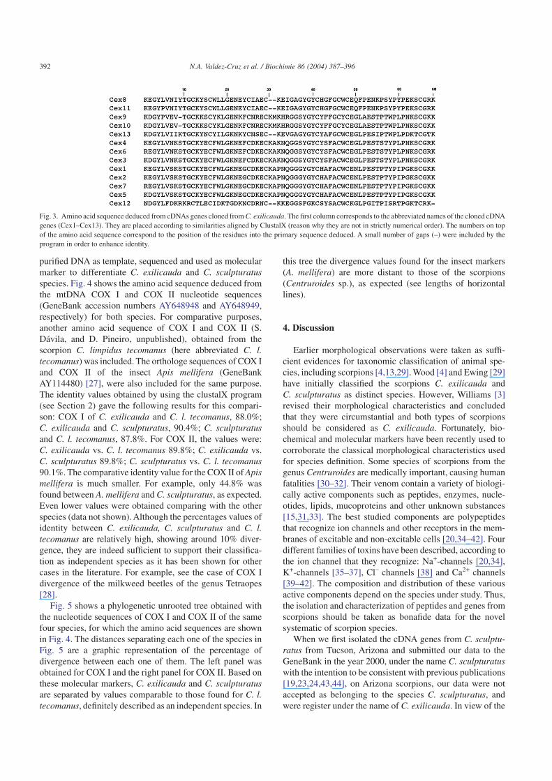
purified DNA as template, sequenced and used as molecularmarker to differentiate C. exilicauda and C. sculpturatusspecies. Fig. 4 shows the amino acid sequence deduced fromthe mtDNA COX I and COX II nucleotide sequences(GeneBank accession numbers AY648948 and AY648949,respectively) for both species. For comparative purposes,another amino acid sequence of COX I and COX II (S.Dávila, and D. Pineiro, unpublished), obtained from thescorpion C. limpidus tecomanus (here abbreviated C. l.tecomanus) was included. The orthologe sequences of COX Iand COX II of the insect Apis mellifera (GeneBankAY114480) [27], were also included for the same purpose.The identity values obtained by using the clustalX program(see Section 2) gave the following results for this compari-son: COX I of C. exilicauda and C. l. tecomanus, 88.0%;C. exilicauda and C. sculpturatus, 90.4%; C. sculpturatusand C. l. tecomanus, 87.8%. For COX II, the values were:C. exilicauda vs. C. l. tecomanus 89.8%; C. exilicauda vs.C. sculpturatus 89.8%; C. sculpturatus vs. C. l. tecomanus90.1%. The comparative identity value for the COX II of Apismellifera is much smaller. For example, only 44.8% wasfound between A. mellifera and C. sculpturatus, as expected.Even lower values were obtained comparing with the otherspecies (data not shown). Although the percentages values ofidentity between C. exilicauda, C. sculpturatus and C. l.tecomanus are relatively high, showing around 10% diver-gence, they are indeed sufficient to support their classifica-tion as independent species as it has been shown for othercases in the literature. For example, see the case of COX Idivergence of the milkweed beetles of the genus Tetraopes[28].
Fig. 5 shows a phylogenetic unrooted tree obtained withthe nucleotide sequences of COX I and COX II of the samefour species, for which the amino acid sequences are shownin Fig. 4. The distances separating each one of the species inFig. 5 are a graphic representation of the percentage ofdivergence between each one of them. The left panel wasobtained for COX I and the right panel for COX II. Based onthese molecular markers, C. exilicauda and C. sculpturatusare separated by values comparable to those found for C. l.tecomanus, definitely described as an independent species. In
this tree the divergence values found for the insect markers(A. mellifera) are more distant to those of the scorpions(Centruroides sp.), as expected (see lengths of horizontallines).
4. Discussion
Earlier morphological observations were taken as suffi-cient evidences for taxonomic classification of animal spe-cies, including scorpions [4,13,29]. Wood [4] and Ewing [29]have initially classified the scorpions C. exilicauda andC. sculpturatus as distinct species. However, Williams [3]revised their morphological characteristics and concludedthat they were circumstantial and both types of scorpionsshould be considered as C. exilicauda. Fortunately, bio-chemical and molecular markers have been recently used tocorroborate the classical morphological characteristics usedfor species definition. Some species of scorpions from thegenus Centruroides are medically important, causing humanfatalities [30–32]. Their venom contain a variety of biologi-cally active components such as peptides, enzymes, nucle-otides, lipids, mucoproteins and other unknown substances[15,31,33]. The best studied components are polypeptidesthat recognize ion channels and other receptors in the mem-branes of excitable and non-excitable cells [20,34–42]. Fourdifferent families of toxins have been described, according tothe ion channel that they recognize: Na+-channels [20,34],K+-channels [35–37], Cl– channels [38] and Ca2+ channels[39–42]. The composition and distribution of these variousactive components depend on the species under study. Thus,the isolation and characterization of peptides and genes fromscorpions should be taken as bonafide data for the novelsystematic of scorpion species.
When we first isolated the cDNA genes from C. sculptu-ratus from Tucson, Arizona and submitted our data to theGeneBank in the year 2000, under the name C. sculpturatuswith the intention to be consistent with previous publications[19,23,24,43,44], on Arizona scorpions, our data were notaccepted as belonging to the species C. sculpturatus, andwere register under the name of C. exilicauda. In view of the
Fig. 3. Amino acid sequence deduced from cDNAs genes cloned from C. exilicauda. The first column corresponds to the abbreviated names of the cloned cDNAgenes (Cex1–Cex13). They are placed according to similarities aligned by ClustalX (reason why they are not in strictly numerical order). The numbers on topof the amino acid sequence correspond to the position of the residues into the primary sequence deduced. A small number of gaps (–) were included by theprogram in order to enhance identity.
392 N.A. Valdez-Cruz et al. / Biochimie 86 (2004) 387–396
results presented here and those earlier published by Coronaet al. [8], the information presently available in theGeneBank corresponding to cloned cDNA genes of C. exili-cauda must be revised. It is clear from the Section 3, thatbiochemical, genetic and evolutionary differences betweenthese two species of scorpions justify the original classifica-tion of Wood [4] and Ewing [29], as different species.
4.1. Lethality testing
The results showed that the toxicity of the venom fromC. sculpturatus is not significantly different from that re-ported by Stahnke [13] and Pete et al. [14]. Our data compare
quite well with those obtained with C. l. limpidus, anotherscorpion dangerous to humans [16]. These authors foundvariation on the value of LD50 (50% lethal doses) for differ-ent strains of mice. Depending on the strain of mice the LD50can vary from 0.61 (strain SSA white) to 3.31 mg/kg(BALB/k, white). Most of the other strains tested by Alagonet al. [16] resulted in LD50 close to 1–2 mg/kg mouse weight.It is also important to note that our data do not reflect exactdeterminations of LD50 (again, to avoid usage of many ani-mals) but is a good indication of the toxic potency of bothvenoms. The results of the lethality tests using the venomfrom C. exilicauda from Baja California are significantlydifferent. Furthermore, these data confirm medical observa-
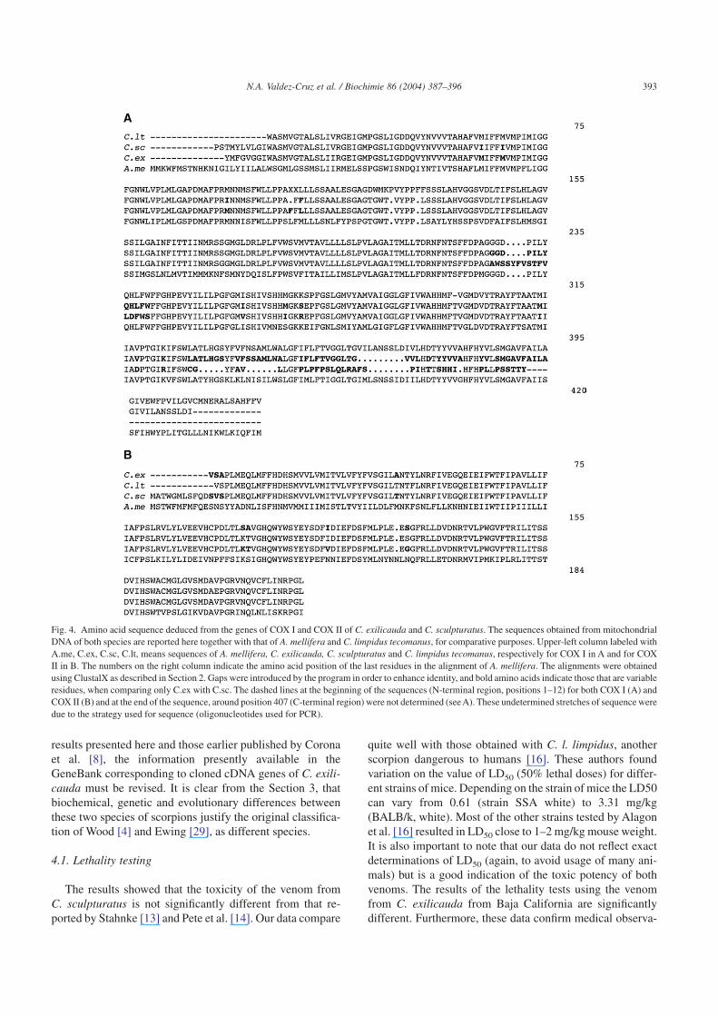
Fig. 4. Amino acid sequence deduced from the genes of COX I and COX II of C. exilicauda and C. sculpturatus. The sequences obtained from mitochondrialDNA of both species are reported here together with that of A. mellifera and C. limpidus tecomanus, for comparative purposes. Upper-left column labeled withA.me, C.ex, C.sc, C.lt, means sequences of A. mellifera, C. exilicauda, C. sculpturatus and C. limpidus tecomanus, respectively for COX I in A and for COXII in B. The numbers on the right column indicate the amino acid position of the last residues in the alignment of A. mellifera. The alignments were obtainedusing ClustalX as described in Section 2. Gaps were introduced by the program in order to enhance identity, and bold amino acids indicate those that are variableresidues, when comparing only C.ex with C.sc. The dashed lines at the beginning of the sequences (N-terminal region, positions 1–12) for both COX I (A) andCOX II (B) and at the end of the sequence, around position 407 (C-terminal region) were not determined (see A). These undetermined stretches of sequence weredue to the strategy used for sequence (oligonucleotides used for PCR).
393N.A. Valdez-Cruz et al. / Biochimie 86 (2004) 387–396

tions of human accidents occurred with C. sculpturatus ofArizona vs. C. exilicauda of Baja California. The first onehas a definitely medical importance [28,45], confirmed byunpublished observations by one of the co-authors of thiswork (L.B.), from the Poison Center in Arizona, whereas thesecond one from Baja California represents no risk for hu-man (personal experience of the first author of this commu-nication). This fact was additionally confirmed by reviewingthe medical records, at the National Institute of PublicHealth, in Cuernavaca, Mexico. This Institute has a completestatistical record of scorpion intoxication cases in the country(Mexico) and there is no record of fatal accidents in BajaCalifornia, due to scorpions (data not shown). Thus, thevenom from C. sculpturatus is dangerous to humans, but thevenom of C. exilicauda is not.
4.2. Electrophysiological characterization
Using MaxiK channels, expressed in oocytes, it was pos-sible to show the existence of marked physiological differ-ences, when both venoms were comparatively assayed, inthis specific ion-channel. It is worth mentioning that block-ade of MaxiK channel by some of these toxins do not neces-sarily causes toxicity symptoms on experimental animals,assayed in vivo. Charybdotoxin was the first scorpion toxinspecific for MaxiK described [21]. Injection of charybdot-oxin in experimental mice does not cause observable intoxi-cation symptoms (C. Miller, personal communication). Thisresult does not contradict our data. Although C. exilicaudalethality tests (see Table 1) show to be less toxic than that ofC. sculpturatus, it is the one that have an effective componentfor the blockade of the MaxiK (Fig. 2). The clinical manifes-tations found when injecting venom from C. sculpturatussuggest that the toxic effect seeing in vivo, is probably due topeptides that affect other ion-channels, as it is certainlyknown for the Na+-channel specific toxins [20].
4.3. cDNA cloning
If we compare the amino acid sequence of toxins directlydetermined by Edman degradation with those deduced from
the cDNA clones, it might be possible to find certain degreeof identity, even between different species of scorpions.When the full amino acid sequences of all these peptidesbecome available it would be not surprising if some peptidesfrom both species turn out to be identical. This was found forthe case of butantoxin, a K+-channel specific toxin present inidentical form in the venom of four different species ofscorpions of the genus Tityus: T. serrulatus, T. bahiensis, T.stigmurus [46] and T. trivittatus [47]. The diversity andvariations of the cDNA gene sequences found when compar-ing C. exilicauda from Baja California with C. sculpturatusfrom Arizona are a reasonable indication that they belong totwo distinct species of scorpions.
4.4. Phylogenetic analysis of peptide toxinsand cytochrome oxidases
A phylogenetic unrooted tree, using the neighbour-joiningmethod [12] was generated with the amino acid sequencescoded by all the cDNA genes cloned from C. exilicauda,whose sequences corresponded to peptides similar to theNa+-channel specific toxins (Fig. 6, left). Similarly, for com-parative purposes, the deduced amino acid sequences from16 genes cloned from the scorpion C. sculpturatus [8] wereanalysed together with the 13 peptides of C. exilicauda(Fig. 6, right).
The left panel in Fig. 6 shows that the peptides obtainedfrom C. exilicauda have important similarities, althoughsituated in at least two separated branches. When this analy-sis is performed with all the genes cloned from both species(right panel), it is clear that most of C. exilicauda sequencesare clustered around the middle of the graphic, whereas in theupper and lower extremes are those from C. sculpturatus,except for Cex12 and Cex13 that commonly show short
Fig. 5. Unrooted phylogenetic tree of COX I and COX II from C. exilicaudaand C. sculpturatus. Left panel: Multiple alignment of nucleotidic sequen-ces of COX I from C. sculpturatus, C. exilicauda, C. l. tecomanus and A.mellifera. This phylogram shows the distances of divergence for the foursequences, where the scale (0.1) means the probability of one changeoccurrence in 1000 nucleotides. Right panel: The same results for COX II.
Fig. 6. Unrooted phylogenetic three of toxins from C. exilicauda andC. sculpturatus. Left panel: Multiple alignments of 13 amino acid sequencesfrom C. exilicauda were used to calculate a matrix with genetic distances(slanted cladogram), using the neighbour-joining algorithm mentioned inSection 2. The abbreviations (Cex with numbers) correspond to the 13 clo-ned cDNAs. Right panel: Multiple alignments of 29 amino acid sequences,of which 13 are from C. exilicauda (this work) and 16 from C. sculpturatus[8], using the same software as for the left picture. The results are shown ina rectangular cladogram. Abbreviations on the right column correspondeither to C. exilicauda (Cex) or to C. sculpturatus (abbreviated CsE, plus aletter or number according to Corona et al. [8].
394 N.A. Valdez-Cruz et al. / Biochimie 86 (2004) 387–396

phylogenetic distances. These results point out to the samedirection as those previously discussed above, when compar-ing the N-terminal amino acid sequences of the peptidespurified from both species. There is no doubt that both spe-cies have similar genes coding for similar peptides, althoughnot identical. The genes from a given species are more relatedamong themselves than to the genes of the other species,when simultaneously compared. These results again, suggestthat we are dealing here with two distinct species of scorpi-ons.
5. Conclusion
This communication reports data on mice toxicity tests,chromatographic separations, cloned genes and electrophysi-ological characteristics of venom from two scorpions, whosetaxonomic classification biochemical, genetic and electro-physiolgical data are under discussion. One of the scorpionsis endemic on the area of the California Peninsula, whereasthe other is from the Continental region. Concerning thissubject, recent data published by Riddle et al. [48], on aphylogeographic population structure across a suite of12 mammalian, avian, amphibian, and reptilian species andsubspecies of the Baja California desert area, give evidencethat this Peninsular Desert can no longer be considered asubset of the Sonoran Desert. Thus, the presence of C. exili-cauda in Baja California and that of C. sculpturatus inArizona (and Sonoran desert), are also not consistent with theproposal that both species are synonyms.
For these reason we can conclude that C. exilicauda foundin Baja California is a bonafide different species than that ofC. sculpturatus found in the Arizona or Sonoran geographi-cal area. Similar studies are expected to be performed forclarification of the existence of a monophylogenetic lineageamong the C. exilicauda scorpions found along the Peninsula(North area of Baja California vs. South Baja California).
Acknowledgments
The authors acknowledge Dr. Jude McNally from theUniversity of Arizona for collecting C. sculpturatus scorpi-ons, and Dr. Ligia Toro from the University of California(Los Angeles, CA, USA) for providing us with the programto analyse our electrophysiological data. This work was par-tially supported by grants Z-005 from the CONACyT (Na-tional Council of Science and Technology—Mexican Gov-ernment), IN206003 of Dirección General de Asuntos delPersonal Académico of UNAM and Laboratorios SilanesS.A. de C.V. to LDP. NAVC is a recipient of a Ph.D. programscholarship from the CONACyT.
References
[1] V. Fet, W.D. Sissom, G. Lowe, M.E. Braunwalder, Catalog of theScorpions of the World (1758–1998), The New York EntomologicalSociety, New York, 2000.
[2] P. Brownell, G. Polis, Scorpion Biology and Research, Oxford Uni-versity Press Inc. New York, 2001.
[3] S.C. Williams, Scorpions of Baja California, Mexico and adjacentislands, Occ. Papers Cal. Acad. Sci. 135 (1980) 1–127.
[4] H.C. Wood, Descriptions of new species of NorthAmerica, Proc. Natl.Acad. Sci. (1863) 107–112.
[5] C.V.F. Batista, F. Gómez-Lagunas, S. Lucas, L.D. Possani, Tc1, fromTityus cambridgei, is the first member of a new sub-family of scorpiontoxin that blocks K+-channels, FEBS Lett. 486 (2000) 117–120.
[6] J. Garcia-Valdes, F.Z. Zamudio, L. Toro, L.D. Possani, Slotoxin,alphaKTx1.11, a new scorpion peptide blocker of MaxiK channelsthat differentiates between alpha and alpha + beta (beta1 or beta4)complexes, FEBS Lett. 505 (2001) 369–373 Correction 507 (1) 122.
[7] J.M. Chirwing, A.E. Przybyla, R.J. McDonald, W.J. Rutter, Isolationof biologically active ribonucleic acid from source enriched in ribo-nuclease, Biochemistry 18 (1979) 5294–5299.
[8] M. Corona, N.A. Valdez-Cruz, M. Merino, L.D. Possani, Genes andpeptides from the scorpion Centruroides sculpturatus Ewing, thatrecognize Na+-channels, Toxicon 39 (2001) 1893–1898.
[9] H.G. Hall, D.R. Smith, Distinguishing African and European honey-bee matrilines using amplified mitochondrial DNA, Proc. Natl. Acad.Sci. USA 88 (1991) 4548–4552.
[10] J. Sambrook, E.F. Fritsch, T. Maniatis, Molecular Cloning a Labora-tory Manual, Cold Spring Harbor Laboratory Press, New York, USA,1989.
[11] J. Felsenstein, PHYLIP (Phylogeny Interference Package), Version3.57c, Department of Genetics, University of Washington, Seattle,USA, 1995.
[12] J. Zhang, M. Nei, Accuracies of ancestral amino acid sequencesinferred by the parsimony likelihood, and distance methods, J. Mol.Evol. 4 (1997) S139–S146.
[13] H.L. Stahnke, Arizona lethal scorpion, Pap. Ariz. Med. 29 (1972)490–493.
[14] M.J. Pete, M.J. Conlon, R.F. Murphy, Isolation and primary structureof a potent toxin from the venom of the scorpion Centruroides sculp-turatus Ewing, Int. J. Peptide Protein Res. 40 (1992) 582–586.
[15] L.D. Possani, Structure of scorpion toxin, in: in: A.T. Tu (Ed.),Handbook of Natural Toxins, 2, Marcel Dekker Inc. New York, 1984,pp.513–550.
[16] A.C. Alagon, H.S. Guzmán, B.M. Martin, A.N. Ramírez, E. Carbone,L.D. Possani, Isolation and characterization of two toxins from theMexican scorpion Centruroides limpidus limpidus Karsch, Comp.Biochem. Physiol. 89B (1988) 153–161.
[17] H.H. Valdivia, B.M. Martin, A.N. Ramírez, P.L. Fletcher, L.D. Pos-sani, Isolation and pharmacological characterization of four novelNa+-channel-blocking toxins from the scorpion Centruroides noxiusHoffmann, J. Biochem. (Japan) 116 (1994) 1383–1391.
[18] N.A. Valdez-Cruz, Morphological analysis of the scorpion Centruroi-des exilicauda and characterization of some of its toxins (Análisismorfológico del alacrán Centruroides exilicauda y caracterización dealgunas de sus toxinas), University thesis presented at the Autono-mous University of Baja California, School of Chemical Sciences,Tijuana, Baja California, México, 1998 (in Spanish).
[19] H. Meves, N. Rubly, D.D. Watt, Effect of toxins isolated from thevenom of the scorpion Centruroides sculpturatus on the Na currentsof the Node of Ranvier, Pflügers. Arch. 393 (1982) 56–62.
[20] L.D. Possani, B. Becerril, M. Delepierre, J. Tytgat, Scorpion toxinsspecific for Na+-channels, Eur. J. Biochem. 264 (1999) 287–300.
[21] C. Miller, The charybdotoxin family of channel-blocking peptides,Neuron 15 (1995) 5–10.
395N.A. Valdez-Cruz et al. / Biochimie 86 (2004) 387–396
miguelcv33
Highlight

[22] B. Becerril, A. Vázquez, C. García, M. Corona, F. Bolivar, L.D. Pos-sani, Cloning and characterization of cDNAs that code for Na+-channel-blocking toxins of the scorpion Centruroides noxius Hoff-mann, GENE 128 (1993) 165–171.
[23] D.R. Babin, D.D. Watt, S.M. Goos, R.V. Mlejnek, Amino acidsequences of neurotoxic protein variants from the venom of Centru-roides sculpturatus Ewing, Arch. Biochem. Biophys. 164 (1974)694–706.
[24] D.R. Babin, D.D. Watt, S.M. Goos, R.V. Mlejnek, Amino acidsequence of neurotoxin I from Centruroides sculpturatus Ewing,Arch. Biochem. Biophys. 166 (1975) 125–134.
[25] F.H. Sheldon, C.E. Jones, K.G. McCracken, Relative patterns andrates of evolution in Heron nuclear and mitochondrial DNA, Mol.Biol. Evol. 17 (2000) 437–450.
[26] D.D. Espiritu, M. Watkins, V. Dia-Monje, G.E. Cartier, L.J. Cruz,B.M. Olivera, Venoms cone snails: molecular phylogeny and thegeneration of toxin diversity, Toxicon 39 (2001) 1899–1916.
[27] A. Marino, B. Mantovani, E. Carpana, A.G. Sabatini, M. Lodesani,ND2 and CO1 mitochondrial genes in Apis mellifera L.: a molecularapproach to Mediterranean populations monitoring, BiologiaEvoluzionistica Sperimentale, University of Bologna, via Selmi 3,Bologna 40126, Italy, 2002.
[28] B.D. Farrell, Evolutionary Assembly of the milkweed fauna: cyto-chrome oxidase I and the age of Tetraopes beetles, Mol. Phylogenet.Evol. 18 (2001) 467–478.
[29] H.E. Ewing, The scorpion of the western part of the United States withnotes on those occurring in Northern Mexico, Proc. US NationalMuseum 73 (9) (1928) 1–24.
[30] S.C. Curry, M.B. Vance, P.J. Ryan, D.B. Kunkel, W.T. Northey,Envenomation by the scorpion Centruroides sculpturatus, J. Toxicol.Clin. Toxicol. 21 (1983) 417–449.
[31] M. Dehesa-Davila, L.D. Possani, Scorpionism and serotherapy inMexico, Toxicon 32 (1994) 1015–1018.
[32] N. Osnaya-Romero, T.J. Medina-Hernández, S.S. Flores-Hernández,G. León-Rojas, Clinical symptoms observed in children envenomatedby scorpion stings, at the children’s hospital from the State of More-los, Mexico, Toxicon 39 (2001) 781–785.
[33] E. Zlotkin, F. Miranda, H. Rochat, Chemistry and pharmacology ofButhidae scorpion venoms, in: in: S. Bettini (Ed.), Handbook ofExperimental Pharmacology, 48, Springer Verlag, Berlin, 1978,pp.317–369.
[34] D. Gordon, P. Savarin, M. Gurevitz, S. Zinn-Justin, Functionalanatomy of scorpion toxins affecting sodium channels, J. Toxicol.Toxin Rev. 17 (1998) 131–159.
[35] C. Miller, E. Moczydlowski, R. Latorre, M. Phillips, Charybdotoxin, aprotein inhibitor of single Ca2+ activated K channels from mammalianskeletal muscle, Nature 213 (1985) 316–318.
[36] M.L. Garcia, M. Hanner, H.G. Knaus, R. Koch, W. Schmalhofer,R.S. Slaughter, G.J. Kaczorowski, Pharmacology of potassium chan-nels, Adv. Pharmacol. 39 (1997) 425–471.
[37] L.D. Possani, E. Merino, M. Corona, B. Becerril, Scorpion genes andpeptides specific for potassium channels: structure, function and evo-lution, in: in: A. Ménez (Ed.), Perspectives in Molecular Toxinology,John Wiley & Sons Ltd. New York, 2002, pp.201–214.
[38] J.A. De Bin, J.E. Maggio, G.R. Strichartz, Purification and character-ization of chlorotoxin, chloride channel ligand from the venom of thescorpions, Am. J. Physiol. Soc. 264 (1993) C361–C369.
[39] H.H. Valdivia, L.D. Possani, Peptide toxins as probes of ryanodinereceptor structure and function, Trends Cardiovasc. Med. 8 (1998)111–118.
[40] R.S. Chuang, H. Jaffe, L. Cribbs, E. Perez-Reyes, K.J. Swartz, Inhi-bition of T-type voltage-gated calcium channels by a new scorpiontoxin, Nature Neurosci. 1 (8) (1998) 668–674.
[41] S.S. Sidach, I.M. Mintz, Kurtoxin, a gating modifier of neuronal high-and low-threshold Ca channels, J. Neurosci. 22 (2002) 2023–2034.
[42] T. Olamendi-Portugal, B.I. García, I. López-González, J. Van derWalt, K. Dyason, C. Ulens, J. Tytgat, R. Felix, A. Darszon, L.D. Pos-sani, Two new scorpion toxins target voltage-gated Ca2+ and Na+
channels, Biochem. Biophys. Res. Comun. 299 (2002) 562–568.[43] D.D. Watt, J.M. Simard, D.R. Babin, R.V. Mlejnek, Physiological
characterization of toxins isolated from scorpion venom, in: in:P. Rosenberg (Ed.), Toxins: Animal, Plant and Microbial, PergamonPress, New York, 1978, pp.647–660.
[44] J.C. Fortecilla-Camps, R.J. Almassy, F.L. Suddath, D.D. Watt,C.E. Bugg, Three dimensional structure of a protein from scorpionvenom: a new structural class of neurotoxins, Proc. Natl. Acad. Sci.USA 77 (1980) 6496–6500.
[45] F.E. Russell, M.B. Madon, Introduction of the scorpion C. exilicaudainto California and its public health significance, Toxicon 22 (1984)658–664.
[46] S.K. Holaday, B.M. Martin, P.L. Fletcher, N.R. Krishna, NMR-solution structure of butantoxin, Arch. Biochem. Biophys. 379 (2000)18–27.
[47] F.V. Coronas, A.R. Roodt, T. Olamendi-Portugal, F.Z. Zamudio,C.V.F. Batista, F. Gómez-Lagunas, L.D. Possani, Disulfide bridgesand blockage of Shaker B K+-channels by another butantoxin peptidepurified from the Argentinean scorpion Tytius trivittatus, Toxicon 41(2003) 173–179.
[48] B.R. Riddle, D.J. Hafner, L.F.Alexander, J.R. Jaeger, Cryptic variancein the historical assembly of a Baja California Peninsular Desert biota,Proc. Natl. Acad. Sci. USA 97 (2000) 14438–14443.
396 N.A. Valdez-Cruz et al. / Biochimie 86 (2004) 387–396