Behavioral effects of manganese injected in the rat substantia nigra are potentiated by dicumarol, a...
7
Behavioral effects of manganese injected in the rat substantia nigra are potentiated by dicumarol, a DT-diaphorase inhibitor Gabriela Dı ´az-Ve ´liz * , Sergio Mora, Patricia Go ´mez, Ma Teresa Dossi, Juan Montiel, Christian Arriagada, Francisco Aboitiz, Juan Segura-Aguilar Programa de Farmacologı ´a Molecular y Clı ´nica, Instituto de Ciencias Biolo ´gicas (ICBM), Facultad de Medicina, Universidad de Chile, P.O. Box 16038, Santiago 9, Chile Abstract The purpose of this study was to evaluate the contribution of DT-diaphorase inhibition to in vivo neurodegenerative effects of dopamine (DA) oxidation to the corresponding o-quinones. The neurotoxicity to nigrostriatal DA neurons was induced by injection of manganese pyrophosphate (Mn 3+ ) complex as a prooxidizing agent alone or together with the DT-diaphorase inhibitor dicumarol into the right rat substantia nigra. The behavioral effects were compared with those induced after selective lesions of dopaminergic neurons with 6- hydroxydopamine (6-OHDA). Intranigral injection of Mn 3+ and Mn 3+ plus dicumarol produced significant impairment in motor behavior compared with control animals. However, the effect seen in the Mn 3+ plus dicumarol injected group was significantly more severe than that observed in the Mn 3+ alone injected group. In motor activity and rearing behavior, the simultaneous injection of Mn 3+ plus dicumarol produced a 6-OHDA-like impairment. Similar effects were observed in the acquisition of a conditioned avoidance response (CAR). Dicumarol significantly impaired avoidance conditioning although without affecting the motor behavior. The behavioral effects were correlated to the extent of striatal tyrosine hydroxylase (TH)-positive fiber loss. Rats receiving unilateral intranigral Mn 3+ and Mn 3+ plus dicumarol injections exhibited a significant reduction in nigrostriatal TH-positive fiber density in medial forebrain bundle compared with the contralateral noninjected side. In conclusion, this study provides evidence that the neurotoxicity of Mn 3+ in vivo is potentiated by DT- diaphorase inhibition, suggesting that this enzyme could play a neuroprotective role in the nigrostriatal DA systems. Keywords: Manganese; DT-diaphorase; Dopamine; Neurodegeneration; Parkinsonism 1. Introduction Although it is generally accepted that free radicals are involved in the neurodegeneration of the nigrostriatal dopa- mine (DA) system observed in Parkinson’s disease, the exact mechanism of neurodegeneration in vivo is still unknown. Much attention has focused on the fact that oxidation of DA results in the formation of cytotoxic compounds, which can cause massive brain damage if they are allowed to accumulate. However, the rate at which oxygen oxidizes DA at neutral pH is very low (Graham et al., 1978) and probably does not produce any significant amount of reactive oxygen intermediates. Manganese in the Mn 3+ state is a potent oxidizing agent and can accelerate the oxidation of DA to its o-quinone (Barbeau, 1984; Archibald and Tyree, 1987), which instantaneously cyclizes to form aminochrome (Segura-Aguilar and Lind, 1989; Shen and Dryhurst, 1998; Brenneman et al., 1999; Dorman et al., 2000; Lee, 2000). This observation may explain the drastic decrease in the level of DA because this reaction appears to be irreversible (Segura-Aguilar and Lind, 1989). Previously, it was demonstrated that aminochrome resulting from oxidation by DA with Mn 3+ was toxic in a mouse- derived neuronal cell line (CNh) (Arriagada et al., 2000) and also induces a significant behavioral impairment in vivo (Dı ´az-Ve ´liz et al., 2002). The one-electron reduction of aminochrome to leukoaminochrome o-semiquinone radicals can be one possible source of reactive species (Stokes et al., 1999; Segura-Aguilar et al., 2001; Smythies, 2002). Leu- koaminochrome o-semiquinone radical is a very reactive metabolite (Segura-Aguilar et al., 1998) that autoxidizes in the presence of oxygen or transition metal ions like man- ganese, copper or iron (Segura-Aguilar and Lind, 1989; * Corresponding author. Fax: +56-2-2741628. E-mail address: [email protected] (G. Dı ´az-Ve ´liz).
-
Upload
independent -
Category
Documents
-
view
6 -
download
0
Transcript of Behavioral effects of manganese injected in the rat substantia nigra are potentiated by dicumarol, a...

Behavioral effects of manganese injected in the rat substantia nigra are
potentiated by dicumarol, a DT-diaphorase inhibitor
Gabriela Dıaz-Veliz*, Sergio Mora, Patricia Gomez, Ma Teresa Dossi, Juan Montiel,Christian Arriagada, Francisco Aboitiz, Juan Segura-Aguilar
Programa de Farmacologıa Molecular y Clınica, Instituto de Ciencias Biologicas (ICBM), Facultad de Medicina, Universidad de Chile,
P.O. Box 16038, Santiago 9, Chile
www.elsevier.com/locate/pharmbiochembeh
Pharmacology, Biochemistry and Behavior 77 (2004) 245–251
Abstract
The purpose of this study was to evaluate the contribution of DT-diaphorase inhibition to in vivo neurodegenerative effects of dopamine
(DA) oxidation to the corresponding o-quinones. The neurotoxicity to nigrostriatal DA neurons was induced by injection of manganese
pyrophosphate (Mn3 + ) complex as a prooxidizing agent alone or together with the DT-diaphorase inhibitor dicumarol into the right rat
substantia nigra. The behavioral effects were compared with those induced after selective lesions of dopaminergic neurons with 6-
hydroxydopamine (6-OHDA). Intranigral injection of Mn3 + and Mn3 + plus dicumarol produced significant impairment in motor behavior
compared with control animals. However, the effect seen in the Mn3 + plus dicumarol injected group was significantly more severe than that
observed in the Mn3 + alone injected group. In motor activity and rearing behavior, the simultaneous injection of Mn3 + plus dicumarol
produced a 6-OHDA-like impairment. Similar effects were observed in the acquisition of a conditioned avoidance response (CAR).
Dicumarol significantly impaired avoidance conditioning although without affecting the motor behavior. The behavioral effects were
correlated to the extent of striatal tyrosine hydroxylase (TH)-positive fiber loss. Rats receiving unilateral intranigral Mn3 + and Mn3 + plus
dicumarol injections exhibited a significant reduction in nigrostriatal TH-positive fiber density in medial forebrain bundle compared with the
contralateral noninjected side. In conclusion, this study provides evidence that the neurotoxicity of Mn3 + in vivo is potentiated by DT-
diaphorase inhibition, suggesting that this enzyme could play a neuroprotective role in the nigrostriatal DA systems.
Keywords: Manganese; DT-diaphorase; Dopamine; Neurodegeneration; Parkinsonism
1. Introduction
Although it is generally accepted that free radicals are
involved in the neurodegeneration of the nigrostriatal dopa-
mine (DA) system observed in Parkinson’s disease, the
Archibald and Tyree, 1987), which instantaneously cyclizes
to form aminochrome (Segura-Aguilar and Lind, 1989;
Shen and Dryhurst, 1998; Brenneman et al., 1999; Dorman
et al., 2000; Lee, 2000). This observation may explain the
drastic decrease in the level of DA because this reaction
appears to be irreversible (Segura-Aguilar and Lind, 1989).
exact mechanism of neurodegeneration in vivo is stillunknown. Much attention has focused on the fact that Previously, it was demonstrated that aminochrome resulting
oxidation of DA results in the formation of cytotoxic
compounds, which can cause massive brain damage if they
are allowed to accumulate. However, the rate at which
oxygen oxidizes DA at neutral pH is very low (Graham et
from oxidation by DA with Mn3 + was toxic in a mouse-
derived neuronal cell line (CNh) (Arriagada et al., 2000) and
also induces a significant behavioral impairment in vivo
(Dıaz-Veliz et al., 2002). The one-electron reduction of
al., 1978) and probably does not produce any significant
amount of reactive oxygen intermediates. Manganese in the
Mn3 + state is a potent oxidizing agent and can accelerate
the oxidation of DA to its o-quinone (Barbeau, 1984;
* Corresponding author. Fax: +56-2-2741628.
E-mail address: [email protected] (G. Dıaz-Veliz).
aminochrome to leukoaminochrome o-semiquinone radicals
can be one possible source of reactive species (Stokes et al.,
1999; Segura-Aguilar et al., 2001; Smythies, 2002). Leu-
koaminochrome o-semiquinone radical is a very reactive
metabolite (Segura-Aguilar et al., 1998) that autoxidizes in
the presence of oxygen or transition metal ions like man-
ganese, copper or iron (Segura-Aguilar and Lind, 1989;
G. Dıaz-Veliz et al. / Pharmacology, Biochem246
Shen and Dryhurst, 1998; Paris et al., 2001), initiating a
redox cycling process (Baez et al., 1995). This aberrant one-
electron metabolism of aminochrome can be prevented by a
two-electron reduction of aminochrome to leukoamino-
chrome, catalyzed by DT-diaphorase (Segura-Aguilar and
Lind, 1989; Segura-Aguilar et al., 1998, 2001). There is
evidence that DT-diaphorase, an enzyme that in rat sub-
stantia nigra constitutes the 98% of the total quinone
reductase activity (Schultzberg et al., 1988), prevents ami-
nochrome one-electron reduction by reducing aminochrome
with two electrons to leukoaminochrome (Segura-Aguilar
and Lind, 1989). The selective inhibition of this enzyme
leads to an autoxidative cascade due to the ability of
leukoaminochrome o-semiquinone to induce redox cycling
(Baez et al., 1995). Thus, very low concentrations of amino-
chrome can produce a large amount of reactive oxygen
species (Segura-Aguilar et al., 2001). In vitro studies have
demonstrated that DT-diaphorase is inhibited by dicumarol
(Schultzberg et al., 1988; Segura-Aguilar and Lind, 1989;
Paris et al., 2001). Recently, we demonstrated that inhibition
of DT-diaphorase is a requirement for Mn3 + to produce a 6-
hydroxydopamine (6-OHDA)-like rotational behavior in
rats (Segura-Aguilar et al., 2002).
To evaluate the contribution of DT-diaphorase inhibition
to in vivo neurodegenerative effects of oxidation products of
DA, we injected into substantia nigra (1) Mn3 + as a general
prooxidizing agent to accelerate the oxidation of endoge-
nous DA and (2) dicumarol as a selective inhibitor of DT-
diaphorase. We evaluated the degeneration of the nigros-
triatal pathway through the expression of spontaneous motor
activity and avoidance conditioning, considering the influ-
ence of integrity of DA systems on these behaviors.
2. Methods
2.1. Animals
Fifty adult, male Sprague–Dawley rats, weighing 180–
220 g, were housed six per cage in a temperature-controlled
vivarium under a 12:12 light/dark cycle (lights on from
08:00 to 20:00 h) with free access to food and water. The
experimental protocols followed the Guide for Care and Use
of Laboratory Animals and were approved by the Faculty of
Medicine Committee. The rats were assigned to five exper-
imental groups injected with (1) Tris-HCl vehicle, (2)
dicumarol, (3) Mn3 + , (4) dicumarol plus Mn3 + and (5)
6-OHDA.
2.2. Drugs
The following compounds were purchased from Sigma
(St. Louis, MO): dicumarol (3,3V-methylene-bis-4-hydrox-
ycoumarin) and 6-OHDA hydrobromide. The Mn3 + pyro-
phosphate complex was prepared according to Archibald
and Fridovich (1982).
2.3. Intranigral injections
Rats were anesthetized with an intraperitoneal injection
of sodium pentobarbital (30 mg/kg) and placed in a David
Kopf stereotaxic frame. With the rat skull oriented accord-
ing to Paxinos and Watson (1986), 2 Al of one of the
following solutions were injected into the right substantia
nigra (coordinates relatives to bregma were AP=� 4.8,
L=� 1.8, V=� 8.2): dicumarol (0.7 Ag), Mn3 + (6 Ag),dicumarol plus Mn3 + and 6-OHDA (8 Ag). Each drug was
dissolved in 0.1 M of Tris-HCl (pH 7.9) and injected at a
rate of 1 Al/min during 2 min. Doses of Mn3 + and
dicumarol were selected based on previous studies
(Segura-Aguilar et al., 2002). 6-OHDA, a neurotoxin that
may produce selective DA denervation following intranigral
administration (Ungerstedt et al., 1974), was dissolved in
physiological saline containing 0.1% ascorbic acid. To
minimize the possibility of back flow, the needle was kept
in place for an additional minute on completion of the
injection. Control animals were injected with a similar
volume of Tris-HCl vehicle. Ten rats were assigned to each
of five experimental groups. Fourteen days after injection,
behavioral end points were evaluated.
2.4. Spontaneous motor activity
Spontaneous motor activity was evaluated as described
previously (Mora and Dıaz-Veliz, 1993). Each rat was
individually placed into a Plexiglas cage (30� 30� 30
cm). The floor of the cage was an activity platform
(Lafayette Instrument, Lafayette, IN) connected to an
electromechanical counter. To avoid the influence of dis-
turbing noises, the platform was placed into a soundproof
chamber and the observations were made through a closed
TV circuit. Spontaneous motor activity was recorded every
5 min during a 15-min period. Simultaneously, the number
of times each rat reared, the number of headshakes
and the time (s) spent in grooming behavior were also
recorded.
2.5. Active avoidance conditioning
Active avoidance conditioning was as described previ-
ously (Mora and Dıaz-Veliz, 1993). Immediately after the
spontaneous motor activity test, each rat was individually
placed in a two-way shuttle box (Lafayette Instrument)
composed of two stainless steel modular testing units. Each
unit was equipped with an 18-bar insulated shock grid floor,
two 28-V DC lights and a tone generator (Mallory Sonalert
2800 Hz; Lafayette Instrument). Electric shocks were pro-
vided to the grid floor by a Master shock supply (Lafayette
Instrument). The rats were trained over 50 trials after a 5-
min period of habituation. The trial consisted of the presen-
tation of a tone that after 5 s was overlapped with a 0.20-mA
footshock until the animal escaped to the opposite chamber,
with maximum shock duration of 10 s. A conditioned
istry and Behavior 77 (2004) 245–251
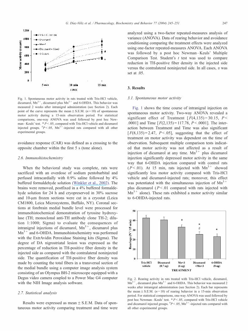
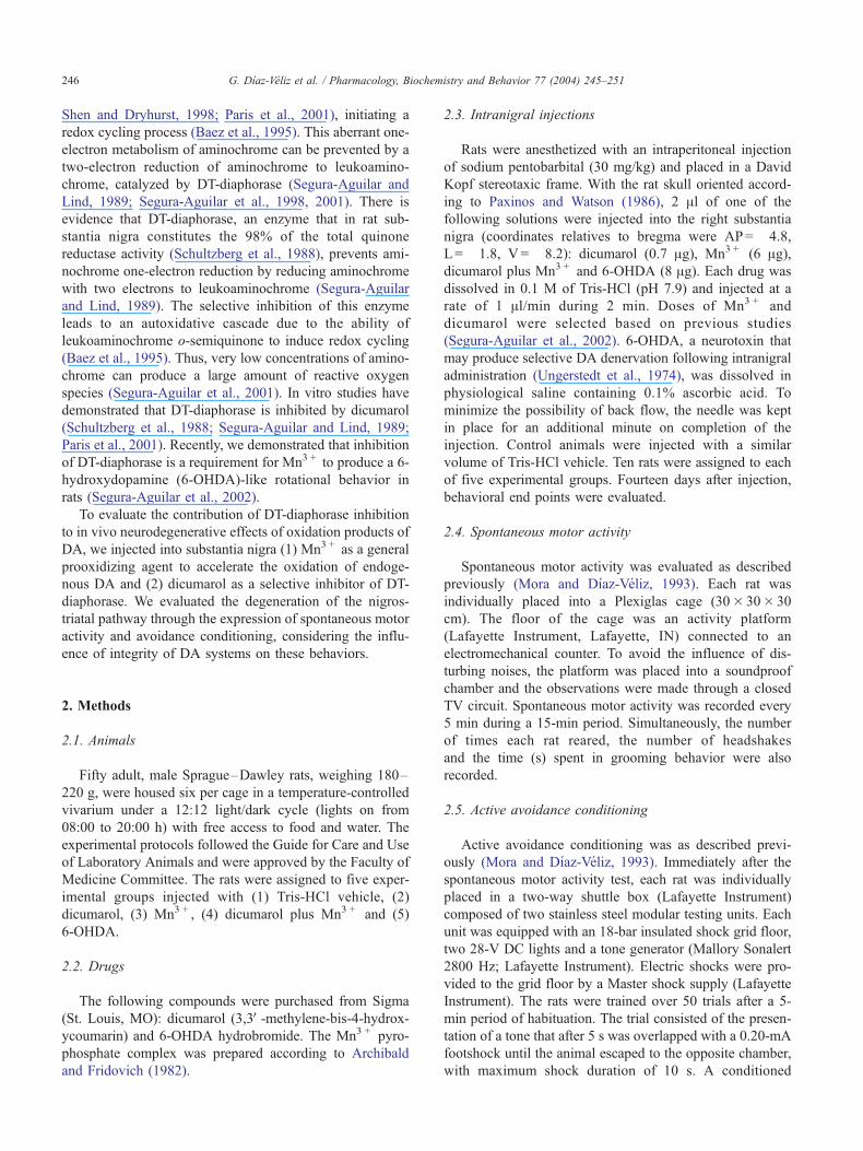
Fig. 1. Spontaneous motor activity in rats treated with Tris-HCl vehicle,
dicumarol, Mn3 + , dicumarol plus Mn3 + and 6-OHDA. This behavior was
measured 2 weeks after intranigral administration (see Section 2). Each
point of the curve represents the meanF S.E.M. (n= 10) of spontaneous
motor activity during a 15-min observation period. For statistical
comparisons, one-way ANOVA was used followed by post hoc New-
man–Keuls’ test. *P < .05, compared with Tris-HCl vehicle and dicumarol
injected groups. #P < .05, Mn3 + -injected rats compared with all other
experimental groups.
Fig. 2. Rearing activity in rats treated with Tris-HCl vehicle, dicumarol,
Mn3 + , dicumarol plus Mn3 + and 6-OHDA. This behavior was measured 2
weeks after intranigral administration (see Section 2). Each bar represents
the meanF S.E.M. (n= 10) of rearing behavior in a 15-min observation
period. For statistical comparisons, one-way ANOVAwas used followed by
post hoc Newman–Keuls’ test. *P < .05, compared with Tris-HCl vehicle
and dicumarol injected groups. #P< .05, Mn3 + -injected rats compared with
all other experimental groups.
G. Dıaz-Veliz et al. / Pharmacology, Biochemistry and Behavior 77 (2004) 245–251 247
avoidance response (CAR) was defined as a crossing to the
opposite chamber within the first 5 s (tone alone).
2.6. Immunohistochemistry
When the behavioral study was complete, rats were
sacrificed with an overdose of sodium pentobarbital and
perfused intracardially with 0.9% saline followed by 4%
buffered formaldehyde solution (Winkler et al., 2002). The
brains were removed, postfixed in a 4% buffered formalde-
hyde solution for 24 h and cryopreserved in 30% sucrose
and 10-Am frozen sections were cut in a cryostat (Leica
CM1800, Leica Microsystems, Buffalo, NY). Coronal sec-
tions at forebrain medial bundle level were processed for
immunohistochemical demonstration of tyrosine hydroxy-
lase (TH; monoclonal anti-TH antibody clone TH-2; dilu-
tion 1:1000; Sigma) to evaluate the consequences of
intranigral injections of dicumarol, Mn3 + , dicumarol plus
Mn3 + and 6-OHDA. Immunohistochemistry was performed
with the ExtrAvidin Peroxidase Staining kits (Sigma). The
degree of DA nigrostriatal lesion was expressed as the
percentage of reduction in TH-positive fiber density in the
injected side as compared with the contralateral noninjected
side. The quantification of TH-positive fiber density was
made by counting the total fibers in a transversal section of
the medial bundle using a computer image analysis system
consisting of an Olympus BH-2 microscope equipped with a
Degus video camera coupled to a Power Mac G4 computer
with the NIH Image analysis software.
2.7. Statistical analysis
Results were expressed as meanF S.E.M. Data of spon-
taneous motor activity comparing treatment and time were
analyzed using a two-factor repeated-measures analysis of
variance (ANOVA). Data of rearing behavior and avoidance
conditioning comparing the treatment effects were analyzed
using one-factor repeated-measures ANOVA. Each ANOVA
was followed by a post hoc Newman–Keuls’ Multiple
Comparison Test. Student’s t test was used to compare
reduction in TH-positive fiber density in the injected side
versus the contralateral noninjected side. In all cases, a was
set at .05.
3. Results
3.1. Spontaneous motor activity
Fig. 1 shows the time course of intranigral injection on
spontaneous motor activity. Two-way ANOVA revealed a
significant effect of Treatment [F(4,135) = 30.15, P <
.0001] and Time [F(2,135) = 117.78, P < .0001]. The inter-
action between Treatment and Time was also significant
[F(8,135) = 2.47, P < .05], suggesting that the effect of
treatment on motor activity was dependent on the time of
observation. Subsequent multiple comparison tests indicat-
ed that motor activity was not affected as a result of
injection of dicumarol at any time. Mn3 + plus dicumarol
injection significantly depressed motor activity in the same
way that 6-OHDA injection compared with control rats
(P < .01). At 15 min, rats injected with Mn3 + showed
significantly less motor activity compared with Tris-HCl
vehicle and dicumarol-injected rats; moreover, this effect
was potentiated with the simultaneous injection of Mn3 +
plus dicumarol (P < .01 compared with rats injected with
Mn3 + alone). These rats exhibited a motor activity similar
to 6-OHDA-injected rats.
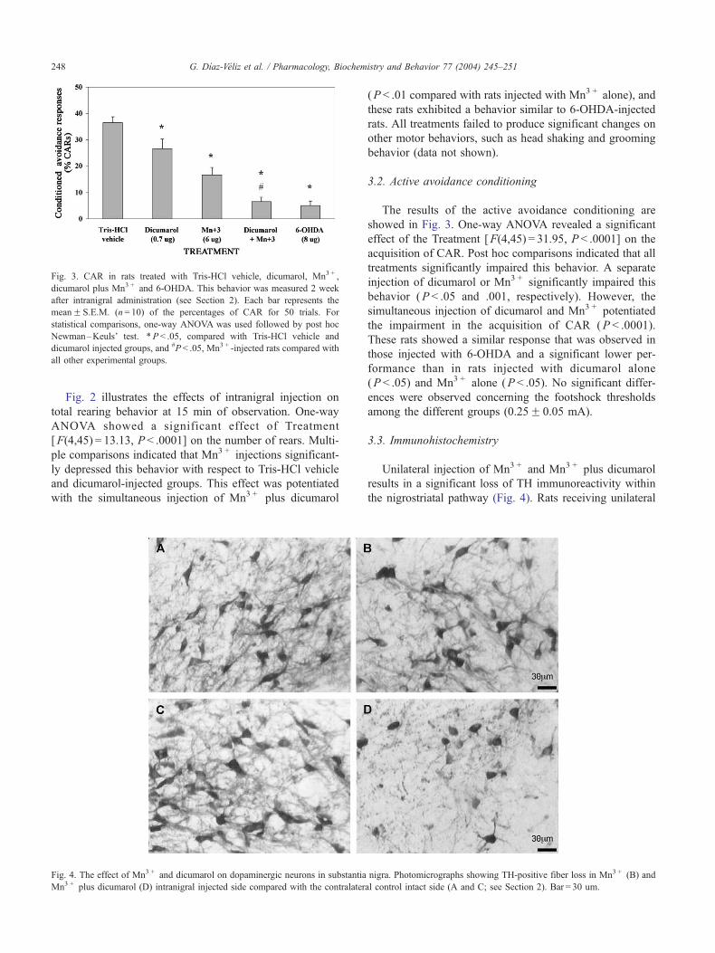
Fig. 3. CAR in rats treated with Tris-HCl vehicle, dicumarol, Mn3 + ,
dicumarol plus Mn3 + and 6-OHDA. This behavior was measured 2 week
after intranigral administration (see Section 2). Each bar represents the
meanF S.E.M. (n= 10) of the percentages of CAR for 50 trials. For
statistical comparisons, one-way ANOVA was used followed by post hoc
Newman–Keuls’ test. *P < .05, compared with Tris-HCl vehicle and
dicumarol injected groups, and #P < .05, Mn3 + -injected rats compared with
all other experimental groups.
G. Dıaz-Veliz et al. / Pharmacology, Biochemistry and Behavior 77 (2004) 245–251248
Fig. 2 illustrates the effects of intranigral injection on
total rearing behavior at 15 min of observation. One-way
ANOVA showed a significant effect of Treatment
[F(4,45) = 13.13, P < .0001] on the number of rears. Multi-
ple comparisons indicated that Mn3 + injections significant-
ly depressed this behavior with respect to Tris-HCl vehicle
and dicumarol-injected groups. This effect was potentiated
with the simultaneous injection of Mn3 + plus dicumarol
Fig. 4. The effect of Mn3 + and dicumarol on dopaminergic neurons in substantia
Mn3 + plus dicumarol (D) intranigral injected side compared with the contralater
(P < .01 compared with rats injected with Mn3 + alone), and
these rats exhibited a behavior similar to 6-OHDA-injected
rats. All treatments failed to produce significant changes on
other motor behaviors, such as head shaking and grooming
behavior (data not shown).
3.2. Active avoidance conditioning
The results of the active avoidance conditioning are
showed in Fig. 3. One-way ANOVA revealed a significant
effect of the Treatment [F(4,45) = 31.95, P < .0001] on the
acquisition of CAR. Post hoc comparisons indicated that all
treatments significantly impaired this behavior. A separate
injection of dicumarol or Mn3 + significantly impaired this
behavior (P < .05 and .001, respectively). However, the
simultaneous injection of dicumarol and Mn3 + potentiated
the impairment in the acquisition of CAR (P < .0001).
These rats showed a similar response that was observed in
those injected with 6-OHDA and a significant lower per-
formance than in rats injected with dicumarol alone
(P < .05) and Mn3 + alone (P < .05). No significant differ-
ences were observed concerning the footshock thresholds
among the different groups (0.25F 0.05 mA).
3.3. Immunohistochemistry
Unilateral injection of Mn3 + and Mn3 + plus dicumarol
results in a significant loss of TH immunoreactivity within
the nigrostriatal pathway (Fig. 4). Rats receiving unilateral
nigra. Photomicrographs showing TH-positive fiber loss in Mn3 + (B) and
al control intact side (A and C; see Section 2). Bar = 30 um.

Fig. 5. Photomicrograph of Nissl-stained section showing the dicumarol
injection site. Bar = 100 um. Signs of hemorrhage or inflammatory damage
around the site of dicumarol injection were not observed by histological
analysis using Nissl stain.
G. Dıaz-Veliz et al. / Pharmacology, Biochemistry and Behavior 77 (2004) 245–251 249
intranigral Mn3 + and Mn3 + plus dicumarol injections
exhibited a 61.9F 5.9% ( P < .005) and 78.2F 5.1%
(P < .005) reduction in nigrostriatal TH-positive fiber den-
sity compared with the contralateral noninjected side. The
simultaneous injection of dicumarol and Mn3 + potentiated
the loss of TH-positive fiber density compared with the
injection of Mn3 + alone (P < .05). The reduction of TH
immunoreactivity in rats receiving unilateral injection of
dicumarol and 6-OHDA was 22.7F 2.5% (P < .05) and
85.1F 4.7% (P < .005), respectively. Signs of hemorrhage
or inflammatory damage around the site of dicumarol
injection were not observed by histological analysis using
Nissl stain (Fig. 5).
4. Discussion
The results of the current study support the in vivo
neurotoxicity of Mn3 + and the contribution of DT-diapho-
rase inhibition by dicumarol to the behavioral consequences
of the nigrostriatal pathway degeneration. In fact, Mn3 +
injected into the rat substantia nigra decreased spontaneous
motor activity, rearing behavior and acquisition of an avoid-
ance response. These effects were potentiated by the con-
comitant administration of dicumarol; in this condition, they
were not significantly different to that induced by the
injection of 6-OHDA. The rationality for using Mn3 + as a
prooxidant agent was to accelerate the autoxidation cascade
involving oxidation of endogenous DA to aminochrome and
leukoaminochrome o-semiquinone radical. One-electron re-
duction of aminochrome to leukoaminochrome o-semiqui-
none radical is responsible for the generation of reactive
species involved in the neurodegenerative process (Segura-
Aguilar and Lind, 1989; Segura-Aguilar et al., 1998, 2001;
Kostrzewa and Segura-Aguilar, 2002). The brain is an
important target of attack for transition metal ions, such as
Mn3 + , due to its great catecholamine concentration and the
high speed of oxidative metabolism catalyzed by these
metals (Stokes et al., 1999). The present findings are
consistent with a recent study that provided evidence that
oxidation of DA to aminochrome appears to be an important
mediator of the behavioral consequences of oxidative dam-
age (Dıaz-Veliz et al., 2002). Our results confirm the sug-
gestion that the neurodegenerative events in dopaminergic
systems depend on overproduction of o-quinones in vivo.
The autoxidation of DA to aminochrome (Segura-Aguilar et
al., 1998, 2001) has been postulated to be a normal process
because DT-diaphorase prevent one-electron reduction of
aminochrome to leukoaminochrome o-semiquinone radical
(Baez et al., 1995; Segura-Aguilar et al., 1998; Paris et al.,
2001). DT-diaphorase is a flavoprotein found in central DA
neurons that prevents the formation of leukoaminochrome o-
semiquinone radicals by reducing aminochrome with two
electrons to leukoaminochrome (Segura-Aguilar and Lind,
1989). Previous in vitro studies have demonstrated that in the
rat brain DT-diaphorase is inhibited by dicumarol (Schultz-
berg et al., 1988; Segura-Aguilar and Lind, 1989; Paris et al.,
2001). The dense network of DT-diaphorase-immunoreac-
tive fibers in the striatum disappeared along with the dopa-
minergic innervation after 6-OHDA lesion (Schultzberg et
al., 1988). Leukoaminochrome o-semiquinone radical has
been reported to be responsible for neurotoxic effects of
aminochrome in dopaminergic RCSN-3 cells derived from
rat substantia nigra when DT-diaphorase was inhibited by
dicumarol (Paris et al., 2001). Recently, we informed that
Mn3 + administration together with dicumarol into the left
medial forebrain bundle produced a behavioral pattern char-
acterized by contralateral rotations when the rats were
stimulated with apomorphine. This effect was not observed
when Mn3 + was administered alone (Segura-Aguilar et al.,
2002). The rotational model developed by Ungerstedt et al.
(1974) is the most established animal assay to study the
unilateral nigrostriatal degeneration. In addition, in the
present study, we observed that the concomitant intranigral
administration of the DT-diaphorase inhibitor dicumarol
potentiates the neurobehavioral effects of Mn3 + , suggesting
that dicumarol enhanced the Mn3 + denervation of the
nigrostriatal DA system. These behavioral observations were
in concordance with the significant loss of TH immunore-
activity within the nigrostriatal pathway after Mn3 + and
Mn3 + plus dicumarol injection.
Both motor activity and associative learning are depen-
dent on the integrity of the DA nigrostriatal system. Then,
degeneration of the nigrostriatal dopaminergic system could
led to severe disruption of motor and associative behavior
(Dubois and Pillon, 1997; Schneider and Pope-Coleman,

Fig. 6. Possible mechanisms for toxic effects of intracerebral injection of Mn3 + together with dicumarol into substantia nigra.
G. Dıaz-Veliz et al. / Pharmacology, Biochemistry and Behavior 77 (2004) 245–251250
1995; Kulisevsky, 2000). Two-way avoidance is a type of
conditioning that results in associative learning. The rat
learns to avoid a signaled noxious stimulus (electrical shock)
by initiating a locomotor response for moving to another
compartment. Then, acquisition of avoidance responses
could be altered by changes in locomotor activity. However,
the present behavioral data suggest that the influence of
intranigral injection on avoidance response was not neces-
sarily a consequence of equivalent changes in spontaneous
motor activity. In 6-OHDA intranigral-injected rats, de-
pressed motor activity was clearly accompanied by a de-
crease in avoidance conditioning. Similar effects were shown
by Mn3 + plus dicumarol injection and with a lesser effect in
Mn3 + injection. However, in dicumarol-injected rats, both
behaviors were dissociated. In fact, the significant impair-
ment in avoidance behavior was not accompanied by any
change in motor activity. In the present study, immunohis-
tochemical assays demonstrate a correlation between percent
reduction in nigrostriatal TH-positive fiber density and
behavioral consequences. Intranigral injection of dicumarol
alone, which induced 22.7% reduction in TH-positive fiber
density, led to a significant inhibition of the avoidance
acquisition that was not accompanied with any change in
spontaneous motor activity. Increased damage of nigrostria-
tal DA fibers, as observed in Mn3 + and Mn3 + plus dicu-
marol intranigral-injected rats, depressed motor activity and
decreased avoidance conditioning in 6-OHDA-lesioned rats.
These findings led us to suggest that cognitive functions
could be more sensitive than motor performance to disrup-
tions in DA nigrostriatal neurotransmission. These data agree
with several experimental and clinical investigations of
Parkinson’s disease, which have shown that cognitive deficit
precedes motor impairment (Schneider and Pope-Coleman,
1995; Dubois and Pillon, 1997).
In conclusion, DT-diaphorase inhibition after dicumarol
injection is able to induce moderate behavioral changes in
the rat and also potentiates the neurotoxic effect of Mn3 +
when they are injected simultaneously into substantia nigra.
These results support the view of neurodegeneration as a
consequence of the oxidation of endogenous DA into
reactive cytotoxic leukoaminochrome o-semiquinone,
which is accelerated by metal transition ions like Mn3 +
(Fig. 6). DA is oxidized to aminochrome by reducing Mn3 +
to Mn2 + (Segura-Aguilar and Lind, 1989), which may react
with superoxide radicals to generate hydrogen peroxide and
Mn3 + (Archibald and Fridovich, 1982). The requirement of
the simultaneous injection of Mn3 + and inhibition of DT-
diaphorase by dicumarol for producing 6-OHDA-like
behaviors supports the proposed role of DT-diaphorase as
an antioxidant and neuroprotective enzyme of the dopami-
nergic systems. Thus, the degeneration of DA neurons in
parkinsonism may be caused by an imbalance between the
factors promoting the oxidation of DA and the availability
of neuroprotective defenses.
Acknowledgements
This work was supported by grant 1020672 from
Fondecyt, Chile.

G. Dıaz-Veliz et al. / Pharmacology, Biochemistry and Behavior 77 (2004) 245–251 251
References
Archibald FS, Fridovich I. The scavenging of superoxide radical by
manganous complexes: in vitro. Arch Biochem Biophys 1982;214:
452–63.
Archibald FS, Tyree C. Manganese poising and the attack of trivalent
manganese upon catecholamines. Arch Biochem Biophys 1987;256:
638–50.
Arriagada C, Dagnino-Subiabre A, Caviedes P, Martin Armero J, Caviedes
R, Segura-Aguilar J. Studies of aminochrome toxicity in a mouse de-
rived neuronal cell line: is this toxicity mediated via glutamate trans-
mission? Amino Acids 2000;18:363–73.
Baez S, Linderson Y, Segura-Aguilar J. Superoxide dismutase and catalase
enhance autoxidation during one-electron reduction of aminochrome by
NADPH-cytochrome P-450 reductase. Biochem Mol Med 1995;54:
12–8.
Barbeau A. Manganese and extrapyramidal disorders. Neurotoxicology
1984;5:13–36.
Brenneman KA, Cattley RC, Ali SF, Dorman DC. Manganese-induced
developmental neurotoxicity in the CD rat: is oxidative damage a mech-
anism of action? Neurotoxicology 1999;20:477–87.
Dıaz-Veliz G, Mora S, Dossi MT, Gomez P, Arriagada C, Montiel J, et al.
Behavioral effects of aminochrome and dopachrome injected in the rat
substantia nigra. Pharmacol Biochem Behav 2002;73:843–50.
Dorman DC, Struve MF, Vitarella D, Byerly FL, Goetz J, Miller R. Neuro-
toxicity of manganese chloride in neonatal and adult CD rats following
subchronic (21-days) high-dose oral exposure. J Appl Toxicol 2000;20:
179–87.
Dubois B, Pillon B. Cognitive deficit in Parkinson’s disease. J Neurol
1997;244:2–8.
Graham DG, Tiffany SM, Bell Jr WB, Guthnecht WF. Autooxidation ver-
sus covalent binding of quinones as the mechanism of toxicity of dop-
amine, 6-hydroxydopamine, and related compounds toward C1300
neuroblastoma cells in vitro. Mol Pharmacol 1978;14:644–53.
Kostrzewa R, Segura-Aguilar J. Neurotoxicological and neuroprotective
elements in Parkinson’s disease. Neurotoxicol Res 2002;4:83–6.
Kulisevsky J. Role of dopamine in learning and memory: implications for
the treatment of cognitive dysfunction in patients with Parkinson’s dis-
ease. Drugs Aging 2000;16:365–79.
Lee JW. Manganese intoxication. Arch Neurol 2000;57:597–9.
Mora S, Dıaz-Veliz G. Intracerebral administration of neuropeptides: an
assessment of behavioral change. In: Michael Conn P, editor. Paradigms
for the study of behavior. Methods in neurosciences, vol. 14. San Diego,
CA: Academic Press; 1993. p. 180–93.
Paris I, Dagnino-Subiabre A, Marcelain K, Bennett LB, Caviedes P, Cav-
iedes R, et al. Cooper neurotoxicity is dependent on dopamine-mediated
uptake and one-electron reduction of aminochrome in a rat substantia
nigra neuronal cell line. J Neurochem 2001;77:519–29.
Paxinos G, Watson C. The rat brain in stereotaxic coordinates. 2nd ed. San
Diego, CA: Academic Press; 1986.
Schneider JS, Pope-Coleman A. Cognitive deficits precede motor deficits in
a slowly progressing model of parkinsonism in the monkey. Neuro-
degeneration 1995;4:245–55.
Schultzberg M, Segura-Aguilar J, Lind C. Distribution of DT-diaphorase in
the rat brain: biochemical and immunohistochemical studies. Neuro-
science 1988;27:763–76.
Segura-Aguilar J, Lind C. On the mechanism of the Mn3 + -induced neuro-
toxicity of dopamine prevention of quinone-derived oxygen toxicity by
DT-diaphorase and superoxide dismutase. Chem Biol Interact 1989;72:
309–24.
Segura-Aguilar J, Metodiewa D, Welch CJ. Metabolic activation of dopa-
mine o-quinones to o-semiquinones by NADPH cytochrome P450 re-
ductase may play an important role in oxidative stress and apoptotic
effects. Biochim Biophys Acta 1998;1381:1–6.
Segura-Aguilar J, Metodiewa D, Baez S. The possible role of one-electron
reduction of aminochrome in the neurodegenerative process of the dop-
amine system. Neurotoxicol Res 2001;3:157–66.
Segura-Aguilar J, Dıaz-Veliz G, Mora S, Herrera-Marschitz M. Inhibi-
tion of DT-diaphorase is a requirement for Mn3 + to produce a 6-
OH-dopamine like rotational behaviour. Neurotoxicity Res 2002;4:
127–31.
Shen XM, Dryhurst G. Iron- and manganese-catalyzed autoxidation of
dopamine in the presence of L-cysteine: possible insights into iron-
and manganese-mediated dopaminergic neurotoxicity. Chem Res Tox-
icol 1998;11:824–37.
Smythies J. The biochemical basis of Parkinson’s disease: the role of cat-
echolamine o-quinones: a review-discussion. Neurotoxicol Res 2002;4:
77–81.
Stokes AH, Hastings TG, Vrana KE. Cytotoxic and genotoxic potential of
dopamine. J Neurosci Res 1999;55:659–65.
Ungerstedt U, Ljunggberg T, Sterg G. Behavioral, physiological and neuro-
chemical changes after 6-hydroxydopamine-induced degeneration of
the nigro-striatal dopamine neurone. Adv Neurol 1974;5:421–6.
Winkler C, Kirik D, Bjorklund A, Censi MA. L-DOPA-induced dyskinesia
in the intrastriatal 6-hydroxydopamine model of Parkinson’s disease:
relation to motor and cellular parameters of nigrostriatal function. Neu-
robiol Dis 2002;10:165–86.