Motor asymmetry and substantia nigra volume are related to spatial delayed response performance in...
18
Motor asymmetry and substantia nigra volume are related to spatial delayed response performance in Parkinson disease Erin R. Foster a,b,d , Kevin J. Black a,c,d,e , Jo Ann V. Antenor-Dorsey c , Joel S. Perlmutter c,d,e,f , and Tamara Hershey a,d,e,* a Department of Psychiatry, Washington University School of Medicine, St. Louis, MO, USA b Department of Occupational Therapy, Washington University School of Medicine, St. Louis, MO, USA c Department of Anatomy and Neurobiology, Washington University School of Medicine, St. Louis, MO, USA d Department of Neurology, Washington University School of Medicine, St. Louis, MO, USA e Department of Radiology, Washington University School of Medicine, St. Louis, MO, USA f Department of Physical Therapy, Washington University School of Medicine, St. Louis, MO, USA Abstract Studies suggest motor deficit asymmetry may help predict the pattern of cognitive impairment in individuals with Parkinson disease (PD). We tested this hypothesis using a highly validated and sensitive spatial memory task, spatial delayed response (SDR), and clinical and neuroimaging measures of PD asymmetry. We predicted SDR performance would be more impaired by PD- related changes in the right side of the brain than in the left. PD (n = 35) and control (n = 28) participants performed the SDR task. PD participants either had worse motor deficits on the right (RPD) or left (LPD) side of the body. Some participants also had magnetic resonance imaging for measurement of their substantia nigra (SN) volumes. The LPD group performed worse on the SDR task than the RPD and control groups. Right SN volume accounted for a unique and significant portion of the variance in SDR error, with smaller volume predicting poorer performance. In conclusion, left motor dysfunction and smaller right SN volume are associated with poorer spatial memory. Keywords Parkinson disease; Working memory; Spatial; Laterality; Substantia nigra; Magnetic resonance imaging 1. Introduction Cognitive dysfunction is well-established in non-demented persons with Parkinson disease (PD). Aspects of executive control, such as working memory (the maintenance and manipulation of information online to guide behavioral response), are particularly affected, but there is significant variability from patient to patient (for review, see Pillon, Boller, © 2007 Elsevier Inc. All rights reserved. * Corresponding author. Address: Department of Psychiatry, Washington University School of Medicine, Campus Box 8225, 4525 Scott Avenue, St. Louis, MO 63110, USA. [email protected] (T. Hershey). NIH Public Access Author Manuscript Brain Cogn. Author manuscript; available in PMC 2010 November 29. Published in final edited form as: Brain Cogn. 2008 June ; 67(1): 1–10. doi:10.1016/j.bandc.2007.10.002. NIH-PA Author Manuscript NIH-PA Author Manuscript NIH-PA Author Manuscript
Transcript of Motor asymmetry and substantia nigra volume are related to spatial delayed response performance in...

Motor asymmetry and substantia nigra volume are related tospatial delayed response performance in Parkinson disease
Erin R. Fostera,b,d, Kevin J. Blacka,c,d,e, Jo Ann V. Antenor-Dorseyc, Joel S.Perlmutterc,d,e,f, and Tamara Hersheya,d,e,*aDepartment of Psychiatry, Washington University School of Medicine, St. Louis, MO, USAbDepartment of Occupational Therapy, Washington University School of Medicine, St. Louis, MO,USAcDepartment of Anatomy and Neurobiology, Washington University School of Medicine, St. Louis,MO, USAdDepartment of Neurology, Washington University School of Medicine, St. Louis, MO, USAeDepartment of Radiology, Washington University School of Medicine, St. Louis, MO, USAfDepartment of Physical Therapy, Washington University School of Medicine, St. Louis, MO, USA
AbstractStudies suggest motor deficit asymmetry may help predict the pattern of cognitive impairment inindividuals with Parkinson disease (PD). We tested this hypothesis using a highly validated andsensitive spatial memory task, spatial delayed response (SDR), and clinical and neuroimagingmeasures of PD asymmetry. We predicted SDR performance would be more impaired by PD-related changes in the right side of the brain than in the left. PD (n = 35) and control (n = 28)participants performed the SDR task. PD participants either had worse motor deficits on the right(RPD) or left (LPD) side of the body. Some participants also had magnetic resonance imaging formeasurement of their substantia nigra (SN) volumes. The LPD group performed worse on theSDR task than the RPD and control groups. Right SN volume accounted for a unique andsignificant portion of the variance in SDR error, with smaller volume predicting poorerperformance. In conclusion, left motor dysfunction and smaller right SN volume are associatedwith poorer spatial memory.
KeywordsParkinson disease; Working memory; Spatial; Laterality; Substantia nigra; Magnetic resonanceimaging
1. IntroductionCognitive dysfunction is well-established in non-demented persons with Parkinson disease(PD). Aspects of executive control, such as working memory (the maintenance andmanipulation of information online to guide behavioral response), are particularly affected,but there is significant variability from patient to patient (for review, see Pillon, Boller,
© 2007 Elsevier Inc. All rights reserved.*Corresponding author. Address: Department of Psychiatry, Washington University School of Medicine, Campus Box 8225, 4525Scott Avenue, St. Louis, MO 63110, USA. [email protected] (T. Hershey).
NIH Public AccessAuthor ManuscriptBrain Cogn. Author manuscript; available in PMC 2010 November 29.
Published in final edited form as:Brain Cogn. 2008 June ; 67(1): 1–10. doi:10.1016/j.bandc.2007.10.002.
NIH
-PA Author Manuscript
NIH
-PA Author Manuscript
NIH
-PA Author Manuscript

Levy, & Dubois, 2001). Several groups have hypothesized that the nigrostriatal dopaminedepletion present in PD leads to dysfunction of the prefrontal cortex, a region critical foroptimal working memory (D’Esposito et al., 1998; Pillon et al., 2001; Taylor & Saint-Cyr,1995).
Because motor dysfunction may directly reflect damage to the nigrostriatal dopaminergicsystem, investigators have explored the relationship between dopamine deficiency andcognition in PD by correlating patterns of cognitive performance with motor deficits.Cognitive impairment is almost always related to increased overall motor severity (Green etal., 2002; Locascio, Corkin, & Growdon, 2003), but its association with more specificaspects of motor dysfunction such as asymmetry is less clear. The motor manifestations ofPD typically begin and persist asymmetrically (Hoehn & Yahr, 1967; Lee et al., 1995),reflecting asymmetric dopaminergic degeneration in the substantia nigra (SN) (Kempster,Gibb, Stern, & Lees, 1989). This pattern of asymmetry makes PD a useful model in which toinvestigate the effects of subcortical degeneration on cognitive functions associated witheach hemisphere. Cognitive deficits may in part depend on which hemisphere of the brain ismore affected and how much asymmetry is present.
This possibility has been addressed in previous studies, but results have been mixed. Anumber of studies fully or partially support the expected pattern of lateralized cognitivedeficits: PD participants with worse left-sided motor dysfunction perform more poorly onvisuospatial (right hemisphere) tasks and those with worse right-sided motor dysfunctionperform more poorly on verbally mediated (left-hemisphere) tasks (Amick, Grace, & Chou,2006; Blonder, Gur, Gur, Saykin, & Hurtig, 1989; Huber, Miller, Bohaska, Christy, &Bornstein, 1992; Spicer, Roberts, & LeWitt, 1988; Starkstein, Leiguarda, Gershanik, &Berthier, 1987; Taylor, Saint-Cyr, & Lang, 1986). Others found widespread cognitivedeficits in participants with worse left-sided dysfunction while participants with worse right-sided dysfunction were relatively cognitively spared (Direnfeld et al., 1984; Tomer, Levin,& Weiner, 1993). Still others found no cognitive differences in regard to motor asymmetry(Barber, Tomer, Sroka, & Myslobodsky, 1985; Huber, Freidenberg, Shuttleworth, Paulson,& Clapp, 1989; St. Clair, Borod, Sliwinski, Cote, & Stern, 1998) or suggest that type, ratherthan side, of predominant or initial motor manifestation is the most important factor (Riklan,Stellar, & Reynolds, 1990; Zetusky & Jankovic, 1985).
Some of this work was limited by use of non-specific or poorly validated tasks, smallsample sizes or participants in varied stages of disease progression. Additionally, researchersused different scales for measuring motor deficits and based group inclusion criteria ondifferent aspects of asymmetry, which may contribute to the controversy. For example, someinvestigators chose to categorize participants according to initial side of symptom onset(Amick et al., 2006; Katzen, Levin, & Weiner, 2006; Tomer et al., 1993) while others usedcurrent ratings of absolute motor asymmetry (Barber et al., 1985; Blonder et al., 1989;Riklan et al., 1990); relatively little attention has been paid to the degree of motorasymmetry at the time of cognitive testing (Huber et al., 1992; Tomer et al., 1993).
The purpose of this study was to determine whether PD asymmetry affects short term spatialmemory performance. To help clarify previous disparate findings, we chose to measurecognitive deficits in PD with a sensitive memory paradigm—the spatial delayed response(SDR) task—which has been validated extensively in animal and human studies asreflecting dorsolateral prefrontal cortex and dopaminergic system functioning (Funahashi,Bruce, & Goldman-Rakic, 1989, 1993; Gibbs & D’Esposito, 2005; Goldman-Rakic, Muly,& Williams, 2000; Leung, Gore, & Goldman-Rakic, 2002; Luciana, Depue, Arbisi, & Leon,1992; McCarthy et al., 1996; Müller, von Cramon, & Pollmann, 1998; Williams &Goldman-Rakic, 1995). The dorsolateral prefrontal cortex is a target of the striatofrontal
Foster et al. Page 2
Brain Cogn. Author manuscript; available in PMC 2010 November 29.
NIH
-PA Author Manuscript
NIH
-PA Author Manuscript
NIH
-PA Author Manuscript

circuitry that is disrupted by dopamine loss in PD (Alexander, DeLong, & Strick, 1986), andSDR tasks have been shown to be affected by PD (Postle, Jonides, Smith, Corkin, &Growdon, 1997). To reduce extraneous variables, we included only mildly affected PDparticipants with consistent and clear motor asymmetry since onset and considered absoluteside of symptom onset as well as the current degree of motor asymmetry in our analyses.Also unique to our study is the use of in vivo measurement of SN volumes as a possibleadditional indicator of disease severity or asymmetry.
We hypothesized that SDR performance would be more impaired by PD-related changes inthe right side of the brain compared to the left due to the right hemisphere’s preference forhandling spatial material. Therefore, we predicted that participants with worse left-sidedmotor dysfunction would perform worse on the SDR task than those with worse right-sidedmotor dysfunction, and we speculated that poorer SDR performance would be accompaniedby smaller right SN volumes.
2. Methods2.1. Participants
This study was approved by the institutional review board at Washington University Schoolof Medicine (WUSM), and all participants gave written informed consent. Study participantsincluded 35 PD and 28 healthy control volunteers who performed cognitive testing. A subsetof these participants (19 PD, 15 control) also underwent magnetic resonance imaging (MRI)on the same day as cognitive testing. Participants with self-reported psychiatric diagnoses orcurrent significant psychiatric symptoms, head injury, neurosurgery or other neurologicalconditions were excluded.
PD participants were diagnosed with clinically definite idiopathic PD by a neurologist in theMovement Disorders Clinic at WUSM. All had Hoehn and Yahr stage I or II (Hoehn et al.,1967), indicating relatively mild predominantly unilateral signs of disease. PD participantswere classified as having symptoms that started on the right (RPD) or left (LPD) side of thebody and remained more severe on that side of the body. This was determined by detailedclinical chart review and corroborated by patient report. The Unified Parkinson’s DiseaseRating Scale Motor subscale was used as a measure of current motor severity (UPDRSm)(Fahn, Elton, & Members of the UPDRS Development Committee, 1987). Each PDparticipant was rated while off PD medications overnight.
2.2. Materials2.2.1. SDR task—The SDR task was administered to each participant to assess short termspatial memory. Participants focused on a central fixation cross that appeared on a computerscreen placed approximately 60 cm away from them. While fixated, a cue (open dot 1 cm indiameter) appeared for 150 ms in any of 32 possible unmarked locations at an 11.43 cmradius from the central fixation. Cues were evenly distributed between left and right sides ofthe screen. A delay period (5 or 15 s) was then imposed. During the delay, participantsperformed a continuous performance task in which a series of geometric shapes (triangle,square, and diamond) appeared in place of the fixation cross. Participants pressed thespacebar whenever the diamond shape appeared. This task engaged the participants andreduced their ability to rehearse information during the delay. After the delay, the fixationcue returned and the participant touched the computer screen where s/he remembered seeingthe cue. Responses were then coded by the experimenter while the participant’s finger wasstill on the screen. Responses were measured in X and Y coordinates and compared with theactual location of the cue. Delay trials and trials with no mnemonic load (cue-present trials)were presented in random order. On the cue-present trials, the cue was present during the
Foster et al. Page 3
Brain Cogn. Author manuscript; available in PMC 2010 November 29.
NIH
-PA Author Manuscript
NIH
-PA Author Manuscript
NIH
-PA Author Manuscript

response phase. This set of trials gave an indication of participants’ pointing and raters’coding accuracies, accounting for error associated with motor deficits and measurement.Mean error in pixels (distance between recall and actual cue) was calculated for eachparticipant for each type of trial. There were four practice trials that could be repeated ifnecessary and 24 experimental trials (eight trials at each delay and eight cue-present trials).
2.2.2. FAS—Verbal fluency was tested using the FAS (Lezak, 1995). Participants wereasked to say as many words they could recall that started with each of those letters (F, A,and S) in three separate 60 s trials, excluding proper nouns, numbers and the same wordwith different suffixes. The score is the sum of all acceptable words produced in the threeone-minute trials. Comparisons were made after controlling for age and premorbid ability(WRAT-3R, see below).
2.2.3. Wide range achievement test III, reading subtest (WRAT-3R)—TheWRAT-3R measures oral reading ability and is an accurate predictor for overall verbalintelligence, especially within the range of average intelligence, in normal and neurologicalpopulations (Griffin, Mindt, Rankin, Ritchie, & Scott, 2002; Johnstone, Callahan, Kapila, &Bouman, 1996). Because oral reading ability is thought to be a fairly stable skill, we usedthis test as an estimate of premorbid intellectual functioning. We administered the testaccording to standard instructions and computed the age-corrected standard score for eachparticipant (Wilkinson, 1993).
2.3. Degree of asymmetry of motor dysfunctionDegree of asymmetry of motor dysfunction for each PD participant was determined bycalculating a motor asymmetry score according to the following formula, which divides thedifference between right and left UPDRSm scores by the average of those scores:
UPDRSm right and left scores were calculated by summing each side’s ratings for rigidity,tremor and bradykinesia (finger tapping, foot tapping, hand agility, and pronation–supination movement). This formula yields a score with absolute values ranging from 0(symmetric) to 2 (exclusively unilateral symptoms), with negative scores indicating moresevere deficits on the left side of the body and positive scores indicating more severe deficitson the right side of the body.
2.4. AnatomySince histologically-defined SN is not discernible with current MR technology, the volumewe measured was defined as a practical compromise between the desired region (i.e.anatomic SN) and reliable landmarks visible on the MR scans. One can calculate from thedata of Damier, Hirsch, Agid, and Graybiel (1999b) that more than 80% of the dopaminergicneurons that degenerate in PD are located in the region described as follows. Only that partof SN that appears on the same slices on which the red nucleus appears was included. Oneach transverse slice the SN region boundary is defined by a simple closed curve with foursegments (see Fig. 1). Segment 1 is formed by a line tangent to the anterior border of the rednucleus and to the posterior border of SN pars reticulata (SNr). Segment 3 is formed by thesagittal line tangent to the medial border of the SNr. Segments 2 and 4 are the curvedanterior border of red nucleus and SNr respectively, connecting the endpoints of Segments 1and 3.
Foster et al. Page 4
Brain Cogn. Author manuscript; available in PMC 2010 November 29.
NIH
-PA Author Manuscript
NIH
-PA Author Manuscript
NIH
-PA Author Manuscript
We decided to include part of SNr because small fronds of SN pars compacta (SNc) extendinto SNr and include a substantial number of dopaminergic neurons (Damier, Hirsch, Agid,& Graybiel, 1999a). This interdigitating boundary between SNc and SNr cannot be reliablyvisualized at the MRI resolution available. The region we define also excludes a smallportion of SN pars dorsalis, but this excluded portion represents only about 10% of alldopaminergic cells in SN and only about 7% of the dopaminergic cells that die in PD(Damier et al., 1999b).
2.5. VolumetryCavalieri’s theorem (Cavalieri, 1653; Gundersen & Jensen, 1987) demonstrates that anunbiased estimate of the volume of a structure is produced by summing cross-sectional areason equally spaced parallel planes and multiplying the sum by the distance between adjacentplanes. For this measurement to be unbiased, the position of the first slice must be uniformlyrandomly distributed along the axis perpendicular to the planes (Gundersen & Jensen, 1987;Mayhew & Olsen, 1991). Also, stereologic volume measurement is optimized by “slicing”the object to be measured in the same orientation relative to each participant’s anatomy(Gundersen, 1992).
The left and right SN were considered independently. The intersection area on each planewas determined by one rater who traced the SN on each slice using software developed inour laboratory, according to the anatomical rules described above. The rater was blind toparticipant diagnosis and age at the time of the tracing. Image intensity was scaled linearlyfor each participant to minimize across-subject variability in visual edge-finding. The modalintensity of within-brain voxels on the most inferior slice on which red nucleus appearedwas determined and multiplied by 2.25. This product was chosen as the upper threshold forthe grayscale display, with zero as the lower threshold.
2.6. Image acquisitionMagnetic resonance images were acquired with the Siemens Allegra 3T head-only scannerusing online correction for anatomical distortion introduced by the short coils. High-resolution T1-weighted images (3D sagittal MP-RAGE) were acquired first and used todetermine the acquisition plane for the primary images used in this study. Specifically, forcomparison with a published autopsy study, we determined from the MP-RAGE the plane ofsection used by Damier and colleagues (1999a) in their autopsy study of the midbrain, i.e.the plane perpendicular to the midsagittal plane that passes through the ponsmedullajunction anteriorly in the midline, and the inferior edge of the rostral two bodies of thecorpora quadrigemina posteriorly. A T2-weighted spin echo image was acquired parallel tothis plane (TR = 5000 ms, TE = 96 ms, flip angle = 180°, effective voxel size = 0.47 × 0.47× 2.0 mm3, 2 acquisitions, acquisition time = 10 min 46 s).
This T2-weighted image was used to define the SN volume of interest. To produce anunbiased volume measure, the slice position along the neuraxis was randomized. Thedorsal–rostral position of the center of the slab was randomly chosen at 0.1 mm intervalsbetween 0.0 and 1.9 mm dorsal to the anatomical plane described in the precedingparagraph. In other words, relative to each participant’s brainstem anatomy, the sliceorientation was nearly identical across participants, but the slice position along the rostral–caudal axis was chosen randomly to the nearest 0.1 mm for each participant.
2.7. AnalysisStatistical analyses were carried out using SPSS for Windows version 12.0. Demographicand clinical characteristics were compared across participant subgroups. Mean values ofcontinuous variables were compared using ANOVAs (PD and control groups) and unpaired
Foster et al. Page 5
Brain Cogn. Author manuscript; available in PMC 2010 November 29.
NIH
-PA Author Manuscript
NIH
-PA Author Manuscript
NIH
-PA Author Manuscript

t-tests (PD groups only), and χ2 tests were used for categorical variables. Separate generallinear models with length of SDR delay (cue-present, 5 and 15 s) and SN side (right, left) asthe repeated measure were used to determine the effect of subject group on SDRperformance and SN volumes, respectively. To explore asymmetry predictors of SDRperformance, the measures of motor (UPDRSm) and brain (SN volumes) asymmetry wereevaluated against SDR 15 s delay error in a series of simple bivariate correlations (Pearsonr). Significant variables were then used as predictors in separate hierarchical regressionmodels with SDR 15 s delay error as the dependent variable and age, WRAT-3R score,whole brain volume, disease duration, and UPDRSm score forced-entered as knowninfluential variables. The proportion of additional variance explained (change in R2) by eachmeasure of asymmetry was tested for significance to determine its unique contribution toSDR performance. All tests were 2-tailed. A p-value of <0.05 was considered significant.
3. Results3.1. Participant characteristics
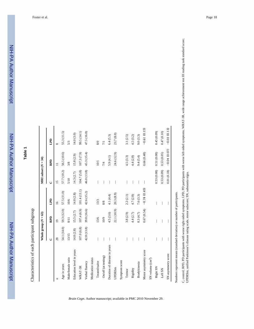
Demographic and clinical characteristics of the sample are in Table 1. There were nosignificant differences between the participant subgroups for any of these variables (p >0.19). Of the PD participants, 26 were chronically treated with medication and 18 of thesewere on medications at the time of testing. Of the 26 treated PD participants, 15 werereceiving carbidopa–levodopa exclusively (RPD = 8, LPD = 7), 5 were receiving adopamine agonist exclusively (i.e. pramipexole, pergolide; RPD = 3; LPD = 2) and 6 werereceiving carbidopa–levodopa with a COMT inhibitor (i.e. entacapone) or dopamine agonist,or both (RPD = 3; LPD = 3). χ2 and Fisher’s exact tests indicated that the PD subgroupswere not significantly different in the number of participants treated with medications vs.medication naïve and in the number of participants on vs. off medications during cognitivetesting (p > 0.34). The PD subgroups were also equivalent in total UPDRSm, tremor,rigidity and bradykinesia scores (p > 0.21). There were no significant differences in verbalfluency performance between LPD, RPD and controls after accounting for age andWRAT-3R score for the entire group as well as for the MRI subset (p > 0.46). These effectsremained the same after comparing only the LPD and RPD groups and additionallycovarying disease duration and UPDRSm score (p > 0.89).
3.2. Group comparisons3.2.1. SDR performance—SDR performance comparisons between LPD, RPD andcontrols were done after controlling for age and WRAT-3R score. When comparing only theLPD and RPD groups, we also controlled for disease duration and total UPDRSm score.
There was a significant within-subjects effect of SDR delay length on performance, F(2,116) = 6.02, p = 0.004. As expected, mean error increased as the length of time between cuepresentation and retrieval increased. This is consistent with the design of the task, wherebylonger delays are hypothesized to be more demanding cognitively and thus produce highererror rate. More interesting was the interaction effect between SDR delay length and worseside of symptoms, F(4, 116) = 4.18, p = 0.005), and the between-subjects effect of worseside of symptoms on SDR performance, F(2, 58) = 8.46, p = 0.001. Follow-up testingshowed that the between-subjects effect was significant in both delay conditions but not inthe cue-present condition (5 s: F(2, 60) = 5.82, p = 0.005; 15 s: F(2, 60) = 7.29, p = 0.001;cue present: F(2, 60) = 0.24, p = 0.79), which indicates there was no fundamental differencein pointing accuracy among the groups. Post hoc tests showed that the LPD group hadsignificantly higher error rate in the delay conditions than the RPD and control groups,which did not differ from each other (Fig. 2A). All of these effects remained the same whencomparing the LPD and RPD groups only.
Foster et al. Page 6
Brain Cogn. Author manuscript; available in PMC 2010 November 29.
NIH
-PA Author Manuscript
NIH
-PA Author Manuscript
NIH
-PA Author Manuscript

Results were similar for the group of participants who had an MRI on the same day ascognitive testing. There was no significant within-subjects effect of SDR delay length onperformance, F(2, 58) = 1.23, p = 0.30; however, there was a trend toward higher error ratewith increasing delay length. The interaction between delay and more affected motor sideand the between-subjects effect of worse side of motor deficits on SDR performanceremained significant (interaction: F(4, 58) = 5.20, p = 0.003; main effect of group: F(2, 29)= 6.68, p = 0.004) again for both delay conditions but not for the cue-present condition (5 s:F(2, 31) = 3.73, p = 0.03 ; 15 s: F(2,31) = 7.80, p = 0.002; cue present: F(2, 31) = 1.97, p =0.16). Post hoc tests showed that the LPD group had significantly higher error rate in thedelay conditions than the RPD and control groups, which did not differ from each other (Fig.2B). As with the larger group, these effects remained the same when comparing the LPDand RPD groups only.
We performed subgroup analyses to explore the possible effects of medication status onSDR performance. Within the entire group of PD participants and within the LPD and RPDgroups, there were no differences in SDR performance between participants on and offmedications (p > 0.56). Within the on and off medications sub groups, the differencesbetween the LPD and RPD groups were consistent with those described above: LPDparticipants had significantly higher error rate in the delay conditions of the SDR task thanRPD participants (p < 0.04).
3.2.2. Motor asymmetry—Motor asymmetry scores were significantly different betweenthe RPD and LPD groups for the whole sample as well as for the MRI subset (p < 0.001; seeTable 1). The groups differed in the expected direction such that RPD participants’ scoreswere positive and LPD participants’ scores were negative, indicating our initialdichotomization for worse side of motor symptoms was congruent with current motordysfunction asymmetry scores. We then compared the absolute values of group motorasymmetry scores to investigate possible differences in degree of motor asymmetry betweenthe PD subgroups. For the whole sample and for the MRI subset, there were no significantdifferences in degree of motor asymmetry between the RPD and LPD groups (p > 0.51).Thus, although we had two distinct groups of PD participants separable by absolute side ofworse motor function, the groups were equivalent in the degree of asymmetry of their motorsigns.
3.2.3. SN volume—Mean SN volumes for each of the participant groups in the MRIsubset are displayed in Table 1. There were no significant differences between subjectgroups in right or left SN volumes after controlling for whole brain volume, F(2,30) = 2.85,p = 0.07. There was no within-subjects effect of SN side (left vs. right) on SN volume (p =0.55) nor was there an interaction effect between SN side and subject group on SN volume(p = 0.66). This pattern remained the same when comparing the LPD and RPD groups only.
We calculated an SN volume asymmetry score for each participant according to the formulaused to calculate the motor asymmetry score (see above). This calculation yields a scorewith absolute values ranging from 0 (symmetric) to 2 (exclusively unilateral SNdegeneration), with negative scores indicating more right SN degeneration and positivescores indicating more left SN degeneration. There were no significant differences betweengroups in SN volume asymmetry (p = 0.63) or in degree of SN volume asymmetry (i.e.absolute value of SN volume asymmetry score; p = 0.44), nor did SN volume asymmetryscore correlate significantly with motor asymmetry score (r = −0.05, p = 0.83). The inversecorrelation between total SN volume and total UPDRSm score was not significant (r =−0.36, p = 0.10).
Foster et al. Page 7
Brain Cogn. Author manuscript; available in PMC 2010 November 29.
NIH
-PA Author Manuscript
NIH
-PA Author Manuscript
NIH
-PA Author Manuscript

3.3. Asymmetry predictors of SDR performance3.3.1. Motor asymmetry—Across all of the PD participants, SDR 15 s delay error wassignificantly correlated with motor symptom asymmetry score (r = −0.49, p = 0.02) suchthat more negative motor asymmetry scores were associated with higher error rate. In alinear regression, motor asymmetry score accounted for a unique and significant portion ofthe variance in SDR 15 s delay performance after controlling for age, WRAT-3R, durationof disease and UPDRSm (R2 change = 0.12; F change (1, 29) = 5.20, p = 0.03), and theoverall model was significant (R2 = 0.33; F(5, 29) = 2.90, p = 0.03). However, when thecorrelation between motor asymmetry and SDR 15 s delay error was examined within theRPD and LPD subgroups separately, the correlation coefficients were markedly reduced andnot significant (RPD: r = −0.20, p = 0.41; LPD: r = 0.11, p = 0.69). Furthermore, visualinspection of the data revealed distinct clusters corresponding to the two groups, whichindicated a bimodal rather than linear association between motor symptom asymmetry andSDR performance (Fig. 3). Total, right and left UPDRSm scores did not correlate with SDRperformance across or within the PD subgroups (p > 0.14). There were no significantcorrelations between the severity of specific motor signs (i.e. tremor, rigidity andbradykinesia scores) and SDR performance across or within the PD subgroups (p > 0.21).
3.3.2. SN volume asymmetry—Across the MRI subset of PD and control participants,SDR 15 s delay error was significantly correlated with right SN volume (r = −0.42, p =0.01) while the correlation with left SN volume did not reach significance (r = −0.31, p =0.07). Smaller SN volumes were associated with higher error rate. In a linear regression,right SN volume accounted for a unique and significant portion of the variance in SDR 15 sdelay performance after controlling for age, WRAT-3R and whole brain volume (R2 change= 0.16, F change (1, 29) = 5.75, p = 0.02, Fig. 4). The overall model did not reachsignificance (R2 = 0.22, F(4, 29) = 2.04, p = .11). The additional proportion of varianceaccounted for by left SN volume was not significant (R2 change = 0.07; F change (1, 29) =2.36, p = 0.14). SN volume asymmetry score did not correlate with SDR performance (r =−0.26, p = 0.25).
4. DiscussionPerformance on spatial working memory relates to asymmetry of motor function and MRmeasurements of SN volume in people with PD. PD participants with left predominantmotor dysfunction performed significantly worse on a spatial delayed response task than PDparticipants with right predominant motor dysfunction, who performed similarly to controls.In addition, poorer spatial delayed response performance was related to smaller right SNvolumes. These effects were independent of other contributors to cognition in normal agingand in PD (age, premorbid intelligence, duration of disease, medication status and motorseverity). They were also independent of type of motor dysfunction, as our groups hadsimilar degrees of tremor, rigidity and bradykinesia and the severity of these motor signswas not related to spatial delayed response performance. Thus, our data support thehypothesis that worse right-brain disease severity is related to worse spatial workingmemory performance.
Our motor asymmetry findings are consistent with a number of studies where LPDparticipants performed more poorly than RPD participants on visuospatial tasks (Amick etal., 2006; Blonder et al., 1989; Katzen et al., 2006; Tomer et al., 1993), or where RPDparticipants’ performance was comparable to that of controls (Direnfeld et al., 1984; Katzenet al., 2006). However, only a few researchers looked at visuospatial memory specifically(Amick et al., 2006; Blonder et al., 1989; St. Clair et al., 1998; Starkstein et al., 1987; Tomeret al., 1993) and none of them used tasks validated to measure short term spatial memory.
Foster et al. Page 8
Brain Cogn. Author manuscript; available in PMC 2010 November 29.
NIH
-PA Author Manuscript
NIH
-PA Author Manuscript
NIH
-PA Author Manuscript
Rather, previous tasks involved memory for objects or may have been susceptible to theemployment of verbal strategies, such as rehearsal or mnemonics, to mediate taskperformance. This perceptual mixing could have attenuated LPD and RPD differences inprevious studies, which may explain why we found such marked differences in performancewith a relatively small sample in the early stages of disease progression. The use of non-specific tasks confounds interpretation of cognitive results in terms of specific brainnetworks and functions.
Tasks that assess single cognitive processes and isolated neural systems are preferred toaddress questions about specific brain-behavior relationships. The SDR task used in thisstudy is modeled specifically after the oculo-motor delayed response task (OMDR)(Funahashi et al., 1989) and requires the participant to maintain purely spatial informationover a delay. Single-cell recording and lesion studies in non-human primates show thatperformance on the OMDR task relies on the principal sulcus region—the area analogous tothe dorsolateral prefrontal cortex in humans (Funahashi et al., 1989, 1993). Humanneuroimaging studies using the SDR task consistently demonstrate involvement of the rightdorsolateral prefrontal cortex as well (D’Esposito et al., 1998; Jonides et al., 1993; Leung etal., 2002; McCarthy et al., 1996). Additionally, task-related neuronal activity in thedorsolateral prefrontal cortex and, consequently, task performance are modulated bydopamine and gamma-aminobutyric acid (GABA) in monkeys and in humans (Gibbs et al.,2005; Goldman-Rakic et al., 2000; Lewis & Moghaddam, 2006; Luciana et al., 1992; Mülleret al., 1998; Rao, Williams, & Goldman-Rakic, 2000).
Right-sided PD neuropathology may disrupt performance on the SDR task by a number ofpossible pathways. Traditional hypotheses would posit that dopaminergic degeneration inthe right SNc leads to decreased dopamine in the right caudate nucleus, which disrupts rightprefrontal dorsolateral cortex function. This would account for our association betweenmotor dysfunction and spatial memory, as dopamine cell loss in the SNc is also the putativemechanism for motor impairment. Another possibility is that right SNr degeneration disruptsthe spatial tuning effects of GABAergic transmission in the right dorsolateral prefrontalcortex and impairs the maintenance of spatial information (Rao et al., 2000). This hypothesisis supported by monkey studies showing that SNr output indirectly targets the dorsolateralprefrontal cortex and that neurons in the SNr demonstrate activity corresponding to theactivity observed in the dorsolateral prefrontal cortex during spatial memory tasks(Middleton & Strick, 2000). There is evidence that the SNr begins to degenerate early in PD(Anik, Iseri, Demirci, Komsuoglu, & Inan, 2007). Our volumetric measurements include aportion of the SNr along with the SNc, which may help to explain why they are more relatedto SDR than to motor performance.
Support for our SN asymmetry hypothesis was mixed. In support of our hypothesis, smallerright SN was predictive of worse SDR performance. In addition, there was an inverserelationship between SN volume asymmetry score and SDR performance such that moredegeneration on the right compared to the left side of the brain (i.e. more negative SNvolume asymmetry score) tended to be associated with worse performance (i.e. higher errorrate). However, we cannot conclude from our data that the effect is driven entirely byasymmetric degeneration rather than by severity of overall nigral degeneration since left SNvolume was moderately correlated with SDR performance and the relationship between SNvolume asymmetry score and SDR performance was not statistically significant.Nonetheless, the strong correlation between smaller right SN volumes and poorer SDRperformance suggests there is a lateralized relationship.
Our volumetric analysis of SN is less clear regarding the relationship between brain andmotor asymmetry. SN volume measurements were not congruent with our group
Foster et al. Page 9
Brain Cogn. Author manuscript; available in PMC 2010 November 29.
NIH
-PA Author Manuscript
NIH
-PA Author Manuscript
NIH
-PA Author Manuscript

categorization according to predominant side of motor dysfunction (RPD vs. LPD), nor didthey discriminate PD participants as a whole from controls. These results are consistent withprevious MR studies that have not detected SN volume loss in PD (Geng, Li, & Zee, 2006;Oikawa, Sasaki, Tamakawa, Ehara, & Tohyama, 2002), although others have shown theopposite (Sohmiya, Tanaka, Aihara, & Okamoto, 2004). From autopsy studies, it is clearthat there is substantial neuronal loss in SN which correlates with contralateral motordysfunction (Kempster et al., 1989); however, neuronal loss may not necessarily translate tomeasurable volume loss.
Our data do not support the notion put forth by Huber and colleagues (1992) that the degreeof motor asymmetry should parallel the degree of cognitive impairment. They found a linearrelationship between motor asymmetry and verbal cognition, whereby increasing right-sidedmotor asymmetry was associated with decreasing performance on verbal tasks. In thepresent study, degree of left-sided asymmetry appeared to predict degree of spatial memoryimpairment, but closer examination revealed that this association was driven by qualitativedifferences in performance between the groups rather than by a linear effect of relativeasymmetry. This finding, along with the observations that motor signs tend to become morebilateral and cognition tends to worsen as the disease progresses, suggests that categorizingparticipants according to side of initial clinical predominance is sufficient when exploringthese concepts.
One methodological limitation of our study is the lack of a congruent “left-hemisphere”working memory task with which to demonstrate a true divergence of cognitive profilesbetween the LPD and RPD groups. This restricts our ability to exclude the possibility thatdegeneration of right subcortical structures relates to widespread cognitive deficits acrossmany domains, rather than to circumscribed right hemisphere memory processes. Theformer has been proposed (Direnfeld et al., 1984; Tomer et al., 1993), as the righthemisphere may mediate overall activation and attentional control, thus forming thefoundation for cognitive processing (Mesulam, 1981). However, our verbal fluency resultsshow that the LPD group was not globally cognitively impaired relative to the control andRPD groups. Other studies have demonstrated worse performance by RPD groups on avariety of cognitive tasks and specifically those that rely on verbal abilities (e.g. Blonder etal., 1989; Spicer et al., 1988; Williams et al., 2007). These points reduce the likelihood ofwidespread impairment in individuals with predominantly left-body motor dysfunction.
Other limitations include the relatively small sample size and the possibility that PDmedications could have influenced behavior. The relationship between dopaminergicmedication and cognition is complex (for review, see Cools, 2006) and a recent study hasshown that dopaminergic medication may interact with asymmetry to influence cognitivefunction in PD (Tomer, Aharon-Peretz, & Tsitrinbaum, 2007). Nevertheless, our cognitivefindings did not differ according to medication status. SDR performance was similarbetween participants on and off medications and was impaired in LPD relative to RPDparticipants regardless of their medication status. Future studies of this type will need tomanipulate medication status explicitly.
In conclusion, we demonstrated that PD participants with worse left-sided motordysfunction are impaired in spatial memory compared to those with worse right-sided motordysfunction, whose spatial memory is equivalent to that of controls. Moreover, poorerspatial memory is related to right SN volume loss. These findings indicate that diseaseasymmetry should be considered when interpreting patterns of cognitive performance inpersons with PD. Overlooking this factor in studies of cognition—especially those withunbalanced samples or with tasks that employ hemispherically lateralized cognitivefunctions—can lead to inconsistent results and faulty interpretations regarding the nature of
Foster et al. Page 10
Brain Cogn. Author manuscript; available in PMC 2010 November 29.
NIH
-PA Author Manuscript
NIH
-PA Author Manuscript
NIH
-PA Author Manuscript

cognitive impairment in PD and its neurological basis. By using the SDR task, which hasbeen validated carefully across animal and human studies to measure the maintenance ofspatial information and the integrity of the right dorsolateral prefrontal cortex, we are betterable to infer the effects of right nigral degeneration on spatial working memory in PD. Theuse of MRI-based volumetry to investigate the association between asymmetrical subcorticaldegeneration, motor symptoms and cognitive performance warrants further exploration.
ReferencesAlexander GE, DeLong MR, Strick PL. Parallel organization of functionally segregated circuits
linking basal ganglia and cortex. Annual Reviews in Neuroscience 1986;9:357–381.Amick MM, Grace J, Chou KL. Body side of motor symptom onset in Parkinson’s disease is
associated with memory performance. Journal of the International Neuropsychological Society2006;12:736–740. [PubMed: 16961953]
Anik Y, Iseri P, Demirci A, Komsuoglu S, Inan N. Magnetization transfer ratio in early period ofParkinson disease. Academic Radiology 2007;14:189–192. [PubMed: 17236991]
Barber J, Tomer R, Sroka H, Myslobodsky MS. Does unilateral dopamine deficit contribute todepression? Psychiatry Research 1985;15:17–24. [PubMed: 3859881]
Blonder LX, Gur RE, Gur RC, Saykin AJ, Hurtig HI. Neuropsychological functioning inhemiparkinsonism. Brain and Cognition 1989;9:244–257. [PubMed: 2923715]
Cavalieri, B. Geometria Indivisibilibus Continuorum Nova quadam ratione promota. Bologna, Italy:Duciis; 1653.
Cools R. Dopaminergic modulation of cognitive function-implications for l-DOPA treatment inParkinson’s disease. Neuroscience and Biobehavioral Reviews 2006;30:1–23. [PubMed: 15935475]
Damier P, Hirsch EC, Agid Y, Graybiel AM. The substantia nigra of the human brain. I. Nigrosomesand the nigral matrix, a compartmental organization based on calbindin D(28K)immunohistochemistry. Brain 1999a;122(Pt. 8):1421–1436. [PubMed: 10430829]
Damier P, Hirsch EC, Agid Y, Graybiel AM. The substantia nigra of the human brain. II. Patterns ofloss of dopamine-containing neurons in Parkinson’s disease. Brain 1999b;122(Pt. 8):1437–1448.[PubMed: 10430830]
D’Esposito M, Aguirre GK, Zarahn E, Ballard D, Shin RK, Lease J. Functional MRI studies of spatialand nonspatial working memory. Cognitive Brain Research 1998;7:1–13. [PubMed: 9714705]
Direnfeld LK, Albert ML, Volicer L, Langlais PJ, Marquis J, Kaplan E. Parkinson’s disease. Thepossible relationship of laterality to dementia and neurochemical findings. Archives of Neurology1984;41:935–941. [PubMed: 6477229]
Fahn, S.; Elton, RL. Members of the UPDRS Development Committee. Unified Parkinson’s diseaserating scale. In: Fahn, S.; Marsden, CD.; Goldstein, M.; Calne, DB., editors. Recent developmentsin Parkinson’s disease. New York: Macmillan; 1987. p. 153-163.
Funahashi S, Bruce CJ, Goldman-Rakic PS. Mnemonic coding of visual space in the monkey’sdorsolateral prefrontal cortex. Journal of Neurophysiology 1989;61:331–349. [PubMed: 2918358]
Funahashi S, Bruce CJ, Goldman-Rakic PS. Dorsolateral prefrontal lesions and oculomotor delayed-response performance: Evidence for mnemonic “scotomas”. Journal of Neuroscience1993;13:1479–1497. [PubMed: 8463830]
Geng DY, Li YX, Zee CS. Magnetic resonance imaging-based volumetric analysis of basal ganglianuclei and substantia nigra in patients with Parkinson’s disease. Neurosurgery 2006;58:256–262.[PubMed: 16462479]
Gibbs SE, D’Esposito M. Individual capacity differences predict working memory performance andprefrontal activity following dopamine receptor stimulation. Cognitive, Affective, and BehavioralNeuroscience 2005;5:212–221.
Goldman-Rakic PS, Muly EC III, Williams GV. D(1) receptors in prefrontal cells and circuits. BrainResearch: Brain Research Reviews 2000;31:295–301. [PubMed: 10719156]
Green J, McDonald WM, Vitek JL, Evatt M, Freeman A, Haber M, et al. Cognitive impairments inadvanced PD without dementia. Neurology 2002;59:1320–1324. [PubMed: 12427877]
Foster et al. Page 11
Brain Cogn. Author manuscript; available in PMC 2010 November 29.
NIH
-PA Author Manuscript
NIH
-PA Author Manuscript
NIH
-PA Author Manuscript

Griffin SL, Mindt MR, Rankin EJ, Ritchie AJ, Scott JG. Estimating premorbid intelligence:Comparison of traditional and contemporary methods across the intelligence continuum. Archivesof Clinical Neuropsychology 2002;17:497–507. [PubMed: 14592003]
Gundersen HJG. Stereology: The fast lane between neuroanatomy and brain function—or still only atightrope? Acta Neurologica Scandinavica Supplementum 1992;137:8–13. [PubMed: 1357908]
Gundersen HJ, Jensen EB. The efficiency of systematic sampling in stereology and its prediction.Journal of Microscience 1987;147(Pt. 3):229–263.
Hoehn MM, Yahr MD. Parkinsonism: Onset, progression and mortality. Neurology 1967;17:427–442.[PubMed: 6067254]
Huber SJ, Freidenberg DL, Shuttleworth EC, Paulson GW, Clapp LE. Neuropsychological similaritiesin lateralized parkinsonism. Cortex 1989;25:461–470. [PubMed: 2805731]
Huber SJ, Miller H, Bohaska L, Christy JA, Bornstein RA. Asymmetrical cognitive differencesassociated with hemiparkinsonism. Archives of Clinical Neuropsychology 1992;7:471–480.[PubMed: 14591398]
Johnstone B, Callahan CD, Kapila CJ, Bouman DE. The comparability of the WRAT-R reading testand NAART as estimates of premorbid intelligence in neurologically impaired patients. Archivesof Clinical Neuropsychology 1996;11:513–519. [PubMed: 14588456]
Jonides J, Smith EE, Koeppe RA, Awh E, Minoshima S, Mintun M. Letters to Nature: Spatial workingmemory in humans as revealed by PET. Nature 1993;363:623–625. [PubMed: 8510752]
Katzen HL, Levin BE, Weiner W. Side and type of motor symptom influence cognition in Parkinson’sdisease. Movement Disorders 2006;21:1947–1953. [PubMed: 16991155]
Kempster PA, Gibb WR, Stern GM, Lees AJ. Asymmetry of substantia nigra neuronal loss inParkinson’s disease and its relevance to the mechanism of levodopa related motor fluctuations.Journal of Neurology, Neurosurgery, and Psychiatry 1989;52:72–76.
Lee CS, Schulzer M, Mak E, Hammerstad JP, Calne S, Calne DB. Patterns of asymmetry do notchange over the course of idiopathic parkinsonism: Implications for pathogenesis. Neurology1995;45:435–439. [PubMed: 7898691]
Leung HC, Gore JC, Goldman-Rakic PS. Sustained mnemonic response in the human middle frontalgyrus during on-line storage of spatial memoranda. Journal of Cognitive Neuroscience2002;14:659–671. [PubMed: 12126506]
Lewis DA, Moghaddam B. Cognitive dysfunction in schizophrenia: Convergence of gamma-aminobutyric acid and glutamate alterations. Archives of Neurology 2006;63:1372–1376.[PubMed: 17030651]
Lezak, MD. Neuropsychological Assessment. 3rd ed.. Oxford: Oxford University Press; 1995.Locascio JJ, Corkin S, Growdon JH. Relation between clinical characteristics of Parkinson’s disease
and cognitive decline. Journal of Clinical and Experimental Neuropsychology 2003;25:94–109.[PubMed: 12607175]
Luciana M, Depue RA, Arbisi P, Leon A. Facilitation of working memory in humans by a D2dopamine receptor agonist. Journal of Cognitive Neuroscience 1992;4:58–68.
Mayhew TM, Olsen DR. Magnetic resonance imaging (MRI) and model-free estimates of brainvolume determined using the Cavalieri principle. Journal of Anatomy 1991;178:133–144.[PubMed: 1810922]
McCarthy G, Puce A, Constable RT, Krystal JH, Gore JC, Goldman-Rakic P. Activation of humanprefrontal cortex during spatial and nonspatial working memory tasks measured by functionalMRI. Cerebral Cortex 1996;6:600–611. [PubMed: 8670685]
Mesulam MM. A cortical network for directed attention and unilateral neglect. Annals of Neurology1981;10:309–325. [PubMed: 7032417]
Middleton FA, Strick PL. Basal ganglia output and cognition: Evidence from anatomical, behavioral,and clinical studies. Brain and Cognition 2000;42:183–200. [PubMed: 10744919]
Müller U, von Cramon DY, Pollmann S. D1- versus D2- receptor modulation of visuospatial workingmemory in humans. Journal of Neuroscience 1998;18:2720–2728. [PubMed: 9502829]
Oikawa H, Sasaki M, Tamakawa Y, Ehara S, Tohyama K. The substantia nigra in Parkinson disease:Proton density-weighted spin-echo and fast short inversion time inversion-recovery MR findings.American Journal of Neuroradiology 2002;23:1747–1756. [PubMed: 12427635]
Foster et al. Page 12
Brain Cogn. Author manuscript; available in PMC 2010 November 29.
NIH
-PA Author Manuscript
NIH
-PA Author Manuscript
NIH
-PA Author Manuscript

Pillon, B.; Boller, F.; Levy, R.; Dubois, B. Cognitive deficits and dementia in Parkinson’s disease. In:Boller, F.; Cappa, SF., editors. Handbook of neuropsychology. 2nd ed.. Amsterdam: Elsevier;2001. p. 311-371.
Postle BR, Jonides J, Smith EE, Corkin S, Growdon JH. Spatial, but not object, delayed response isimpaired in early Parkinson’s disease. Neuropsychology 1997;11:171–179. [PubMed: 9110324]
Rao SG, Williams GV, Goldman-Rakic PS. Destruction and creation of spatial tuning by disinhibition:GABA(A) blockade of prefrontal cortical neurons engaged by working memory. Journal ofNeuroscience 2000;20:485–494. [PubMed: 10627624]
Riklan M, Stellar S, Reynolds C. The relationship of memory and cognition in Parkinson’s disease tolateralisation of motor symptoms. Journal of Neurology, Neurosurgery, and Psychiatry1990;53:359–360.
Sohmiya M, Tanaka M, Aihara Y, Okamoto K. Structural changes in the midbrain with aging andParkinson’s disease: An MRI study. Neurobiology of Aging 2004;25:449–453. [PubMed:15013565]
Spicer KB, Roberts RJ, LeWitt PA. Neuropsychological performance in lateralized parkinsonism.Archives of Neurology 1988;45:429–432. [PubMed: 3355399]
St. Clair J, Borod JC, Sliwinski M, Cote LJ, Stern Y. Cognitive and affective functioning inParkinson’s disease patients with lateralized motor signs. Journal of Clinical and ExperimentalNeuropsychology 1998;20:320–327. [PubMed: 9845159]
Starkstein S, Leiguarda R, Gershanik O, Berthier M. Neuropsychological disturbances inhemiparkinson’s disease. Neurology 1987;37:1762–1764. [PubMed: 3670613]
Taylor AE, Saint-Cyr JA. The neuropsychology of Parkinson’s disease. Brain and Cognition1995;28:281–296. [PubMed: 8546855]
Taylor AE, Saint-Cyr JA, Lang AE. Frontal lobe dysfunction in Parkinson’s disease. The cortical focusof neostriatal outflow. Brain 1986;109(Pt. 5):845–883.
Tomer R, Aharon-Peretz J, Tsitrinbaum Z. Dopamine asymmetry interacts with medication to affectcognition in Parkinson’s disease. Neuropsychologia 2007;45:357–367. [PubMed: 16876208]
Tomer R, Levin BE, Weiner WJ. Side of onset of motor symptoms influences cognition in Parkinson’sdisease. Annals of Neurology 1993;34:579–584. [PubMed: 8215246]
Wilkinson, GW. WRAT-3: Wide range achievement test administration manual. 3rd ed.. Wilmington,DE: Western psychological services; 1993.
Williams GV, Goldman-Rakic PS. Modulation of memory fields by dopamine D1 receptors inprefrontal cortex. Nature 1995;376:572–575. [PubMed: 7637804]
Williams LN, Seignourel P, Crucian GP, Okun MS, Rodriguez RL, Skidmore FM, et al. Laterality,region, and type of motor dysfunction correlate with cognitive impairment in Parkinson’s disease.Movement Disorders 2007;22:141–145. [PubMed: 17089386]
Zetusky WJ, Jankovic J. Laterality and symptom association in Parkinson’s disease. Archives ofNeurology 1985;42:1132–1133. [PubMed: 4062609]
Foster et al. Page 13
Brain Cogn. Author manuscript; available in PMC 2010 November 29.
NIH
-PA Author Manuscript
NIH
-PA Author Manuscript
NIH
-PA Author Manuscript
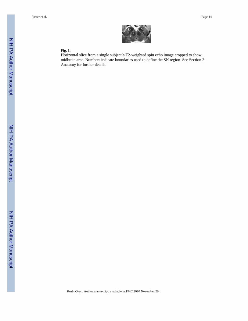
Fig. 1.Horizontal slice from a single subject’s T2-weighted spin echo image cropped to showmidbrain area. Numbers indicate boundaries used to define the SN region. See Section 2:Anatomy for further details.
Foster et al. Page 14
Brain Cogn. Author manuscript; available in PMC 2010 November 29.
NIH
-PA Author Manuscript
NIH
-PA Author Manuscript
NIH
-PA Author Manuscript

Fig. 2.Group differences in SDR performance error (mean ± SEM) across delay conditions for (A)the entire group of participants (control, n = 28; RPD, n = 19; LPD, n = 16), and (B) theMRI subset (control, n = 15; RPD, n = 11; LPD, n = 8) after controlling for age andWRAT-3R score. * LPD>RPD and control, p < 0.01.
Foster et al. Page 15
Brain Cogn. Author manuscript; available in PMC 2010 November 29.
NIH
-PA Author Manuscript
NIH
-PA Author Manuscript
NIH
-PA Author Manuscript

Fig. 3.Relationship between motor asymmetry score and SDR 15 s delay error after accounting forage, WRAT-3R score, disease duration and UPDRSm for the PD participants (RPD, n = 19;LPD, n = 16).
Foster et al. Page 16
Brain Cogn. Author manuscript; available in PMC 2010 November 29.
NIH
-PA Author Manuscript
NIH
-PA Author Manuscript
NIH
-PA Author Manuscript
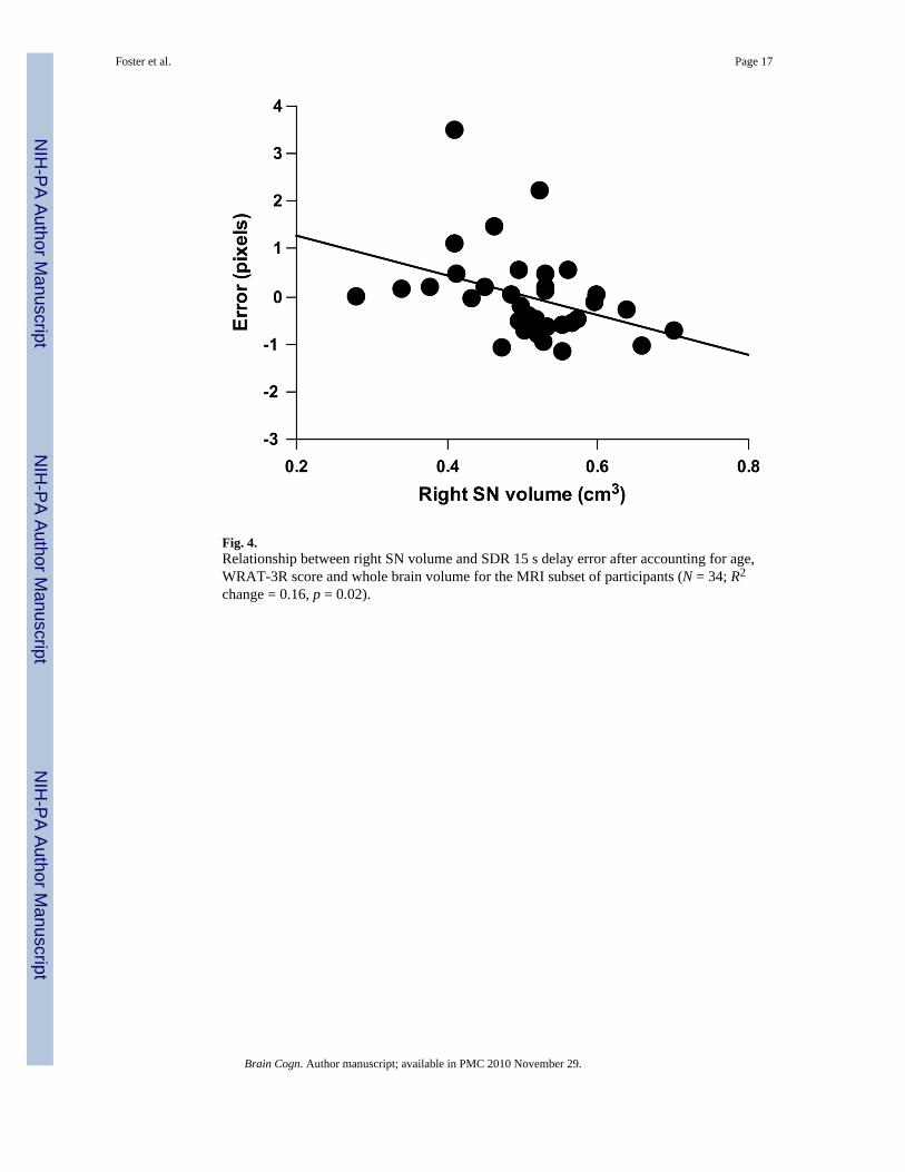
Fig. 4.Relationship between right SN volume and SDR 15 s delay error after accounting for age,WRAT-3R score and whole brain volume for the MRI subset of participants (N = 34; R2
change = 0.16, p = 0.02).
Foster et al. Page 17
Brain Cogn. Author manuscript; available in PMC 2010 November 29.
NIH
-PA Author Manuscript
NIH
-PA Author Manuscript
NIH
-PA Author Manuscript
NIH
-PA Author Manuscript
NIH
-PA Author Manuscript
NIH
-PA Author Manuscript
Foster et al. Page 18
Tabl
e 1
Cha
ract
eris
tics o
f eac
h pa
rtici
pant
subg
roup
Who
le g
roup
(N =
63)
MR
I sub
set (
N =
34)
CR
PDL
PDC
RPD
LPD
n28
1916
1511
8
Age
in y
ears
54.1
(14.
0)59
.3 (1
2.0)
57.5
(11.
0)57
.7 (1
0.2)
56.5
(10.
6)55
.3 (1
1.5)
Mal
e/fe
mal
e ra
tio13
/15
8/11
10/6
5/10
3/8
5/3
Educ
atio
n le
vel i
n ye
ars
14.8
(2.8
)15
.5 (2
.7)
14.8
(2.8
)14
.3 (2
.7)
15.8
(2.9
)14
.0 (3
.0)
WR
AT-
3R10
7.0
(6.8
)10
7.4
(6.9
)10
1.4
(13.
1)10
4.7
(5.8
)10
7.3
(7.9
)99
.5 (1
4.1)
Ver
bal f
luen
cy42
.8 (1
3.8)
39.8
(16.
6)42
.6 (1
5.2)
46.0
(13.
8)45
.3 (1
5.4)
47.5
(16.
8)
Med
icat
ion
stat
us
Tre
ated
/naï
ve—
13/6
13/3
—10
/18/
0
On/
off (
at te
stin
g)—
10/9
8/8
—7/
47/
1
Dur
atio
n of
dis
ease
in y
ears
—4.
5 (3
.6)
4.1
(4.4
)—
5.9
(4.1
)6.
4 (5
.3)
UPD
RSm
—22
.1 (1
0.9)
20.3
(8.9
)—
24.6
(12.
9)23
.7 (8
.0)
Sym
ptom
scor
e
Tre
mor
—3.
8 (2
.9)
2.2
(2.1
)—
4.5
(3.3
)3.
1 (2
.5)
Rig
idity
—4.
4 (2
.7)
4.7
(2.8
)—
4.4
(2.8
)4.
5 (3
.2)
Bra
dyki
nesi
a—
8.3
(4.7
)7.
6 (3
.3)
—9.
4 (5
.4)
9.0
(3.3
)
Mot
or a
sym
met
ry sc
ore
—0.
67 (0
.54)
−0.
78 (0.
45)
—0.
66 (0
.48)
−0.
61 (0.
15)
SN v
olum
e (c
m3 )
Rig
ht S
N—
——
0.53
(0.0
8)0.
51 (0
.08)
0.45
(0.0
9)
Lef
t SN
——
—0.
53 (0
.09)
0.53
(0.0
5)0.
47 (0
.10)
SN a
sym
met
ry sc
ore
——
—0.
01 (0
.18)
−0.
04 (0.
07)
−0.
03 (0.
13)
Num
bers
repr
esen
t mea
ns (s
tand
ard
devi
atio
n) o
r num
ber o
f par
ticip
ants
.
C, c
ontro
l; R
PD, P
D p
artic
ipan
ts w
ith w
orse
righ
t-sid
ed sy
mpt
oms;
LPD
, PD
par
ticip
ants
with
wor
se le
ft-si
ded
sym
ptom
s; W
RA
T-3R
, wid
e ra
nge
achi
evem
ent t
est I
II re
adin
g ta
sk st
anda
rd sc
ore;
UPD
RSm
, uni
fied
Park
inso
n’s d
isea
se ra
ting
scal
e, m
otor
subs
core
; SN
, sub
stan
tia n
igra
.
Brain Cogn. Author manuscript; available in PMC 2010 November 29.