Modulation of nociceptive and acoustic startle responses to an unpredictable threat in men and women
Upload
independentCategory
view
3download
0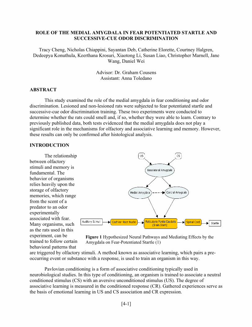
ROLE OF THE MEDIAL AMYGDALA IN FEAR POTENTIATED STARTLE AND SUCCESSIVE-CUE ODOR DISCRIMINATION
Tracy Cheng, Nicholas Chiappini, Sayantan Deb, Catherine Elorette, Courtney Halgren,
Dedeepya Konuthula, Keerthana Krosuri, Xiaotong Li, Susan Liao, Christopher Marnell, Jane Wang, Daniel Wei
Advisor: Dr. Graham Cousens
Assistant: Anna Toledano
ABSTRACT
This study examined the role of the medial amygdala in fear conditioning and odor discrimination. Lesioned and non-lesioned rats were subjected to fear potentiated startle and successive-cue odor discrimination training. These two experiments were conducted to determine whether the rats could smell and, if so, whether they were able to learn. Contrary to previously published data, both tests evidenced that the medial amygdala does not play a significant role in the mechanisms for olfactory and associative learning and memory. However, these results can only be confirmed after histological analysis.
INTRODUCTION
The rbetween olfactory stimuli and memory isfundamentabehavior of organisrelies heavily upon the storage of olfactory memories, which ranfrom the scent of a predator to an odor experimentally associated with fMany organisms, suchas the rats used in this experiment, can be trained to follow cerbehavioral patterns that are triggered by olfactory stimuli. A method known as associative learning, which pairs a poccurring event or substance with a response, is used to train an organism in this way.
elationship
l. The
ms
ge
ear.
tain
re-
Pavlovian conditioning is a form of associative conditioning typically used in neurob e a neutral
serve as the basis of emotional learning in US and CS association and CR expression.
Figure 1 Hypothesized Neural Pathways and Mediating Effects by the Amygdala on Fear-Potentiated Startle (1)
iological studies. In this type of conditioning, an organism is trained to associatconditioned stimulus (CS) with an aversive unconditioned stimulus (US). The degree of associative learning is measured in the conditioned response (CR). Gathered experiences
[4-1]
Olfactory fear conditioning has become an increasingly popular method of analyzing the neural pathways that involve olfactory learning. This type of conditioning utilizes a neutral odor (CS) paired with a negative stimulus (US). Later exposure to this odor in the absence of the US can cau
s an important component in the neurological basis of fear and anxiety. Responses to fear may be indicated by physiological changes such as tachycardia, tachypnea or
ustic
s
loidal
se heightened fear, which is indicated by freezing behavior, increased respiration rate andstartle. In this study, an olfactory stimulus (CS) was paired with a mild electrical foot shock (US). The startle response to auditory pulses was measured during re-exposure to the odor in theabsence of the shock. The amplification of the startle response in the presence of the CS compared to the US was a fear potentiated startle (CR) that demonstrated the effects of associative learning.
The fear-potentiated startle pathway (Figure 1) has been studied extensively in experimental settings a
behavioral responses like freezing or jumping. According to a previous study on the acostartle response in rats, the acoustic startle pathway is a simple reflexive neural pathway, indicated by its remarkably short latency (8 ms in lab settings) (2). By nature, reflex pathways are particularly useful in experiments, as the experimenter can make simple changes in stimuluintensity to ascertain the degree of fear experienced by the subjects. Auditory informationproceeds through cochlear root neurons within the cochlear nucleus to the reticularis pontis caudalis (PnC) of the brainstem. At this point, information from the nuclei of the amygdala may mediate the response intensities at the PnC. After being enhanced or suppressed by amygdamediation, the startle information continues on towards the spinal cord and is expressed as physiological/behavioral startle (2). The parts of the amygdala that have the greatest effect on this startle response, however, have remained an area of discussion.
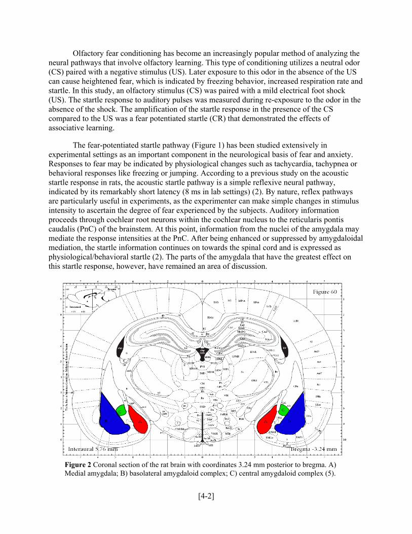
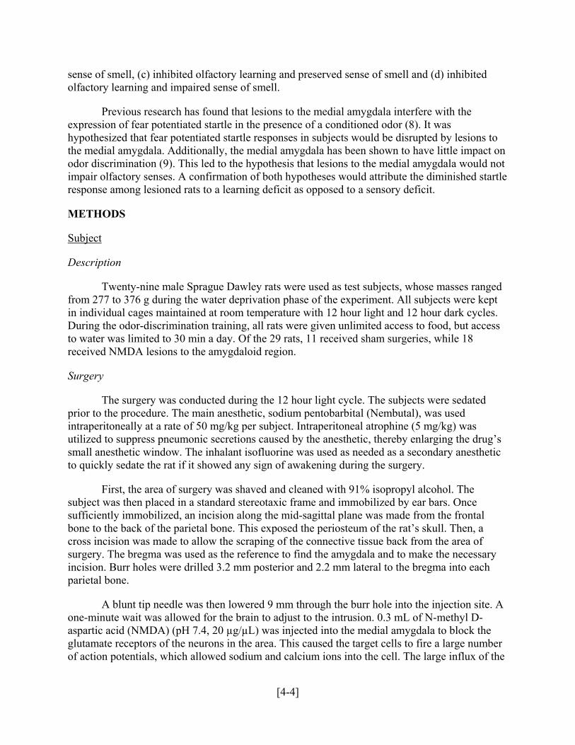
Figure 2 Coronal section of the rat brain with coordinates 3.24 mm posterior to bregma. A) Medial amygdala; B) basolateral amygdaloid complex; C) central amygdaloid complex (5).
[4-2]
The amygdala, a roughly almond-shaped nucleus in the medial temporal lobe of the brain, is associated with fear and responses to fear-based learning (3, 4). Subdivisions of the amygda (B,
.
is closely associated with fear conditioning and expression. These studies have revealed that lesions in this area ca se
Studies of the medial nucleus of the amygdala have been far less conclusive. There is much debate about the connection, if any, between olfactory discrimination learning and express
hall,
nd odor discrimination testing in order to determine the role of the medial amygdala. Lesioning the medial
lved
ther or not lesions to the medial amygdala could impair or enhance sense of smell, resulting in a more accurate interpre n
the role of
ither n
ing and
la have different purposes within the fear response pathway. The basolateral nucleusFigure 2) is responsible for the intake of stimuli, which include olfactory and auditory signalsThese signals travel to the medial (A, Figure 2) and then to the central amygdala (C, Figure 2), the latter of which is responsible for the generation of fear potentiated responses.
Studies of the basolateral amygdaloid complex have shown that the nucleus
n lead to difficulties in olfactory discrimination learning and fear expression (6, 7). Thefunctional areas have been traced to the amygdala through experiments that render nuclear glutamate receptors ineffectual by infusing these receptors with NMDA and other antagonists (8).
ion of fear and the medial nucleus (6, 8). LeDoux et al. did not find evidence of a role played by the medial amygdala in olfactory fear conditioning (6). Walker, Davis and Paschowever, found that the amygdala plays “an essential role” in learning fear conditioning (8). While there are afferent connections from the olfactory cortex towards the medial amygdala, little is known about how the area actually supports olfactory learning and expression.
In this study, both lesioned and SHAM rats were subjected to fear conditioning a
amygdala prior to training allowed for the analysis of the effects of the medial amygdala on memory and recall. After the procedure, the rats underwent fear conditioning, which invoexposing them to a certain odor and initiating electrical shocks to develop an association betweenthat specific scent and fear. The rats’ startle potentiation was observed in response to auditory pulses in the presence of the odor. To ensure the integrity of the results, a control odor discrimination test was run by training the rats to remember the consequences of reward and punishment associated with specific odors. In order to pass this successive cue go/no-goolfactory discrimination task, the rats had to actively respond to the positive odor to receive a reward and not respond to the negative odor to avoid a punishment.
The goal of the odor discrimination test was to determine whe
tation of the fear conditioning data. Interpreting the fear conditioning data in conjunctiowith the odor discrimination test could result in several possible conclusions regarding the medial amygdala in memory and learning. Prevention of the functionality of the medial amygdala may either enhance or inhibit the process of olfactory learning, and thereby alter the rats’ ability to respond to fear and startle. Similarly, inactivating the medial amygdala may epreserve or impair the sense of smell, potentially altering the rats’ ability to discriminate betweeodors. Because the learning mechanisms associated with fear conditioning and odor discrimination are mutually exclusive, the rats could theoretically learn the fear conditioning and not the odor discrimination task, or vice versa. The combination of the fear conditionodor discrimination testing could therefore result in any one of four outcomes: (a) enhanced olfactory learning and preserved sense of smell, (b) enhanced olfactory learning and impaired
[4-3]
sense of smell, (c) inhibited olfactory learning and preserved sense of smell and (d) inhibitedolfactory learning and impaired sense of smell.
Previous research has found that lesions
to the medial amygdala interfere with the expression of fear potentiated startle in the presence of a conditioned odor (8). It was
ns to impact on
hypothesized that fear potentiated startle responses in subjects would be disrupted by lesiothe medial amygdala. Additionally, the medial amygdala has been shown to have littleodor discrimination (9). This led to the hypothesis that lesions to the medial amygdala would notimpair olfactory senses. A confirmation of both hypotheses would attribute the diminished startle response among lesioned rats to a learning deficit as opposed to a sensory deficit.
METHODS
Subject
Description
ty-nine male Sprague Dawley rats were used as test subjects, whose masses ranged from 277 to 376 g during the water deprivation phase of the experiment. All subjects were kept in indiv
he surgery was conducted during the 12 hour light cycle. The subjects were sedated prior to the procedure. The main anesthetic, sodium pentobarbital (Nembutal), was used intrape
drug’s etic
hol. The subject was then placed in a standard stereotaxic frame and immobilized by ear bars. Once sufficie l
y
tip needle was then lowered 9 mm through the burr hole into the injection site. A one-minute wait was allowed for the brain to adjust to the intrusion. 0.3 mL of N-methyl D-aspartic
ber e
Twen
idual cages maintained at room temperature with 12 hour light and 12 hour dark cycles. During the odor-discrimination training, all rats were given unlimited access to food, but access to water was limited to 30 min a day. Of the 29 rats, 11 received sham surgeries, while 18 received NMDA lesions to the amygdaloid region.
Surgery
T
ritoneally at a rate of 50 mg/kg per subject. Intraperitoneal atrophine (5 mg/kg) was utilized to suppress pneumonic secretions caused by the anesthetic, thereby enlarging thesmall anesthetic window. The inhalant isofluorine was used as needed as a secondary anesthto quickly sedate the rat if it showed any sign of awakening during the surgery.
First, the area of surgery was shaved and cleaned with 91% isopropyl alco
ntly immobilized, an incision along the mid-sagittal plane was made from the frontabone to the back of the parietal bone. This exposed the periosteum of the rat’s skull. Then, across incision was made to allow the scraping of the connective tissue back from the area of surgery. The bregma was used as the reference to find the amygdala and to make the necessarincision. Burr holes were drilled 3.2 mm posterior and 2.2 mm lateral to the bregma into eachparietal bone.
A blunt
acid (NMDA) (pH 7.4, 20 µg/µL) was injected into the medial amygdala to block the glutamate receptors of the neurons in the area. This caused the target cells to fire a large numof action potentials, which allowed sodium and calcium ions into the cell. The large influx of th
[4-4]
sodium and calcium into cells then caused a large amount of water to enter the cell. The ions and water from this lysed cell would then enter the neighboring cells and restart the cycle of lesion.
After the injection of the drug into the amygdala, the subject was allowed to rest for two minute
In the sham surgeries, the needle was only inserted 7 mm below the cranium and NMDA was no
n
Fear Conditioning
s and then the needle was raised. The skull was then cleaned with glycol and the skin was stapled shut. The rats were then allowed to recover from the surgery for one week to prevent any post-surgical discomfort from affecting the startle results.
t injected. This was done to make sure that the amygdala would not be affected by the injection. The sham surgeries served as a control to rule out the possibility that the outcome ofthe study was caused by damaging the brain cells above the amygdala rather than the inactivatioof the medial amygdala itself.
Apparatus
The Pavlovian fear conditioning ilt
rm
o e
f
The
A testing chamber was position top
he
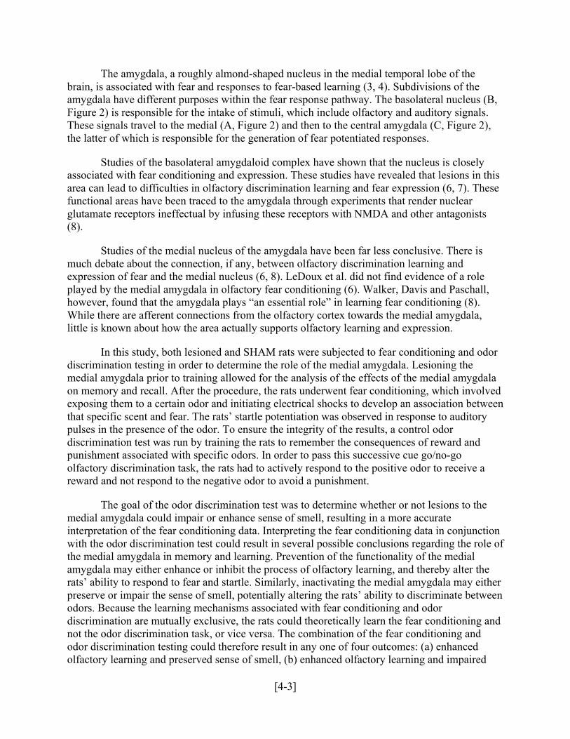
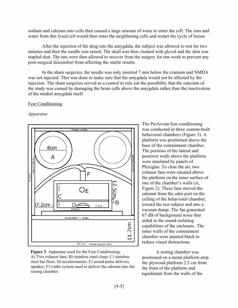
Figure 3 Apparatus used for the Fear Conditioning.
was conducted in three custom-bubehavioral chambers (Figure 3). A platform was positioned above the base of the containment chamber. The portions of the lateral and posterior walls above the platfowere insulated by panels of Plexiglas. To clear the air, twexhaust fans were situated abovthe platform on the inner surface oone of the chamber’s walls (A, Figure 2). These fans moved theodorant from the odor port on theceiling of the behavioral chamber, toward the test subject and into a vacuum dump. The fan generated 67 dB of background noise that aided in the sound-isolating capabilities of the enclosure. inner walls of the containment chamber were painted black to reduce visual distractions.
A) Two exhaust fans; B) stainless steel clasp; C) stainless
e steel bar floor; D) accelerometer; E) sound-pulse delivery speaker; F) t-tube system used to deliver the odorant into thtesting chamber.
ed on a metal platform athe plywood platform 2.5 cm from the front of the platform and equidistant from the walls of t
[4-5]
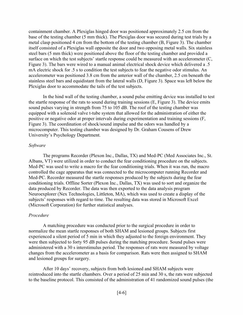
containment chamber. A Plexiglas hinged door was positioned approximately 2.5 cm from the base of the testing chamber (5 mm thick). The Plexiglas door was secured during test trials by a metal clasp positioned 4 cm from the bottom of the testing chamber (B, Figure 3). The chitself consisted of a Plexiglas wall opposite the door and two opposing metal walls. Six stainlessteel bars (5 mm thick) were positioned above the floor of the testing chamber and provided a surface on which the test subjects’ startle response could be measured with an accelerometer (C,Figure 3). The bars were wired to a manual animal electrical shock device which delivered mA electric shock for .5 s to condition the test subjects to fear the negative odor stimulus. Aaccelerometer was positioned 3.8 cm from the anterior wall of the chamber, 2.5 cm beneath thestainless steel bars and equidistant from the lateral walls (D, Figure 3). Space was left below tPlexiglas door to accommodate the tails of the test subjects.
amber s
a .5 n
he
In the hind wall of the testing chamber, a sound pulse emitting device was installed to test the star
the
Software
The programs Recorder (Plexon Inc., Dallas, TX) and Med-PC (Med Associates Inc., St. Albans
e
of the
Procedure
A matching procedure was conducted prior to the surgical procedure in order to normal
hey
After 10 days’ recovery, subjects from both lesioned and SHAM subjects were reintrod ubjected
tle response of the rats to sound during training sessions (E, Figure 3). The device emits sound pulses varying in strength from 75 to 105 dB. The roof of the testing chamber was equipped with a solenoid valve t-tube system that allowed for the administration of either positive or negative odor at proper intervals during experimentation and training sessions (F, Figure 3). The coordination of shock/sound impulse and the odors was handled by a microcomputer. This testing chamber was designed by Dr. Graham Cousens of DrewUniversity’s Psychology Department.
, VT) were utilized in order to conduct the fear conditioning procedure on the subjects. Med-PC was used to write a macro for the fear conditioning trials. When it was run, the macro controlled the cage apparatus that was connected to the microcomputer running Recorder and Med-PC. Recorder measured the startle responses produced by the subjects during the fear conditioning trials. Offline Sorter (Plexon Inc., Dallas, TX) was used to sort and organize thdata produced by Recorder. The data was then exported to the data analysis program Neuroexplorer (Nex Technologies, Littleton, MA), which was used to create a displaysubjects’ responses with regard to time. The resulting data was stored in Microsoft Excel (Microsoft Corporation) for further statistical analyses.
ize the mean startle responses of both SHAM and lesioned groups. Subjects first experienced a silent period of 5 min in which they adjusted to the foreign environment. Twere then subjected to forty 95 dB pulses during the matching procedure. Sound pulses were administered with a 30 s interstimulus period. The responses of rats were measured by voltagechanges from the accelerometer as a basis for comparison. Rats were then assigned to SHAM and lesioned groups for surgery.
uced into the startle chambers. Over a period of 25 min and 30 s, the rats were sto the baseline protocol. This consisted of the administration of 41 randomized sound pulses (the
[4-6]
final pulse was not counted). Exactly 10 pulses of each decibel level were given to the test subjects. Decibel levels, as measured from within the startle chamber, were 75, 85, 95 and 1dB. 3 sessions were conducted over a period of 3 days. To develop meaningful figures, average startle responses (as recorded from accelerometer data) were calculated from the individual response figures. The pulses were then isolated and used to plot the rats’ data using peaks antroughs within 100 ms of the pulse. The baseline startle was used to develop meaningful areas for comparison and used trials 11-30 to establish error margins.
05
d
The subjects were then introduced to the shock apparatus. After an acclimation period of 5 min,
dor
The subjects were given a 24 hour resting period before the test adapted from Paschal and Davis w
e
d
Successive Cue Go/No-Go Olfactory Discrimination Task
subjects were exposed to an amyl acetate odor. The odor was released into the shock chamber at a rate of 1.5 mL/min over a period of 4 s. A mild foot shock was paired with the o3.5 s after the release of the odorant. The shocks, measured at 0.5 mA and administered for .5 s duration as scramble pulses, were designed to cause no tissue damage under any circumstances, but to elicit a startle response from the rats. The odor and foot shock stimuli co-terminated; 3.56 min passed before the release of the odor resumed. There was a 4 min interstimulus period between each foot shock. The odor and shock procedure was run 6 times, at which point theprocess concluded.
as run (10). The subjects were reintroduced to the startle response chambers and given a 5 min acclimation period. A sequence of 30 baseline pulses of 95 dB each was administered to the subjects and their responses were recorded. Each sonic pulse lasted .5 s. 3.5 s of amyl acetatpreceded the 31st pulse and co-terminated with it after the standard pulse duration (.5 s). The 31st pulse was preceded by 3.5 s of amyl acetate odor, which lasted 4 s and co-terminated with the pulse. This was followed by a regular sequence of 3 pulses absent of the odor and 1 pulse pairewith the odor. The test ran for 70 trials, at which point the test concluded. The data produced by the test was then analyzed. The results from the first 10 pulses were disregarded, as these represented the subject acclimating to the stimuli.
Apparatus
All odor discrimination trials were carried out in a sealed Plexiglas chamber (Figure 4). This ex
ly 20
perimental vessel was enclosed in a sound-muffling chamber. The pseudo-soundproofingcompartment was equipped with a ventilation/exhaust fan that helped to quickly remove the odor from the rat’s chamber and provide background noise to drown out external sounds. The compartment was also equipped with a view window. One 6 W bulb, located approximatecm above the floor of the box, was used to illuminate the test chamber and provide a mechanismfor punishment for a negative response.
[4-7]

The chamber was also outfitted with two blind portals. The first, situated 3 cm above the floor of the testing chamber, received an influx of odorant when the photo electric beam at its entrance was broken (A, Figure 4Whenever the beam was brokfresh air passed through one of two solenoid valves (B, Figure 4of the olfactometer (C, Figure 4). The solenoid valves directed the air into one of two glass vessels(D, Figure 4) containing 2 meither cineole (MP Biomedicals, LLC) or n-heptane (Sigma-Aldrich Co). The odorant passed through the aforementioned dilution olfactometer into the conical odor port (E, Figure 4). The second portal, situated 1.5 below the odor portal, was equipped with a water port 1.5 cmin diameter (F, Figure 4). Water delivery was controlled by a syringe pump situated atop the testing chamber (G, Figure 4). The photoelectric beams monitored nose poke entries to both portals throughout the entire experiment. An exhaust/ventilation fan in the wall of the chamber could quickly evacuate odorized air when necessary (H, Figure 4). The delivery of both odor and water to the test subject was coordinated by a microcomputer. This apparatus was identical in structure to that used in previous olfactory discrimination learning studies (9).
Figure 4 Apparatus used for Odor Discrimination Learning. A) Photosensitive odor portal; B) solenoid valve system that regulates which odor is passed into the testing chamber; C) olfactometer; D) scent jars; E) conical port for tubing; F) photosensitive water portal; G) syringe pump; H) exhaust fan.
). en,
)
L of
flow
cm
Software
A number of different software programs were utilized to obtain data from the successive-cue odor discrimination trials. The odor discrimination test was coordinated by a microcomputer using a macro designed and run using Med-PC (Med Associates Inc., St. Albans, VT). A related program, Med Test (Med Associates Inc., St. Albans, VT), was used to test the macro before the actual odor discrimination took place. Med-PC recorded the raw data from the odor discrimination trials and exported the data to Notepad (Microsoft Corporation), a text editing program. The program Matlab (The Mathworks Inc., Natick, MA) was used to execute custom data analyses from the data stored in Notepad. The resulting information was stored using Microsoft Excel (Microsoft Corporation) to run further tests as well as statistical analyses.
[4-8]
Procedure
Prior to training sessions, the rats’ access to water was restricted to 30 min/day. The rats were handled for approximately 5 min/day to assimilate the subjects to human touch. Subjects were then trained to poke their nose in an odor port to receive a water reward. After subjects reached criterion (12 hours or 100 rewards), the S+ stimulus was introduced and associated with the reward. Subjects received 3 days of positive odor training prior to the test at a rate of 60 trials/day. Every time the rat test subject poked its nose into the odor port, the breaking of a photoelectric beam triggered an influx of air scented with cineol. The rat subjects were then presented with a 5 s window to poke their noses into the water portal below. If the rat succeeded in making a nose poke entry to the water port within this window, 2.5 s (.125 mL/response) of water was given as a reward. If the rat failed to make a nose poke entry to the water portal within the 5 s window, the response was counted as an error of omission and the rat was punished with a 2 s blackout of the testing box.
The rats were at first only exposed to the positive scent so as to correlate the positive scent with a reward of water access. The negative odor was then presented in the same way as the positive odor, except the subject did not receive any water reward. Instead, if the subject made a nose entry into the water cup, it was punished with a blackout of the testing box for the remainder of the 5 s trial. This response was counted as an error of commission.
During the actual successive-cue odor discrimination tests, the rats were given 75 positive and 75 negative pseudorandom trials that were initiated by a nose poke entry to the odor portal. The tests ended either after 150 trials or after the subject made 10 consecutive correct responses.
RESULTS
Fear Conditioning
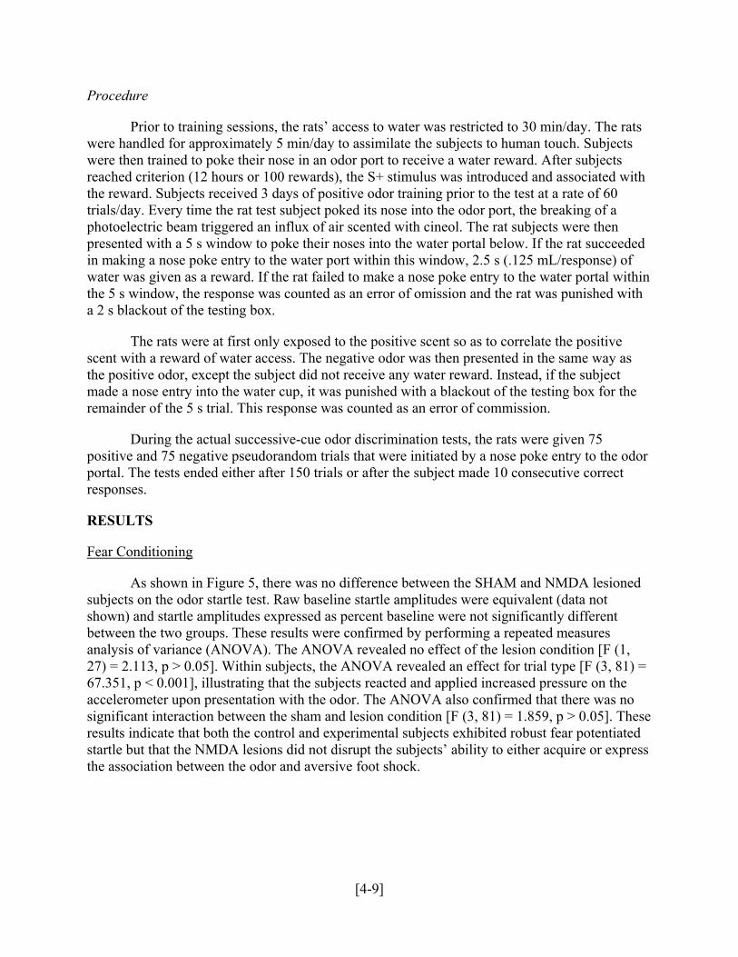
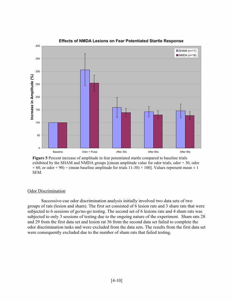
As shown in Figure 5, there was no difference between the SHAM and NMDA lesioned subjects on the odor startle test. Raw baseline startle amplitudes were equivalent (data not shown) and startle amplitudes expressed as percent baseline were not significantly different between the two groups. These results were confirmed by performing a repeated measures analysis of variance (ANOVA). The ANOVA revealed no effect of the lesion condition [F (1, 27) = 2.113, p > 0.05]. Within subjects, the ANOVA revealed an effect for trial type [F (3, 81) = 67.351, p < 0.001], illustrating that the subjects reacted and applied increased pressure on the accelerometer upon presentation with the odor. The ANOVA also confirmed that there was no significant interaction between the sham and lesion condition [F (3, 81) = 1.859, p > 0.05]. These results indicate that both the control and experimental subjects exhibited robust fear potentiated startle but that the NMDA lesions did not disrupt the subjects’ ability to either acquire or express the association between the odor and aversive foot shock.
[4-9]
Effects of NMDA Lesions on Fear Potentiated Startle Response
0
50
100
150
200
250
300
350
400
Baseline Odor + Pulse After 30s After 60s After 90s
Incr
ease
in A
mpl
itude
(%)
SHAM (n=11)NMDA (n=18)
Figure 5 Percent increase of amplitude in fear potentiated startle compared to baseline trials exhibited by the SHAM and NMDA groups [(mean amplitude value for odor trials, odor + 30, odor + 60, or odor + 90) ÷ (mean baseline amplitude for trials 11-30) × 100]. Values represent mean ± 1 SEM.
Odor Discrimination
Successive-cue odor discrimination analysis initially involved two data sets of two groups of rats (lesion and sham). The first set consisted of 6 lesion rats and 3 sham rats that were subjected to 6 sessions of go/no-go testing. The second set of 6 lesions rats and 4 sham rats was subjected to only 3 sessions of testing due to the ongoing nature of the experiment. Sham rats 28 and 29 from the first data set and lesion rat 36 from the second data set failed to complete the odor discrimination tasks and were excluded from the data sets. The results from the first data set were consequently excluded due to the number of sham rats that failed testing.
[4-10]
Distribution of Responses by Type (Session 3)
0
10
20
30
40
50
60
Positiveodor,
Response
PositiveOdor, NoResponse
NegativeOdor,
Response
NegativeOdor, NoResponse
Type of Response
Freq
uenc
y of
Res
pons
e (%
)
Sham (n=4)
Lesion (n=5)
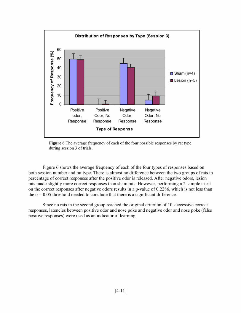
Figure 6 The average frequency of each of the four possible responses by rat type during session 3 of trials.
Figure 6 shows the average frequency of each of the four types of responses based on both session number and rat type. There is almost no difference between the two groups of rats in percentage of correct responses after the positive odor is released. After negative odors, lesion rats made slightly more correct responses than sham rats. However, performing a 2 sample t-test on the correct responses after negative odors results in a p-value of 0.2286, which is not less than the α = 0.05 threshold needed to conclude that there is a significant difference.
Since no rats in the second group reached the original criterion of 10 successive correct responses, latencies between positive odor and nose poke and negative odor and nose poke (false positive responses) were used as an indicator of learning.
[4-11]

Figure 7 The average latencies before both hits and false positives for both types of rats. Values represent means ± SEM.
Figure 7 shows average latencies before both types of responses. While both groups of rats had a longer latency period after receiving the negative odor than after receiving the positive odor, there is little evidence to suggest that the two groups of rats performed differently. The p-values of 2 sample t-tests of the latency times were 0.7044 for the positive and 0.7770 for the negative odors. Neither of these values are below the α < 0.05 threshold necessary to obtain a significant result. Since the p-values are so high, it can be concluded that lesions in the medial amygdala do not affect latency times before responses.
The mean latency to reach the final trial in the second data set was 2094.48 seconds for lesion rats and 2078.33 seconds for sham rats. The similarity in total latency times between the two groups further supports a lack of difference in odor discrimination post-surgery.
Percent Correct in Odor2 Trial
0
5
15
20
25
30
1 2
Rat Group
nt C
orre
ct
10
Per
ce
[4-12]
Figure 8 Comparison of percent correct in odor2 between lesion and sham rats is inconclusive due to inconsistencies in data. Values represent means ± SEM.
Percent correct in odor2 trials is calculated by recording the absence of head entry in thepresence
of a negative stimulus over total odor2 trials. The percentages (Figure 8) suggest a
significant difference in number of correct trials between lesion and sham rats. However, a t-test compar .
higher mean negative latency times with 1.725 seconds for lesion rats and 1.591 seconds for sham rats, a statistical t-test revealed no significant difference between the sham and lesion
The differences in expr ed responses betw lesion and sham rats are too insignificant for the medial amygdala to be considered an essential factor in olfaction. A difference in both latency times between initial and final sessions suggests the successful conditionin ecrease in positive la egative latencies across sessions in both groups of rats indicates similar ability in odor discrimination and equal post-surgery retention of olfaction ability.
ion
ison revealed no significant difference between the two groups [t(7)= 1.189, p= 0.273]
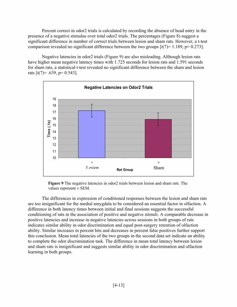
Negative latencies in odor2 trials (Figure 9) are also misleading. Although lesion rats have
rats [t(7)= .639, p= 0.543].
ession of condition een the
g of rats in the association of positive and negative stimuli. A comparable dtencies and increase in n
Similar increases in percent hits and decreases in percent false positives further support this conclusion. Mean total latencies of the two groups in the second data set indicate an ability to complete the odor discrimination task. The difference in mean total latency between lesand sham rats is insignificant and suggests similar ability in odor discrimination and olfaction learning in both groups.
Lesion Sham Figure 9
Negative Latencies on Odor2 Trials
10
11
12
13
14
15
16
17
18
19
Tim
e (.1
s)
1 2
Rat GroupLesion Sham
Figure 9 The negative latencies in odor2 trials between lesion and sham rats. The values represent ± SEM.
[4-13]
DISCUSSION
General Discussion
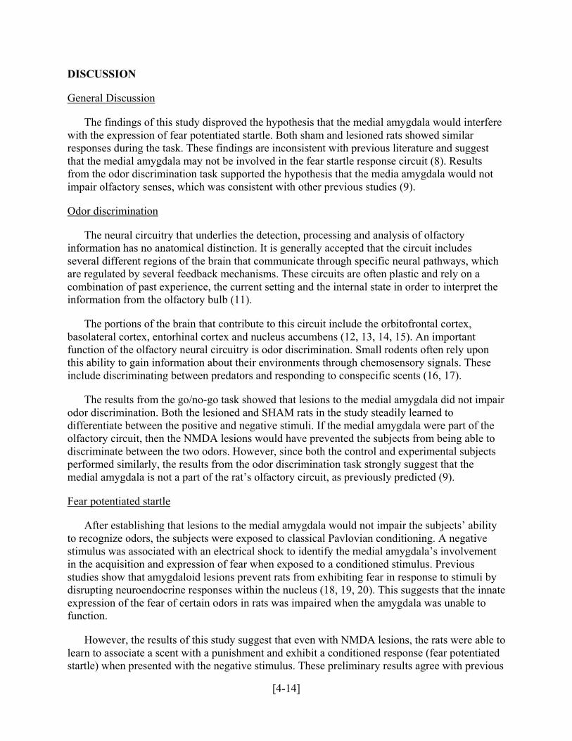
The findings of this study disproved the hypothesis that the medial amygdala would interfere f fear potentiated startle. Both sham and lesioned rats showed similar
responses during the task. These findings are inconsistent with previous literature and suggest that
not
with the expression o
the medial amygdala may not be involved in the fear startle response circuit (8). Results from the odor discrimination task supported the hypothesis that the media amygdala would impair olfactory senses, which was consistent with other previous studies (9).
Odor discrimination
The neural circuitry that underlies the detection, processing and analysis of olfactory atomical distinction. It is generally accepted that the circuit includes
several different regions of the brain that communicate through specific neural pathways, which are a
t the
ucleus accumbens (12, 13, 14, 15). An important function of the olfactory neural circuitry is odor discrimination. Small rodents often rely upon this ese
ir
differentiate between the positive and negative stimuli. If the medial amygdala were part of the olfa
subjects
information has no an
regulated by several feedback mechanisms. These circuits are often plastic and rely oncombination of past experience, the current setting and the internal state in order to interpreinformation from the olfactory bulb (11).
The portions of the brain that contribute to this circuit include the orbitofrontal cortex, basolateral cortex, entorhinal cortex and n
ability to gain information about their environments through chemosensory signals. Thinclude discriminating between predators and responding to conspecific scents (16, 17).
The results from the go/no-go task showed that lesions to the medial amygdala did not impaodor discrimination. Both the lesioned and SHAM rats in the study steadily learned to
ctory circuit, then the NMDA lesions would have prevented the subjects from being able to discriminate between the two odors. However, since both the control and experimental performed similarly, the results from the odor discrimination task strongly suggest that the medial amygdala is not a part of the rat’s olfactory circuit, as previously predicted (9).
Fear potentiated startle
After establishing that lesions to the medial amygdala would not impair the subjects’ ability ubjects were exposed to classical Pavlovian conditioning. A negative
stimulus was associated with an electrical shock to identify the medial amygdala’s involvement in t
o
to sociate a scent with a punishment and exhibit a conditioned response (fear potentiated
startle) when presented with the negative stimulus. These preliminary results agree with previous
to recognize odors, the s
he acquisition and expression of fear when exposed to a conditioned stimulus. Previous studies show that amygdaloid lesions prevent rats from exhibiting fear in response to stimuli bydisrupting neuroendocrine responses within the nucleus (18, 19, 20). This suggests that the innateexpression of the fear of certain odors in rats was impaired when the amygdala was unable tfunction.
However, the results of this study suggest that even with NMDA lesions, the rats were ablelearn to as
[4-14]
scie
entiated startle as an embedded component of the conditioned response pathway (10, 22).
Although brain histologies are necessary to confirm these results, it appears that lesions on the med
hin the larger context of fear potentiated startle research. Through a series of fear conditioning and successive-cue odor discrimination tests, the effects of the medial amygda l
lved
(1) Zhao Z, Yang Y, Walker DL, Davis M. Effects of Substance P in the Amygdala, us, and Periaqueductal Gray on Fear-Potentiated Startle.
europsychopharamacology. 2008;34:331-340.
(2) ty.
on hippocampal system differentiates anxiety from fear in startle paradigms.
Neuroscience. 2009;not yet published; unedited.
(5)
ion learning but not classical fear conditioning.
Journal of Neuroscience. 2000;20(18):7059-66.
ntific literature on medial amygdaloid lesions (8, 21). Therefore, it can be concluded that the medial amygdala is not involved with olfactory learning or memory recall within the olfactory circuit.
This study is unique compared to others that suggest that the medial amygdala is involved infear-pot
ial amygdala had no effect on the subject’s ability to learn a conditioned stimulus and exhibitthe conditioned response. This suggests that the possible error identified by Paschall and Davis, the disruption of performance because of NBQX infusions before training, may have occurred.
CONCLUSION
The goal of this study was to investigate the role of the medial amygdala in odor discrimination wit
la on olfactory learning were assessed. The results show that lesions to the mediaamygdala neither affected the expression of fear potentiated startle nor impaired the ability to discriminate between odors. While a larger group of subjects and histological studies arerequired to confirm these results, this research reveals that the medial amygdala is not invoin the neural pathways involved in olfactory learning. The significance of this discovery lies inits elucidation of the role of the medial amygdala in the formation of memories through associative learning.
REFERENCES
Ventromedial HypothalamN
Davis M. Are different parts of the extended amygdala involved in fear versus anxieBiological Psychiatry. 1998;44:1239-1247.
(3) Bullock TH. Introduction to nervous systems. Cambridge: Harvard University Press;
1959. 559p. (4) Veening JG, Böcker KBE, Verdouw PM, Olivier B, de Jongh R, Groenink L. Activati
of the Septo-
Paxinos G, Watson C. The rat brain in stereotaxic coordinates: the new coronal set. Elsevier Academic Press; 2005. 167p.
(6) Wilensky AE, Schafe GE, LeDoux JE. The amygdala modulates memory consolidat
of fear-motivated inhibitory avoidance
[4-15]
(7) Schettino LF, Otto T. Patterns of Fos expression in the amygdala and ventral perirhinal cortex induced by training in an olfactory fear conditioning paradigm. Behavioral Neuroscience. 2001;115(6):1257-1272
(8) Walker DL, Paschall GY, Davis M. Glutamate receptor antagonist infusions into the
basolateral and medial amygdala reveal differential contributions to olfactory vs. cfear conditioning and expression. Learn
ontext ing and Memory. 2005;12:120-129
1) Wilson DA, Best AR, Sullivan RM. Plasticity in the olfactory system: lessons for the
2) Schoenbaum G, Nugent SL, Saddoris MP, Setlow B. Orbitofrontal lesions in rats impair
4) Schoenbaum G, Setlow B, Nugent SL, Saddoris MP, Gallagher M. Lesions of
):129-40.
ctory
1;105(1):111-9.
(16) n
7) Takahashi LK, Chan MM, Pilar ML. Predator odor fear conditioning: current
n of
in rats. Brain Research. Uncorrected Proof: To be published Aug. 4, 2009, Brain Research 1283.
(9) Cousens G, Otto T. Both pre- and posttraining excitotoxic lesions of the basolateral
amygdale abolish the expression of olfactory and contextual fear conditioning. Behavioral Neuroscience. 1998;112(5):1092-1102
(10) Paschall GY, Davis M. Olfactory-mediated fear-potentiated startle. Behavioral
Neuroscience. 2002;116(1):4-12
(1neurobiology of memory. Neuroscientist. 2004;10(6):513
(1reversal but not acquisition of go, no-go odor discriminations. Neuroreport. 2002;13(6):885-90.
(13) Schoenbaum G, Setlow B. Lesions of nucleus accumbens disrupt learning about aversive
outcomes. The Journal of Neuroscience. 2003;23(30):9833-41.
(1orbitofrontal cortex and basolateral amygdala complex disrupt acquisition of odor-guided discriminations and reversals. Learning and Memory. 2003;10(2
(15) Otto T, Schottler F, Staubli U, Eichenbaum H, Lynch G. Hippocampus and olfa
discrimination learning: effects of entorhinal cortex lesions on olfactory learning and memory in a successive-cue, go-no-go task. Behavioral Neuroscience. 199
Samuelson CL, Meredith M. Categorization of biologically relevant chemical signals ithe medial amygdala. Brain Research. 2009;1263:33-42.
(1perspectives and new directions. Neuroscience Biobehavioral Review. 2008;32(7):1218-1227
(18) Masini CV, Sasse SK, Garcia RJ, Nyhuis TJ, Day HEW, Campeau S. Disruptio
neuroendocrine stress responses to acute ferret odor by medial, but not central amygdala lesions
[4-16]
(19) Lin JY, Roman C, St Andre J, Reilly S. Taste, olfactory and trigeminal neophobia inwith forebrain lesions. Brain Research. 2008;1251:195-203.
Slotnick BM. Ol
rats
(20) factory discrimination in rats with anterior amygdala lesions. Behavioral
Neuroscience. 1986;99(5):956-963.
(21) ygdaloid circuitries in the rat: an emerging framework for understanding functions of the amygdala. Trends in
2) Eichenbaum H, Fagan A, Cohen NJ. Normal olfactory discrimination learning set and
Pitkänen A, Savander V, LeDoux JE. Organization of intra-am
Neuroscience. 1997;20:517-523
(2facilitation of reversal leaning after medial-temporal damage in rats: implications for an account of preserved learning abilities in amnesia. The Journal of Neuroscience. 1986;6(7):1876-1884
[4-17]
Copyright © 2022 FDOKUMEN