GABAergic neurons expressing p75 in rat substantia innominata and nucleus basalis
Upload
independentCategory
view
3download
0
SYNAPSE 17~16-25 (1994)
Relationship Between Asymmetries in Striatal Dopamine Release and the Direction of Amphetamine-Induced Rotation During
the First Week Following a Unilateral 6-OHDA Lesion of the Substantia Nigra TERRY E. ROBINSON, MAURA NOORDHOORN, EMILY M. CHAN, ZOLTAN MOCSARY,
DIANNE M. CAMP, AND IAN Q. WHISHAW Department of Psychology and Neuroscience Program, The University of Michigan, Ann Arbor, Michigan 48104-1687 (T.E.R., M.N., E.M.C., Z.M., D.M.C.) and Department of Psychology, University of Lethbridge,
Lethbridge AB, Canada (I.Q. W.)
KEY WORDS
ABSTRACT In animals with a large unilateral 6-hydroxydopamine (6-OHDA) lesion of the nigrostriatal dopamine (DA) system the traditional “rotational behavior model” states that amphetamine will induce circling behavior towards the denervated striatum (ipsiversive), that is, away from the side where there is greater amphetamine-stimulated DA release and greater DA receptor stimulation. It is puzzling, therefore, why amphet- amine induces contraversive rotation in rats tested 4 days after a unilateral 6-OHDA lesion, despite a 90-95% loss of the dopaminergic input to the striatum by this time. Rats reverse their direction of amphetamine-induced rotation by 8 days post-lesion and turn in the ipsiversive direction thereafter. To try and resolve this paradox, bilateral striatal microdialysis was used to estimate the effects of amphetamine on DA neurotransmission on Day 4 and Day 8 following a large unilateral 6-OHDA lesion of the substantia nigra. On Day 4 post-lesion, amphetamine produced a moderate (around 50% of control) in- crease in the extracellular concentration of DA in the denervated striatum. This amphet- amine-releasable pool of DA was exhausted by a single amphetamine challenge, because a second injection of amphetamine given 3 h after the first did not produce a comparable increase in DA. It is suggested that on Day 4 post-lesion the amount of DA released by amphetamine in the denervated striatum is sufficient to produce greater DA receptor stimulation on that side, because of DA receptor supersensitivity, and this leads to contraversive rotation. On Day 8 post-lesion, amphetamine induced DA release in the intact striatum but had no effect on extracellular DA in the denervated striatum (DA was nondetectable). On Day 8, therefore, DA receptor stimulation would be greatest in the intact striatum, leading to ipsiversive rotation. In conclusion, it is suggested that the seemingly paradoxical reversal in the direction of amphetamine-induced rotation that occurs over the first week following a unilateral 6-OHDA lesion is consistent with the traditional rotational model, and is due to time-dependent changes in the ability of amphetamine to release DA in the denervated striatum.
Microdialysis, Recovery, Caudate nucleus, Striatum
o 1994 Wiley-Liss, Inc.
INTRODUCTION Drug-induced behavioral asymmetries seen in ani-
mals with unilateral damage to the nigrostriatal DA system are among the most extensively studied behav- ioral phenomena in neuroscience. The “rotational be- havior model,” first described by Ungerstedt (1971a,b; Ungerstedt and Arbuthnott, 19701, provides a gener-
era1 destruction of the nigrostriatal DA system, drugs that act to release DA, such as amphetamine, produce turning towards the denervated striatum (ipsiversive). Drugs that act as DA receptor agonists, such as apo- morphine, produce turning away from the denervated
ally accepted rule regarding the effects of dopaminergic drugs On On rotational or turning behavior. In animals with unilat- 0 1994 WILEY-LISS, INC
Received June 14,1993, accepted in revlsed form October 26,1993 Address correspondence to Dr Terry E Robinson, Neuroscience Laboratory
Building, The University of Michigan, 1103 East Huron Street, Ann Arbor, MI asymmetries and’ in 48104-1687

LESION-ASSOCIATED CHANGES IN DA RELEASE 17
striatum (contraversive). The rule, therefore, is that animals turn away from the striatum with the greatest DA receptor stimulation, which in the case of amphet- amine is on the intact side (where intact presynaptic terminals can release DA), and in the case of DA recep- tor agonists is on the denervated side (where DA recep- tors are supersensitive).
There are, however, apparent exceptions to this rule. For example, it has been known since Ungerstedt's (1971) original studies that for 24-36 h after a unilat- eral 6-OHDA lesion amphetamine often induces contra- versive rotation (Oberlander et al., 1980, as well). This is usually explained in terms of a transient accumula- tion of DA during the degenerative process. Of more interest are reports that in rats with a very large 6-OHDA lesion, amphetamine can induce contraver- sive rotation for up to 6 days post-lesion (Carey, 1992; Lynch and Carey, 1989; Mintz et al., 19861, despite an almost complete loss of the dopaminergic input to the striatum within 3-4 days post-lesion (Altar et al., 1984). By 3-4 days post-lesion there is already a >90- 95% decrease in DA histofluorescence (Hokfelt and Un- gerstedt, 1973), in the postmortem tissue content of DA (Altar et al., 1984; Carey, 1992; Neve et al., 1982; Staunton et al., 1981), in DA uptake sites (Altar et al., 19871, and in boutons with small granular vesicles (Hokfelt and Ungerstedt, 1973).
There are two possible interpretations of this phe- nomenon. First, for 4-6 days after a unilateral 6-OHDA lesion amphetamine may not induce turning towards the striatum with the greatest DA receptor stimulation. If this is true the basic tenet of the rotational behavior model would be violated. This could have important implications, because the model is widely used to esti- mate, for example, the actions of drugs on DA systems, the degree of recovery of function following DA deple- tion, and the effectiveness of neural tissue grafts (Zig- mond et al., 1990). A second possibility is that a t 4 days post-lesion amphetamine does result in greater DA re- ceptor stimulation in the denervated striatum. If this is true, there must be as of yet unidentified time-depen- dent changes in the ability of amphetamine to release DA, despite a >90-95% lesion of the nigrostriatal DA system. Carey (1992) suggested, for example, there may be a small residual amphetamine-releasable pool of DA that persists for 4-6 days post-lesion, and that DA released from this pool is sufficient to induce con- traversive rotation because of its action on supersensi- tive DA receptors. The purpose of the present study was to test this hypothesis.
METHODS Experiment 1
Subjects and surgical procedures Adult female rats (Holtzman Sprague-Dawley)
weighing approximately 225 g were housed individu-
ally in wire mesh cages in a room maintained on a 14:lO h 1ight:dark cycle (lights on at 08:00), with free access to food and water. The animals were pretreated with 5 mgkg of atropine methyl nitrate, anesthetized with 30 m&g of sodium pentobarbital (i.p.1, supplemented with methoxyflurane, and then using standard stereo- taxic procedures, one 23 gauge stainless steel guide cannula was positioned on the dural surface above the nigrostriatal bundle (coordinates: 3.0 mm posterior to bregma, 1.8 mm lateral, and 1.0 mm ventral from skull surface; Paxinos and Watson, 1986). The guide cannula was fixed in place with dental cement and jeweler's screws attached to the skull, and closed with a stainless steel stylet. After recovery from the anaesthetic the animals were returned to their home cages.
6-OHDA lesions At least 2 days following surgery the animals were
pretreated with 15 mgkg desipramine-HC1, and be- tween 30 and 60 rnin later a 30 gauge injection cannula was inserted into the guide cannula to the level of the nigrostriatal bundle. An infusion pump was used to infuse 8 kg of 6-hydroxydopamine HBr (6-OHDA) dis- solved in 4 p,l of vehicle (0.9% NaCl solution containing 0.1 mg/ml ascorbic acid) over a period of 8 min. Follow- ing the infusion the injection cannula was left in place for 2 min before it was removed and the stylet was replaced. The animals were not anaesthetized during this procedure. This day (the day of the lesion) was defined as Day 0.
Test procedure Two days following the 6-OHDA lesion (post-lesion)
animals were observed for spontaneous rotational be- havior for 10 rnin on a small (12" X 1 2 ) elevated plat- form, and then were returned to their home cage. Four days post-lesion each animal was placed in an auto- mated hemispherical rotometer described previously (McFarlane et al., 1992). This device recorded the num- ber of quarter (90") turns during each 5 min interval. These data were converted to full rotations, which are defined as four consecutive 90" turns in the same direc- tion, and net rotations, which are defined as full rota- tions in the dominant direction minus full rotations in the nondominant direction. Following a 15 min habitu- ation period animals received an i.p. injection of 1.5 mg/kg d-amphetamine sulfate (weight of the salt) and rotational behavior was recorded for 1 h. They were then replaced in their home cages, until this procedure was repeated, on Days 6, 8, and 10 post-lesion. Two weeks post-lesion the animals were again tested for rotational behavior, but now received 0.05 mgkg of apomorphine HC1 (weight of salt). This latter test was done to determine whether the animals had a nearly complete depletion of striatal DA, because animals with less than a 90-95% DA depletion do not respond to this dose of apomorphine (Marshall and Ungerstedt, 1977).

18 T.E. ROBINSON ET AL
To estimate the size of the lesion, and to confirm the reliability of the apomorphine test, the animals were killed 3 weeks post-lesion and striatal samples ob- tained for postmortem tissue assay.
To determine the extent of DA depletion in postmor- tem tissue specifically on Day 4 and Day 8 post-lesion, two additional independent groups of rats were given a 6-OHDA lesion, as described above, and striatal tissue was obtained at these points in time. These latter ani- mals did not undergo any behavioral testing.
Tissue assay Animals were killed by decapitation and their brains
were removed and placed in ice-cold saline. After the brain cooled (30-45 sec), it was placed in a chilled cut- ting block and brain slices were obtained as described by Heffner et al. (1980). Using a 2 mm tissue punch a sample from the corpus of each striatum was taken, weighed, and placed into a tube containing 0.05 N per- chloric acid and dihydroxybenzylamine (internal stan- dard). The samples were homogenized and centrifuged and the supernatant was assayed by HPLC-EC, using procedures similar to those described previously (Rob- inson et al., 1987).
Experiment 2 The subjects and surgical procedures were the same
as are described for Experiment 1, except in addition to implantation of a chronic guide cannula for the 6-OHDA lesion, a guide cannula was also placed on the dural surface above each striatum (i.e., bilaterally: co- ordinates, 0.6 mm anterior to bregma, 2.8 mm lateral, and 1 mm ventral from the skull surface). At least 3 days following surgery 15 animals received a 6-OHDA lesion, as described above, and 6 control animals re- ceived a “sham lesion” (i.e., the injection cannula was simply lowered and left in place for 10 min). Of the rats given a 6-OHDA lesion, eight underwent dialysis on Day 4 post-lesion and seven underwent dialysis on Day 8 post-lesion. Control animals were tested with each of these groups, using the following procedures.
The day prior to microdialysis testing, animals were lightly anaesthetized with ether and a dialysis probe was inserted into the guide cannula overlying each stri- atum. The exposed surface of the dialysis membrane was 4 mm long, and was positioned to sample the entire dorso-ventral extent of the striatum (see Fig. 4 in Rob- inson and Whishaw, 1988). The dialysis probes and pro- cedures have been described in detail previously (Rob- inson and Camp, 1991). Each rat was then placed in a hemispherical testing chamber equipped for both mi- crodialysis sampling and the automated measurement of rotational behavior. The probes were perfused with a perfusion solution containing 145 mM NaCl, 2.7 mM KCI, 1.2 mM CaC12, 1.0 mM MgCI,, and 0.2 mM ascor- bic acid, pH 7.3 (Moghaddam and Bunney, 19891, a t a flow rate of 0.3 pVmin. The following day, the pump
speed was increased to 1.5 pJmin, and after a t least a 30 min equilibration period, a minimum of three 20 minute basal samples of dialysate were collected. Fol- lowing this, each rat was given an injection of 1.5 mg/kg of d-amphetamine sulfate (i.p.1, and ten additional 20 min samples of dialysate were collected. At the same time, rotational behavior was monitored using the de- vice described above. At the completion of the experi- ment, the dialysis probes were removed, the stylets were replaced, and the rats were returned to their home cages.
Ten days post-lesion, animals with a 6-OHDA lesion were screened for apomorphine-induced rotation, as de- scribed above. Only animals that made at least 100 contraversive rotations within the 30 min test session were included in the experiment, to insure a homoge- nous group with >95% DA depletions. Following this, animals were given an overdose of sodium pentobar- bital, perfused through the heart with 0.9% saline fol- lowed by 10% formalin in saline, and the brains ob- tained for histological analysis. Sections of each brain were obtained through the striatum, mounted, stained with cresyl violet, and examined to reconstruct the track formed by each dialysis probe. Only animals with at least 75% of the dialysis surface of each probe located within the corpus of the striatum were included in the experiment.
Experiment 3 The procedures for this experiment were the same as
for Experiment 2, with the following modifications: 1) all animals were tested only 4 days following a unilat- eral 6-OHDA lesion (n = 81, and 2) after collecting basal dialysate all animals received two successive in- jections of 1.5 mg/kg of d-amphetamine, 200 min apart, and ten 20 min samples of dialysate were collected fol- lowing each injection.
RESULTS Experiment 1
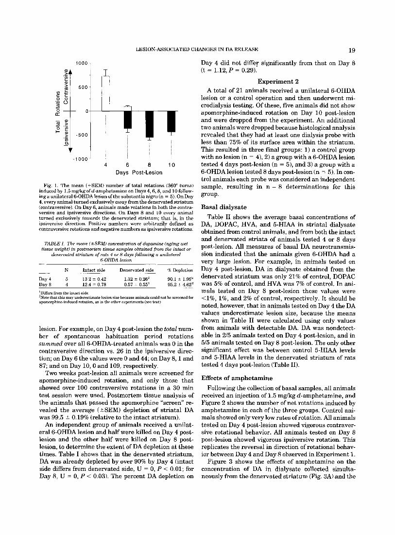
The direction and rate of amphetamine-induced rota- tion seen in animals tested 4,6,8, and 10 days following a unilateral 6-OHDA lesion is illustrated in Figure 1. Four days post-lesion amphetamine produced rotation predominantly away from the denervated striatum (contraversive) in every animal. By 6 days post-lesion, animals showed predominately contraversive rotation for the first 15 min of the test session and then switched direction and showed predominately ipsiversive rota- tion for the remainder of the test session (time course data not shown). Finally, by 8 and 10 days post-lesion, amphetamine produced ipsiversive rotation exclusively in every animal.
It needs to be emphasized, however, that during the 15 min habituation period, prior to amphetamine ad- ministration, spontaneous rotation was almost exclu- sively in the ipsiversive direction on all days post-
LESION-ASSOCIATED CHANGES IN DA RELEASE 19
Day 4 did not differ significantly from that on Day 8 (t = 1.12, P = 0.29).
1000 1
- 1 o o o J
I
4 6 8 10 Days Post-Lesion
Fig. 1. The mean (+SEM) number of total rotations (360" turns) induced by 1.5 mgkg ofd-amphetamine on Days 4,6,8, and 10 follow- ing a unilateral 6-OHDA lesion of the substantia nigra (n = 5). On Day 4, every animal turned exclusively away from the denervated striatum (contraversive). On Day 6, animals made rotations in both the contra- versive and ipsiversive directions. On Days 8 and 10 every animal turned exclusively towards the denervated striatum; that is, in the ipsiversive direction. Positive numbers were arbitrarily defined as contraversive rotations and negative numbers as ipsiversive rotations.
TABLE I . The mean ( H E M ) concentration of dopamine (nglmg wet tissue weight) in postmortem tissue samples obtained from the intact or
denervated striatum of rats 4 or 8 days following a unilateral 6-OHDA lesion
N Intact side Denervated side % DeDletion
Day 4 5 13.2 ? 0.42 1.32 ? 0.26l 90.1 t- 1.95' Day 8 4 12.4? 0.78 0.57 t- 0 . 5 ~ 5 ~ 95.2 2 4.62'
'Differs from the intact side. 'Note that this may underestimate lesion size because animals could not he screened for apomorphine-induced rotation, as in the other experiments (see text).
lesion. For example, on Day 4 post-lesion the total num- ber of spontaneous habituation period rotations summed over all 6-OHDA-treated animals was 0 in the contraversive direction vs. 26 in the ipsiversive direc- tion; on Day 6 the values were 0 and 44; on Day 8 , l and 87; and on Day 10,O and 109, respectively.
Two weeks post-lesion all animals were screened for apomorphine-induced rotation, and only those that showed over 100 contraversive rotations in a 30 min test session were used. Postmortem tissue analysis of the animals that passed the apomorphine "screen" re- vealed the average (&SEM) depletion of striatal DA was 99.5 k 0.19% (relative to the intact striatum).
An independent group of animals received a unilat- eral 6-OHDA lesion and half were killed on Day 4 post- lesion and the other half were killed on Day 8 post- lesion, to determine the extent of DA depletion at these times. Table I shows that in the denervated striatum, DA was already depleted by over 90% by Day 4 (intact side differs from denervated side, U = 0, P < 0.01; for Day 8, U = 0, P < 0.03). The percent DA depletion on
Experiment 2 A total of 21 animals received a unilateral 6-OHDA
lesion or a control operation and then underwent mi- crodialysis testing. Of these, five animals did not show apomorphine-induced rotation on Day 10 post-lesion and were dropped from the experiment. An additional two animals were dropped because histological analysis revealed that they had at least one dialysis probe with less than 75% of its surface area within the striatum. This resulted in three final groups: 1) a control group with no lesion (n = 4),2) a group with a 6-OHDA lesion tested 4 days post-lesion (n = 51, and 3) a group with a 6-OHDA lesion tested 8 days post-lesion (n = 5). In con- trol animals each probe was considered an independent sample, resulting in n = 8 determinations for this group.
Basal dialysate Table I1 shows the average basal concentrations of
DA, DOPAC, HVA, and 5-HIAA in striatal dialysate obtained from control animals, and from both the intact and denervated striata of animals tested 4 or 8 days post-lesion. All measures of basal DA neurotransmis- sion indicated that the animals given 6-OHDA had a very large lesion. For example, in animals tested on Day 4 post-lesion, DA in dialysate obtained from the denervated striatum was only 21% of control, DOPAC was 5% of control, and HVA was 7% of control. In ani- mals tested on Day 8 post-lesion these values were <1%, 1%, and 2% of control, respectively. It should be noted, however, that in animals tested on Day 4 the DA values underestimate lesion size, because the means shown in Table I1 were calculated using only values from animals with detectable DA. DA was nondetect- able in 215 animals tested on Day 4 post-lesion, and in 515 animals tested on Day 8 post-lesion. The only other significant effect was between control 5-HIAA levels and 5-HIAA levels in the denervated striatum of rats tested 4 days post-lesion (Table 11).
Effects of amphetamine Following the collection of basal samples, all animals
received an injection of 1.5 mgkg d-amphetamine, and Figure 2 shows the number of net rotations induced by amphetamine in each of the three groups. Control ani- mals showed only very low rates of rotation. All animals tested on Day 4 post-lesion showed vigorous contraver- sive rotational behavior. All animals tested on Day 8 post-lesion showed vigorous ipsiversive rotation. This replicates the reversal in direction of rotational behav- ior between Day 4 and Day 8 observed in Experiment 1.
Figure 3 shows the effects of amphetamine on the concentration of DA in dialysate collected simulta- neously from the denervated striatum (Fig. 3A) and the

20 T.E. ROBINSON ET A L
TABLE II. Basal dialysate values (mean t SEMI in pglpl (corrected for recouery')
200
100
- 0
-100
N DA DOPAC HVA 5-HIAA
Control 8 1.39 t 0.22 805 i 79.3 587 ? 86.5 197 lr 13.9 Day 4
(Intact) 5 1.35 ? 0.25 732 t 90.4 718 + 102.5 210 t 25.1 (97%) (91%) (122%) (107%)
(70%) (Denervated) 5 0.29 t 0.112 39.8 ? 6.07' 43.1 2 6.37' 138 -c 21.23
~~
(7%) -~ (21%) 2 ND (5%) ___-- Day 8
(Intact) 5 1.93 i 0.65 784 t 88.6 689 * 171 230 t 35.9
(Denervated) 5 5ND2 7.49 ? 0.112 8.82 t 5.74' 173 t 26.0 (139%) (97%) (117%) (117%)
(<1%) (1%) 3 ND (2%) 3 ND (88%)
'Numbers in parentheses indicate the experimental mean expressed as a percent of the control mean, and the number of animals in which values were nondetectable is indicated by the number followed by ND where applicable. 2Differs significantly from the intact side and fmm control (U = 0 to 4, P < 0.01-0.001). 3Differs significantly from control (U = 4, P < 0.01).
20 Minute Intervals
Fig. 2. The mean (kSEM) number of net rotations per 20 min inter- val induced by 1.5 mgkg of d-amphetamine in animals undergoing microdialysis. There are three independent groups: 1) a group with a unilateral 6-OHDA lesion that was tested on Day 4 post-lesion (n = 5; closed circles), 2) a group with a unilateral 6-OHDA lesion group that was tested on day 8 post-lesion (n = 5; closed squares), and 3) a control group (n = 4; open circles). On Day 4 post-lesion, all animals with a 6-OHDA lesion turned predominantly in the contraversive direction. On Day 8, all animals with a 6-OHDA lesion turned predominantly in the ipsiversive direction. Control animals showed only very low rates of rotational behavior with no significant directional bias. Positive numbers were arbitrarily defined as contraversive rotations and neg- ative numbers as ipsiversive rotations. B, baseline; A1 to A10, succes- sive 20 min intervals following amphetamine. All group comparisons were highly significant (two-way ANOVAs).
intact striatum (Fig. 3B), as well as from control ani- mals, as they engaged in the rotational behavior sum- marized in Figure 2. Amphetamine produced a large increase in the concentration of DA in dialysate ob- tained from the intact striatum of all animals, and there were no significant group differences (Fig. 3B). In animals tested 4 days post-lesion amphetamine pro- duced a moderate increase in DA on the denervated side (Fig. 3A), although this was significantly less than oh the intact side, or in control animals. In animals tested 8 days post-lesion, DA was nondetectable in the denervated striatum at all points in time (Fig. 3A).
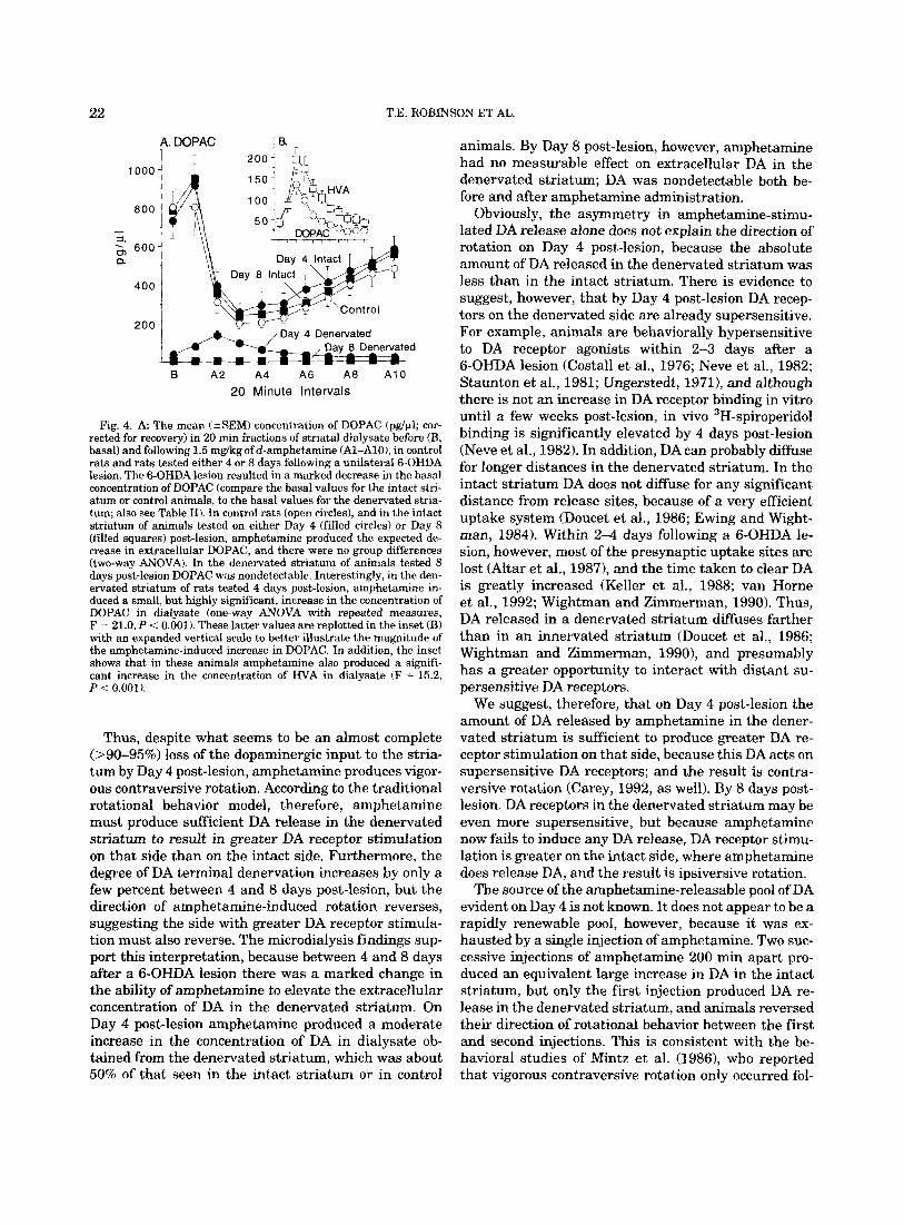
Figure 4A shows the effects of amphetamine on the concentration of DOPAC in dialysate collected simulta- neously from the intact and denervated striata of rats with a 6-OHDA lesion, and from control animals. In control animals, and in the intact striatum of animals with a 6-OHDA lesion, amphetamine produced the ex- pected decrease in DOPAC concentrations. In the den- ervated striatum of animals tested 8 days post-lesion DOPAC was detectable in only two of five animals, and therefore, no effect of amphetamine was evident. In the denervated striatum of animals tested 4 days post- lesion, however, amphetamine produced a small, but highly significant, increase in both DOPAC and HVA. This is best illustrated by the inset in Figure 4 (4B), in which the vertical scale is expanded.
Experiment 3 Figure 5 shows the effects of two successive injections
of 1.5 mgkg of d-amphetamine on rotational behavior. Following the first injection of amphetamine all ani- mals showed contraversive rotation for approximately 1.5 h, after which time they started to show low levels of ipsiversive rotation. Following the second injection of amphetamine all animals showed predominantly ip- siversive rotation.
Figure 6 shows the effects of amphetamine on the concentration of DA in dialysate collected from the in- tact and denervated striata, as animals engaged in the behavior summarized in Figure 5. Both injections of amphetamine produced an equivalent large increase in the concentration of DA in dialysate collected from the intact striatum. In the denervated striatum, however, the first injection of amphetamine produced a modest but significant increase in DA, but the second injection of amphetamine had very little effect.
Figure 7 shows the effects of amphetamine on the concentration of DOPAC in dialysate collected simulta- neously from the intact and denervated striata. In the intact striatum amphetamine produced the expected decrease in DOPAC. By the end of the first injection period DOPAC concentrations were returning to base-

LESION-ASSOCIATED CHANGES IN DA RELEASE 21
A. Denervated Striaturn -f- Day 4 Post-lesion + Day 8 Post-lesion -0- Controls
I
LLm&* o;,
B A 2 A 4 A 6 A 8 A 1 0 B A 2 A 4 A 6 A 8 A 1 0
20 Minute intervals
Fig. 3. The mean (rSEM) concentration of DA (pg/pl; corrected for recovery) in 20 min fractions of striatal dialysate collected before (B, basal) and following 1.5 mgkg of &hetamine (Al-AlO), in control rats (n = 8 determinations) and rats tested either 4 days (n = 5) or 8 days (n = 5) following a unilateral 6-OHDA lesion. A: Denervated Striatum. Amphetamine induced a significant increase in extracellu- lar DA in the striatum of control rats, and in the denervated striatum of rats tested on Day 4 post-lesion (for the Day 4 post-lesion group: one-way ANOVA with repeated measures, F = 4.52, P < 0.001). The amount of amphetamine-stimulated DA release in the denervated
line, but they declined again following the second injec- tion of amphetamine. In the denervated striatum, how- ever, the first injection of amphetamine produced a small, but highly significant, increase in DOPAC, and this was not seen following the second injection of am- phetamine (see inset in Fig. 7).
DISCUSSION This study confirms previous reports that rats with a
large unilateral 6-OHDA lesion of the substantia nigra reverse their direction of amphetamine-induced rota- tional behavior between 4 and 8 days post-lesion (Carey, 1992; Lynch and Carey, 1989; Mintz et al., 1986). For 4-6 days post-lesion, amphetamine produces contraversive rotational behavior. By 8 days post-lesion rats reverse their direction of amphetamine-induced rotation, and turn in the ipsiversive direction there- after.
According to the traditional rotational behavior model, animals should not turn in the contraversive direction following amphetamine administration (Un- gerstedt, 1971a, 1971b; Ungerstedt and Arbuthnott, 1970), at least if the striatum on the side with a lesion is denervated of DA terminals. There is considerable evi- dence, however, suggesting that the striatum ipsilat- era1 to a 6-OHDA lesion is already denervated of 90- 95% of its dopaminergic input within 3-4 days post- lesion. For example, in this, and other studies, the postmortem tissue content of DA was reduced by over 90% by Day 4 post-lesion (Carey, 1992; Mishra et al., 1980; Neve et al., 1984; Staunton et al., 1981), and this
striatum of animals tested 4 days post-lesion was significantly less than in their intact striatum, and in controls (Day 4, denervated stria- tum vs. controls, F = 9.54, P < 0.01; Day 4, denervated striatum vs. Day 4, intact striatum, F = 7.29, P < 0.001). In animals tested 8 days post-lesion amphetamine failed to release DA (DA was nondetectable both before and after amphetamine administration). B: Intact Stria- tum. Amphetamine produced a large, statistically significant increase in extracellular DA in all groups, and there were no significant group differences (two-way ANOVA). Note that the same control values shown in A are simply replotted in B to facilitate comparisons.
is thought to provide a good index of DA terminal den- sity (Altar et al., 1987). Others have also reported that within 2 4 days following a 6-OHDA lesion there is already a >90-95% decrease in the density of DA up- take sites (Altar et al., 1987), in catecholamine histoflu- orescence (Hokfelt and Ungerstedt, 19731, and in the number of boutons containing small granular vesicles (Hokfelt and Ungerstedt, 1973). Furthermore, in the present study, basal DOPAC concentrations in dialy- sate from the denervated striatum were reduced by 95% within 4 days post-lesion, indicative of a massive loss of dopaminergic input at this time. There was a further decrease in DOPAC of around 4% between Day 4 and Day 8, which is consistent with postmortem tis- sue measures reported here and elsewhere (Carey, 1992). Finally, during the first week post-lesion rats with a unilateral 6-OHDA lesion show marked behav- ioral deficits, including spontaneous ipsiversive rota- tional behavior and contralateral sensorimotor neglect (Altar et al., 1984; present study). [As an aside, it is worth noting that the basal concentration of DOPAC in dialysate probably provides a more accurate index of DA terminal density in this situation than does DA itself, because DOPAC is formed primarily intraneu- ronally, independent of DA release (Kuczenski and Segal, 1989; Zetterstrom et al., 19881, and because DOPAC is not influenced by the same compensatory neuroadaptations that serve to normalize the extracel- lular concentration of DA (Castaneda et al., 1990; Rob- inson et al,, 1990; Robinson and Whishaw, 1988; Zig- mond et al., 19901.1
22 T.E. ROBINSON ET AL.
1000 -
800 -
- 5. & 600- Q
400 -
200
- B A 2 A4 A6 A8 A 1 0
20 Minute Intervals
Fig. 4. A. The mean ("SEM) concentration of DOPAC (pg/pl; cor- rected for recovery) in 20 min fractions of striatal dialysate before (B, basal) and following 1.5 mgkg of d-amphetamine (Al-AlO), in control rats and rats tested either 4 or 8 days following a unilateral 6-OHDA lesion. The 6-OHDA lesion resulted in a marked decrease in the basal concentration of DOPAC (compare the basal values for the intact stri- atum or control animals, to the basal values for the denervated stria- turn; also see Table 11). In control rats (open circles), and in the intact striatum of animals tested on either Day 4 (filled circles) or Day 8 (filled squares) post-lesion, amphetamine produced the expected de- crease in extracellular DOPAC, and there were no group differences (two-way ANOVA). In the denervated striatum of animals tested 8 days post-lesion DOPAC was nondetectable. Interestingly, in the den- ervated striatum of rats tested 4 days post-lesion, amphetamine in- duced a small, but highly significant, increase in the concentration of DOPAC in dialysate (one-way ANOVA with repeated measures, F = 21.0, P < 0.001). These latter values are replotted in the inset (B) with an expanded vertical scale to better illustrate the magnitude of the amphetamine-induced increase in DOPAC. In addition, the inset shows that in these animals amphetamine also produced a signifi- cant increase in the concentration of HYA in dialysate (F = 15.2, P < 0.001).
Thus, despite what seems to be an almost complete (>90-95%) loss of the dopaminergic input to the stria- tum by Day 4 post-lesion, amphetamine produces vigor- ous contraversive rotation. According to the traditional rotational behavior model, therefore, amphetamine must produce sufficient DA release in the denervated striatum to result in greater DA receptor stimulation on that side than on the intact side. Furthermore, the degree of DA terminal denervation increases by only a few percent between 4 and 8 days post-lesion, but the direction of amphetamine-induced rotation reverses, suggesting the side with greater DA receptor stimula- tion must also reverse. The microdialysis findings sup- port this interpretation, because between 4 and 8 days after a 6-OHDA lesion there was a marked change in the ability of amphetamine to elevate the extracellular concentration of DA in the denervated striatum. On Day 4 post-lesion amphetamine produced a moderate increase in the concentration of DA in dialysate ob- tained from the denervated striatum, which was about 50% of that seen in the intact striatum or in control
animals. By Day 8 post-lesion, however, amphetamine had no measurable effect on extracellular DA in the denervated striatum; DA was nondetectable both be- fore and after amphetamine administration.
Obviously, the asymmetry in amphetamine-stimu- lated DA release alone does not explain the direction of rotation on Day 4 post-lesion, because the absolute amount of DA released in the denervated striatum was less than in the intact striatum. There is evidence to suggest, however, that by Day 4 post-lesion DA recep- tors on the denervated side are already supersensitive. For example, animals are behaviorally hypersensitive to DA receptor agonists within 2-3 days after a 6-OHDA lesion (Costal1 et al., 1976; Neve et al., 1982; Staunton et al., 1981; Ungerstedt, 1971), and although there is not an increase in DA receptor binding in vitro until a few weeks post-lesion, in vivo 3H-spiroperidol binding is significantly elevated by 4 days post-lesion (Neve et al., 1982). In addition, DA can probably diffuse for longer distances in the denervated striatum. In the intact striatum DA does not diffuse for any significant distance from release sites, because of a very efficient uptake system (Doucet et al., 1986; Ewing and Wight- man, 1984). Within 2 4 days following a 6-OHDA le- sion, however, most of the presynaptic uptake sites are lost (Altar et al., 19871, and the time taken to clear DA is greatly increased (Keller et al., 1988; van Horne et al., 1992; Wightman and Zimmerman, 1990). Thus, DA released in a denervated striatum diffuses farther than in an innervated striatum (Doucet et al., 1986; Wightman and Zimmerman, 1990), and presumably has a greater opportunity to interact with distant su- persensitive DA receptors.
We suggest, therefore, that on Day 4 post-lesion the amount of DA released by amphetamine in the dener- vated striatum is sufficient to produce greater DA re- ceptor stimulation on that side, because this DA acts on supersensitive DA receptors; and the result is contra- versive rotation (Carey, 1992, as well). By 8 days post- lesion, DA receptors in the denervated striatum may be even more supersensitive, but because amphetamine now fails to induce any DA release, DA receptor stimu- lation is greater on the intact side, where amphetamine does release DA, and the result is ipsiversive rotation.
The source of the amphetamine-releasable pool of DA evident on Day 4 is not known. It does not appear to be a rapidly renewable pool, however, because it was ex- hausted by a single injection of amphetamine. Two suc- cessive injections of amphetamine 200 min apart pro- duced an equivalent large increase in DA in the intact striatum, but only the first injection produced DA re- lease in the denervated striatum, and animals reversed their direction of rotational behavior between the first and second injections. This is consistent with the be- havioral studies of Mintz et al. (19861, who reported that vigorous contraversive rotation only occurred fol-

LESION-ASSOCIATED CHANGES IN DA RELEASE 23
6o 1 contraversive T
c a - 2 0 -
- 4 0 -
0 10 20 30 40 50 60 70 80
5 Minute intervals
Fig. 5. The mean (+SEM) number of net rotations per 5 min inter- val following two successive injections of 1.5 mgkg of d-amphetamine (n = 5). Amphetamine was given at the time indicated by the arrow- heads. Positive numbers were arbitrarily defined as rotations in the contraversive direction and negative numbers as rotations in the ip-
siversive direction. Note that following the first injection of amphet- amine all animals turned predominantly in the contraversive direc- tion, whereas following the second injection all animals turned predominantly in the ipsiversive direction.
40
a, S .- 30 E x g 20
10
1 B.,Percent of Baseline
10000
8000
1 l \ T I Denervated
4 8 12 16 20
20 Minute Intervals
Fig. 6. The mean (2SEM) concentration of DA in successive 20 min fractions of striatal dialysate obtained simultaneously from the intact and denervated striata of rats tested 4 days following a unilateral 6-OHDA lesion (n = 5). Samples were obtained both before (first 3 intervals) and following two successive injections of 1.5 mgkgofd-am- phetamine. Amphetamine was administered at the time indicated by the arrowheads. A DA expressed in pg/pl. In the intact striatum, the first and second injection of amphetamine induced an equivalent large increase in the concentration of DA (comparison of 1st vs. 2nd injec- tion, two-way ANOVA, non-significant). In the denervated striatum,
lowing the first exposure to amphetamine, and primar- ily ipsiversive rotation occurred with subsequent injec- tions of amphetamine.
It is interesting to speculate that the residual pool of DA seen on Day 4 may be contained in structures some- what analogous to “synaptosomes.” In this experiment 6-OHDA was infused near the DA cell bodies, and the soma and axons of most DA cells may have degenerated by Day 4 post-lesion. This may leave, however, a few isolated DA terminals in the striatum that have not yet degenerated. These “in vivo synaptosomes” may con-
the first injection of amphetamine produced a smaller, but highly significant, increase in extracellular DA. The second injection of am- phetamine produced very little DA release in the denervated striatum, relative to either the intact striatum, or that seen on the denervated side following the first injection (two-way ANOVA comparing 1st vs. 2nd injection, F,,,,,,, = 14.3, P < 0.004, F,,,,,,,,,, = 11.2, P < 0.001). B: DA expressed as a percent of baseline. The decrease in the magnitude of DA release in the denervated striatum between the first and second injections of amphetamine is especially evident when the data are plotted as a percent of baseline (Fig. 6B).
tain DA, but would not show spontaneous impulse-de- pendent DA release, because there is no soma to initiate impulses, and no axon to conduct them. Amphetamine may release DA, however, because the DA transporter is still functional, allowing for accelerative exchange- diffusion (Fischer and Cho, 1979). This would explain why on Day 4 post-lesion spontaneous rotational be- havior is in the ipsiversive direction, but amphetamine- induced rotation in the contraversive direction. Whether other ion channels are operative, as in synap- tosomes, is not known, but if they are, the local applica-

24 T.E. ROBINSON ET AL.
1500
f- 2. 0) . ,a 1000
2 s a
500
20 Minute Intervals
Fig. 7. The mean (?SEMI concentration of DOPAC in successive 20 min fractions of striatal dialysate (pg/pl) obtained simultaneously from the intact (closed circles) and denervated striata (open circles) of rats tested 4 days following a unilateral 6-OHDA lesion (n = 5). Sam- ples were obtained both before (first 3 intervals) and following two successive injections of 1.5 mgkg of &hetamine. Amphetamine was administered at the time indiated by the arrowheads. The 6-OHDA lesion resulted in a marked decrease in the basal concentra- tion of DOPAC in dialysate (compare basal intact side to basal dener- vated side values). In the intact striatum, amphetamine produced the expected decrease in extracellular DOPAC following both the first and second treatments. In the denervated striatum, the first injection of amphetamine produced a small, but highly significant, increase in DOPAC (one-way ANOVA, F = 13.9, P < 0.001). In contrast, the sec- ond injection of amphetamine did not produce a comparable increase in DOPAC (P < 0.01). The values for DOPAC for just the denervated side are replotted in the inset, with an expanded vertical scale, to better illustrate the magnitude of the amphetamine-induced increase in DOPAC. Values for intervals 19-23 are not given for the denervated striatum because DOPAC was nondetectable in too many samples to calculate meaningful averages.
tion of other releasing agents should also evoke DA release. This pool of DA may not be renewable, how- ever, because without a cell body the enzymes critical for neurotransmitter synthesis, and the proteins re- quired for neurotransmission, could not be replenished. Ultrastructurally, this pool of DA may be located in 'boutons of the "dense" type' described by Hokfelt and Ungerstedt (1969), which are most numerous 2-4 days after a 6-OHDA lesion.
A final interesting observation in the present study was the amphetamine-induced increase in the concen- tration of DOPAC in the denervated striatum seen 4 days post-lesion. Amphetamine normally produces a decrease in extracellular DOPAC, as seen here in con- trol animals and in the intact striatum of animals with a 6-OHDA lesion. Amphetamine is thought to decrease DOPAC because it depletes a cytoplasmic pool of newly synthesized DA that is normally subject to degradation by intraneuronal monoamine oxidase (Zetterstrom et al., 1988). The amount of DOPAC formed from re- leased DA that is taken back up into the presynaptic terminal, and then metabolized, is thought to be insig- nificant normally, relative to the amount of DOPAC produced intraneuronally, independent of release. This
is why DOPAC usually is considered to be a poor indica- tor of DA release (Wood and Altar, 1988). In the dener- vated striatum, however, the contribution of DOPAC formed from released DA may be relatively large, be- cause most cytoplasmic DOPAC is lost when the DA terminals degenerate. Thus, in the denervated stria- tum the metabolism of released DA may be "unmasked" in a sense, and under these conditions the extracellular concentration of DOPAC reflects DA release.
In conclusion, we suggest that the seemingly para- doxical reversal in the direction of amphetamine-in- duced rotational behavior between 4 and 8 days after a unilateral 6-OHDA lesion is not inconsistent with the traditional rotational behavior model (Ungerstedt, 1971a,b; Ungerstedt and Arbuthnott, 1970). Our re- sults suggest that on Day 4 post-lesion the amount of DA released by amphetamine in the denervated stria- tum is sufficient to produce greater DA receptor stimu- lation on that side, because this DA acts on supersensi- tive DA receptors. By Day 8 post-lesion animals reverse their direction of amphetamine-induced rotational be- havior, because now amphetamine fails to release any DA in the denervated striatum, and therefore, there is greater DA receptor stimulation on the intact side, where amphetamine does release DA.
ACKNOWLEDGMENTS This research was supported by a grant to T.E.R.
from the National Parkinson Foundation (U.S.A.) and to I.Q.W. from the Medical Research Council of Canada.
REFERENCES Altar, C.A., Marien, M.R., and Marshall, J.F. 11987) Time course of
adaptations in dopamine biosynthesis, metabolism, and release fol- lowing nigrostriatal lesions: Implications for behavioral recovery from brain injury. J. Neurochem., 48:390-399.
Altar, C.A., O'Neil, S., and Marshall, J.F. (1984 ) Sensorimotor impair- ment and elevated levels of dopamine metabolites in the neostria- tum occur rapidly after intranigral injection of 6-hydroxydopamine or gamma-hydroxybutyrate in awake rats. Neuropharmacology, 23: 309-318.
Carey, R.J. (1992) Factors in amphetamine-induced contralateral ro- tation in the unilateral 6-OHDA lesion rat model during the first- week postoperative: Implications for neuropathology and neural grafting. Brain Res., 570:ll-20.
Castaneda, E., Whishaw, I.Q., and Robinson, T.E. (1990) Changes in striatal dopamine neurotransmission assessed with microdialysis following recovery from a bilateral 6-OHDA lesion: Variation as a function of lesion size. J . Neurosci., 10:1847-1854.
Costall, B., Marsden, C.D., Naylor, R.J., and Pycock, C.J. (1976) The relationship between striatal and mesolimbic dopamine dysfunction and the nature of circling responses following 6-hydroxydopamine and electrolytic lesions of the ascending dopamine systems of rat brain. Brain Res., 118:87-113.
Doucet, G., Descarries, L., and Garcia, S. (1986) Quantification of the dopamine innervation in adult rat neostriatum. Neuroscience, 19: 427-445.
Ewing, A.G., and Wightman, R.M. (1984) Monitoring the stimulated release of dopamine with in vivo voltammetry. 11. Clearance of re- leased dopamine from extracellular fluid. J. Neurochem., 43:570- 577.
Fischer, J.F., and Cho, A.K. 11979) Chemical release of dopamine from striatal homogenates: Evidence for an exchange diffusion model. J . Pharmacol. Exp. Ther., 2082203-209.
Heffner, T.F., Hartman, J.A., and Seiden, L.S. (1980) A rapid method for the regional dissection of the rat brain. Pharmacol. Biochem. Behav., 13:453-456.

Hokfelt, T., and Ungerstedt, U. (1969) Electron and fluorescence mi- croscopical studies on the nucleus caudatus putamen of the rat after unilateral lesions of ascending nigro-neostriatal dopamine neurons. Acta Physiol. Scand., 76:415-426.
Hokfelt, T., and Ungerstedt, U. (1973) Specificity of 6-hydroxydopam- ine induced degeneration of central monoamine neurones: An elec- tron and fluorescence microscopic study with special reference to intracerebral injection on the nigrostriatal dopamine system. Brain Res., 60:269-297.
Keller, R.W., Jr, Kuhr, W.G., Wightman, R.M., and Zigmond, M.J. (1988) The effect of L-dopa on in vivo dopamine release from nigro- striatal bundle neurons. Brain Res., 447:191-194.
Kuczenski, R., and Segal, D. (1989) Concomitant characterization of behavioral and striatal neurotransmitter response to amphetamine using in vivo microdialysis. J . Neurosci., 9:2051-2065.
Lynch, M.R., and Carey, R. J. (1989) Amphetamine-induced rotation reveals post 6-OHDA lesion neurochemical reorganization. Behav. Brain Res., 32:69-74.
Marshall, J.F., and Ungerstedt, U. (1977) Supersensitivity to apomor- phine following destruction of the ascending dopamine neurons: Quantification using the rotational model. Eur. J . Pharmacol., 41: 361-367.
McFarlane, D.K., Martonyi, B.J., and Robinson, T.E. (1992) An inex- pensive automated system for the measurement of rotational behav- ior in small animals. Behav. Res. Methods Inst. Computers, 24:414- 419.
Mintz, M., Douglas, R.J., Tomer, R., de Villiers, A.S., and Kellaway, L. (1986) Transient contralateral rotation following unilateral sub- stantia nigra lesion reflects susceptibility of the nigrostriatal sys- tem to exhaustion by amphetamine. Life Sci., 39:69-76.
Mishra, R.K., Marshall, A.M., and Varmuza, S.L. (1980) Supersensi- tivity in rat caudate nucleus: effects of 6-hydroxydopamine on the time course of dopamine receptor and cyclic AMP changes. Brain
-
Res., 200:47-57. Moghaddam, B., and Bunney, B.S. (1989) Ionic composition of microdi-
alysis perfusing solution alters the pharmacological responsiveness and basal outflow of striatal dopamine. J. Neurochem., 536524354.
Neve, K.A., Altar, C.A., Wong, C.A., and Marshall, J.F. (1984) Quanti- tative analysis of [3H]spiroperidol binding to rat forebrain sections: plasticity of neostriatal dopamine receptors after nigrostriatal in- jury. Brain Res., 3029-18.
Neve, K.A., Kozlowski, M.R., and Marshall, J.F. (1982) Plasticity of neostriatal dopamine receptors after nigrostriatal injury: relation- ship to recovery of sensorimotor functions and behavioral supersen- sitivity. Brain Res., 244:33-44.
Oberlander, C., Dumont, C., and Boissier, J.R. (1980) Time course of apomorphine-induced circling behaviour after striatal dopamine re- ceptor denervation. Eur. J . Pharmacol., 62:107-110.
Paxinos, G.. and Watson, C. (1986) The Rat Brain in Stereotaxic Coor-
LESION-ASSOCIATED CHANGES IN DA RELEASE 25
dinates. Raven Press, New York.
Robinson, T.E., Becker, J.B., Young, E.A., Akil, H., and Castaneda, E. (1987) The effects of footshock stress on regional brain dopamine metabolism and pituitary beta-endorphin release in rats previously sensitized to amphetamine. Neuropharmacology, 26:679-691.
Robinson, T.E., and Camp, D.M. (1991) The feasibility of repeated microdialysis for within-subjects design experiments: Studies on the mesostriatal dopamine system. In: Microdialysis in the Neuro- sciences. T.E. Robinson and J.B. Justice Jr . , eds. Elsevier, Amster- dam, pp. 189-234.
Robinson. T.E.. Castaneda. E.. and Whishaw. LQ. (1990) ComDensa- tory changes' in striatal dopamine neurons following recovery from injury induced by 6-OHDA or methamphetamine: A review of evi- dence of microdialysis studies. Can. J. Psychol., 44:253-275.
Robinson, T.E., and Whishaw, I.Q. (1988) Normalization of extracellu- lar dopamine in striatum following recovery from a partial unilat- eral 6-OHDA lesion of the substantia nigra: A microdialysis study in freely moving rat. Brain Res., 450:209-224.
Staunton, D.A., Wolfe, B.B., Groves, P.M., and Molinoff, P.B. (1981) Dopamine receptor changes following destruction of the nigrostri- atal pathway: Lack of a relationship to rotational behavior. Brain Res., 211:315-327.
Ungerstedt, U. (1971a) Postsynaptic supersensitivity after 6-hydroxy- dopamine induced degeneration of the nigro-striatal dopamine sys- tem. Acta Physiol. Scand., 367:69-93.
Ungerstedt, U. (1971b) Striatal dopamine release after amphetamine or nerve degeneration revealed by rotational behaviour. Acta Phys- iol. Scand. 367:49-68.
Ungerstedt, U., and Arbuthnott, G.W. (1970) Quantitative recording of rotational behavior in rats after 6-hydroxy-dopamine lesions of the nigrostriatal dopamine system. Brain Res., 24:485-493.
van Horne, C., Hoffer, B.J., Stromberg, I., and Gerhardt, G.A. (1992) Clearance and diffusion of locally applied dopamine in normal and 6-hydroxydopamine-lesioned rat striatum. J . Pharmacol. Exp. Ther., 263:1285-1292.
Wightman, R.M. and Zimmerman, J.B. (1990) Control of dopamine extracellular concentration in rat striatum by impulse flow and uptake. Brain Res. Rev., 15:135-144.
Wood, P.L., and Altar, C.A. (1988) Dopamine release in vivo from nigrostriatal, mesolimbic, and mesocortical neurons: utility of 3-methoxytyramine measurements. Pharmacol. Rev., 40:163-187.
Zetterstrom, T., Sharp, T., Collin, A.K., and Ungerstedt, U. (1988) In vivo measurement of extracellular dopamine and DOPAC in rat striatum after various dopamine-releasing drugs; implications for the origin of extracellular DOPAC. Eur. J . Pharmacol., 148:327-
-
- 334.
Zigmond, M.J., Abercrombie, E.D., Berger, T.W., Grace, A.A., and Stricker, E.M. (1990) Compensations after lesions of central dopam- inergic neurons: Some clinical and basic implications. Trends Neu- rosci., 13:290-296.
Copyright © 2022 FDOKUMEN