Melatonin protects against 6-OHDA-induced neurotoxicity in rats: a role for mitochondrial complex I...
7
Melatonin protects against 6-OHDA-induced neurotoxicity in rats: a role for mitochondrial complex I activity FEDERICA DABBENI-SALA, STEFANIA DI SANTO, DAVIDE FRANCESCHINI, STEPHEN D. SKAPER,* AND PIETRO GIUSTI 1 Department of Pharmacology, University of Padova, 35131 Padova, Italy; and *Department of Neuroscience Research, SmithKline Beecham Pharmaceuticals, Harlow, Essex CM19 5AW, U.K. ABSTRACT Unilateral injection into the right sub- stantia nigra of the catecholaminergic neurotoxin 6-hy- droxydopamine (6-OHDA) produces extensive loss of dopaminergic cells (‘hemi-parkinsonian rat’). The pi- neal hormone melatonin, which is a potent antioxidant against different reactive oxygen species and has been reported to be neuroprotective in vivo and in vitro, was evaluated for potential anti-Parkinson effects in this model. Imbalance in dopaminergic innervation be- tween the striata produced by intranigral administra- tion of 6-OHDA results in a postural asymmetry causing rotation away from the nonlesioned side. Melatonin given systemically prevented apomorphine-induced cir- cling behavior in 6-OHDA-lesioned rats. Reduced activ- ity of mitochondrial oxidative phosphorylation en- zymes has been suggested in some neurodegenerative diseases; in particular, selective decrease in complex I activity is observed in the substantia nigra of Parkin- son’s disease patients. Analysis of mitochondrial oxida- tive phosphorylation enzyme activities in nigral tissue from 6-OHDA-lesioned rats by a novel BN-PAGE histo- chemical procedure revealed a clear loss of complex I activity, which was protected against in melatonin- treated animals. A good correlation between behavioral parameters and enzymatic (complex I) analysis was observed independent of melatonin administration. A deficit in mitochondrial complex I could conceivably contribute to cell death in parkinsonism via free radical mechanisms, both directly via reactive oxygen species production and by decreased ATP synthesis and energy failure. Melatonin may have potential utility in the treatment of neurodegenerative disorders where oxida- tive stress is a participant.—Dabbeni-Sala, F., Di Santo, S., Franceschini, D., Skaper, S. D., Giusti, P. Melatonin protects against 6-OHDA-induced neurotoxicity in rats: a role for mitochondrial complex I activity. FASEB J. 15, 164 –170 (2001) Key Words: Parkinson’s disease z neurodegeneration z pineal hormone z neuroprotection z oxidative phosphorylation Parkinson’s disease (PD) is an age-related disorder characterized by a progressive degeneration of dopami- nergic neurons of the substantia nigra (SN) pars com- pacta. The underlying cause of this selective cell death is not understood, although several hypotheses have been advanced (1). Generation of reactive oxygen species (ROS) caused by oxidative stress, together with a relative paucity of antioxidant defenses in the SN and nigrostriatal dopaminergic pathway, is widely consid- ered as the final cause of neuronal death (2, 3). A similar loss in nigro-striatal dopaminergic neurons is produced on intracerebral administration of the catecholaminergic neurotoxin 6-hydroxydopamine (6- OHDA). In this animal model of PD, termed ‘the hemi-parkinsonian rat’ (4, 5), unilateral injection of 6-OHDA into the nigro-striatal pathway results in ex- tensive loss of dopaminergic cells in the ipsilateral SN (6 – 8). The resulting imbalance in dopaminergic inner- vation between the striata produces a postural asymme- try that manifests itself in the animal rotating away from the nonlesioned side (6). The 6-OHDA lesion model has been used to evaluate potential anti-Parkinson agents (6). The clinical relevance of 6-OHDA toxicity has been strengthened by the detection of 6-OHDA in urine of parkinsonian patients treated with l-DOPA (9). Damage induced by 6-OHDA in vivo and in cultured neuronal cells is similar to that induced by ROS, i.e., increased lipid peroxidation, a decrease in reduced glutathione, and an increased iron content (10). In fact, 6-OHDA-induced release iron from ferritin (11) may enhance oxidative stress through induction of hydroxyl radical formation via the Fenton reaction (reviewed in ref 12). Increasing evidence points to a correlation between neurodegenerative diseases and reduced activity of mitochondrial oxidative phosphor- ylation (OXPHOS) enzymes (13, 14). For instance, a selective 30 – 40% decrease in complex I activity has been found in the SN of PD patients (15). Such a defect could be due to an inherited mutation, although disease-related mitochondrial DNA mutations are not known; more probably, toxic insult secondary to oxi- dant stress is responsible. A mitochondrial complex I deficit could contribute to cell degeneration in PD via 1 Correspondence: Department of Pharmacology, L.go Me- neghetti, 2, 35131 Padova, Italy. E-mail: [email protected] 164 0892-6638/01/0015-0164/$02.25 © FASEB
Transcript of Melatonin protects against 6-OHDA-induced neurotoxicity in rats: a role for mitochondrial complex I...

Melatonin protects against 6-OHDA-inducedneurotoxicity in rats: a role for mitochondrialcomplex I activity
FEDERICA DABBENI-SALA, STEFANIA DI SANTO, DAVIDE FRANCESCHINI,STEPHEN D. SKAPER,* AND PIETRO GIUSTI1
Department of Pharmacology, University of Padova, 35131 Padova, Italy; and *Department ofNeuroscience Research, SmithKline Beecham Pharmaceuticals, Harlow, Essex CM19 5AW, U.K.
ABSTRACT Unilateral injection into the right sub-stantia nigra of the catecholaminergic neurotoxin 6-hy-droxydopamine (6-OHDA) produces extensive loss ofdopaminergic cells (‘hemi-parkinsonian rat’). The pi-neal hormone melatonin, which is a potent antioxidantagainst different reactive oxygen species and has beenreported to be neuroprotective in vivo and in vitro, wasevaluated for potential anti-Parkinson effects in thismodel. Imbalance in dopaminergic innervation be-tween the striata produced by intranigral administra-tion of 6-OHDA results in a postural asymmetry causingrotation away from the nonlesioned side. Melatoningiven systemically prevented apomorphine-induced cir-cling behavior in 6-OHDA-lesioned rats. Reduced activ-ity of mitochondrial oxidative phosphorylation en-zymes has been suggested in some neurodegenerativediseases; in particular, selective decrease in complex Iactivity is observed in the substantia nigra of Parkin-son’s disease patients. Analysis of mitochondrial oxida-tive phosphorylation enzyme activities in nigral tissuefrom 6-OHDA-lesioned rats by a novel BN-PAGE histo-chemical procedure revealed a clear loss of complex Iactivity, which was protected against in melatonin-treated animals. A good correlation between behavioralparameters and enzymatic (complex I) analysis wasobserved independent of melatonin administration. Adeficit in mitochondrial complex I could conceivablycontribute to cell death in parkinsonism via free radicalmechanisms, both directly via reactive oxygen speciesproduction and by decreased ATP synthesis and energyfailure. Melatonin may have potential utility in thetreatment of neurodegenerative disorders where oxida-tive stress is a participant.—Dabbeni-Sala, F., Di Santo,S., Franceschini, D., Skaper, S. D., Giusti, P. Melatoninprotects against 6-OHDA-induced neurotoxicity in rats:a role for mitochondrial complex I activity. FASEB J. 15,164–170 (2001)
Key Words: Parkinson’s disease z neurodegeneration z pinealhormone z neuroprotection z oxidative phosphorylation
Parkinson’s disease (PD) is an age-related disordercharacterized by a progressive degeneration of dopami-nergic neurons of the substantia nigra (SN) pars com-
pacta. The underlying cause of this selective cell deathis not understood, although several hypotheses havebeen advanced (1). Generation of reactive oxygenspecies (ROS) caused by oxidative stress, together witha relative paucity of antioxidant defenses in the SN andnigrostriatal dopaminergic pathway, is widely consid-ered as the final cause of neuronal death (2, 3).
A similar loss in nigro-striatal dopaminergic neuronsis produced on intracerebral administration of thecatecholaminergic neurotoxin 6-hydroxydopamine (6-OHDA). In this animal model of PD, termed ‘thehemi-parkinsonian rat’ (4, 5), unilateral injection of6-OHDA into the nigro-striatal pathway results in ex-tensive loss of dopaminergic cells in the ipsilateral SN(6–8). The resulting imbalance in dopaminergic inner-vation between the striata produces a postural asymme-try that manifests itself in the animal rotating away fromthe nonlesioned side (6). The 6-OHDA lesion modelhas been used to evaluate potential anti-Parkinsonagents (6). The clinical relevance of 6-OHDA toxicityhas been strengthened by the detection of 6-OHDA inurine of parkinsonian patients treated with l-DOPA(9).
Damage induced by 6-OHDA in vivo and in culturedneuronal cells is similar to that induced by ROS, i.e.,increased lipid peroxidation, a decrease in reducedglutathione, and an increased iron content (10). Infact, 6-OHDA-induced release iron from ferritin (11)may enhance oxidative stress through induction ofhydroxyl radical formation via the Fenton reaction(reviewed in ref 12). Increasing evidence points to acorrelation between neurodegenerative diseases andreduced activity of mitochondrial oxidative phosphor-ylation (OXPHOS) enzymes (13, 14). For instance, aselective 30–40% decrease in complex I activity hasbeen found in the SN of PD patients (15). Such a defectcould be due to an inherited mutation, althoughdisease-related mitochondrial DNA mutations are notknown; more probably, toxic insult secondary to oxi-dant stress is responsible. A mitochondrial complex Ideficit could contribute to cell degeneration in PD via
1 Correspondence: Department of Pharmacology, L.go Me-neghetti, 2, 35131 Padova, Italy. E-mail: [email protected]
164 0892-6638/01/0015-0164/$02.25 © FASEB
a direct generation of ROS together with a decrease inATP synthesis leading to energy failure. Cat-echolamines themselves are potent inhibitors of mito-chondrial respiration (16). Moreover, 6-OHDA toxicitycan be attenuated by free radical scavengers (16–18).
We have shown that the pineal hormone melatoninprevents the neurotoxic effects of ROS triggered bykainic acid receptor activation (19–22). Melatonin an-tioxidant effects directed to different ROS species (23,24) have been documented in various in vitro and invivo models (25–28). Moreover, this pineal productincreased mRNA levels and activity of several antioxi-dant enzymes and inhibited apoptosis caused by6-OHDA in PC12 cells (29).
In the present study we have used the hemi-Parkin-son rat model of intranigral 6-OHDA administration toanalyze the effects of melatonin on apomorphine-induced circling behavior and mitochondrial OXPHOSenzyme activities. Furthermore, a blue native-polyacryl-amide gel electrophoresis (BN-PAGE) histochemicalmethod was used for the first time to evaluate damageand repair in a central nervous system degenerativepathology.
MATERIALS AND METHODS
Animals
Adult male Sprague-Dawley rats (body weight 250620 g) wereobtained from Harlan Italy (S. Pietro al Natisone, UD).Animals were housed under conditions of controlled temper-ature (2362°C) and illumination (12 h light; 12 h darkness;darkness: 19:00–07:00) with free access to standard diet andwater. Experiments were performed between 10:00 and 17:00in compliance with the European Communities Councildirective of 24 November 1986 (86/609/IIC) concerning theexperimental use of animals.
6-OHDA lesions
Animals (seven per group) were anesthetized intramuscularwith a solution of ketamine (87 mg/kg) plus xylazine (13mg/kg) and secured in a Kopf stereotaxic instrument withthe tooth bar set 15 mm above the interaural line. Lesionswere made by the unilateral injection of 6-OHDA (8 mg in 2ml) into the right SN at the following coordinates: AP, 24.8;L, 22.2; DV, 28.7 mm from Bregma (30). The sham-operated animals received vehicle only (0.1% ascorbate in0.9% saline) at the same coordinates. 6-OHDA administrationwas performed using a 27 gauge Hamilton syringe connectedto an infusion minipump (Harvard Compact Infusion Pump,Holliston, Mont.) at a rate of 0.5 ml/min. The syringe was leftin place for 5 min before slowly retracting it to allow for toxininfusion and prevent reflux.
Melatonin administration and measurement
Melatonin was administered immediately after intranigral6-OHDA injection. Osmotic minipumps filled with a solutionof melatonin (50 mg/ml in saline plus 50% of DMSO; 220 ml)were placed in the subcutaneous (s.c.) tissue between thescapulae. The delivery rate was constant at 1 6 0.15 ml/h(5067.5 mg melatonin/h). This produced a plasma concen-
tration of 1660 6 240 pg melatonin/ml for at least 7 days.Other groups of seven animals received a bolus injection ofmelatonin s.c. (5.0 mg/kg) immediately after 6-OHDA; con-trols were administered vehicle.
Melatonin was measured as described by Chanut et al. (31).Plasma samples of 1 ml were added to 5 ml of dichlorometh-ane after alkalinization with 100 ml 1N KOH; 6-fluoro-tryptamine (10 ml of a 0.05 mM solution) was added asinternal standard. Samples were shaken for 10 min and thencentrifuged (10 min, 1500 g, 4°C). The aqueous phase wasremoved and the lower (organic) phase taken to drynessunder a nitrogen stream. The residue was dissolved of 100 mlmobile phase buffer and 50 ml subjected to high-performanceliquid chromatography using a Beckman System Gold PSM125 pump equipped with a Rheodyne injection valve and areversed-phase C18 (150 mm 3 4.6 mm) Ultrasphere column.The mobile phase consisted of water-acetonitrile (80:20, v/v)containing 0.01 mM EDTA, 0.1 mM KH2PO4, and 0.5 mMoctan-sulfonic acid (pH 4.7): flow rate 1.4 ml/min. Electro-chemical detection was performed with an amperometricdetector (BAS, LC-4B) at a working potential of 10.9 V.
Behavioral testing
Two weeks after right intranigral stereotaxic injection of6-OHDA, animals were subjected to rotational behavior test-ing (6–8). Rats were injected s.c. with R-(2)-apomorphinehydrochloride, melatonin, or vehicle, placed in a cylindricalcage (240 mm in diameter, 300 mm high), and the number ofrotations over a 1 h period, both ipsilateral and contralateral,were recorded using an automatic rotometer (Rota-Count 8;Columbus Instruments, Columbus, Ohio).
Electrophoretic analysis
Five rats each from groups receiving 6-OHDA plus melatonin(minipump) or 6-OHDA plus vehicle were used. Animalswere killed after behavioral testing; the right and left SN wereexcised according to Heffner et al. (32) and immediatelyfrozen in liquid nitrogen. On the day of analysis, 5 mg offrozen SN were homogenized in 1 ml of 0.44 M sucrose, 1 mMEDTA, 0.2 mM phenylmethylsulfonyl fluoride, and 20 mM3-(N-morpholino)propanesulfonic acid, pH 7.2, using a tight-fitting Teflon pestle and Beckman polycarbonate centrifugetube. The homogenate was centrifuged at 20,000 g for 20 min;the supernatant was discarded and the pellet resuspended in1 ml buffer and centrifuged as above. The pellet was thensuspended in 40 ml 1M aminocaproic acid, 50 mM Bis-Tris/HCl pH 7.0 (4°C) containing protease inhibitors (20 mMphenylmethylsulfonyl fluoride, 20 mM N-tosyl-L-phenylala-nine chloromethyl ketone, 1 mg/ml pepstatin and 1 mg/mlleupeptin) and membranes solubilized by adding 15 ml 10%freshly prepared dodecylmaltoside. After centrifugation for25 min at 100,000 g, the supernatants were collected and thepellets were re-extracted with the same volume of buffereddetergent and centrifuged for 25 min at 100,000 g. The firstand second extracts were pooled, the final volume being100–120 ml for each tissue sample. The extracts were usedimmediately for BN-PAGE or stored in small aliquots at280°C for several months. BN-PAGE was performed using aminigel apparatus (miniprotean, Bio-Rad, 1370382 mm). A5–11% polyacrylamide gradient was used; 14 ml of superna-tant plus 1 ml 5% Serva Blue G in 1M aminohexanoic acid wasapplied to each lane. BN-PAGE was performed as described(33), with the blue cathode buffer being replaced by a clearbuffer immediately after the sample passed the stacking gel.Gels were sliced into individual lanes for histochemical andCoomassie staining.
1656-OHDA-INDUCED PARKINSONISM AND MELATONIN PROTECTION
Histochemical evaluation of BN-PAGE gels
Histochemical staining of BN-PAGE was performed as de-scribed (34). Complex I (NADH, NADH dehydrogenase)activity was determined by incubating gel slices with 2 mMTris-HCl pH 7.4, 0.1 mg/ml NADH, and 2.5 mg/ml NTB(Nitro Blue Tetrazolium) at room temperature. Complex II(succinate dehydrogenase) activity was evaluated by incubat-ing gel slices at room temperature with 4.5 mM EDTA, 10 mMKCN, 0.2 mM phenazine methosulfate, 84 mM succinic acid,and 50 mM NTB in 1.5 mM phosphate buffer (pH 7.4).Complex IV (cytochrome oxidase) activity was assessed byincubating gel slices with 5 mg 3,39-diaminobenzidine tetra-hydrochloride dissolved in 9 ml phosphate buffer (0.05 M,pH 7.4), 1 ml catalase (20 mg/ml), 10 mg cytochrome c, and750 mg sucrose. Maximum color was achieved with a 3 hincubation. Color development for complex I, II, and IVreacting bands was preserved by fixing gels in 50% methanol/10% acetic acid for 15 min; the fixed gel was stored in 10%acetic acid. Complex V (ATPase) activity was measured byincubating gel slices overnight in 35 mM Tris, 270 mMglycine, 14 mM MgSO4, 0.2% Pb(NO3)2, and 8 mM ATP, pH7.8, at room temperature. The gel was then washed andstored in distilled water. The remaining gel slice from eachsample group was fixed and stained with Coomassie blue G(33). Color intensity of the stained bands was assessed byscanning the still wet gel with a HP3 Scanjet; areas wereintegrated using Sigma-plot Jandel (Sigma Chemical Co., St.Louis, Mo.). Band area was expressed either in arbitrary unitsor relative to the area for Coomassie blue-stained complex V.This allowed for BN-PAGE results to be given quantitatively(34).
Materials
6-OHDA, melatonin, and xylazine HCl were purchased fromSigma. 6-OHDA HBr was dissolved in sterile saline containing0.1% ascorbic acid; melatonin in 0.9% saline/50% DMSO.R-(2)-apomorphine HCl was obtained from ICN BiomedicalInc. (Costa Mesa, Calif.) and dissolved in sterile saline.Ketamine HCl was obtained from Virbac (Milan, Italy).ALZET mini-osmotic pumps (model 2001) were purchasedfrom Alza Pharmaceuticals (Palo Alto, Calif.). Serva Blue G(Coomassie blue) was from by Serva (Heidelberg, Germany),dodecylmaltoside from Boehringer (Mannheim, Germany),digitonin from Merck (Darmstad, Germany), 6-aminocaproicacid from Fluka (Milan, Italy), acrylamide and bis-acrylamidefrom Bio-Rad (Milan, Italy), and nitrocellulose filters forWestern blotting from Pharmacia (Uppsala, Sweden). Allother chemicals were purchased from Sigma-Aldrich (Milan,Italy).
Statistics
Behavioral test results were subjected to Kruskall-Wallis non-parametric analysis of variance, followed by a two-tailedMann-Whitney U test. Data were expressed as the mean 6 sedespite the probable non-normality of the distribution ofscores. OXPHOS respiratory chain enzyme activities on rightand left sides and the effects of 6-OHDA and 6-OHDA plusmelatonin were analyzed by a t test.
RESULTS
Apomorphine induces circling behavior after 6-OHDAlesioning
Rats with unilateral (right SN) 6-OH-DA lesions exhib-ited apomorphine-induced contralateral rotations:
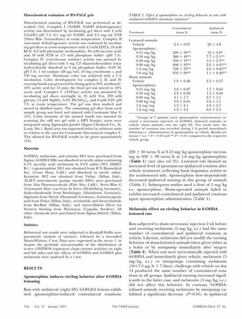
220 6 50 turns/h at 0.15 mg/kg apomorphine increas-ing to 856 6 99 turns/h at 1.6 mg/kg apomorphine(Table 1) (see also ref 35). Lesioned rats showed anincreased level of spontaneous ipsilateral rotation aftervehicle treatment, reflecting basal dopamine activity inthe nonlesioned side. Apomorphine dose-dependentlydecreased ipsilateral turning in this group of animals(Table 1). Subsequent studies used a dose of 1 mg/kgs.c. apomorphine. Sham-operated animals failed toexhibit increased contralateral and ipsilateral rotationsupon apomorphine administration (Table 1).
Melatonin effect on circling behavior in 6-OHDAlesioned rats
Rats subjected to sham stereotaxic injection 2 wk beforeand receiving melatonin (5 mg/kg, s.c.) had the samenumber of contralateral and ipsilateral rotations asvehicle. Likewise, melatonin did not modify the turningbehavior of sham-lesioned animals when given either asa bolus or by minipump immediately after surgery(Table 2). When rats were stereotaxically injected with6-OHDA and immediately given vehicle, melatonin (5mg/kg, s.c.) or minipumps containing melatonin(5067.5 mg/h 3 7 days), challenge with vehicle on day14 produced the same number of contralateral rota-tions in all groups. Ipsilateral turning increased signif-icantly in the latter case, and melatonin (5 mg/kg, s.c.)did not affect this behavior. In contrast, 6-OHDA-infused animals receiving melatonin by minipump ex-hibited a significant decrease (P,0.01) in ipsilateral
TABLE 1. Effect of apomorphine on circling behavior in rats withunilateral 6-OHDA stereotaxic injectionsa
TreatmentContralateral
turns/hIpsilateralturns/h
Lesioned animalsVehicle 2.4 6 0.97 23 6 4.0Apomorphine:
0.15 mg/kg 220 6 50** 12 6 2.8*0.30 mg/kg 380 6 49** 7.7 6 2.1**0.60 mg/kg 450 6 31** 5.4 6 2.3**0.80 mg/kg 608 6 83** 2.6 6 0.81**1.2 mg/kg 720 6 78** 2.0 6 0.65**1.6 mg/kg 856 6 99** 1.1 6 0.40**
Sham animalsVehicle 1.9 6 0.46 2.3 6 0.57Apomorphine:
0.15 mg/kg 3.6 6 0.87 1.7 6 0.640.30 mg/kg 2.9 6 0.99 1.3 6 0.680.60 mg/kg 4.1 6 1.7 2.7 6 1.10.80 kg/mg 3.9 6 0.94 3.6 6 1.51.2 mg/mg 5.3 6 2.1 4.9 6 2.11.6 mg/mg 5.9 6 2.2 6.0 6 2.3
a Groups of 7 animals (each apomorphine concentration) re-ceived a stereotaxic injection of 6-OHDA (lesioned animals) orvehicle (sham animals) into the right SN. Two weeks later, thenumber of rotations was recorded during 1 h period immediatelyfollowing s.c. administration of apomorphine or vehicle. Results aremeans 6 s.e. * P , 0.05 and ** P , 0.01 compared to the respectivevehicle group.
166 Vol. 15 January 2001 DABBENI-SALA ET AL.The FASEB Journal
rotations (6.363.5 vs. 2364.0) when challenged withvehicle (Table 2). Minipumps were not implanted priorto 6-OHDA administration, as damage caused by theneurotoxin is fully evident after 2 wk and still present10 months later (36). The first week, and not the initialhours after 6-OHDA treatment, appears critical.
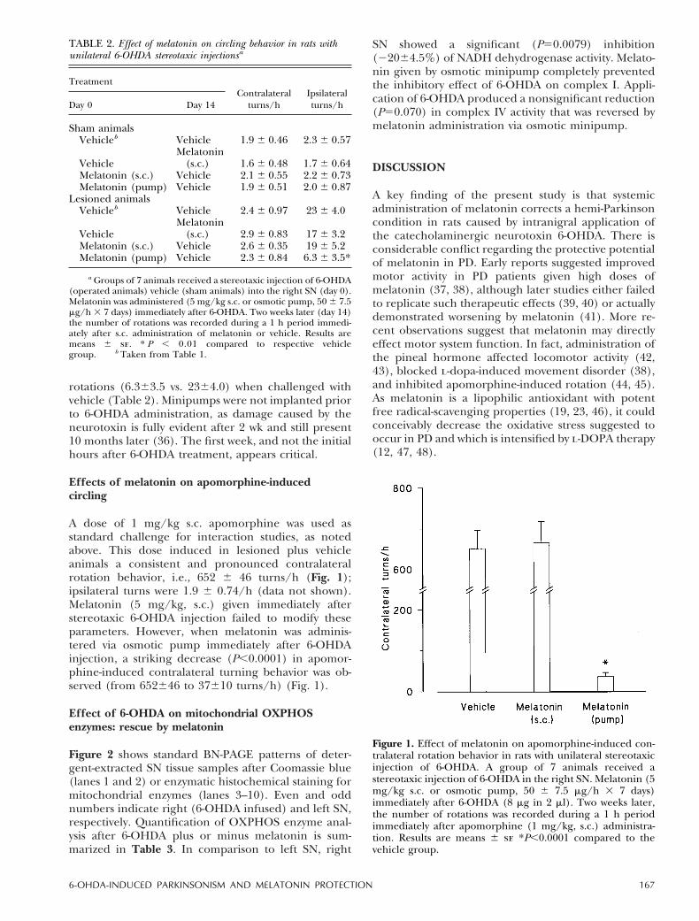
Effects of melatonin on apomorphine-inducedcircling
A dose of 1 mg/kg s.c. apomorphine was used asstandard challenge for interaction studies, as notedabove. This dose induced in lesioned plus vehicleanimals a consistent and pronounced contralateralrotation behavior, i.e., 652 6 46 turns/h (Fig. 1);ipsilateral turns were 1.9 6 0.74/h (data not shown).Melatonin (5 mg/kg, s.c.) given immediately afterstereotaxic 6-OHDA injection failed to modify theseparameters. However, when melatonin was adminis-tered via osmotic pump immediately after 6-OHDAinjection, a striking decrease (P,0.0001) in apomor-phine-induced contralateral turning behavior was ob-served (from 652646 to 37610 turns/h) (Fig. 1).
Effect of 6-OHDA on mitochondrial OXPHOSenzymes: rescue by melatonin
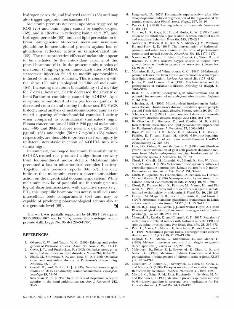
Figure 2 shows standard BN-PAGE patterns of deter-gent-extracted SN tissue samples after Coomassie blue(lanes 1 and 2) or enzymatic histochemical staining formitochondrial enzymes (lanes 3–10). Even and oddnumbers indicate right (6-OHDA infused) and left SN,respectively. Quantification of OXPHOS enzyme anal-ysis after 6-OHDA plus or minus melatonin is sum-marized in Table 3. In comparison to left SN, right
SN showed a significant (P50.0079) inhibition(22064.5%) of NADH dehydrogenase activity. Melato-nin given by osmotic minipump completely preventedthe inhibitory effect of 6-OHDA on complex I. Appli-cation of 6-OHDA produced a nonsignificant reduction(P50.070) in complex IV activity that was reversed bymelatonin administration via osmotic minipump.
DISCUSSION
A key finding of the present study is that systemicadministration of melatonin corrects a hemi-Parkinsoncondition in rats caused by intranigral application ofthe catecholaminergic neurotoxin 6-OHDA. There isconsiderable conflict regarding the protective potentialof melatonin in PD. Early reports suggested improvedmotor activity in PD patients given high doses ofmelatonin (37, 38), although later studies either failedto replicate such therapeutic effects (39, 40) or actuallydemonstrated worsening by melatonin (41). More re-cent observations suggest that melatonin may directlyeffect motor system function. In fact, administration ofthe pineal hormone affected locomotor activity (42,43), blocked l-dopa-induced movement disorder (38),and inhibited apomorphine-induced rotation (44, 45).As melatonin is a lipophilic antioxidant with potentfree radical-scavenging properties (19, 23, 46), it couldconceivably decrease the oxidative stress suggested tooccur in PD and which is intensified by l-DOPA therapy(12, 47, 48).
Figure 1. Effect of melatonin on apomorphine-induced con-tralateral rotation behavior in rats with unilateral stereotaxicinjection of 6-OHDA. A group of 7 animals received astereotaxic injection of 6-OHDA in the right SN. Melatonin (5mg/kg s.c. or osmotic pump, 50 6 7.5 mg/h 3 7 days)immediately after 6-OHDA (8 mg in 2 ml). Two weeks later,the number of rotations was recorded during a 1 h periodimmediately after apomorphine (1 mg/kg, s.c.) administra-tion. Results are means 6 se *P,0.0001 compared to thevehicle group.
TABLE 2. Effect of melatonin on circling behavior in rats withunilateral 6-OHDA stereotaxic injectionsa
TreatmentContralateral
turns/hIpsilateralturns/hDay 0 Day 14
Sham animalsVehicleb Vehicle 1.9 6 0.46 2.3 6 0.57
VehicleMelatonin
(s.c.) 1.6 6 0.48 1.7 6 0.64Melatonin (s.c.) Vehicle 2.1 6 0.55 2.2 6 0.73Melatonin (pump) Vehicle 1.9 6 0.51 2.0 6 0.87
Lesioned animalsVehicleb Vehicle 2.4 6 0.97 23 6 4.0
VehicleMelatonin
(s.c.) 2.9 6 0.83 17 6 3.2Melatonin (s.c.) Vehicle 2.6 6 0.35 19 6 5.2Melatonin (pump) Vehicle 2.3 6 0.84 6.3 6 3.5*
a Groups of 7 animals received a stereotaxic injection of 6-OHDA(operated animals) vehicle (sham animals) into the right SN (day 0).Melatonin was administered (5 mg/kg s.c. or osmotic pump, 50 6 7.5mg/h 3 7 days) immediately after 6-OHDA. Two weeks later (day 14)the number of rotations was recorded during a 1 h period immedi-ately after s.c. administration of melatonin or vehicle. Results aremeans 6 se. * P , 0.01 compared to respective vehiclegroup. b Taken from Table 1.
1676-OHDA-INDUCED PARKINSONISM AND MELATONIN PROTECTION

To explore further a role for melatonin in PD, weused the stereotaxic intranigral injection of 6-OHDA, awidely accepted experimental model for PD (4, 5).Lesion function was verified by administering the do-paminergic agonist apomorphine, which induced acontralateral rotation behavior reflecting an action atsupersensitive, denervated dopamine receptors withinthe striatum of the lesioned site (6–8). Apomorphinereduction in ipsilateral turning was likely caused byresidual dopamine in the nonlesioned side. BN-PAGEhistochemical analysis was used to provide quantitativeevaluation of OXPHOS enzyme activities in nigraltissue (34). In 6-OHDA-treated nigra, a 20% decreasein mitochondrial complex I activity was detected. Sim-ilar reductions have been reported after 6-OHDA ap-plication in vitro (49) and in vivo (50), and in ratstreated chronically with l-DOPA (51). Complex I activ-ity was reduced in postmortem nigral tissue frompatients treated with l-DOPA (15, 52). It is noteworthythat mitochondrial complex I is of critical importancein the control of oxidative phosphorylation. In isolatedbrain mitochondria, a 25% reduction in enzyme activityis sufficient to impair ATP synthesis (53). Further, asignificant reduction in complex IV activity was re-ported in experimental PD (14). In the present modelno significant decrease in complex IV activity wasobserved. However, in isolated brain mitochondria, a70% loss of complex IV activity is needed to producemajor reductions in oxygen consumption and ATPsynthesis (53).
Inappropriate activation of apoptosis by dopamineand/or its oxidation products has been hypothesized toinitiate nigral cell loss in PD (54–56). Elevated ROSmay participate in 6-OHDA neurotoxicity, as evidencedby reductions in brain GSH (e.g., 222% in striatum)and loss in SOD activity (222% in striatum) (57–59).Furthermore, 6-OHDA appears to be more toxic tocomplex I than 1-methyl-4-phenylpyridinium ion(MPP1) (49). Inhibition of complex I stimulates mito-chondrial production of superoxide free radicals (60),
Figure 2. Histochemical enzymatic staining of rat nigral tissueon BN-PAGE: effect of 6-OHDA lesion. A) Lanes 1 and 2:Coomassie blue staining; lanes 3, 4: NADH dehydrogenaseactivity staining; lanes 5, 6: cytochrome oxidase activity stain-ing; lanes 7, 8: succinate dehydrogenase activity staining;lanes 9, 10: ATPase activity staining. Odd numbers, left SN(control); even numbers, right SN (6-OHDA injection). B) Asin panel A, with odd numbers indicating left SN and evennumbers right SN (6-OHDA injection 1 osmotic minipumpwith melatonin).
TABLE 3. OXPHOS activity in rat nigral tissue: effect of 6-OHDA lesiona
Enzyme Left SN
Right SN
Difference P6-OHDA6-OHDA plus
melatonin
Arbitrary unitsNADH 8.4 6 0.55 6.7 6 0.40 — 1.7 6 0.22 0.00798.2 6 0.56 — 9.1 6 0.26 20.91 6 0.70COX 4.6 6 0.61 3.1 6 0.28 — 1.4 6 0.5 0.0704.7 6 0.53 — 4.7 6 0.26 20.03 6 0.43SDH 4.3 6 0.59 3.9 6 0.48 — 0.43 6 0.26 0.194.2 6 0.56 — 5.1 6 0.54 20.92 6 0.65ATPase 1.4 6 0.10 1.42 6 0.18 — 0.01 6 0.083 0.491.3 6 0.056 — 1.3 6 0.052 0.01 6 0.074
a Animals (n 5 5) received 6-OHDA (8 mg) into right SN or 6-OHDA plus melatonin (osmotic pump infusion, 50 6 7.5 mg/h 3 7 days).Left SN served as control in each group. Results are means 6 s.e. Values are expressed as a ratio between areas of enzymatic stained OXPHOScomplex (BN-PAGE) and Coomassie blue-stained complex V (internal standard). Differences between right and left SN were analyzed by t test.
168 Vol. 15 January 2001 DABBENI-SALA ET AL.The FASEB Journal
hydrogen peroxide, and hydroxyl radicals (63) and mayalso trigger apoptotic mechanisms (1).
Melatonin prevents neuronal apoptosis triggered byROS (26) and brain injury caused by singlet oxygen(62), and is effective in reducing kainic acid (27) andhydrogen peroxide (63) -induced lipid peroxidation inbrain homogenates. Moreover, melatonin maintainsglutathione homeostasis and protects against loss ofglutathione reductase activity in kainate-treated rats(22). The neuroprotective effects of melatonin appearto be mediated by the antioxidant capacity of thispineal hormone (64). In the present study, a bolus ofmelatonin (5 mg/kg) given immediately after 6-OHDAstereotaxic injection failed to modify apomorphine-induced contralateral rotations. This is consistent withthe short (20 min) biological half-life of melatonin(64). Increasing melatonin bioavailability (1.2 mg/dayfor 7 days), however, clearly decreased the severity ofhemi-Parkinson conditions caused by 6-OHDA. Apo-morphine administered 14 days postlesion significantlydecreased contralateral turning in these rats. BN-PAGEhistochemical analysis of melatonin-treated animals re-vealed a sparing of mitochondrial complex I activitywhen compared to contralateral (untreated) nigra.Plasma concentrations of 1660 6 240 pg/ml melatonin,i.e., ;80- and 30-fold above normal daytime (2066.4pg/ml) (65) and night (5064.7 pg/ml) (65) values,respectively, are thus able to protect rats from injury byunilateral stereotaxic injection of 6-OHDA into sub-stantia nigra.
In summary, prolonged melatonin bioavailability in6-OHDA-treated rats produced a significant recoveryfrom lesion-induced motor deficits. Melatonin alsoprevented a loss in mitochondrial complex I activity.Together with previous reports (66, 67), the dataindicate that melatonin exerts a potent antioxidantaction on the nigrostriatal dopaminergic system. Whilemelatonin may be of potential use in treating neuro-logical disorders associated with oxidative stress (e.g.,PD), this lipophilic hormone has access to all cells andintracellular body compartments (68) and may becapable of producing pharmacological actions also atthe genomic level (69).
This work was partially supported by MURST 1998_prot.9805089988_007 and by ‘Programma Biotecnologie: azionipreviste dalla Legge No. 95 del 29–3-1995’.
REFERENCES
1. Olanow, C. W., and Tatton, W. G. (1999) Etiology and patho-genesis of Parkinson’s disease. Annu. Rev. Neurosci. 22, 123–144
2. Coyle, J. T., and Puttfarcken, P. (1993) Oxidative stress, gluta-mate, and neurodegenerative disorders. Science 262, 689–695
3. Ebadi, M., Srinivasan, S. K., and Baxi, M. D. (1996) Oxidativestress and antioxidant therapy in Parkinson’s disease. Prog.Neurobiol. 48, 1–19
4. Costall, B., and Naylor, R. J. (1975) Neuropharmacologicalstudies on D145 (1,3-dimethyl-5-aminoadamantan). Psychophar-macologia 43, 53–56
5. Silverman, P. B. (1993) On-off effects of dopamine receptoragonists in the hemi-parkinsonian rat. Eur. J. Pharmacol. 242,31–36
6. Ungerstedt, U. (1971) Postsynaptic supersensitivity after 6-hy-droxy-dopamine induced degeneration of the nigro-striatal do-pamine system. Acta Physiol. Scand. (Suppl.) 367, 69–93
7. Pycock, C. J. (1980) Turning behaviour in animals. Neuroscience5, 461–514
8. Carman, L. S., Gage, F. H., and Shults, C. W. (1991) Partiallesion of the substantia nigra: relation between extent of lesionand rotational behavior. Brain Res. 553, 275–283
9. Andrew, R., Watson, D. G., Best, S. A., Midgley, J. M., Wenlong,H., and Petty, R. K. (1993) The determination of hydroxydo-pamines and other trace amines in the urine of parkinsonianpatients and normal controls. Neurochem. Res. 18, 1175–1177
10. Naveilhan, P., Neveu, I., Jehan, F., Baudet, C,, Wion, D., andBrachet, P. (1994) Reactive oxygen species influence nervegrowth factor synthesis in primary rat astrocytes. J. Neurochem.62, 2178–2186
11. Monteiro, H. P., and Winterbourn, C. C. (1989) 6-Hydroxydo-pamine releases iron from ferritin and promotes ferritin-depen-dent lipid peroxidation. Biochem. Pharmacol. 38, 4177–4182
12. Jenner, P., and Olanow, C. W. (1996) Oxidative stress and thepathogenesis of Parkinson’s disease. Neurology 47 (Suppl. 3),S161–S170
13. Beal, M. F. (1999) Coenzyme Q10 administration and itspotential for treatment of neurodegenerative diseases. Biofactors9, 261–266
14. Schapira, A. H. (1999) Mitochondrial involvement in Parkin-son’s disease, Huntington’s disease, hereditary spastic paraple-gia and Friedreich’s ataxia. Biochim. Biophys. Acta 1410, 159–170
15. Schapira, A. H. (1998) Human complex I defects in neurode-generative diseases. Biochim. Biophys. Acta 1364, 261–270
16. Ben-Shachar, D., Riederer, P., and Youdim, M. B. (1991)Iron-melanin interaction and lipid peroxidation: implicationsfor Parkinson’s disease. J. Neurochem. 57, 1609–1614
17. Rojas, P., Cerutis, D. R., Happe, H. K., Murrin, L. C., Hao, R.,Pfeiffer, R. F., and Ebadi, M. (1996) 6-Hydroxydopamine-mediated induction of rat brain metallothionein I mRNA.Neurotoxicology 17, 323–334
18. Hou, J. G., Cohen, G., and Mytilineou, C. (1997) Basic fibroblastgrowth factor stimulation of glial cells protects dopamine neu-rons from 6-hydroxydopamine toxicity: involvement of theglutathione system. J. Neurochem. 69, 76–83
19. Giusti, P., Gusella, M., Lipartiti, M., Milani, D., Zhu, W., Vicini,S., and Manev, H. (1995) Melatonin protects primary cultures ofcerebellar granule neurons from kainate but not from N-methyl-D-aspartate excitotoxicity. Exp. Neurol. 131, 39–46
20. Giusti, P., Lipartiti, M., Franceschini, D., Schiavo, N., Floreani,M., and Manev, H. (1996) Neuroprotection by melatonin fromkainate-induced excitotoxicity in rats. FASEB J. 10, 891–896
21. Giusti, P., Franceschini, D., Petrone, M., Manev, H., and Flo-reani, M. (1996) In vitro and in vivo protection against kainate-induced excitotoxicity by melatonin. J. Pineal Res. 20, 226–231
22. Floreani, M., Skaper, S. D., Facci, L., Lipartiti, M., and Giusti, P.(1997) Melatonin maintains glutathione homeostasis in kainicacid-exposed rat brain tissues. FASEB J. 11, 1309–1315
23. Reiter, R. J., Tang, L., Garcia, J. J., and Munoz-Hoyos, A. (1996)Pharmacological actions of melatonin in oxygen radical patho-physiology. Life Sci. 60, 2255–2271
24. Matuszak, Z., Reszka, K., and Chignell, C. F. (1997) Reaction ofmelatonin and related indoles with hydroxyl radicals: EPR andspin trapping investigations. Free Radic. Biol. Med. 23, 367–372
25. Pieri, C., Marra, M., Moroni, F., Recchioni, R., and Marcheselli,F. (1994) Melatonin: a peroxyl radical scavenger more effectivethan vitamin E. Life Sci. 55, PL271–PL276
26. Cagnoli, C. M., Atabay, C., Kharlamova, E., and Manev, H.(1995) Melatonin protects neurons from singlet oxygen-in-duced apoptosis. J. Pineal Res. 18, 222–226
27. Melchiorri, D., Reiter, R. J., Sewerynek, E., Chen, L. D., andNistico, G. (1995) Melatonin reduces kainate-induced lipidperoxidation in homogenates of different brain regions. FASEBJ. 9, 1205–1210
28. Melchiorri, D., Reiter, R. J., Sewerynek, E., Hara, M., Chen, L.,and Nistico, G. (1996) Paraquat toxicity and oxidative damage.Reduction by melatonin. Biochem. Pharmacol. 51, 1095–1099
29. Mayo, J. C., Sainz, R. M., Uria, H., Antolin, I., Esteban, M. M.,and Rodriguez, C. (1998) Melatonin prevents apoptosis inducedby 6-hydroxydopamine in neuronal cells: implications for Par-kinson’s disease. J. Pineal Res. 24, 179–192
1696-OHDA-INDUCED PARKINSONISM AND MELATONIN PROTECTION
30. Paxinos, G., and Watson, C. (1986) The Rat Brain In StereotaxicCoordinates, 2nd Ed, Academic Press, Sydney, Australia
31. Chanut, E., Nguyen-Legros, J., Versaux-Botteri, C., Trouvin,J. H., and Launay, J. M. (1998) Determination of melatonin inrat pineal, plasma and retina by high-performance liquid chro-matography with electrochemical detection. J. Chromatogr. B.Biomed. Sci. Appl. 709, 11–18
32. Heffner, T. G., Hartman, J. A., and Seiden, L. S. (1980) A rapidmethod for the regional dissection of the rat brain. Pharmacol.Biochem. Behav. 13, 453–456
33. Schagger, H., and von Jagow, G. (1991) Blue native electro-phoresis for isolation of membrane protein complexes in enzy-matically active form. Anal. Biochem. 199, 223–231
34. Zerbetto, E., Vergani, L., and Dabbeni-Sala, F. (1997) Quantifi-cation of muscle mitochondrial oxidative phosphorylation en-zymes via histochemical staining of blue native polyacrylamidegels. Electrophoresis 18, 2059–2066
35. Gerlach, M., and Riederer, P. (1996) Animal models of Parkin-son’s disease: an empirical comparison with the phenomenol-ogy of the disease in man. J. Neural Transm. 103, 987–1041
36. Ichitani, Y., Okamura, H., Nakahara, D., Nagatsu, I., and Ibata,Y. (1994) Biochemical and immunochemical changes inducedby intrastriatal 6-hydroxydopamine injection in the rat nigrostri-atal dopamine neuron system: evidence for cell death in thesubstantia nigra. Exp. Neurol. 130, 269–278
37. Anton-Tay, F., Diaz, J. L., and Fernandez-Guardiola, A. (1971)On the effect of melatonin upon human brain. Its possibletherapeutic implications. Life Sci. 10, 841–850
38. Cotzias, G. C., Tang, L. C., Miller, S. T., and Ginos, J. Z. (1971)Melatonin and abnormal movements induced by l-DOPA inmice. Science 173, 450–452
39. Papavasiliou, P. S., Cotzias, G. C., Duby, S. E., Steck, A. J., Bell,M., and Lawrence, W. H. (1972) Melatonin and parkinsonism.J. Am. Med. Assn. 221, 88–89
40. Shaw, K. M., Stern, G. M., and Sandler, M. (1973) Melatoninand parkinsonism. Lancet 1, 271
41. Willis, G. L., and Armstrong, S. M. (1999) A therapeutic role formelatonin antagonism in experimental models of Parkinson’sdisease. Physiol. Behav. 66, 785–795
42. Chesworth, M. J., Cassone, V. M., and Armstrong, S. M. (1987)Effects of daily melatonin injections on activity rhythms of ratsin constant light. Am. J. Physiol. 253, R101–R107
43. Redman, J., Armstrong, S., and Ng, K. T. (1983) Free-runningactivity rhythms in the rat: entrainment by melatonin. Science219, 1089–1091
44. Burton, S., Daya, S., Potgeiter, B. (1991) Melatonin modulatesapomorphine-induced rotational behaviour. Experentia 47, 466–469
45. Tenn, C. C., and Niles, L. P. (1997) Mechanisms underlying theantidopaminergic effect of clonazepam and melatonin in sero-tonin. Neuropharmacology 36, 1659–1663
46. Pierrefiche, G., and Laborit, H. (1995) Oxygen free radicals,melatonin, and aging. Exp. Gerontol. 30, 213–227
47. Jenner, P., Schapira, A. H., and Marsden, C. D. (1992) Newinsights into the cause of Parkinson’s disease. Neurology 42,2241–2250
48. Ogawa, N., Edamatsu, R., Mizukawa, K., Asanuma, M., Kohno,M., and Mori, A. (1993) Degeneration of dopaminergic neuronsand free radicals. Possible participation of levodopa. Adv. Neurol.60, 242–250
49. Glinka, Y. Y., and Youdim, M. B. (1995) Inhibition of mitochon-drial complexes I and IV by 6-hydroxydopamine. Eur. J. Pharma-col. 292, 329–332
50. Ben-Shachar, D., Zuk, R., and Glinka, Y. (1995) Dopamineneurotoxicity: inhibition of mitochondrial respiration. J. Neuro-chem. 64, 718–23
51. Przedborski, S., Jackson-Lewis, V., Muthane, U., Jiang, H.,Ferreira, M., Naini, A. B., and Fah, S. (1993) Chronic levodopaadministration alters cerebral mitochondrial respiratory chainactivity. Ann. Neurol. 34, 715–723
52. Gu, M., Cooper, J. M., Taanman, J. W., and Schapira, A. H.(1998) Mitochondrial DNA transmission of the mitochondrialdefect in Parkinson’s disease. Ann. Neurol. 44, 177–86
53. Davey, G. P., Peuchen, S., and Clark, J. B. (1998) Energythresholds in brain mitochondria. Potential involvement inneurodegeneration. J. Biol. Chem. 273, 12753–127577
54. Offen, D., Ziv, I., Sternin, H., Melamed, E., and Hochman, A.(1996) Prevention of dopamine-induced cell death by thiolantioxidants: possible implications for treatment of Parkinson’sdisease. Exp. Neurol. 141, 32–39
55. Zamostiano, R., Pinhasov, A., Bassan, M., Perl, O., Steingart,R. A., Atlas, R., Brenneman, D. E., and Gozes, I. (1999) Afemtomolar-acting neuroprotective peptide induces in-creased levels of heat shock protein 60 in rat cortical neu-rons: a potential neuroprotective mechanism. Neurosci. Lett.264, 9 –12
56. Ziv, I., Melamed, E., Nardi, N., Luria, D., Achiron, A., Offen, D.,and Barzilai, A. (1994) Dopamine induces apoptosis-like celldeath in cultured chick sympathetic neurons—a possible novelpathogenetic mechanism. Neurosci. Lett. 170, 136–140
57. Perumal, A. S., Gopal, V. B., Tordzro, W. K., Cooper, T.B.,andCadet, J. L. (1992) Vitamin E attenuates the toxic effects of6-hydroxydopamine on free radical scavenging systems in ratbrain. Brain Res. Bull. 29, 699–701
58. Hodgson, E. K., and Fridovich, I. (1975) The interaction ofbovine erythrocyte superoxide dismutase with hydrogen perox-ide: chemiluminescence and peroxidation. Biochemistry 14,5299–5303
59. Kumar, R., Agarwal, A. K., and Seth, P. K. (1995) Free radical-generated neurotoxicity of 6-hydroxydopamine. J. Neurochem.64, 1703–1707 (published erratum appears in J. Neurochem. 65,p. 1906, 1995)
60. Hasegawa, E., Takeshige, K., Oishi, T., Murai, Y., and Mi-nakami, S. (1990) 1-Methyl-4-phenylpyridinium (MPP1) in-duces NADH-dependent superoxide formation and enhancesNADH-dependent lipid peroxidation in bovine heart submi-tochondrial particles. Biochem. Biophys. Res. Commun. 170,1049 –1055
61. Adams J. D., Jr, Klaidman, L. K., and Leung, A. C. (1993) MPP1and MPDP1 induced oxygen radical formation with mitochon-drial enzymes. Free Radic. Biol. Med. 15, 181–186
62. Manev, H., Cagnoli, C. M., Kharlamov, A., Atabay, C., andKharlamov, E. (1995) In vitro and in vivo neuroprotection withmelatonin against toxicity of singlet oxygen Soc. Neurosci. Abstr.21, 1518
63. Sewerynek, E., Melchiorri, D., Ortiz, G. G., Poeggeler, B., andReiter, R. J. (1995) Melatonin reduces H2O2-induced lipidperoxidation in homogenates of different rat brain regions. J.Pineal Res. 1, 51–56
64. Reiter, R. J. (1998) Oxidative damage in the central nervoussystem: protection by melatonin. Prog. Neurobiol. 56, 359–384
65. Wolden-Hanson, T., Mitton, D. R., McCants, R. L., Yellon, S. M.,Wilkinson, C. W., Matsumoto, A. M., Rasmussen, D. D. (2000)Daily melatonin administration to middle-aged male rats sup-presses body weight, intraabdominal adiposity, and plasmaleptin and insulin independent of food intake and total bodyfat. Endocrinology 141, 487–497
66. Acuna-Castroviejo, D., Coto-Montes, A., Gaia Monti, M., Ortiz,G. G., and Reiter, R. J. (1997) Melatonin is protective againstMPTP-induced striatal and hippocampal lesions. Life Sci. 60,PL23–PL29
67. Kim, Y. S., Joo, W. S., Jin, B. K., Cho, Y. H., Baik, H. H., and Park,C. W. (1998) Melatonin protects 6-OHDA-induced neuronaldeath of nigrostriatal dopaminergic system. NeuroReport 9, 2387–2390
68. Yeleswaram, K., McLaughlin, L. G., Knipe, J. O., and Schabdach,D. (1997) Pharmacokinetics and oral bioavailability of exoge-nous melatonin in preclinical animal models and clinical impli-cations. J. Pineal Res. 22, 45–51
69. Reiter, R. J., Chang-Seok, O., and Fujimori, O. (1996) Melato-nin: its intracellular and genomic actions. Trends Endocrinol.Metab. 7, 22–27
Received for publication March 6, 2000.Revised for publication June 19, 2000.
170 Vol. 15 January 2001 DABBENI-SALA ET AL.The FASEB Journal