Assessment of the sensitivity and specificity of tissue-specific-based and anatomical-based optical...
9
Assessment of the sensitivity and specificity of tissue-specific-based and anatomical-based optical biomarkers for rapid detection of human head and neck squamous cell carcinoma Fangyao Hu a,1 , Karthik Vishwanath a,1 , H. Wolfgang Beumer b , Liana Puscas b,c , Hamid R. Afshari d , Ramon M. Esclamado b , Richard Scher b , Samuel Fisher b , Justin Lo a , Christine Mulvey a , Nirmala Ramanujam a , Walter T. Lee b,c,⇑ a Duke University, Biomedical Engineering Department, Durham, NC, USA b Division of Otolaryngology – Head and Neck Surgery, Duke University Medical Center, Durham, NC, USA c Section of Otolaryngology – Head and Neck Surgery, Durham Veterans Administration Medical Center, Durham, NC, USA d Dental Service, Durham Veterans Administration Medical Center, Durham, NC, USA article info Article history: Received 31 October 2013 Received in revised form 22 May 2014 Accepted 19 June 2014 Available online 16 July 2014 Keywords: Diffuse reflectance Head and neck cancer Cancer detection Hypoxia Global health Cancer screening summary Objectives: We propose the use of morphological optical biomarkers for rapid detection of human head and neck squamous cell carcinoma (HNSCC) by leveraging the underlying tissue characteristics in aero- digestive tracts. Materials and Methods: Diffuse reflectance spectra were obtained from malignant and contra-lateral nor- mal tissues of 57 patients undergoing panendoscopy and biopsy. Oxygen saturation, total hemoglobin concentration, and the reduced scattering coefficient were extracted. Differences in malignant and nor- mal tissues were examined based on two different groupings: anatomical site and morphological tissue type. Results and Conclusions: Measurements were acquired from 252 sites, of which 51 were pathologically classified as SCC. Optical biomarkers exhibited statistical differences between malignant and normal samples. Contrast was enhanced when parsing tissues by morphological classification rather than ana- tomical subtype for unpaired comparisons. Corresponding linear discriminant models using multiple optical biomarkers showed improved predictive ability when accounting for morphological classification, particularly in node-positive lesions. The false-positive rate was retrospectively found to decrease by 34.2% in morphologically- vs. anatomically-derived predictive models. In glottic tissue, the surgeon exhibited a false-positive rate of 45.7% while the device showed a lower false-positive rate of 12.4%. Addi- tionally, comparisons of optical parameters were made to further understand the physiology of tumor staging and potential causes of high surgeon false-positive rates. Optical spectroscopy is a user-friendly, non-invasive tool capable of providing quantitative information to discriminate malignant from normal head and neck tissues. Predictive models demonstrated promising results for real-time diagnostics. Fur- thermore, the strategy described appears to be well suited to reduce the clinical false-positive rate. Published by Elsevier Ltd. Introduction In 2012, there were 52,610 new cases of mucosal head and neck cancers (HNC) in the United States [1]. These cancers develop through a spectrum of changes that can be pathologically identi- fied as progressing from hyperplasia to dysplasia to carcinoma in situ, and finally, to invasive carcinoma [2–4]. Once carcinoma is identified, treatment currently involves modalities of surgery, radiation, and chemotherapy [3]. Early detection of new and locally recurrent cancers is clinically important to reduce not only cancer related mortality, but also treatment associated morbidity, as it impacts multiple organ functions including respiration, olfaction, hearing, eating, swallowing, and speaking [5]. The gold standard for diagnosing cancer is dependent on path- ological examination. Thus, currently all patients with clinically suspicious lesions undergo surgical biopsies. Although these http://dx.doi.org/10.1016/j.oraloncology.2014.06.015 1368-8375/Published by Elsevier Ltd. ⇑ Corresponding author at: Division of Otolaryngology – Head and Neck Surgery, Duke University Medical Center, Duke Clinics Blue Zone – 3561b, Durham, NC 27710, USA. Tel.: +1 919 681 8449 (O); fax: +1 919 681 7949. E-mail address: [email protected] (W.T. Lee). 1 Contributed equally to this research. Oral Oncology 50 (2014) 848–856 Contents lists available at ScienceDirect Oral Oncology journal homepage: www.elsevier.com/locate/oraloncology
Transcript of Assessment of the sensitivity and specificity of tissue-specific-based and anatomical-based optical...
Oral Oncology 50 (2014) 848–856
Contents lists available at ScienceDirect
Oral Oncology
journal homepage: www.elsevier .com/locate /ora loncology
Assessment of the sensitivity and specificity of tissue-specific-based andanatomical-based optical biomarkers for rapid detection of human headand neck squamous cell carcinoma
http://dx.doi.org/10.1016/j.oraloncology.2014.06.0151368-8375/Published by Elsevier Ltd.
⇑ Corresponding author at: Division of Otolaryngology – Head and Neck Surgery,Duke University Medical Center, Duke Clinics Blue Zone – 3561b, Durham, NC27710, USA. Tel.: +1 919 681 8449 (O); fax: +1 919 681 7949.
E-mail address: [email protected] (W.T. Lee).1 Contributed equally to this research.
Fangyao Hu a,1, Karthik Vishwanath a,1, H. Wolfgang Beumer b, Liana Puscas b,c, Hamid R. Afshari d,Ramon M. Esclamado b, Richard Scher b, Samuel Fisher b, Justin Lo a, Christine Mulvey a,Nirmala Ramanujam a, Walter T. Lee b,c,⇑a Duke University, Biomedical Engineering Department, Durham, NC, USAb Division of Otolaryngology – Head and Neck Surgery, Duke University Medical Center, Durham, NC, USAc Section of Otolaryngology – Head and Neck Surgery, Durham Veterans Administration Medical Center, Durham, NC, USAd Dental Service, Durham Veterans Administration Medical Center, Durham, NC, USA
a r t i c l e i n f o
Article history:Received 31 October 2013Received in revised form 22 May 2014Accepted 19 June 2014Available online 16 July 2014
Keywords:Diffuse reflectanceHead and neck cancerCancer detectionHypoxiaGlobal healthCancer screening
s u m m a r y
Objectives: We propose the use of morphological optical biomarkers for rapid detection of human headand neck squamous cell carcinoma (HNSCC) by leveraging the underlying tissue characteristics in aero-digestive tracts.Materials and Methods: Diffuse reflectance spectra were obtained from malignant and contra-lateral nor-mal tissues of 57 patients undergoing panendoscopy and biopsy. Oxygen saturation, total hemoglobinconcentration, and the reduced scattering coefficient were extracted. Differences in malignant and nor-mal tissues were examined based on two different groupings: anatomical site and morphological tissuetype.Results and Conclusions: Measurements were acquired from 252 sites, of which 51 were pathologicallyclassified as SCC. Optical biomarkers exhibited statistical differences between malignant and normalsamples. Contrast was enhanced when parsing tissues by morphological classification rather than ana-tomical subtype for unpaired comparisons. Corresponding linear discriminant models using multipleoptical biomarkers showed improved predictive ability when accounting for morphological classification,particularly in node-positive lesions. The false-positive rate was retrospectively found to decrease by34.2% in morphologically- vs. anatomically-derived predictive models. In glottic tissue, the surgeonexhibited a false-positive rate of 45.7% while the device showed a lower false-positive rate of 12.4%. Addi-tionally, comparisons of optical parameters were made to further understand the physiology of tumorstaging and potential causes of high surgeon false-positive rates. Optical spectroscopy is a user-friendly,non-invasive tool capable of providing quantitative information to discriminate malignant from normalhead and neck tissues. Predictive models demonstrated promising results for real-time diagnostics. Fur-thermore, the strategy described appears to be well suited to reduce the clinical false-positive rate.
Published by Elsevier Ltd.
Introduction
In 2012, there were 52,610 new cases of mucosal head and neckcancers (HNC) in the United States [1]. These cancers developthrough a spectrum of changes that can be pathologically identi-
fied as progressing from hyperplasia to dysplasia to carcinomain situ, and finally, to invasive carcinoma [2–4]. Once carcinomais identified, treatment currently involves modalities of surgery,radiation, and chemotherapy [3]. Early detection of new and locallyrecurrent cancers is clinically important to reduce not only cancerrelated mortality, but also treatment associated morbidity, as itimpacts multiple organ functions including respiration, olfaction,hearing, eating, swallowing, and speaking [5].
The gold standard for diagnosing cancer is dependent on path-ological examination. Thus, currently all patients with clinicallysuspicious lesions undergo surgical biopsies. Although these

F. Hu et al. / Oral Oncology 50 (2014) 848–856 849
lesions are identified during an initial clinical exam, obtaining aspecimen via biopsy for analysis can be uncomfortable for patients.This may further include the use of labor, facility, and monetaryresources that are expended on patients, some of whom may ulti-mately have no malignancy. It would be of benefit to clinicians andpatients if it were possible to have an ‘‘adjunct’’ technique thatcould suitably identify those patients that would benefit from fur-ther surgical biopsy from those that likely only need follow-upclinical observations. For those patients identified as having alow likelihood of cancer, this would prevent unnecessary proce-dures, pain, and better utilize limited resources. The clinical valueof this tool would depend on it providing rapid, non-invasive feed-back could be obtained during the patient’s visit. This would befacilitated by a portable unit such that it can be used in ambulatorysettings, and quantitative such that data obtained is consistentacross operators and patients.
Several techniques show potential in non-invasive cancer diag-noses. Fluorescence endoscopy relies on contrast agents for stain-ing and imaging cell nuclei [6,7]. Time-domain Optical coherencetomography (OCT) system requires a Michelson interferometer tosample lesions in z-direction [8]. Frequency-domain OCT systemimprove the image acquisition time, where information in the z-direction is sampled in the spatial frequency domain [9]. Reflec-tance confocal microscopy utilizes a pinhole to reject the out-of-focus light [10,11]. Hence, the cellular structures could be imagedin nontransparent tissues. Although these techniques show highpotential in cancer diagnoses [6,12,13], the systems is expensiveor requiring extra procedures. Moreover, these techniques provideonly the morphological information but not the physiologicalinformation.
Diffuse reflectance spectroscopy (DRS) can provide informationabout tissue composition including physiological, metabolic andstructural properties [14]. DRS has been used to show that malig-nant and normal tissues of the head and neck can be differentiatedwhen analyzed using a variety of different techniques [15–32]. Ourteam has developed a quantitative DRS technique that incorporatesa portable fiber-based spectrometer and a robust inverse MonteCarlo (MC) algorithm capable of extracting tissue optical properties[33,34]. The inverse model can rapidly compute total hemoglobinconcentration (THb), hemoglobin oxygen saturation (SO2), andmean reduced tissue scattering coefficient (l0s), which reflects thecellularity of the tissue within the probed volume. The feasibilityof using DRS in patients undergoing panendoscopy has been dem-onstrated [16].
Although the majority of HNC are squamous cell carcinomas(>90%), signals collected from DRS might be sensitive to both SCCand the underlying tissues (the tissue below the epithelial cancer)since DRS usually lacks of optical sectioning ability. Currentdescription of head and neck squamous cell carcinomas (HNSCC)is often based on anatomical boundaries (i.e. oropharynx, oral cav-ity, pharynx, larynx) [35]. Therefore, contrast detected by DRSbetween the tumor and normal tissues might be diminished whenmixing samples with different surrounding tissue types. Othergroups have tried to eliminate the effects of collecting light frombeneath the epithelium when trying to diagnose epithelial cancers.Perelman et al. employed a physical model for the diffusive back-ground originated from the stroma [36]. After removing the back-ground, density and size distribution of the nuclei can becomputed, though this method is time-consuming. Nieman et al.combined an angled illumination-collection strategy with thepolarization illumination technique for reducing the optical back-ground signals from the stromal layer [37]. Nevertheless, an angledillumination-collection probe with polarization sensitivity tech-nique is hard to fabricate. We simply propose that by groupingthe samples with similar tissue types, contrast between the SCCand normal tissues could be enhanced.
In this manuscript, the diagnostic accuracy between the ana-tomical and tissue-specific grouping strategies using the opticalbiomarkers obtained via the quantitative DRS technique was inves-tigated for the HNSCC detection during staging panendoscopy. Inaddition, a comparison was made among the optical biomarkersof normal, node-positive, and node-negative malignant tissues.Finally, optical biomarkers of the malignant, pathologically con-firmed normal, and clinically observed normal tissue samples werecompared.
Materials and methods
Clinical study design
This study was approved by the Duke University School of Med-icine Institutional Review Board (Pro00021026) and was open toall patients who were scheduled for panendoscopy and biopsyfor suspected HNC at the Duke University Hospital during 2010–2012. Patients with suspicious lesions were approached and con-sented to undergo a non-invasive evaluation of sites to be biopsiedusing the optical probe. All patients were included in the studywith no further sub-selection. Tissue biopsies were only obtainedfrom the site suspected of disease and all measurements from nor-mal appearing unaffected tissues were assumed to be normal. Nobiopsies were taken from normal appearing unaffected tissues.Several surgeons specialized in head and neck surgery participatedin this study taking biopsies. The locations of distant normal tissuemeasurements were supervised by the same surgeon (W.L.) whoparticipated in a previously published study at the Durham Veter-ans Administration Hospital [16] in which 25 normal tissues werebiopsied and submitted for histopathology. In this study, the sur-geon demonstrated 100% accuracy in correctly identifying clini-cally-appearing distant normal sites as histologically normal.
In each consented patient, the optical probe was placed on thesurface of at least two tissue sites (‘‘tumor’’ and ‘‘normal appearingunaffected tissue’’). Five diffuse reflectance scans were obtained ateach site and data was recorded. To co-localize the optical scansand clinical biopsies, the suspicious sites were biopsied immedi-ately after the optical measurements were completed with theattending physician visually marking the spot of optical measure-ments for biopsy (which was approximately 2–5 mm in diameter).
The measurements were obtained from anatomical sites in thelarynx, pharynx, or oral cavity. Four groups were built based on thestructures underneath the epithelium layer and their epithelialvariations. The first group consisted of measurements from theglottis. Specialized structures existed such as vocal cord in the lar-ynx area. The true and false vocal cords are covered with stratifiedsquamous epithelium and the ciliated pseudostratified columnarepithelium respectively [38]. The supporting ligament and muscle,vocalis muscle, also makes the underlying of the vocal cord differ-ent from the other structures in head and neck. The second lym-phoid group was formed by combining measurements from theoropharynx, tonsil and base of the tongue due to the rich lymphaticdrainage in the region [39]. The third muscle group consisted ofmeasurements from tongues which consisted of majorly with stri-ated muscle fibers. The tongue epithelium, which is modified intofiliform papillae and fungiform papilla, is also distinct from otherepitheliums in head and neck. The last group combined measure-ments from all remaining sites into a mucosal group.
Optical spectroscopy instrumentation
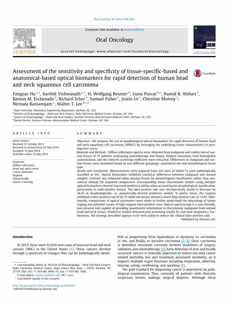
A portable fiber optic instrument (Fig. 1) was used to measuretissue diffuse reflectance spectra. Light from a 40 W halogen lamp(HL2000HP; Ocean Optics, Dunedin, FL), was coupled to an optical

Figure 1. A picture of the portable system which consists of a laptop, USBspectrometer, halogen light source, heat filter and a bendable gooseneck probecontaining the two optical fibers. The system is computer controlled.
850 F. Hu et al. / Oral Oncology 50 (2014) 848–856
fiber (400 lm diameter) for illumination. Another fiber (400 lmdiameter) was placed 475 lm away from the source fiber and cou-pled the diffuse reflectance from tissue into a spectrometer(USB4000, Ocean Optics, Dunedin, FL). Tissue reflectance measure-ments were normalized to a 99% reflectance standard measure-ment (Labsphere, Inc.) obtained each day. Tissue sensing depthswere determined using forward MC simulation [40] and werefound to be 1.1 mm and 1.6 mm at 480 nm and 600 nm, respec-tively, for the median absorption and scattering coefficientsderived from the previous study [16].
Extraction of tissue biomarkers from optical reflectance data
Diffuse reflectance spectra (480–650 nm) were collected andanalyzed using an inverse MC model to obtain the scattering andabsorption coefficients [33]. THb (lM), SO2 (%) and l0s (1/cm) wereextracted from the absorption and scattering coefficients [16].
Statistical analysis
Tissue samples were divided into malignant and normal groups,based on histopathological diagnosis of biopsied tissues and clini-cal impression of the non-biopsied normal tissues. The histopa-thological slides were read and analyzed by several pathologistsfrom Duke Universality Medical Center Pathological laboratory.The pathologists and surgeons were blinded to the DRS results.Mean contributions per tissue/site for all extracted optical bio-markers were compared between groups using the Wilcox rank-sum test to determine if there were statistically significant differ-ences (p < 0.05) between the two groups. Linear-discriminantmodels were used to classify the tissue as malignant or normal,by using all or a subset of (all pair-wise combinations of THb,SO2, and l0s) the three optical biomarkers. This was carried outfor tissues grouped by the anatomical sites classification (larynx,pharynx, oral) and for tissues grouped by the tissue-specific classi-fication (lymphoid, glottis, mucosal and muscle). The sensitivity(Se), specificity (Sp), positive predict value (PPV) and negative pre-dict value (NPV) were determined by comparing the model predic-tion to the pathological diagnosis. The discriminant algorithm wasvalidated using a leave-one-out cross-validation technique. Inaddition, the optical biomarkers of the normal, the lymph-node-positive, and the lymph-node-negative samples were comparedwith the Wilcox rank-sum test. Finally, differences between the
optical biomarkers of the normal, pathologically-confirmed normaland the clinical observed normal samples were also evaluated withthe Wilcox rank-sum test. All data processing and statistical dataanalysis was performed using MATLAB (MathWorks Inc., NatickMA).
Results
Ninety-nine biopsies co-localized with optical probe measure-ments were obtained from the 57 enrolled patients. Fifty-one ofthe 99 biopsies were diagnosed as SCC. These 51 SCC samples wereobtained from 37 of the enrolled patients. Forty of the 99 biopsieswere pathologically classified as negative for SCC (30 pathologi-cally-confirmed normal tissue, 10 inflammation with no atypia).No specific information regarding other pathology such as lichenplanus, ulceration were specifically given in the pathology report.Eight of the 99 biopsies were pathologically classified as dysplastic,and these were excluded from this study due to the small samplesize. Another 158 optical probe measurements were measured atdistant normal epithelium from all 57 patients. These 158 siteswere not biopsied and were considered to be normal based on clin-ical appearance and distance from the tumor.
Fig. 2 shows the representative normalized reflectance scans ofmalignant and nonmalignant tissues for the 3 anatomical sites andthe 4 morphological categories. All SCC-positive tissues were fur-ther separated into 4 groups by their tumor size and lymph nodestatus according to the TMN staging information. A breakdownby anatomical site, morphological categories, tumor stage andlymph node status for all measurements in this study is shownin Table 1.
Contrast between SCC and normal samples is significantly enhancedwhen grouped by tissue morphology
Fig. 3 shows SO2, THb and l0s as boxplots when the sampleswere grouped together by anatomical sites or by morphologi-cally-similar sites for pathologically-confirmed SCC and normalsites. When the samples were grouped by their anatomical loca-tions, the SO2 of the malignant tissues was significantly lower thanthat in normal tissues in the larynx and the pharynx (p < 0.01 forboth). The l0s of the malignant tissues was significantly lower thanthat in the normal tissues in the pharynx (p = 0.03) and in the oralcavity (p < 0.01). When the samples were grouped by morphologi-cally-similar tissue types, SO2 showed statistically significant dif-ferences between SCC and normal tissues for all four tissuegroups (glottis p < 0.01; lymphoid p < 0.01; muscle p = 0.03; andmucosal p < 0.01). Further, statistical differences in l0s were alsoobserved between SCC and normal tissues of lymphoid and muco-sal origin (p = 0.02 and p < 0.01, respectively) but not in the glottis.The l0s of the SCC tissues is nearly significantly lower than the l0s ofthe normal tissues (p = 0.058) in the muscle group. In summary,contrast between SCC and normal tissue is significantly enhancedwhen parsing tissues by morphological rather than by anatomicalsubtypes.
Linear discriminant model has better predictive power with themorphological tissue grouping
The sensitivity, specificity, PPV and NPV were computed bycomparing predictions from the discriminant model to thoseobtained from pathology and are listed for each set of optical vari-ables used (Table 2). The numbers of false negative (FN) and falsepositive (FP) samples are also provided. The FNs were separatedinto node-positive SCC and node-negative SCC for Tis/T1/T2 stageand T3/T4 stage. The FPs were separated into pathologically nor-

Figure 2. Representative normalized reflectance scans for the 3 anatomical sites (A) and the 4 morphological categories (B).
Table 1Breakdown of anatomical site, sub-site and morphologically similar tissue types for all measurements. All tumor sites were confirmed by histopathological confirmation ofbiopsies. 37 patients had pathologically-confirmed SCC. The number of patients included in each sample type was indicated in the parentheses.
Pathology confirmed SCC Normal
Tis/T1/T2a T3/T4 Total Pathology confirmed Clinical observation Total
Anatomical site Anatomical sub-site Morphological groups N+b N�c N+ N�
Larynx Glottic Glottic 1(1) 11(8) 2(1) 0(0) 14(10) 21(14) 23(18) 47(23)Supraglottic Mucosal 2(2) 1(1) 2(2) 0(0) 5(5) 0(0) 5(4) 5(4)Site-total 3(2) 12(8) 4(3) 0(0) 19(13) 21(14) 28(21) 52(25)
Pharynx Oropharynx Lymphoid 2(2) 0(0) 1(1) 0(0) 3(3) 1(1) 7(6) 8(6)Hypopharynx Mucosal 1(1) 0(0) 0(0) 0(0) 1(1) 3(2) 1(1) 4(3)Base of Tongue Lymphoid 5(5) 3(3) 2(2) 0(0) 10(10) 11(7) 15(14) 26(15)Tonsil Lymphoid 3(3) 1(1) 1(1) 1(1) 6(6) 0(0) 5(4) 5(4)Site-total 11(11) 4(4) 4(3) 1(1) 20(19) 15(8) 28(22) 43(23)
Oral cavity Tongue Muscle 3(1) 4(3) 0(0) 0(0) 7(4) 3(2) 34(29) 37(29)Floor of Mouth Mucosal 0(0) 0(0) 1(1) 0(0) 1(1) 0(0) 23(22) 23(22)Alveolus Mucosal 0(0) 0(0) 0(0) 2(1) 2(1) 0(0) 2(2) 2(2)Palate Mucosal 0(0) 0(0) 0(0) 0(0) 0(0) 1(1) 21(19) 22(19)Cheek Mucosal 0(0) 1(1) 0(0) 0(0) 1(1) 0(0) 22(22) 22(22)RMTd Mucosal 0(0) 1(1) 0(0) 0(0) 1(1) 0(0) 0(0) 0(0)Site-total 3(1) 6(5) 1(1) 2(1) 12(8) 4(3) 102(34) 106(34)
Total 17(14) 22(17) 9(6) 3(2) 51(37) 40(25) 158(56) 198(57)
a Tis: carcinoma in situ.b N+: node-positive SCC.c N�: node-negative SCC.d RMT: retromolar trigone.
F. Hu et al. / Oral Oncology 50 (2014) 848–856 851
mal and clinically normal groups. Overall, for every pair of opticalvariables selected, there were improvements in the sensitivity,specificity, PPV, and NPV when discriminant models were builtbased on morphological groupings, relative to a model that wasbuilt based on the anatomical location. The total number of FNsamples was lowest when using all three optical biomarkers for
classification. Importantly, the total number of FNs and FPs derivedfrom the morphologically-grouped algorithm was less than thetotal number of FNs and FPs derived from anatomically-groupedalgorithm. Further, the algorithm based on morphological group-ings was particularly more effective in identifying node-positivevs. node-negative disease.

Table 2Diagnostic accuracy based on linear-discriminant models using two or more measured optical biomarkers for sites grouped by anatomical sites or by tissue types (shaded cells).The sensitivity, specificity, positive predictive value and negative predictive value values are reported for leave-one-out cross-validation. The standard error of the diagnosticaccuracies were computed and provided in the parentheses. The numbers in the site column in parenthesis represents the number of node positive SCC, node-negative SCC,pathologically-confirmed normal, clinically-observed normal samples in each group. See Table 1 for tissue groupings used. The numbers of false negatives and false positives arealso shown. The FN was further broken down into N+ and N� groups. The FP was broken down into pathological confirmed normal or clinical observed normal groups. Thecollective Se, Sp, PPVs and NPVs when all subgroups are combined are also shown. This table suggests that while the same optical biomarkers were used to build the discriminantmodel, the models generally perform better when the samples were grouped by tissue type (shaded) than by anatomical site (non-shaded). Moreover, the discriminant modelswith three optical biomarkers generally classify the samples better than those with only two optical biomarkers. The number in parenthesis indicates the standard error of thecorresponding diagnostic accuracy.
FN FP
Tis/T1/T2 T3/T4 Total aPath. bClin. Total
Biomarker Pairs Site Se. Sp. PPV. NPV. N+ N- N+ N-
SO2&THb Larynx (7/12/24/28) 63%(11.1%) 94%(3.4%) 80%(10.3%) 88%(4.7%) 1 4 2 0 7 2 1 3
Pharynx (15/5/15/28) 65%(10.6%) 86%(5.3%) 68%(10.7%) 84%(5.5%) 4 1 1 1 7 4 2 6
Oral Cavity (4/8/4/102) 50%(14.4%) 73%(4.3%) 17%(6.4%) 93%(2.8%) 1 4 0 1 6 1 28 29
Total 61%(6.8%) 81%(2.8%) 45%(6.0%) 89%(2.3%) 6 9 3 2 20 7 31 38
Glottic (3/11/24/23) 57%(13.2%) 94%(3.8%) 73%(13.4%) 88%(4.7%) 1 4 1 0 6 2 1 3
Lymphoid (14/5/12/27) 68%(10.6%) 87%(5.4%) 72%(10.6%) 85%(5.6%) 3 1 1 1 6 3 2 5
Muscle (3/4/3/34) 71%(17.1%) 68%(7.7%) 29%(11.1%) 93%(5.0%) 1 1 0 0 2 0 12 12
Mucosal (6/5/4/74) 73%(13.4%) 90%(3.4%) 50%(12.5%) 96%(2.3%) 0 2 0 1 3 2 6 8
Total 67%(6.6%) 86%(2.5%) 55%(6.3%) 91%(2.1%) 5 8 2 2 17 7 21 28
SO2&µs’ Larynx
(7/12/24/28) 63%(11.1%) 94%(3.4%) 80%(10.3%) 88%(4.7%) 1 4 2 0 7 2 1 3
Pharynx (15/5/15/28) 75%(9.7%) 86%(5.4%) 71%(9.9%) 88%(5.0%) 2 1 1 1 5 5 1 6
Oral Cavity (4/8/4/102) 67%(13.6%) 74%(4.3%) 22%(6.9%) 95%(2.4%) 1 2 0 1 4 2 26 28
Total 69%(6.5%) 82%(2.8%) 49%(5.9%) 91%(2.2%) 4 7 3 2 16 9 28 37
Glottic (3/11/24/23) 57%(13.2%) 94%(3.8%) 73%(13.4%) 88%(4.9%) 1 4 1 0 6 2 1 3
Lymphoid (14/5/12/27) 74%(10.1%) 87%(5.4%) 74%(10.1%) 87%(5.4%) 2 1 1 1 5 4 1 5
Muscle (3/4/3/34) 71%(17.1%) 73%(7.3%) 33%(12.1%) 93%(4.7%) 1 1 0 0 2 0 10 10
Mucosal (6/5/4/74) 73%(13.4%) 92%(3.0%) 57%(13.2%) 96%(2.3%) 0 2 0 1 3 3 3 6
Total 69%(6.5%) 88%(2.6%) 59%(6.5%) 92%(2.0%) 4 8 2 2 16 9 15 24
SO2&THb&µs
Larynx (7/12/24/28) 63%(11.6%) 94%(3.4%) 80%(10.3%) 88%(4.7%) 1 4 2 0 7 2 1 3
Pharynx (15/5/15/28) 80%(8.9%) 86%(5.3%) 73%(9.5%) 90%(4.6%) 1 1 1 1 4 5 1 6
Oral Cavity (4/8/4/102) 67%(13.6%) 73%(4.3%) 22%(6.8%) 95%(2.4%) 1 2 0 1 4 2 27 29
Total 71%(6.4%) 81%(2.8%) 49%(5.8%) 92%(2.1%) 3 7 3 2 15 9 29 38
Glottic (3/11/24/23) 57%(13.2%) 94%(3.8%) 73%(13.4%) 88%(4.9%) 1 4 1 0 6 2 1 3
Lymphoid (14/5/12/27) 79%(9.4%) 90%(4.9%) 79%(9.4%) 90%(4.9%) 1 1 1 1 4 3 1 4
Muscle (3/4/3/34) 71%(17.1%) 73%(7.3%) 33%(12.2%) 93%(4.7%) 1 1 0 0 2 0 10 10
Mucosal (6/5/4/74) 73%(13.4%) 90%(3.4%) 50%(12.5%) 96%(2.3%) 0 2 0 1 3 3 5 8
Total 71%(6.4%) 88%(2.4%) 59%(6.3%) 92%(2.0%) 3 8 2 2 15 8 17 25
a Path.: pathological confirmed normal sample.b Clin.: clinical observed normal samples.
852 F. Hu et al. / Oral Oncology 50 (2014) 848–856

Figure 3. Range and median values of extracted biomarkers for normal and SCC for different anatomic and tissue classifications. Numbers in parentheses indicate samplenumbers. Significant p-values are reported from unpaired Wilcoxon rank sum tests. Contrast between SCC and normal tissues is enhanced when parsing tissues bymorphological rather than by anatomical subtypes (*p < 0.05, **p < 0.01) (N: normal).
F. Hu et al. / Oral Oncology 50 (2014) 848–856 853
Tissue scattering and hemoglobin oxygenation saturation are lower atthe primary tumor with node positive disease
Fig. 4 shows boxplots of the optical biomarkers for normal sam-ples, node-positive and node-negative SCC samples. The SO2 of thenormal samples was significantly higher than that of the node-positive and node-negative SCC groups (p < 0.01 for both). The l0sof the node-positive SCC samples was nearly significantly lowerthan that of normal group (p = 0.055) thus providing additionalcontrast when differentiating normal from node-positive SCC sam-ples. Moreover, the l0s of the node-positive SCC samples was signif-icantly lower than that of node-negative SCC samples (p < 0.01)and the l0s of the node-negative SCC samples was significantlylower than that of the normal samples (p < 0.01).
Optical spectroscopy shows contrast between SCC and pathologically-confirmed normal tissues
Fig. 5 shows boxplots of the optical parameters for the malig-nant, pathologically-confirmed normal and clinically-observednormal tissues. The SO2 of the malignant tissues were significantlylower than both the pathological normal tissues and the clinicalobserved normal tissues. The SO2 of the pathological normal tis-sues were also significantly lower than the clinically-observed nor-mal tissues. No statistical significance was observed in the THb
results. The l0s of the clinically-observed normal tissues werehigher than both the pathologically-confirmed normal and themalignant tissues.
Discussion
Our results show that when using SO2, THb and l0s for buildingthe discriminant models, the overall PPV and NPV increased from49% and 92%, respectively, to 59% and 92%, when tissue-specificclassification was used instead of anatomically-based classifica-tion. Moreover, this change resulted in a 34% decrease in the num-ber of FP classifications. This supports the hypothesis that a strongpredictive algorithm would need to be tissue-specific, since differ-ent tissues are expected to have different baseline physiologicaland morphological properties. It has been demonstrated that thesedifferences in morphological and chemical properties affects thetissue optical properties. For instance, Bashkatov et al. showed thatthe absorption and the scattering spectra of human skin, subcuta-neous adipose, and mucosa exhibit different spectral features [41].Therefore, mixing different types of tissues could be expected todiminish contrast between the malignant and normal tissues asobserved here.
Forty-three clinically-appearing malignant tissues were con-firmed to be histologically benign. While the tissue absorptionand scattering were similar between the 51 malignant and the

Figure 4. Boxplots of SO2 (A), THb (B) and l0s(C) for normal and SCC samples. TheSCC samples were separated into two groups by lymph node status. The significantp values from Wilcoxon rank sum tests are shown. This figure suggests that headand neck malignant tissue is generally hypoxic. Moreover, as the tumor progressfrom node-negative to node-positive, the tissue scattering generally decreases.(**p < 0.01) (N: normal).
Figure 5. Boxplots of SO2 (A), THb (B) and l0s(C) for malignant, pathologically-confirmed normal and clinically-observed normal tissue. The significant p valuescomputed from the Wilcoxon rank sum tests are shown. (**p < 0.01) This figuresuggests that the contrast between the malignant and pathologically-confirmednormal was less than the contrast between the malignant and the clinically-observed normal tissues. (Path.: pathologically-confirmed normal. Clinical: clinicalobserved normal).
854 F. Hu et al. / Oral Oncology 50 (2014) 848–856
43 pathologically-confirmed normal tissues, the SO2 was signifi-cantly different. The algorithm based on morphological groupingsclassified 81% of these samples correctly. In addition, 23 glotticsamples were biopsied and confirmed as pathologically normal.With the morphologically based algorithm, only 2 pathologically-confirmed benign samples were classified as malignant. Thisimplies that the vast majority of these glottic biopsies if obtainedfor the concern of cancer may have been avoided. These data illus-trate the potential utility of using optical spectroscopy for reducingthe number of biopsies and their related patient discomfort/painand use of limited resources.
The effect of enhancing contrast when building the linear dis-criminant model based on tissue type is obvious when comparingthe oral cavity group to its constituent tissue groups (muscle andmucosal) since they had comparable sample sizes (37% muscle,63% mucosal). The linear discriminant models have better sensitiv-ities and PPVs and similar specificities and NPVs regardless of theoptical parameters chosen (Table 2) when using tissue type overanatomical location. In addition, the SO2 showed no statistical dif-ferences between the malignant and normal tissues in the oral cav-ity. On the other hand, both the SO2 of the malignant tissues inmuscle and the mucosal group were significantly lower than theSO2 of the normal tissues in the corresponding group (Fig. 3).
Optical techniques have been widely used to detect and charac-terize differences between dysplastic and malignant tissue frombenign tissue, as reviewed previously [35,42,43]. Although capable
of providing high discriminatory power, these approaches rely onheuristic algorithms for classification, and do not necessarily lever-age information about the underlying tissue physiology or mor-phology for tissue classification, as has been demonstrated in thisstudy. Further, only one of the three groups mentioned above haspreviously reported on differences between benign and dysplasticand/or cancerous lesions based on anatomical sub groups. How-ever, this was limited to just the oral cavity, which is a very smallsubset of the head and neck lesions observed clinically.
Hypoxia is a general characteristic of solid tumors in the headand neck [44] due to a less-ordered and leaky vascular supply, ascompared with that of normal tissues [45]. In this manuscript,the oxygenation of the malignant head and neck tissues are shownto be generally lower than that of normal head and neck tissues inthe unpaired comparisons (Figs. 3–5). This result was also observedin our previous spectroscopy study of HNCC [16]. In another study,Terris and Dunphy found that the pO2 of the primary tumor(22.7 ± 16.0 mm Hg) was lower than that of normal subcutaneoustissue (57.2 ± 12.8 mm Hg) [46]. This result is concordant with ourfindings. In addition to providing contrast for the detection of SCC,knowledge of tumor SO2 will be invaluable in therapy planning, astumor hypoxia renders the tumor resistant to radiation treatment,which plays an important role in HNC therapy.
Tissue scattering likely reflects the morphology of the HNC. Tis-sue scattering of the malignant tissues was significantly lower thanthat of the clinical observed normal tissues. Moreover, tissue scat-tering of lymph-node-positive malignant tissues was lower thanboth normal tissues and node-negative malignant tissues. Thiscould be caused by the loss of collagen in the basement membraneduring the invasion of the SCC cells. Breach of the basement

F. Hu et al. / Oral Oncology 50 (2014) 848–856 855
membrane is considered to be the first step for the HNSCC cells tobecome an invasive carcinoma [47,48]. Both urokinase-type plas-minogen activator and metalloproteinases (MMPs, also known ascollagenases) play important roles allowing SCC cells to degradethe basement membrane. Activating the urokinase or the collagen-ases can result in the hydrolysis of collagen [49,47,50], whichserves as an important component in the basement membraneand one major tissue scatterer as well [51]. Georgakoudi et al.observed a decrease of tissue scattering during the progression ofdysplasia in esophagus tissue with reflectance spectroscopy [52].Loss of collagen due to neoplasia can also be seen in wide-fieldautofluorescence imaging in oral cavity [53].
Although we failed to achieve 100% sensitivity and specificity,our technique is low-cost, label-free and portable, and thus haspotential in countries where histopathology services are limited.If our technique proves reliable and rapid screening, it couldreduce the current burden of trained pathologists. In addition,the classification outcome may be improved by further refiningthe tissue grouping strategy. This strategy could also be appliedto other technologies and organ sites. Moreover, our results showthat the tissue scattering at the primary tumor might be relatedto the nodal status. This is important since whether the cancerhas metastasized will significantly change the treatment strategy.Finally, current histopathology results of the biopsies at the pri-mary tumor would not reveal the lymph node status.
Our group demonstrates a quantitative optical spectroscopytechnique and tissue grouping strategy capable of enhancing con-trast between malignant and normal tissues from different regionsof the head and neck. In this preliminary diagnostic study, our datasuggests that a predictive algorithm that combines optical end-points and that is stratified by tissue type improves accuracy, par-ticularly in node-positive disease. This is based on accounting fordifferences in tissue physiology and morphology within the upperaerodigestive tract. Further, the strategy described here appears tobe particularly well suited to reduce the clinical false-positive rate,particularly for glottic tissue, where the surgeon had the highestfalse-positive rate, thereby potentially reducing the need forunnecessary surgical procedures that could significantly impactpatient morbidity.
Role of the funding source
This work was funded by the Alexander and Margaret StewartTrust, Duke Cancer Institute, and the NIH (5K99CA140783-02)(KV). This project was supported by Career Development Award(IK2BX001398) from the Biomedical Laboratory Research andDevelopment Service of the Department of Veterans Affairs Officeof Research and Development (WTL). The views expressed in thisarticle are those of WTL and LP and do not necessarily representthe views of the Department of Veterans Affairs or the UnitedStates government.
Conflict of Interest
None declared.
References
[1] Cancer Facts and Figures. Atlanta, GA: American Cancer Society; 2008.[2] Argiris A, Karamouzis MV, Raben D, et al. Head and neck cancer. Lancet
2008;371:1695–709.[3] Forastiere A, Koch W, Trotti A, et al. Head and neck cancer. New Engl J Med
2001;345:1890–900.[4] Hunter KD, Parkinson EK, Harrison PR. Profiling early head and neck cancer.
Nat Rev Cancer 2005;5:127–35.[5] Thompson LW. Head and neck cancer: early detection. Semin Surg Oncol
1989;5:168–75.
[6] Vila PM, Park CW, Pierce MC, et al. Discrimination of benign and neoplasticmucosa with a high-resolution microendoscope (HRME) in head and neckcancer. Ann Surg Oncol 2012;19:3534–9.
[7] Parikh ND, Perl D, Lee MH, et al. In vivo diagnostic accuracy of high-resolutionmicroendoscopy in differentiating neoplastic from non-neoplastic colorectalpolyps: a prospective study. Am J Gastroenterol 2014;109:68–75.
[8] Brezinski ME, Fujimoto JG. Optical coherence tomography: high-resolutionimaging in nontransparent tissue. IEEE J Sel Top Quant 1999;5:1185–92.
[9] Yun SH, Tearney GJ, de Boer JF, et al. High-speed optical frequency-domainimaging. Opt Express 2003;11:2953–63.
[10] Clark AL, Gillenwater AM, Collier TG, et al. Confocal microscopy for real-timedetection of oral cavity neoplasia. Clin Cancer Res 2003;9:4714–21.
[11] Rajadhyaksha M, Anderson RR, Webb RH. Video-rate confocal scanning lasermicroscope for imaging human tissues in vivo. Appl Opt 1999;38:2105–15.
[12] Ridgway JM, Armstrong WB, Guo S, et al. In vivo optical coherence tomographyof the human oral cavity and oropharynx. Arch Otolaryngol2006;132:1074–81.
[13] Gerger A, Koller S, Weger W, et al. Sensitivity and specificity of confocal laser-scanning microscopy for in vivo diagnosis of malignant skin tumors. Cancer2006;107:193–200.
[14] Ramanujam N. Fluorescence spectroscopy of neoplastic and non-neoplastictissues. Neoplasia 2000;2:89–117.
[15] Amelink A, Kaspers OP, Sterenborg HJCM, et al. Non-invasive measurement ofthe morphology and physiology of oral mucosa by use of optical spectroscopy.Oral Oncol 2008;44:65–71.
[16] Beumer HW, Vishwanath K, Puscas L, et al. Detection of squamous cellcarcinoma and corresponding biomarkers using optical spectroscopy.Otolaryngol Head Neck 2011;144:390–4.
[17] Ghanate AD, Kothiwale S, Singh SP, et al. Comparative evaluation ofspectroscopic models using different multivariate statistical tools in amulticancer scenario. J Biomed Opt 2011;16.
[18] Jayanthi JL, Nisha GU, Manju S, et al. Diffuse reflectance spectroscopy:diagnostic accuracy of a non-invasive screening technique for earlydetection of malignant changes in the oral cavity. BMJ Open 2011;1:e000071.
[19] Jayanthi JL, Subhash N, Stephen M, et al. Comparative evaluation of thediagnostic performance of autofluorescence and diffuse reflectance in oralcancer detection: a clinical study. J Biophotonics 2011;4:696–706.
[20] Kamath SD, Mahato KK. Optical pathology using oral tissue fluorescencespectra: classification by principal component analysis and k-means nearestneighbor analysis. J Biomed Opt 2007;12.
[21] Kan CW, Jiang BC, Nieman LT, et al. Comparison of linear and non-linearclassifiers for oral cancer screening by optical spectroscopy. AMIA Ann SympProc/AMIA Symp AMIA Symp 2007: 1003.
[22] Majumder SK, Ghosh N, Gupta PK. Support vector machine for opticaldiagnosis of cancer. J Biomed Opt 2005;10.
[23] Majumder SK, Gupta A, Gupta S, et al. Multi-class classification algorithm foroptical diagnosis of oral cancer. J Photochem Photobiol B 2006;85:109–17.
[24] Mallia R, Thomas SS, Mathews A, et al. Oxygenated hemoglobin diffusereflectance ratio for in vivo detection of oral pre-cancer. J Biomed Opt 2008;13.
[25] Mallia RJ, Narayanan S, Madhavan J, et al. Diffuse reflection spectroscopy: analternative to autofluorescence spectroscopy in tongue cancer detection. ApplSpectrosc 2010;64:409–18.
[26] Mallia RJ, Subhash N, Mathews A, et al. Clinical grading of oral mucosa bycurve-fitting of corrected autofluorescence using diffuse reflectance spectra.Head Neck – J Sci Spec 2010;32:763–79.
[27] Manhas S, Swami MK, Patel HS, et al. Polarized diffuse reflectancemeasurements on cancerous and noncancerous tissues. J Biophotonics2009;2:581–7.
[28] Mcgee S, Mardirossian V, Elackattu A, et al. Anatomy-based algorithms fordetecting oral cancer using reflectance and fluorescence spectroscopy. AnnOtol Rhinol Laryn 2009;118:817–26.
[29] Schwarz RA, Gao W, Weber CR, et al. Noninvasive evaluation of oral lesionsusing depth-sensitive optical spectroscopy. Cancer 2009;115:1669–79.
[30] Sharwani A, Jerjes W, Salih V, et al. Assessment of oral premalignancy usingelastic scattering spectroscopy. Oral Oncol 2006;42:343–9.
[31] Subhash N, Mallia JR, Thomas SS, et al. Oral cancer detection using diffusereflectance spectral ratio R540/R575 of oxygenated hemoglobin bands. JBiomed Opt 2006;11.
[32] Vishwanath K, Yuan H, Barry WT, et al. Using optical spectroscopy tolongitudinally monitor physiological changes within solid tumors. Neoplasia2009;11:889–900.
[33] Palmer GM, Ramanujam N. Monte Carlo-based inverse model for calculatingtissue optical properties. Part I: theory and validation on synthetic phantoms.Appl Opt 2006;45:1062–71.
[34] Vishwanath K, Chang K, Klein D, et al. Portable, fiber-based, diffuse reflectionspectroscopy (DRS) systems for estimating tissue optical properties. ApplSpectrosc 2011;65:206–15.
[35] Hughes ORSN, Kraft M, Arens C, Birchall MA. Optical and moleculartechniques to identify tumor margins within the larynx. Head Neck 2010;32:1544–53.
[36] Perelman LT, Backman V, Wallace M, et al. Observation of periodic finestructure in reflectance from biological tissue: a new technique for measuringnuclear size distribution. Phys Rev Lett 1998;80:627–30.
[37] Nieman L, Myakov A, Aaron J, et al. Optical sectioning using a fiber probe withan angled illumination-collection geometry: evaluation in engineered tissuephantoms. Appl Opt 2004;43:1308–19.

856 F. Hu et al. / Oral Oncology 50 (2014) 848–856
[38] Ross MH, Pawlina W. Histology: a text and atlas: with correlated cell andmolecular biology. 5th ed. Baltimore, MD: Lippincott Williams & Wilkins;2006. p. xvii, 906 p.
[39] Koch WM. Head and neck cancer. In: Early diagnosis and treatment ofcancer. Philadelphia, PA: Saunders/Elsevier; 2010. p. 1 online resource (xii, p.255).
[40] Liu Q, Ramanujam N. Scaling method for fast Monte Carlo simulation of diffusereflectance spectra from multilayered turbid media. J Opt Soc Am A2007;24:1011–25.
[41] Bashkatov AN, Genina EA, Kochubey VI, et al. Optical properties of human skin,subcutaneous and mucous tissues in the wavelength range from 400 to2000 nm. J Phys D Appl Phys 2005;38:2543–55.
[42] Upile T, Jerjes W, Betz CS, et al. Optical diagnostic techniques in the head andneck. Dent Update 2007;34:410–2. 15-6, 19-20 passim.
[43] Swinson B, Jerjes W, El-Maaytah M, et al. Optical techniques in diagnosis ofhead and neck malignancy. Oral Oncol 2006;42:221–8.
[44] Nordsmark M, Bentzen SM, Rudat V, et al. Prognostic value of tumoroxygenation in 397 head and neck tumors after primary radiation therapy.An international multi-center study. Radiother Oncol: J Eur Soc Ther RadiolOncol 2005;77:18–24.
[45] Janssen HL, Haustermans KM, Balm AJ, et al. Hypoxia in head and neck cancer:how much, how important? Head Neck 2005;27:622–38.
[46] Terris DJ, Dunphy EP. Oxygen tension measurements of head and neck cancers.Arch Otolaryngol – Head Neck Surg 1994;120:283–7.
[47] Tryggvason K, Hoyhtya M, Salo T. Proteolytic degradation of extracellular-matrix in tumor invasion. Biochim Biophys Acta 1987;907:191–217.
[48] Nicolson GL. Cancer metastasis – organ colonization and the cell-surfaceproperties of malignant-cells. Biochim Biophys Acta 1982;695:113–76.
[49] Matrisian LM, Bowden GT. Stromelysin/transin and tumor progression. SeminCancer Biol 1990;1:107–15.
[50] Bejarano PA, Noelken ME, Suzuki K, et al. Degradation of basement membranesby human matrix metalloproteinase 3 (stromelysin). Biochem J1988;256:413–9.
[51] Saidi IS, Jacques SL, Tittel FK. Mie and Rayleigh modeling of visible-lightscattering in neonatal skin. Appl Opt 1995;34:7410–8.
[52] Georgakoudi I, Jacobson BC, Van Dam J, et al. Fluorescence, reflectance, andlight-scattering spectroscopy for evaluating dysplasia in patients withBarrett’s esophagus. Gastroenterology 2001;120:1620–9.
[53] Pierce MC, Schwarz RA, Bhattar VS, et al. Accuracy of in vivo multimodaloptical imaging for detection of oral neoplasia. Cancer Prev Res (Phila)2012;5:801–9.