Design, Synthesis, and Biological Evaluation of Thiazoles Targeting Flavivirus Envelope Proteins
Upload
independentCategory
view
4download
0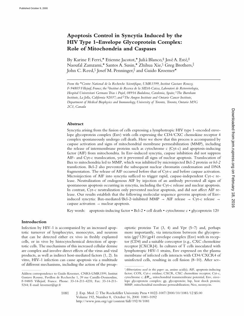
J. Exp. Med.
The Rockefeller University Press • 0022-1007/2000/10/1081/12 $5.00Volume 192, Number 8, October 16, 2000 1081–1092http://www.jem.org/cgi/content/full/192/8/1081
1081
Apoptosis Control in Syncytia Induced by theHIV Type 1–Envelope Glycoprotein Complex:Role of Mitochondria and Caspases
By Karine F. Ferri,
*
Etienne Jacotot,
*
Julià Blanco,
‡
José A. Esté,
‡
Naoufal Zamzami,
*
Santos A. Susin,
*
Zhihua Xie,
§
Greg Brothers,
i
John C. Reed,
§
Josef M. Penninger,
i
and Guido Kroemer
*
From the
*
Centre National de la Recherche Scientifique, UMR1599, Institut Gustave Roussy,F-94805 Villejuif, France; the
‡
Institut de Recerca de la SIDA-Caixa, Laboratori de Retrovirologia, Hospital Universitari Germans Trias i Pujol, 08916 Badalona, Catalonia, Spain;
§
The Burnham Institute, La Jolla, California 92037; and
i
The Amgen Institute and Ontario Cancer Institute, Department of Medical Biophysics and Immunology, University of Toronto, Toronto, Ontario M5G 2C1, Canada
Abstract
Syncytia arising from the fusion of cells expressing a lymphotropic HIV type 1–encoded enve-lope glycoprotein complex (Env) with cells expressing the CD4/CXC chemokine receptor 4complex spontaneously undergo cell death. Here we show that this process is accompanied bycaspase activation and signs of mitochondrial membrane permeabilization (MMP), includingthe release of intermembrane proteins such as cytochrome c
(Cyt-c) and apoptosis-inducingfactor (AIF) from mitochondria. In Env-induced syncytia, caspase inhibition did not suppressAIF- and Cyt-c translocation, yet it prevented all signs of nuclear apoptosis. Translocation ofBax to mitochondria led to MMP, which was inhibited by microinjected Bcl-2 protein or
bcl-2
transfection. Bcl-2 also prevented the subsequent nuclear chromatin condensation and DNAfragmentation. The release of AIF occurred before that of Cyt-c and before caspase activation.Microinjection of AIF into syncytia sufficed to trigger rapid, caspase-independent Cyt-c re-lease. Neutralization of endogenous AIF by injection of an antibody prevented all signs ofspontaneous apoptosis occurring in syncytia, including the Cyt-c release and nuclear apoptosis.In contrast, Cyt-c neutralization only prevented nuclear apoptosis, and did not affect AIF re-lease. Our results establish that the following molecular sequence governs apoptosis of Env-
induced syncytia: Bax-mediated/Bcl-2–inhibited MMP
→
AIF release
→
Cyt-c release
→
caspase activation
→
nuclear apoptosis.
Key words: apoptosis-inducing factor • Bcl-2 • cell death • cytochrome c • glycoprotein 120
Introduction
Infection by HIV-1 is accompanied by an increased apop-totic turnover of lymphocytes, monocytes, and neuronsthat can be detected either ex vivo in freshly explantedcells, or in vivo by histocytochemical detection of apop-totic cells. The mechanisms of this increased cellular demiseare complex and involve direct effects of the virus and viralproducts, as well as indirect host-mediated factors (1, 2). Invitro, HIV-1 infection can cause apoptosis via a multitudeof different mechanisms, including the action of the proap-
optotic proteins Tat (3, 4) and Vpr (5–7) and, perhapsmore importantly, via interactions between the glycopro-tein (gp)
1
120/gp41 envelope complex (Env) with its recep-tor (CD4) and a suitable coreceptor (e.g., CXC chemokinereceptor [CXCR]4). In cultures of T cells inoculated withlymphotropic HIV-1 strains, Env expressed on the plasmamembrane of infected cells interacts with CD4/CXCR4 ofuninfected cells, resulting in cell fusion (8–10). After sev-
Address correspondence to Guido Kroemer,
CNRS-UMR1599, InstitutGustave Roussy, Pavillon de Recherche 1, 39 rue Camille-Desmoulins,F-94805 Villejuif, France. Phone: 33-14-211-4235; Fax: 33-14-211-5244; E-mail: [email protected]
1
Abbreviations used in this paper:
aa, amino acid(s); AIF, apoptosis-inducingfactor; COX, Cyt-c oxidase; CXCR, CXC chemokine receptor; Cyt-c,
cytochrome c;
DC
m
, mitochondrial transmembrane potential; Env, enve-lope glycoprotein complex; gp, glycoprotein; hsp, heat shock protein;MMP, mitochondrial membrane permeabilization; Neo, neomycin.
on February 18, 2016
jem.rupress.org
Dow
nloaded from
Published October 9, 2000

1082
Syncytial Apoptosis
eral rounds of fusion, syncytia attain volumes equivalent toseveral dozens or hundreds of individual cells and ulti-mately lyse, while exhibiting several biochemical character-istics of apoptosis and/or signs of necrosis such as cytoplas-mic vacuolization (11–14). According to several studies,syncytium formation is the principal cause of HIV-1–medi-ated T cell destruction in vitro (11, 15). Env variants inter-acting with CD4/CXCR4 (rather than those having apreference for CD4/CCR5) are mostly encoded by syncy-tium-inducing HIV-1 strains, and a strong correlation be-tween CD4
1
T cell decline and infection by syncytium-inducing HIV-1 variants has been established by someauthors in vitro and in vivo (8, 9, 15, 16).
Apoptosis of single cells is generally associated with theactivation of caspases (17), a set of specific proteases whicheither can serve as molecular switches initiating cell deathpathways or, when activated in a massive fashion, can me-diate the degradation of essential structural and regulatoryproteins, culminating in the activation of the caspase-acti-vated DNase, the enzyme responsible for oligonucleosomalDNA fragmentation (18, 19). In addition, apoptosis is ac-companied by signs of mitochondrial membrane permeabi-lization (MMP), including a loss of the inner mitochondrialtransmembrane potential (
DC
m
) and the release of solubleintermembrane proteins via the outer mitochondrial mem-brane (20–24). The functional hierarchy among theseevents depends on the apoptosis-inducing trigger. In the“extrinsic” pathway, caspase activation is induced as an up-stream event, e.g., by recruiting apical caspases to thedeath-induced signaling complex formed upon the ligationof death receptors expressed on the cell surface (e.g., CD95and TNFR). Depending on the cell type, receptor-proxi-mal caspase activation then either suffices to set off thecaspase activation cascade in an MMP-independent fashion(“type 1 cells”) or must relay to MMP via cleavage of Bidand/or production of ceramide (“type 2 cells” [25–27]). Incontrast, most “intrinsic” apoptosis triggers (e.g., DNAdamage, glucocorticoids, and reactive oxygen species) acti-vate caspases in an indirect fashion, namely by first perme-abilizing mitochondrial membranes in a Bcl-2–inhibitablefashion (20–23, 28, 29). MMP results in the translocationof cytochrome c (Cyt-c) from the mitochondrial inter-membrane space to the cytosol, where Cyt-c triggers theassembly of a caspase-9–caspase-3 activation complex, theapoptosome (18, 21). In addition to Cyt-c, mitochondriacan release a variety of intermembrane proteins (30), in-cluding a cell type–specific set of procaspases (31–33) andthe apoptosis-inducing factor (AIF [34]), a flavoprotein ox-idoreductase that translocates to the cytosol as well as to thenucleus and stimulates apoptosis via an as yet unknown,caspase-independent mechanism.
As in other models of apoptosis induction, signaling viaCD4/CXCR4 induces both caspase activation (35–39) andsigns of MMP (14, 40). However, the precise mechanismsof Env-mediated apoptosis induction and the molecular or-der of these events have remained elusive in syncytia. Stim-ulated by these premises, we studied the role of caspasesand MMP in syncytia arising from the fusion between Env-
and CD4/CXCR4-expressing cells. Here we report thatthe death of Env-induced syncytia obeys the rules of theintrinsic pathway of apoptosis induction in the sense thatcaspase activation occurs downstream of MMP. Our resultsindicate that MMP is regulated by members of the Bcl-2/Baxfamily and establish a molecular order of apoptotic signal-ing in which AIF release occurs upstream of that of Cyt-c,which in turn is required for caspase-dependent nuclear ap-optosis.
Materials and Methods
Cells and Culture Conditions.
HeLa cells stably transfectedwith a vector containing the
env
gene of HIV-1 LAI (HeLa 243Env [41]) were cultured in complete culture medium (DMEMsupplemented with 2 mM glutamine, 10% FCS, 1 mM pyruvate,10 mM Hepes, and 100 U/ml penicillin/streptomycin) contain-ing 2
m
M methotrexate. HeLa cells stably transfected with CD4(HeLa P4; a gift from P. Charneau, Pasteur Institute, Paris,France [42]) were selected in medium containing 500
m
g/mlG418. Jurkat cells expressing CD4 and CXCR4 and stably trans-fected with the human Bcl-2 gene or a neomycin (Neo) resis-tance vector (43) only were provided by N. Israel (Pasteur Insti-tute, Paris, France). Neo and Bcl-2 U937 cells (44) were a giftfrom F. Hirsch (Centre National de la Recherche Scientifique,ERS1984, Villejuif, France). Cocultures of different cell typeswere performed in complete culture medium in the absence ofselecting antibiotics by adding trypsinized HeLa cells to adherentHeLa CD4 or HeLa Env cells (density: 1–1.5
3
10
3
cells/mm
2
)at an
z
1:1 ratio or by adding Jurkat or U937 cells to adherentHeLa Env cells (National Institutes of Health AIDS Research andReference Reagent Program, Bethesda, MD). Apoptosis wasinduced or inhibited by staurosporin (2
m
M; Sigma-Aldrich),
N
-benzyloxycarbonyl-Val-Ala-Asp-fluoromethylketone (Z-VAD.fmk), Boc-Asp-fluoromethylketone (Boc-D.fmk), or
N
-benzyl-oxycarbonyl-Phe-Ala-fluoromethylketone (Z-FA.fmk) (all usedat 100
m
M added every 24 h; Enzyme Systems).
HIV-1 Infection.
5
3
10
6
HeLa CD4 cells were cultured inthe presence of 2.5
3
10
6
chronically HIV-1–infected H9/IIIBcells obtained from Dr. R.C. Gallo (National Institutes of Health,Bethesda, MD) in 5 ml of RPMI 1640 supplemented with 10%heat-inactivated FCS, 2 mM glutamine, and 25 IU/ml recombi-nant human IL-2. Before the addition of chronically infectedcells, target cells were preincubated for 30 min at 37
8
C in thepresence or absence of Z-VAD.fmk (50
m
M), and Z-VAD.fmkwas readded every 12 h throughout the culture period.
Fluorescence Staining of Live Cells and Immunofluorescence.
Forthe assessment of mitochondrial and nuclear features of apoptosis,cells cultured on a coverslip were stained with 5,5
9
,6,6
9
-tetra-chloro-1,1
9
, 3,3
9
-tetraethylbenzimidazolylcarbocyanine iodide(JC-1, 2
m
M; Molecular Probes) and Hoechst 33342 (2
m
M;Sigma-Aldrich) for 30 min at 37
8
C in complete culture medium.A rabbit antiserum generated against a mixture of three peptidesderived from the mouse AIF amino acid (aa) sequence (aa 151–170, 166–185, 181–200, coupled to KLH [34]) was used (diluted1:1,000) on paraformaldehyde (4% wt/vol) and picric acid–fixed(0.19% vol/vol) cells and revealed with a goat anti–rabbit IgGconjugated to PE (Southern Biotechnology Associates, Inc.).Cells were also stained for the detection of Cyt-c
(mAb 6H2.B4from BD PharMingen; revealed by a goat anti–mouse IgG1FITC conjugate from Southern Biotechnology Associates, Inc.),heat shock protein (hsp)60 (mAb H4149 from Sigma-Aldrich; re-
on February 18, 2016
jem.rupress.org
Dow
nloaded from
Published October 9, 2000
1083
Ferri et al.
vealed by a goat anti–mouse IgG1 FITC), Cyt-c oxidase (COXsubunit IV, mAb 2038C12 from BD PharMingen; revealed by agoat anti–mouse IgG2a FITC conjugate), Bax (66241A; BDPharMingen), and/or chromatin (Hoechst 33342, 2
m
M, 15 minof incubation at room temperature). Several stages of nuclear ap-optosis were distinguished by staining with Hoechst 33342: stageI with rippled nuclear contours and a rather partial chromatincondensation, stage IIa with marked peripheral chromatin con-densation, and stage IIb with formation of nuclear bodies (45). Arabbit polyclonal antiserum, CM1 (detected as for anti-AIF),which recognizes the p18 subunit of cleaved caspase-3 but notthe zymogen (46), was employed to detect the proteolytic activa-tion of caspase-3, followed by detection of the fluorescence in-tensity by confocal microscopy.
DNA Gel Electrophoresis.
For pulse field gel electrophoresis,DNA was prepared from agarose plugs (2
3
10
6
nuclei [47]), fol-
lowed by electrophoresis in a Bio-Rad Laboratories CHEF-DRII (1% agarose, TBE, 200 V, 24 h, pulse wave 60 s, 120
8
angle).
Subcellular Fractionation and Immunoblotting.
Cells werewashed once in PBS, resuspended in isotonic HIM buffer (200mM mannitol, 70 mM sucrose, 1 mM EGTA, 10 mM Hepes,pH 7.5) supplemented with a protease inhibitor mixture (addedat a 1:100 dilution; Sigma-Aldrich), and homogenized using aPolytron homogenizer (Brinkmann Instruments) at setting 6.5 for10 s. Nuclei were separated at 600
g
for 10 min as the low speedpellet and washed twice (1,200
g
, 10 min). The supernatant wascentrifuged at 10,000
g
for 10 min to collect the heavy membranepellet enriched in mitochondria. This supernatant was centri-fuged at 100,000
g
for 30 min to yield the organelle-free cytosols.Samples were stored at
2
80
8
C until analysis by SDS-PAGE un-der reducing conditions (40
m
g protein/lane), Western blot, andimmunodetection of AIF and Cyt-c as described (34).
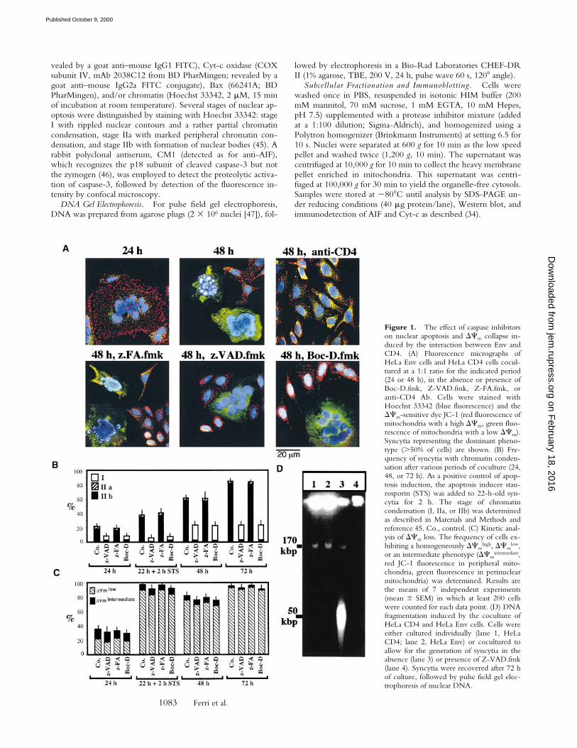
Figure 1. The effect of caspase inhibitorson nuclear apoptosis and DCm collapse in-duced by the interaction between Env andCD4. (A) Fluorescence micrographs ofHeLa Env cells and HeLa CD4 cells cocul-tured at a 1:1 ratio for the indicated period(24 or 48 h), in the absence or presence ofBoc-D.fmk, Z-VAD.fmk, Z-FA.fmk, oranti-CD4 Ab. Cells were stained withHoechst 33342 (blue fluorescence) and theDCm-sensitive dye JC-1 (red fluorescence ofmitochondria with a high DCm, green fluo-rescence of mitochondria with a low DCm).Syncytia representing the dominant pheno-type (.50% of cells) are shown. (B) Fre-quency of syncytia with chromatin conden-sation after various periods of coculture (24,48, or 72 h). As a positive control of apop-tosis induction, the apoptosis inducer stau-rosporin (STS) was added to 22-h-old syn-cytia for 2 h. The stage of chromatincondensation (I, IIa, or IIb) was determinedas described in Materials and Methods andreference 45. Co., control. (C) Kinetic anal-ysis of DCm loss. The frequency of cells ex-hibiting a homogeneously DCm
high, DCmlow,
or an intermediate phenotype (DCmintermediate,
red JC-1 fluorescence in peripheral mito-chondria, green fluorescence in perinuclearmitochondria) was determined. Results arethe means of 7 independent experiments(mean 6 SEM) in which at least 200 cellswere counted for each data point. (D) DNAfragmentation induced by the coculture ofHeLa CD4 and HeLa Env cells. Cells wereeither cultured individually (lane 1, HeLaCD4; lane 2, HeLa Env) or cocultured toallow for the generation of syncytia in theabsence (lane 3) or presence of Z-VAD.fmk(lane 4). Syncytia were recovered after 72 hof culture, followed by pulse field gel elec-trophoresis of nuclear DNA.
on February 18, 2016
jem.rupress.org
Dow
nloaded from
Published October 9, 2000
1084
Syncytial Apoptosis
Microinjection.
All syncytia growing on a premarked V-shapedarea of a coverslip (
.
200 per experiment) were microinjectedinto the cytoplasm (1–3 injections per syncytium depending ontheir size, 1 injection per
z
4 nuclei) using a computer-controlledmicroinjector (pressure 200 hPa, 2 s; Eppendorf) with PBS only(pH 7.2), recombinant AIF protein (500 ng/
m
l [34]), horse Cyt-c(12
m
g/
m
l; Sigma-Aldrich), a neutralizing anti-AIF rabbit Ab (ti-ter
z
10
5
[34]; diluted 1:1 with PBS) optionally neutralized bypreincubation with 10
m
M of AIF immunogenic peptides, a pre-immune rabbit antiserum, a neutralizing Cyt-c–specific IgG1mAb (6H2.B4, 250 ng/
m
l; BD PharMingen), an irrelevant iso-type-matched control mAb (anti-hsp60), Koenig’s polyanion (2.5
m
M; a gift from Dieter Brdizcka, University of Konstanz, Kon-stanz, Germany), recombinant human Bcl-2 (aa 1–218, 500 ng/
m
l), Bcl-2
Da
5/6 (Bcl-2
D
143–184, 500 ng/
m
l), or murine Bax(aa 1–171, 250 ng/
m
l), Bax
Da
5/6 (
D
106–153, 250 ng/
m
l [48]).After microinjection, cells were cultured for 3–24 h and stainedfor 30 min with the
DC
m
-sensitive dye JC-1 (2
m
M) and theDNA-intercalating dye Hoechst 33342 (2
m
M), followed by fixa-tion (which removes the JC-1 staining) and immunostaining forCyt-c
and/or AIF as described above.
Data Analysis.
The quantitation of different parameters byfluorescence microscopy was performed on at least 200 syncytiafor each data point, and was repeated at least 3 times in indepen-dent experiments, as stated in the figure legends. In at least oneexperiment out of each series, quantitations were performed in ablinded fashion, and in an additional experiment quantitationswere performed independently by two individuals. Interexperi-mental variability was generally
,
15%.
ResultsCaspase-dependent Nuclear Apoptosis of Syncytia Induced by
the Interaction between Env- and CD4-expressing Cells.HeLa cells stably transfected with human CD4 (HeLaCD4) formed syncytia when cocultured with HeLa cellsexpressing a lymphotropic HIV-1 Env gene (HeLa Env[42]; Fig. 1). After 24 h of coculture, several morphologi-cally normal nuclei (detected with Hoechst 33342, bluefluorescence) could be clearly distinguished within a com-mon cytoplasm of HeLa CD4/HeLa Env hybrids (Fig. 1
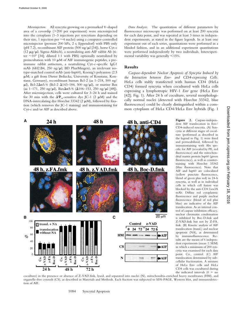
Figure 2. Caspase-indepen-dent AIF translocation in Env/CD4-induced syncytia. (A) Syn-cytia at different stages of cocul-ture (performed as described inthe legend to Fig. 1) were fixedand permeabilized, followed byimmunostaining with Abs spe-cific for AIF (revealed by PE, redfluorescence) and the mitochon-drial matrix protein hsp60 (greenfluorescence), as well as counter-staining with Hoechst 33342(blue fluorescence). Note thatAIF and hsp60 are colocalized(yellow punctate fluorescence,blend of green plus red) in 24-hsyncytia, as well as in individualcells in which cell fusion wasblocked by the anti-CD4 Leu3AmAb. Diffuse red cytoplasmicfluorescence and purple nuclearfluorescence (blend of red plusblue) are indicative of the AIFtranslocation. As an internal con-trol of caspase inhibition efficacy,nuclear chromatin condensationis inhibited by Boc-D.fmk andZ-VAD.fmk but not by Z-FA.fmk. (B) Kinetic analysis of AIFtranslocation (transl.) and nuclearapoptosis (NA), as determinedby immunofluorescence. Re-sults are the means of 6 indepen-dent experiments (mean 6 SEM)in which a minimum of 200 syn-cytia was examined for each datapoint. Co., control. (C) AIFtranslocation determined by sub-cellular fractionation. A mixtureof HeLa Env cells and HeLaCD4 cells was cocultured duringthe indicated intervals (0 5 no
coculture) in the presence or absence of Z-VAD.fmk, lysed, and separated into nuclei (N), mitochondria-enriched heavy membranes (HM), andorganelle-free cytosols (CS), as described in Materials and Methods. Each fraction was subjected to SDS-PAGE, Western blot, and immunodetec-tion of AIF.
on February 18, 2016
jem.rupress.org
Dow
nloaded from
Published October 9, 2000

1085 Ferri et al.
A). However, after prolonged culture (48–72 h) an increas-ing percentage of nuclei manifested apoptotic chromatincondensation (Fig. 1, A and B). This chromatin condensa-tion was restricted to syncytia, and inhibition of cell fusionby addition of the anti-CD4 mAb Leu3A at the beginningof coculture prevented all signs of apoptosis (Fig. 1 A). Asin other models of apoptosis (45), chromatin condensationrapidly evolved to a stage with strong Hoechst 33342–detec-table condensation of most of the chromatin (stage II),without (stage IIa, in a minority of syncytia) or with theformation of nuclear apoptotic bodies (stage IIb, in mostsyncytia) (Fig. 1 B). Nuclear apoptosis attained all nucleiwithin the same heterokaryon in a coordinated fashion(Fig. 1 A), and was accompanied by oligonucleosomalDNA degradation (not shown) as well as by “large scale”(z50 kbp) DNA fragmentation (Fig. 1 D). All morpholog-ical and biochemical signs of nuclear apoptosis werestrongly reduced by the two pancaspase inhibitors Boc-D.fmk and Z-VAD.fmk, but not by the chemically relatedcathepsin inhibitor Z-FA.fmk (Fig. 1 A). Only a minorityof cells exhibited a partial peripheral chromatin condensa-tion (stage I) in the presence of caspase inhibitors (Fig. 1 B).In conclusion, Env-induced syncytia spontaneously un-dergo caspase-dependent nuclear apoptosis.
Caspase-independent Signs of MMP of Env-induced Syncy-tia. Staining of Env-induced syncytia with the DCm-sen-sitive dye JC-1 revealed a progressive DCm loss. Thus,mitochondria from most newly formed syncytia (24 h) pos-sessed a high DCm (red JC-1 fluorescence; Fig. 1 A),whereas mitochondria from aging syncytia (48–72 h)mostly have a low DCm (green JC-1 fluorescence; Fig. 1, Aand C). The DCm loss progressed from the perinuclear areato the periphery (not shown), giving rise to a transient in-termediate phenotype (Fig. 1 C), and was accompanied bya moderate perinuclear clustering of mitochondria (stainedfor the matrix protein hsp60, which is not released duringapoptosis [30], or the sessile inner membrane proteinCOX; green fluorescence in Fig. 2 A and Fig. 3 A, respec-tively). In addition, syncytia progressively manifested signsof outer MMP, as indicated by the translocation of AIF(red fluorescence in Fig. 2 A) from mitochondria to the cy-tosol and to the nucleus (blue fluorescence in Fig. 2 A andFig. 3 A), or that of Cyt-c to the cytosol (red fluorescencein Fig. 3 A). The translocation of AIF and Cyt-c from mi-tochondria to the extramitochondrial compartment wasconfirmed by subcellular fractionation followed by immu-noblot. In single cells or 24-h-old syncytia, AIF and Cyt-care confined to the mitochondrial compartment. 48 h after
Figure 3. Caspase-independent Cyt-ctranslocation in syncytia. (A) Syncytia aris-ing from the coculture of HeLa Env andHeLa CD4 cells, treated as described in thelegends to Figs. 1 and 2, were stained withmAbs specific for Cyt-c (revealed by PE)and COX (revealed by FITC) as well aswith Hoechst 33342. Representative pho-tomicrographs are shown. Note the diffusered fluorescence indicative of Cyt-c releasefrom mitochondria occurring in the major-ity of 48-h-old syncytia. (B) Kinetic analysisof AIF, Cyt-c release, and nuclear apoptosis.Results (obtained as described in A) are themeans of six independent experiments(mean 6 SEM). NA, nuclear apoptosis;transl., translocation; Co., control. (C) Cyt-ctranslocation determined by immunoblot-ting. A mixture of HeLa Env cells and HeLaCD4 cells was cocultured during the indi-cated intervals in the presence or absence ofZ-VAD.fmk, lysed, subjected to subcellularfractionation (HM, heavy membranes; CS,cytosols; N, nuclei), and immunodetectionof Cyt-c (same samples as described in thelegend to Fig. 2).
on February 18, 2016
jem.rupress.org
Dow
nloaded from
Published October 9, 2000

1086 Syncytial Apoptosis
initiation of coculture, ectopic AIF becomes detectable inboth cytosols and nuclei (Fig. 2 C), whereas ectopic Cyt-ccan be detected only in the cytosol (Fig. 3 C). NeitherBoc-D.fmk nor Z-VAD.fmk (added every 24 h at a con-centration of 100 mM) prevented the mitochondrialmanifestations of apoptosis (DCm collapse, AIF and Cyt-ctranslocation; Figs. 1–3). Hence, MMP proceeds in acaspase-independent fashion in HIV-1–induced syncytia.
Kinetics of AIF and Cyt-c Translocation. The percentageof cells manifesting DCm dissipation and AIF translocationwas higher than that of cells positive for Cyt-c translocation(compare percentage values in Figs. 1 C, 2 B, and 3 B), anddouble immunofluorescence staining of cells for AIF andCyt-c confirmed the existence of cells having translocatedAIF to the nucleus and still retaining Cyt-c in mitochon-dria (but not vice versa; not shown), indicating that DCm
loss and AIF release occurred before Cyt-c release. AIF andCyt-c translocation were also observed in heterokaryonsgenerated by coculturing HeLa CD4 cells with a lymphoidcell line chronically infected with a syncytium-inducingHIV-1 isolate. Caspase inhibition with Z-VAD.fmk failedto prevent signs of MMP, although it did inhibit nuclearapoptosis as an internal control of its efficacy (Fig. 4). Ki-netic analyses confirmed that mitochondria from HIV-1–induced syncytia translocate AIF before Cyt-c and beforecaspase-3 activation or nuclear chromatin condensationcould be detected (Fig. 4). Hence, immunodetectabletranslocation of AIF precedes that of Cyt-c.
Bax and Bcl-2 Regulate MMP in Env-induced Syncytia.Members of the Bcl-2/Bax family regulate apoptosis viatheir capacity to modulate MMP (21–23, 49). To addressthe mechanisms by which MMP occurs in Env-inducedsyncytia, cells were stained with Abs directed against Baxand Bcl-2. Whereas no changes in Bcl-2 staining were ob-
served upon prolonged culture of syncytia (not shown),Bax (red fluorescence) was found to translocate from a cy-toplasmic, preponderantly nonmitochondrial to a punctate,mitochondrial (counterstained with anti-COX, green fluo-rescence) localization (Fig. 5 A). This finding is reminiscentof other models of apoptosis in which insertion of Bax intomitochondrial membranes causes MMP (50–53). Microin-jection of recombinant Bax (but not microinjection of themutant BaxDa5/6 protein lacking the putative membraneinsertion domain) induced a rapid (3 h) DCm dissipation,Cyt-c translocation, and nuclear apoptosis (Fig. 5 B). Thiseffect of Bax was reduced by coinjection of Bcl-2 orKoenig’s polyanion (Fig. 5 B), an inhibitor of the mito-
Figure 4. Kinetics of AIF translocation, Cyt-c translocation, andcaspase-3 activation in HIV-1–infected syncytia. CD4-expressing HeLacells were cocultured with chronically HIV-1–infected H9/IIIB cells at a2:1 ratio. At different time points, syncytia were fixed and permeabilizedto assess the translocation of AIF (as described in the legend to Fig. 2) orthat of Cyt-c (as described in the legend to Fig. 3), as well as the activa-tion of caspase-3, using an antiserum specific for the active caspase-3 p18subunit. Moreover, the frequency of syncytia exhibiting different stages ofnuclear apoptosis was assessed (as described in the legend to Fig. 1). Re-sults are the means of three experiments (mean 6 SEM). NA, nuclear ap-optosis; transl., translocation.
Figure 5. Bax/Bcl-2–mediated control of MMP in Env/CD4-inducedsyncytia. (A) Subcellular redistribution of Bax during spontaneous apop-tosis in syncytia. Syncytia were obtained by the coculture of HeLa Envand HeLa CD4 cells for 24 or 48 h, followed by staining with anti-COX(green fluorescence), an anti-Bax mAb (revealed by PE, red fluorescence),and Hoechst 33342 (blue fluorescence). At 24 h, 82 6 3% of syncytiamanifest a preponderantly nonmitochondrial distribution of Bax, whereasat 48 h, 61 6 5% cells demonstrate a mainly mitochondrial localization ofBax (88 6 4% at 72 h, n 5 3). (B) Microinjection of recombinant pro-teins from the Bax/Bcl-2 family. 24-h-old syncytia were microinjectedwith recombinant Bax, Bcl-2, inactive mutant proteins (BaxDa5/6, Bcl-2Da5/6), and/or Koenig’s polyanion (KPA), followed by determinationof the frequency of cells with a low DCm (green JC-1 fluorescence), Cyt-ctranslocation, AIF translocation (only determined after 24 h), and nuclearchromatin condensation. Results are the means of three experiments(mean 6 SEM). N.A., nuclear apoptosis; Co., control.
on February 18, 2016
jem.rupress.org
Dow
nloaded from
Published October 9, 2000
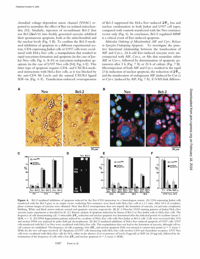
1087 Ferri et al.
chondrial voltage–dependent anion channel (VDAC) re-ported to neutralize the effect of Bax on isolated mitochon-dria (53). Similarly, injection of recombinant Bcl-2 (butnot Bcl-2Da5/6) into freshly generated syncytia inhibitedtheir spontaneous apoptosis, both at the mitochondrial andthe nuclear levels (Fig. 5 B). To confirm the Bcl-2–medi-ated inhibition of apoptosis in a different experimental sys-tem, CD4-expressing Jurkat cells or U937 cells were cocul-tured with HeLa Env cells, a manipulation that resulted inrapid syncytium formation and apoptosis (in the case of Jur-kat Neo cells; Fig. 6, A–D) or syncytium-independent ap-optosis (in the case of U937 Neo cells [54]; Fig. 6 E). Thislatter type of apoptosis requires CD4- and CXCR4-medi-ated interactions with HeLa Env cells, as it was blocked bythe anti-CD4 Ab Leu3a and the natural CXCR4 ligandSDF-1a (Fig. 6 F). Transfection-enforced overexpression
of Bcl-2 suppressed the HeLa Env–induced DCm loss andnuclear condensation in both Jurkat and U937 cell types,compared with controls transfected with the Neo resistancevector only (Fig. 6). In conclusion, Bcl-2–regulated MMPis a critical event of Env-induced apoptosis.
Molecular Ordering of Mitochondrial AIF and Cyt-c Releasein Syncytia Undergoing Apoptosis. To investigate the puta-tive functional relationship between the translocation ofAIF and Cyt-c, 24-h-old Env-induced syncytia were mi-croinjected with AIF, Cyt-c, or Abs that neutralize eitherAIF or Cyt-c, followed by determination of apoptotic pa-rameters after 3 h (Fig. 7 A) or 24 h of culture (Fig. 7 B).Microinjection of both AIF and Cyt-c resulted in the rapid(3 h) induction of nuclear apoptosis, the reduction of DCm,and the translocation of endogenous AIF (induced by Cyt-c)or Cyt-c (induced by AIF; Fig. 7 A). Z-VAD.fmk differen-
Figure 6. Bcl-2–mediated inhibition of apoptosis induced by the Env–CD4 interaction in a heterologous system. (A) CD4-expressing Jurkat cellstransfected with the Bcl-2 gene or an empty vector conferring Neo resistance were fused with HeLa Env cells at a 1:1 ratio. After 18 h of coculture,phase–contrast images of syncytia were obtained. Note that Bcl-2 overexpression does not impede the formation of syncytia, yet prevents cytoplasmicblebbing. White and black arrows indicate normal and apoptotic syncytia, respectively. (B) JC-1/Hoechst 33342 staining patterns of Jurkat/HeLa Envsyncytia (same experiment as described in A). (C) Kinetic analysis of apoptosis induction by fusion of Bcl-2 or Neo Jurkat cells to HeLa Env cells. Thefrequency of cells demonstrating a JC-1–detectable DCm reduction and nuclear apoptosis was determined after the indicated period of coculture (mean 6SEM, n 5 3). (D) DNA fragmentation pattern induced by coculture of HeLa Env cells with Neo Jurkat or Bcl-2 cells. Cells were recovered after 18 hand nuclear DNA was analyzed by pulse field gel electrophoresis. (E) Bcl-2–mediated inhibition of HeLa Env–induced apoptosis of U937 cells. U937cells transfected with Bcl-2 or Neo were cocultured with HeLa Env cells. This manipulation does not lead to the formation of syncytia, although cell-to-cell contacts are established. The frequency of cells acquiring a low DCm and nuclear apoptosis (NA) was assessed at various time points (n 5 3, mean 6SEM) for the two cell types involved. (F) Apoptosis of U937 cells interacting with HeLa Env cells involves CD4 and chemokine receptors. U937 Neocells were cocultured with HeLa Env cells for 24 h, either in the absence (Co) or presence of Leu3a (5 mg/ml) or SDF-1a (10 mg/ml), followed by de-termination of the frequency of cells with a low DCm and nuclear apoptosis (n 5 3, mean 6 SEM).
on February 18, 2016
jem.rupress.org
Dow
nloaded from
Published October 9, 2000

1088 Syncytial Apoptosis
tially affected the mitochondrial effects of ectopic (extrami-tochondrial) AIF and Cyt-c. It suppressed the Cyt-c–inducedDCm loss and release of AIF. In contrast, Z-VAD.fmkfailed to inhibit the AIF-triggered DCm dissipation andCyt-c translocation (Fig. 7 A). As an internal control, aneutralizing anti-AIF Ab prevented the acute (3 h) AIF ef-fects when microinjected together with AIF (Fig. 7 A).This Ab also prevents the spontaneous DCm loss, Cyt-c re-lease, and nuclear apoptosis of syncytia (Fig. 7 B). This ef-fect is specific, as it was not observed with a preimmuneantiserum nor when the AIF Ab was neutralized by prein-cubation with an excess of AIF-derived immunogenic pep-tides (Fig. 7 B). In sharp contrast, neutralization of Cyt-cby microinjection of a specific mAb did not prevent theDCm change nor did it affect the AIF translocation occur-ring during syncytial aging (Fig. 7 B). However, Cyt-cneutralization did inhibit the Hoechst 33342–detectablechromatin condensation (Fig. 7 B). Altogether, these resultssuggest that ectopic AIF is sufficient and necessary forcaspase-independent mitochondrial Cyt-c release, whereasCyt-c is critical for caspase-dependent nuclear apoptosis.
DiscussionThe Intrinsic Pathway Governs Apoptosis Triggered by
Env. Here we demonstrate that Env-induced syncytiaspontaneously undergo apoptosis and that this apoptoticprocess obeys the rules of the intrinsic (rather than the ex-trinsic) cell death pathway. This demonstration is based onseveral lines of evidence. First, syncytia manifest signs of in-ner MMP (DCm dissipation; Fig. 1) and outer MMP (re-lease of AIF and Cyt-c; Figs. 2 and 3), in line with the factthat syncytial mitochondria frequently are dilated (13, 14).Swelling of mitochondria is associated with MMP and oc-curs both in early apoptosis of individual cells (before cellshrinkage [55–57]) and in necrosis (58). Second, outer andinner MMP occurs well before caspases are activated andbefore nuclear chromatin is condensed in a caspase-depen-dent fashion (Figs. 1–4). Third, inhibition of caspases byoligo- or monopeptidic inhibitors does not prevent MMP,confirming that caspases act downstream of MMP (Figs.1–3). Fourth, inhibition of MMP by microinjection of re-combinant Bcl-2 (Fig. 5 B) or by coculture of Env-positivecells with CD41 cells expressing a Bcl-2 transgene (Fig. 6)prevents nuclear apoptosis, as this may be expected for theintrinsic (but not the extrinsic) pathway of death induction(25–27, 36).
Formation of syncytia is a nonphysiological process (withthe exception of a few cell types such as syncytiotropho-blasts, spermatogonia, osteoclasts, and myocytes [59]), sup-porting the idea that syncytia could be intrinsically con-demned to undergo apoptosis. However, HeLa cells drivento form syncytia by culture with methotrexate or by trans-fection with fusion-competent proteins from humanparainfluenza virus type 4a (60) die more slowly than Env/CD4-induced HeLa syncytia (Ferri, K.F., and G. Kroemer,unpublished observation), suggesting that the receptors in-volved in the fusion process contribute to the triggering ofthe intrinsic pathway. Accordingly, engagement of CD4and CXCR4 can induce lymphocyte death without syncy-tium formation (38, 40, 61), and U937 cells die upon con-tact with Env-transfected cells without prior cell fusion (54,62; Fig. 6 E). Although it remains elusive whether Env-in-duced syncytium-dependent and syncytium-independentapoptosis are mediated by identical pathways, it appearsclear that Bcl-2–regulated, presumably Bax-triggered MMPis a critical event of different types of cell death stimulatedvia the Env–CD4/CXCR4 interaction (Figs. 5 and 6).
Cell Type–specific Contribution of AIF and Caspases to Nu-clear Apoptosis. In dying syncytia arising from the fusion ofEnv- and CD4/CXCR4-expressing cells, mitochondriarelease both Cyt-c and AIF (Figs. 2 and 3). Microinjectionof Abs neutralizing either AIF or Cyt-c prevents chromatincondensation (Fig. 7 B), suggesting that both factors con-tribute to nuclear apoptosis: AIF in a caspase-independentfashion, and Cyt-c in a caspase-dependent fashion (Fig. 7A). In a cell-free system, recombinant AIF causes caspase-independent large scale (z50 kbp) DNA fragmentation andperipheral chromatin condensation (34) when added to pu-rified nuclei. Paradoxically, however, in Z-VAD.fmk–
Figure 7. Molecular hierarchy between the mitochondrial release ofAIF and Cyt-c. 24-h-old syncytia, generated by coculture of HeLa Envand HeLa CD4 cells, were microinjected with the indicated combinationof AIF, Cyt-c and/or anti-AIF, and anti–Cyt-c Abs, followed by an addi-tional culture period of 3 (A) or 24 h (B), optionally in the presence ofZ-VAD.fmk, as indicated. Unfixed cells were then stained with Hoechst33324 and JC-1 or, alternatively, cells were fixed and stained with anti-AIF plus anti-hsp60 or anti–Cyt-c plus anti-COX to determine the nu-clear and mitochondrial parameters of apoptosis. After microinjection ofanti-AIF or anti–Cyt-c Abs, the subcellular localization of AIF or Cyt-c,respectively, could not be determined (asterisks). Each point representsthe mean of at least 200 microinjected syncytia (n 5 3, mean 6 SEM).Nucl. Apopt., nuclear apoptosis; transl., translocation; Co., control; pept.,peptide.
on February 18, 2016
jem.rupress.org
Dow
nloaded from
Published October 9, 2000
1089 Ferri et al.
treated 72-h-old syncytia, AIF is clearly present in the nu-cleus (Fig. 2), yet no z50 kbp DNA fragmentation patterncan be detected (Fig. 1 D) and chromatin condensation isstrongly reduced (Fig. 1, A and B). Thus, in contrast to fi-broblast cell lines in which large scale DNA fragmentationis caspase independent (and presumably AIF mediated [34,45]), HeLa syncytia large scale DNA fragmentation appearsto be fully caspase dependent, as this has been reported forJurkat cells in which the caspase-activated DNase accountsfor both oligonucleosomal and large scale DNA fragmenta-tion (63). Of note, microinjection of an excess of exoge-nous recombinant AIF into freshly formed (24 h) syncytiacan trigger acute (3 h) chromatin condensation in acaspase-independent fashion (Fig. 7 A). Thus, in principle,AIF can exert an (presumably direct) effect on nuclei fromHeLa syncytia. Nonetheless, in the slow (24–48 h) ad-vancement of chromatin condensation observed in HeLasyncytia spontaneously undergoing apoptosis, AIF clearlyacts in a caspase-dependent fashion (Fig. 2). Future workwill unravel whether AIF-inhibitory factors and/or theabundance of the nuclear AIF target account for these celltype–specific differences.
Hierarchy of Mitochondrial AIF and Cyt-c Release. Cyt-crelease has been reported to occur in a coordinated, nearlysimultaneous fashion in most if not all mitochondria of thesame cell (64). Accordingly, syncytia retaining Cyt-c orAIF in a fraction of mitochondria were infrequently ob-served (,1% of the entire population; Figs. 2 A and 3 A) atany time point, whereas a heterogeneity in the DCm losswas evident in a substantial fraction of cells (Fig. 1 C). In-ternal feed-forward amplification loops could contribute tothe rapid kinetics of Cyt-c release. One such amplificationloop has been proposed to be provided by caspases, which,once activated as a consequence of MMP, stimulate MMP(65, 66). However, in Env-induced syncytia, caspase inhi-bition does not affect the kinetics of Cyt-c release (Fig. 3B). A caspase-independent amplification loop may be pro-vided by AIF based on the observation that the microinjec-tion of an anti-AIF Ab retards all signs of MMP, includingDCm collapse and the release of Cyt-c (Fig. 7 B), whereasmicroinjection of recombinant AIF induces MMP (Fig. 7A). Intriguingly, the mitochondrial release of AIF precedesthat of Cyt-c in Env-induced syncytia by several hours(compare the percentage values in Figs. 2 B and 3 B, and inFig. 4). This appears counterintuitive because protein-per-meant pores formed in the outer mitochondrial membraneshould favor the release of small proteins such as Cyt-c(14.5 kD) over that of the much larger AIF (56 kD [53]).Cyt-c is known to be associated with inner membranechristae, in electrostatic interaction with cardiolipin (67). Itis tempting to speculate that, in addition to outer MMP,(AIF-induced?) changes in inner membrane physicochem-istry such as cardiolipin oxidation (44, 68) must occur to al-low for full Cyt-c release.
Irrespective of the exact mechanism, our results indicatethat, at least in the model studied herein, the mitochondrialrelease of Cyt-c is subordinate to that of AIF. Thus, micro-injection of AIF into the cytoplasm of syncytia suffices to
cause Cyt-c release in a caspase-independent fashion (Fig. 7A), mimicking that of the spontaneous (caspase-indepen-dent; Fig. 3) Cyt-c release. Moreover, neutralization of AIFby microinjection of a specific Ab prevents the release ofCyt-c as well as nuclear apoptosis, whereas neutralization ofCyt-c has no effect on the AIF translocation and only im-pedes nuclear condensation (Fig. 7 B). Taken together,these results delineate the following sequence of events:Bax-mediated/Bcl-2–inhibited MMP → AIF release →Cyt-c release → caspase activation → nuclear apoptosis.Future research will unravel whether this hierarchy is onlyapplicable to syncytial apoptosis or whether it can be in-scribed into a more general pathway.
This work establishes that MMP is a critical step in Env-induced syncytial apoptosis. Other proapoptotic proteinsencoded by HIV-1 (PR, Tat, and Vpr) also favor MMP.Vpr exerts its proapoptotic effect, at least in part, by bind-ing to the mitochondrial adenine nucleotide translocator,thereby directly inducing MMP (7). The HIV-1 protease(PR) can cleave Bcl-2, thereby abolishing its MMP-inhibi-tory function (69). Tat reduces the expression of the mito-chondrial superoxide dismutase 2 isoenzyme (4, 70), whichis another endogenous MMP inhibitor (71). Tat may alsofavor apoptosis by physically interacting with mitochondria(72). It thus emerges that HIV-1 employs several indepen-dent strategies to induce MMP and apoptosis via the intrin-sic pathway. It remains an ongoing conundrum whetherthese manifold strategies are designed to cooperate amongeach other in an additive or synergistic fashion, in the samecell, or whether they rather reflect the capacity of HIV-1 tokill a wide array of distinct cell types.
We are indebted to Dr. Dominique Piatier-Tonneau for constantsupport and to Drs. Marc Alizon, Dieter Brdiczka, Pierre Char-neau, Nicole Israel, and Anu Srinivasen, as well as the National In-stitutes of Health AIDS Research and Reference Reagent Program,for gifts of reagents.
This work was supported by a special grant by the Ligue Nation-ale contre le Cancer, as well as by grants from Association Nation-ale pour la Recherche sur le SIDA, Fondation pour la RechercheMedicale, the European Commission, the Picasso Program (to G.Kroemer), Fundació irsiCaixa (to J.A. Este), FIS 00/0893 (to J.Blanco), and grants GM 60554 and National Institutes of HealthCA69381 (to J.C. Reed). K.F. Ferri receives a fellowship from theFrench Ministry of Science, and E. Jacotot receives an AssociationNationale pour la Recherche sur le SIDA fellowship. J. Blanco is aresearcher from the Fundació per a la Recerca Biomèdica GermansTrias i Pujol.
Submitted: 12 April 2000Revised: 17 August 2000Accepted: 28 August 2000
References1. Fauci, A.S. 1996. Host factors and the pathogenesis of HIV-
induced disease. Nature. 384:529–534.2. Gougeon, M.L., and L. Montagnier. 1999. Programmed cell
death as a mechanism of CD4 and CD8 T cell depletion inAIDS. Molecular control and effect of highly active anti-ret-
on February 18, 2016
jem.rupress.org
Dow
nloaded from
Published October 9, 2000

1090 Syncytial Apoptosis
roviral therapy. Ann. NY Acad. Sci. 887:199–212.3. Li, C.J., D.J. Friedman, C. Wang, V. Metelev, and A.B. Par-
dee. 1995. Induction of apoptosis in uninfected lymphocytesby HIV-1 Tat protein. Science. 268:429–431.
4. Westendorp, M.O., V.A. Shatrov, K. Schulze-Osthoff, R.Frank, M. Kraft, M. Los, P.H. Krammer, W. Dröge, and V.Lehrmann. 1995. HIV-1 Tat potentiates TNF-induced NF-kB activation and cytotoxicity by altering the cellular redoxstate. EMBO (Eur. Mol. Biol. Organ.) J. 14:546–554.
5. Stewart, S.A., B. Poon, J.B.M. Jowett, and I.S.Y. Chen.1997. Human immunodeficiency virus type 1 Vpr inducesapoptosis following cell cycle arrest. J. Virol. 71:5579–5592.
6. Stewart, S.A., B. Poon, J.B.M. Jowett, Y. Xie, and I.S.Y.Chen. 1999. Lentiviral delivery of HIV-1 Vpr protein in-duces apoptosis in transformed cells. Proc. Natl. Acad. Sci.USA. 96:12039–12043.
7. Jacotot, E., L. Ravagnan, M. Loeffler, K.F. Ferri, H.L.A.Vieira, N. Zamzami, P. Costantini, S. Druillennec, J. Hoe-beke, J.P. Brian, et al. 2000. The HIV-1 viral protein R in-duces apoptosis via a direct effect on the mitochondrial per-meability transition pore. J. Exp. Med. 191:33–45.
8. Lifson, J.D., G.R. Reyes, M.S. McGrath, B.S. Stein, andE.G. Engleman. 1986. AIDS retrovirus-induced cytopathol-ogy: giant cell formation and involvement of CD4 antigen.Science. 232:1123–1127.
9. Sodroski, J.G., W.C. Goh, A. Rosen, K. Campbell, andW.A. Haseltine. 1986. Role of the HTLV/LAV envelope insyncytia formation and cytopathicity. Nature. 322:470–474.
10. Chirmule, N., and S. Pahwa. 1996. Envelope glycoproteinsof human immunodeficiency virus type 1: profound influ-ences on immune function. Microbiol. Rev. 60:386–406.
11. Laurent-Crawford, A.G., B. Krust, Y. Riviere, C. Des-granges, S. Muller, M.P. Kieny, C. Dauguet, and A.G. Hova-nessian. 1993. Membrane expression of HIV envelope glyco-proteins triggers apoptosis in CD4 cells. AIDS Res. Hum.Retroviruses. 9:761–773.
12. Sandstrom, P.A., D. Pardi, C.S. Goldsmith, C.Y. Duan, A.M.Diamond, and T.M. Folks. 1996. Bcl-2 expression facilitateshuman immunodeficiency virus type 1-mediated cytopathiceffects during acute spreading infections. J. Virol. 70:4617–4622.
13. Kolesnitchenko, V., L. King, A. Riva, V. Tani, S.J. Kors-meyer, and D.I. Cohen. 1997. A major human immunodefi-ciency virus type 1-initiated killing pathway distinct from ap-optosis. J. Virol. 71:9753–9763.
14. Plymale, D.R., D.S.N. Tang, A.M. Comardelle, C.D. Fer-min, D.E. Lewis, and R.G. Garry. 1999. Both necrosis andapoptosis contribute to HIV-1-induced killing of CD4 cells.AIDS. 13:1827–1839.
15. Sylwester, A., S. Murphy, D. Shutt, and D.R. Soll. 1997.HIV-induced T cell syncytia are self-perpetuating and theprimary cause of T cell death in culture. J. Immunol. 158:3996–4007.
16. Blaak, H., A.B. van’t Wout, M. Brouwer, B. Hoolbrink, E.Hovenkamp, and H. Schuitemaker. 2000. In vivo HIV-1 in-fection of CD45RA1 CD41 T cells is established primarily bysyncytium-inducing variants and correlates with the rate ofCD41 T cell decline. Proc. Natl. Acad. Sci. USA. 97:1269–1274.
17. Nicholson, D.W., and N.A. Thornberry. 1997. Caspases:killer proteases. Trends Biochem. Sci. 22:299–306.
18. Budijardjo, I., H. Oliver, M. Lutter, X. Luo, and X. Wang.1999. Biochemical pathways of caspase activation during apop-
tosis. Annu. Rev. Cell Dev. Biol. 15:269–290.19. Nagata, S. 1999. Fas ligand-induced apoptosis. Annu. Rev.
Genet. 33:29–55.20. Kroemer, G., B. Dallaporta, and M. Resche-Rigon. 1998.
The mitochondrial death/life regulator in apoptosis and ne-crosis. Annu. Rev. Physiol. 60:619–642.
21. Green, D.R., and J.C. Reed. 1998. Mitochondria and apop-tosis. Science. 281:1309–1312.
22. Gross, A., J.M. McDonnell, and S.J. Korsmeyer. 1999. Bcl-2family members and the mitochondria in apoptosis. GenesDev. 13:1899–1911.
23. Vander Heiden, M.G., and C.B. Thompson. 1999. Bcl-2proteins: inhibitors of apoptosis or regulators of mitochondrialhomeostasis? Nat. Cell Biol. 1:E209–E216.
24. Kroemer, G., and J.C. Reed. 2000. Mitochondrial control ofcell death. Nat. Med. 6:513–519.
25. Scaffidi, C., S. Fulda, A. Srinivasan, C. Friesen, F. Li, K.J.Tomaselli, K.-M. Debatin, P.H. Krammer, and M.E. Peter.1998. Two CD95 (APO-1/Fas) signaling pathways. EMBO(Eur. Mol. Biol. Organ.) J. 17:1675–1687.
26. Scaffidi, C., S. Kirchhoff, P.H. Krammer, and M.E. Peter.1999. Apoptosis signaling in lymphocytes. Curr. Opin. Immu-nol. 11:277–285.
27. Yin, X.-M., K. Wang, A. Gross, Y. Zhao, S. Zinkel, B.Klocke, K.A. Rothe, and S.J. Korsmeyer. 1999. Bid-deficientmice are resistant to Fas-induced hepatocellular apoptosis.Nature. 400:886–891.
28. Susin, S.A., N. Zamzami, M. Castedo, T. Hirsch, P. Mar-chetti, A. Macho, E. Daugas, M. Geuskens, and G. Kroemer.1996. Bcl-2 inhibits the mitochondrial release of an apopto-genic protease. J. Exp. Med. 184:1331–1342.
29. Kluck, R.M., E. Bossy-Wetzel, D.R. Green, and D.D. New-meyer. 1997. The release of cytochrome c from mitochon-dria: a primary site for Bcl-2 regulation of apoptosis. Science.275:1132–1136.
30. Patterson, S., C.S. Spahr, E. Daugas, S.A. Susin, T. Irinopou-los, C. Koehler, and G. Kroemer. 2000. Mass spectrometricidentification of proteins released from mitochondria under-going permeability transition. Cell Death Differ. 7:137–144.
31. Mancini, M., D.W. Nicholson, S. Roy, N.A. Thornberry,E.P. Peterson, L.A. Casciola-Rosen, and A. Rosen. 1998.The caspase-3 precursor has a cytosolic and mitochondrialdistribution: implications for apoptotic signaling. J. Cell Biol.140:1485–1495.
32. Susin, S.A., H.K. Lorenzo, N. Zamzami, I. Marzo, N. Laro-chette, P.M. Alzari, and G. Kroemer. 1999. Mitochondrialrelease of caspases-2 and -9 during the apoptotic process. J.Exp. Med. 189:381–394.
33. Krajewski, S., M. Krajewska, L.M. Ellerby, K. Welsh, Z.H.Xie, Q.L. Deveraux, G.S. Salvesen, D.E. Bredesen, R.E.Rosenthal, G. Fiskum, and J.C. Reed. 1999. Release ofcaspase-9 from mitochondria during neuronal apoptosis andcerebral ischemia. Proc. Natl. Acad. Sci. USA. 96:5752–5757.
34. Susin, S.A., H.K. Lorenzo, N. Zamzami, I. Marzo, B.E.Snow, G.M. Brothers, J. Mangion, E. Jacotot, P. Costantini,M. Loeffler, et al. 1999. Molecular characterization of mito-chondrial apoptosis-inducing factor. Nature. 397:441–446.
35. Chinnaiyan, A.M., C. Woffendin, V.M. Dixit, and G.J. Na-bel. 1997. The inhibition of pro-apoptotic ICE-like proteasesenhances HIV replication. Nat. Med. 3:333–337.
36. Ohnimus, H., M. Heinkelein, and C. Jassoy. 1997. Apoptoticcell death upon contact of CD41 T lymphocytes with HIVglucoprotein-expressing cells is mediated by caspases but by-
on February 18, 2016
jem.rupress.org
Dow
nloaded from
Published October 9, 2000

1091 Ferri et al.
passes CD95 (Fas/Apo-1) and TNF receptor 1. J. Immunol.159:5246–5252.
37. Kaul, M., and S.A. Lipton. 1999. Chemokines and activatedmacrophages in HIV gp120-induced neuronal apoptosis. Proc.Natl. Acad. Sci. USA. 96:8212–8216.
38. Blanco, J., E. Jacotot, C. Cabrera, A. Cardona, B. Clotet, E.De Clercq, and J.A. Este. 1999. The implication of thechemokine receptor CXCR4 in HIV-1 envelope protein-induced apoptosis is independent of the G protein-mediatedsignalling. AIDS. 13:909–917.
39. Cicala, C., J. Arthos, A. Rubbert, S. Selig, K. Wildt, O.J. Co-hen, and A.S. Faus. 2000. HIV-1 envelope induces activationof caspase-3 and cleavage of focal adhesion kinase in primaryhuman CD41 T cells. Proc. Natl. Acad. Sci. USA. 97:1178–1183.
40. Berndt, C., B. Möpps, S. Angermüller, P. Gierschik, andP.H. Krammer. 1998. CXCR4 and CD4 mediate a rapidCD95-independent cell death in CD41 cells. Proc. Natl. Acad.Sci. USA. 95:12556–12561.
41. Schwartz, O., M. Alizon, J.M. Heard, and O. Danos. 1994.Impairment of T cell receptor-dependent stimulation inCD41 lymphocytes after contact with membrane-boundHIV-1 envelope glycoprotein. Virology. 198:360–365.
42. Dragic, T., P. Charneau, F. Clavel, and M. Alizon. 1992.Complementation of murine cells for human immunodefi-ciency virus envelope/CD4-mediated fusion in human/mu-rine heterokaryons. J. Virol. 66:4794–4802.
43. Aillet, F., H. Masutani, C. Elbim, H. Raoul, L. Chene, M.T.Nugeyre, C. Paya, F. Barre-Sinoussi, M.A. Gougerot-Poci-dalo, and N. Israel. 1998. Human immunodeficiency virusinduces a dual regulation of Bcl-2, resulting in persistent in-fection of CD41 T- or monocytic cell lines. J. Virol. 72:9698–9705.
44. Zamzami, N., P. Marchetti, M. Castedo, D. Decaudin, A.Macho, T. Hirsch, S.A. Susin, P.X. Petit, B. Mignotte, andG. Kroemer. 1995. Sequential reduction of mitochondrialtransmembrane potential and generation of reactive oxygenspecies in early programmed cell death. J. Exp. Med. 182:367–377.
45. Daugas, E., S.A. Susin, N. Zamzami, K. Ferri, T. Irinopoulos,N. Larochette, M.C. Prevost, B. Leber, D. Andrews, J. Pen-ninger, and G. Kroemer. 2000. Mitochondrio-nuclear redis-tribution of AIF in apoptosis and necrosis. FASEB (Fed. Am.Soc. Exp. Biol.) J. 14:729–739.
46. Srinivasan, A., K.A. Roth, R.O. Sayer, K.S. Shindler, A.N.Wong, L.C. Fritz, and K.J. Tomaselli. 1998. In situ immuno-detection of activated caspase-3 in apoptotic neurons in thedeveloping nervous system. Cell Death Differ. 5:1004–1016.
47. Brown, D.G., X.M. Sun, and G.M. Cohen. 1993. Dexa-methasone-induced apoptosis involves cleavage of DNA tolarge fragments prior to internucleosomal fragmentation. J.Biol. Chem. 268:3037–3039.
48. Xie, Z.H., S. Schendel, S. Matsuyama, and J.C. Reed. 1998.Acidic pH promotes dimerization of Bcl-2 family proteins.Biochemistry. 37:6410–6418.
49. Zamzami, N., C. Brenner, I. Marzo, S.A. Susin, and G. Kroe-mer. 1998. Subcellular and submitochondrial mechanisms ofapoptosis inhibition by Bcl-2-related proteins. Oncogene. 16:2265–2282.
50. Wolter, K.G., Y.-T. Hsu, C.L. Smith, A. Nechushtan, X.-G.Xi, and R.J. Youle. 1997. Movement of Bax from the cytosolto mitochondria during apoptosis. J. Cell Biol. 139:1281–1292.
51. Marzo, I., C. Brenner, N. Zamzami, J. Jürgensmeier, S.A.Susin, H.L.A. Vieira, M.-C. Prévost, Z. Xie, S. Mutsiyama,J.C. Reed, and G. Kroemer. 1998. Bax and adenine nucle-otide translocator cooperate in the mitochondrial control ofapoptosis. Science. 281:2027–2031.
52. Gross, A., J. Jockel, M.C. Wei, and S.J. Korsmeyer. 1998.Enforced dimerization of Bax results in its translocation, mi-tochondrial dysfunction and apoptosis. EMBO (Eur. Mol.Biol. Organ.) J. 17:3878–3885.
53. Shimizu, S., M. Narita, and Y. Tsujimoto. 1999. Bcl-2 familyproteins regulate the release of apoptogenic cytochrome c bythe mitochondrial channel VDAC. Nature. 399:483–487.
54. Asjo, N., I. Ivhed, M. Gidlund, S. Fuerstenberg, E. Fenyo,M. Nilsson, and J. Wigzell. 1987. Susceptibility to infectionby the human immunodeficiency virus correlates with T4 ex-pression in a parental monocytoid cell line and its subclones.Virology. 157:359–365.
55. Vander Heiden, M.G., N.S. Chandal, E.K. Williamson, P.T.Schumacker, and C.B. Thompson. 1997. Bcl-XL regulatesthe membrane potential and volume homeostasis of mito-chondria. Cell. 91:627–637.
56. Dallaporta, B., P. Marchetti, M. de Pablo, C. Maisse, H.T.Duc, D. Metivier, N. Zamzami, M. Geuskens, and G. Kroe-mer. 1999. Plasma membrane potential in thymocyte apopto-sis. J. Immunol. 162:6534–6542.
57. Feldmann, G., D. Haouzi, A. Moreai, A.M. Durand-Schneider, A. Bringuier, A. Berson, A. Mansouri, D. Fau,and D. Pessayre. 2000. Opening of the mitochondrial perme-ability transition pore causes matrix expansion and outermembrane rupture in Fas-mediated hepatic apoptosis in mice.Hepatology. 31:674–683.
58. Lemasters, J.J., A.-L. Nieminen, T. Qjan, L.C. Trost, S.P.Elmore, Y. Nishimura, R.A. Crowe, W.E. Cascio, C.A.Bradham, D.A. Brenner, and B. Herman. 1998. The mito-chondrial permeability transition in cell death: a commonmechanism in necrosis, apoptosis and autophagy. Biochim.Biophys. Acta. 1366:177–196.
59. Anderson, J.M. 2000. Multinucleated giant cells. Curr. Opin.Hematol. 7:40–47.
60. Nishio, M., M. Tsurudome, H. Komada, M. Kawano, N.Tabata, H. Matsumura, N. Ikemura, N. Watanabe, and Y.Ito. 1994. Fusion properties of cells constitutively expressinghuman parainfluenza virus type 4a hemagglutinin-neuramini-dase and fusion glycoproteins. J. Gen. Virol. 75:3517–3523.
61. Newell, M.K., L.J. Haughn, C.R. Maroun, and M.H. Julius.1990. Death of mature T cells by separate ligation of CD4and the T-cell receptor for antigen. Nature. 347:286–289.
62. Xiao, X., D. Norwood, Y.R. Feng, M. Morluchi, A. Jones-Trower, T.S. Stantchev, H. Moriuchi, C.C. Broder, and D.S.Dimitrov. 2000. Inefficient formation of a complex amongCXCR4, CD4 and gp120 in U937 clones resistant to X4gp120-gp41-mediated fusion. Exp. Mol. Pathol. 68:139–146.
63. Sakahira, H., M. Enari, Y. Ohsawa, Y. Uchiyama, and S. Na-gata. 1999. Apoptotic nuclear morphological change withoutDNA fragmentation. Curr. Biol. 9:543–546.
64. Goldstein, J.C., N.J. Waterhouse, P. Juin, G.I. Evan, andD.R. Green. 2000. The coordinate release of cytochrome cis rapid, complete and kinetically invariant. Nat. Cell Biol.2:156–162.
65. Susin, S.A., N. Zamzami, M. Castedo, E. Daugas, H.-G.Wang, S. Geley, F. Fassy, J. Reed, and G. Kroemer. 1997.The central executioner of apoptosis. Multiple links betweenprotease activation and mitochondria in Fas/Apo-1/CD95–
on February 18, 2016
jem.rupress.org
Dow
nloaded from
Published October 9, 2000

1092 Syncytial Apoptosis
and ceramide-induced apoptosis. J. Exp. Med. 186:25–37.66. Chen, Q., B. Gong, and A. Almasan. 2000. Distinct stages of
cytochrome c release from mitochondria: evidence for a feed-back amplification loop linking caspase activation to mito-chondrial dysfunction in genotoxic stress induced apoptosis.Cell Death Differ. 7:227–233.
67. Kim, S.M., K.H. Shin, T. Fujiwara, and H. Akutsu. 1998.The interaction of ferric and ferrous cytochrome c with cardio-lipin in phospholipid membranes studied by solid-state H-2and P-31 NMR. J. Mol. Struct. 441:183–188.
68. Shidoji, Y., K. Hayashi, S. Komura, N. Ohishi, and K. Yagi.1999. Loss of molecular interaction between cytochrome cand cardiolipin due to lipid peroxidation. Biochem. Biophys.Res. Commun. 264:343–347.
69. Strack, P.R., M.W. Frey, C.J. Rizzo, B. Cordova, H.J.George, R. Meade, S.W. Jo, J. Corman, R. Tritch, and B.D.
Korant. 1996. Apoptosis mediated by HIV protease is pre-ceded by cleavage of Bcl-2. Proc. Natl. Acad. Sci. USA. 93:9571–9576.
70. Creaven, M., F. Hans, V. Mutskov, E. Col, C. Caron, S.Dimitrov, and S. Khochbin. 1999. Control of the histone-acetyltransferase activity of Tip60 by the HIV-1 transactivatorprotein, Tat. Biochemistry. 38:8826–8830.
71. Williams, M.D., H. Van Remmen, C.C. Conrad, T.T.Huang, C.J. Epstein, and A. Richardson. 1998. Increased ox-idative damage is correlated to altered mitochondrial functionin heterozygous manganese superoxide dismutase knockoutmice. J. Biol. Chem. 273:28510–28515.
72. Macho, A., M.A. Calzado, L. Jimenez-Reina, E. Ceballos, J.Leon, and E. Munoz. 1999. Susceptibility of HIV-1-TATtransfected cells to undergo apoptosis. Biochemical mecha-nisms. Oncogene. 18:7543–7551.
on February 18, 2016
jem.rupress.org
Dow
nloaded from
Published October 9, 2000
Copyright © 2022 FDOKUMEN