Design, fabrication and characterization of a VMOS monolithic ...
Upload
independentCategory
view
0download
0
Antibody Purification Using Porous Metal–ChelatedMonolithic Columns
Nilay Bereli,1 Lokman Uzun,1 Handan Yavuz,1 Assem Elkak,2 Adil Denizli1
1Department of Chemistry, Biochemistry Division, Hacettepe University, Ankara, Turkey2Department of Medical Biology, Lebanese University, Beirut, Lebanon
Received 22 July 2005; accepted 29 November 2005DOI 10.1002/app.23894Published online in Wiley InterScience (www.interscience.wiley.com).
ABSTRACT: A novel monolithic material was developedto obtain efficient and cost-effective purification of IgG fromhuman plasma. The porous monolith was obtained by bulkpolymerization in a glass tube of 2-hydroxyethyl methacry-late (HEMA) and N-methacryloyl-(l)-histidine methyl ester(MAH). The poly(HEMA-MAH) monolith had a specificsurface area of 214.6 m2/g and was characterized by swell-ing studies, porosity measurement, FTIR, scanning electronmicroscopy, and elemental analysis. Then the monolith wasloaded with Cu2� ions to form the metal chelate. Poly-(HEMA-MAH) monolith with a swelling ratio of 74% andcontaining 20.9 �mol MAH/g was used in the adsorption/
desorption of IgG from aqueous solutions and humanplasma. The maximum adsorption of IgG from an aqueoussolution in phosphate buffer was 10.8 mg/g at pH 7.0.Higher adsorption was obtained from human plasma (up to104.2 mg/g), with a purity of 94.1%. It was observed thatIgG could be repeatedly adsorbed and desorbed with thepoly(HEMA-MAH) monolith without significant loss of ad-sorption capacity. © 2006 Wiley Periodicals, Inc. J Appl Polym Sci101: 395–404, 2006
Key words: adsorption; chromatography; protein separationtechniques
INTRODUCTION
Packed-bed columns have inherent limitations such asslow diffusional mass transfer and a large void vol-ume between the beads, leading to the efficiency ofconventional columns remaining in the range of10,000–30,000 plates/column in the past two de-cades.1 Although some new stationary phases such asmonosize nonporous beads2–4 and perfusion chroma-tography packings have been designed to resolvethese limitations, in essence they have not been over-come.5 Recently, monolithic columns have been con-sidered a novel generation of stationary phases for theseparation of biomolecules because of their easy prep-aration, excellent flow properties, and high perfor-mance compared to those of conventional beads.6–10
Antibodies are biologically active proteins pro-duced by plasma cells in response to the presence offoreign substances. The growing role of antibodies inbiomedical research and devolopment is widely ac-knowledged. Antibody-based in vivo diagnostics andtherapeutics are gaining wider approval from regula-tory agencies around the world.11 At present, the mostwidely used technique for antibody purification isaffinity chromatography on protein A adsorbents.12–14
The high specificity of protein A for the Fc antibody
domain provides excellent chromatographic selec-tivity.15 Despite this, protein A sorbent has drawbacksworth considering: (1) it could leak from the matrix,causing contamination, which is intolerable in clinicalapplications; and (2) it would tend to be very high incost. Ligands such as protein A or G are difficult toimmobilize in the proper orientation. They also aresusceptible to degradation during cleaning proce-dures. To avoid complications from the use of proteinA sorbents, several available alternative purificationtechniques can be used: ion exchange chromatogra-phy, hydrophobic interaction chromatography, dye–ligand chromatography, thiophilic chromatography,histidine affinity chromatography, and molecularsieving.16–18 However, a comparison of these tech-niques is of little significance because they lack theselectivity of protein A. Among the techniques, metal-chelate affinity chromatography is a promising alter-native in downstream processing for the purificationof antibodies.19–21
Immobilized metal affinity chromatography (IMAC)is a sensitive technique for protein separation thatenables distinguishing between proteins differing byonly a single histidine residue on the surface.22–27 It isassumed that proteins interact mainly through theimidazole group of histidine and, to a lesser extent, theindoyl group of tryptophan and the thiol group ofcysteine. Cooperation between neighboring aminoacid side chains and local conformations play impor-tant roles in protein binding. Aromatic amino acids
Correspondence to: A. Denizli ([email protected]).
Journal of Applied Polymer Science, Vol. 101, 395–404 (2006)© 2006 Wiley Periodicals, Inc.

and the amino terminal of the peptides also contrib-ute.28 The low cost of metals and the ability to reuseadsorbents hundreds of times without any detectableloss of metal-chelating properties are the attractivefeatures of metal affinity separation.
This article reports on the purification of an IgGantibody from human plasma by metal-chelate affinitychromatography with a monolith column. Poly-(HEMA-MAH) monolith is a copolymer of 2-hydroxy-ethyl methacrylate (HEMA) and N-methacryloyl-(l)-histidine-methylester (MAH), which was obtained bybulk polymerization. The poly(HEMA-MAH) mono-lith was characterized by scanning electron micros-copy (SEM), porosity measurement, FTIR, elementalanalysis, and swelling tests. Then Cu2� ions werechelated through imidazole groups on the MAH reac-tive functional groups of the polymeric structure. Theability of the monoliths to adsorb IgG from aqueoussolutions containing different IgG concentrations atdifferent pHs and ionic strengths and from humanplasma was investigated. Desorption of IgG and ma-terial stability also were tested.
EXPERIMENTAL
Materials
Immunoglobulin G (IgG) (Sigma, cat. no. 160101), l-histidine methylester, and methacryloyl chloride weresupplied by Sigma (St. Louis, MO). Hydroxyethylmethacrylate (HEMA) and ethylene glycol dimethac-rylate (EGDMA) were obtained from Fluka A.G.(Buchs, Switzerland), distilled under reduced pressurein the presence of hydroquinone inhibitor, and storedat 4°C until use. Potassium persulfate (KPS) was ob-tained from Fluka (Switzerland). All other chemicalswere of reagent grade and were purchased fromMerck AG (Darmstadt, Germany). All water used inthe adsorption experiments was purified using a Barn-stead (Dubuque, IA) ROpure LP� reverse osmosis unitwith a high-flow cellulose acetate membrane (Barn-stead D2731) and then a Barnstead D3804 NANO-pure� organic/colloid removal and ion exchangepacked-bed system.
Synthesis of MAH
The synthesis and characterization of MAH were per-formed as described previously.29 In the experimentalprocedure for synthesis of MAH, 5.0 g of l-histidinehydrochloride and 0.2 g of hydroquinone were dis-solved in 100 mL of a dichloromethane solution,which was cooled to 0°C. Then 12.7 g of triethylaminewas added to the solution, followed by the addition of5.0 mL of methacryloyl chloride, which was poured inslowly. Then this solution was stirred magnetically atroom temperature for 2 h, after which hydroquinone
and unreacted methacryloyl chloride were extractedwith a 10% NaOH solution. The aqueous phase wasevaporated in a rotary evaporator. The residue (i.e.,MAH) was crystallized in an ether–cyclohexane mix-ture and then dissolved in ethyl alcohol. 1H-NMR,performed in CDCl3 on a JEOL GX-400 300 MHz in-strument, was used to determine if the MAH structurewas synthesized. The residual nondeuterated solvent(CHCl3) served as an internal reference. Chemicalshifts are reported in parts per million downfield rel-ative to CHCl3. The 1H-NMR spectrum shows thecharacteristic peaks of the groups in the MAH mono-mer as follows—1H-NMR (CDCl3): � � 1.99 (t; 3H, J� 7.08 Hz, CH3), 1.42 (m; 2H, CH2), 3.56 (t; 3H,OOCH3) 4.82–4.87 (m; 1H, methin), 5.26 (s; 1H, vinylH), 5.58 (s; 1H, vinyl); 6.86 (�; 1H, J � 7.4 Hz, NH), 7.82(�; 1H, J � 8.4 Hz, NH), 6.86–7.52 (m; 5H, aromatic).
Preparation of poly(HEMA-MAH) monolithiccolumn
The poly[hydroxyethyl methacrylate-N-methacryloyl-(l)-histidinemethylester] [poly(HEMA-MAH)] mono-lithic column was prepared by in situ polymerizationin a glass tube, using potassium persulfate as theinitiator. Toluene and EGDMA were the pore formerand the crosslinker, respectively. Potassium persulfate(15 mg) was dissolved in a mixture of monomers(HEMA: 1.0 mL; EGDMA: 250 �L) and porogenicdiluent (toluene: 750 �L). MAH (10 mg) was dissolvedin HEPES (500 �L). These monomer solutions weremixed, and then the final solution was purged withnitrogen for 15 min. The glass tube (100 � 10 mminside diameter) was filled with the above mixtureand then sealed with pieces of silicone rubber tubingplugged with silicone stoppers. Polymerization wasallowed to proceed at 75°C for 45 min. The tube wasthen attached to a chromatographic system. Ethyl al-cohol (50 mL) and water (50 mL) were pumpedthrough the column at a flow rate of 1.0 mL/min inorder to remove the unreacted monomers and poro-genic diluents in the monolith after completion ofpolymerization. The monolith was stored in buffercontaining 0.02% sodium azide at 4°C until use.
Incorporation of Cu2� ions
The investigation of Cu2� chelation was carried out ina recirculating system equipped with a water jacketfor temperature control. The monolith was washedwith 30 mL of water. Then 40 mL of a Cu2� solution[50 mg/L (pH 4.1), adjusted with HCl and NaOH] waspumped through the column under recirculation atroom temperature for 2 h. A 1000-ppm atomic absorp-tion standard solution (containing 10% HNO3) wasthe source of the Cu2� ions. The concentration of theCu2� ions in the resulting solution was determined
396 BERELI ET AL.

with a graphite furnace atomic absorption spectrom-eter (GFAAS, Analyst 800/Perkin Elmer, USA). Theinstrument response was periodically checked withknown metal solution standards. The experimentswere performed in triplicate, as were analyses of thesamples. For each set of data, standard statisticalmethods were used to determine the mean and stan-dard deviation. A 95% confidence interval was calcu-lated for each set of samples in order to determine themargin of error. The Cu2� concentrations in the initialand final solutions were used to calculate the amountof Cu2� ions adsorbed.
Cu2� leakage from the poly(HEMA-MAH) mono-lith was investigated in media whose pH varied be-tween 5.0 and 8.0 and also in a medium containing1.0M NaCl. The monolith was stirred for 24 h at roomtemperature. Then the concentration of Cu2� ions inthe supernatants was determined using an atomic ab-sorption spectrophotometer. Note that the metal-che-lated monolith was stored at 4°C in a 10 mM Trisbuffer (pH 7.4).
Characterization of monoliths
Surface area and porosity measurements
The surface area of the monolith sample was deter-mined in a BET isotherm of nitrogen using anASAP2000 instrument (Micromeritics, Norcross, GA).Pore volume and average pore diameter greater than20 Å were determined up to 2000 kg/cm2 with amercury porosimeter (Carlo Erba, model 200).
Swelling test
Monolith water uptake ratios were determined in dis-tilled water as follows: A dry monolith was carefullyweighed and then placed in a 50-mL vial containingdistilled water. The vial was put into an isothermalwater bath at 25°C for 24 h. The monolith was re-moved from the water, wiped with a filter paper, andweighed. The water content of the monolith (ratio ofthe dry sample mass to the wet sample mass) wascalculated using the following expression:
Water uptake ratio % � ��Ws � Wo�/Wo� � 100 (1)
where Wo and Ws are the mass of the monolith beforeand after uptake of water, respectively.
Surface morphology
Scanning electron microscopy was used to analyze themorphology of cross sections of dried monolith. Thesamples were initially dried in air at 25°C for 7 daysbefore use in the SEM analysis. A fragment of driedmonolith was mounted on a SEM sample mount and
sputter-coated for 2 min. The sample was thenmounted in a scanning electron microscope (RasterElectronen Microscopy, Leitz-AMR-1000, Germany),and the surface was scanned at the desired magnifi-cation.
Elemental analysis
Elemental analysis (Leco elemental analyzer, ModelCHNS-932, USA) was performed to evaluate howmuch MAH was incorporated into the poly(HEMA-MAH) monolith.
FTIR
The FTIR spectrum of the poly(HEMA-MAH) mono-lith was obtained with an FTIR spectrophotometer(FTIR 8000 Series, Shimadzu, Japan). Dry monolith(about 0.1 g) was thoroughly mixed with KBr (0.1 g, IRGrade, Merck, Germany) and pressed into pellet form,after which the FTIR spectrum was recorded.
Chromatographic procedures
IgG adsorption from aqueous solutions
Investigation of IgG adsorption was carried out in arecirculating system equipped with a water jacket fortemperature control. The monolith was washed with30 mL of water and then equilibrated with 25 mMphosphate buffer containing 0.1M NaCl (pH 7.4). Thenthe prepared IgG solution was pumped through thecolumn under recirculation for 2 h. The adsorptionwas followed by monitoring of the decrease in UVabsorbance at 280 nm. The effects of flow rate, IgGconcentration, pH of the medium, and ionic strengthon adsorption capacity were studied. The flow rate ofthe solution (i.e., 50 mL of the aqueous IgG solution)was varied in the range of 0.5–4.0 mL/min. To ob-serve the effects of the initial concentration of IgG onadsorption, it was varied between 0.05 and 2.0 mg/mL. To determine the effects of pH and temperatureon adsorption, they were varied between 4.0 and 8.5and 4°C and 37°C, respectively. To observe the effectsof ionic strength, NaCl was used at ionic strengths of0.01 and 0.1.
Desorption and repeated use
In all cases, the adsorbed IgG molecules were de-sorbed using a 1.0M NaCl solution. In a typical de-sorption experiment, 50 mL of the desorption agentwas pumped through the monolith column at a flowrate of 1.0 mL/min for 1 h. The final IgG concentrationin the desorption medium was spectroscopically de-termined by a solid-phase enzyme-linked immunosor-bent assay (ELISA) method. When desorption was
ANTIBODY PURIFICATION USING METAL-CHELATED COLUMNS 397

achieved, the monolith was cleaned with 50 mM so-dium hydroxide and then reequilibrated with 25 mMphosphate buffer containing 0.1M NaCl (pH 7.4). Thedesorption ratio was calculated as the ratio of theamount of IgG adsorbed on the monolith to the finalIgG concentration in the desorption medium.
To test the repeated use of monoliths, the IgG ad-sorption–desorption cycle was repeated 10 times us-ing the same monolith. To regenerate and sterilize,after desorption, the monolith was washed with 1Msodium hydroxide solution.
IgG adsorption from human plasma
Human blood was collected in EDTA-containing va-cutainers. Centrifugation at 4000 g for 30 min at roomtemperature was used to separate the red blood cellsfrom the plasma, which was then filtered (3-�m Sar-torius filter) and frozen at �20°C. Prior to use, theplasma was thawed for 1 h at 37°C. Before application,the viscous sample was diluted with 25 mM phos-phate buffer containing 0.1M NaCl (pH 7.4) to ratios of1:2 and 1:10. Then 50 mL of the human plasma with anIgG content of 14.6 mg/mL was pumped through themonolith column at a flow rate of 1.0 mL/min for 1 h.The amount of IgG adsorbed on the monoliths wasdetermined using the solid-phase ELISA method. Hu-man anti-IgG (Sigma, I-9384) diluted 1:1000 in 50 mMNaHCO3 (pH 9.6) was adsorbed to PVC microtiterplates at 4°C for 12 h. The plates were washed withPBS containing 0.05% Tween 20 (wash buffer) andblocked with PBS containing 0.05% Tween 20, 1.5%BSA, and 0.1% sodium azide (blocking buffer). Sam-ples (2.5 mL, neutralized with 0.5 mL of 1.0M triso-dium citrate) or controls containing known amountsof IgG were added and incubated at 37°C for 1 h.Bound IgG was detected with biotin-labeled anti-IgGfollowed by peroxidase-conjugated streptavidin ando-phenylenediamine. Absorbance was measured at492 nm.
Adsorption of albumin and fibrinogen also wasmonitored. The monolith had contact with humanplasma containing albumin (37.2 mg/mL), fibrinogen(2.2 mg/mL), and �-globulin (14.6 mg/mL) at roomtemperature for 2 h in a continuous system describedpreviously. The flow rate was kept constant at 1.0mL/min. Total protein concentration was measuredusing a total protein reagent (Ciba Corning Diagnos-tics Ltd., Halstead, Essex, UK; catalog ref. no. 712076)at 540 nm, which is based on the Biuret reaction.Chronometric determination of fibrinogen on plasmawas performed according to the Clauss method usingFibrinogene-Kit (Ref. Nos. 68452 and 68582, bi-oMerieux Laboratory Reagents and Instruments,Marcy-l’Etoile, France). Human serum albumin con-centration was determined according to the bromocre-sol green (BCG) dye method using Ciba Corning al-
bumin reagent (catalog ref. no. 229241). IgG concen-tration was determined by ELISA as described above.
The purity of IgG was assayed by sodium dodecyl-sulfate–polyacrylamide gel electrophoresis using 10%separating gels (9 � 7.5 cm) and 6% stacking gelsstained with 0.25% (w/v) Coomassie Brillant R 250 inacetic acid/methanol/water [1:5:5 (v/v/v)] anddestained in ethanol/acetic acid/water [1:4:6 (v/v/v)]. Electrophoresis was run for 2 h with a voltage of110 V. Human serum albumin, lysozyme, and IgGwere used as standards.
RESULTS AND DISCUSSION
In this study, we prepared a monolith for specificmetal-chelating affinity separation of IgG from humanplasma. MAH was used as the metal-chelating affinityligand to chelate Cu2� ions for specific binding of IgGmolecules. According to mercury porosimetry data,the average pore size of the monolith was 820 nm.Total pore volume was 3.85 mL/g, and porosity wasmore than 83%. These results indicated that the porevolume and pore size were sufficiently large to ensuremodest resistance to the mobile phase. The equilib-rium swelling ratio of the poly(HEMA-MAH) mono-lith was 74%. Compared to the equilibrium swellingratio of poly(HEMA) (35%), the water uptake ratio ofthe poly(HEMA-MAH) monolith was increased. In-creasing of surface area may affect the swelling ratio ofthe matrix. The specific surface area of the monolithwas found to be 214.6 m2/g by the BET method. Thepoly(HEMA-MAH) monolith had a larger surface areathan did the poly(HEMA) monolith (65.8 m2/g). TheMAH content of the polymerization mixture was re-sponsible for this large surface area. Therefore, morewater molecules penetrated the entanglement poly-mer chains, resulting in increased polymer water up-take in aqueous solutions.
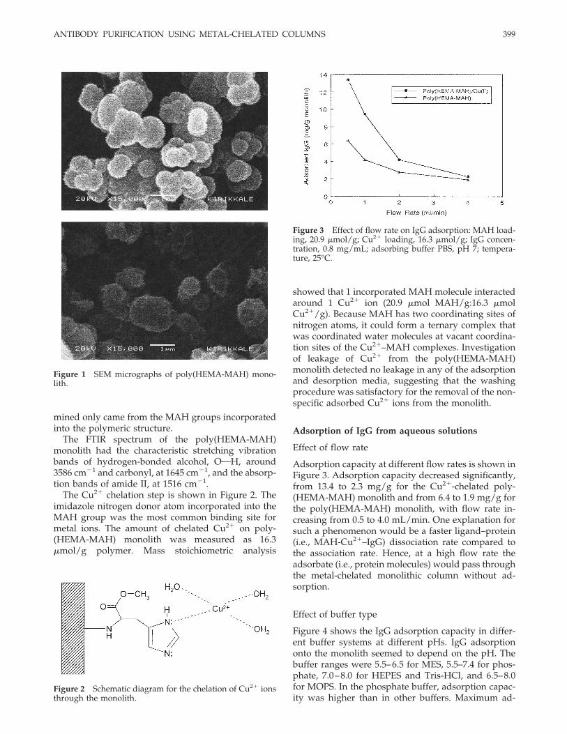
The monolithic columns prepared in glass tubeswere observed by scanning electron microscopy(SEM), which showed (Fig. 1) a structure typical ofmonolithic materials. The monoliths were composedof small and interconnected globules that formed amonolithic porous structure. The globules were 2 �min size and irregular. There also were many 2-�m-diameter pores on the bulk structure of the globules.The large pores between the clusters were 1 �m insize. Using flow rates suitable for chromatographicpurposes, this open structure allowed liquid to beforced through the polymer without compressing it.The back pressure of the monolith was only about 5.8MPa when the flow rate reached 5.0 mL/min.
Concentration of the incorporated MAH was foundto be 20.9 �mol/g polymer using nitrogen stoichiom-etry. Note that HEMA and other chemicals in thepolymerization formula do not contain nitrogen. Ele-mental analysis determined that this nitrogen deter-
398 BERELI ET AL.
mined only came from the MAH groups incorporatedinto the polymeric structure.
The FTIR spectrum of the poly(HEMA-MAH)monolith had the characteristic stretching vibrationbands of hydrogen-bonded alcohol, OOH, around3586 cm�1 and carbonyl, at 1645 cm�1, and the absorp-tion bands of amide II, at 1516 cm�1.
The Cu2� chelation step is shown in Figure 2. Theimidazole nitrogen donor atom incorporated into theMAH group was the most common binding site formetal ions. The amount of chelated Cu2� on poly-(HEMA-MAH) monolith was measured as 16.3�mol/g polymer. Mass stoichiometric analysis
showed that 1 incorporated MAH molecule interactedaround 1 Cu2� ion (20.9 �mol MAH/g:16.3 �molCu2�/g). Because MAH has two coordinating sites ofnitrogen atoms, it could form a ternary complex thatwas coordinated water molecules at vacant coordina-tion sites of the Cu2�–MAH complexes. Investigationof leakage of Cu2� from the poly(HEMA-MAH)monolith detected no leakage in any of the adsorptionand desorption media, suggesting that the washingprocedure was satisfactory for the removal of the non-specific adsorbed Cu2� ions from the monolith.
Adsorption of IgG from aqueous solutions
Effect of flow rate
Adsorption capacity at different flow rates is shown inFigure 3. Adsorption capacity decreased significantly,from 13.4 to 2.3 mg/g for the Cu2�-chelated poly-(HEMA-MAH) monolith and from 6.4 to 1.9 mg/g forthe poly(HEMA-MAH) monolith, with flow rate in-creasing from 0.5 to 4.0 mL/min. One explanation forsuch a phenomenon would be a faster ligand–protein(i.e., MAH-Cu2�–IgG) dissociation rate compared tothe association rate. Hence, at a high flow rate theadsorbate (i.e., protein molecules) would pass throughthe metal-chelated monolithic column without ad-sorption.
Effect of buffer type
Figure 4 shows the IgG adsorption capacity in differ-ent buffer systems at different pHs. IgG adsorptiononto the monolith seemed to depend on the pH. Thebuffer ranges were 5.5–6.5 for MES, 5.5–7.4 for phos-phate, 7.0–8.0 for HEPES and Tris-HCl, and 6.5–8.0for MOPS. In the phosphate buffer, adsorption capac-ity was higher than in other buffers. Maximum ad-
Figure 1 SEM micrographs of poly(HEMA-MAH) mono-lith.
Figure 2 Schematic diagram for the chelation of Cu2� ionsthrough the monolith.
Figure 3 Effect of flow rate on IgG adsorption: MAH load-ing, 20.9 �mol/g; Cu2� loading, 16.3 �mol/g; IgG concen-tration, 0.8 mg/mL; adsorbing buffer PBS, pH 7; tempera-ture, 25°C.
ANTIBODY PURIFICATION USING METAL-CHELATED COLUMNS 399

sorption capacity were observed at a pH of 6.5 forMOPS (6.9 mg/g), 7.0 for HEPES (4.2 mg/g), 7.0 forTris-HCl (5.3 mg/g), 7.0 for phosphate (9.5 mg/g),and 6.0 for MES (5.0 mg/g). Below and above thesemaximum adsorption pHs, the adsorption capacitydecreased significantly. The pKa values for MOPS,HEPES, and MES were pHs of 6.5, 7.0, and 5.4, respec-tively. From the structure of the buffer ions used, itwas obvious that Tris-HCl and phosphate carried oneor more charges of the same sign —only positive oronly negative, whereas the zwitter ionic buffers MES,MOPS, and HEPES carried two charges of oppositesign below their pKa. It could be clearly observed thatmaximum adsorption was obtained with phosphate.
Effect of concentration of IgG
Figure 5 shows the effect of IgG concentration onadsorption. IgG adsorption on the poly(HEMA-MAH)monolith was low (about 4.2 mg/g), although adsorp-tion of IgG molecules onto the Cu2�-chelated poly-(HEMA-MAH) monolith through Cu2� ions was sig-nificant (up to 10.8 mg/g). As expected, the amount ofIgG coupled to monoliths almost reached a plateau ofaround 1.0 mg/mL because of saturation of the activebinding sites.
Effect of ionic strength
Adsorption of IgG by the Cu2� chelated-poly(HEMA-MAH) and poly(HEMA-MAH) monoliths was per-formed at different NaCl concentrations. The effect ofionic strength on IgG adsorption is shown in Figure 6.As seen here, IgG adsorption capacity decreased withincreasing salt concentration. The decrease in adsorp-tion capacity as ionic strength increased can be attrib-uted to the repulsive electrostatic forces between the
Cu2�-chelated poly(HEMA-MAH) monolith and pro-tein molecules. When the salt concentration increasedin the adsorption medium, this could have led tocoordination of the deprotonated amino groups of thehistidine with the cations of the salts, resulting in lowprotein adsorption. The distortion of existing salt bridgesbetween protein molecules and the pseudospecific met-al-complexing affinity ligand in the presence of salt alsocontributed to the low protein adsorption at high ionicstrength.
Effect of temperature
The effect of temperature on IgG adsorption was stud-ied in the range of 4°C–37°C. At all temperatures,adsorption of IgG by the poly(HEMA-MAH) monolithwas lower than that by the Cu2�-chelated-poly-
Figure 4 Effect of buffer type on IgG adsorption: MAHloading, 20.9 �mol/g; Cu2� loading, 16.3 �mol/g; IgG con-centration, 0.8 mg/mL; flow rate, 1.0 mL/min; temperature,25°C.
Figure 5 Effect of IgG concentration on adsorption capac-ity: MAH loading, 20.9 �mol/g; Cu2� loading, 16.3 �mol/g;adsorbing buffer PBS, pH 7; flow rate, 1.0 mL/min; temper-ature, 25°C.
Figure 6 Effect of ionic strength on IgG adsorption: MAHloading, 20.9 �mol/g; Cu2� loading, 16.3 �mol/g; IgG con-centration, 0.8 mg/mL; flow rate, 1.0 mL/min; adsorbingbuffer PBS, pH 7; temperature, 25°C.
400 BERELI ET AL.
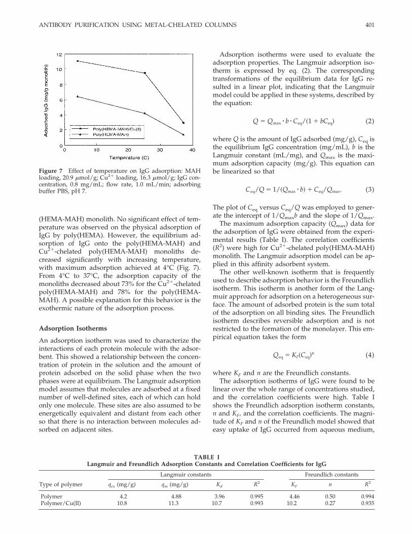
(HEMA-MAH) monolith. No significant effect of tem-perature was observed on the physical adsorption ofIgG by poly(HEMA). However, the equilibrium ad-sorption of IgG onto the poly(HEMA-MAH) andCu2�-chelated poly(HEMA-MAH) monoliths de-creased significantly with increasing temperature,with maximum adsorption achieved at 4°C (Fig. 7).From 4°C to 37°C, the adsorption capacity of themonoliths decreased about 73% for the Cu2�-chelatedpoly(HEMA-MAH) and 78% for the poly(HEMA-MAH). A possible explanation for this behavior is theexothermic nature of the adsorption process.
Adsorption Isotherms
An adsorption isotherm was used to characterize theinteractions of each protein molecule with the adsor-bent. This showed a relationship between the concen-tration of protein in the solution and the amount ofprotein adsorbed on the solid phase when the twophases were at equilibrium. The Langmuir adsorptionmodel assumes that molecules are adsorbed at a fixednumber of well-defined sites, each of which can holdonly one molecule. These sites are also assumed to beenergetically equivalent and distant from each otherso that there is no interaction between molecules ad-sorbed on adjacent sites.
Adsorption isotherms were used to evaluate theadsorption properties. The Langmuir adsorption iso-therm is expressed by eq. (2). The correspondingtransformations of the equilibrium data for IgG re-sulted in a linear plot, indicating that the Langmuirmodel could be applied in these systems, described bythe equation:
Q � Qmax � b � Ceq/�1 � bCeq� (2)
where Q is the amount of IgG adsorbed (mg/g), Ceq isthe equilibrium IgG concentration (mg/mL), b is theLangmuir constant (mL/mg), and Qmax is the maxi-mum adsorption capacity (mg/g). This equation canbe linearized so that
Ceq/Q � 1/�Qmax � b� � Ceq/Qmax. (3)
The plot of Ceq versus Ceq/Q was employed to gener-ate the intercept of 1/Qmaxb and the slope of 1/Qmax.
The maximum adsorption capacity (Qmax) data forthe adsorption of IgG were obtained from the experi-mental results (Table I). The correlation coefficients(R2) were high for Cu2�-chelated poly(HEMA-MAH)monolith. The Langmuir adsorption model can be ap-plied in this affinity adsorbent system.
The other well-known isotherm that is frequentlyused to describe adsorption behavior is the Freundlichisotherm. This isotherm is another form of the Lang-muir approach for adsorption on a heterogeneous sur-face. The amount of adsorbed protein is the sum totalof the adsorption on all binding sites. The Freundlichisotherm describes reversible adsorption and is notrestricted to the formation of the monolayer. This em-pirical equation takes the form
Qeq � KF�Ceq�n (4)
where KF and n are the Freundlich constants.The adsorption isotherms of IgG were found to be
linear over the whole range of concentrations studied,and the correlation coefficients were high. Table Ishows the Freundlich adsorption isotherm constants,n and KF, and the correlation coefficients. The magni-tude of KF and n of the Freundlich model showed thateasy uptake of IgG occurred from aqueous medium,
Figure 7 Effect of temperature on IgG adsorption: MAHloading, 20.9 �mol/g; Cu2� loading, 16.3 �mol/g; IgG con-centration, 0.8 mg/mL; flow rate, 1.0 mL/min; adsorbingbuffer PBS, pH 7.
TABLE ILangmuir and Freundlich Adsorption Constants and Correlation Coefficients for IgG
Type of polymer
Langmuir constants Freundlich constants
qex (mg/g) qm (mg/g) Kd R2 KF n R2
Polymer 4.2 4.88 3.96 0.995 4.46 0.50 0.994Polymer/Cu(II) 10.8 11.3 10.7 0.993 10.2 0.27 0.935
ANTIBODY PURIFICATION USING METAL-CHELATED COLUMNS 401

with the metal-chelated monolith having a high ad-sorption capacity.
Adsorption kinetics modeling
To determine the controlling mechanism of the ad-sorption process such as mass transfer or chemicalreaction, kinetic models were used to test the experi-mental data. The kinetic models (pseudo-first- and-second-order equations) can be used in this case as-suming the measured concentrations equal the adsor-bent surface concentrations. The first-order rate equa-tion of Lagergren is one of the most widely used forthe adsorption of solute from a liquid solution. It maybe represented as
dqt/dt � k1�qeq � qt� (5)
where k1 is the rate constant of pseudo-first-orderadsorption (1/min) and qeq and qt are the amounts ofprotein adsorbed at equilibrium and at time t (mg/g),respectively. After integration by applying boundaryconditions, qt � 0 at t � 0 and qt � qt at t � t, gives
log[qeq/�qeq � qt�] � �k1t�/2.303 (6)
Equation (6) can be rearranged to obtain the linearform
log(qeq � qt) � log(qeq) � �k1t�/2.303 (7)
where a plot of log (qeq � qt) versus t should give astraight line to confirm the applicability of the kineticmodel. In a true first-order process, log qeq shouldequal the interception point of a plot of log(qeq � qt)via t.
In addition, a pseudo-second-order equation basedon equilibrium adsorption capacity may be expressedin the form
dqt/dt � k2�qeq � qt�2 (8)
where k2 (g mg�1 min�1) is the rate constant of thepseudo-first-order adsorption process. Integrating eq.(8) and applying the boundary conditions, qt � 0 at t� 0 and qt � qt at t � t, leads to
�1/�qeq � qt�� � �1/qeq� � k2t (9)
or, equivalently, the linear form
�t/qt� � �1/k2qeq2 � � �1/qeq�t (10)
where a plot of t/qt versus t should give a linearrelationship for the applicability of the second-orderkinetics. The rate constant (k2) and adsorption at equi-librium (qeq) can be obtained from the intercept andslope, respectively. A comparison of the experimentaland theoretical adsorption capacity values is pre-sented in Table II. The theoretical qe values estimatedfrom the pseudo-first- and -second-order kinetic mod-els were very close to the experimental values, and thecorrelation coefficients were high. The results indicatethis metal-chelated monolith was described by boththe first-order and the second-order kinetic models.
Adsorption from human plasma
Table III gives the adsorption data. As seen here, therewas a pronounced adsorption of IgG (up to 104.2mg/g) onto the Cu2�-chelated poly(HEMA-MAH)
TABLE IIFirst- and Second-order Kinetic Constants for Cu2�-Chelated Poly(HEMA-MAH) Monolith
Initialconcentration
(mg/mL)
Experimental First-order kinetic
R2
Second-order kinetic
R2qeq
(mg/g)k1
(L/min)qeq
(mg/g)k2
(g mg–1 min–1)qeq
(mg/g)
0.05 2.61 0.050 2.34 0.959 0.035 2.18 0.9460.1 3.92 0.053 3.28 0.978 0.027 4.17 0.9940.2 4.89 0.037 5.29 0.988 0.003 7.36 0.9390.3 5.55 0.041 5.25 0.921 0.007 6.86 0.9740.5 8.16 0.023 8.24 0.970 0.008 15.10 0.8310.8 9.47 0.038 10.60 0.965 0.014 14.50 0.9371.0 10.12 0.040 11.30 0.976 0.012 15.80 0.9021.5 10.77 0.046 12.40 0.966 0.009 17.70 0.8142.0 10.80 0.062 11.80 0.995 0.004 12.80 0.979
TABLE IIIIgG Adsorption from Human Plasma IgG Concentration
Before Dilution: 14.6 mg/mL; MAH Loading: 20.9�mol/g; Cu2� Loading: 16.3 �mol/g; Flow Rate:
1.0 mL/min; T: 25°C
Dilution agentAdsorption capacity
(mg/g)
Plasma (undiluted) 104.2 2.411:2 diluted plasma (phosphate pH: 7.4) 62.3 2.801:10 diluted plasma (phosphate pH: 7.4) 41.8 2.63
402 BERELI ET AL.

monolith for plasma diluted with PBS. The purity ofthe IgG, assayed by SDS-PAGE, was found to be94.1%. It is worth noting that because of the highinitial concentration of IgG in the plasma, the adsorp-tion of IgG by the Cu2�-chelated poly(HEMA-MAH)monolith was higher than that obtained with aqueoussolutions. IgG has a molecular mass of 150,000 andconsists of four peptide chains; two identical lightchains are linked by strong disulfide bonds to make aY- or T-shaped structure with hingelike flexible arms.Thus, an IgG molecule would expand and contractsignificantly with variation in the ionizable groups inthe molecule. This high IgG adsorption may also havebeen a result of suitable conformation of IgG mole-cules in their native medium [i.e., human plasma (pH7.4)] for interaction with the histidine groups of thepoly(HEMA-MAH) monolith.
Adsorptions of albumin and fibrinogen also weredetermined. There was a pronounced adsorption ofIgG by the Cu2�-chelated poly(HEMA-MAH) mono-lith for undiluted plasma (104.2 mg/g). Adsorptioncapacity was found to be 6.2 mg/g for fibrinogen and8.5 mg/g for albumin. The total protein adsorptionwas determined as 119.1 mg/g. The IgG adsorptionratio was around 87.5% (104.2 mg IgG/g adsorbent;119.1 mg total protein/g adsorbent). The fibrinogenand albumin adsorption ratios were 5.2% (6.2 mg fi-brinogen/g sorbent; 119.1 mg total protein/g sorbent)and 7.1% (8.5 mg albumin/g sorbent; 119.1 mg totalprotein/g sorbent). IgG adsorbed more than HSA un-der physiological conditions, although the initial con-centration ratio of IgG to HSA (mg/mL) was 11.9:37.2.Most of the described methods for IgG capture re-sulted in coadsorption of several other proteins,among which adsorption of albumin was significant.Adsorption of IgG was significant compared to ad-sorption of albumin and fibrinogen. Transition metalions have a high affinity to the peptide sequencesHis-Gly-His, His-Tyr-NH2, and His-Trp. The signifi-cant IgG adsorption by the Cu2�-chelated monolithcould have been a result of its having a greater num-ber of histidine residues, which could interact with themetal ions.
Comparison with related literature
Different IgG adsorption capacities have been re-ported in the literature. Fuglistaller was interested indetermining the dynamic binding capacities of differ-ent commercial protein A affinity chromatographymatrices, including Affi-Gel, Eupergit, Ultragel, theSepharose series, and Prosep A.15 He found that ad-sorption capacity varied between 0.7 and 20 mgIgG3/g. Bueno et al. used poly(ethylene vinyl alcohol)hollow-fiber cartridges carrying l-histidine to deter-mine adsorption; they reported dynamic adsorption ofup to 77.7 mg IgG/g polymer.30 Klein et al. used
microporous poly(caprolactam) hollow fibers and flat-sheet membranes as the carrier matrices and immobi-lized recombinant protein A as the specific bioli-gand.31 They reported IgG adsorption capacities ofaround 12.4–28.3 mg/cm3. With the use of hydropho-bic amino acids (e.g., phenylalanine and tryptophan)containing membranes based on polyethylene, Kim etal. obtained an adsorption capacity of 50 mg/g poly-mer for bovine gamma globulin.32 Muller-Shulte et al.used several polymeric carriers made of differentpolymers, including Biograft, Sepharose 4B, Superose,and Spherosil, with histidine as the pseudospecificligand.33 They found maximum IgG1 adsorption in therange of 0.05–0.23 mg IgG1/mL sorbent. Denizli et al.reported an adsorption capacity of 24 mg IgG/g withprotein A–immobilized PHEMA beads.34 Using mi-croporous immunoaffinity hollow fibers composed ofpolysulfone/protein A, Charcosset et al. obtained anadsorption capacity of 8.8 mg/g.35 Langotz and Kro-ner reported an adsorption capacity of 0.5 mg/mLrabbit IgG with commercially available Sartobind Ep-oxy sorbents.36 Dancette et al. studied the perfor-mance of recombinant protein A/G affinity mem-branes based on poly(methyl methacrylate), and poly-acrylonitrile for human and mouse IgG purification,and they obtained a static binding capacity of 6.6 mgIgG/mL membrane.37 Teng et al. described a fullycharacterized IgG-binding ligand comprising a tri-azine scaffold substituted with 3-aminophenol and4-amino-1-naphthol.38 They showed that this syntheticligand mimicking protein A interacted with HIgG andwas able to selectively purify IgG from diluted humanplasma. They achieved an adsorption capacity of 52mg/g moist weight gel. Ozkara et al. immobilizedl-histidine covalently on PHEMA beads, obtainingadsorption of 3.5 mg IgG/g polymer in a batch sys-tem.39 Protein A mimetic synthetic ligands carryingSepharose 4B and Sepharose CL-6B sorbents wereused in the purification of IgG from diluted humanplasma, with the maximum adsorption capacity foundto be in the range of 7–25 mg/g.40,41 The IgG adsorp-tion capacity obtained in the present study (up to 104mg IgG/g) would seem to be sufficient for proposingthat these metal-chelated affinity monoliths be used asIMAC supports. Differences in IgG adsorption are aresult of the specific properties of each adsorbent, suchas structure, functional groups, ligand loading, andsurface area.
Desorption and repeated use
In the last step of the affinity separation, the mainconcern was to desorb the adsorbed protein in theshortest time and the highest amount possible. It wasthus necessary to evaluate the regeneration efficiencyof the affinity adsorbents after each cycle. In thisstudy, more than 95% of the adsorbed IgG molecules
ANTIBODY PURIFICATION USING METAL-CHELATED COLUMNS 403
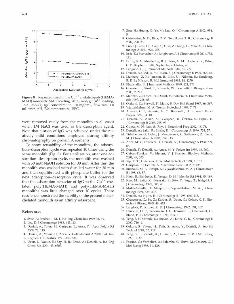
were removed easily from the monolith in all caseswhen 1M NaCl was used as the desorption agent.Note that elution of IgG was achieved under the rel-atively mild conditions employed during affinitychromatography on protein A sorbents.
To show reusability of the monoliths, the adsorp-tion–desorption cycle was repeated 10 times using thesame monolith (Fig. 8). For sterilization, after one ad-sorption–desorption cycle, the monolith was washedwith 50 mM NaOH solution for 30 min. After this, themonolith was washed with distilled water for 30 minand then equilibrated with phosphate buffer for thenext adsorption–desorption cycle. It was observedthat the adsorption behavior of IgG to the Cu2�-che-lated poly(HEMA-MAH) and poly(HEMA-MAH)monoliths was little changed over 10 cycles. Theseresults demonstrated the stability of the present metal-chelated monolith as an affinity adsorbent.
References
1. Svec, F.; Frechet, J. M. J. Ind Eng Chem Res 1999 38, 34.2. Lee, D. J Chromatogr 1988, 443,143.3. Denizli, A.; Yavuz, H.; Garipcan, B.; Arıca, Y. J Appl Polym Sci
2000, 76, 115.4. Denizli, A.; Yavuz, H.; Arıca, Y. Colloids Surf A 2000, 174, 147.5. Regnier, F. E. Nature 1991, 350, 634.6. Uzun, L.; Yavuz, H.; Say, H. R.; Ersoz, A.; Denizli, A. Ind Eng
Chem Res 2004, 43, 6507.
7. Zou, H.; Huang, X.; Ye, M.; Luo, Q. J Chromatogr A 2002, 954,5.
8. Ostryanina, N. D.; Ilina, O. V.; Tennikova, T. B. J Chromatogr B2002, 770, 35.
9. Luo, Q.; Zou, H.; Xiao, X.; Guo, Z.; Kong, L.; Mao, X. J Chro-matogr A 2001, 926, 255.
10. Josic, D.; Buchacher, A.; Jungbauer, A. J Chromatogr B 2001, 752,191.
11. Duffy, S. A.; Moellering, B. J.; Prior, G. M.; Doyle, K. R.; Prior,C. P. Biopharm 1989, September/October, 46.
12. Langone, J. J. J Immunol Methods 1982, 55, 277.13. Denizli, A.; Rad, A. Y.; Piskin, E. J Chromatogr B 1995, 668, 13.14. Ljunberg, U. K.; Jansson, B.; Niss, U.; Nilsson, R.; Sandberg,
B. E. B.; Nilsson, B. Mol Immunol 1993, 14, 1279.15. Fuglistaller, P. J Immunol Methods 1989, 124, 171.16. Guerrier, L.; Girot, P.; Schwartz, W.; Boschetti, E. Bioseparation
2000, 9, 211.17. Manzke, O.; Tesch, H.; Dischl, V.; Bohlen, H. J Immunol Meth-
ods 1997, 208, 65.18. Ostlund, C.; Borwell, P.; Malm, B. Dev Biol Stand 1987, 66, 367.19. Vijayalakshmi, M. A. Trends Biotechnol 1989, 7, 71.20. Alvarez, C. I.; Strumia, M. C.; Bertorello, H. E. React. Funct
Polym 1997, 34, 103.21 Denizli, A.; Alkan, M.; Garipcan, B.; Ozkara, S.; Piskin, E.
J Chromatogr B 2003, 795, 93.22. Gupta, M. N.; Jain, S.; Roy, I. Biotechnol Prog 2002, 18, 78.23. Denizli, A.; Salih, B.; Piskin, E. J Chromatogr A 1996, 731, 57.24. Tishchenko, G.; Dybal, J.; Meszaroova, K.; Sedlakova, Z.; Bleha,
M. J Chromatogr A 2002, 954, 115.25. Arıca, M. Y.; Testereci, H.; Denizli, A. J Chromatogr A 1998, 799,
83.26. Denizli, F.; Denizli, A.; Arıca, M. Y. Polym Int 1999, 48, 360.27. Gaberc-Porekar, V.; Menart, V. J Biochem Biophys Methods
2001, 49, 335.28. Yip, T. T.; Hutchens, T. W. Mol Biotechnol 1994, 1, 151.29. Garipcan, B.; Denizli, A. Macromol Biosci 2002, 2, 135.30. Bueno, S. M. A.; Haupt, K.; Vijayalakshmi, M. A. J Chromatogr
B 1995, 66, 57.31. Klein, E.; Eichholtz, E.; Yeager, D. H. J Membr Sci 1994, 95, 199.32. Kim, M.; Saito, K.; Furusaki, S.; Sato, T.; Sugo, T.; Ishigaki, I.
J Chromatogr 1991, 585, 45.33. Muller-Schulte, D.; Manjini, S.; Vijayalakshmi, M. A. J Chro-
matogr 1991, 539, 307.34. Denizli, A.; Piskin, E. J Chromatogr B 1995, 666, 215.35. Charcosset, C.; Su, Z.; Karoor, S.; Daun, G.; Colton, C. K. Bio-
technol Bioeng 1995, 48, 415.36. Langlotz, P.; Kroner, K. H. J Chromatogr 1992, 591, 107.37. Dancette, O. P.; Taboureau, J. L.; Tournier, E.; Charcosset, C.;
Blond, P. J Chromatogr B 1999, 723, 61.38. Teng, S. F.; Sproule, K.; Husain, A.; Lowe, C. R. J Chromatogr B
2000, 740, 1.39. Ozkara, S.; Yavuz, H.; Patir, S.; Arıca, Y.; Denizli, A. Sep Sci
Technol 2002, 37, 717.40. Teng, S. F.; Sproule, K.; Hussain, A.; Lowe, C. R. J Mol Recog
1999, 12, 67.41. Fassina, G.; Verdoliva, A.; Palombo, G.; Ruvo, M.; Cassani, G. J
Mol Recog 1998, 11, 128.
Figure 8 Repeated used of the Cu�2 chelated-poly(HEMA-MAH) monolith: MAH loading, 20.9 �mol/g; Cu2� loading,16.3 �mol/g; IgG concentration, 0.8 mg/mL; flow rate, 1.0mL/min; pH, 7.0; temperature, 25°C.
404 BERELI ET AL.
Copyright © 2022 FDOKUMEN