Androgen receptor expresion in breast cancer: Relationship with clinicopathological characteristics...
10
BioMed Central Page 1 of 10 (page number not for citation purposes) BMC Cancer Open Access Research article Androgen receptor expresion in breast cancer: Relationship with clinicopathological characteristics of the tumors, prognosis, and expression of metalloproteases and their inhibitors Luis O Gonzalez 1,2,3 , Maria D Corte 1 , Julio Vazquez 2,4 , Sara Junquera 1 , Rosario Sanchez 1 , Ana C Alvarez, Juan C Rodriguez 1,2,5 , Maria L Lamelas 1,2,6 and Francisco J Vizoso* 1,2,5 Address: 1 Unidad de Investigación, Hospital de Jove, Gijón, Spain, 2 Instituto Universitario de Oncología del Principado de Asturias, Oviedo, Spain, 3 Servicio de Anatomía Patológica, Hospital de Jove, Gijón, Spain, 4 Servicio de Ginecología, Hospital Álvarez Buylla, Mieres, Spain, 5 Servicio de Cirugía General, Hospital de Jove, Gijón, Spain and 6 Servicio de Ginecología, Hospital de Jove, Gijón, Spain Email: Luis O Gonzalez - [email protected]; Maria D Corte - [email protected]; Julio Vazquez - [email protected]; Sara Junquera - [email protected]; Rosario Sanchez - [email protected]; Ana C Alvarez - [email protected]; Juan C Rodriguez - [email protected]; Maria L Lamelas - [email protected]; Francisco J Vizoso* - [email protected] * Corresponding author Abstract Background: In the present study we analyze, in patients with breast cancer, the tumor expression of androgen receptors (AR), its relationship with clinicopathological characteristics and with the expression of several matrix metalloproteases (MMPs) and their inhibitors (TIMPs), as well as with prognosis. Methods: An immunohistochemical study was performed using tissue microarrays and specific antibodies against AR, MMPs -1, -2, -7, -9, -11, -13, -14, and TIMPs -1, -2 and -3. More than 2,800 determinations on tumor specimens from 111 patients with primary invasive ductal carcinoma of the breast (52 with axillary lymph node metastases and 59 without them) and controls were performed. Staining results were categorized using a score based on the intensity of the staining and a specific software program calculated the percentage of immunostained cells automatically. Results: A total of 83 cases (74.8%) showed a positive immunostaining for AR, but with a wide variation in the staining score values. There were no significant associations between the total immunostaining scores for AR and any clinicopathological parameters. However, score values for MMP-1, -7 and -13, were significantly higher in AR-positive tumors than in AR-negative tumors. Likewise, when we considered the cellular type expressing each factor, we found that AR-positive tumors had a higher percentage of cases positive for MMP-1, -7, -11, and TIMP-2 in their malignant cells, as well as for MMP-1 in intratumoral fibroblasts. On the other hand, multivariate analysis demonstrated that patients with AR-positive tumors have a significant longer overall survival than those with AR-negative breast carcinomas (p = 0.03). Conclusion: Our results confirm that AR are commonly expressed in breast cancer, and are correlated with the expression of some MMPs and TIMP-2. Although we found a specific value of AR expression to be a prognostic indicator in breast cancer, the functional role of AR in these neoplasms is still unclear and further data are needed in order to clarify their biological signification in breast cancer. Published: 28 May 2008 BMC Cancer 2008, 8:149 doi:10.1186/1471-2407-8-149 Received: 28 December 2007 Accepted: 28 May 2008 This article is available from: http://www.biomedcentral.com/1471-2407/8/149 © 2008 Gonzalez et al; licensee BioMed Central Ltd. This is an Open Access article distributed under the terms of the Creative Commons Attribution License (http://creativecommons.org/licenses/by/2.0 ), which permits unrestricted use, distribution, and reproduction in any medium, provided the original work is properly cited.
Transcript of Androgen receptor expresion in breast cancer: Relationship with clinicopathological characteristics...

BioMed CentralBMC Cancer
ss
Open AcceResearch articleAndrogen receptor expresion in breast cancer: Relationship with clinicopathological characteristics of the tumors, prognosis, and expression of metalloproteases and their inhibitorsLuis O Gonzalez1,2,3, Maria D Corte1, Julio Vazquez2,4, Sara Junquera1, Rosario Sanchez1, Ana C Alvarez, Juan C Rodriguez1,2,5, Maria L Lamelas1,2,6 and Francisco J Vizoso*1,2,5Address: 1Unidad de Investigación, Hospital de Jove, Gijón, Spain, 2Instituto Universitario de Oncología del Principado de Asturias, Oviedo, Spain, 3Servicio de Anatomía Patológica, Hospital de Jove, Gijón, Spain, 4Servicio de Ginecología, Hospital Álvarez Buylla, Mieres, Spain, 5Servicio de Cirugía General, Hospital de Jove, Gijón, Spain and 6Servicio de Ginecología, Hospital de Jove, Gijón, Spain
Email: Luis O Gonzalez - [email protected]; Maria D Corte - [email protected]; Julio Vazquez - [email protected]; Sara Junquera - [email protected]; Rosario Sanchez - [email protected]; Ana C Alvarez - [email protected]; Juan C Rodriguez - [email protected]; Maria L Lamelas - [email protected]; Francisco J Vizoso* - [email protected]
* Corresponding author
AbstractBackground: In the present study we analyze, in patients with breast cancer, the tumor expression ofandrogen receptors (AR), its relationship with clinicopathological characteristics and with the expressionof several matrix metalloproteases (MMPs) and their inhibitors (TIMPs), as well as with prognosis.
Methods: An immunohistochemical study was performed using tissue microarrays and specific antibodiesagainst AR, MMPs -1, -2, -7, -9, -11, -13, -14, and TIMPs -1, -2 and -3. More than 2,800 determinations ontumor specimens from 111 patients with primary invasive ductal carcinoma of the breast (52 with axillarylymph node metastases and 59 without them) and controls were performed. Staining results werecategorized using a score based on the intensity of the staining and a specific software program calculatedthe percentage of immunostained cells automatically.
Results: A total of 83 cases (74.8%) showed a positive immunostaining for AR, but with a wide variationin the staining score values. There were no significant associations between the total immunostainingscores for AR and any clinicopathological parameters. However, score values for MMP-1, -7 and -13, weresignificantly higher in AR-positive tumors than in AR-negative tumors. Likewise, when we considered thecellular type expressing each factor, we found that AR-positive tumors had a higher percentage of casespositive for MMP-1, -7, -11, and TIMP-2 in their malignant cells, as well as for MMP-1 in intratumoralfibroblasts. On the other hand, multivariate analysis demonstrated that patients with AR-positive tumorshave a significant longer overall survival than those with AR-negative breast carcinomas (p = 0.03).
Conclusion: Our results confirm that AR are commonly expressed in breast cancer, and are correlatedwith the expression of some MMPs and TIMP-2. Although we found a specific value of AR expression tobe a prognostic indicator in breast cancer, the functional role of AR in these neoplasms is still unclear andfurther data are needed in order to clarify their biological signification in breast cancer.
Published: 28 May 2008
BMC Cancer 2008, 8:149 doi:10.1186/1471-2407-8-149
Received: 28 December 2007Accepted: 28 May 2008
This article is available from: http://www.biomedcentral.com/1471-2407/8/149
© 2008 Gonzalez et al; licensee BioMed Central Ltd. This is an Open Access article distributed under the terms of the Creative Commons Attribution License (http://creativecommons.org/licenses/by/2.0), which permits unrestricted use, distribution, and reproduction in any medium, provided the original work is properly cited.
Page 1 of 10(page number not for citation purposes)

BMC Cancer 2008, 8:149 http://www.biomedcentral.com/1471-2407/8/149
BackgroundIn the last two decades, the molecular mechanisms relatedto the hormone dependence of breast tumors have beenextensively investigated and the role of the estrogen andprogesterone receptors (ER and PgR) in promoting breastcancer has been well documented. However, the role ofandrogens and their receptors (AR) in breast cancer etiol-ogy and progression has been less profoundly studied andremains an unanswered question [1,2]. There is evidenceshowing that androgens can directly stimulate the growthof human breast cancer cell lines [3]. In addition, both ret-rospective and prospective studies have reported statisti-cally significant associations between increased levels oftestosterone and higher breast cancer risk in both pre- andpostmenopausal women [4-6]. Likewise, AR is expressedin approximately 70% to 90% of invasive breast cancers,a frequency comparable with or higher than the onereported for ER (70–80%) and PgR (50–70%) [1,7-10].Although a relationship between AR and both ER and PgRstatus has been demonstrated [10-14], a significant per-centage of tumors are positive for AR and negative for ERand PgR [13]. This finding reveals the independent expres-sion of AR in human breast cancer. However, there areapparently divergent data on the biological and clinicalsignification of AR in breast cancer. AR have also beendetected in a significantly higher percentage of AR-posi-tive ductal carcinomas "in situ" (DCIS) adjacent to inva-sive carcinomas of the breast than in pure DCIS lesions[15], suggesting that AR correlates with tumor invasive-ness, at least in the early phases of tumor progression. Ininvasive breast carcinomas, AR-positive tumors have beenassociated with a low or intermediate histological grade(G1, G2) [10,13,14,16,17]. In addition, certain types ofbreast carcinoma, even high grade ones, are typically ER-and PR-negative, but AR-positive; a typical example ofsuch tumors is the apocrine breast carcinoma [18,19].However, the expression level of both the AR gene and theAR protein in breast cancer was found to be positively cor-related with axillary lymph node involvement [20]. Inaddition, it is remarkable that among the steroid hor-mone receptors, the androgen receptor is the best pre-served one during metastases development and isexpressed in the majority of metastatic tumors [8,21].There is evidence as well indicating that AR/steroids areable to up-regulate matrix metalloproteases (MMPs), con-tributing to invasiveness via destruction of basementmembrane and extracellular matrix [22,23]. Nevertheless,only a few studies have examined the impact of AR expres-sion on patient prognosis in early breast cancer. Patientswith AR-positive tumors were shown to have a significanttrend toward longer relapse-free and/or overall survival inthe univariate analysis than those patients with AR-nega-tive tumors [1,16,24], but none in the multivariate analy-sis [1,16]. In addition, other studies have not found any
significance of AR expression in predicting prognosis inbreast cancer [20,25].
Since AR are expressed by an important percentage ofbreast carcinomas and there are evidences pointing theirrole in tumor progression, in the present study we analyzethe tumor expression of AR, its relationship with clinico-pathological characteristics, with several MMPs and theirinhibitors (TIMPs) and with prognosis, in patients withbreast cancer.
MethodsPatient characteristics and tissue specimen handlingThis study comprised 111 women with a histologicallyconfirmed diagnosis of breast cancer and treated between1990 and 2001. We selected women with the followinginclusion criteria: invasive ductal carcinoma, at least tenhistopathologically-assessed axillary lymph nodes, and aminimum of five years of follow-up in those women with-out tumor recurrence. The exclusion criteria were the fol-lowing: metastatic disease at presentation, prior history ofany type of malignant tumor, bilateral breast cancer atpresentation, having received any type of neoadjuvanttherapy, development of loco-regional recurrence duringthe follow-up period, development of a second primarycancer, and absence of sufficient tissue in the paraffinblocks used for manufacturing the TMAs. From a total of1053 patients fulfilling these criteria, we selected ran-domly a sample size of 111 patients, in accordance to fourdifferent groups of similar size and stratified with regardto nodal status and the development of metastatic disease,which were the key measure variables of this study. Thus,we included an important number of events in both node-negative and node-positive patients subgroups (half of thecases with distant metastasis during the follow-up periodin each one of these subgroups) in order to warrant thestatistical power of the survival analysis. Patients charac-teristics included in the two main groups, with or withoutdistant metastases, are listed in Table 1. Histological gradewas determined according to the criteria reported byElston and Ellis [26].
Women were treated according to the guidelines used inour institution. The study adhered to national regulationsand was approved by our institution's Ethics and Investi-gation Committee. The end-point was death from tumorprogression. The median follow-up period in patientswithout metastases was of 87.5 months, and of 52.7months in patients with them.
Tissue microarrays and immunohistochemistryRoutinely fixed (overnight in 10% buffered formalin),paraffin-embedded tumor samples stored in our pathol-ogy laboratory files were used in this study. Histopatho-logically representative tumor areas were defined on
Page 2 of 10(page number not for citation purposes)

BMC Cancer 2008, 8:149 http://www.biomedcentral.com/1471-2407/8/149
Table 1: Relationship between AR expression and different clinicopathological parameters in 111 breast carcinomas.
Patient and tumor characteristics N Median (range) p N° positive cases (score >0) (%) p
Total cases 111 69 (0–288) 83 (74.8)
Age (years) n.s. n.s.
≤ 58 55 68(0–279) 44(80)>58 56 76(0–288) 39(69.6)
Menopausal status n.s. n.s.
Premenopausal 29 92(0–279) 25(86.2)Postmenopausal 82 59(2–288) 58(70.4)
Tumor size n.s. n.s.
T1 50 80(0–288) 40(80)T2 61 64(0–279) 43(70.5)
Nodal status n.s. n.s.
Positive 52 53(0–273) 38(73.1)Negative 59 100(0–288) 45(76.3)
Stage n.s. n.s.
I 32 71(0–273) 25 (78.1)II 51 64(0–243) 40 (78.4)III 28 83.5(0–288) 18 (64.3)
Histologic grade n.s. n.s.
Well Dif. 30 66.5(0–273) 22(73.3)Mod. Dif. 57 88(0–288) 44(77.2)Poorly Dif. 24 40.5(0–267) 17(70.8)
Estrogen receptor n.s. n.s.
Negative 52 55.5(0–273) 34(65.4)Positive 59 80(0–288) 49(83.1)
Progesterone receptor n.s. n.s.
Negative 59 58(0–279) 41(69.5)Positive 52 76(0–288) 42(80.8)
Desmoplastic reaction n.s. n.s.
No 35 48(0–270) 23(65.7)Yes 76 80.5(0–288) 60(78.9)
Peritumoral inflammation n.s. n.s.
No 65 69.5(0–279) 48(75)Yes 46 86(0–288) 34(75.6)
Tumor advancing edge n.s. n.s.
Page 3 of 10(page number not for citation purposes)

BMC Cancer 2008, 8:149 http://www.biomedcentral.com/1471-2407/8/149
haematoxylin and eosin-stained sections and marked onthe slides. Tumor tissue array blocks were obtained bypunching a tissue cylinder (core) with a diameter of 1.5mm through a histologically representative area of each'donor' tumor block, which was then inserted into anempty 'recipient' tissue array paraffin block using a man-ual tissue arrayer (Beecker Instruments, Sun Praerie, Win-consin, USA) as described elsewhere [27]. Collection oftissue cores was carried out under highly controlled con-ditions. Areas of non-necrotic cancerous tissue wereselected for arraying by two experienced pathologists(L.O. González and A. M. Merino). Two cores wereemployed for each case. From the 111 tumor samplesavailable, three tissue array blocks were prepared, eachone containing 37 tumors samples, as well as internalcontrols including four normal breast tissue samples fromtwo healthy women who had undergone reductive mam-mary surgery.
Three composite high-density TMA blocks were designed,and serial 5-µm sections were consecutively cut with amicrotome (Leica Microsystems GmbH, Wetzlar, Ger-many) and transferred to adhesive-coated slides. One sec-tion from each tissue array block was stained with H&E,and these slides were then reviewed to confirm that thesample was representative of the original tumor. Immu-nohistochemistry was done on these sections of TMAfixed in 10% buffered formalin and embedded in paraffinusing a TechMate TM50 autostainer (Dako, Glostrup,Denmark). Antibodies for MMPs and TIMPs wereobtained from Neomarker (Lab Vision Corporation, Fre-mont, CA, USA). The dilution for each antibody wasestablished based on negative and positive controls (1/50for MMP-2, -7, 14, and TIMP-2; 1/100 for 9, 13, TIMP-1and -3; 1/200 for MMP-1, MMP-11); and anti-AR cloneAR 441 (Dako) at a dilution of 1/50. The positive controlwas prostate carcinoma, previously tested. The negativecontrol was DakoCytomation mouse serum diluted to thesame mouse IgG concentration as the primary antibody.
Tissue sections were deparaffinized in xylene, and thenrehydrated in graded concentrations of ethyl alcohol(100%, 96%, 80%, 70%, then water). To enhance antigenretrieval only for some antibodies, TMA sections weremicrowave-treated (H2800 Microwave Processor,EBSciences, East Granby, Connecticut, USA) in citrate
buffer pH 6 (Target Retrieval Solution, Dako) at 99°C for15 min. Endogenous peroxidase activity was blocked byincubating the slides in peroxidase-blocking solution(Dako) for 5 min. The EnVision Detection Kit (Dako) wasused as the staining detection system. Sections were coun-terstained with hematoxilin, dehydrated with ethanol,and permanently coverslipped.
TMA analysisFor each antibody preparation studied, the location ofimmunoreactivity, percentage of stained cells and inten-sity were determined. All the cases were semiquantifiedfor each protein-stained area. An image analysis systemmade up by the Olympus BX51 microscope and soft anal-ysis (analySIS®, Soft imaging system, Münster, Germany)was employed as follows: tumor sections were stainedwith antibodies according to the method explained aboveand counterstained with hematoxilin. There were differ-ent optical thresholds for both stains. Each core wasscanned with a 400× power objective in two fields percore. Fields were selected searching for the protein-stainedareas. The computer program selects and traces a linearound antibody-stained areas (higher optical threshold:red spots), with the remaining, non-stained areas (hema-toxilin-stained tissue with lower optical threshold) stand-ing out as a blue background. Any field has an area ratioof stained (red) versus non-stained areas (blue). A finalarea ratio was obtained after averaging two fields. To eval-uate immunostaining intensity we used a numeric scoreranging from 0 to 3, reflecting the intensity as follows: 0,no staining; 1, weak staining; 2, moderate staining; and 3,intense staining. Using an Excel spreadsheet, the meanscore was obtained by multiplying the intensity score (I)by the percentage of stained cells (PC) and the results wereadded together (total score: I × PC). This overall score wasthen averaged with the number of cores that were done foreach patient. If there was no tumor in a particular core,then no score was given. In addition, for each tumor, themean score of two core biopsies was calculated.
Furthermore, whole-tissue sections from tumor blocksfrom a subset of ten cases were compared with the corre-sponding TMA discs, regarding AR expression. These caseswere selected randomly, and the obtained clinicopatho-logical data were very similar to those from the wholeseries. Each whole-tissue section was scanned with a 400×
Expansive 49 50.5(0–270) 33(68.8)Infiltrating 62 84.5(0–288) 48(80)
Vascular invasion n.s. n.s.
No 72 64.5(0–288) 52(72.2)Yes 39 81(0–279) 31(79.5)
Table 1: Relationship between AR expression and different clinicopathological parameters in 111 breast carcinomas. (Continued)
Page 4 of 10(page number not for citation purposes)

BMC Cancer 2008, 8:149 http://www.biomedcentral.com/1471-2407/8/149
power lens in ten different fields. Fields were selectedsearching for the protein-stained areas, such as it wasdescribed above. Previously, we described a similar vali-dation study for the evaluated MMPs and TIMPs, in inva-sive breast cancer [28].
Data analysis and statistical methodsDifferences in percentages were calculated with the chi-square test. Immunostaining score values for each proteinwere expressed as median (range). Comparison of immu-nostaining values between groups was made with theMann-Whitney or Kruskall-Wallis tests. For metastasis-free survival analysis we used the Cox's univariatemethod. Cox's regression model was used to examineinteractions of different prognostic factors in a multivari-ate analysis. The SPSS 11.5 program was used for all cal-culations.
ResultsMore than 2,800 determinations were performed onTMAs of cancer specimens from 111 patients with primaryinvasive ductal carcinoma of the breast and controls. Min-imal internal variance of score data between duplicate tis-sue cores from the same patients was detected in the tissuearrays, showing a high agreement for each protein (r >0.95 and p < 0.0001, for each protein). In the validationstudy there was a total concordance in the global expres-sion, as well as in the intensity of immunostaining for AR,between TMA cases and the corresponding whole-tissuesections. In addition, there were highly significant correla-
tions in the immunostaining scores between these twopaired sets (r > 0.90 and p < 0.0001, for each protein).
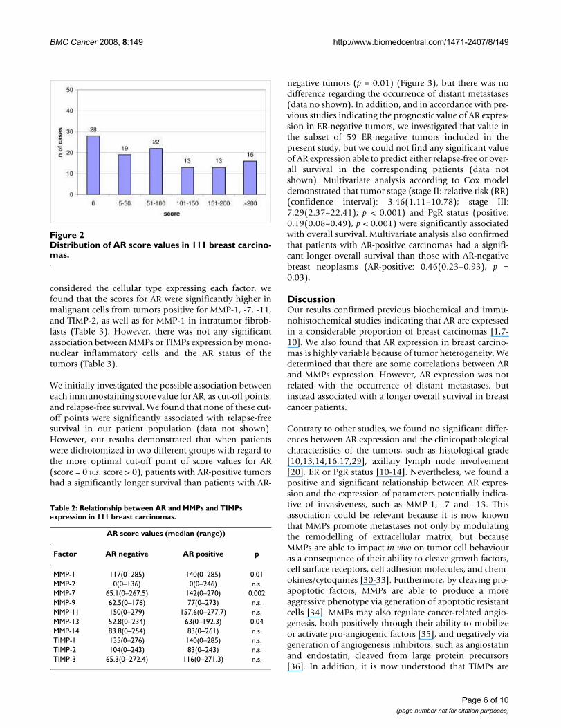
Figure 1 shows examples of tissue immunostained for theproteins evaluated. As it was expected, AR immunostain-ing was of nuclear localization. A total of 83 cases (74.8%)showed a positive immunostaining for AR, although witha wide variation in the immunostaining score values (Fig-ure 2). There were only five cases with a score value of lessthan ten points (three with a score of 5, and two with ascore of 8). Immunostaining for MMPs and TIMPs waslocalized predominantly in the cytoplasm of the malig-nant cells, but also in stromal cells in a considerable per-centage of cases.
There were not any significant associations between thetotal immunostaining scores for AR and clinicopatholog-ical parameters such as tumor size, lymph node involve-ment, stage, histological grade, high Nottinghamprognostic index, infiltrating edge, vascular invasion,desmoplastic reaction or peritumoral inflammation, ERor PgR status (Table 1).
In the present study we also investigated the possible rela-tionship between AR expression and both MMPs andTIMPs expression in tumors, which have been associatedwith an aggressive behaviour and a poor prognosis inbreast cancer patients. Our data demonstrated some sig-nificant associations. Thus, score values for MMP-1, -7and -13, were significantly higher in AR-positive tumorsthan in AR-negative tumors (Table 2). Likewise, when we
Microphotographs representative of malignant cells positive for AR (A), MMP-1 (B), MMP-7 (C), MMP-13 (D) and TIMP-2 (E) and fibroblasts positive for MMP-1 (F)Figure 1Microphotographs representative of malignant cells positive for AR (A), MMP-1 (B), MMP-7 (C), MMP-13 (D) and TIMP-2 (E) and fibroblasts positive for MMP-1 (F). Magnification 400×.
Page 5 of 10(page number not for citation purposes)
BMC Cancer 2008, 8:149 http://www.biomedcentral.com/1471-2407/8/149
considered the cellular type expressing each factor, wefound that the scores for AR were significantly higher inmalignant cells from tumors positive for MMP-1, -7, -11,and TIMP-2, as well as for MMP-1 in intratumor fibrob-lasts (Table 3). However, there was not any significantassociation between MMPs or TIMPs expression by mono-nuclear inflammatory cells and the AR status of thetumors (Table 3).
We initially investigated the possible association betweeneach immunostaining score value for AR, as cut-off points,and relapse-free survival. We found that none of these cut-off points were significantly associated with relapse-freesurvival in our patient population (data not shown).However, our results demonstrated that when patientswere dichotomized in two different groups with regard tothe more optimal cut-off point of score values for AR(score = 0 v.s. score > 0), patients with AR-positive tumorshad a significantly longer survival than patients with AR-
negative tumors (p = 0.01) (Figure 3), but there was nodifference regarding the occurrence of distant metastases(data no shown). In addition, and in accordance with pre-vious studies indicating the prognostic value of AR expres-sion in ER-negative tumors, we investigated that value inthe subset of 59 ER-negative tumors included in thepresent study, but we could not find any significant valueof AR expression able to predict either relapse-free or over-all survival in the corresponding patients (data notshown). Multivariate analysis according to Cox modeldemonstrated that tumor stage (stage II: relative risk (RR)(confidence interval): 3.46(1.11–10.78); stage III:7.29(2.37–22.41); p < 0.001) and PgR status (positive:0.19(0.08–0.49), p < 0.001) were significantly associatedwith overall survival. Multivariate analysis also confirmedthat patients with AR-positive carcinomas had a signifi-cant longer overall survival than those with AR-negativebreast neoplasms (AR-positive: 0.46(0.23–0.93), p =0.03).
DiscussionOur results confirmed previous biochemical and immu-nohistochemical studies indicating that AR are expressedin a considerable proportion of breast carcinomas [1,7-10]. We also found that AR expression in breast carcino-mas is highly variable because of tumor heterogeneity. Wedetermined that there are some correlations between ARand MMPs expression. However, AR expression was notrelated with the occurrence of distant metastases, butinstead associated with a longer overall survival in breastcancer patients.
Contrary to other studies, we found no significant differ-ences between AR expression and the clinicopathologicalcharacteristics of the tumors, such as histological grade[10,13,14,16,17,29], axillary lymph node involvement[20], ER or PgR status [10-14]. Nevertheless, we found apositive and significant relationship between AR expres-sion and the expression of parameters potentially indica-tive of invasiveness, such as MMP-1, -7 and -13. Thisassociation could be relevant because it is now knownthat MMPs promote metastases not only by modulatingthe remodelling of extracellular matrix, but becauseMMPs are able to impact in vivo on tumor cell behaviouras a consequence of their ability to cleave growth factors,cell surface receptors, cell adhesion molecules, and chem-okines/cytoquines [30-33]. Furthermore, by cleaving pro-apoptotic factors, MMPs are able to produce a moreaggressive phenotype via generation of apoptotic resistantcells [34]. MMPs may also regulate cancer-related angio-genesis, both positively through their ability to mobilizeor activate pro-angiogenic factors [35], and negatively viageneration of angiogenesis inhibitors, such as angiostatinand endostatin, cleaved from large protein precursors[36]. In addition, it is now understood that TIMPs are
Distribution of AR score values in 111 breast carcinomasFigure 2Distribution of AR score values in 111 breast carcino-mas.
Table 2: Relationship between AR and MMPs and TIMPs expression in 111 breast carcinomas.
AR score values (median (range))
Factor AR negative AR positive p
MMP-1 117(0–285) 140(0–285) 0.01MMP-2 0(0–136) 0(0–246) n.s.MMP-7 65.1(0–267.5) 142(0–270) 0.002MMP-9 62.5(0–176) 77(0–273) n.s.MMP-11 150(0–279) 157.6(0–277.7) n.s.MMP-13 52.8(0–234) 63(0–192.3) 0.04MMP-14 83.8(0–254) 83(0–261) n.s.TIMP-1 135(0–276) 140(0–285) n.s.TIMP-2 104(0–243) 83(0–243) n.s.TIMP-3 65.3(0–272.4) 116(0–271.3) n.s.
Page 6 of 10(page number not for citation purposes)

BMC Cancer 2008, 8:149 http://www.biomedcentral.com/1471-2407/8/149
Page 7 of 10(page number not for citation purposes)
Table 3: Relationship between the expression of AR, MMPs and TIMPs by each cellular type in breast cancer.
Factor N AR score values median (range) p value
MMP-1
TC (-) vs. (+) 7/104 0(0–70)/80(0–288) 0.004FC (-) vs. (+) 14/97 2.5(0–225)/80(0–288) 0.03MIC (-) vs. (+) 30/81 53(0–225)/81(0–288) n.s.
MMP-2
TC (-) vs. (+) 70/41 59.5(0–279)/90(0–288) n.s.FC (-) vs. (+) 83/28 69(0–288)/88(0–270) n.s.MIC (-) vs. (+) 108/3 68.5(0–288)/66(0–132) n.s.
MMP-7
TC (-) vs. (+) 8/103 0(0–156)/80(0–288) 0.01FC (-) vs. (+) 27/84 68(0–267)/74.5(0–288) n.s.MIC (-) vs. (+) 50/61 66(0–267)/81(0–288) n.s.
MMP-9
TC (-) vs. (+) 27/84 48(0–270)/80.5(0–288) n.s.FC (-) vs. (+) 93/18 68(0–288)/83.5(0–270) n.s.MIC (-) vs. (+) 98/13 80(0–279)/30(0–288) n.s.
MMP-11
TC (-) vs. (+) 11/100 8(0–168)/80(0–288) 0.03FC (-) vs. (+) 33/78 53(0–264)/81(0–288) n.s.MIC (-) vs. (+) 72/39 68(0–270)/87(0–288) n.s.
MMP-13
TC (-) vs. (+) 29/82 48(0–225)/74.5(0–288) n.s.FC (-) vs. (+) 54/57 56(0–267)/81(0–288) n.s.MIC (-) vs. (+) 70/41 64.5(0–279)/92(0–288) n.s.
MMP-14
TC (-) vs. (+) 10/101 64.5(0–273)/69(0–288) n.s.FC (-) vs. (+) 21/90 92(0–273)/66.5(0–288) n.s.MIC (-) vs. (+) 50/61 64.5(0–273)/81(0–288) n.s.
TIMP-1
TC (-) vs. (+) 6/105 0(0–225)/72(0–288) n.s.FC (-) vs. (+) 56/55 70(0–288)/69(0–273) n.s.MIC (-) vs. (+) 81/30 64(0–279)/96(0–288) n.s.
TIMP-2
TC (-) vs. (+) 13/98 0(0–174)/80(0–288) 0.01FC (-) vs. (+) 59/52 80(0–270)/63(0–288) n.s.MIC (-) vs. (+) 63/48 80(0–288)/68.5(0–279) n.s.
TIMP-3
TC (-) vs. (+) 15/96 48(0–210)/76(0–288) n.s.FC (-) vs. (+) 42/69 50.5(0–225)/88(0–288) n.s.MIC (-) vs. (+) 53/58 64(0–279)/87(0–288) n.s.

BMC Cancer 2008, 8:149 http://www.biomedcentral.com/1471-2407/8/149
multifactorial proteins also involved in the induction ofproliferation and the inhibition of apoptosis [37,38]. In aprior report we found an increase in AR expression in thetransition from pure DCIS to DCIS adjacent to the inva-sive component of breast carcinoma, which led us to con-sider that there could be a relationship between both ARand MMPs/TIMPs expression in breast carcinoma [39].There are evidences indicating the existence of a steroidregulation of the gelatinases (MMP -2 and MMP -9) inboth breast [40,41] and prostate cancer [42,43]. Likewise,it is of note that the expression of MMP -13 (collagenase-3), which has been associated with the microinvasivecomponent of "in situ" carcinomas [44], has been foundto be up-regulated by androgens in prostate cancerderived the cell line LNCaP [23]. Nevertheless, the regula-tion of MMPs production by androgens seems to be aquite complex process. Thus, experimental studiesshowed that androgens, via AR-Ets, negatively regulate theexpression of interstitial collagenase (MMP -1), stromeli-sin-1 (MMP -3), and matrilysin -1 (MMP -7) [45]. Even so,in despite of assuming an association between AR expres-sion and some MMPs/TIMPs production in the context ofbreast cancer, we could not determine any significant rela-tionship between AR status and the occurrence of distantmetastases. This may be due to the consideration of thecellular type expressing each factor in the tumor scene. Inaccordance with other authors, we found that AR immu-noreactivity is localized in the nuclei of tumor cells andno stromal staining was observed [1,13]. However, thereis a biological variability with regard to the cellular type
expressing MMPs or TIMPs (cancerous cells and/or stro-mal cells -fibroblasts or mononuclear inflammatory cells-). When we considered this morphological aspect, wefound that AR-positive tumors had a higher percentage ofcases positive for MMP-1, -7, -11, and TIMP-2 in theirmalignant cells, when compared to AR-negative tumors.The only association with AR-positive status in stromalcells was for MMP-1 in intratumor fibroblasts. We believethat these findings could explain our results pointing thelack of any significant association between AR status andthe occurrence of distant metastases because, such as itwas recently reported from our group, the expression ofthese MMPs and the TIMP-2 correlate with distant metas-tases mainly when those are expressed by stromal cells[15,28]. Thus, our results led us to consider the existenceof a regulation of MMPs/TIMPs expression via AR in thesame tumor cells, but without a significant influence inthe development of distant metastases. We consideredthat both AR and MMPs/TIMPs expression could be moreimportant in the early phases of tumor progression, butless in primary invasive breast carcinomas.
On the other hand, our data demonstrated that the AR sta-tus correlates significantly and independently with overallpatient survival. Other authors have also found that breastcancer patients with AR-negative tumors show a trendtoward a shorter overall survival than those patients withAR-positive tumors [24]. It has been proposed that thistrend may be secondary to the AR-positive tumors' capa-bility to retain a hormone-sensibility that confers a lowbiological aggressiveness. In fact, among the steroid hor-mone receptors, AR is the best preserved one duringmetastases development and is expressed in the majorityof metastatic tumors [21]. Furthermore, the effects oftamoxifen [46] and medroxyprogesterone acetate aremediated by AR [47]. A recent study showed that reducedlevels of AR or impaired AR function contribute to the fail-ure of medroxyprogesterone acetate therapy, potentiallydue to the abrogation of the inhibitory effect of AR on ERsignaling [48]. In addition, Aggof et al. have reported asignificant association in the univariate analysis (p =0.049) between AR expression and relapse-free survival inpatients with ER-negative tumors (n = 57), but none withoverall survival [16]. In the present study we did not findthis association. Nevertheless, there are possible explana-tions for that discrepancy with our results due to differ-ences in the studied patient populations. Thus, althoughwe have a similar number of patients with ER-negativetumors (n = 59), it is of note that our study included ahigher number of events (tumor relapses) (61%) than inthe study of Aggof et al, (33%), because our populationwas selected stratifying on the basis of the occurrence ofdistant metastases. Likewise, in our study we applied dif-ferent criteria for patient selection, such as ductal beingthe chosen histological type, considering distant metas-
Relapse-free survival and overall survival as a function of AR values in 111 breast carcinomasFigure 3Relapse-free survival and overall survival as a func-tion of AR values in 111 breast carcinomas.
Page 8 of 10(page number not for citation purposes)

BMC Cancer 2008, 8:149 http://www.biomedcentral.com/1471-2407/8/149
tases as the only type of tumor recurrence, and includingonly T1 and T2 tumors. On the other hand it is remarkablethat Schippinger et al., did not find in their multivariateanalysis any independent prognostic value for AR-expres-sion in patients with metastatic breast cancer [25]. Never-theless, this patient population differs clinically of that ofnon-metastatic breast cancer included in our study. Evenso, the latter finding seems to indicate that the prognosticsignificance of AR status may be lost once distant meta-static disease occurs.
ConclusionOur results confirm that AR are commonly expressed inbreast cancer, and correlate with the expression of someMMPs and TIMP-2. Although we found a value of ARexpression to be a prognostic indicator in breast cancer,the functional role of AR in these neoplasms is stillunclear and further data are needed to determine theirbiological signification in breast cancer, i.e. if AR could beused as a marker for efficiency of endocrine therapy andfor new hormonal therapeutic strategies in women withER-negative carcinomas, as well as on the prognostic sig-nification of AR microsatellites polymorphism in breastcancer, which could affect the transactivation capacity ofthe receptor [49], and whether or not patients with ER-negative, PR-negative, but AR-positive cancers behave dif-ferently from those with triple negative breast carcinomas.
Competing interestsThe authors declare that they have no competing interests.
Authors' contributionsLOG carried out the immunohistochemical analysis.MDC carried out the immunohistochemical assays andparticipated in drafting the manuscript. JV participated inthe study's design and performed the statistical analysis. SJcarried out the immunohistochemical analysis. RS partic-ipated in the recording of clinical data. ACA participatedin the recording of clinical data and in the immunohisto-chemical analysis. JCR conceived the study, and partici-pated in its design and coordination and helped draftingthe manuscript. MLL participated in the recording of clin-ical data. FJV conceived the study, participated in itsdesign and coordination, helped drafting the manuscriptand revising it critically for important intellectual content.
All the authors read and approved the final manuscript.
AcknowledgementsWe thank Dr. Antonio Merino, for his collaboration in the histopathologic evaluation of the tumors.
This work was supported by grant FIS-PI040137, from the Fondo de Inversión Sanitaria del Instituto Carlos III (FIS-Spain), and by Obra Social Cajastur.
References1. Kuenen-Boumeester V, Van der Kwast TH, Claassen CC, Look MP,
Liem GS, Klijn JG, Henzen-Logmans SC: The clinical significanceof androgen receptors in breast cancer and their relation tohistological and cell biological parameters. Eur J Cancer 1996,32A(9):1560-1565.
2. Nicolas Diaz-Chico B, German Rodriguez F, Gonzalez A, Ramirez R,Bilbao C, Cabrera de Leon A, Aguirre Jaime A, Chirino R, Navarro D,Diaz-Chico JC: Androgens and androgen receptors in breastcancer. J Steroid Biochem Mol Biol 2007, 105(1-5):1-15.
3. Lippman M, Bolan G, Huff K: The effects of androgens andantiandrogens on hormone-responsive human breast cancerin long-term tissue culture. Cancer Res 1976, 36(12):4610-4618.
4. Dorgan JF, Longcope C, Stephenson HE Jr., Falk RT, Miller R, Franz C,Kahle L, Campbell WS, Tangrea JA, Schatzkin A: Relation of predi-agnostic serum estrogen and androgen levels to breast can-cer risk. Cancer Epidemiol Biomarkers Prev 1996, 5(7):533-539.
5. Wysowski DK, Comstock GW, Helsing KJ, Lau HL: Sex hormonelevels in serum in relation to the development of breast can-cer. Am J Epidemiol 1987, 125(5):791-799.
6. Hankinson SE, Willett WC, Manson JE, Colditz GA, Hunter DJ,Spiegelman D, Barbieri RL, Speizer FE: Plasma sex steroid hor-mone levels and risk of breast cancer in postmenopausalwomen. J Natl Cancer Inst 1998, 90(17):1292-1299.
7. Lillie EO, Bernstein L, Ursin G: The role of androgens and poly-morphisms in the androgen receptor in the epidemiology ofbreast cancer. Breast Cancer Res 2003, 5(3):164-173.
8. Lea OA, Kvinnsland S, Thorsen T: Improved measurement ofandrogen receptors in human breast cancer. Cancer Res 1989,49(24 Pt 1):7162-7167.
9. Hall RE, Aspinall JO, Horsfall DJ, Birrell SN, Bentel JM, Sutherland RL,Tilley WD: Expression of the androgen receptor and anandrogen-responsive protein, apolipoprotein D, in humanbreast cancer. Br J Cancer 1996, 74(8):1175-1180.
10. Riva C, Dainese E, Caprara G, Rocca PC, Massarelli G, Tot T, CapellaC, Eusebi V: Immunohistochemical study of androgen recep-tors in breast carcinoma. Evidence of their frequent expres-sion in lobular carcinoma. Virchows Arch 2005, 447(4):695-700.
11. Miller WR, Telford J, Dixon JM, Hawkins RA: Androgen receptoractivity in human breast cancer and its relationship with oes-trogen and progestogen receptor activity. Eur J Cancer ClinOncol 1985, 21(4):539-542.
12. Brentani MM, Franco EL, Oshima CT, Pacheco MM: Androgen,estrogen, and progesterone receptor levels in malignant andbenign breast tumors: a multivariate analysis approach. Int JCancer 1986, 38(5):637-642.
13. Isola JJ: Immunohistochemical demonstration of androgenreceptor in breast cancer and its relationship to other prog-nostic factors. J Pathol 1993, 170(1):31-35.
14. Moinfar F, Okcu M, Tsybrovskyy O, Regitnig P, Lax SF, Weybora W,Ratschek M, Tavassoli FA, Denk H: Androgen receptors fre-quently are expressed in breast carcinomas: potential rele-vance to new therapeutic strategies. Cancer 2003,98(4):703-711.
15. Gonzalez LO, Pidal I, Junquera S, Corte MD, Vazquez J, Rodriguez JC,Lamelas ML, Merino AM, Garcia-Muniz JL, Vizoso FJ: Overexpres-sion of matrix metalloproteinases and their inhibitors inmononuclear inflammatory cells in breast cancer correlateswith metastasis-relapse. Br J Cancer 2007, 97(7):957-963.
16. Agoff SN, Swanson PE, Linden H, Hawes SE, Lawton TJ: Androgenreceptor expression in estrogen receptor-negative breastcancer. Immunohistochemical, clinical, and prognostic asso-ciations. Am J Clin Pathol 2003, 120(5):725-731.
17. Bieche I, Parfait B, Tozlu S, Lidereau R, Vidaud M: Quantitation ofandrogen receptor gene expression in sporadic breasttumors by real-time RT-PCR: evidence that MYC is an AR-regulated gene. Carcinogenesis 2001, 22(9):1521-1526.
18. Bratthauer GL, Lininger RA, Man YG, Tavassoli FA: Androgen andestrogen receptor mRNA status in apocrine carcinomas.Diagn Mol Pathol 2002, 11(2):113-118.
19. Gatalica Z: Immunohistochemical analysis of apocrine breastlesions. Consistent over-expression of androgen receptoraccompanied by the loss of estrogen and progesteronereceptors in apocrine metaplasia and apocrine carcinoma insitu. Pathol Res Pract 1997, 193(11-12):753-758.
Page 9 of 10(page number not for citation purposes)
http://www.ncbi.nlm.nih.gov/entrez/query.fcgi?cmd=Retrieve&db=PubMed&dopt=Abstract&list_uids=8911118
http://www.ncbi.nlm.nih.gov/entrez/query.fcgi?cmd=Retrieve&db=PubMed&dopt=Abstract&list_uids=8911118
http://www.ncbi.nlm.nih.gov/entrez/query.fcgi?cmd=Retrieve&db=PubMed&dopt=Abstract&list_uids=8911118
http://www.ncbi.nlm.nih.gov/entrez/query.fcgi?cmd=Retrieve&db=PubMed&dopt=Abstract&list_uids=1000506
http://www.ncbi.nlm.nih.gov/entrez/query.fcgi?cmd=Retrieve&db=PubMed&dopt=Abstract&list_uids=1000506
http://www.ncbi.nlm.nih.gov/entrez/query.fcgi?cmd=Retrieve&db=PubMed&dopt=Abstract&list_uids=1000506
http://www.ncbi.nlm.nih.gov/entrez/query.fcgi?cmd=Retrieve&db=PubMed&dopt=Abstract&list_uids=8827358
http://www.ncbi.nlm.nih.gov/entrez/query.fcgi?cmd=Retrieve&db=PubMed&dopt=Abstract&list_uids=8827358
http://www.ncbi.nlm.nih.gov/entrez/query.fcgi?cmd=Retrieve&db=PubMed&dopt=Abstract&list_uids=8827358
http://www.ncbi.nlm.nih.gov/entrez/query.fcgi?cmd=Retrieve&db=PubMed&dopt=Abstract&list_uids=3565354
http://www.ncbi.nlm.nih.gov/entrez/query.fcgi?cmd=Retrieve&db=PubMed&dopt=Abstract&list_uids=3565354
http://www.ncbi.nlm.nih.gov/entrez/query.fcgi?cmd=Retrieve&db=PubMed&dopt=Abstract&list_uids=3565354
http://www.ncbi.nlm.nih.gov/entrez/query.fcgi?cmd=Retrieve&db=PubMed&dopt=Abstract&list_uids=9731736
http://www.ncbi.nlm.nih.gov/entrez/query.fcgi?cmd=Retrieve&db=PubMed&dopt=Abstract&list_uids=9731736
http://www.ncbi.nlm.nih.gov/entrez/query.fcgi?cmd=Retrieve&db=PubMed&dopt=Abstract&list_uids=9731736
http://www.ncbi.nlm.nih.gov/entrez/query.fcgi?cmd=Retrieve&db=PubMed&dopt=Abstract&list_uids=2582456
http://www.ncbi.nlm.nih.gov/entrez/query.fcgi?cmd=Retrieve&db=PubMed&dopt=Abstract&list_uids=2582456
http://www.ncbi.nlm.nih.gov/entrez/query.fcgi?cmd=Retrieve&db=PubMed&dopt=Abstract&list_uids=8883401
http://www.ncbi.nlm.nih.gov/entrez/query.fcgi?cmd=Retrieve&db=PubMed&dopt=Abstract&list_uids=8883401
http://www.ncbi.nlm.nih.gov/entrez/query.fcgi?cmd=Retrieve&db=PubMed&dopt=Abstract&list_uids=8883401
http://www.ncbi.nlm.nih.gov/entrez/query.fcgi?cmd=Retrieve&db=PubMed&dopt=Abstract&list_uids=4007019
http://www.ncbi.nlm.nih.gov/entrez/query.fcgi?cmd=Retrieve&db=PubMed&dopt=Abstract&list_uids=4007019
http://www.ncbi.nlm.nih.gov/entrez/query.fcgi?cmd=Retrieve&db=PubMed&dopt=Abstract&list_uids=4007019
http://www.ncbi.nlm.nih.gov/entrez/query.fcgi?cmd=Retrieve&db=PubMed&dopt=Abstract&list_uids=3770993
http://www.ncbi.nlm.nih.gov/entrez/query.fcgi?cmd=Retrieve&db=PubMed&dopt=Abstract&list_uids=3770993
http://www.ncbi.nlm.nih.gov/entrez/query.fcgi?cmd=Retrieve&db=PubMed&dopt=Abstract&list_uids=3770993
http://www.ncbi.nlm.nih.gov/entrez/query.fcgi?cmd=Retrieve&db=PubMed&dopt=Abstract&list_uids=8100853
http://www.ncbi.nlm.nih.gov/entrez/query.fcgi?cmd=Retrieve&db=PubMed&dopt=Abstract&list_uids=8100853
http://www.ncbi.nlm.nih.gov/entrez/query.fcgi?cmd=Retrieve&db=PubMed&dopt=Abstract&list_uids=8100853
http://www.ncbi.nlm.nih.gov/entrez/query.fcgi?cmd=Retrieve&db=PubMed&dopt=Abstract&list_uids=9521507
http://www.ncbi.nlm.nih.gov/entrez/query.fcgi?cmd=Retrieve&db=PubMed&dopt=Abstract&list_uids=9521507

BMC Cancer 2008, 8:149 http://www.biomedcentral.com/1471-2407/8/149
Publish with BioMed Central and every scientist can read your work free of charge
"BioMed Central will be the most significant development for disseminating the results of biomedical research in our lifetime."
Sir Paul Nurse, Cancer Research UK
Your research papers will be:
available free of charge to the entire biomedical community
peer reviewed and published immediately upon acceptance
cited in PubMed and archived on PubMed Central
yours — you keep the copyright
Submit your manuscript here:http://www.biomedcentral.com/info/publishing_adv.asp
BioMedcentral
20. Soreide JA, Lea OA, Varhaug JE, Skarstein A, Kvinnsland S: Andro-gen receptors in operable breast cancer: relation to othersteroid hormone receptors, correlations to prognostic fac-tors and predictive value for effect of adjuvant tamoxifentreatment. Eur J Surg Oncol 1992, 18(2):112-118.
21. Bayer-Garner IB, Smoller B: Androgen receptors: a marker toincrease sensitivity for identifying breast cancer in skinmetastasis of unknown primary site. Mod Pathol 2000,13(2):119-122.
22. Nelson AR, Fingleton B, Rothenberg ML, Matrisian LM: Matrix met-alloproteinases: biologic activity and clinical implications. JClin Oncol 2000, 18(5):1135-1149.
23. Pang ST, Flores-Morales A, Skoog L, Chuan YC, Nordstedt G,Pousette A: Regulation of matrix metalloproteinase 13expression by androgen in prostate cancer. Oncol Rep 2004,11(6):1187-1192.
24. Bryan RM, Mercer RJ, Bennett RC, Rennie GC, Lie TH, Morgan FJ:Androgen receptors in breast cancer. Cancer 1984,54(11):2436-2440.
25. Schippinger W, Regitnig P, Dandachi N, Wernecke KD, BauernhoferT, Samonigg H, Moinfar F: Evaluation of the prognostic signifi-cance of androgen receptor expression in metastatic breastcancer. Virchows Arch 2006, 449(1):24-30.
26. Elston EW, Ellis IO: Method for grading breast cancer. J ClinPathol 1993, 46(2):189-190.
27. Parker RL, Huntsman DG, Lesack DW, Cupples JB, Grant DR, AkbariM, Gilks CB: Assessment of interlaboratory variation in theimmunohistochemical determination of estrogen receptorstatus using a breast cancer tissue microarray. Am J Clin Pathol2002, 117(5):723-728.
28. Vizoso FJ, Gonzalez LO, Corte MD, Rodriguez JC, Vazquez J, LamelasML, Junquera S, Merino AM, Garcia-Muniz JL: Study of matrix met-alloproteinases and their inhibitors in breast cancer. Br J Can-cer 2007, 96(6):903-911.
29. Narita D, Raica M, Suciu C, Cimpean A, Anghel A: Immunohisto-chemical expression of androgen receptor and prostate-spe-cific antigen in breast cancer. Folia Histochem Cytobiol 2006,44(3):165-172.
30. Egeblad M, Werb Z: New functions for the matrix metallopro-teinases in cancer progression. Nat Rev Cancer 2002,2(3):161-174.
31. Turk V, Kos J, Turk B: Cysteine cathepsins (proteases)--on themain stage of cancer? Cancer Cell 2004, 5(5):409-410.
32. Manes S, Llorente M, Lacalle RA, Gomez-Mouton C, Kremer L, MiraE, Martinez AC: The matrix metalloproteinase-9 regulates theinsulin-like growth factor-triggered autocrine response inDU-145 carcinoma cells. J Biol Chem 1999, 274(11):6935-6945.
33. Noe V, Fingleton B, Jacobs K, Crawford HC, Vermeulen S, SteelantW, Bruyneel E, Matrisian LM, Mareel M: Release of an invasionpromoter E-cadherin fragment by matrilysin and strome-lysin-1. J Cell Sci 2001, 114(Pt 1):111-118.
34. Fingleton B, Vargo-Gogola T, Crawford HC, Matrisian LM: Matri-lysin [MMP-7] expression selects for cells with reduced sen-sitivity to apoptosis. Neoplasia 2001, 3(6):459-468.
35. Stetler-Stevenson WG: Matrix metalloproteinases in angiogen-esis: a moving target for therapeutic intervention. J Clin Invest1999, 103(9):1237-1241.
36. Cornelius LA, Nehring LC, Harding E, Bolanowski M, Welgus HG,Kobayashi DK, Pierce RA, Shapiro SD: Matrix metalloproteinasesgenerate angiostatin: effects on neovascularization. J Immunol1998, 161(12):6845-6852.
37. Jiang Y, Goldberg ID, Shi YE: Complex roles of tissue inhibitorsof metalloproteinases in cancer. Oncogene 2002,21(14):2245-2252.
38. Wurtz SO, Schrohl AS, Sorensen NM, Lademann U, Christensen IJ,Mouridsen H, Brunner N: Tissue inhibitor of metalloprotein-ases-1 in breast cancer. Endocr Relat Cancer 2005, 12(2):215-227.
39. Gonzalez LO, Corte MD, Junquera S, Bongera M, Rodriguez JC,Vizoso FJ: Expression of androgen receptor and two andro-gen-induced proteins (apolipoprotein D and pepsinogen C)in ductal carcinoma in situ of the breast. Histopathology 2007,50(7):866-874.
40. Mitropoulou TN, Tzanakakis GN, Kletsas D, Kalofonos HP, Kara-manos NK: Letrozole as a potent inhibitor of cell proliferationand expression of metalloproteinases (MMP-2 and MMP-9)
by human epithelial breast cancer cells. Int J Cancer 2003,104(2):155-160.
41. Di GH, Lu JS, Song CG, Li HC, Shen ZZ, Shao ZM: Over expressionof aromatase protein is highly related to MMPs levels inhuman breast carcinomas. J Exp Clin Cancer Res 2005,24(4):601-607.
42. Wilson SR, Gallagher S, Warpeha K, Hawthorne SJ: Amplificationof MMP-2 and MMP-9 production by prostate cancer celllines via activation of protease-activated receptors. Prostate2004, 60(2):168-174.
43. Miyamoto H, Altuwaijri S, Cai Y, Messing EM, Chang C: Inhibition ofthe Akt, cyclooxygenase-2, and matrix metalloproteinase-9pathways in combination with androgen deprivation ther-apy: potential therapeutic approaches for prostate cancer.Mol Carcinog 2005, 44(1):1-10.
44. Nielsen BS, Rank F, Lopez JM, Balbin M, Vizoso F, Lund LR, Dano K,Lopez-Otin C: Collagenase-3 expression in breast myofibrob-lasts as a molecular marker of transition of ductal carcinomain situ lesions to invasive ductal carcinomas. Cancer Res 2001,61(19):7091-7100.
45. Schneikert J, Peterziel H, Defossez PA, Klocker H, Launoit Y, CatoAC: Androgen receptor-Ets protein interaction is a novelmechanism for steroid hormone-mediated down-modula-tion of matrix metalloproteinase expression. J Biol Chem 1996,271(39):23907-23913.
46. Zhou J, Ng S, Adesanya-Famuiya O, Anderson K, Bondy CA: Testo-sterone inhibits estrogen-induced mammary epithelial pro-liferation and suppresses estrogen receptor expression.Faseb J 2000, 14(12):1725-1730.
47. Birrell SN, Bentel JM, Hickey TE, Ricciardelli C, Weger MA, HorsfallDJ, Tilley WD: Androgens induce divergent proliferativeresponses in human breast cancer cell lines. J Steroid BiochemMol Biol 1995, 52(5):459-467.
48. Buchanan G, Birrell SN, Peters AA, Bianco-Miotto T, Ramsay K, CopsEJ, Yang M, Harris JM, Simila HA, Moore NL, Bentel JM, Ricciardelli C,Horsfall DJ, Butler LM, Tilley WD: Decreased androgen receptorlevels and receptor function in breast cancer contribute tothe failure of response to medroxyprogesterone acetate.Cancer Res 2005, 65(18):8487-8496.
49. Beilin J, Ball EM, Favaloro JM, Zajac JD: Effect of the androgenreceptor CAG repeat polymorphism on transcriptionalactivity: specificity in prostate and non-prostate cell lines. JMol Endocrinol 2000, 25(1):85-96.
Pre-publication historyThe pre-publication history for this paper can be accessedhere:
http://www.biomedcentral.com/1471-2407/8/149/prepub
Page 10 of 10(page number not for citation purposes)
http://www.ncbi.nlm.nih.gov/entrez/query.fcgi?cmd=Retrieve&db=PubMed&dopt=Abstract&list_uids=1582503
http://www.ncbi.nlm.nih.gov/entrez/query.fcgi?cmd=Retrieve&db=PubMed&dopt=Abstract&list_uids=1582503
http://www.ncbi.nlm.nih.gov/entrez/query.fcgi?cmd=Retrieve&db=PubMed&dopt=Abstract&list_uids=1582503
http://www.ncbi.nlm.nih.gov/entrez/query.fcgi?cmd=Retrieve&db=PubMed&dopt=Abstract&list_uids=6498736
http://www.ncbi.nlm.nih.gov/entrez/query.fcgi?cmd=Retrieve&db=PubMed&dopt=Abstract&list_uids=6498736
http://www.ncbi.nlm.nih.gov/entrez/query.fcgi?cmd=Retrieve&db=PubMed&dopt=Abstract&list_uids=8459046
http://www.ncbi.nlm.nih.gov/entrez/query.fcgi?cmd=Retrieve&db=PubMed&dopt=Abstract&list_uids=9862716
http://www.ncbi.nlm.nih.gov/entrez/query.fcgi?cmd=Retrieve&db=PubMed&dopt=Abstract&list_uids=9862716
http://www.ncbi.nlm.nih.gov/entrez/query.fcgi?cmd=Retrieve&db=PubMed&dopt=Abstract&list_uids=8798622
http://www.ncbi.nlm.nih.gov/entrez/query.fcgi?cmd=Retrieve&db=PubMed&dopt=Abstract&list_uids=8798622
http://www.ncbi.nlm.nih.gov/entrez/query.fcgi?cmd=Retrieve&db=PubMed&dopt=Abstract&list_uids=8798622
http://www.ncbi.nlm.nih.gov/entrez/query.fcgi?cmd=Retrieve&db=PubMed&dopt=Abstract&list_uids=7748811