Analytical Thesis
53
Analytical Thesis The Production and Crystallization of Penicillin 1
-
Upload
independent -
Category
Documents
-
view
2 -
download
0
Transcript of Analytical Thesis
AnalyticalThesis
The Production and Crystallization of Penicillin
1

Submitted by: Mark Escander, Maliha Shaikh, David Kingsbury,Amanda Deon, Tiran YahathugodaInstructors: Jack Najzer & Denzil DesouzaAbstract
The primary intention of this thesis project was to investigate the production of penicillin from the mold Penicillium Chrysogenum and if successful, test and compare its antibiotic effects with specific bacterial species. We wouldthen attempt to crystallize the penicillin that we obtained.Our group was able to prove that penicillin works well against Enterobacter Cloacae and Escherichia Coli. Although we were unable to get the growth patterns that we wanted, the tests were positive. The lack of a proper growth pattern could be a result of the saturation of the penicillin medium on the agar. Our attempt to crystallize the penicillin was a successful failure. The reason for this statement is becausethe process of crystallization in itself is very difficult, and is known to yield a small amount; hence it is usually mass manufactured to have a greater yield.
2
Table of Contents
Page #
Title Page………………………………………………………………………………….1
Abstract………………………….…………………………………………………….…..2
Table of Contents…………………………………………………………………..……...3
Objective…………………………………………………………………………………..4
3
Theory………………………………………………………………………………….5-11
Procedure……………………………………………………………………………..12-14
Results………………………………………………………………………………..16-27
Discussion…………………………………………………………………………….28-34
Conclusion……………………………………………………………………………35-37
Objective
Our main objective is to grow penicillin, test it against different types of bacteria and to obtain it in a crystal form. We are going to grow penicillin from a natural source,which is lemon. The performing tests of penicillin on bacteria will be against gram positive bacteria.
4
Theory
Penicillin is an antibiotic that is derived from Penicillium fungi. It is the first naturally occurring antibiotic that was discovered by Sir Alexander Fleming. Penicillin is used to treat the infection that is caused by mostly Gram positive bacteria.
General descriptionPenicillin is divided into two categories, biosynthetic and semisynthetic. Biosynthetic penicillin is the natural penicillin that is harvested from the naturally grown mold. Semisynthetic penicillin is the penicillin obtained from thechanging in the structure of aminopenicillianic acid. Different types of penicillin can be synthesized according to the desired task and purpose. Penicillin G, or benzyl penicillin, is the only natural penicillin that is used today. It has to be injected by intramuscular injection because it tends to break down very easily in the stomach acid, so it can’t be taken orally. Most types of penicillin have to be injected because of the stomach acid, only some semisynthetic penicillin can be taken orally. There are manytypes of penicillin; they all contain a common nucleus but different structure side chain.
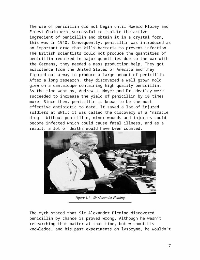
History of penicillinPenicillin was discovered by the Scottish Nobel Laureate Alexander Fleming (Figure 1.1), he discovered it in 1928, yet, he was never able to obtain it in a pure crystalline form. The Australian Nobel Laureate Howard Walter Florey was the first scientist to develop penicillin in order to use it as a medicine. As a matter of fact, penicillin was discovered earlier by John Tyndall but his paper was rejected by the Institute Pasteur due to his young age. Another scientist, Clodomiro Picado Twight, reported his observations on the actions of the genus Penic, but it was rejected as well.
5
Sir Fleming’s discovery started in his laboratory in London;he was researching the properties of Staphylococcus. He suffered from the continued contamination of his petri dishes with the airborne molds and bacteria. Sir Fleming wasknown for his carelessness during his experiments.
Sir Fleming returned from some time off to observe the fungus contamination that has grown on the plates and it wasblue-green in color. He started discarding the contaminated plates. Fortunately, a former member was on a visit to the lab, Sir Fleming started to show the member his contaminatedplates. He observed the empty zone around the mold, which was the inhibiting bacterial growth effect of the mold. Fromthat point, Sir Fleming concentrated his research on this phenomenon, and he continued studying the anti-bacterial studies. He started his trials to obtain the penicillin in acrystalline form, but he was unsuccessful. On the other hand, he was the first scientist that introduced the power of germ killing; he proved his theory by many experiments against different type of infectious bacteria.
Sir Fleming did not realize that he was on the edge of a great discovery. He did not have the knowledge about fungus,although, and after a lot of experiments, he was able to identify it as a species of Penicillum. He also named it as penicillin rubra, which was lately proved wrong by Charles Thom.After a lot of investigation, Charles Thom identified it as penicillin notatum.
In WW1 many soldiers died from the wounds received during the war; he concluded that blood poisoning was the reason behind these deaths.
Baron Joseph Lister, a British surgeon, argued that surgery can’t be performed without antiseptics. He stated that the infected wounds have to be sterilized. Sir Fleming adapted his theory and he was convinced that Baron Lister’s theory was correct.
6
The use of penicillin did not begin until Howard Florey and Ernest Chain were successful to isolate the active ingredient of penicillin and obtain it in a crystal form, this was in 1940. Consequently, penicillin was introduced asan important drug that kills bacteria to prevent infection. The British scientists could not produce the quantities of penicillin required in major quantities due to the war with the Germans, they needed a mass production help. They got assistance from the United States of America and they figured out a way to produce a large amount of penicillin. After a long research, they discovered a well grown mold grew on a cantaloupe containing high quality penicillin. As the time went by, Andrew J. Moyer and Dr. Heatley were succeeded to increase the yield of penicillin by 10 times more. Since then, penicillin is known to be the most effective antibiotic to date. It saved a lot of injured soldiers at WWII; it was called the discovery of a “miracle drug.” Without penicillin, minor wounds and injuries could become infected which could cause fatal illness, and as a result; a lot of deaths would have been counted.
The myth stated that Sir Alexander Fleming discovered penicillin by chance is proved wrong. Although he wasn’t researching that matter at that time, but without his knowledge, and his past experiments on lysozyme, he wouldn’t
7
Figure 1.1 – Sir Alexander Fleming
have took this matter to further investigation. He reacted differently to the contamination of his Petri plates, other scientist just concluded that their experiments were ruined by contamination and they discarded their cultures.
Chemical Structure:
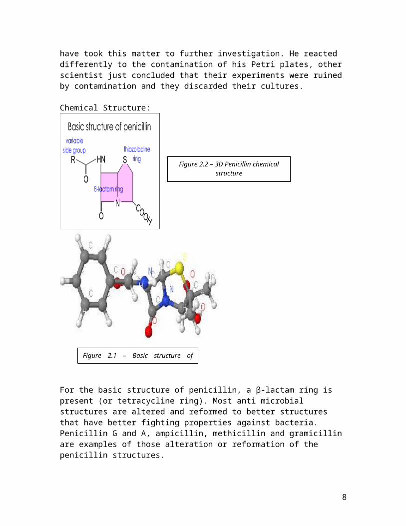
For the basic structure of penicillin, a β-lactam ring is present (or tetracycline ring). Most anti microbial structures are altered and reformed to better structures that have better fighting properties against bacteria. Penicillin G and A, ampicillin, methicillin and gramicillin are examples of those alteration or reformation of the penicillin structures.
8
Figure 2.2 – 3D Penicillin chemicalstructure
Figure 2.1 – Basic structure ofpenicillin
Penicillin has both single and double bonds, all double bonds are connected to oxygen and carbon atoms, in addition,all bonds are covalent. (Figure 2.2)In the early 1940s, Ernst Boris Chain and Howard Florey investigated the work of Sir Fleming on penicillin. Chain and Ernest discovered the chemical composition of penicillinand its therapeutic action. Chain theorized the chemical structure of penicillin. After few years, Dorothy Hodgkin confirmed the structure by her X-ray crystallography technique.
Physical propertiesChemical Formula: C16H18N2O5SMelting point: 97°CPercent composition by mass:Element: Carbon Hydrogen Nitrogen Oxygen SulphurPercentage:
57.4% 5.43% 8.38% 19.14% 9.59%
Percent composition by number:Element: Carbon Hydrogen Nitrogen Oxygen SulphurPercentage:
38.1% 42.9% 4.8% 11.9% 2.3%
Chemical propertiesPenicillin is an organic acids and it is soluble in organic solvents. Esters, chloroform and ethers are good example. Onthe other hand, penicillin is insoluble or hardly soluble inhydrocarbons. In water, penicillin is stable only when it is in the salt form and in a pH ranging between 5 and 8. Penicillin is inactivated by some reagents; it loses its biological activity when it is present in aqueous solutions of higher acidity or alkalinity. In addition to acid and alkali, penicillin is inactivated by most heavy-metal ions, like metal ions of zinc and cadmium. Other reagents that inactivate penicillin are primary alcohols, amines, thiols, aldehydic reagents, ketonic reagents and oxidizing reagents.
9
An enzyme, penicillinase, inactivates penicillin as well, itis a part of the bacterial strains that resists penicillin.
Penicillin mode of action – Enzyme inhibitionPenicillin and its derivatives works by inhibiting the cell wall synthesis of bacteria. To be more specific, the cross linking of peptides on the monosaccharide chains is prohibited. This action will allow water to enter the cell resulting in the damage of the cell.
Mechanism of drug action – Enzyme inhibitionPenicillin works better on Gram positive bacteria than Gram negative bacteria; the reasons will be discussed shortly. Penicillin also blocks the division of bacteria, most importantly, cyanobacteria and cyanelles.Gram positive bacteria have very thick cell walls that are made from sugar polymers that covalently bound to peptide units. The peptidoglycan contains a portion of polysaccharide which is made of repeating units of N-acetylglucosamine that is linked to N-acetylmuramic acid (NAG-NAM) (Figure 3.1).
10
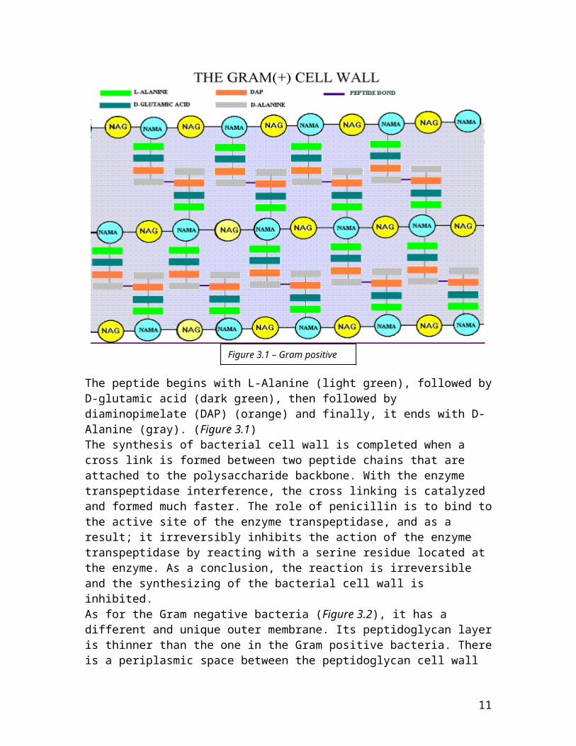
The peptide begins with L-Alanine (light green), followed byD-glutamic acid (dark green), then followed by diaminopimelate (DAP) (orange) and finally, it ends with D-Alanine (gray). (Figure 3.1)The synthesis of bacterial cell wall is completed when a cross link is formed between two peptide chains that are attached to the polysaccharide backbone. With the enzyme transpeptidase interference, the cross linking is catalyzed and formed much faster. The role of penicillin is to bind tothe active site of the enzyme transpeptidase, and as a result; it irreversibly inhibits the action of the enzyme transpeptidase by reacting with a serine residue located at the enzyme. As a conclusion, the reaction is irreversible and the synthesizing of the bacterial cell wall is inhibited.As for the Gram negative bacteria (Figure 3.2), it has a different and unique outer membrane. Its peptidoglycan layeris thinner than the one in the Gram positive bacteria. Thereis a periplasmic space between the peptidoglycan cell wall
11
Figure 3.1 – Gram positive cell wall
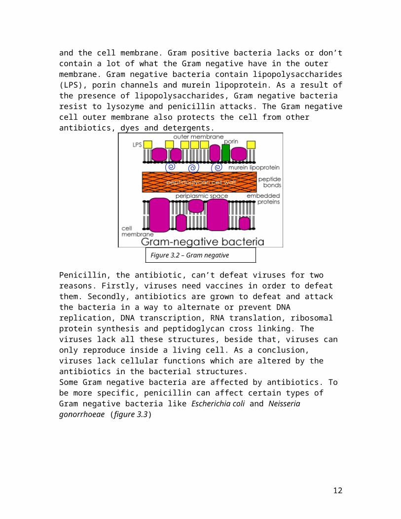
and the cell membrane. Gram positive bacteria lacks or don’tcontain a lot of what the Gram negative have in the outer membrane. Gram negative bacteria contain lipopolysaccharides(LPS), porin channels and murein lipoprotein. As a result ofthe presence of lipopolysaccharides, Gram negative bacteria resist to lysozyme and penicillin attacks. The Gram negativecell outer membrane also protects the cell from other antibiotics, dyes and detergents.
Penicillin, the antibiotic, can’t defeat viruses for two reasons. Firstly, viruses need vaccines in order to defeat them. Secondly, antibiotics are grown to defeat and attack the bacteria in a way to alternate or prevent DNA replication, DNA transcription, RNA translation, ribosomal protein synthesis and peptidoglycan cross linking. The viruses lack all these structures, beside that, viruses can only reproduce inside a living cell. As a conclusion, viruses lack cellular functions which are altered by the antibiotics in the bacterial structures.Some Gram negative bacteria are affected by antibiotics. To be more specific, penicillin can affect certain types of Gram negative bacteria like Escherichia coli and Neisseria gonorrhoeae (figure 3.3)
12
Figure 3.2 – Gram negative structure
Bacterial resistance to penicillinIn early 1940s, some bacteria started to resist and fight the effect of penicillin. The reason is the presence of penicillinases, which are beta lactamases enzymes. Those enzymes are produced by susceptible bacteria which causes the effectiveness of penicillin to fade away. The theory behind this enzyme action is that it hydrolysis the peptide bond that is present in the β-lactam ring, this is present in the nucleus. The penicillinase enzyme is formed by the response of bacterial adaption to the unpleasant environmentthat the bacteria is present in. Those bacteria become stronger and more resistant. In addition, the nutrients thatare present will be available to the resister bacteria. Those nutrients are not eaten by the other bacteria because they will be dead once the antibacterial agent, penicillin, is present. Some bacteria develop mechanisms to resist the antibiotic agents by mutating existing genes or acquiring new ones. The condition of the mutation is that the genes can encode for efflux pumps (figure 4.1) or deactivating enzymes (as penicillinase).
13
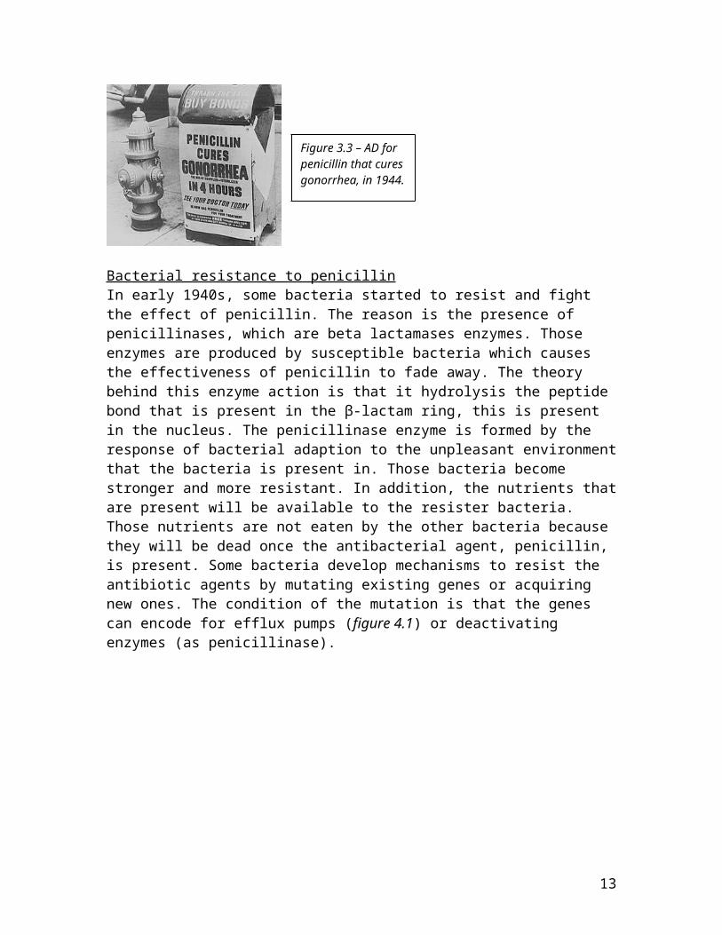
Figure 3.3 – AD for penicillin that cures gonorrhea, in 1944.
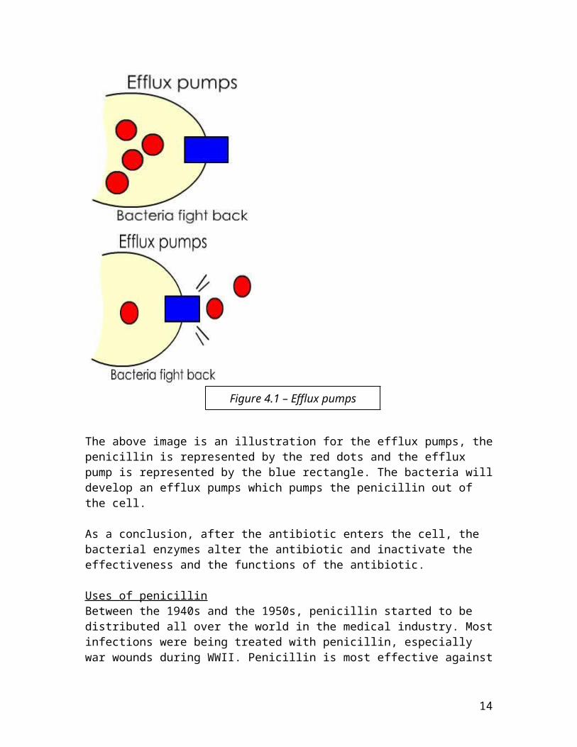
The above image is an illustration for the efflux pumps, thepenicillin is represented by the red dots and the efflux pump is represented by the blue rectangle. The bacteria willdevelop an efflux pumps which pumps the penicillin out of the cell.
As a conclusion, after the antibiotic enters the cell, the bacterial enzymes alter the antibiotic and inactivate the effectiveness and the functions of the antibiotic.
Uses of penicillinBetween the 1940s and the 1950s, penicillin started to be distributed all over the world in the medical industry. Mostinfections were being treated with penicillin, especially war wounds during WWII. Penicillin is most effective against
14
Figure 4.1 – Efflux pumps
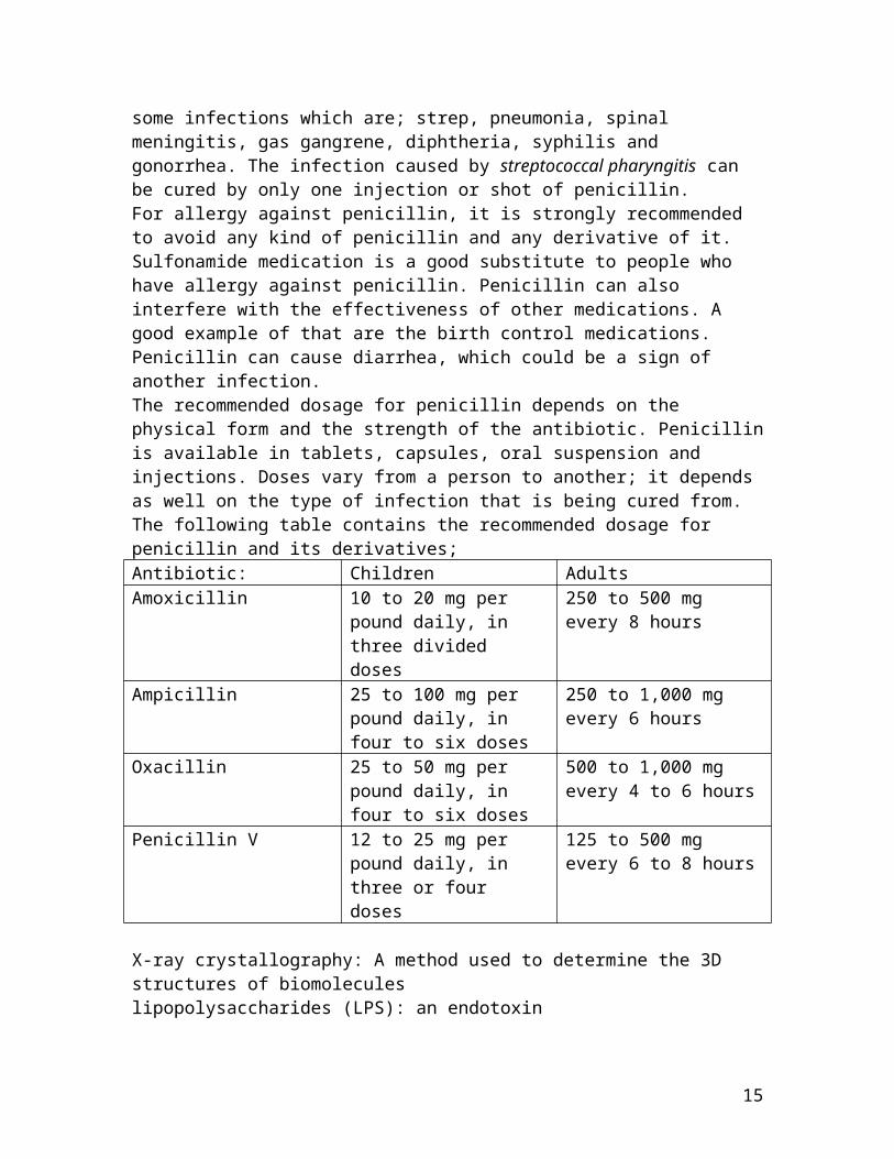
some infections which are; strep, pneumonia, spinal meningitis, gas gangrene, diphtheria, syphilis and gonorrhea. The infection caused by streptococcal pharyngitis can be cured by only one injection or shot of penicillin.For allergy against penicillin, it is strongly recommended to avoid any kind of penicillin and any derivative of it. Sulfonamide medication is a good substitute to people who have allergy against penicillin. Penicillin can also interfere with the effectiveness of other medications. A good example of that are the birth control medications. Penicillin can cause diarrhea, which could be a sign of another infection.The recommended dosage for penicillin depends on the physical form and the strength of the antibiotic. Penicillinis available in tablets, capsules, oral suspension and injections. Doses vary from a person to another; it depends as well on the type of infection that is being cured from.The following table contains the recommended dosage for penicillin and its derivatives;Antibiotic: Children AdultsAmoxicillin 10 to 20 mg per
pound daily, in three divided doses
250 to 500 mg every 8 hours
Ampicillin 25 to 100 mg per pound daily, in four to six doses
250 to 1,000 mg every 6 hours
Oxacillin 25 to 50 mg per pound daily, in four to six doses
500 to 1,000 mg every 4 to 6 hours
Penicillin V 12 to 25 mg per pound daily, in three or four doses
125 to 500 mg every 6 to 8 hours
X-ray crystallography: A method used to determine the 3D structures of biomoleculeslipopolysaccharides (LPS): an endotoxin
15
Procedure
In order for our group to perform this experiment, we had tofirst discuss our objectives and gather parts of experimentsto help us meet our objectives. As we performed the experiment we came across some obstacles, hence we had to augment our procedures accordingly. The following are 9 major steps we had in order to fulfill our objectives. They are all discussed in detail below.
Steps to Production and Crystallization of Penicillin 1. Making the P. Chrysogenum specimen2. Creating the penicillin production medium3. Making P.Chrysogenum seed medium and inoculum
suspension4. Penicillin production procedure
16
5. Production of Staphylococcus6. Testing of Staphylococcus7. Testing penicillin produced8. Test Penicillin vs. various bacteria’s9. Crystallization of Penicillin
Making the P.Chrysogenum Specimen1. Obtain pure culture of P. Chrysogenum and P. Notatum.2. Produce a streak plate using Sabouraud’s agar and
incubate at 25C for 5-7days.3. Take a mouldy lemon and streak potato dextrose and rose
bangle agar.4. Repeat “step 2” after each incubation period in order to
possess fresh growth through the experiment.
Creating the Penicillin Production Medium1. Prepare simulated corn steep liquor
a. Add 50 ml ground corn meal to 100ml water and let soakof night in the refrigerator.
2. To a 1L beaker, add:a. 40.0ml of the simulated corn steep liquorb. 20.0g of lactosec. 1.5g of sodium acetated. 3.0g sodium nitratee. 0.5g potassium dihydrogen phosphatef. 0.25g magnesium sulphate heptahydrateg. 0.4g zinc sulphateh. 0.25g Tween 80i. 950ml distilled water
3. Prepare a calcium carbonate solution by adding 5.0g calcium carbonate to 50.0ml of water.
4. Sterilize the mixture from “step 2” and the calcium carbonate solution in the autoclave
5. Aseptically transfer the calcium carbonate solution into the mixture from step 2 and keep the container sealed.
Making P. Chrysogenum Seed Medium & Inoculum Suspension
17
1. Into four sterilized 125ml Erlenmeyer flasks (with sterile sponge stoppers), add 25ml of sterile penicillin production medium to each.
2. To each flask, add 0.5g glucose or molasses (20g per 1Lof production medium).
3. Obtain a sterile test tube and add 2-3ml of sterile distilled water.
4. Aseptically suspend P. Chyrosgenum spores in the test tube, close the lid, and shake.
Penicillin Production Procedure1. Incubate the flasks for 2 to 3 days at 25C with
agitation.2. Prepare two sterile 250ml Erlenmeyer flasks with 100ml
of penicillin production medium. Transfer content in 125ml flasks previously on the agitator into the 250 mlflask. Incubate for 5-7 days at 25C with agitation.
3. After incubation, put flask in refrigerator to be cooled; penicillin is unstable at room temperature.
4. Remove the mould pellets by filtration with normal filter paper and a membrane filter.
5. Acidify the product with phosphoric acid to a pH of 2.56. Store in the refrigerator when not in use
** note: sponge stoppers must be used in flasks to prevent contamination and allow oxygen circulation in the bioreactor.
Production of S. Aureus1. Add the following
a. Yeast extract = 1.25b. Gelatin = 15.25c. D-mannitol = 5d. Dipotassium phosphate = 2.5e. Casein peptone = 5
18
f. Lactose = 1g. Sodium chloride 37.5h. Agar = 7.5i. 500 ml distilled water
2. Autoclave the mixture 3. Pour in petri dishes allow it to solidify4. Take swab samples from different people and streak them
onto the petri dishes and incubate for 5-7 days at 37°C
Testing of Staphylococcus1. Use the S. Aureus that we previously incubated and streak
a plate of Mannitol salt agar.
**Mannitol contains 7.5% sodium chloride, and phenol red pH indicator. Pathogenic species of Staphylococcus ferment mannitol and produce acid, which turns the pH indicator yellow. Non- pathogenic staphylococcal species grow, but produce no color change.**
Testing Penicillin Produced1. Prepare bacterial lawns with Staphylococcus aureus on
nutrient agar plates.2. Aseptically dip a fritted disc or piece of filter paper in
the penicillin product and place in the centre of the S. Aureus inoculated lawn plate.
3. Repeat “step 2” using sterile water instead of penicillin ascontrol.
4. Incubate the plates at 37C for 48hours5. Compare the growth inhibitition on the plate with the
penicillin product with the growth on the control plate.
**We first tested our penicillin on 8 different bacteria’s. Bacillus coagulus Salmonella typhimulum Klebsiella pneumoniac Enterobacter cloacae
19
Escherichia coli Streptococcus Citrobacteur freudi
All those results were contaminated, and we augmented our procedure to get results for 3 specific bacteria’s listed below.**
Test Penicillin vs. Various Bacteria’s1. Prepare bacterial lawns with nutrient agar in
triplicate:a. Enterobacter Cloacaeb. Bacillius Coagulusc. E. Coli
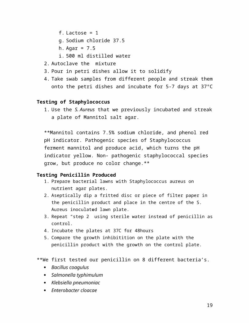
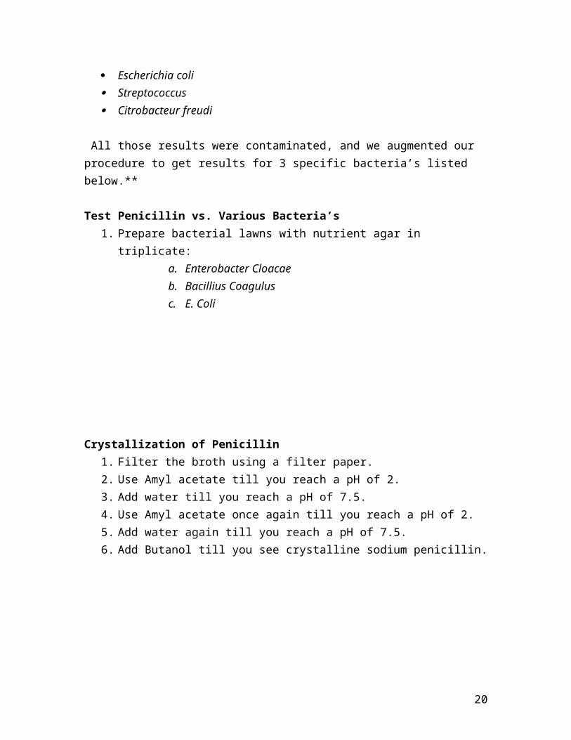
Crystallization of Penicillin1. Filter the broth using a filter paper.2. Use Amyl acetate till you reach a pH of 2.3. Add water till you reach a pH of 7.5.4. Use Amyl acetate once again till you reach a pH of 2.5. Add water again till you reach a pH of 7.5.6. Add Butanol till you see crystalline sodium penicillin.
20
Results
Making the P. Chrysogenum specimen
21
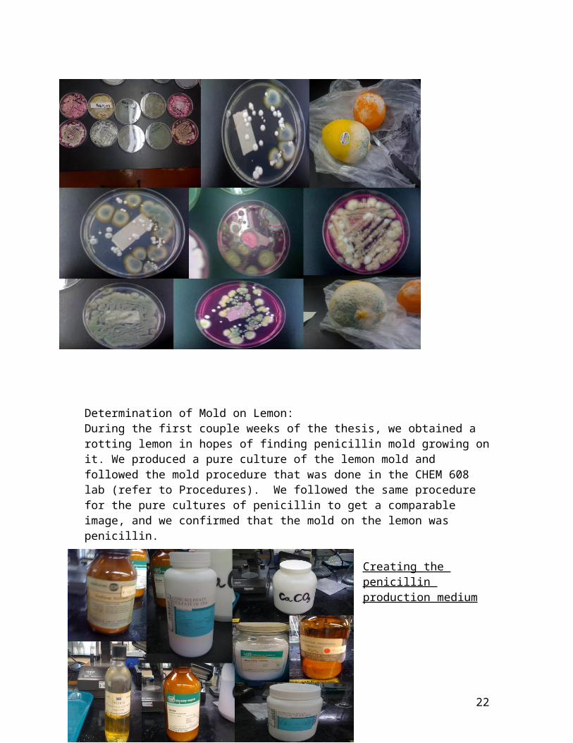
Determination of Mold on Lemon:During the first couple weeks of the thesis, we obtained a rotting lemon in hopes of finding penicillin mold growing onit. We produced a pure culture of the lemon mold and followed the mold procedure that was done in the CHEM 608 lab (refer to Procedures). We followed the same procedure for the pure cultures of penicillin to get a comparable image, and we confirmed that the mold on the lemon was penicillin.
Creating the penicillin production medium
22
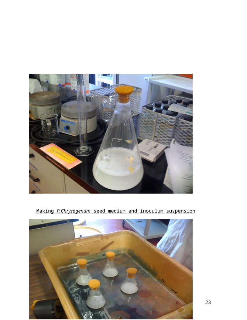
Making P. Chrysogenum seed medium and inoculum suspension
23
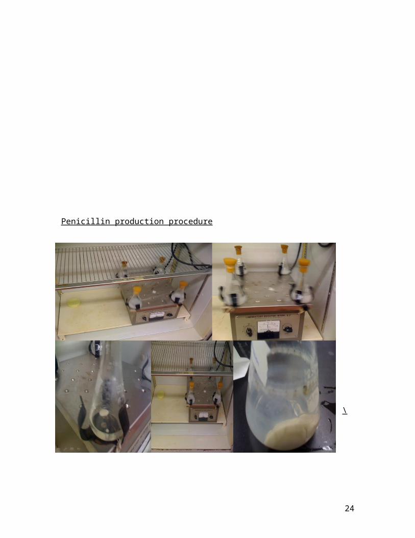
Penicillin production procedure
\
24
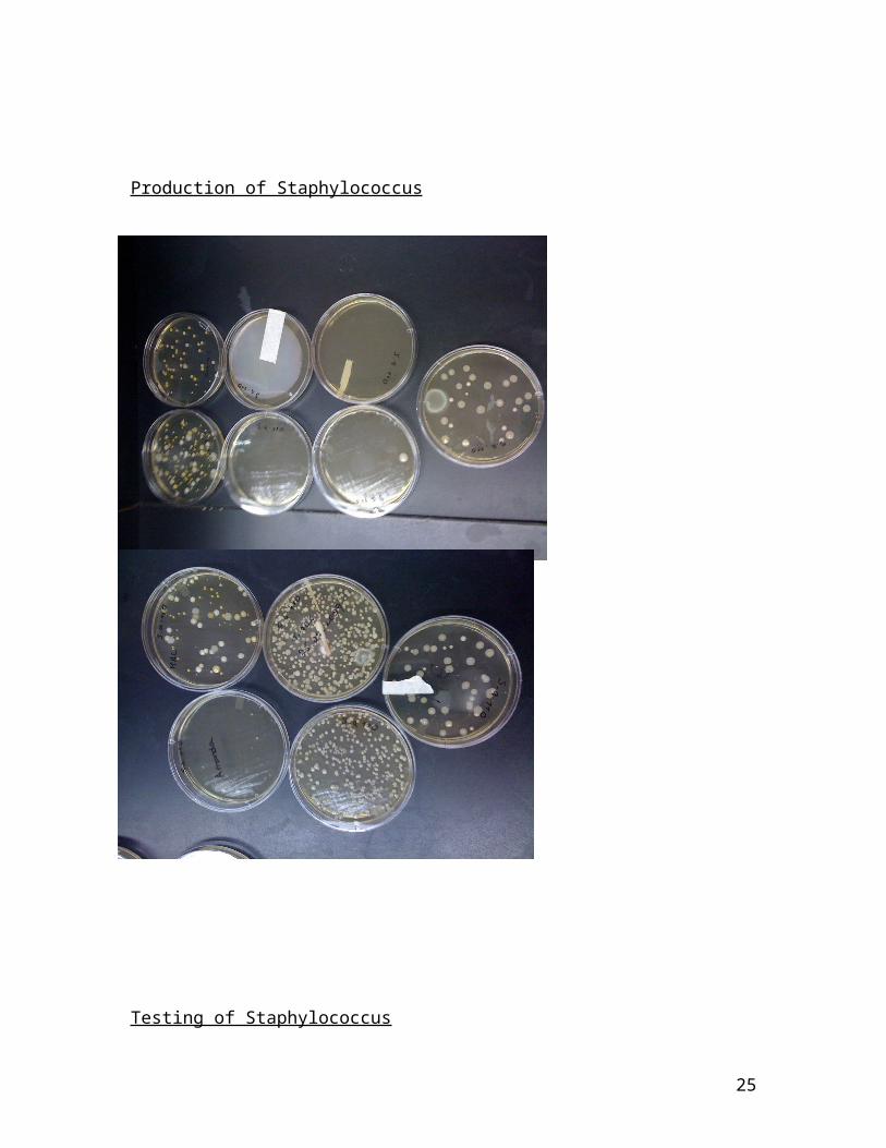
Production of Staphylococcus
Testing of Staphylococcus
25
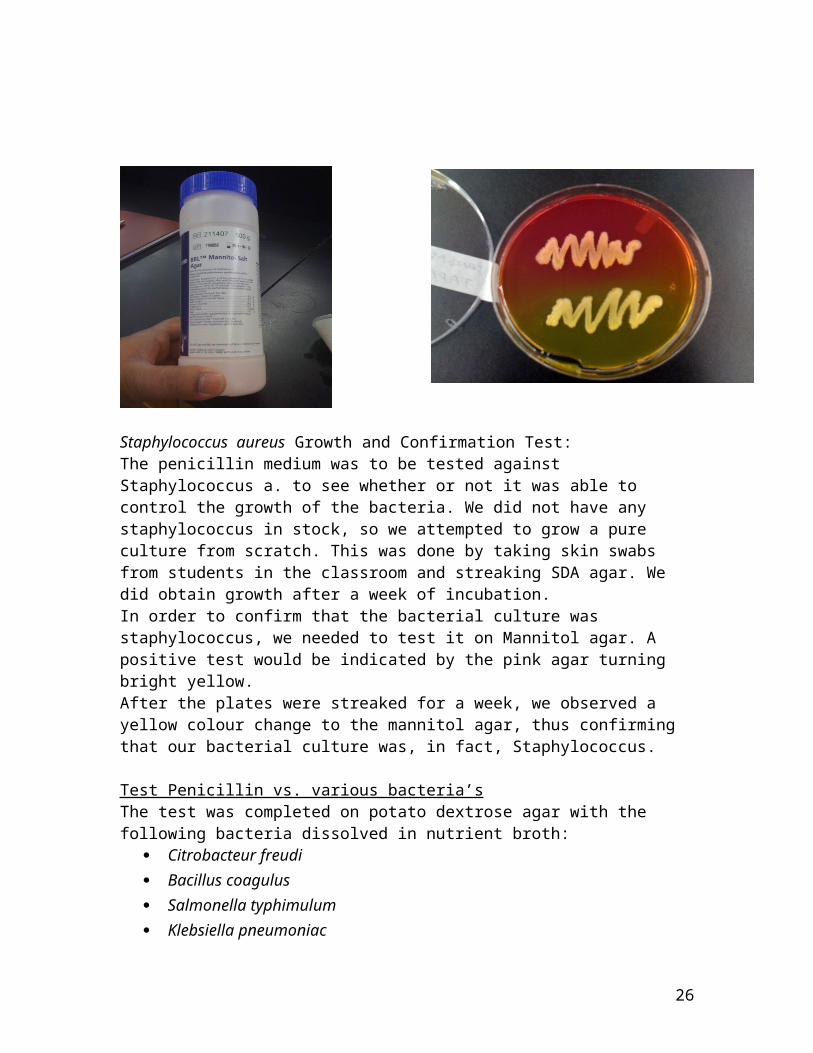
Staphylococcus aureus Growth and Confirmation Test:The penicillin medium was to be tested against Staphylococcus a. to see whether or not it was able to control the growth of the bacteria. We did not have any staphylococcus in stock, so we attempted to grow a pure culture from scratch. This was done by taking skin swabs from students in the classroom and streaking SDA agar. We did obtain growth after a week of incubation.In order to confirm that the bacterial culture was staphylococcus, we needed to test it on Mannitol agar. A positive test would be indicated by the pink agar turning bright yellow. After the plates were streaked for a week, we observed a yellow colour change to the mannitol agar, thus confirming that our bacterial culture was, in fact, Staphylococcus.
Test Penicillin vs. various bacteria’sThe test was completed on potato dextrose agar with the following bacteria dissolved in nutrient broth:
Citrobacteur freudi Bacillus coagulus Salmonella typhimulum Klebsiella pneumoniac
26
Enterobacter cloacae Escherichia coli Streptococcus
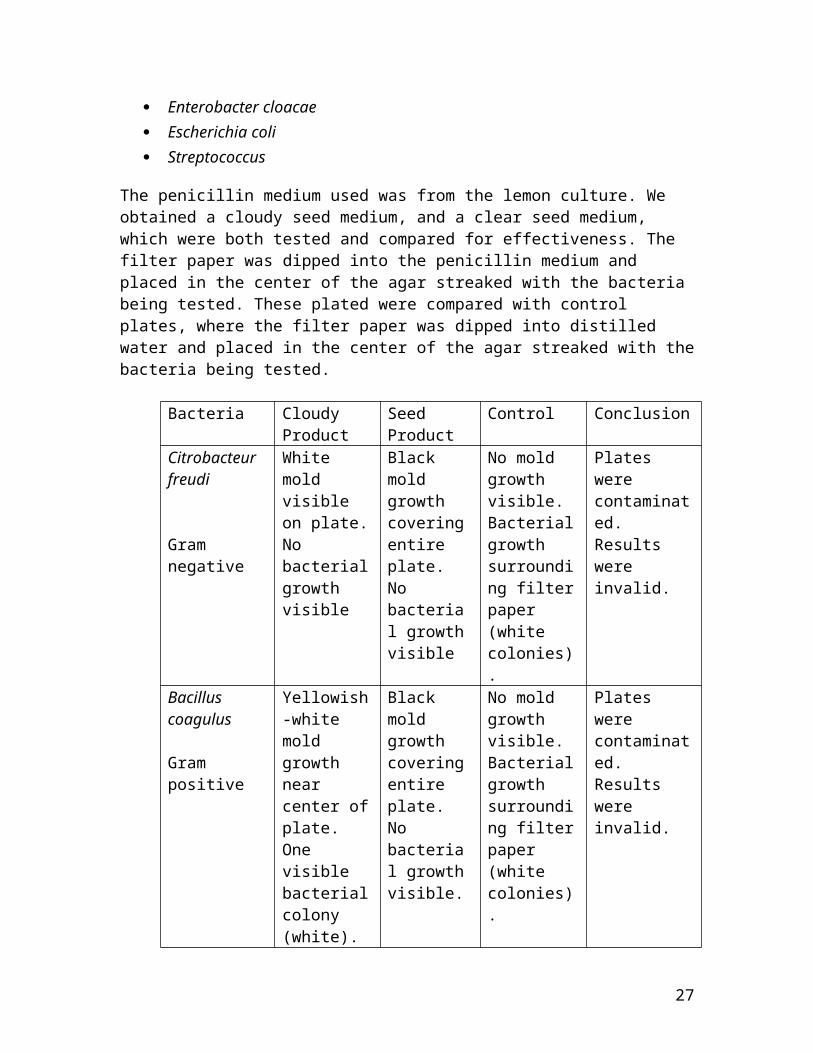
The penicillin medium used was from the lemon culture. We obtained a cloudy seed medium, and a clear seed medium, which were both tested and compared for effectiveness. The filter paper was dipped into the penicillin medium and placed in the center of the agar streaked with the bacteria being tested. These plated were compared with control plates, where the filter paper was dipped into distilled water and placed in the center of the agar streaked with thebacteria being tested.
Bacteria Cloudy Product
Seed Product
Control Conclusion
Citrobacteur freudi
Gram negative
White mold visible on plate.No bacterialgrowth visible
Black mold growth coveringentire plate. No bacterial growthvisible
No mold growth visible. Bacterialgrowth surrounding filterpaper (white colonies).
Plates were contaminated. Results were invalid.
Bacillus coagulus
Gram positive
Yellowish-white mold growth near center ofplate. One visible bacterialcolony (white).
Black mold growth coveringentire plate. No bacterial growthvisible.
No mold growth visible. Bacterialgrowth surrounding filterpaper (white colonies).
Plates were contaminated. Results were invalid.
27
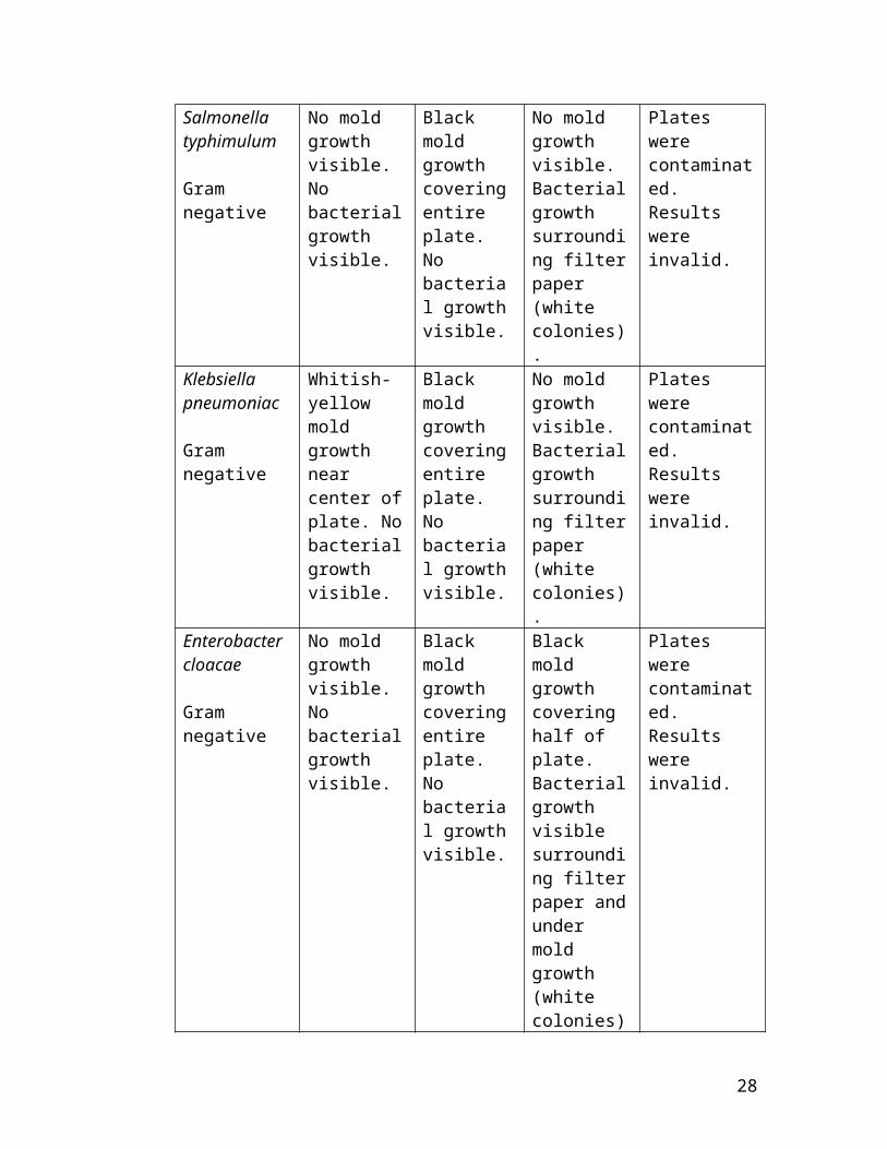
Salmonella typhimulum
Gram negative
No mold growth visible. No bacterialgrowth visible.
Black mold growth coveringentire plate. No bacterial growthvisible.
No mold growth visible. Bacterialgrowth surrounding filterpaper (white colonies).
Plates were contaminated. Results were invalid.
Klebsiella pneumoniac
Gram negative
Whitish-yellow mold growth near center ofplate. Nobacterialgrowth visible.
Black mold growth coveringentire plate. No bacterial growthvisible.
No mold growth visible. Bacterialgrowth surrounding filterpaper (white colonies).
Plates were contaminated. Results were invalid.
Enterobacter cloacae
Gram negative
No mold growth visible. No bacterialgrowth visible.
Black mold growth coveringentire plate. No bacterial growthvisible.
Black mold growth covering half of plate. Bacterialgrowth visible surrounding filterpaper andunder mold growth (white colonies)
Plates were contaminated. Results were invalid.
28
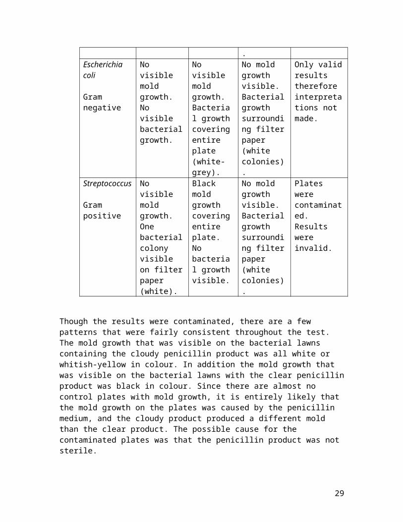
.Escherichia coli
Gram negative
No visible mold growth. No visible bacterialgrowth.
No visible mold growth. Bacterial growthcoveringentire plate (white-grey).
No mold growth visible. Bacterialgrowth surrounding filterpaper (white colonies).
Only validresults therefore interpretations not made.
Streptococcus
Gram positive
No visible mold growth. One bacterialcolony visible on filterpaper (white).
Black mold growth coveringentire plate. No bacterial growthvisible.
No mold growth visible. Bacterialgrowth surrounding filterpaper (white colonies).
Plates were contaminated. Results were invalid.
Though the results were contaminated, there are a few patterns that were fairly consistent throughout the test. The mold growth that was visible on the bacterial lawns containing the cloudy penicillin product was all white or whitish-yellow in colour. In addition the mold growth that was visible on the bacterial lawns with the clear penicillinproduct was black in colour. Since there are almost no control plates with mold growth, it is entirely likely that the mold growth on the plates was caused by the penicillin medium, and the cloudy product produced a different mold than the clear product. The possible cause for the contaminated plates was that the penicillin product was not sterile.
29
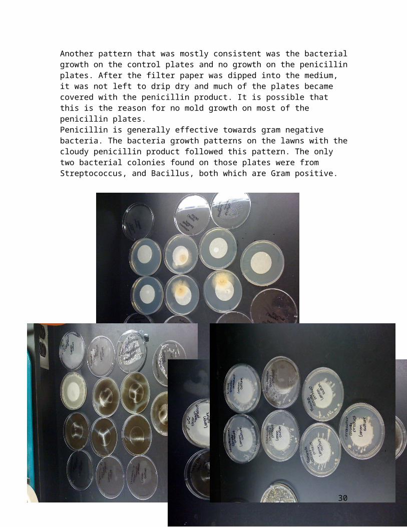
Another pattern that was mostly consistent was the bacterialgrowth on the control plates and no growth on the penicillinplates. After the filter paper was dipped into the medium, it was not left to drip dry and much of the plates became covered with the penicillin product. It is possible that this is the reason for no mold growth on most of the penicillin plates. Penicillin is generally effective towards gram negative bacteria. The bacteria growth patterns on the lawns with thecloudy penicillin product followed this pattern. The only two bacterial colonies found on those plates were from Streptococcus, and Bacillus, both which are Gram positive.
30
Penicillin Tests on Bacterial Lawns (2nd attempt)The test was completed on nutrient agar with the following bacteria:
Enterobacter cloacae Escherichia coli
The bacteria were dissolved in three different broths to ensure valid growth patterns on the plates.
Nutrient broth Saline broth Distilled water
Each of the bacterial lawns was done in duplicates and the controls were done in singles. At this point in the thesis, most of the penicillin medium had been used for the crystallization attempt. Only the clear penicillin medium from the lemon culture remained, so the test was only done using this product.Bacteria Enterobacter cloacae Escherichia coliControl Nutrient broth
Bacterial colonies covering entire
Bacterial colonies covering entire
31
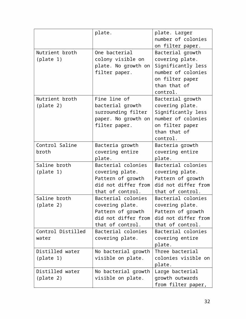
plate. plate. Larger number of colonies on filter paper.
Nutrient broth (plate 1)
One bacterial colony visible on plate. No growth onfilter paper.
Bacterial growth covering plate. Significantly less number of colonies on filter paper than that of control.
Nutrient broth (plate 2)
Fine line of bacterial growth surrounding filter paper. No growth onfilter paper.
Bacterial growth covering plate. Significantly less number of colonies on filter paper than that of control.
Control Saline broth
Bacteria growth covering entire plate.
Bacteria growth covering entire plate.
Saline broth (plate 1)
Bacterial colonies covering plate. Pattern of growth did not differ fromthat of control.
Bacterial colonies covering plate. Pattern of growth did not differ fromthat of control.
Saline broth (plate 2)
Bacterial colonies covering plate. Pattern of growth did not differ fromthat of control.
Bacterial colonies covering plate. Pattern of growth did not differ fromthat of control.
Control Distilled water
Bacterial colonies covering plate.
Bacterial colonies covering entire plate.
Distilled water (plate 1)
No bacterial growthvisible on plate.
Three bacterial colonies visible onplate.
Distilled water (plate 2)
No bacterial growthvisible on plate.
Large bacterial growth outwards from filter paper,
32
but no growth on filter paper.
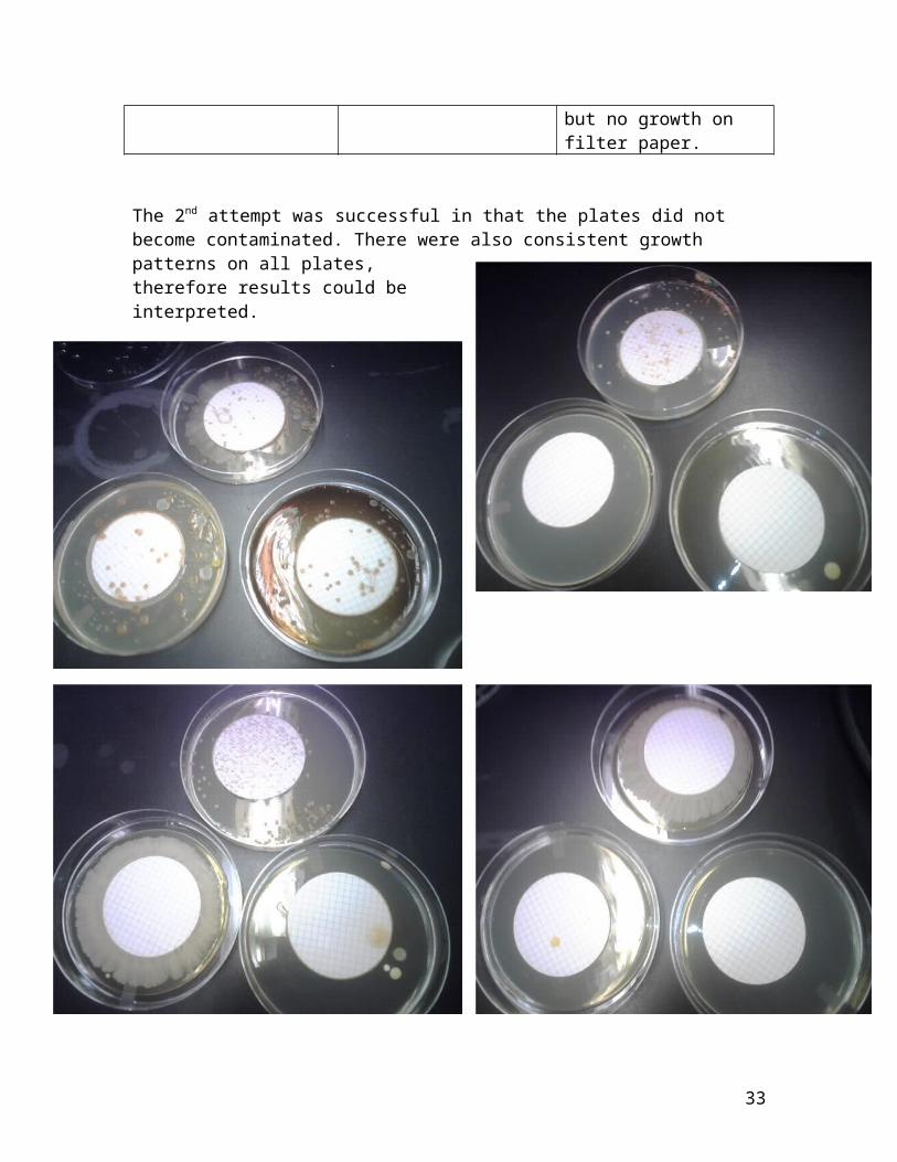
The 2nd attempt was successful in that the plates did not become contaminated. There were also consistent growth patterns on all plates,therefore results could beinterpreted.
33
Interpretations:Enterobacter cloacae
Nutrient broth – The growth on the control plate was significantly greater than that of the penicillin plates. Weexpected to see growth on the entire plate, but no growth surrounding the filter paper. This, however, was not the case. We did see a reduced amount of growth on the lawns with the penicillin medium which confirms that the penicillin did control the growth of the bacteria. It is likely that there was an excess of penicillin medium covering the agar on the plate which resulted us not obtaining the desired growth patterns. Saline broth – The growth patterns on the control plate and the penicillin plates did not differ. It is possible that the saline broth affected the performance of the penicillin product. It is also possible that the saline broth provided perfect growth conditions for the bacteria allowing the bacteria to continue to grow in large numbers. To conclude, the penicillin was ineffective on the bacteria dissolved in the saline broth. This was also the case with the E.coli in the saline broth. Distilled water – There were bacterial colonies covering theentire control plate, and no growth on either of the penicillin plates. Once again, the growth patterns that we expected to see on the penicillin plates were not there. Since there is a large amount of growth on the control plateand no growth on the penicillin plates, it is confirmed thatthe penicillin did prevent the growth of bacteria on these plates. There may have also been an excess of the penicillinmedium on the agar which prevented us from seeing the desired growth patterns.
Escherichia coli
34
Nutrient broth – There was bacterial growth on the control plate and the penicillin plates, but there were a significantly larger number of colonies on the filter paper of the control plate than that of the penicillin plates. Thegrowth patterns on the penicillin plates were not what we expected to see. However, there were significantly less colonies on the filter papers of the penicillin lawns; therefore we can confirm that the penicillin did work to reduce the bacterial growth. Saline broth – As seen with the Enterobacter lawns, the growth patterns on the control plate and the penicillin plates did not differ. As mentioned, there is a possibility that the saline broth affected the penicillin medium performance, or the right growth conditions were provided for the E.coli to continue to grow in large numbers. We can say that the penicillin was ineffective on the bacteria dissolved in the saline broth.
Distilled water – There were bacterial colonies covering theentire control plate and very few colonies on both of the penicillin plates. The expected growth patterns were not visible on the penicillin plates, but we did see a reduced amount of growth on the penicillin plates. It is possible that there was an excess of penicillin medium on the agar which prevented the bacteria from growing in the pattern we desired. We can confirm that the penicillin did reduce the growth of the bacteria.
Crystallization of Penicillin
35
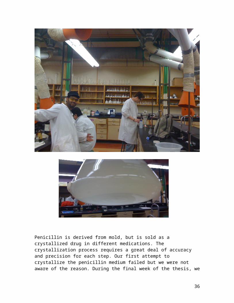
Penicillin is derived from mold, but is sold as a crystallized drug in different medications. The crystallization process requires a great deal of accuracy and precision for each step. Our first attempt to crystallize the penicillin medium failed but we were not aware of the reason. During the final week of the thesis, we
36
attempted to be even more accurate to get some results. After several attempts, we did not obtain any results. We concluded that the experiment was a successful failure, and were pleased with our efforts.
Discussion
TIME MANAGEMENTIn almost any situation imaginable, time management is
a major contributing factor to the success and credible outcome of an experiment or process (or series of processes). One must play the responsible role in start andanalyze the components and different sections of an experiment and ensure that enough time has been allotted to the appropriate procedures and analyses.
Naturally, the method of approach to this particular thesis project was in fact to determine the amount of time necessary to complete all of the individual procedures throughout the project. Some of these processes included acquiring the necessary materials, preparation of bacterial lawns and inoculating broth tubes, attempting to recrystalize penicillin from produced medium ( a notoriouslycomplicated procedure), sterilizing the proper equipment (which was a rather time consuming process), and cultivatingfresh growths every week. These are some of the procedures among an extensive list of tasks.
To approach this situation in an appropriate manor, it was felt that the crystallization method should be allocatedample time to ensure a proper attempt at the procedure. Since it is notoriously difficult and little to no results were expected to begin with, it was known that more than oneattempt would be needed. Therefore, the time consuming nature of this portion of the overall project was taken intoaccount, and it was made sure that the last 3 weeks of the allotted research time and analysis time (in the laboratories) was available for the crystallization attempt.
Of course, one must not forget that mistakes should be anticipated. That being said, one must take into
37
consideration the fact that unforeseeable incidences will most likely occur, and as a responsible group member, one must give enough time to deal with any setbacks these occurrences may cause. Fortunately, this group did encounter some setbacks and had planned well enough in advance for these occurrences as to not suffer significantlyin terms of valid results.
Time management becomes an extremely critical incident once a mistake has been made, and the setback is enough to throw off the whole experiment, or make it impossible to actually finish the experiment in the time allotted. If however, responsible actions are taken and the undesired occurrence/contamination/machine (or human) error occurs andthere is enough time to get back on track, this error will simply act as a valuable learning experience.
One would gain knowledge from this error and would in turn become more efficient as a result; constantly being more aware of the reactions taking place and the real necessity of time management.
DETERMINATION AND PREPARATION OF NECESSARY TYPES OF AGARAlthough not a large obstacle, this task was an
obstacle none the less. This particular project consisted of the use of not only several different types of agar, but ample amounts of them.
One reason for the rather large amounts of agar was theneed to cultivate fresh growths every week the group met in the laboratory. Therefore, we had to make some simple calculations to determine how many plates (i.e. how many litres) of which types of agar would be needed throughout the 13 weeks of analysis and experimentation.
Much like the time management element, contamination was anticipated, so the theoretical amount of plates was never the amount being made: sometimes these amounts would double (if necessary) if the group had reason to believe that the plates would be subjected to highly infectious areas of the laboratory. Just as well, it’s always better safe than sorry.
38
An issue came up early on when there was some confusionas to whether or not the right agar was accounted for. The specific desired agar was nowhere to be found in the laboratory, and as the determined group we were, we took it amongst ourselves to formulate this agar ourselves. Some ofthe many components included calcium carbonate and sodium chloride, common chemicals that can easily be found in the laboratory. Upon the tracking down and retrieval of set items, it came to one’s attention that the desired agar was in fact accounted for in the laboratory and the production of it from scratch was not necessary. Although again, thiswas not a huge obstacle, it was an early sign of the group’sconfidence and ambition and overall motivation to deal with any discrepancies and obstacles head on, and under no circumstance accept defeat as a result. This also demonstrated initiative on the groups’ behalf to go to theselengths for the sake of a thesis project.
In some cases, certain types of agar that were desired were only actually desired/needed in small quantities. A perfect example of such a case is when the group realized that to positively identify the cultivated bacteria (though a precise procedure) as Staphylococcus Aureus (needed for testing against the produced penicillin), a very specific agar was needed (one containing sodium chloride; most bacteria are not resistant to this compound, where as Staphylococcus Aureus is), which is known as Mannitol agar. Allthat was needed of this agar was about ten plates, and the directions on the container gave the procedure for making one litre. Through previous experience, it was safe to assume that one litre of agar would yield approximately forty plates. That being said, it was only a matter of performing a simple calculation to determine the amount needed. The method was as such: since one litre gave about forty plates, and what was needed was approx. ten plates, the amount given on the container was simply divided by four.
This is significant when considering working in the professional field; one must be efficient with company resources and not waste any product. Therefore, one must
39
determine the amount of necessary nutrient agars accurately and precisely before carrying out an experiment.
This calculation did in fact work out to one’s advantage.
COLLECTING AND ACQUIRING NECESSARY MATERIALSAs learned from hundreds of hours in the Humber
laboratories, any successful experiment must be planned thoroughly, including which chemicals and equipment are necessary. This must be done not only for efficiency, but specifically in this case, to ensure that the chemicals/bacteria/equipment is in fact accounted for in thelaboratories.
This being said the first step was to meet with the coordinator to confirm the presence of the desired bacteria and equipment. Most of the bacteria were accounted for as well as all of the equipment (more detail for this section will be presented further on in the report).
This is a significant portion of a successful experiment as well as a practical lesson in thinking critically and reacting quickly in a potentially hazardous situations. One may be faced with a situation in which the experiment is already underway, and a vital chemical for a certain part of the experiment is nowhere to be found. Either the technician determines an appropriate alternative, or the experiment is lost.
Equipment can cause equally as upsetting situations, inthe sense that a procedure might call for a specific apparatus or set of instruments which are simply just not available in the laboratory in question. This obstacle was a definite issue, especially when considering the penicillincrystallization process. The only way around this that one could think of is to seek out an alternate method. This uncertainty would be a definite attribute to the “successfulfailure” that was our crystallization attempt.
To improve in this area for future experiments, one would consult a professional or someone with experience in this field, before attempting a procedure that could
40
potentially cost a company or organization unnecessary amounts of money, resources, and time.
AGITATOR COMPLICATIONS AND THE IMPORTANCE OF DUPLICATESHaving very random and unforeseeable errors occur
because of a malfunction of an instrument / machine in the laboratory is a very real possibility. These possible occurrences must be anticipated if any credible results are to come from an experiment of this calibre.
This specific obstacle presented itself very abruptly one day in the laboratory, when it was heard that in the incubator where the agitator is kept there was a noise of something “banging” around.
Right away it was known that this could not have been agood sound and the group investigated this problem. Upon opening the door of the incubator, it was seen that the agitator, which contained four flasks (each containing the penicillin seed medium) that had stoppers in them. The agitator had been placed on an extremely high setting of agitation, and had somehow been pushed right up against the door and side of the incubator, causing it to make the “banging” sound and to ultimately cause a big mess.
The big mess was the solutions in the flasks that had spilled all over the place. Now, this is leads to the next crucial point: duplication is essential.
What is meant by this statement is that duplicating solutions or bacterial lawns, or inoculations (etc.), is never a bad idea. In fact, if the group had not duplicated the solutions, all of the product would have been lost and this would have dealt a major blow to the flow of the experiment. One would have been forced to repeat a portion of the procedure that easily consumes a week’s worth of lab work, had there not been duplications.
The attitude originally was essentially “duplicating can’t hurt if the resources are present” (which they were), and this attitude ultimately saved a great deal of time. This is a rather practical learning experience because when working in the industry, time is costly, and to have to repeat unnecessary portions of an experiment can be
41
considered simply unacceptable (unacceptable waste of money,time, and resources).
There are no real suggestions as to how to avoid this situation for future references. All one could say is to keep the agitator away from the sides of the incubator and to make duplicates. This was a strange incident and the group can still not account for what went wrong. This can be dubbed a “indeterminate error”.
CONTAMINATION OF AGAR AND BACTERIAL LAWNSOn a few different occasions, one would enter the lab
only to find that several (if not all) of the plates of agarkept in the fridge from the week before had been contaminated. The contamination was evident because on the surface of the agar, it was seen that large amounts of mouldwere growing. This mould was not our penicillin because theculture had not been introduced to that set of agar plates yet.
Several factors could have been responsible for this contamination, including improper (if any at all) sterilization techniques and methods (with the equipment), mould particles flowing through the air in the laboratory (from previous mould labs performed in biotechnology the past days) and landing on the surface of the plates, as wellas the possibility that the agar was contaminated before it even was poured and left to solidify.
The action taken was to completely disregard these plates (throw them out and make new ones). Since making agar is a rather lengthy process, this was a significant setback in the flow of the experiment. The actual process of combining the mixture with water and mixing is not the issue, it is the sterilization process that was time consuming, and ultimately the whole reason why the contamination of these lawns and agar plates were consideredsignificant setbacks (mainly in terms of time wasted and resources wasted).
In the future, many more precautions will be taken, such as wearing gloves at all times and sterilizing the sponge stoppers before using them in flasks, as well as
42
ensuring that there were no recent experiments that would lead one to assume that there are potentially damaging chemicals floating in the air. As well, one should take extra care in seeing that any part of instruments used (i.e.pipettes and burettes) should not come in contact with benchtops or other instruments, as both these present the possibility of direct contamination.
MEMBRANE FILTRATION COMPLICATIONSAt one critical point in the experiment, a technique
known as membrane filtration was called for by the specific procedure. What was being filtered was the penicillin pure culture suspensions (which were to be further acidified – pH2.5) and what was noticed immediately was the unusual amountof trouble that came with trying to work with the membrane filtration apparatus. The syringe pump action required an unusual amount of strength and was taking an extremely long time to filter volumes as small as 3 millilitres.
As students in an engineering program, we took it upon ourselves to think of a different and more efficient way of filtering the necessary solutions. What came to mind was the usage of the vacuum pump (located in the instrumentationlab). This did prove to be more effective; however one can’t help but wonder if the constant degassing of these solutions contributed to the lack of result (crystallization), or any possible source of future contamination.
CONTAMINATION OF EQUIPMENT The first suspicions of the equipment being used being
the source of contamination arose right about the time of the first appearance of the contaminated bacterial lawns andagar plates. Upon the discovery of further contamination inthe plates and the seed medium, it became clear that the source of contamination was most likely the equipment.
Just as well, this point was the point at which we realized that the “innovative” idea we had (which was to usethe vacuum pump instead of the non-cooperative membrane filtration manual device) turned out to be rather useless
43
because the membrane and the giant flask had not been sterilized and therefore could not be fully trusted.
This being determined, the process was repeated while sterilizing absolutely everything including the membrane, sponge stoppers, and flasks. This did consume extra time.
HANDLING OF HAZARDOUS MATERIALSIn order to test the penicillin that was cultivated
from the rotten lemon, certain bacteria had to be acquired and tested against the produced penicillin strain. These bacterium, however are very hazardous microorganisms.
In plain English, these bacteria are dangerous and could potentially pose huge health risks and therefore indicate that the utmost respect and caution is to be exercised while handling these materials. Gloves were worn while working with the bacterium and necessary precautions were taken.
I feel as though working with these bacteria provided some very valuable and practical experience in terms of handling hazardous materials. This allowed me be at a real risk and therefore test my skills as a technologist. Not tomention, the results and documentation are fascinating.
This kind of work is so valuable because in industries that concern themselves with these types of bacteria value caution and knowledge of the materials. This group has gained firsthand experience of treating the potentially pathogenic substances with respect and testing with them with confidence and extreme caution.
TROUBLES WITH THE MICROSCOPEThe concern here is not with the microscope at all
actually, it is more so the method preparing the slide to beviewed. The obstacle presented here was this: when the mould scraped from the agar (which was used to cultivate thepenicillin) was placed on the microscope, one would expect to get a clear view of what penicillin chrysogenum is supposed to look like. This was not the case.
What was seen under the microscope was simply circular shaped molecules. What this meant was that all that was
44
being seen were the spores, however the structure of the chrysogenum strain is highly branched and much more complex then what was seen under the microscope. After doing some research, it was found that the mould actually has to grow on the microscope for a couple of days, and then it was suitable for viewing and in turn identification.
This being said, the slide was prepared by cutting a small piece of agar (PDA) from a plate (already prepared) and placing it (coated with a layer of culture) over a smallfew drops of water (also inoculated with the mould). This is kept on slant in a Petri dish with a lot of moisture in the bottom of the dish (water is kept in the dish to promotemoisture and therefore allow the mould to grow). The dish was placed in the incubator for a week (7 days exactly).
After the week had passed, the slant was examined underthe microscope and viewed the structure that was expected. This matched perfectly with what was the accepted structure of this particular strain of penicillin (penicillin chrysogenum). Drawings were made of what was viewed for clear and appropriate documentation.
THE ABSENCE OF STAPHYLOCOCCUS AUREUS What was felt to be one of the biggest scares in this
project was the point at which the group realized that one of the main bacterium which was supposed to be used to test against the penicillin was not accounted for. This bacterium is known as Staphylococcus Aureus and is a rather harmful substance in high concentrations.
Staphylococcus is actually found on the surface of the skin of human beings, which was actually quite fortunate forthe group for one reason; this made it possible to isolate our own staphylococcus and use it for experimentation purposes.
The first step in this procedure was to determine whichagar would be used to develop this bacteria. The research performed demonstrated that an agar with a sodium chloride concentration (NaCl) of at least 10% (13.5% to be specific).The reason for this sodium chloride is that most bacteria are not resistant to sodium chloride, where as Staphylococcus
45
Aureus, is. This eliminates the possibility of any other typeof bacteria growing in the agar.
The next logical step was to figure out how to actuallytransfer the staph. from human to petri dish. This wasn’t too big an obstacle, as the group quickly came to find that a simple cotton swab would suffice. The idea was to swab the skin of someone who hadn’t showered that day, since the staphylococcus wouldn’t have been washed off as recently or as much. The cotton swab was to be swabbed against the forearm of the person in question and then simply streaked across the plate in every direction.
Since the group had made duplicates of this specific type of agar plate (after having learned from the previous occurrences – see earlier in this section), it was possible to collect a sample (swab the forearm) from everyone that was in the laboratory. The cotton swabs were individually streaked against Petri dishes (one sample per dish) and leftto incubate for a week in the incubator.
This, however, was not the end of the task. It was recommended that the extra mile be taken and have yet another type of agar be developed.
The next step was to make a type of agar with the specific purpose of confirming the identity of staphylococcus aureus. Thankfully, there just happened to be the right chemicals in the laboratory to make this agar. It is known as Mannitol agar and is reddish pink in color. The idea behind this is to take the cultivated sample from the nutrient agar (in the previous step) and streak it onto the mannitol agar, and let to incubate for a few days. If the color of either the agar or the streaked area turns yellow, then the identity of the staphylococcus aureus couldbe confirmed.
Fortunately enough, we did receive much growth in the nutrient agar for the growing of the staphylococcus, and a positive test (yellow color) for the Mannitol agar. After this lengthy process, it was fair to say that thegroup had a strong pride about them, as this completely demonstrated a huge accomplishment; cultivating from scratchtheir own bacteria and confirming the identity in an agar
46
made from the start. Everything went smoothly and clearly showed that the procedure had been followed with extreme accuracy and care/caution.
Upon completion of this rather lengthy step, the staphylococcus aureus was then ready for testing.
CRYSTALLIZATION OF PENICILLINBy far the hardest part of the whole thesis project,
the crystallization of the penicillin product was ultimatelywritten off as a successful failure. This was not too much an upset, or a surprise for that matter.
The reason for this statement is that the process of crystallizing penicillin is notoriously difficult and is also notorious for yielding extremely small amounts of product. This being said, we were optimistic, but realistic. We did not immediately blame ourselves, rather, we reflected upon the method and the degree of suitability in terms of our conditions and resources in the laboratories.
Three different methods were consulted for this portionof the experiment, again due to the lack of options in our laboratory setting. One of the main issues was that the main product (liquid form) from which the crystals were supposed to have formed had to be acidified, brought back toa pH of 7, and then acidified again. The materials used forthe first trial consisted of ethyl acetate and sodium hydroxide.
The reason we used sodium hydroxide is because that particular procedure called for the solution to be made alkaline using regular water. Ample amounts of water were added with barely any change in the pH. Upon closer inspection of the procedure, it became evident that that procedure was directed more at mass production (several thousand litres worth).
Therefore, this method was not plausible. We moved on to method 2, one using amyl acetate and again water was supposed to be used as the base. Again, this would not workfor our cause, and so we used sodium hydroxide as a means ofmaking the solution more alkaline. This did work for that
47
purpose , however one cannot help but wonder if this caused any discrepancies in terms of desired reactions within the solution.
Upon addition of 1-Butanol (called for by method #3 after acidification/alkalinity), two separate layers were formed, which was thought to be a good sign. The procedure did call for extraction using 10 ml portions of water, so wesaw to separating the layers in a separating funnel and allowing the gas to escape before the separation.
The two separate layers were collected and because of some minor confusion as to which layer was actually containing the desired crystals, both layers were kept and tested on. Filtrations using a Buchner funnel, cooling in an ice bath, scraping with a crystal rod, extraction method,all of these were used, yet no visible result.
As mentioned, this process was performed three times and no sign of any success (at least in this portion of the experiment). It was unfortunate for the group, but the attempt to crystallize the penicillin product was ultimatelyconsidered a “successful failure”.
Conclusion
Present day, the use of penicillin and other antibiotics take a common place. A cut or a more serious wound can easily be infected and to prevent the onset of infection various antibiotics are used. These types of antibiotics were not available in the early 1900’s. However, the discovery of penicillin in the early 1940’s gave hope to allof us; in terms of curing infectious diseases.
In our knowledge, we have encountered lemon or lime as the best source to find penicillin chrysogenum mold/fungi. Therefore, we have used a moldy lemon to isolate penicillin
48
chrysogenum. Even though, we have taken a moldy lemon to isolate penicillin, we came to a bumpy road which not all molds on a lemon are penicillin chrysogenum. In this case we must isolate the mold by cultivation it on a growth medium which is specify for growth of mold and examine this mold under a microscope to identify if the mold is penicillin chrysogenum. What we have isolated in our experiment was identified as penicillin, but whether to say it was chrysogenum or notatum we did not know. Due to the fact it has been some form of penicillin we have decided to continueour experiment. However, future research done later in the experiment on where we can find penicillin chrysogenum mold. Most of the web sites indicated that cantaloupes are the best source of fruit to isolate higher percentage of penicillinchrysogenum fungi.
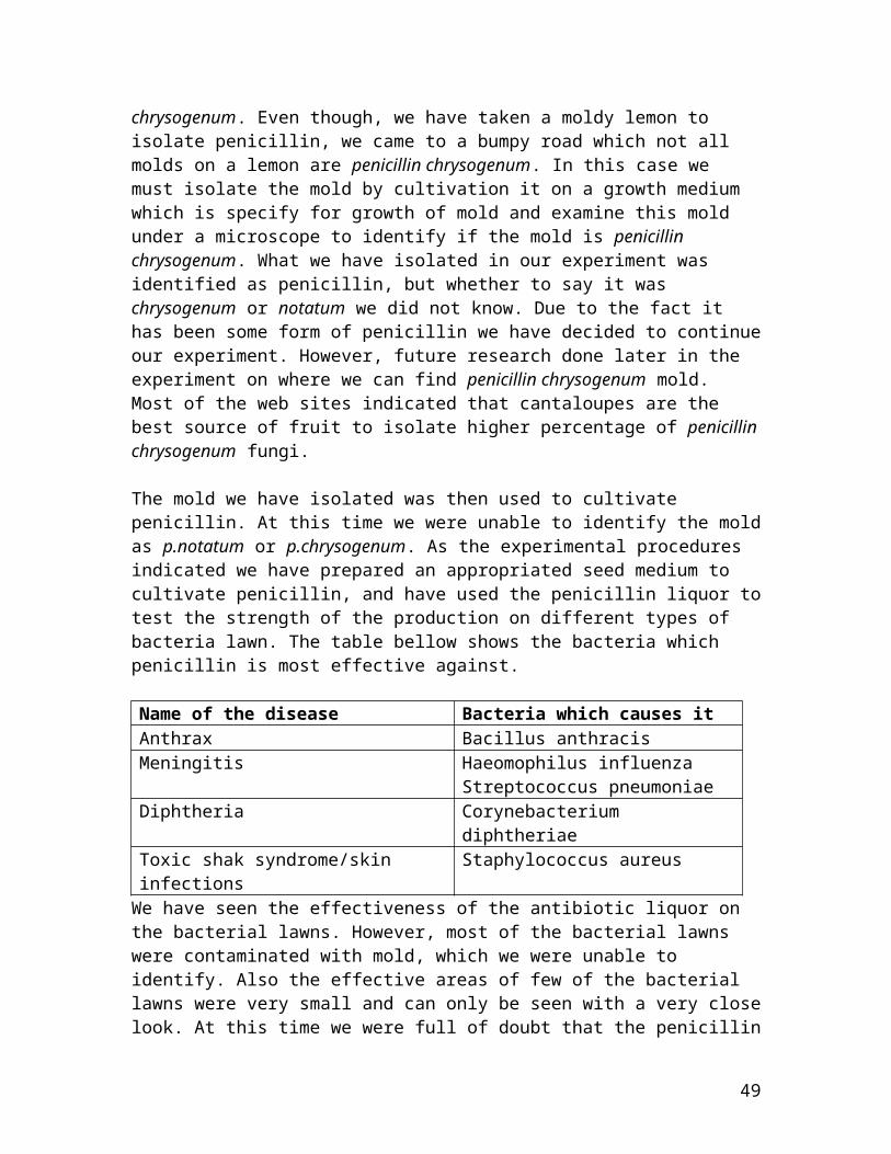
The mold we have isolated was then used to cultivate penicillin. At this time we were unable to identify the moldas p.notatum or p.chrysogenum. As the experimental procedures indicated we have prepared an appropriated seed medium to cultivate penicillin, and have used the penicillin liquor totest the strength of the production on different types of bacteria lawn. The table bellow shows the bacteria which penicillin is most effective against.
Name of the disease Bacteria which causes itAnthrax Bacillus anthracisMeningitis Haeomophilus influenza
Streptococcus pneumoniaeDiphtheria Corynebacterium
diphtheriaeToxic shak syndrome/skin infections
Staphylococcus aureus
We have seen the effectiveness of the antibiotic liquor on the bacterial lawns. However, most of the bacterial lawns were contaminated with mold, which we were unable to identify. Also the effective areas of few of the bacterial lawns were very small and can only be seen with a very closelook. At this time we were full of doubt that the penicillin
49
liquor is not strong enough to fight the bacteria that was growing on the medium. Since, we have done the liquor in duplicate; we have given a second try on the bacterial lawn.This time the results were disappointing as well because almost all the bacterial lawn were contaminated. Here, we have encountered we have made a mistake on not sterilizing the equipment we have used. A third try was attempted on thebacterial lawn using a newly and properly produce penicillinliquor. Even the last try was failed due to the bacterial lawn being contaminated, but some had a growth of bacteria and the effect of the penicillin liquor was unable to see. At this point we were able to conclude that the laboratory was contaminated with air born mold by the mold used on a previous laboratory experiment, and the antibiotic liquor did not have enough penicillin to affect the bacterial lawns.
Synthesis of penicillin is not as simple as a procedure would elaborate. Procedure for manufacturing of penicillin was done in large scales and the experiment we have performed is done in laboratory scale. Therefore, when we done the calculation from the lager scales to smaller scales, in order to perform the production in laboratory scale might have cause a discrepancy in the seed medium. This can be very difficult because the production of penicillin mold is very limited when grown in small scale seed mediums. Even the high yield recombinant penicillin G amidase production and export into the growth medium using bacillus megaterium gives only 40mg of penicillin to 1 L of the medium. Reverse calculation of the growth medium from largerscale to fit our laboratory scale experiment gave us only 100-150 mL growth medium. In calculation the production of penicillin on this medium would be;
40 mg of penicillin / 1 L of medium × 1L / 1000 mL × 150 mL = 6 mg.
50
So we can conclude that the theoretical value of the yield of penicillin on our medium would be very small. Even a 250 mg antibiotic tablet has about 28.06 mg of penicillin.
When comparing these numbers we would believe the amount of medium we have used is not enough to cultivate enough penicillin to crystallize it. This is also can be the cause of the minimal effect on the bacterial lawns because the penicillin liquor did not contain enough antibiotic to fightout the bacteria on the lawns. However, there was penicillinpresent in the liquor and it only had a minimal effect on the bacteria; therefore, we were unable to call this a very successful experiment.
As we mentioned before, when isolating penicillin mold it also can be difficult because the other type of mold also can grow on lemon. We have used moldy lemon as the source ofthe mold to inoculate the mold mediums. This can give us a deviation on the penicillin production, which we might have known to use a similar mold that has the similar color patterns, or the similar biological structure. However, thiswould not be the fact for the failure because we have examined the mold under a microscope and have identified themold as penicillin. But another question rises at this time, was the entire mold was concluded to be penicillin or there was only a presence of penicillin and other species present as well, when look under a microscope. Even though, we have observe the mold under 100 times magnification, it did not clear prove that the entire mold that was grown on the medium was penicillin because we only tend to isolate minimal amount of penicillin mold, there were other species present as well. This also could have been due to contamination; cause of this was the laboratory been used for mold experiment prior to starting of our experiment, andthe molds tend to air born. This could be a factor in contamination of the mediums we have grown the penicillin mold. We did not take the time to isolate other species to determine whether lemon was the best source to isolate penicillin. However, further research has given us a bit of
51

information about cantaloupes being the best candidate to isolate penicillin.
With few ends tied up, more research was done on why the penicillin we isolated from the lemon did not give us enoughyields to test the production against chosen set of bacteria. Here we are faced with another loose end of our very exciting project of production of penicillin. We were left with nothing but to do more and more research to find what went wrong in the production medium. Since, we were able to conclude that the production yield was minimal to test it against bacteria or to crystallize penicillin; the only confusion was that the penicillin isolated from the lemon mold was the correct penicillin.
A new door was opened for us, and we were able to conclude why the production of penicillin would have been less than what we have expected. It was do to the fact we have used a lemon mold which most often carry penicillin notatum, and this form of penicillin has a very low production yield. It was also known that penicillin notatum would not live on a culture medium for no longer than 4-5 days. However, we left the culture medium incubated for about 7 days. This might have cause a discrepancy in the growth of penicillin. As previously mentioned cantaloupes was the best candidate for the isolation of penicillin, and it also known to carry highyield penicillin chrysogenum. So if we were to use mold from a cantaloupe, even though we have used a 150 mL seed medium, this would have given us acceptable results. What we were able to conclude here, is that penicillin notatum is a low yieldpenicillin and a penicillin chrysogenum is a high yield penicillin, and we should have used a penicillin chrysogenum in order to get a better yield. This also ties up the end wherewhen we look the mold under a microscope we were unable to identify between the two penicillin because their biologicalstructure is very similar to each other and only way to findout the difference is to look under a high resolution microscope because the differences are only can be identified by the size (p.notatum is about 5.4 Mb and
52
p.chrysogenum is 6.8 Mb) and the lack of a certain DNA regionthat penicillin chrysogenum has and the penicillin notatum lack. It shows that it is very difficult to identify the non commonalities between the two strains of penicillin. So the only way to isolate penicillin chrysogenum is by specific vegetation that grows the penicillin chrysogenum mold.
As to our final thought we come to a conclusion that the penicillin that we had cultivated was penicillin notatum, and this form of penicillin should not be used to isolate and cultivate under laboratory conditions to test it against bacteria or to crystallize penicillin.
53