An investigation into the efficacy of ozone for inactivation of microalgae cells in photobioreactors
15
1 AN INVESTIGATION INTO THE EFFICACY OF OZONE FOR INACTIVATION OF THE MICROALGAE DUNALIELLA TERTIOLECTA IN PHOTOBIOREACTORS Rachel M. Clarke 1 , Patrick J. Solan 1 , Brijesh. K. Tiwari 2 , Colm P. O’Donnell 1 and Thomas P. Curran 1 [Email: [email protected]. Ph +353 7167362 1 UCD School of Biosystems Engineering, University College Dublin, Ireland 2 Food and Consumer Technology, Manchester Metropolitan University, Manchester, UK Abstract Microalgae cultivation in photobioreactors has many advantages such as reduced risk of contamination; more controlled cultivation and increased illuminated area to volume ratio. However, due to the adhesive nature of microalgae, fouling of the light transmitting surfaces can be problematic. Traditional methods of sterilising photobioreactors between batches involve ultrasound, autoclaving or as seen in recent times, the photobioreactor incorporates a cleaning system. The objective of this research was to look at an alternative method of sterilisation. The efficacy of ozone was investigated to treat the algae strain Dunaliella tertiolecta (Butcher). Algae cultures at 10 4 cells CFU mL -1 were exposed to 3-29 μg mL -1 of ozone at varying time periods (0-80 sec) and constant gas flow rates of 0.125 and 0.25 L min -1 . Inactivation rates for total reduction of D. tertiolecta were found to increase significantly with an increase in ozone concentration and treatment time. The effectiveness of ozone treatment is dependent on various factors such as contact time, ozone concentration and the ozone demand of the algae.
Transcript of An investigation into the efficacy of ozone for inactivation of microalgae cells in photobioreactors
1
AN INVESTIGATION INTO THE EFFICACY OF OZONE FOR INACTIVATION OF THE MICROALGAE DUNALIELLA TERTIOLECTA IN PHOTOBIOREACTORS
Rachel M. Clarke1, Patrick J. Solan1, Brijesh. K. Tiwari2, Colm P. O’Donnell1 and Thomas P. Curran1
[Email: [email protected]. Ph +353 7167362
1UCD School of Biosystems Engineering, University College Dublin, Ireland
2Food and Consumer Technology, Manchester Metropolitan University, Manchester, UK
Abstract
Microalgae cultivation in photobioreactors has many advantages such as reduced risk of contamination; more controlled cultivation and increased illuminated area to volume ratio. However, due to the adhesive nature of microalgae, fouling of the light transmitting surfaces can be problematic. Traditional methods of sterilising photobioreactors between batches involve ultrasound, autoclaving or as seen in recent times, the photobioreactor incorporates a cleaning system. The objective of this research was to look at an alternative method of sterilisation. The efficacy of ozone was investigated to treat the algae strain Dunaliella tertiolecta (Butcher). Algae cultures at 104 cells CFU mL-1 were exposed to 3-29 μg mL-1 of ozone at varying time periods (0-80 sec) and constant gas flow rates of 0.125 and 0.25 L min -1. Inactivation rates for total reduction of D. tertiolecta were found to increase significantly with an increase in ozone concentration and treatment time. The effectiveness of ozone treatment is dependent on various factors such as contact time, ozone concentration and the ozone demand of the algae.
2
Keywords: cell inactivation, Dunaliella tertiolecta, flow, microalgae, ozone, oxygen, photobioreactor
1. Introduction
Rising worldwide demand for motor and power generation fuels, along with environmental concerns in terms of greenhouse gases (GHG) has motivated policy makers, scientists and technologists alike to consider alternative sources of energy [1,2,3]. Within the European Union (EU), the transportation and energy sectors are among the major anthropogenic sources responsible for more than 20% and 60% of greenhouse gas (GHG) emissions respectively [4]. First generation fuels have been mainly extracted from food and oil crops such as sugarcane, maize, rapeseed oil and sugar beet. This has caused controversy as the ‘food versus fuel’ debate has become one of the most contentious topics of the 21st Century [5,6]. While first generation crops alone cannot meet the current demand for fuel, third generation fuels which are derived from microorganisms such as yeast, fungi and microalgae appear to be a more promising feedstock [7]. The applications of microalgae include potential biofuel production, CO2 fixation, biohydrogen production and bio-treatment of wastewater [8]. Microalgae have higher photosynthetic efficiencies, higher yields and growth rates compared to plant harvest [9]. In terms of CO2 mitigation, one kilogram of algal dry weight utilises around 1.83 kg CO2 [10]. Microalgae require less cultivation space and can be cultivated in saline waters and in arid land areas [11], they also have the potential to produce more oil per hectare than any other feedstock being used to make biodiesel and they can be grown on land that is unsuitable for food crops [12]. Within 3–5 days, microalgae can start producing oil with a minimum yield oil of approximately 6.21 g of oil m-2 day (~ 23 t oil ha-1 yr-1) [13].
The genetic engineering of marine algae has been attracting an increasing amount of interest as a crucial technology in which to overcome the biomass problem in industrial applications [14]. With the absence of cell differentiation, genetic manipulation of microalgae can be a much simpler system compared with higher plants as the allelic genes are usually absent because of the haploid nature of most vegetative stages of microalgae [15]. One of the major disadvantages to microalgae cultivation is the rapid fouling of the light transmitting surfaces of the reactor due to the adhesive nature of microalgae [12,16].This can lead to the reactor frequently being “taken offline” for mechanical cleaning and sterilisation, thus reducing production [6,17]. Some of the traditional methods of mechanical cleaning and sterilisation of
3
photobioreactors involve autoclaving, ultrasonic cleaning or inbuilt systems that internally ‘self-clean’ the bioreactor [6,18]. Sterilisation is important to prevent photobioreactor systems from undesired microorganisms that might reduce the system’s efficiency [18]. The use of ozone is an alternative sterilisation method.
1.1 Ozone
Ozone is a highly reactive form of oxygen, consisting of three oxygen atoms. It is a potent disinfectant that quickly decomposes to diatomic oxygen (O2), while reacting with targeted organic matter or microorganisms. Ozone can effectively kill viruses, bacteria, fungi and parasites, including those causing food spoilage or human diseases. Commercial applications of ozone include purification of drinking water, sterilisation of containers for aseptic packaging, decontamination of fresh produce, and food preservatives in cold storage. Ozone is also an effective deodorizer in air and water [19]. It is generally accepted that the oxidant potential of ozone induces the destruction of cell walls and cytoplasmic membranes of bacteria and fungi, making ozone a powerful and reliable antimicrobial agent against bacteria, fungi, protozoa and viruses [20]. Huang and Wu [21] proposed ozonation as a new method for controlling membrane fouling of the membrane bioreactor (MBR) in a waste water treatment plant. Results showed that an ozone dose of only 0.18 mg/l ozone per mg total organic content (TOC) of biopolymer clusters (BPC) reduced the membrane fouling rate by up to 70%. Bullock et al. [22] found that adding ozone at a lower rate (25g ozone/kg) to recirculating water culture system just before it entered the culture tanks was as effective as adding ozone at a higher dosing rate (35-39g ozone/kg). This finding was supported by Summerfelt et al. [23] who also found that at the same lower rate, the water quality was greatly improved, hence using lower rates of ozone also has lower capital and operating costs. Garvand et al. [1] reported that low dose of ozone (100 ppm) and a longer exposure time was more effective at causing cell mortality (40%) of algae than sonication and hydrogen peroxide.
1.2 Dunaliella tertiolecta (Butcher)
Dunaliella tertiolecta is a marine unicellular green algal species; it has the capability to utilize inorganic nutrients present in wastewater and solar energy to produce biomass from carbon dioxide [24]. The algae strain D. tertiolecta is commonly used for microalgae cultivation [25], it can contain more than 60 w % lipids under certain growth conditions [11], it is also fast growing and that means it has a high CO2 sequestration rate [26]. Since the algal cells of D. tertiolecta do not clump together or form chains, their morphology makes it ideal to perform a cell count [24].
As it lacks a rigid cell wall and has a thin cytoplasmic membrane, D. tertiolecta is considered a sensitive strain of algae [11]. These factors may contribute to rapid cell viability when exposed to ozone. Shear stress as a result of high gas velocities would rupture the already weak cell membrane along with the bactericidal effect of ozone. There are three regions in a bubble column where cell death might occur: 1) At the diffuser where the bubbles are formed; 2) In the region where the bubbles rise, and 3) At the surface where the bubble disengagement occurs [27].
4
The objective of this study was to investigate this hypothesis by investigating the efficacy of ozone for reduction of Dunaliella tertiolecta for the sterilisation of photobioreactors. 2. Materials and methods
2.1. The culturing of algae
The alga Dunaliella tertiolecta which is commonly found in saline waters was chosen as a model organism because of its abundant distribution and ease of culture [1]. Parent cultures of the algae strain D. tertiolecta were sourced from the Scottish Association for Marine Science’s (SAMS) Culture Collection of Algae and Protozoa (CCAP) (Argyll, Scotland). The sea water used to culture the algae was sourced from EIRCOD (Cod Broodstock and Breeding Programme) of the Martin Ryan Institute (MRI), National University of Ireland, Galway. This water had been filtered through a 1.5 µm screen and treated with UV to kill any microorganisms. This seawater also provided the basis for creating the artificial seawater growth medium (F/2).
The treated media was inoculated with an F/2 medium which includes trace metals, vitamins solution and macronutrients (NaNO3, NaHPO4 H2O) adopted from Guillard and Ryther [28]. To prepare the F/2 liquid growth medium for the stock algal cultures, 1 mL of macronutrients solution and 1 mL of trace metals solution were added to the pre-treated Atlantic seawater and the medium was then autoclaved. After autoclaving, 1 mL of the vitamins solution was added to prevent denaturing. The F/2 medium and stock cultures were maintained in a culture chamber at 20 ± 2 °C under cool-white fluorescent lighting. Concentrations of algal cells in culture were determined using a haemocytometer. Cultures were aseptically sterile-transferred every two-three days.
2.2. Media preparation
Seawater was collected from the Lower Falcorrib Pier, Co. Donegal, Ireland (54°53’37.15’’N, 8°24’ 20.91’’W) for the preparation of agar media. This location was chosen due to the exceptional water quality of the coast which was compliant with EU guide values [29]. The collected seawater was first analysed for salinity using a digital salinity meter (Inolab, WTW series) and for pH using a hand held pH monitor (Thermo Scientific Orion 5- star plus, Cambridgeshire, England). A salinity reading using practical salinity units (PSU) equivalent to parts per thousand on a mass basis, was recorded. The seawater was filtered using a Whatman filter No. 1 filter paper disc, (pore size 10 µm, 5.5 cm in diameter; Whatman INC, Clifton, NJ) and a manual vacuum filtration method to remove all debris and organisms. The filtered seawater was then used to make nutrient agar.
To prepare the agar, 28 g of agar nutrient (Oxoid CMO325, Thermo Scientific), 1 L of the Donegal filtered seawater and 1 mL of F/2 medium using the methods of Guillard and Ryther, (1963) were placed together in a 1 L glass beaker and autoclaved (Rodwell scientific Instruments, MP 24, Essex, England) at 121°C for 15 minutes. When cooled, 0.5 mL of vitamin solution (B12, biotin and Thiamine HCL) was added to ensure that adequate nutrients were available for algal growth. The agar was then poured into 90 mm petri plates under the cover of a laminar hood (Astec Microflow, Class II, UK) to maintain a sterile environment. The agar petri plates were allowed to set in a sterile environment for at least 24 hours before use.
5
2.3. Ozone system design
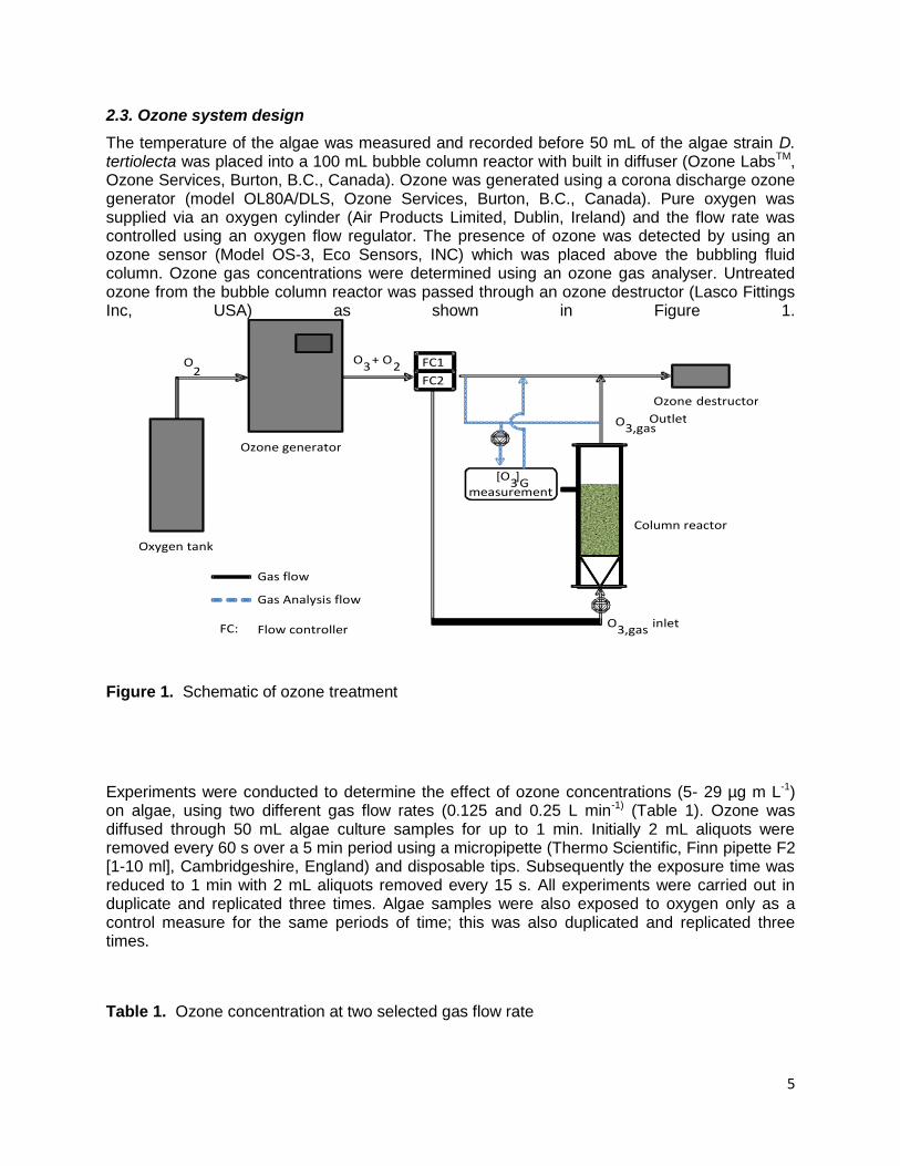
The temperature of the algae was measured and recorded before 50 mL of the algae strain D. tertiolecta was placed into a 100 mL bubble column reactor with built in diffuser (Ozone LabsTM, Ozone Services, Burton, B.C., Canada). Ozone was generated using a corona discharge ozone generator (model OL80A/DLS, Ozone Services, Burton, B.C., Canada). Pure oxygen was supplied via an oxygen cylinder (Air Products Limited, Dublin, Ireland) and the flow rate was controlled using an oxygen flow regulator. The presence of ozone was detected by using an ozone sensor (Model OS-3, Eco Sensors, INC) which was placed above the bubbling fluid column. Ozone gas concentrations were determined using an ozone gas analyser. Untreated ozone from the bubble column reactor was passed through an ozone destructor (Lasco Fittings Inc, USA) as shown in Figure 1.
Figure 1. Schematic of ozone treatment
Experiments were conducted to determine the effect of ozone concentrations (5- 29 µg m L-1) on algae, using two different gas flow rates (0.125 and 0.25 L min-1) (Table 1). Ozone was diffused through 50 mL algae culture samples for up to 1 min. Initially 2 mL aliquots were removed every 60 s over a 5 min period using a micropipette (Thermo Scientific, Finn pipette F2 [1-10 ml], Cambridgeshire, England) and disposable tips. Subsequently the exposure time was reduced to 1 min with 2 mL aliquots removed every 15 s. All experiments were carried out in duplicate and replicated three times. Algae samples were also exposed to oxygen only as a control measure for the same periods of time; this was also duplicated and replicated three times.
Table 1. Ozone concentration at two selected gas flow rate
O
Oxygen tank
Ozone generator
Ozone destructor
[ O 3 ] G
measurement
O 3 + O
2 2
O 3,gas
Outlet
O 3,gas inlet
Gas flow
Gas Analysis flow
Flow controller FC:
FC1
FC2
Column reactor
6
Flow rate (L min-1)
Ozone concentration (µg mL-1)
0.125 5
17 29
0.25 3 9 13
2.4. Laboratory analysis
Following ozone treatment all ozonated aliquots underwent a dilution series up to a dilution of 10-4 under a laminar hood. To ensure proper mixing the test tubes were placed on a vortex (Scientific Industries, INC, USA) and 200 µL aliquots of appropriate dilutions were surface spread onto labelled agar nutrient petri plates. To prepare a salinity buffer for the dilution series, 42 g of sodium chloride (FLINN, Scientific, Inc, 2008) was added to 350 mL of distilled water. A sodium chloride solution (9 mL) was placed into each of the test tubes before being autoclaved at 121°C for 15 min. The test tubes were allowed to cool for a period of up to 24 hours before use.
Each dilution was plated in duplicate to ensure accuracy and consistency. All completed plates were placed into an incubation chamber (Thermo Scientific, Precision compact, Cambridgeshire, England) set at 37°C for 72 hours. The efficacy of treatments was determined in terms of viable cell counts following the 72 hour period in the incubation chamber. The results were reported as colony forming units (CFU mL-1). The data was converted exponentially using Microsoft Excel for all graphical representations. All data was pooled and values averaged and standard deviations determined. 2.5. Cell inactivation kinetics
Weibull modelling
The reductions in the algal cell count (log10 CFU mL-1) population following ozone treatment were fitted to Weibull kinetic model to analyse the algal cell inactivation. Weibull models [Eq.1] were fitted using GInaFit software tool as described by Geeraerd [30].
( ) ( ) [
] [1]
Herein, Nt represents the algal cell counts (Log10 CFU mL-1); N0 is the initial algal cell counts (Log10 CFU mL-1); β is the shape factor. The shape parameter provides a flexibility which takes concave shape (β<1) or convex shape (β>1).The δ parameter is the decimal reduction time (min), i.e. the time taken to achieve a 1 log or 90% reduction in the algal cell count.
4. Results and discussion
The effects of ozone on algae species D. tertiolecta were determined by measuring the cell counts of the algae after a 72 h incubation period. Preliminary tests showed that after ozone exposure the number of algae cells in cultivation at 104 CFU mL-1 decreased in a time-
7
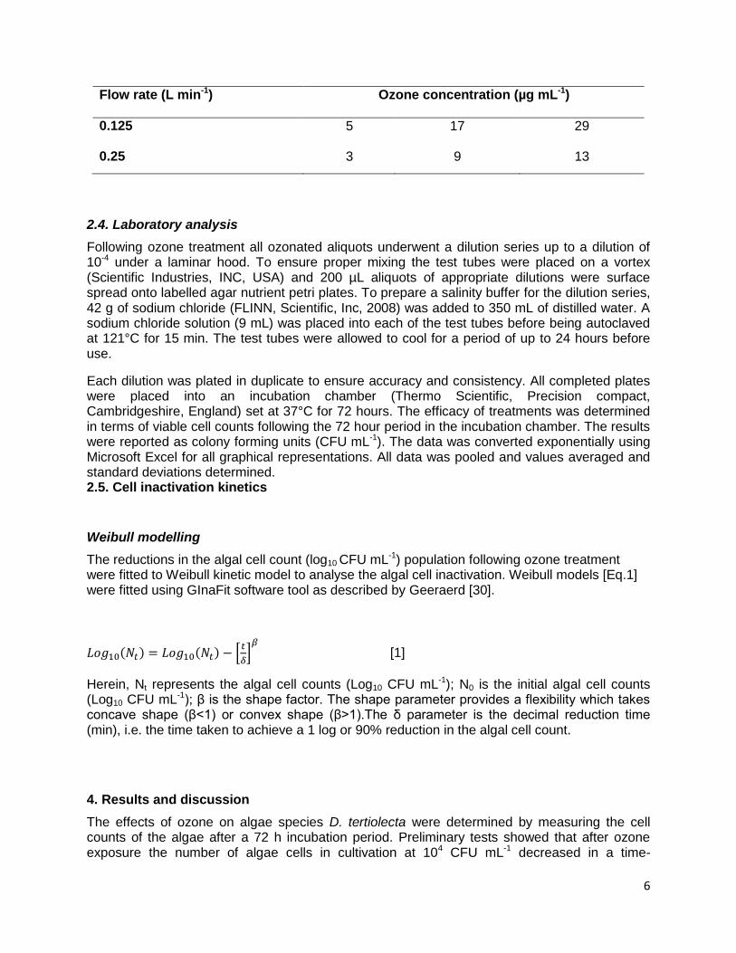
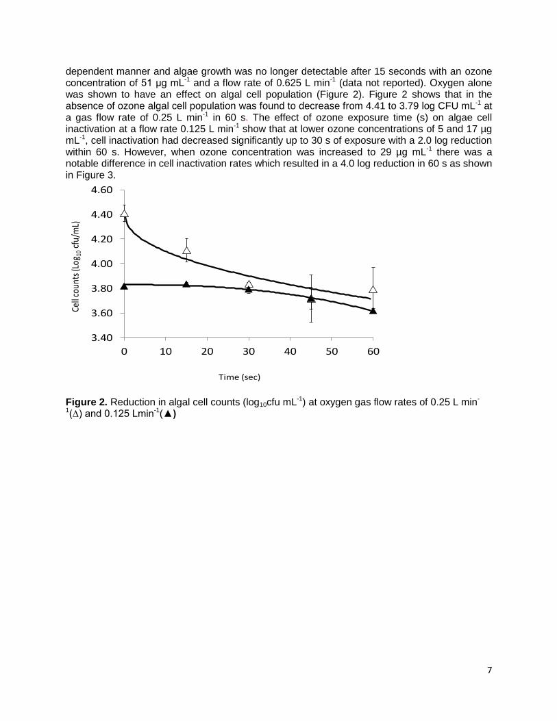
dependent manner and algae growth was no longer detectable after 15 seconds with an ozone concentration of 51 μg mL-1 and a flow rate of 0.625 L min-1 (data not reported). Oxygen alone was shown to have an effect on algal cell population (Figure 2). Figure 2 shows that in the absence of ozone algal cell population was found to decrease from 4.41 to 3.79 log CFU mL-1 at a gas flow rate of 0.25 L min-1 in 60 s. The effect of ozone exposure time (s) on algae cell inactivation at a flow rate 0.125 L min-1 show that at lower ozone concentrations of 5 and 17 µg mL-1, cell inactivation had decreased significantly up to 30 s of exposure with a 2.0 log reduction within 60 s. However, when ozone concentration was increased to 29 µg mL-1 there was a notable difference in cell inactivation rates which resulted in a 4.0 log reduction in 60 s as shown in Figure 3.
Figure 2. Reduction in algal cell counts (log10cfu mL-1) at oxygen gas flow rates of 0.25 L min-
1(∆) and 0.125 Lmin-1(▲)
3.40
3.60
3.80
4.00
4.20
4.40
4.60
0 10 20 30 40 50 60
Time (sec)
Cell
coun
ts (L
og10
cfu/
mL)
8
Figure 3. Reduction in algal cell counts (log10CFU ml-1) at gas flow rate of 0.125 L min-1 and ozone concentrations of 5 μg mL-1 (∆), 17 μg mL-1 (□) and 29 μg mL-1 (○) respectively.
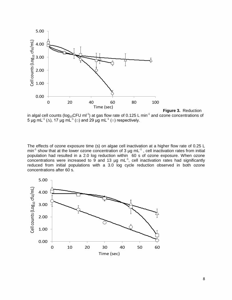
The effects of ozone exposure time (s) on algae cell inactivation at a higher flow rate of 0.25 L min-1 show that at the lower ozone concentration of 3 µg mL-1 , cell inactivation rates from initial population had resulted in a 2.0 log reduction within 60 s of ozone exposure. When ozone concentrations were increased to 9 and 13 µg mL-1, cell inactivation rates had significantly reduced from initial populations with a 3.0 log cycle reduction observed in both ozone concentrations after 60 s.
0.00
1.00
2.00
3.00
4.00
5.00
0 20 40 60 80 100Time (sec)
Cel
l co
un
ts (L
og 1
0cf
u/m
L)
0.00
1.00
2.00
3.00
4.00
5.00
0 10 20 30 40 50 60
Time (sec)
Cell
coun
ts (L
og10
cfu/
mL)
9
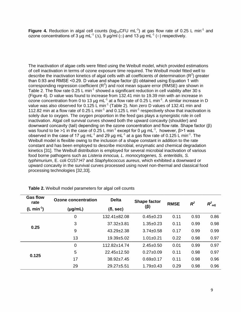
Figure 4. Reduction in algal cell counts (log10CFU mL-1) at gas flow rate of 0.25 L min-1 and ozone concentrations of 3 μg mL-1 (∆), 9 μg/ml (□) and 13 μg mL-1 (○) respectively. The inactivation of algae cells were fitted using the Weibull model, which provided estimations of cell inactivation in terms of ozone exposure time required. The Weibull model fitted well to describe the inactivation kinetics of algal cells with all coefficients of determination (R2) greater than 0.93 and RMSE <0.29. D value and shape factor (β) obtained using Equation 1 with corresponding regression coefficient (R2) and root mean square error (RMSE) are shown in Table 2. The flow rate 0.25 L min-1 showed a significant reduction in cell viability after 30 s (Figure 4). D value was found to increase from 132.41 min to 19.39 min with an increase in ozone concentration from 0 to 13 μg mL-1 at a flow rate of 0.25 L min-1. A similar increase in D value was also observed for 0.125 L min-1 (Table 2). Non zero D values of 132.41 min and 112.82 min at a flow rate of 0.25 L min-1 and 0.125 L min-1 respectively show that inactivation is solely due to oxygen. The oxygen proportion in the feed gas plays a synergistic role in cell inactivation. Algal cell survival curves showed both the upward concavity (shoulder) and downward concavity (tail) depending on the ozone concentration and flow rate. Shape factor (β) was found to be >1 in the case of 0.25 L min-1 except for 0 μg mL-1, however, β>1 was observed in the case of 17 μg mL-1 and 29 μg mL-1 at a gas flow rate of 0.125 L min-1. The Weibull model is flexible owing to the inclusion of a shape constant in addition to the rate constant and has been employed to describe microbial, enzymatic and chemical degradation kinetics [31]. The Weibull distribution is employed for several microbial inactivation of various food borne pathogens such as Listeria innocua, L. monocytogenes, S. enteritidis, S. typhimurium, E. coli O157:H7 and Staphylococcus aureus, which exhibited a downward or upward concavity in the survival curves processed using novel non-thermal and classical food processing technologies [32,33].
Table 2. Weibull model parameters for algal cell counts
Gas flow rate
Ozone concentration Delta Shape factor (β)
RMSE R2 R2adj
(L min-1) (μg/mL) (δ, sec)
0.25
0 132.41±82.08 0.45±0.23 0.11 0.93 0.86
3 37.32±3.81 1.35±0.23 0.11 0.99 0.98
9 43.29±2.38 3.74±0.58 0.17 0.99 0.99
13 19.39±5.02 1.01±0.21 0.22 0.98 0.97
0.125
0 112.82±14.74 2.45±0.50 0.01 0.99 0.97
5 22.45±12.50 0.27±0.09 0.11 0.98 0.97
17 38.92±7.45 0.69±0.17 0.11 0.98 0.96
29 29.27±5.51 1.79±0.43 0.29 0.98 0.96
10
3.1. Effect of ozonation on algae colour
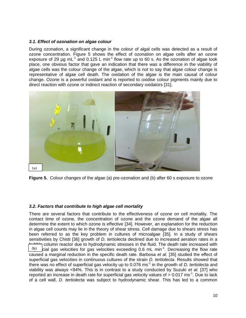
During ozonation, a significant change in the colour of algal cells was detected as a result of ozone concentration. Figure 5 shows the effect of ozonation on algae cells after an ozone exposure of 29 µg mL-1 and 0.125 L min-1 flow rate up to 60 s. As the ozonation of algae took place, one obvious factor that gave an indication that there was a difference in the viability of algae cells was the colour change of the algae, which is not to say that algae colour change is representative of algae cell death. The oxidation of the algae is the main causal of colour change. Ozone is a powerful oxidant and is reported to oxidise colour pigments mainly due to direct reaction with ozone or indirect reaction of secondary oxidators [31].
Figure 5. Colour changes of the algae (a) pre-ozonation and (b) after 60 s exposure to ozone
3.2. Factors that contribute to high algae cell mortality
There are several factors that contribute to the effectiveness of ozone on cell mortality. The contact time of ozone, the concentration of ozone and the ozone demand of the algae all determine the extent to which ozone is effective [34]. However, an explanation for the reduction in algae cell counts may lie in the theory of shear stress. Cell damage due to shears stress has been referred to as the key problem in cultures of microalgae [35]. In a study of shears sensitivities by Chisti [36] growth of D. tertiolecta declined due to increased aeration rates in a bubble column reactor due to hydrodynamic stresses in the fluid. The death rate increased with superficial gas velocities for gas velocities exceeding 0.6 mL min-1. Decreasing the flow rate caused a marginal reduction in the specific death rate. Barbosa et al. [35] studied the effect of superficial gas velocities in continuous cultures of the strain D. tertiolecta. Results showed that there was no effect of superficial gas velocity up to 0.076 ms-1 in the growth of D. tertiolecta and viability was always <94%. This is in contrast to a study conducted by Suzuki et al. [37] who reported an increase in death rate for superficial gas velocity values of > 0.017 ms-1. Due to lack of a cell wall, D. tertiolecta was subject to hydrodynamic shear. This has led to a common
(a)
(b)
11
opinion among researchers within the field of microalgae cultivation that microalgae are shear sensitive.
Other considerations for reduced mean cell counts are the oxygen flow rate and bubble formation. The results show that the optimum flow rate is 0.125 L min-1 at an ozone concentration of 29 µg mL-1. This is because at lower flow rates, ozone concentration increases [38]. Higher oxygen flow rates usually produce larger bubbles whereas lower flow rates produce smaller bubbles [39]. At high flow rates, a small number of large bubbles are produced that rapidly rise to the surface and escape the media at an uncontrollable rate [40]. If an anti-foaming agent is not used at higher oxygen flow rates, most of the liquid volume can be lost through the head of the bubble column, No anti-foam agents were used in this project, foaming of bubbles and loss of liquid volume was experienced at the higher oxygen flow rate of 0.25 L min-1.
Barbosa [35] found that ozone transfer from the bubble to the algae culture happens only on the surface of the bubble where ozone is in direct contact with the solution. Ozone may be trapped inside the bubble in which it does not do anything but creates a strong ozone gas odour. Smaller bubbles are more preferable as the surface to volume ratio is increased as more ozone in the bubbles will be in direct contact with the algae solution. Chisti [36] found that shear stress caused by high gas velocities can cause injury or even cell death to algae populations. Wang et al. [41] concluded that the principal determinants of cell damage were cell-bubble encounter rate which is the rate of bubble break up within the fluid and the bursting rate at the surface. It has also been suggested that column height results in an increase of cell death. Jobses et al. [42] reported that the bubble break up at the liquid surface is the cause for cell damage; the death rate will be proportional to the gas flow rate per unit volume. Camacho et al. [43] found that there was a relationship between fluid height and cell attachment to bubbles. A greater height of rise means that more cells can be captured by the rising bubbles and carried to the surface where cells die as the bubbles rupture.
It may be argued that the algae were already near the 'death phase’ but the premise of the project was to investigate the effects of ozone on algae cell viability after harvesting within a photobioreactor. It was imperative to the study to simulate photobioreactor conditions; therefore ten-day old algae were engaged. However studies on algae found that younger algae are most resistant to ozone [44]. Based on the results of this experiment, ozone treatment of algae was found to be a reliable method to cause cell mortality. As an organism that is relatively susceptible to the effects of ozone, D. tertiolecta is an ideal organism in which to cultivate for biofuel or as a mitigation factor for carbon dioxide.
4. Conclusion
The algae strain Dunaliella tertiolecta was exposed to ozone concentrations 3-29 µg mL-1 and flow rates of 0.125, and 0.25 L min-1 at varying time periods. Effective cell death was evident at 45 seconds at a flow rate of 0.125 L min-1 and ozone concentration of 29 μg mL-1 and for up to 60 seconds at a flow rate of 0.125 L min-1 and ozone concentration of 17 μg mL-1. This research has shown that ozone is a feasible technique in which to cause cell death after a short incubation period of 72 hours only. The effectiveness of ozone is dependent on the contact time, ozone concentration and ozone demand of the algae. However, the algae strain, D. tertiolecta does not contain a rigid cell wall. This aspect renders the algae vulnerable to stress. It is
12
uncertain whether ozone alone was responsible for cell viability as other factors such as shear stress, oxygen flow rate and bubble formation must also be considered. The authors recommend that more research needs to be done to eliminate other parameters. Studies using strains of algae that contain rigid cell walls may facilitate this.
Acknowledgements
The authors would like to thank Damien Farrelly for the cultivation of the algae, Azra Czar for her assistance with the experiments and Rachel Halpin for all her help and expertise in microbiology.
References:
1.Gavand, M. R., McClintock, J. B., Amsler, C. D., Peters, R. W. & Angus, R. A. 2007. Effects of sonication and advanced chemical oxidants on the unicellular green alga Dunaliella tertiolecta and cysts, larvae and adults of the brine shrimp Artemia salina: A prospective treatment to eradicate invasive organisms from ballast water. Marine Pollution Bulletin 54:1777-88.
2. Singh, J. & Gu, S. 2010. Commercialization potential of microalgae for biofuels production. Renewable and Sustainable Energy Reviews 14:2596-610.
3. Rösch, C., Skarka, J. & Wegerer, N. 2012. Materials flow modeling of nutrient recycling in biodiesel production from microalgae. Bioresource Technology 107:191-99.
4. Mata, T. M., Martins, A. A. & Caetano, N. S. 2010. Microalgae for biodiesel production and other applications: A review. Renewable and Sustainable Energy Reviews 14:217-32.
5. Kuchler, M., Linnér, B.-O. 2012. Challenging the food vs. fuel dilemma: Genealogical analysis of the biofuel discourse pursued by international organizations. Food Policy, 37(5), 581-588.
13
6. Singh RN, Sharma S (2012) Development of suitable photobioreactor for algae production - A review. Renewable & Sustainable Energy Reviews 16: 2347-2353. DOI: 10.1016/j.rser.2012.01.026
7. Peralta-Ruiz, Y., González-Delgado, A. D. & Kafarov, V. 2013. Evaluation of alternatives for microalgae oil extraction based on exergy analysis. Applied Energy 101:226-36.
8. Brennan, L. & Owende, P. 2010. Biofuels from microalgae—A review of technologies for production, processing, and extractions of biofuels and co-products. Renewable and Sustainable Energy Reviews, pp. 557-77.
9. Harun, R., Singh, M., Forde, G. M. & Danquah, M. K. 2010. Bioprocess engineering of microalgae to produce a variety of consumer products. Renewable and Sustainable Energy Reviews 14:1037-47.
10. Kumar, K., Dasgupta, C. N., Nayak, B., Lindblad, P. & Das, D. 2011. Development of suitable photobioreactors for CO2 sequestration addressing global warming using green algae and cyanobacteria. Bioresource Technology 102:4945-53.
11. Lakaniemi, A. M., Intihar, V. M., Tuovinen, O. H. & Puhakka, J. A. 2012. Growth of Dunaliella tertiolecta and associated bacteria in photobioreactors. J Ind Microbiol Biotechnol 39:1357-65.
12. Rawat, I., Ranjith Kumar, R., Mutanda, T. & Bux, F. 2013. Biodiesel from microalgae: A critical evaluation from laboratory to large scale production. Applied Energy 103:444-67.
13. Moheimani, N. 2013. Long-term outdoor growth and lipid productivity of Tetraselmis suecica, Dunaliella tertiolecta and Chlorella sp (Chlorophyta) in bag photobioreactors. Journal of Applied Phycology 25:167-76.
14. Qin, S., Lin, H. & Jiang, P. 2012. Advances in genetic engineering of marine algae. Biotechnology Advances 30:1602-13.
15. Tabatabaei, M., Tohidfar, M., Jouzani, G. S., Safarnejad, M. & Pazouki, M. 2011. Biodiesel production from genetically engineered microalgae: Future of bioenergy in Iran. Renewable and Sustainable Energy Reviews 15:1918-27.
16. Nguyen, T., Roddick, F.A., Fan, L. 2012. Biofouling of Water Treatment Membranes: A Review of the Underlying Causes, Monitoring Techniques and Control Measures. Membranes, pp. 804-40.
17. Holtermann, T. & Madlener, R. 2011. Assessment of the technological development and economic potential of photobioreactors. Applied Energy 88:1906-19.
18. Wang, B., Lan, C.Q., Horsman, M. 2012. Closed photobioreactors for production of microalgal biomasses. Biotechnology Advances, 30(4), 904-912.
19. Ramaswamy, R., Vurma, M., Balasubramaniam, V. M. & Yousef, A. E. 2007. OZONE TECHNOLOGY, FACT SHEET FOR FOOD PROCESSORS. Food science and technology College of food, agriculture and environmental sciences. The Ohio State University, Columbas, Ohio.
20. Azarpazhooh, A. & Limeback, H. 2008. The application of ozone in dentistry: A systematic review of literature. Journal of Dentistry 36:104-16.
21. Huang, X. & Wu, J. 2008. Improvement of membrane filterability of the mixed liquor in a membrane bioreactor by ozonation. Journal of Membrane Science 318:210-16.
22. Bullock, G. L., Summerfelt, S. T., Noble, A. C., Weber, A. L., Durant, M. D. & . Hankins, J. A. 1997. Ozonation of a Recirculating Rainbow Trout Culture System. I. Effects on Bacterial Gill Disease and Heterotrophic Bacteria. Aquaculture, pp. 43-55.
23. Summerfelt, S. T. & Hochheimer, J. N. 1997. Review of ozone processes and applications as an oxidising agent in aquaculture. The progressive fish culturist., pp. 94-105.
24. Belle, A. J. 2007. Laboratory evaluation of Dunaliella tertiolecta as a candidate algal species for tertiary wastewater treatment of nitrogen and phosphorus-laden effluents impacting marine environments. Thesis B.S. Dillard University.
14
25. Francavilla, M., Trotta, P. & Luque, R. 2010. Phytosterols from Dunaliella tertiolecta and Dunaliella salina: A potentially novel industrial application. Bioresource Technology 101:4144-50.
26. Demirbas, A. & Fatih Demirbas, M. 2011. Importance of algae oil as a source of biodiesel. Energy Conversion and Management 52:163-70.
27. Lopes, R. J. G. & Quinta-Ferreira, R. M. 2011. Detoxification of high-strength liquid pollutants in an ozone bubble column reactor: Gas–liquid flow patterns, interphase mass transfer and chemical depuration. Chemical Engineering Journal 172:476-86.
28. Guillard, R. & Ryther, J. 1963. Studies on marine planktonic diatoms.I. cyclotella nana Husted and Detonula confervacea cleve. Can J Microbial. pp. 229-39.
29. EPA 2008. Estuarine and coastal waters report. Availiable from http://www.epa.ie/downloads/pubs/other/indicators/irlenv/43366%20epa%20report%20chap%2091.pdf [Last accessed 28th February, 2013].
30. Geeraerd, A. H. V., V. P. Van Impe, J.F. 2005. GInaFiT, a freeware tool to assess non-log-linear microbial survivor curves. 102:95–105.
31. Cullen, P. J., Tiwari, B. K., O'Donnell, C. P. & Muthukumarappan, K. 2009. Modelling approaches to ozone processing of liquid foods. Trends in Food Science & Technology 20:125-36.
32. Bialka, K. L., Demirci, A. & Puri, V. M. 2008. Modeling the inactivation of Escherichia coli O157:H7 and Salmonella enterica on raspberries and strawberries resulting from exposure to ozone or pulsed UV-light. Journal of Food Engineering 85:444-49.
33. Buzrul, S. & Alpas, H. 2007. Modeling inactivation kinetics of food borne pathogens at a constant temperature. LWT - Food Science and Technology 40:632-37.
34. Kureshy, N., Davis, D. A. & Arnold, C. K. 1999. Effect of ozone treatment on cultures of Nannochloropis occulata, Isochrysis galbana and Chaetoceros gracilis. Journal of the world aquaculture society, pp. 473-80.
35. Barbosa, M. J. G. V. 2003. Microalgae photobioreactors: scale up and optimisation. PhD, Wageningen University, The Netherlands.
36. Chisti, Y. & Zealand, M. U. S. o. E. P. N. N. 2009. Shear Sensitivity. John Wiley & Sons, Inc.,
37. Suzuki T, Matsuo T, Ohtaguchi K, Koide K. 1995. Gas-sparged bioreactors for CO2 fixation by Dunaliella tertiolecta. J Chem Technol Biotechnol. 62:351–358.
38. Tizaoui, C. & Zhang, Y. 2010. The modelling of ozone mass transfer in static mixers using Back Flow Cell Model. Chemical Engineering Journal 162:557-64.
39. Kukuzaki, M., Fujimoto, K., Kai, S., Ohe, K., Oshima, T. & Baba, Y. 2010. Ozone mass transfer in an ozone–water contacting process with Shirasu porous glass (SPG) membranes—A comparative study of hydrophilic and hydrophobic membranes. Separation and Purification Technology 72:347-56.
40. Vijayan, T. & Patil, J. G. 2010. High concentration ozone generation in the laboratoryfor various applications. International Journal of Science and Technology Education Research. Academic Journals, pp. 132-42.
41. Wang, N. Y. J.-D. Y., Richard V. Calabrese, Kuo-Ching Chang 1994. Unified modeling framework of cell death due to bubbles in agitated and sparged bioreactors. Journal of Biotechnology 33:107–22.
42. Jobses, I., Marker, D. & Ramper, J. A. 1991. Lethal events during gas sparging in animal cell culture. Biotechnol Bioengineering. pp. 484-90.
43. Camacho, F. G., Grima, E. M., Miron, A. S., Pascual, V. G. & Chisti, M. Y. 2001.
Carboxymethyl cellulose protects algal cells against hydrodynamic stress. Enzyme Microb. pp. 602-10.

15
44. Heath, R. L. 1984. Decline in energy reserves of Chlorella Sorokiniana upon exposure to ozone. Plant physiology, pp. 700-04.