Highly valuable microalgae: biochemical and topological aspects
16
REVIEW Highly valuable microalgae: biochemical and topological aspects Olivier Pignolet • Se ´bastien Jubeau • Carlos Vaca-Garcia • Philippe Michaud Received: 23 February 2013 / Accepted: 25 April 2013 / Published online: 10 May 2013 Ó Society for Industrial Microbiology and Biotechnology 2013 Abstract The past decade has seen a surge in the interest in microalgae culture for biodiesel production and other applications as renewable biofuels as an alternative to petroleum transport fuels. The development of new tech- nologies for the culture of these photosynthetic microor- ganisms and improved knowledge of their biochemical composition has spurred innovation in the field of high- value biomolecules. These developments are only eco- nomically viable if all the microalgae fractions are valo- rized in a biorefinery strategy. Achieving this objective requires an understanding of microalgae content and the cellular localization of the main biomolecular families in order to develop efficient harvest and sequential recovery technologies. This review summarizes the state of the art in microalgae compositions and topologies using some examples of the main industrially farmed microalgae. Keywords Biorefinery Cyanobacteria Eukaryotic microalgae Biofuel Lipid Polysaccharide Pigment Protein Introduction Microalgae are unicellular and multicellular photosynthetic microorganisms from freshwater and marine systems that offer extremely interesting industrial potential. Definitions of microalgae include prokaryotic microalgae, i.e., cyano- bacteria (Chloroxybacteria), and eukaryotic microalgae, i.e., diatoms (Bacillariophyta), green algae (Chlorophyta) and red algae (Rhodophyta). Eukaryotic microalgae are recognized as primitive plants and can be either autotrophic or heterotrophic. Autotrophic microalgae require only inorganic compounds such as CO 2 , N, S, P, and light as an energy source for their growth and development. They convert captured solar energy into biomass (photosynthesis) with efficiencies that generally exceed those of terrestrial plants (3 % reported for marine microalgae against 0.2–2 % for terrestrial plants) [108]. Some photosynthetic microal- gae are mixotrophic, meaning they are able to perform photosynthesis and to catabolize exogenous organic nutri- ents. For autotrophic microalgae, photosynthesis is essential to convert solar radiation and CO 2 into reducing power, adenosine triphosphate (ATP), O 2 , and 3-phosphoglycerate, which are then used to support growth. With these simple growth requirements, microalgae can sustainably generate lipids, proteins, and carbohydrates at a large scale, offering promising environmentally friendly alternatives to the current consumer products. The most prominent examples are biofuels, human diet and animal feed supplements, and applications in the pharmaceutics, nutraceutics, and cos- metics industries. Some macroalgal compounds also show interesting functional properties (free radical scavengers) and could provide new treatments against diseases such as cancer or even AIDS [97]. However, these high-value compounds (primary products) are generally extracted individually through expensive processes that waste most of O. Pignolet C. Vaca-Garcia INP-ENSIACET, LCA (Laboratoire de Chimie Agro- Industrielle), INRA, UMR 1010 CAI, Universite ´ de Toulouse, 31030 Toulouse, France S. Jubeau Laboratoire GEPEA, CRTT, UMR CNRS 6144, Universite ´ de Nantes, 44602 Saint-Nazaire, France P. Michaud (&) Institut Pascal UMR CNRS 6602, Polytech’ Clermont Ferrand, Clermont Universite ´, Universite ´ Blaise Pascal, 24 Avenue des Landais, BP-20206, 63174 Aubie `re, France e-mail: [email protected] 123 J Ind Microbiol Biotechnol (2013) 40:781–796 DOI 10.1007/s10295-013-1281-7
-
Upload
independent -
Category
Documents
-
view
2 -
download
0
Transcript of Highly valuable microalgae: biochemical and topological aspects

REVIEW
Highly valuable microalgae: biochemical and topological aspects
Olivier Pignolet • Sebastien Jubeau •
Carlos Vaca-Garcia • Philippe Michaud
Received: 23 February 2013 / Accepted: 25 April 2013 / Published online: 10 May 2013
� Society for Industrial Microbiology and Biotechnology 2013
Abstract The past decade has seen a surge in the interest
in microalgae culture for biodiesel production and other
applications as renewable biofuels as an alternative to
petroleum transport fuels. The development of new tech-
nologies for the culture of these photosynthetic microor-
ganisms and improved knowledge of their biochemical
composition has spurred innovation in the field of high-
value biomolecules. These developments are only eco-
nomically viable if all the microalgae fractions are valo-
rized in a biorefinery strategy. Achieving this objective
requires an understanding of microalgae content and the
cellular localization of the main biomolecular families in
order to develop efficient harvest and sequential recovery
technologies. This review summarizes the state of the art in
microalgae compositions and topologies using some
examples of the main industrially farmed microalgae.
Keywords Biorefinery � Cyanobacteria � Eukaryotic
microalgae � Biofuel � Lipid � Polysaccharide � Pigment �Protein
Introduction
Microalgae are unicellular and multicellular photosynthetic
microorganisms from freshwater and marine systems that
offer extremely interesting industrial potential. Definitions
of microalgae include prokaryotic microalgae, i.e., cyano-
bacteria (Chloroxybacteria), and eukaryotic microalgae,
i.e., diatoms (Bacillariophyta), green algae (Chlorophyta)
and red algae (Rhodophyta). Eukaryotic microalgae are
recognized as primitive plants and can be either autotrophic
or heterotrophic. Autotrophic microalgae require only
inorganic compounds such as CO2, N, S, P, and light as an
energy source for their growth and development. They
convert captured solar energy into biomass (photosynthesis)
with efficiencies that generally exceed those of terrestrial
plants (3 % reported for marine microalgae against 0.2–2 %
for terrestrial plants) [108]. Some photosynthetic microal-
gae are mixotrophic, meaning they are able to perform
photosynthesis and to catabolize exogenous organic nutri-
ents. For autotrophic microalgae, photosynthesis is essential
to convert solar radiation and CO2 into reducing power,
adenosine triphosphate (ATP), O2, and 3-phosphoglycerate,
which are then used to support growth. With these simple
growth requirements, microalgae can sustainably generate
lipids, proteins, and carbohydrates at a large scale, offering
promising environmentally friendly alternatives to the
current consumer products. The most prominent examples
are biofuels, human diet and animal feed supplements, and
applications in the pharmaceutics, nutraceutics, and cos-
metics industries. Some macroalgal compounds also show
interesting functional properties (free radical scavengers)
and could provide new treatments against diseases such as
cancer or even AIDS [97]. However, these high-value
compounds (primary products) are generally extracted
individually through expensive processes that waste most of
O. Pignolet � C. Vaca-Garcia
INP-ENSIACET, LCA (Laboratoire de Chimie Agro-
Industrielle), INRA, UMR 1010 CAI, Universite de Toulouse,
31030 Toulouse, France
S. Jubeau
Laboratoire GEPEA, CRTT, UMR CNRS 6144, Universite de
Nantes, 44602 Saint-Nazaire, France
P. Michaud (&)
Institut Pascal UMR CNRS 6602, Polytech’ Clermont Ferrand,
Clermont Universite, Universite Blaise Pascal, 24 Avenue des
Landais, BP-20206, 63174 Aubiere, France
e-mail: [email protected]
123
J Ind Microbiol Biotechnol (2013) 40:781–796
DOI 10.1007/s10295-013-1281-7

the microalgal material (co-products). The microalgae
industry’s future hinges on enabling first-generation biore-
fineries to valorize the main primary products from a bio-
mass and not only one of them. In a schematic way, first-
generation biorefineries work to a three-step concept high-
lighted in a schematic flowsheet shown in Fig. 1. These
steps are disintegration of cells, fractionation of main
component families, and purification of valuable biomole-
cules. Nevertheless, microalgal biorefineries are hard to set
up due to the broad chemical and morphological diversity of
microalgae and the high-value fractions that need to be
obtained for specific applications. Indeed, each unit opera-
tion has to preserve the specific properties of the obtained
fractions and the stability of the target compounds in the
remaining materials. Given that the main molecular content
groups are often associated with specific intracellular
compartments, unit operations could be performed on a
deconstructed biomass in a conceptual biorefinery (Fig. 2).
These main families are lipids, polysaccharides, proteins,
and pigments. Even though pigments are biochemically
varied, they can be considered a single biochemical family
for the purposes of a valorization strategy. All these
compounds are qualitatively and quantitatively dependent
on microalgal species used. However, some species have
risen to prominence in the industry and are dedicated to
specific applications due to their select biochemical com-
position. The aim of this review is to describe the bio-
chemical composition and topology of selected microalgae
and to highlight the compounds to be valorized in a first-
generation biorefinery strategy.
Lipids
Biological lipids are chemically diverse groups that nev-
ertheless share the common defining feature of being
insoluble in water. Their biological functions, like their
chemistry, are equally diverse. Oil-rich algae (called ole-
aginous species) can be grown either autotrophically and
heterotrophically. In a biorefinery concept, this class of
compounds, and more especially reserve lipids, are at the
origin of biofuel production. Microalgae for biofuel pro-
duction can be grown in several places all year round with
significant advantages such as good oil productivity
exceeding the yields of the best oilseed crops, non-use of
arable land, low water consumption compared to crop
irrigation, and low environmental impact [78]. Microalgae
growing under normal conditions actually produce signif-
icant amounts of lipids, but this lipid accumulation
decreases when cultivation conditions are optimized to
increase biomass yield [76, 109, 124]. Microalgae contain
primarily polar lipids such as phospholipids and glycolipids
together with neutral storage lipids such as monoglycerides
(MAGs), diglycerides (DAGs), and triglycerides (TAGs),
free fatty acids, hydrocarbons, and pigments [57]. Polar
lipids are confined to cell organelle membranes such as the
thylakoid membranes of the chloroplast [57]. TAGs are
neutral lipids mainly stored in vacuoles within the cell [21,
57]. Most microalgae accumulate very few TAGs during
the exponential growth phase but can produce and store
substantial amounts of TAGs during the stationary phase or
under adverse environmental conditions [13, 57, 114].
Fatty acid (FA) composition may differ between lipid
classes. As a rule, saturated fatty acids (SAFA) and
monounsaturated fatty acids (MUFA) comprise the storage
lipid fraction while polyunsaturated fatty acids (PUFA)
largely comprise the structural lipid fraction [57]. How-
ever, culture processes and species selection have a major
influence on lipid composition, accumulation, and distri-
bution. For many microalgae, growth stressors such as
excessive solar energy captured by photosynthesis, nitro-
gen depletion, salinity, or high temperature lead to pri-
marily lipid storage, with high TAGs levels [26, 31, 62,
108, 114] or to a shift in FA composition [38]. Some
microalgae are well-known lipid producers extensively
Photobioreactor
Harvesting
Biomass
Cell disruption
Fractionation
Purification
High-value compounds
Fractions for food, feed and
energy
Residue
Residue
Fig. 1 A microalgae biorefinery
782 J Ind Microbiol Biotechnol (2013) 40:781–796
123

a
b
c
Fig. 2 Ultrastructure of
microalgae: \Emphasis Type=
Chlorophyceae, b Rodophyceae,
and c cyanobacteria (based on
Ref. [70])
J Ind Microbiol Biotechnol (2013) 40:781–796 783
123

studied for biofuel and more especially for biodiesel pro-
duction. Biodiesel is defined as monoalkyl esters of vege-
table oils or animal fats. It is produced by the
transesterification of TAGs with methanol (or other alco-
hols) in the presence of an appropriate catalyst, with
glycerol as by-product. Biodiesel standards around the
world, including the USA (ASTM D 6751) and Europe
(EN 14214), also address storage and handling issues—
linoleic and linolenic acid esters, for example, are prone to
oxidation and so need to be chemically reduced by a
hydrogenation process [63]. Hydrogenation converts dou-
ble bonds to single bonds by H addition. Other microalgae-
producing lipids are exploited for food and feed applica-
tions, notably due to their unsaturated FA or pigment
contents. A majority of the high-value lipid compounds
extracted from these algae are long-chain PUFA and
carotenoid pigments.
Although all microalgae are candidate lipid-producers,
the ‘‘winners’’ for industry applications are the eukaryotic
microalgae Chlorella vulgaris (up to 58 % dw) [2], Nan-
nachloropsis oculata (up to 69 % dw) [19], Botryococcus
braunii (up to 75 % dw) [27], and Scenedesmus obliquus
(up to 50 % dw) [78]. Nonetheless, another eukaryotic
microalgae such as Porphyridium cruentum may have
futures for lipid-based industrial applications as they have
original and/or highly specific lipid profiles, rich in long-
chain PUFA. Indeed, Porphyridium cruentum is rich in
high-value FA such as arachidonic acid (20:4x6) and ei-
cosapentaenoic acid (20:5x3) [32] for a global lipid con-
tent of between 9 and 19 % [78].
Chlorella vulgaris
Lipid accumulation as chloroplastic and cytoplasmic
droplets by the green microalga C. vulgaris in freshwater
(Fig. 2) requires a pre-extraction cell lysis step. Growth on
nitrogen-limiting media resulted in higher lipid accumu-
lation (20–53 %) but lower growth rate, whereas mixo-
trophic cultivation yielded higher lipid content (40–53 %)
and higher lipid productivity (67–144 mg/l/day) in all
culture media [124]. The FA composition of microalgal
lipids comprises over 60–68 % SAFA [124]. More spe-
cifically, the FA most accumulated are described as C18,
C18:1, C18:2, and C18:3 (3.4, 16.3, and 79.4 % of total FA
[68]. Choi et al. [30] compared the lipid production of
mixotrophic and autotrophic cultures of Chlorella sp.
Cultures were carried out in photobioreactors using various
light periods and various CO2 supplementations [30].
Methanol (1 % v/v) was used as sole organic carbon source
in the mixotrophic conditions. The highest lipid accumu-
lation (18.6 % dw) with high proportions of C16:0, C18:0,
and C18:1 was obtained with 5 % CO2 and 450 lE/m2/s of
12:12 (h) irradiation cycles in mixotrophic mode. It is
interesting to note that in this study, mixotrophy gave
better results than autotrophy. Previous data [30, 68, 124]
suggest the lipid composition of C. vulgaris is suitable for
biodiesel production. C. vulgaris was also grown in a
column aeration photobioreactor filled with artificial
wastewater in semicontinuous mode [41]. The highest lipid
production and content were 147 mg/l/day and 42 % dw,
respectively, in the optimal growth conditions, and the
system was characterized as highly efficiency at removing
nutrients from wastewater [41].
Nannochloropsis oculata
Nannochloropsis oculata is well known in marine bio-
technology for its potential interest in biodiesel production.
N. oculata NCTU-3 was grown in a semicontinuous system
aerated with 2 % CO2 and operated by one-day replace-
ment with the objective of optimizing culture for long-term
biomass and lipid productions [28]. A two-stage cultivation
strategy has also been proposed to enhance the lipid pro-
duction of Nannochloropsis oculata [109]. Biomass growth
and lipid production were experimented in two separate
stages. Final lipid yield was 2.82-fold higher in these
conditions than in traditional single-stage batch cultivation
systems [109]. Microalgae lipid content was strongly
influenced by the physical–chemical environment and
composition of the culture media. For example, a temper-
ature increase from 20 to 25 �C doubled the lipid content of
N. oculata (from 7.90 to 14.92 % dw), while a 75 %
decrease in nitrogen concentration in the medium increased
N. oculata lipid content from 7.90 to 15.31 % dw without
impact on growth [33]. The authors identified high amounts
of palmitic acid (16:0), which constituted about 60 % (mol/
mol) of the overall lipid fraction, along with C18:1, C18:2,
and C18:3. This result confirmed the finding that storage
lipids in Nannochloropsis sp. consist largely of the SAFA
palmitic acid (C16:0) and the MUFA palmitoleic acid
(C16:1) [110]. However, Tonon et al. [114] found that
68 % of TAGs in Nannochloropsis oculata consisted of the
PUFA eicosapentaenoic acid (20:5 x3, EPA) at the end of
stationary phase compared to just 8 % in the exponential
phase. Similarly, under nitrogen deprivation, the average
biomass and lipid productivities of Nannochloropsis sp.
reached 9.9 and 6.5 g/m2/day, respectively. Lipid content
was 68.5 % in starved biomass compared to 39.1 % in non-
starved biomass, and the two cultivation conditions led to
significant differences in lipid classes and FA composition
[19]. Effectively, under nitrogen deprivation, polar lipid
content decreased whereas neutral lipid content increased.
The major lipids represented under nitrogen starvation
were TAGs, polar lipids, and hydrocarbons (79, 9, and
2.5 % of total lipids, respectively) [19]. The FA composi-
tion of the different lipid classes (neutral, polar, and
784 J Ind Microbiol Biotechnol (2013) 40:781–796
123

phospholipids) includes mainly C16:0, C16:1, and C18:1.
Note that polar lipids and phospholipids have a significant
ratio of C20:5x3 (eicosapentaenoic acid) in their structure.
Lipid content, FA profiles, MAGs, DAGs, and TAGs were
analyzed for 2 years in Nannochloropsis oculata biomasses
grown outdoors in closed vertical flat-panel photobioreac-
tors [88]. Results showed that the highest lipid content was
recorded during autumn, signaling an optimal non-linear
response to light and temperature. The authors hypothe-
sized that enhanced thylakoid stacking under reduced light
conditions resulted in more structural lipids concomitantly
with the increase in glycerides due to released photo-oxi-
dative stress. The relative increase in MUFA during
autumn suggested a synthesis either of structural FA as
MUFA or a relative increase of C16:1 incorporated into
TAGs and DAGs.
Botryococcus braunii
B. braunii is a colonial hydrocarbon-rich green microalga
[6] widespread in freshwater and brackish lakes at tem-
perate, tropical, and Arctic latitudes. B. braunii has tre-
mendous potential as a renewable biomass feedstock and
high potential for the production of renewable liquid
hydrocarbons. The lipids of B. braunii can be repeatedly
extracted from wet biomass without the usual harvesting
and dewatering steps [18]. By exposing B. braunii to bio-
compatible organic solvent, a substantial fraction of
hydrocarbons was obtained without impairing cell viability
[42], as the majority of biosynthesized lipids in B. braunii
are located in the outer cell wall of the algal material [18].
Note that a high percentage of hydrocarbons in some
petroleum fuels were probably derived exclusively from
this specie [122]. Depending on the type of hydrocarbons
synthesized, B. braunii is classified into A, B, and L strains
but some strains of this species are not classified [61]. For
example, strain B. braunii AP103 grown on open raceway
pond produced a biomass concentration of 1.8 ± 0.13 g/l.
Its biochemical analysis revealed 19 % lipids, 33 % car-
bohydrates, 18 % proteins, and 11 % hydrocarbons [4].
The hydrocarbon profile showed the presence of hepta-
decane (34 %) and hexadecane (12.5 %). The major FA
detected in the lipids extracted were oleic (25.7 %), lino-
lenic (34.26 %), and palmitic (9.42 %) acid [4]. B. braunii
race A is a ubiquitous colonial green microalga that is an
important synthesizer of lipids in fresh and brackish water
ecosystems [82]. Race A produces C23–C33 odd-num-
bered n-alkadienes, mono-, tri-, tetra-, and pentanes from
FA. Linear olefins can account for up to 61 % of the dry
matter of green active-state colonies. Note that the growth
of B. braunii (race A) and its production of hydrocarbons
and FA were influenced by environmental salinity [95].
The L race produces a single tetraterpene hydrocarbon
called lycopadiene (C40H78) and it accounts for up to
2–8 % of dry biomass. The B race produces polyunsatu-
rated and branched C30–C37 terpenoid hydrocarbons
called polymethylated botryococcenes that are promising
renewable energy sources as they accumulate at very high
levels of 26–86 % dry weight in the algae [82]. Photo-
synthesis, growth, and metabolic productivity of B. braunii
BOT-22 cells (race B) were saturated at approximately
1,000, 100, and 200 lmol/m2/s, respectively, under
monochromatic red light. Lipid and hydrocarbon produc-
tion was not directly supported by an increase in photo-
synthetic activity [102].
Scenedesmus obliquus
The green microalgae Scenedesmus obliquus is an oleagi-
nous alga well known for its ability to accumulate lipids,
notably in fresh wastewater [77]. The lipids of S. obliquus
grown under controlled conditions have been characterized
as 7.24 % neutral lipid, 2.45 % glycolipid and 1.48 %
phospholipid on a dry weight basis. The major neutral
lipids were di- and triacylglycerol, free and esterified
sterols, and hydrocarbons. The glycolipids were monoga-
lactosyl diglyceride, digalactosyl diglyceride, free and
esterified sterol glycoside. The phospholipids included
phosphatidyl ethanolamine, phosphatidyl glycerol and
phosphatidyl choline. The 14 FA identified in the lipid
fractions were led by C18:2, C16:0, C18:3(alpha), C18:1,
C16:3, C16:1, and C16:4. Unsaturated and essential FA
were quantified at 80 and 38 % of total algal lipids,
respectively [29]. The C16/C18 FA groups of S. obliquus
lipids obtained from cultivation under nitrogen-starved
conditions could reach up to 92.4 % of total FA [56]. Oleic
acid, an important indicator of biodiesel quality, accounted
for 35.10 % of total FA. The main FA of S. obliquus cul-
tivated in nutrient-rich media with 10 % CO2 were C16:0
(15–16 %), C16:1 (2–3 %), C18:0 (17–18 %), C18:1
(15–16 %), C18:2 (13–14 %), C18:3 (3–4 %), and several
minor component FA with a carbon number lower than
C16 and higher than C18 [56, 76]. These data were con-
firmed by [119]. Note also that under P-deficiency and
thiosulphate supplementation, the lipid content of S. obli-
quus increased up to 30 % [76].
Polysaccharides
Polysaccharides are defined as polymers of monosaccha-
rides linked by glycosidic bonds. As for other organisms,
microalgae polysaccharides demonstrate strong structural
variability and biological functions [97]. Based on their
role in the microalgae physiology, they can be split in three
classes: structural polysaccharides generally associated to
J Ind Microbiol Biotechnol (2013) 40:781–796 785
123

cell walls, energy polysaccharides like starch, and poly-
saccharides implicated in cellular communication and
recognition sites. Structural investigations into polysac-
charides are complicated by the very broad diversity of
their constitutive monosaccharides (pentoses and hexoses),
non-sugar substituents (e.g., sulfate, acetate, or pyruvate)
and glycosidic linkages. If we exclude starch, exopoly-
saccharides (also known as exopolymeric substances or
exopolymers when associated to proteins) have gained
significant attention in the published literature. This pop-
ularity is explained by their extractability, as these bio-
polymers are produced by microalgae in the extracellular
medium without covalent linkages with cell walls. In some
cases, it is difficult to distinguish these exopolysaccharides
from the cell wall in which they are embedded (e.g., exo-
polysaccharides of Porphyridium sp.) [3]. Their physio-
logical role is unclear, but some authors have hypothesized
that they prevent desiccation [94] or offer a mechanical
protection enabling the cells to develop in a range of
environments [3]. Microalgal production of polysaccharide
and especially exopolysaccharide (EPS) could be also one
way to control photosynthetic activity. Algae colony for-
mation is linked to polysaccharide production [113], which
is catalyzed by various factors including C/N ratio. Reports
suggest glyoxylate, a stimulator of carbon metabolism, is
able to inhibit photorespiration and increase photosynthesis
in higher plants [87] and some cyanobacteria [14]. Adding
glyoxylate to algae like Anabaena cylindrica [15] and
Cyanospira capsulate [91] triggers an excess of carbon flux
resulting in intracellular polysaccharide accumulation and
soluble extracellular polysaccharide release. A culture
study by Liu et al. [73] found that Scenedesmus obliquus
supplemented with glyoxylate increased exopolysaccharide
production.
In recent years, there has been of surge of interest in
polysaccharide-producing bacteria, cyanobacteria, and
microalgae for industrial applications. Effectively, EPSs
are regarded as an abundant source of structurally diverse
polysaccharides and some may possess unique properties
for special applications. Excluding the EPSs from Por-
phyridium sp. [46] or Arthrospira platensis [116], which
have been partially characterized, and starch (known as
floridean starch) located in cytoplasm and chloroplasts,
structural characterization data on microalga-produced
polysaccharides remains scarce, even on cell-wall poly-
saccharides. Vilchez et al. [120] tried to document a global
composition of microalgae and described mono- and oli-
gosaccharides as mainly represented by trehalose, glucose,
sucrose, and mannose but also polyols. Polysaccharides
were more or less ignored, and the authors only gave a
global composition for monosaccharides (glucose, galact-
ose, xylose, methylxylose, and glucuronic acid).
Chlorella vulgaris
The most important substance in C. vulgaris for human
health is a cell-wall b-(1,3)-glucan that has potential as an
antitumor agent [85]. This class of polysaccharide is well
known as an active immunostimulator, free radical scav-
enger, and reducer of blood lipids [66]. To our knowledge,
the exopolysaccharides or other cell wall polymers from
this microalgae have not been described. The only excep-
tion is an acidic polysaccharide containing high contents of
glucuronic acid and rhamnose identified as a cell wall
component [86].
Arthrospira platensis
Arthrospira platensis, (Spirulina) is a Gram-negative cya-
nobacteria species. A. platensis grows in tropical and
subtropical waters with pH between 8 and 11 and high
carbonate and bicarbonate concentrations. The EPS of A.
platensis is generally considered as a polyanionic sulfated
heteropolymer containing proteins, carbohydrates, and
sulfated groups. Contents were estimated at 55, 13, and
0.5 %, respectively in A. platensis Compere 1968/3786
strain grown in photoautotrophic-mode for 25 days. Car-
bohydrates are mainly composed of seven neutral sugars
[galactose (14.9 %), xylose (14.3 %), glucose (13.2 %),
fructose (13.2 %), rhamnose (3.7 %), arabinose (1 %),
mannose (0.3 %)], and two uronic acids [galacturonic and
glucuronic acids; 13.5 % together] [116]. Trabelsi et al.
[115] evaluated EPS production in relation to culture
mode: high EPS levels were obtained in culture medium
using mixotrophy with a short cultivation period whereas
photoautotropohy and heterotrophy cultures produced low
EPS levels at longer cultivation periods (290; 219 and
30 mg/l, respectively; 1.5 g/l glucose and/or 100 lmol
photon m-2s-1). Optimizing light and glucose concentra-
tions led to a peak production level of 369 mg EPS/l in
mixotrophic mode.
EPS from A. platensis exhibits interesting functional
properties. It has value to industry due a non-Newtonian
pseudoplastic behavior, but the main applications remain
pharmaceutical in nature due to its wide range of biological
properties. The sulfated EPS from A. platensis acts as
anticoagulant, anti-thrombogenic and anti-atherogenic
[97]. In addition, calcium and sodium spirulans isolated
from hot water-extracted EPS interfere with the adsorption
and penetration of viruses like herpes or HIV [55]. Chal-
louf et al. [25] ran cytotoxicity and antioxidant assays on
solvent-isolated EPS and concluded that its biological
activity is closely tied to the molecular weight and con-
formation of EPS in terms of solvent–polymer interactions
and the presence of sulfated moieties.
786 J Ind Microbiol Biotechnol (2013) 40:781–796
123
Botryococcus braunii
Race-A and race-B strains produce EPS at up to 250 g/m3
whereas L-race strains produce EPS at up to 1 kg/m3 [6].
However, EPS production level varies with strain and
culture conditions such as culture media, illumination,
agitation, and above all salinity [35, 36, 95]. B. braunii
appears to produce carbohydrates as an osmoprotectant.
The authors failed to describe the structures of these exo-
polymers and only quantified them after ethanol precipi-
tation of culture medium supernatant and/or phenol–
sulfuric assay [35, 36, 95].
Dunaliella salina
Dunaliella salina is a type of halophile pink microalgae
especially found in sea salt fields. EPS production by this
strain increases with salt concentration in the culture
medium, and peak production (944 mg/l) was obtained at
5 M NaCl [83]. Isolated EPS showing emulsifying activity
slightly reduces with salinity. 1H NMR, FT-IR, and HPLC
have given structural clues on the presence of uronic acids,
halides, sulfides, alkyl amine, and/or cyclic amine with
polysaccharides. Galactose, glucose, xylose, and fructose
were detected as constitutive monosaccharides [83]. Fine-
grained monosaccharide composition analysis in five
fractions of a polysaccharidic crude extract fractionated by
DEAE chromatography using reverse-phase HPLC showed
that PD1 and PD4a fractions were acidic heteropolysac-
charides mainly containing glucose and galactose, respec-
tively, and that PD4a contained sulfated groups. PD2 and
PD3 were identified as a glucan. PD4b was a complex of
polysaccharide covalently linked with nucleic acids.
Compositional analysis of PD4a confirmed the deduction
made by Fabregas et al. [39] that water extracts with
antivirus activity from D. salina contained sulfated poly-
saccharides with a molecular weight of a hundred-thousand
Da [34]. These EPS proved thermostable up to 270 �C and
their dynamic viscosity was significantly high at pH 3.0 but
decreased with shear rate, confirming pseudoplastic rheo-
logical properties [83].
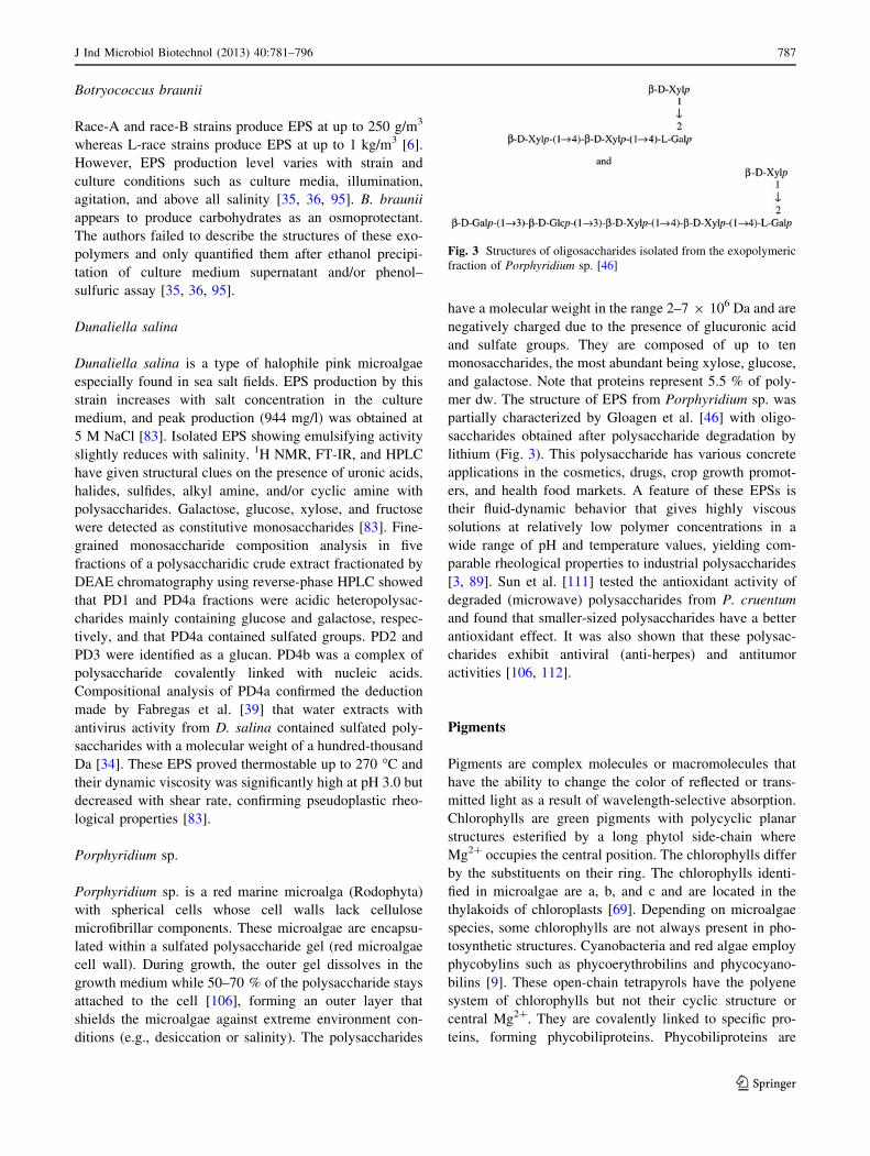
Porphyridium sp.
Porphyridium sp. is a red marine microalga (Rodophyta)
with spherical cells whose cell walls lack cellulose
microfibrillar components. These microalgae are encapsu-
lated within a sulfated polysaccharide gel (red microalgae
cell wall). During growth, the outer gel dissolves in the
growth medium while 50–70 % of the polysaccharide stays
attached to the cell [106], forming an outer layer that
shields the microalgae against extreme environment con-
ditions (e.g., desiccation or salinity). The polysaccharides
have a molecular weight in the range 2–7 9 106 Da and are
negatively charged due to the presence of glucuronic acid
and sulfate groups. They are composed of up to ten
monosaccharides, the most abundant being xylose, glucose,
and galactose. Note that proteins represent 5.5 % of poly-
mer dw. The structure of EPS from Porphyridium sp. was
partially characterized by Gloagen et al. [46] with oligo-
saccharides obtained after polysaccharide degradation by
lithium (Fig. 3). This polysaccharide has various concrete
applications in the cosmetics, drugs, crop growth promot-
ers, and health food markets. A feature of these EPSs is
their fluid-dynamic behavior that gives highly viscous
solutions at relatively low polymer concentrations in a
wide range of pH and temperature values, yielding com-
parable rheological properties to industrial polysaccharides
[3, 89]. Sun et al. [111] tested the antioxidant activity of
degraded (microwave) polysaccharides from P. cruentum
and found that smaller-sized polysaccharides have a better
antioxidant effect. It was also shown that these polysac-
charides exhibit antiviral (anti-herpes) and antitumor
activities [106, 112].
Pigments
Pigments are complex molecules or macromolecules that
have the ability to change the color of reflected or trans-
mitted light as a result of wavelength-selective absorption.
Chlorophylls are green pigments with polycyclic planar
structures esterified by a long phytol side-chain where
Mg2? occupies the central position. The chlorophylls differ
by the substituents on their ring. The chlorophylls identi-
fied in microalgae are a, b, and c and are located in the
thylakoids of chloroplasts [69]. Depending on microalgae
species, some chlorophylls are not always present in pho-
tosynthetic structures. Cyanobacteria and red algae employ
phycobylins such as phycoerythrobilins and phycocyano-
bilins [9]. These open-chain tetrapyrols have the polyene
system of chlorophylls but not their cyclic structure or
central Mg2?. They are covalently linked to specific pro-
teins, forming phycobiliproteins. Phycobiliproteins are
Fig. 3 Structures of oligosaccharides isolated from the exopolymeric
fraction of Porphyridium sp. [46]
J Ind Microbiol Biotechnol (2013) 40:781–796 787
123

associated in order complexes called phycobilisomes.
Phycobilisomes are the primary light-harvesting structures
of these microorganisms and are attached in regular arrays
to the outer surface of the thylakoids [8]. Phycobilisomes
absorb light over a wide range of wavelengths and transfer
the energy to the reaction centers in the photosynthetic
membranes for conversion into chemical energy [16]. In
addition to chlorophylls, thylakoid membranes contain
accessory photosynthetic pigments called carotenoids.
Carotenoids are a class of hydrocarbons (carotenes) and
their oxygenated derivatives (xanthophylls) consist of eight
isoprenoid units (Fig. 4). Carotenoids may be purple, yel-
low, or red. The accumulation of secondary carotenoids is
generally thought to be a survival strategy employed by the
algae under photo-oxidative stress or other adverse envi-
ronmental conditions [58]. The most important are red–
orange b-carotene, an isoprenoid, and the yellow lutein.
This section focuses on xanthophylls and phycobilipro-
teins, which are high-value compounds for industrial
applications. Among the microorganisms detailed for these
molecular families in the literature, the following eukary-
otic microalgae emerge as the most significant: Chlorella
vulgaris, Dunaliella salina, Haematococcus pluvialis,
Porphyridium cruentum, Arthrospira platensis, and Haslea
ostrearia.
Chlorella vulgaris
Like other species of microalgae, C. vulgaris is able to
activate metabolic pathways and accumulate carotenoids in
response to high irradiance stresses, nitrogen availability,
and other changes in environmental conditions. Gouveia
et al. [50] studied carotenoid biosynthesis by C. vulgaris
grown in a reactor for 22 days under a light intensity of 38/
lE/s/m2, saline stress (30 g/l NaCl), and nitrogen starva-
tion. They observed that b-carotene and lutein were first
biosynthesized along with chlorophyll-a during the expo-
nential phase. Carotenogenesis was activated just before
the stationary phase, and b-carotene and lutein concentra-
tions decreased while canthaxanthin, astaxanthin, and as-
taxanthin esters were biosynthesized in stressed cells. It
was suggested that the accumulation of canthaxanthin
resulted from the oxidative transformation of b-carotene
and that hydroxylated/oxidized astaxanthin was biosyn-
thesized from zeaxanthin/lutein precursors towards hy-
droxylative pathways. Lutein and b-carotene content in
Fig. 4 Structure of the main microalgal carotenoids [43]
788 J Ind Microbiol Biotechnol (2013) 40:781–796
123

‘‘green’’ C. vulgaris grown under normal conditions is
about 8 and 5 mg/100 g, respectively [24, 100]. Never-
theless, Mendes et al. [79] extracted pigments of ‘‘orange’’
C. vulgaris obtained from a carotenogenesis-optimized
culture. Supercritical CO2 extraction was estimated to be
efficient at removing carotenoids from well-crushed bio-
mass (content: 547 mg/100 g dw) compared to solvent
(acetone) extraction (content: 133 mg/100 g dw). Asta-
xanthin and canthaxanthin composed 2/3 of the extracted
carotenoids [80]. However, carotenoid content is strain-
dependent. Ruen-ngam et al. [100] estimated the lutein
content from spray-dried high lutein content C. vulgaris to
be 7.9 mg/g dw. Carotenoids from C. vulgaris have
important pharmaceutical and food-industry applications.
Indeed, semi-purified carotenoid (mainly lutein) extracts
showed antiproliferative activity in an MTT assay on
HCT116 cells with interesting IC50 values [24]. Carote-
noids from C. vulgaris have also been tested for their
coloring and antioxidant properties in egg yolk, fish, food
emulsions, and soybean oil [49, 50].
Arthrospira platensis
C-phycocyanin is the major phycobiliprotein of the cya-
nobacterium A. platensis (also known as Spirulina platen-
sis). This bright-blue pigment is part of the phycobilisome
and is used for photosynthetic light collection and transfer
of chemical energy to the photosynthetic apparatus.
C-phycocyanin has an estimated molecular weight of
100–200 kDa according to filtration techniques [60]. It is
formed of two sub-units a and b with molecular weights of
20.5 and 23.5 kDa, respectively [8]. A. platentis has a
chemical composition that shows promising effects on
human and animal health. Indeed, the whole alga is used as
nutrient ingredients, and C-phycocyanin is used as natural
dyes for food and cosmetics. C-phycocyanin also shows
promise as a potential therapeutic agent for oxidative
stress-induced diseases and as a fluorescent marker for
biomedical research. C-phycocyanin content could reach
20 % [103]. However, A. platensis does contain other
phycobiliproteins: Patel et al. [90] determined C-phyco-
cyanin, allophycocyanins, and phycoerythrin contents to be
17.5, 3.8, and 1.2 % dw, respectively. A. platensis is also
rich in carotenoids, 80 % of which are b-carotenes, the rest
being physoxanthin and cryptoxanthin. The b-carotene and
cryptoxanthin contents are very low under normal growth
conditions (just 0.2 and 0.01 % dw, respectively) but these
two carotenoids warrant mention as they are both con-
vertible to vitamin A by mammals [22]. Abd El-Baky et al.
[1] studied the influence of nitrogen and NaCl concentra-
tions on A. platensis growth in batch culture and found that
cyanobacterium responded to nitrogen starvation and high
NaCl levels by accumulating carotenoids. High carotenoid
contents were obtained when A. platensis was grown at a
low nitrogen level (51 ppm N), with values ranging from
19.82 to 24.1 mg/g (dw).
Haematococcus pluvialis and Haematococcus sp.
Haematococcus pluvialis is a freshwater unicellular green
eukaryotic microalgae belonging to the class Chlorophy-
ceae. It can grow under autotrophic or heterotrophic con-
ditions and is commercially well known for its ability to
produce massive amounts of astaxanthin (3,30-dihydroxy-
b,b-carotene-4,40-dione), a red ketocarotenoid accumulated
at up to 2.0 % (dw). Astaxanthin is biosynthesized through
the isoprenoid pathway [51]. It is a high-value product for
pharmaceutical, nutraceutical and animal nutrition appli-
cations [74]. Astaxanthin from Haematococcus algae is
under consideration for clearance by the US FDA and has
already been cleared for sale in several European countries
as a dietary supplement ingredient for human consumption.
H. pluvialis changes from a motile, flagellated cell to a
non-motile, thick-walled aplanospore during the growth
cycle [20, 64]. The astaxanthin is contained in the apl-
anospore. Astaxanthin content in the aplanospores is about
1–2 % of dw, and the thick wall requires physical breakage
before the astaxanthin can be either extracted or made
available to organisms consuming the alga [81, 107].
Zhekisheva et al. [125] suggested that astaxanthin accu-
mulation under a monoesterified form under high light or
nitrogen starvations was linked with FA accumulation. In
both cases, the newly formed FA mostly correspond to
oleic acid. The production of oleic acid-rich triacylglyce-
rols and the esterification of astaxantin enable the oil
globules to maintain a high content of astaxantin esters.
The highest rate of carotenoid biosynthesis was observed
almost simultaneously with the highest rates of cell divi-
sion and chlorophyll biosynthesis after exposure to limited
nitrogen and high irradiance [54]. A photo-protective role
for astaxanthin has been suggested based on comparisons
of astaxanthin-rich cells with astaxanthin-free vegetative
cells, along with lower lipid peroxidation [53], a higher Fv/
Fm value [52], or a higher oxygen evolution rate [54, 121].
Dunaliella salina and Dunaliella sp.
When the halophilic eukaryotic microalgae D. salina is
under appropriate (carotenogenic) cultivation conditions, it
produces more than 10 % (on a dw basis) b-carotene versus
just 0.3 % for other microalgae and higher plants. This
massive accumulation of b-carotene seems to be a pro-
tection mechanism to counteract the negative effects of
solar radiations [44, 123]. High b-carotene content makes
the cell display bright red coloration owing to the charac-
teristic absorption spectrum of b-carotene. Dunaliella
J Ind Microbiol Biotechnol (2013) 40:781–796 789
123

species concentrate b-carotene in the interthylakoid spaces
of the cell’s single chloroplast in the form of lipid globules
under carotenogenic conditions. Few or no globules exist
when the cell is cultivated under non-induced conditions
[12]. One analysis found that this membrane-free lipid
globule was exclusively composed of b-carotene (more
than half), neutral lipids (mainly with triacylglycerols) and
a small amount of protein [12].
Porphyridium sp.
The main accessory light-harvesting complexes of Por-
phyridium cruentum are phycobilisomes. P. cruentum
phycobilisomes are described as hemiellipsoidal, contain-
ing a tricylindrical core subassembly in the semi-spherical
centre and several peripheral rods radiating out from the
center to form the rounded surface of the semi-sphere. The
core contains allophycocyanins, while the peripheral rods
are mainly composed of phycocyanins and phycoerythrins.
It has been shown that P. purpureum (P. cruentum) con-
tains four phycobiliproteins: allophycocyanins (5 %),
R-phycocyanin (11 %), b-phycoerythrin (42 %), and
B-phycoerythrin (42 %) [17]. Porphyridium sp. is the
potential organism for the production of phycoerythrins
and especially B-phycoerythrin. B-phycoerythrin is the
major light-harvesting pigment and the most valuable of
the three main phycoerythrins classes due to its high
fluorescence efficiency (intense unique pink color) and
therapeutic value (immunomodulatory and anticarcino-
genic activity). Rebolloso-Fuentes et al. [98] studied the
influence of dilution rates, residence time, and solar irra-
diation conditions on the biomass profiles of P. cruentum
grown on outdoor photobioreactor systems. The authors
reported peak values of 3.6 % dw PB and 2.8 % dw phy-
coerythrins, but allophycocyanins and phycocyanin were
also detected at up to 0.36 and 0.43 % dw, respectively. It
has also been demonstrated in P. purpureum that phyco-
erythrin production is dependent on salt concentration in
the culture medium. Sodium chloride seems to possess the
maximum effect, and it was established that PB and phy-
coerythrins production can reach over 4.8 and 3.3 % dw,
respectively, under optimal cultivation conditions at lab
scale. Further optimization of specific culture parameters
(light and sodium bicarbonate concentration) of a culture of
P. purpureum led to a pigment-rich content composed of
B-phycoerythrin (12.17 % dw), R-phycocyanin (10.2 %
dw), and allophycocyanins (2.0 % dw) [118].
Haslea ostrearia
Haslea ostrearia, previously known as Navicula ostrearia,
is a diatom involved in the greening of the French oysters
called ‘‘Fines de Claires’’ [65]. This microalga synthesizes
a blue-green water-soluble pigment called marennine that
is first stored at the apex of the cells then released into the
surrounding environment [96]. Marennine is present under
two different location-specific forms: extracellular maren-
nine (EMn; molecular weight: 9,893 Da) and intracellular
marennine (IMn; molecular weight: 10,751 Da) [92].
Although the structure of the molecule remains unknown,
Pouvreau et al. [92] have shown that it is a polyphenolic
compound close to the proanthocyanides. The specific
absorption coefficients of purified EMn and IMn were
12.13 l/g/cm at 677 nm and 6.88 l/g/cm at 672 nm,
respectively. This pigment is not involved in photosyn-
thesis processes but instead acts as a photoprotector that is
produced in response to different stresses such as high light
intensity [84] or nitrogen and silica depletion [84, 99].
Marennine possesses antiproliferative, antiviral, and
photoprotective activities [45, 93].
Proteins
Proteins constitute a large fraction of the actively growing
eukaryotic microalgae and cyanobacteria. They are gener-
ally undervalued compared to minor products such as
unsaturated FA or pigments for medical, pharmaceutical,
and nutraceutical applications. Today, there are still no
significant applications of purified proteins from microal-
gae, since the presence of non-protein components gener-
ally leads to undesired changes in color, taste, and protein
structure [104]. Nevertheless, efforts are now turning to the
impressive genetic potential of microalgae on one hand and
the specific activities of microalgal proteins on the other.
However, some algal biomasses are often used directly as a
supplement in human diet, animal feed, or aquaculture,
largely due to their high protein levels.
Chlorella vulgaris
C. vulgaris cultures grown in semicontinuous photobiore-
actor mode under constant irradiance contained up to 58 %
protein content [10]. Seyfabadi et al. [105] studied the effects
of irradiance and photoperiod on the total FA, pigment and
protein contents of C. vulgaris. Total protein was then esti-
mated to be 46 % at 100 lmol photons/m2/s and 16:8 h
photoperiod. Mahboob et al. [75] determined the biochem-
ical composition of a thermotolerant strain of C. vulgaris
grown in mixotrophic mode with the aim of determining the
biological value of the biomass for further aquaculture
applications. Crude protein production was 2.26 g/l/d with
urea as nitrogen source, and maximum protein content was
60 % dw. The crude protein extracts contained 17 amino
acids, including the essential amino acids (AA) isoleucine,
leucine, lysine, methionine, phenylalanine, and valine.
790 J Ind Microbiol Biotechnol (2013) 40:781–796
123

Comparative analysis against FAO/WAO requirements for
aquaculture development was performed based on AA and
essential AA profile. It was confirmed that the thermotolerant
C. vulgaris grown under mixotrophic mode is suitable for
aquaculture applications. In addition, the high scores
obtained for lysine and threonine suggested that this biomass
may be utilized as a feed supplement in grain-based diets.
Janczyk et al. [59] tested the nutritional quality of pretreated
(cell lysis) C. vulgaris on growing Wistar rats. Note that the
pretreatment was necessary to degrade the rigid cellulosic
cell wall, which is not digestible for humans and other non-
ruminants, and to make the intracellular compounds acces-
sible. Animals were fed with the algal products as sole pro-
tein source (150 mg N per 100 g of bw), and digestion
coefficient, net protein utilization, biological value, and
protein efficiency ratio (PER) were recorded. Ultrasonica-
tion emerged as the best pretreatment, with digestion coef-
ficient, biological value and protein efficiency ratio values of
64, 101, and 2.1, respectively. Protein digestibility was close
to 70 %. Comparable results were obtained by Becker [10]
on drum-dried Chlorella sp. with maximum values for
digestibility, biological value, net protein utilization, and
protein efficiency ratio of 89, 77, 68, and 2.00, respectively,
highlighting that the pretreated biomass was of good nutri-
tive quality.
Arthrospira platensis
A. platensis is considered an alternative protein source and
a very promising food supplement or additive, as its bio-
mass contains up to 70 % dw of proteins [5]. The quality of
powders from A. platensis (drum-dried and sun-dried bio-
masses) was assessed using parameters representative of
nutritional quality. Maximum digestion coefficient, net
protein utilization, biological value, and protein efficiency
ratio values were 77.6, 83.9, 65.0, and 2.10, respectively.
These values are relatively close to those of reference
nutrition proteins such as casein or egg proteins, high-
lighting the quality of A. platensis as protein source for
human nutrition or as a feed supplement [10]. Additional
data indicate that proteins from A. platensis are up to 90 %
digestible and contain about 60 % essential AA [37].
Depleted culture media have a significant impact on the
protein content of A. platensis. In a study on batch cultures
systems, the crude protein content of S. platensis harvested
at the stationary phase was 67.4, 53.5, and 5.6 % dw for
controls, 50 %-nitrogen-depleted media and 100 %-nitro-
gen-depleted media, respectively [117].
Scenedesmus obliquus
S. obliquus is commonly used for biofuel production but is
also important as a source of proteins, with maximum
content reaching 56 % [10]. However, many authors have
described S. obliquus as a good candidate for wastewater
treatment and the conversion of nutrients or waste com-
pounds to biomass. The protein content of S. obliquus
grown in outdoor conditions in artificial wastewater during
3 days under seasonal irradiance levels was ranged from
33.5–38 % dw in winter to 51 % dw in summer [47].
Celekli and Balci [23] evaluated the production of proteins
from Scenedesmus sp. grown in Johnson’s medium as a
function of P and N concentration in the culture medium,
and found that protein production was strongly related to
nutrient concentration. Maximum protein yield was 32 mg/l
for 0.3 mM P and 12 mM N. In another experiment, Ruiz-
Martin et al. [101] showed that a semicontinuous culture of
immobilized S. obliquus did not appear to allow sustainable
culture for wastewater treatment compared to a free-cell
culture. Protein content first increased but then tailed off
toward the end of the treatment (250 h), meaning that the
exponential phase was maintained for a few cycles before
the culture collapsed. However, protein content hit a peak of
30 % dw compared to only 16 % dw in the free-cell culture.
The immobilized system thus appears suitable for separat-
ing biomass from wastewater whereas the free-cell system
appears to be the best option for producing biomasses with
high nutritional potential. This nutritional quality of S.
obliquus was confirmed by the maximum biological value,
digestion coefficient, net protein utilization, and protein
efficiency ratio values (75.0, 88.0, 67.3, and 1.99, respec-
tively) and the essential AA content that ranged from 30 to
45 % of total AA. It was concluded that low concentrations
of metallic pollutants stimulated the production of biomass
and improved total protein content and AA biosynthesis
whereas high concentrations were inhibitory [10].
Dunaliella sp.
The marine microalgae D. salina is mainly identified as a
carotenoid producer, and is widely used in cosmetics.
There has been little research on the nutritional, functional,
and biological properties of the proteins but intensive
research on their specific role in the mechanisms of salt
tolerance. Certain specific proteins (e.g., two membrane-
bound proteins: a 60-kDa carbonic anhydrase and a
150-kDa transferrin-like protein) and enzymes (e.g., dihy-
droxy acetone reductase reduction of dihydroxyacetone in
glycerol) can be induced by different salinities in the
osmotic response processes [72]. More, Dunaliella sp. is
amenable for genetic manipulation and is a good candidate
for the induction, accumulation and production of several
novel bioactive compounds, including valuable enzymes.
For instance, dihydroxy acetone reductase has been
obtained from D. salina at the industrial scale and subse-
quently produced for commercialization [11].
J Ind Microbiol Biotechnol (2013) 40:781–796 791
123

Porphyridium sp.
P. cruentum is not necessarily known for its nutritional
value despite its rich FA composition (eicosapentaenoic
acid, docosahexaenoic acid, etc.) and significant protein
content (up to 35 %) [48], which includes a significant
fraction of phycobiliproteins, EPS-embedded proteins,
biological membrane or cell wall proteins, and free pro-
teins, but more crucially, superoxide dismutase. Superox-
ide dismutase is a free cytoplasmic enzyme that is
synthesized in abundance and considered a high-value
compound for biomedical applications such as the treat-
ment of cancers or HIV involving free radicals and other
active forms of oxygen [7]. Moreover, among the proteins
of the EPS cell wall of Porphyridium sp., the most prom-
inent is a 66-kDa glycoprotein (consisting of a 58-kDa
polypeptide and 8-kDa sugar moieties) that is tightly bound
but not covalently linked to EPSs. This glycoprotein merits
attention due to its role in biorecognition and its potential
biotechnological applications such as for understanding the
N-glycosylation mechanisms linked to the production of
therapeutic proteins, where the algae could be used as a
‘‘cell factory’’ [71].
Tetraselmis suecica
Tetraselmis suecica, a marine green microalgae, is widely
used as a source of nutrients for invertebrates in aqua-
culture due to its homogenous chemical composition and
high protein content ranging from 26 to 69 % dw,
depending on growth conditions [40]. Bondioli et al. [19]
recorded a substantial decrease in protein content when T.
suecica was grown in semicontinuous-mode in outdoor
‘‘green wall panels’’ under both N and P starvation.
Indeed, the protein content of N-sufficient, N-starved, and
N-and-P-starved T. suecica were 52, 8, and 9 % dw,
respectively, in 14-day cultures. Nevertheless, Laws et al.
[67], studying the relationship between growth rate and
biochemical composition, concluded that the control of T.
suecica growth rate in a continuous-mode system and
phosphate-limited conditions is a convenient way to
optimize the biochemical composition of T. suecica and
its nutritional value to invertebrate consumers in aqua-
culture systems. Protein content under this protocol was
estimated to range from a minimum of 7 % at zero
growth rate to a maximum of 59 % at a relative growth
rate of 1.0.
Conclusions
The past two decades have seen huge strides forward in our
understanding of the chemical composition, cellular
compartments, and culture conditions of eukaryotic mic-
roalgae and cyanobacteria, culminating in the development
of new-generation photobioreactors enabling large-scale
microalgae biomass farming. Moreover, the recent industry
drive for microalgae applications in the near future, mainly
for biodiesel production but also for more specific appli-
cations in the fields of food, feed, cosmetics, and thera-
peutics, has prompted a significant increase in funding for
this field of science. However, the fractionation of all
components of microalgae biomasses under a biorefinery
framework is an emergent concept that starts to be trans-
lated into real-world industrial practice. The complexity of
membrane bound organelles, granules, and droplets con-
taining specific biomolecule classes combined with inade-
quate methods for separating them has meant that in many
cases, only a single biomolecular fraction extracted from
specific species actually gets used. A biorefinery approach
generally entails upgrading spent biomass after lipid
extraction at the expense of alternative bulk or fine
chemical production streams. Some breakthroughs are still
needed in the design and development of technologies that
can reduce costs and increase yields, but even more
important is the development of a methodology and tech-
nical processes for the global utilization of microalgae. The
strategy can be considered as the eco-design of an inte-
grated production scheme. Integrated studies will probably
attain the goal of using lipid-producing strains under
adapted growth conditions and optimized processes to
successfully create an economically viable biorefinery of
microalgae.
References
1. Abd El-Baky HH, El Baz FK, El-Baroty GS (2003) Spirulina
species as a source of carotenoids and a-tocopherol and its an-
ticarcinoma factors. Biotechnology 2:222–240
2. Amaro HM, Guedes AC, Malcata FX (2011) Advances and
perspectives in using microalgae to produce biodiesel. Appl
Energy 88:3402–3410
3. Arad SM, Levy-Ontman O (2010) Red microalgal cell-wall
polysaccharides: biotechnological aspects. Curr Opin Biotechnol
21:358–364
4. Ashokkumar V, Rengasamy R (2012) Mass culture of Botryo-
coccus braunii Kutz. under open raceway pond for biofuel
production. Bioresour Technol 104:394–399
5. Avila-Leon I, Chuei Matsudo M, Sato S, de Carvalho JCM
(2012) Arthrospira platensis biomass with high protein content
cultivated in continuous process using urea as nitrogen source.
J Appl Microbiol 112:1086–1094
6. Banerjee A, Sharma R, Chisti Y, Benerjee UC (2002) Botryo-
coccus braunii: a renewable source of hydrocarbons and other
chemicals. Crit Rev Biotechnol 22:245–279
7. Bannister JV, Bannister WH, Rotilio G (1987) Aspects of the
structure, function, and applications of superoxide dismutase.
Ann Rev Biochem 22:110–180
792 J Ind Microbiol Biotechnol (2013) 40:781–796
123

8. Basaca-Loya GA, Valdez MA, Enriquez-Guevara EA, Gut-
ierrez-Millan LE, Burboa MG (2009) Extraction and purification
of B-phycoerythrin from the red microalga Rhodosorus marinus.
Cienc Mar 35:359–368
9. Beale SI (1993) Biosynthesis of phycobilins. Chem Rev
93:785–802
10. Becker EW (2007) Micro-algae as a source of protein. Bio-
technol Adv 25:207–210
11. Ben-Amotz A, Avron M (1990) The biotechnology of cultivat-
ing the halotolerant alga Dunaliella. Trends Biotechnol
8:121–126
12. Ben-Amotz A, Katz A, Avron M (1982) Accumulation of b-
carotene in halotolerant algae: purification and characterization
of b-carotene-rich globules from D. bardawil (Chlorophyceae).
J Phycol 18:529–537
13. Berge JP, Gouygou JP, Dubacq JP, Durand P (1995) Reassess-
ment of lipid-composition of the diatom, skeletonema costatum.
Phytochemistry 39:1017–1021
14. Bergman B (1981) Glyoxylate decreases the oxygen sensitivity
of nitrogenase activity and photosynthesis in the cyanobacterium
Anabaena cylindrical. Planta 152:302–306
15. Bergman B (1986) Glyoxylate induced changes in the carbon
and nitrogen metabolism of the cyanobacterium Anabaena
cylindrical. Plant Physiol 80:698–701
16. Bermejo R, Alvarez-Pez JM, Acien Fernandez FG, Molina
Grima E (2002) Recovery of pure B-phycoerythrin from the
microalga Porphyridium cruentum. J Biotechnol 93:73–85
17. Bermejo R, Talavera EM, Alvarez-Pez JM (2001) Chromato-
graphic purification and characterization of b-phycoerythrin
from Porphyridium cruentum: semipreparative HPLC separation
and characterization of its subunits. J Chromatogr A 917:35–45
18. Bertheas O, Metzger P, Largeau C (1998) A high molecular
weight complex lipid, aliphatic polyaldehyde tetraterpenediol
polyacetal from Botryococcus braunii (L race). Phytochemistry
50:85–96
19. Bondioli P, Bella LD, Rivolta G, Zittelli GC, Bassi N, Rodolfi L,
Casini D, Prussi M, Chiaramonti D, Tredici MR (2012) Oil
production by the marine microalgae Nannochloropsis sp.
F&M-M24 and Tetraselmis suecica F&M-M33. Bioresour
Technol 114:567–572
20. Borowitzka MA, Huisman JM, Osborn A (1991) Culture of the
astaxanthin-producing green alga Haematococcus pluvialis 1.
Effects of nutrients on growth and cell type. J Appl Phycol
3:295–304
21. Brown MR, Dunstan GA, Norwood SJ, Miller KA (1996)
Effects of harvest stage and light on the biochemical composi-
tion of the diatom Thalassiosira pseudonana. J Phycol 32:64–73
22. Bujard E, Baco U, Mauron J, Mottu F, Nabholtz A, Wuhrmann
JJ, Clement G (1970) Composition and nutritive value of blue
green algae (Spirulina) and their possible use in food formula-
tions. In: 3rd International Congress of Food Science and
Technology, Washington, DC
23. Celekli A, Balci M (2009) The influence of different phosphate
and nitrate concentrations on growth, protein and chlorophyll
a content of Scenedesmus obliquus. Fresenius Environ Bull
18:1363–1366
24. Cha KH, Koo SY, Lee DU (2008) Antiproliferative effects of
carotenoids extracted from Chlorella ellipsoidea and Chlorella
vulgaris on human colon cancer cells. J Agric Food Chem
56:10521–10526
25. Challouf R, Trabelsi L, Ben Dhieb R, El Abed O, Yahia A,
Ghozzi K, Ben Ammer G, Omran H, Ben Ouada H (2011)
Evaluation of cytotoxicity and biological activity in extracellular
polysaccharides released by cyanobacterium Arthrospira plat-
ensis. Braz Arch Biol Technol 54:831–838
26. Chen CY, Yeh KL, Aisyah R, Lee DJ, Chang JS (2011) Culti-
vation, photobioreactor design and harvesting of microalgae for
biodiesel production: a critical review. Bioresour Technol
102:71–81
27. Chisti Y (2007) Biodiesel from microalgae. Biotechnol Adv
25:294–306
28. Chiu SY, Kao CY, Tsai MT, Ong SC, Chen CH, Lin CS (2009)
Lipid accumulation and CO2 utilization of Nannochloropsis
oculata in response to CO2 aeration. Bioresour Technol
100:833–838
29. Choi KJ, Nakhost Z, Barzana E, Karel M (1987) Lipid content
and fatty acid composition of green algae Scenedesmus obliquus
grown in a constant cell density apparatus. Food Biotechnol
1:117–128
30. Choi WY, Oh SH, Seo YC, Kim GB, Kang DH, Lee SY, Jung
KH, Cho JS, Ahn JH, Choi GP, Lee HY (2011) Effects of
methanol on cell growth and lipid production from mixotrophic
cultivation of Chlorella sp. Biotechnol Bioprocess Eng
16:946–955
31. Cohen Z (1990) The production potential of eicosapentaenoic
and arachidonic acids by the red alga Porphyridium cruentum.
J Am Oil Chem Soc 67:916–920
32. Cohen Z, Khozin-Goldberg I, Adlerstein D, Bigogno C (2002)
The role of triacylglycerol as a reservoir of polyunsaturated fatty
acids for the rapid production of chloroplastic lipids in certain
microalgae. Biochem Soc Trans 28:740–743
33. Converti A, Casazza AA, Ortiz EY, Perego P, Del Borghi M
(2009) Effect of temperature and nitrogen concentration on the
growth and lipid content of Nannochloropsis oculata and
Chlorella vulgaris for biodiesel production. Chem Eng Process
48:1146–1151
34. Dai J, Wu Y, Chen SW, Zhu S, Yin HP, Wang M, Tang J (2010)
Sugar composition determination of polysaccharides from Du-
nalielle salina by modified RP-HPLC method of precolumn
derivatization with 1-phenyl-3-methyl-5-pyrazolone. Carbohyd
Polym 82:639–635
35. Dayananda C, Sarada R, Kumar V, Ravishankar GA (2007)
Isolation and characterization of hydrocarbon producing green
alga Botryococcus braunii from Indian freshwater bodies.
Electron J Biotechnol 10:1–14
36. Dayananda C, Sarada R, Usha Rani M, Shamala TR, Ravi-
shankar GA (2007) Autotrophic cultivation of Botryococcus
braunii for the production of hydrocarbons and exopolysac-
charides in various media. Biomass Bioenergy 31:87–93
37. Dillon JC, Phan PA (1993) Spirulina as a source of proteins in
human nutrition. In: Doumengue F, Durand-Chastel H, Toule-
mont A (eds) Spiruline algue de vie Musee Oceanographique,
vol 12. Bulletin de l0Institut Oceanographique Monaco,
pp 103–107
38. Durmaz Y, Monteiro M, Bandarra N, Gokpinaret S, Isik O
(2007) The effect of low temperature on fatty acid composition
and tocopherols of the red microalga Porphyridium cruentum.
J Appl Phycol 19:223–227
39. Fabregas J, Garcia D, Fernandez AM, Rocha AI, Gomez P,
Escribano JM, Otero A, Coll JM (1999) In vitro inhibition of the
replication of haemorrhagic septicaemia virus (VHSV) and
African swine fever virus (ASFV) by extracts from marine
microalgae. Antiviral Res 44:67–73
40. Fabregas J, Patifio M, Vecino E, Chfizaro F, Otero A (1995)
Productivity and biochemical composition of cyclostat cultures
of the marine microalga Tetraselmis suecica. Appl Microbiol
Biotechnol 43:617–621
41. Feng Y, Li C, Zhang D (2011) Lipid production of Chlorella
vulgaris cultured in artificial wastewater medium. Bioresour
Technol 102:101–105
J Ind Microbiol Biotechnol (2013) 40:781–796 793
123

42. Frenz J, Largeau C, Casadevall E, Kollerup F, Daugulis AJ
(1989) Hydrocarbon recovery and biocompatibility of solvents
for extraction from cultures of Botryococcus braunii. Biotechnol
Bioeng 34:755–762
43. Guaratini T, Cardozo KHM, Pinto E, Colepicolo P (2009)
Comparison of diode array and electrochemical detection in the
C30 reverse phase HPLC analysis of algae carotenoids. J Braz
Chem Soc 20:1609–1616
44. Garcia-Gonzales M, Moreno J, Manzano JC, Florencio FJ,
Guerrero MG (2005) Production of Dunaliella salina biomass
rich in 9-cis-b-carotene and lutein in closed tubular photobior-
eactor. J Biotechnol 115:81–90
45. Gastineau R, Pouvreau JB, Hellio C, Morancais M, Fleurence J,
Gaudin P, Bourgougnon N, Mouget JL (2012) Biological
activities of purified marennine, the blue pigment responsible for
the greening of oysters. J Agric Food Chem 60:3599–3605
46. Gloaguen V, Ruiz G, Morvan H, Mouradi-Givernaud A, Maes
E, Krausz P, Strecker G (2004) The extracellular polysaccharide
of Porphyridium sp.: an NMR study of lithium-resistant oligo-
saccharidic fragments. Carbohydr Res 339:97–103
47. Gomez-Villa H, Voltolina D, Nieves M, Pina P (2005) Biomass
production and nutrient budget in outdoor cultures of Scene-
desmus obliquus (Chlorophyceae) in artificial wastewater, under
the winter and summer conditions of Mazatlan, Sinaloa, Mexico.
Vie et Milieu 55:121–126
48. Gonzalez Lopez CV, Ceron Garcıa MDC, Acien Fernandez FG,
Segovia Bustos C, Chisti Y, Fernandez Sevilla JM (2010) Pro-
tein measurements of microalgal and cyanobacterial biomass.
Bioresour Technol 101:7587–7591
49. Gouveia L, Nobre BP, Marcelo FM, Mrejen S, Cardoso MT,
Palavra AF (2007) Functional food oil coloured by pigments
extracted from microalgae with supercritical CO2. Food Chem
101:717–723
50. Gouveia L, Veloso V, Reis A, Fernandes H, Novais J, Empis J
(1996) Evolution of pigment composition in Chlorella vulgaris.
Bioresour Technol 57:157–163
51. Grung M, D’Souza F, Borowitzka M, Liaaen-Jensen S (1992)
Algal carotenoids: 1. Secondary carotenoids 2. Haematococcus
pluvialis aplanospores as a source of (3S,30S)-astaxanthin esters.
J Appl Phycol 4:165–171
52. Hagen C, Braune W, Bjorn LO (1994) Functional aspects of
secondary carotenoids in Haematococcus lacustris (Volvocales).
III. Action as a sunshade. J Phycol 30:241–248
53. Hagen C, Braune W, Greulich F (1993) Functional aspects of
secondary carotenoids in Haematococcus lacustris [Girod] Ro-
stafinski (Volvocales). IV. Protection from photodynamic dam-
age. J Photochem Photobiol B Biol 20:153–160
54. Hagen C, Grunewald K (2000) Fosmidomycin as an inhibitor of
the non-mevalonate terpenoid pathway depresses synthesis of
secondary carotenoids in flagellates of the green alga Haema-
tococcus pluvialis. J Appl Bot 74:137–140
55. Hayashi K, Hayashi T, Kojima I (1996) A natural sulfated
polysaccharide, calcium spirulan, isolated from Spirulina plat-
ensis: in vitro and ex vivo evaluation of anti-Herpes Simplex
virus and anti-human immunodeficiency virus activities. AIDS
Res Hum Retroviruses 12:1463–1471
56. Ho SH, Chen CY, Chang JS (2012) Effect of light intensity and
nitrogen starvation on CO2 fixation and lipid/carbohydrate pro-
duction of an indigenous microalga Scenedesmus obliquus
CNW-N. Bioresour Technol 113:244–252
57. Hu Q, Sommerfeld M, Jarvis E, Ghirardi M, Posewitz M, Seibert
M, Darzins A (2008) Microalgal triacylglycerols as feedstocks
for biofuel production: perspectives and advances. Plant J
54:621–639
58. Hu Z, Li Y, Sommerfeld M, Chen F, Hu Q (2008) Enhanced
protection against oxidative stress in an astaxanthin-
overproduction Haematococcus mutant (Chlorophyceae). Eur J
Phycol 43:365–376
59. Janczyk P, Franke H, Souffrant WB (2007) Nutritional value of
Chlorella vulgaris: effects of ultrasonication and electroporation
on digestibility in rats. Anim Feed Sci Technol 132:163–169
60. Jaouen P, Lepine B, Rossignol N, Royer R, Quemeneur F (1999)
Clarification and concentration with membrane technology of a
phycocyanin solution extracted from Spirulina platensis. Bio-
technol Tech 13:877–881
61. Kawachi M, Tanoi T, Demura M, Kaya K, Watanabe MM
(2012) Relationship between hydrocarbons and molecular phy-
logeny of Botryococcus braunii. Algal Res 1:114–119
62. Khozin-Goldberg I, Cohen Z (2011) Unraveling algal lipid
metabolism: recent advances in gene identification. Biochimie
93:91–100
63. Knothe G (2006) Analyzing biodiesel: standards and other
methods. J Am Oil Chem Soc 83:823–833
64. Kobyashi M, Kakizono T, Nagai S (1991) Astaxanthin pro-
duction by a green alga, Haematococcus pluvialis accompanied
with morphological changes in acetate media. J Ferm Bioeng
71:335–339
65. Lankester R (1986) On green oysters. Q J Microsc Sci 26:71–94
66. Laroche C, Michaud P (2007) New developments and pro-
spective applications for b (1,3) glucans. Rec Pat Biotechnol
1:59–73
67. Laws EA, Pei S, Bienfang P, Grant S (2011) Phosphate-limited
growth and uptake kinetics of the marine prasinophyte Tetra-
selmis suecica (Kylin) Butcher. Aquaculture 322–323:117–121
68. Lee JY, Yoo C, Jun SY, Ahn CY, Oh HM (2010) Comparison of
several methods for effective lipid extraction from microalgae.
Bioresour Technol 101:S75–S77
69. Lehninger AL, Nelson DL, Cox MM (2005) Lehninger princi-
ples of biochemistry, 4th edn. W.H. Freeman, New York
70. Lee RE (2008) Phycology, 4th edn. Cambridge University Press,
Cambridge
71. Levy-Ontman O, Arad SM, Harvey DJ, Parsons TB, Fairbanks
A, Tekoah Y (2011) Unique N-glycan moieties of the 66-kDa
cell wall glycoprotein from the red microalga Porphyridium sp.
J Biol Chem 286:24340–21352
72. Liska AJ, Shevchenko A, Pick U, Katz A (2004) Enhanced
photosynthesis and redox energy production contribute to
salinity tolerance in Dunaliella as revealed by homology-based
proteomics. Plant Physiol 136:2806–2817
73. Liu Y, Wang W, Zhang M, Xing P, Yang Z (2010) PSII-effi-
ciency, polysaccharide production, and phenotypic plasticity of
Scenedesmus obliquus in response to changes in metabolic
carbon flux. Biochem Syst Ecol 38:292–299
74. Lorenz RT, Cysewski GR (2000) Commercial potential for
Haematococcus microalgae as a natural source of astaxanthin.
Trends Biotechnol 18:160–167
75. Mahboob S, Rauf A, Ashraf M, Sultana T, Sultana S, Jabeen F,
Rajoka MI, Alkaham Al-Balawi HF, Al-Ghanim KA (2012)
High-density growth and crude protein productivity of a ther-
motolerant Chlorella vulgaris: production kinetics and thermo-
dynamics. Aquacult Int 20:455–466
76. Mandal S, Mallick N (2009) Microalga Scenedesmus obliquus as
a potential source for biodiesel production. Appl Microbiol
Biotechnol 84:281–291
77. Mandal S, Mallick N (2011) Waste utilization and biodiesel
production by the green microalga Scenedesmus obliquus. Appl
Environ Microbiol 77:374–377
78. Mata TM, Martins AA, Caetano NS (2010) Microalgae for
biodiesel production and other applications: a review. Renew
Sust Energ Rev 14:217–232
79. Mendes RL, Fernandes HL, Coelbo JP, Reis EC, Cabral JMS,
Novais JM, Palavra AF (1995) Supercritical CO2 extraction of
794 J Ind Microbiol Biotechnol (2013) 40:781–796
123

carotenoids and other lipids from Chlorella vulgaris. Food
Chem 53:99–103
80. Mendes RL, Nobre BP, Cardoso MT, Pereira AP, Palavra AF
(2003) Supercritical carbon dioxide extraction of compounds
with pharmaceutical importance from microalgae. Inorg Chim
Acta 356:328–334
81. Mendes-Pinto MM, Raposo MFJ, Bowen J, Young AJ, Morais R
(2001) Evaluation of different cell disruption processes on
encysted cells of Haematococcus pluvialis: effects on astaxan-
thin recovery and implications for bio-availability. J Appl
Phycol 13:19–24
82. Metzger P, Allard B, Casadevall E, Berkaloff C, Coute A (1990)
Structure and chemistry of a new chemical race of Botryococcus
braunii (Chlorophyceae) that produces lycopadiene, a tetrat-
erpenoid hydrocarbon. J Phycol 26:258–266
83. Mishra A, Kavita K, Jha B (2011) Characterization of extra-
cellular polymeric substances produced by micro-algae Dunal-
iella salina. Carbohydr Polym 83:852–857
84. Neuville D, Daste PH (1978) Recherche sur le determinisme de
la production de marennine par la diatomee marine Navicula
ostrearia (Gaillon) Bory en culture in vitro. Revue Generale de
Botanique 85:255–303
85. Nomoto K, Yokokura T, Satoh H, Mutai M (1983) Anti-tumor
effect by oral administration of Chlorella extract, PCM-4 by oral
admission. Gan To Kagaku Zasshi 10:781–785
86. Ogawa K, Ikeda Y, Kondo S (1999) A new trisaccharide, alpha-
D-glucopyranuronosyl-(1 ? 3)-alpha-L-rhamnopyranosyl-
(1 ? 2)-alpha-L-rhamnopyranose from Chlorella vulgaris.
Carbohydr Res 321:128–131
87. Oliver DJ, Zelitch I (1977) Metabolic regulation of glycolate
synthesis, photorespiration, and net photosynthesis in tobacco by
L-glutamate. Plant Physiol 59:688–694
88. Olofsson M, Lamela T, Nilsson E, Berge JP, del Pino V, Uronen
P, Legrand C (2012) Seasonal variation of lipids and fatty acids
of the microalgae Nannochloropsis oculata grown in outdoor
large-scale photobioreactors. Energies 5:1577–1592
89. Patel AK, Laroche C, Marcati A, Ursu AV, Jubeau S, Marchal
L, Petit E, Djelveh G, Michaud P (2013) Separation and frac-
tionation of exopolysaccharides from Porphyridium cruentum.
Bioresource Technol (in press)
90. Patel A, Mishr S, Pawar R, Ghosh PK (2005) Purification and
characterization of C-phycocyanin from cyanobacterial species
of marine and freshwater habitat. Protein Expres Purif
40:248–255
91. Philippis R, Sili C, Vincenzini M (1996) Response of an exo-
polysaccharide-producing heterocystous cyanobacterium to
changes in metabolic carbon flux. J Appl Phycol 8:275–281
92. Pouvreau JB, Morancais M, Masse G, Rosa P, Robert JM,
Fleurence J, Pondaven P (2006) Purification of the blue-green
pigment ‘‘marennine’’ from the marine tychopelagic diatom
Haslea ostrearia (Gaillon/Bory) Simonsen. J Appl Phycol
18:769–781
93. Pouvreau JB, Morancais M, Taran F, Rosa P, Dufosse L, Gu-
erard F, Pin S, Fleurence J, Pondaven P (2008) Antioxidant and
free radical scavenging properties of marennine, a blue-green
polyphenolic pigment from the diatom Haslea ostrearia (Gail-
lon/Bory) Simonsen responsible for the natural greening of
cultured oysters. J Agric Food Chem 56:6278–6286
94. Ramus J (1981) The capture and transduction of light energy. In:
Lobban CS, Wynne MJ (eds) The biology of seaweeds. Black-
well Scientific Publications, Boston, pp 458–492
95. Ranga Rao A, Dayananda C, Sarada R, Shamala TR, Ravi-
shankar GA (2007) Effect of salinity on growth of green alga
Botryococcus braunii and its constituents. Bioresour Technol
98:560–564
96. Ranson G (1927) L’absorption de matieres organiques dissoutes
par la surface exterieure du corps chez les animaux aquatiques.
Annales de l’Institut Oceanographique 4:49–174
97. Raposo MPJ, Morais RMSC, Morais AMMB (2013) Bioactivity
and applications of sulphated polysaccharides from marine
microalgae. Mar Drugs 11:233–252
98. Rebolloso Fuentes MM, Acien Fernandez GG, Sanchez Perez
JA, Guil Guerrero JL (2000) Biomass nutrient profiles of the
microalga Porphyridium cruentum. Food Chem 70:345–353
99. Rossignol N, Jaouen P, Robert JM, Quemeneur F (2000) Pro-
duction of exocellular pigment by the marine diatom Haslea
ostrearia Simonsen in a photobioreactor equipped with
immersed ultrafiltration membranes. Bioresour Technol
73:197–200
100. Ruen-Ngam D, Shotipruk A, Pavasant P, Machmudah S, Goto
M (2012) Selective extraction of lutein from alcohol treated
Chlorella vulgaris by supercritical CO2. Chem Eng Technol
35:255–260
101. Ruiz-Martin A, Mendoza-Espinosa G, Stephenson T (2010)
Growth and nutrient removal in free and immobilized green
algae in batch and semi-continuous cultures treating real
wastewater. Bioresour Technol 101:58–64
102. Sakamoto K, Baba M, Suzuki I, Watanabe MM, Shiraiwa Y
(2012) Optimization of light for growth, photosynthesis, and
hydrocarbon production by the colonial microalga Botryococcus
braunii BOT-22. Bioresour Technol 110:474–479
103. Sarada R, Pillai MG, Ravishankar GA (1999) Phycocyanin from
Spirulina sp: influence of processing of biomass on phycocyanin
yield, analysis of efficacy of extraction methods and stability
studies on Phycocyanin. Process Biochem 34:795–801
104. Schwenzfeier A, Wierenga PA, Gruppen H (2011) Isolation and
characterization of soluble protein from the green microalgae
Tetraselmis sp. Bioresour Technol 102:9121–9127
105. Seyfabadi J, Ramezanpouret Z, Khoeyi ZA (2011) Protein, fatty
acid, and pigment content of Chlorella vulgaris under different
light regimes. J Appl Phycol 23:721–726
106. Singh S, Arad SM, Richmond A (2000) Extracellular polysac-
charide production in outdoor mass cultures of Porphyridium sp.
in flat plate glass reactors. J Appl Phycol 12:269–275
107. Sommer TR, Pott WT, Morrisey NM (1991) Utilization of
microalgae astaxanthin by rainbow trout (Oncorhynchus my-
kiss). Aquaculture 94:79–88
108. Stephenson PG, Moore CM, Terry MJ, Zubkov MV, Bibby TS
(2011) Improving photosynthesis for algal biofuels: toward a
green revolution. Trends Biotechnol 29:615–623
109. Su CH, Chien LJ, Gomes J, Lin YS, Yu YK, Liou JS, Syu RJ
(2011) Factors affecting lipid accumulation by Nannochloropsis
oculata in a two-stage cultivation process. J Appl Phycol
23:903–908
110. Sukenik A, Carmeli Y, Berner T (1989) Regulation of fatty acid
composition by irradiance level in the eustigmatophyte Nanno-
chloropsis sp. J Phycol 25:686–692
111. Sun L, Wang C, Shi QJ, Ma CH (2009) Preparation of different
molecular weight polysaccharides from Porphyridium cruentum
and their antioxidant activities. Int J Biol Macromol 45:42–47
112. Sun L, Wang L, Zhou Y (2012) Immunomodulation and anti-
tumor activities of different-molecular-weight polysaccharides
from Porphyridium cruentum. Carbohydr Polym 87:1206–1210
113. Thornton DCO (2002) Diatom aggregation in the sea: mecha-
nisms and ecological implications. Eur J Phycol 37:149–161
114. Tonon T, Harvey D, Larson TR, Graham IA (2002) Long-chain
polyunsaturated fatty acid production and partitioning to tria-
cylglycerols in four microalgae. Phytochemistry 61:15–24
115. Trabelsi L, Ben Houada H, Zili F, Mazhoud N, Ammar J (2013)
Evaluation of Arthrospira platensis extracellular polymeric
J Ind Microbiol Biotechnol (2013) 40:781–796 795
123

substance production in photoautotrophic, heterotrophic and
mixotrophic conditions. Folia Microbiol 58:39–45
116. Trabelsi L, M’sakni NH, Ben Houada H, Bacha H, Roudesli S
(2009) Partial characterization of extracellular polysaccharide
produced by Arthrospira platensis. Biotechnol Bioprocess Eng
14:27–31
117. Uslu L, Isik O, Koc K, Goksan T (2011) The effects of nitrogen
deficiencies on the lipid and protein contents of Spirulina plat-
ensis. Afr J Biotechnol 10:386–389
118. Velea S, Ilie L, Filipescu L (2011) Optimization of Porphyri-
dium purpureum culture growth using two variables experi-
mental design: light and sodium bicarbonate. U.P.B. Sci Bull
Ser B 73:86–94
119. Vigeolas H, Duby F, Kaymak E, Niessen G, Motte P, Franck F,
Remacle C (2012) Isolation and partial characterization of
mutants with elevated lipid content in Chlorella sorokiniana and
Scenedesmus obliquus. J Biotechnol 162:3–12
120. Vılchez C, Garbayo I, Lobato MV, Vega JM (2007) Microalgae-
mediated chemicals production and wastes removal. Enzyme
Microb Technol 20:562–572
121. Wang B, Zarka A, Trebst A, Boussiba S (2003) Astaxanthin
accumulation in Haematococcus pluvialis (Chlorophyceae) as
an active photoprotective process under high irradiance. J Phy-
col 39:1116–1124
122. Wilson MA, Batts BD, Hatcher PG (1988) Molecular compo-
sition and mobility of torbanite precursors: implications for the
structure of coal. Energy Fuels 2:668–672
123. Ye ZW, Jiang JG, Wu GH (2008) Biosynthesis and regulation of
carotenoids in Dunaliella: progresses and prospects. Biotechnol
Adv 26:352–360
124. Yeh KL, Chang JS (2012) Effects of cultivation conditions and
media composition on cell growth and lipid productivity of
indigenous microalga Chlorella vulgaris ESP-31. Biochem Eng
J 64:1–7
125. Zhekisheva M, Boussiba S, Khozin-Goldberg I, Zarka A, Cohen
Z (2002) Accumulation of oleic acid in Haematococcus pluvi-
alis (Chlorophyceae) under nitrogen starvation or high light is
correlated with that of astaxanthin esters. J Phycol 38:325–331
796 J Ind Microbiol Biotechnol (2013) 40:781–796
123