Biodiversity and application of microalgae - Oxford Academic
13
Journal of Industrial Microbiology (1996)17, 477-489 1996 Society for Industrial Microbiology 016%4146/96/$12.00 Biodiversity and application of microalgae FB Metting Jr Battelle Pacific Northwest National Laboratory, MSIN P7-54, PO Box 999, 902 Battelle Boulevard, Richland, Washington 99352, USA The algae are a polyphyletic, artificial assemblage of 02-evolving, photosynthetic organisms (and secondarily non- photosynthetic evolutionary descendants) that includes seaweeds (macroalgae) and a highly diverse group of micro- organisms known as microalgae. Phycology, the study of algae, developed historically as a discipline focused on the morphological, physiological and ecological similarities of the subject organisms, including the prokaryotic blue- green algae (cyanobacteria) and prochlorophytes. Eukaryotic algal groups represent at least five distinct evolution- ary lineages, some of which include protists traditionally recognized as fungi and protozoa. Ubiquitous in marine, freshwater and terrestrial habitats and possessing broad biochemical diversity, the number of algal species has been estimated at between one and ten million, most of which are microalgae. The implied biochemical diversity is the basis for many biotechnological and industrial applications. Keywords: microalgae; cyanobacteria; biodiversity; applied phycology Introduction The phylogenetic diversity of the algae is very broad and is reflected in an equally wide range of metabolisms and biochemical properties. This paper considers the phylogeny (evolutionary relatedness), classification (taxonomy), ecol- ogy and distribution, biochemical diversity and industrial application of eukaryotic and prokaryotic microalgae in an attempt to illustrate the biodiversity of this assemblage of largely microscopic phototrophs. The volume of infor- mation on these subjects is more than sufficient for a num- ber of textbooks and this article is by no means believed or intended to be authoritative or complete. For in depth information, interested readers are encouraged to consult the literature cited in the text and Bold and Wynne [10] and van den Hoek et al [93] on algal biology, systematics, and classification, Cox [26] on photosynthetic flagellates. Margulis et al [59] and O'Kelly [70] for perspectives on phylogeny and classification, Andersen [2], Norton et al [68], and Radmer [77] on biodiversity, Darley [28], Rogers and Gallon [85] and Stewart [91] on physiology and bio- chemistry, and Akatsuka [l], Borowitzka and Borowitzka [14], and Lembi and Waaland [55] on commercial appli- cations. Much of our fundamental knowledge of microalgae is based on research with isolates cultured in the laboratory. For a discussion of culturing and a listing of culture collec- tions throughout the world, see Andersen [3]. Phylogeny and classification Our understanding and interpretation of the phylogeny of algae and related organisms has evolved dramatically in recent years. Molecular and ultrastructural evidence of evolutionarily conserved features (eg ribosomal RNA gene Correspondence: FB Metting Jr, Battelle PacificNorthwest NationalLab- oratory, MSIN P7-54, PO Box 999, 902 Battelle Boulevard, Richland, Washington 99352, USA Received 19 March 1996; accepted 30 August 1996 sequencing, flagellar hairs and roots, plastid and mitochon- drial structure, the mitotic apparatus) has combined to cre- ate an exciting, dynamic field of inquiry. Biodiversity implies that groups of organisms are differentiated by some measure of the extent to which their gene pools are separ- ated and how this is expressed phenotypically. For purposes of classification and for understanding biological and evol- utionary relatedness (phylogeny), the species is the funda- mental unit for classifying groups of organisms. Thus, in order to assess biodiversity, it is important to consider the species concept as it is applied to the algae. With the increasing availability of molecular information (ie protein and nucleic acid sequence data), there has been a movement toward reconciliation of taxonomic and phylogenetic approaches. This is leading to classification systems that reflect some biological reality, such as the degree to which groups of populations are genetically similar, with impli- cations for evolutionary history and speciation. The results of ongoing molecular level interrogation of cultured and natural microbial populations is leading to completely new frameworks for categorization at the higher taxonomic lev- els. For an in depth treatment of the interrelationships among phylogeny and classification based on current debate on the species concept applied to algae, see Manhart and McCourt [58]. Historically, species of microalgae were recognized on the basis of phenotypic properties, such as whole organism morphology, cellular anatomy and ultrastructure, and metabolism and physiology and were described and catego- rized according to the International Code of Botanical Nomenclature. More recently, it has been recommended by some bacteriologists that the taxonomy of prokaryotic blue- green algae (cyanobacteria or cyanophytes*) and prochloro- phytes be treated under the International Code of Nomenclature of Bacteria, with the botanical system serv- *In this article, the terms blue-green algae, cyanophytes,and cyanobac- teria are used interchangeably. Downloaded from https://academic.oup.com/jimb/article/17/5-6/477/5988981 by guest on 02 February 2022
-
Upload
khangminh22 -
Category
Documents
-
view
2 -
download
0
Transcript of Biodiversity and application of microalgae - Oxford Academic
Journal of Industrial Microbiology (1996) 17, 477-489 �9 1996 Society for Industrial Microbiology 016%4146/96/$12.00
Biodiversity and application of microalgae FB Metting Jr
Battelle Pacific Northwest National Laboratory, MSIN P7-54, PO Box 999, 902 Battelle Boulevard, Richland, Washington 99352, USA
The algae are a polyphyletic, artificial assemblage of 02-evolving, photosynthetic organisms (and secondarily non- photosynthetic evolutionary descendants) that includes seaweeds (macroalgae) and a highly diverse group of micro- organisms known as microalgae. Phycology, the study of algae, developed historically as a discipline focused on the morphological, physiological and ecological similarities of the subject organisms, including the prokaryotic blue- green algae (cyanobacteria) and prochlorophytes. Eukaryotic algal groups represent at least five distinct evolution- ary lineages, some of which include protists traditionally recognized as fungi and protozoa. Ubiquitous in marine, freshwater and terrestrial habitats and possessing broad biochemical diversity, the number of algal species has been estimated at between one and ten million, most of which are microalgae. The implied biochemical diversity is the basis for many biotechnological and industrial applications.
Keywords: microalgae; cyanobacteria; biodiversity; applied phycology
Introduction
The phylogenetic diversity of the algae is very broad and is reflected in an equally wide range of metabolisms and biochemical properties. This paper considers the phylogeny (evolutionary relatedness), classification (taxonomy), ecol- ogy and distribution, biochemical diversity and industrial application of eukaryotic and prokaryotic microalgae in an attempt to illustrate the biodiversity of this assemblage of largely microscopic phototrophs. The volume of infor- mation on these subjects is more than sufficient for a num- ber of textbooks and this article is by no means believed or intended to be authoritative or complete. For in depth information, interested readers are encouraged to consult the literature cited in the text and Bold and Wynne [10] and van den Hoek et al [93] on algal biology, systematics, and classification, Cox [26] on photosynthetic flagellates. Margulis et al [59] and O'Kelly [70] for perspectives on phylogeny and classification, Andersen [2], Norton et al [68], and Radmer [77] on biodiversity, Darley [28], Rogers and Gallon [85] and Stewart [91] on physiology and bio- chemistry, and Akatsuka [l], Borowitzka and Borowitzka [14], and Lembi and Waaland [55] on commercial appli- cations. Much of our fundamental knowledge of microalgae is based on research with isolates cultured in the laboratory. For a discussion of culturing and a listing of culture collec- tions throughout the world, see Andersen [3].
Phylogeny and classification
Our understanding and interpretation of the phylogeny of algae and related organisms has evolved dramatically in recent years. Molecular and ultrastructural evidence of evolutionarily conserved features (eg ribosomal RNA gene
Correspondence: FB Metting Jr, Battelle Pacific Northwest National Lab- oratory, MSIN P7-54, PO Box 999, 902 Battelle Boulevard, Richland, Washington 99352, USA Received 19 March 1996; accepted 30 August 1996
sequencing, flagellar hairs and roots, plastid and mitochon- drial structure, the mitotic apparatus) has combined to cre- ate an exciting, dynamic field of inquiry. Biodiversity implies that groups of organisms are differentiated by some measure of the extent to which their gene pools are separ- ated and how this is expressed phenotypically. For purposes of classification and for understanding biological and evol- utionary relatedness (phylogeny), the species is the funda- mental unit for classifying groups of organisms. Thus, in order to assess biodiversity, it is important to consider the species concept as it is applied to the algae. With the increasing availability of molecular information (ie protein and nucleic acid sequence data), there has been a movement toward reconciliation of taxonomic and phylogenetic approaches. This is leading to classification systems that reflect some biological reality, such as the degree to which groups of populations are genetically similar, with impli- cations for evolutionary history and speciation. The results of ongoing molecular level interrogation of cultured and natural microbial populations is leading to completely new frameworks for categorization at the higher taxonomic lev- els. For an in depth treatment of the interrelationships among phylogeny and classification based on current debate on the species concept applied to algae, see Manhart and McCourt [58].
Historically, species of microalgae were recognized on the basis of phenotypic properties, such as whole organism morphology, cellular anatomy and ultrastructure, and metabolism and physiology and were described and catego- rized according to the International Code of Botanical Nomenclature. More recently, it has been recommended by some bacteriologists that the taxonomy of prokaryotic blue- green algae (cyanobacteria or cyanophytes*) and prochloro- phytes be treated under the International Code of Nomenclature of Bacteria, with the botanical system serv-
*In this article, the terms blue-green algae, cyanophytes, and cyanobac- teria are used interchangeably.
Dow
nloaded from https://academ
ic.oup.com/jim
b/article/17/5-6/477/5988981 by guest on 02 February 2022
478
Biodiversity and application of microalgae FB Metting Jr
ing as the baseline [21]. However, very few species have been described under the bacteriological code and most researchers agree to the pressing needs for resolution of this issue. Geitler [32] continues to be used as the baseline authority for the identification and preliminary classi- fication of genera and putative species of blue-green algae. Species are placed in Orders that are defined on the basis of morphology and contrasting modes of cell division.
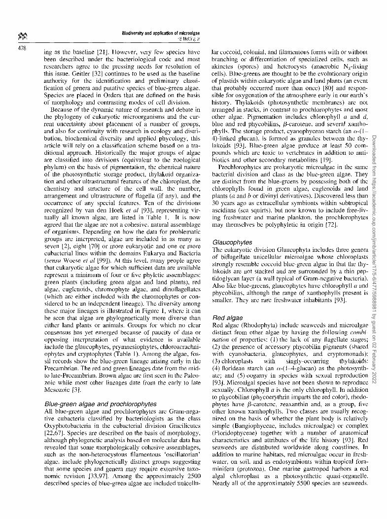
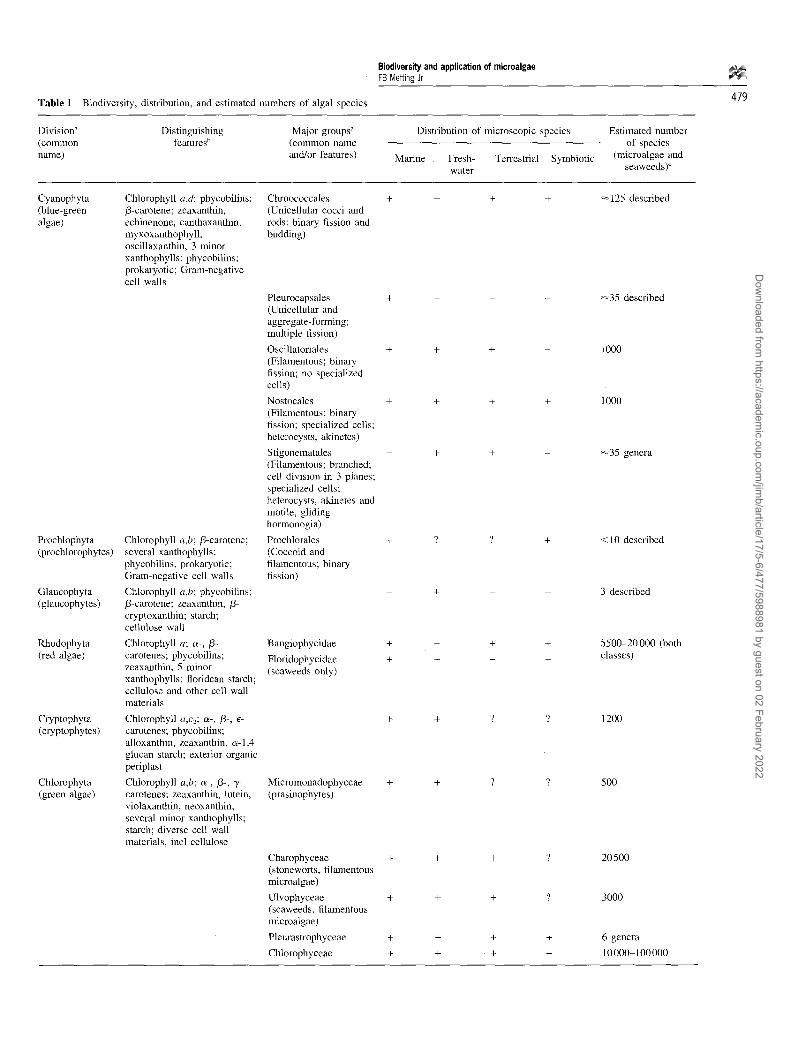
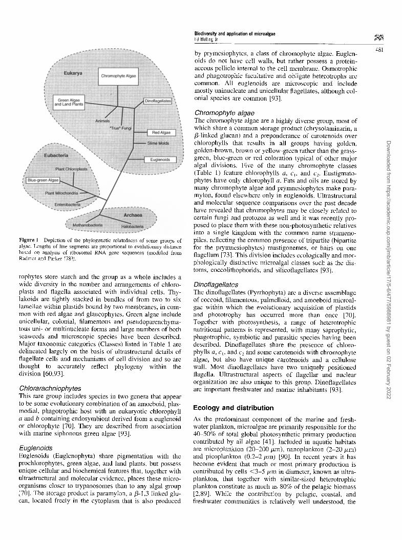
Because of the dynamic nature of research and debate in the phylogeny of eukaryotic microorganisms and the cur- rent uncertainty about placement of a number of groups, and also for continuity with research in ecology and distri- bution, biochemical diversity and applied phycology, this article will rely on a classification scheme based on a tra- ditional approach. Historically the major groups of algae are classified into divisions (equivalent to the zoological phylum) on the basis of pigmentation, the chemical nature of the photosynthetic storage product, thylakoid organiza- tion and other ultrastructural features of the chloroplast, the chemistry and structure of the cell wall, the number, arrangement and ultrastructure of flagella (if any), and the occurrence of any special features. Ten of the divisions recognized by van den Hoek et al [93], representing vir- tually all known algae, are listed in Table 1. It is now agreed that the algae are not a cohesive, natural assemblage of organisms. Depending on how the data for problematic groups are interpreted, algae are included in as many as seven [2], eight [70] or more eukaryotic and one or more eubacterial lines within the domains Eukarya and Bacteria (sensu Woese et al [99]). At this level, many people agree that eukaryotic algae for which sufficient data are available represent a minimum of four or five phyletic assemblages: green plants (including green algae and land plants), red algae, euglenoids, chromophyte algae, and dinoflagellates (which are either included with the chromophytes or con- sidered to be an independent lineage). The diversity among these major lineages is illustrated in Figure 1, where it can be seen that algae are phylogenetically more diverse than either land plants or animals. Groups for which no clear consensus has yet emerged because of paucity of data or opposing interpretation of what evidence is available include the glaucophytes, prymnesiophytes, chloroacrachni- ophytes and cryptophytes (Table 1). Among the algae, fos- sil records show the blue-green lineage arising early in the Precambrian. The red and green lineages date from the mid- to late-Precambrian. Brown algae are first seen in the Paleo- zoic while most other lineages date from the early to late Mesozoic [3].
Blue-green algae and prochlorophytes All blue-green algae and prochlorophytes are Gram-nega- tive eubacteria classified by bacteriologists as the class Oxyphotobacteria in the eubacterial division Gracilicutes [22,67]. Species are described on the basis of morphology, although phylogenetic analysis based on molecular data has revealed that some morphologically cohesive assemblages, such as the non-heterocystous filamentous 'oscillatorian' algae, include phylogenetically distinct groups suggesting that some species and genera may require extensive taxo- nomic revision [33,97]. Among the approximately 2500 described species of blue-green algae are included unicellu-
lar coccoid, colonial, and filamentous forms with or without branching or differentiation of specialized cells, such as akinetes (spores) and heterocysts (anaerobic N2-fixing cells). Blue-greens are thought to be the evolutionary origin of plastids within eukaryotic algae and land plants (an event that probably occurred more than once) [80] and respon- sible for oxygenation of the atmosphere early in our earth's history. Thylakoids (photosynthetic membranes) are not arranged in stacks, in contrast to prochlorophytes and most other algae. Pigmentation includes chlorophyll a and d, blue and red phycobilins, /3-carotene, and several xantho- phylls. The storage product, cyanophycean starch (an c~-(1- 4)-linked glucan), is formed as granules between the thy- lakoids [93]. Blue-green algae produce at least 50 com- pounds which are toxic to vertebrates in addition to anti- biotics and other secondary metabolites [19].
Prochlorophytes are prokaryotic microalgae in the same bacterial division and class as the blue-green algae. They are distinct from the blue-greens by possessing both of the chlorophylls found in green algae, euglenoids and land plants (a and b or divinyl derivatives). Discovered less than 30 years ago as extracellular symbionts within subtropical ascidians (sea squirts), but now known to include free-liv- ing freshwater and marine plankton, the prochlorophytes may themselves be polyphyletic in origin [72].
Glaucophytes The eukaryotic division Glaucophyta includes three genera of biflagellate unicellular microalgae whose chloroplasts strongly resemble coccoid blue-green algae in that the thy- lakoids are not stacked and are surrounded by a thin pep- tidoglycan layer (a wall typical of Gram-negative bacteria). Also like blue-greens, glaucophytes have chlorophyll a and phycobilins, although the range of xanthophylls present is smaller. They are rare freshwater inhabitants [93].
Red algae Red algae (Rhodophyta) include seaweeds and microalgae distinct from other algae by having the following combi- nation of properties: (1) the lack of any flagellate stages; (2) the presence of accessory phycobilin pigments (shared with cyanobacteria, glaucophytes, and cryptomonads); (3) chloroplasts with singly-occurring thylakoids; (4) ftoridean starch (an c~-(1-4-glucan) as the photosynth- ate; and (5)oogamy in species with sexual reproduction [93]. Microalgal species have not been shown to reproduce sexually. Chlorophyll a is the only chlorophyll. In addition to phycobilins (phycoerythrin imparts the red color), rhodo- phytes have /3-carotene, zeaxanthin and, as a group, five other known xanthophylls. Two classes are usually recog- nized on the basis of whether the plant body is relatively simple (Bangiophyceae, includes microalgae) or complex (Floridophyceae) together with a number of anatomical characteristics and attributes of the life history [93]. Red seaweeds are distributed worldwide along coastlines. In addition to marine habitats, red microalgae occur in fresh- water, on soil, and as endosymbionts within tropical fora- minifera (protozoa). One marine gastropod harbors a red algal chloroplast as a photosynthetic quasi-organelle. Nearly all of the approximately 5500 species are seaweeds.
Dow
nloaded from https://academ
ic.oup.com/jim
b/article/17/5-6/477/5988981 by guest on 02 February 2022
Biodiversity and application of microalgae FB Metting Jr
Table 1 Biodiversity, distribution, and estimated numbers of algal species 479
Division a Distinguishing Major groups a (common features t' (common name name) and/or features)
Distribution of microscopic species
Marine Fresh- Terrestrial Symbiotic water
Estimated number of species
(microalgae and seaweeds) ~
Cyanophyta (blue-green algae)
Prochlophyta (prochlorophytes)
Glaucophyta (glaucophytes)
Rhodophyta (red algae)
Cryptophyta (cryptophytes)
Chlorophyta (green algae)
Chlorophyll a,d: phycobilins: /3-carotene; zeaxanthin, echinenone, canthaxanthin, myxoxanthophyll, oscillaxanthin, 3 minor xanthophylls; phycobilins; prokaryotic; Gram-negative cell walls
Chlorophyll a,b; /3-carotene; several xanthophylls; phycobilins, prokaryotic; Gram-negative cell walls
Chlorophyll a,b; phycobilins; /3-carotene; zeaxanthin, /3- cryptoxanthin; starch; cellulose wall
Chlorophyll a; c~-, /3- carotenes; phycobilins; zeaxanthin, 5 minor xanthophylls; ftoridean starch; cellulose and other cell wall materials
Chlorophyll a,c2; ee-, /3-, e- carotenes; phycobilins; alloxanthin, zeaxanthin, c~-1,4 glucan starch; exterior organic periplast
Chlorophyll a,b; oe-, /3-, "y- carotenes; zeaxanthin, lutein, violaxanthin, neoxanthin; several minor xanthophylls; starch; diverse cell wall materials, incl cellulose
Chroococcales (Unicellular cocci and rods; binary fission and budding)
+ + + +
Pleurocapsales + + - - (Unicellular and aggregate-forming; multiple fission)
Oscillatoriales + + + - (Filamentous; binary fission; no specialized cells)
Nostocales + + + + (Filamentous; binary fission; specialized cells; heterocysts, akinetes)
Stigonematales - + + - (Filamentous; branched; cell division in 3 planes; specialized cells; heterocysts, akinetes and motile, gliding hormonogia)
Prochlorales + ? ? + (Coccoid and filamentous; binary fission)
Bangiophycidae +
Floridophycidae + (seaweeds only)
Micromonadophyceae (prasinophytes)
+ + +
~125 described
~35 described
1000
iooo
+ + ? ? 1200
+ + ? ? 500
~35 genera
<10 described
3 described
5500-20 000 (both classes)
Charophyceae + + + ? 20 500 (stoneworts, filamentous microalgae)
Ulvophyceae + + + ? 3000 (seaweeds, filamentous microalgae)
Pleurastrophyceae + + + + 6 genera
Chlorophyceae + + + + 10 000-100 000
Dow
nloaded from https://academ
ic.oup.com/jim
b/article/17/5-6/477/5988981 by guest on 02 February 2022

Biodiversity and application of microalgae FB Metting Jr
480 Table 1 Continued
Division a (common name)
Distinguishing Major groups ~ Distribution of microscopic species Estimated number features b (common name of species
and/or features) Marine Fresh- Terrestrial Symbiotic (microalgae and water seaweeds)C
Euglenophyta (euglenoids)
Chlorarachnio- phytes
PyiThophyta d (dinoflagellates)
Chromophyta (heterokont algae) ~
Chlorophyll a,b; /3- W- + + + ? 2000 carotenes; neoxanthin, diadinoxanthin, diatoxanthin; paramylon; wall-like organic pellicle internal to cell membrane
Chlorophyll a,b; unknown + - - + 2 genera, each with 1 carotenoids; amoeboid with species zoospores
Chlorophyll a,c; /3-carotene; + + ? + 3500-11000 diadinoxanthin, diatoxanthin, fucoxantbin, peridinin, 5 minor xanthophylls; a- l ,4 glucan starch; cellulose walls
Chlorophyll a,c,,c2,Cz; o~-, ,6- Chrysophyceae (golden- + + ? ? 3400 and e-carotenes; several major brown algae) and minor xanthophylls; chrysolaminarin, other glucans, oils; variety of cell wall materials
B acill ariophyceae + + + ? 100 000-10 000 000 (diatoms)
Xantbophyceae (yellow- + + + ? 2000 green algae)
Eustigmatopbyceae + + + 9 1000-10 000 (eustigmatophytes)
Raphidophyceae + + ? ? 100 (raphidophytes, cbloromonads)
Prymnesiophyceae + + ? ? 2000 (baptophytes or prymnesiophytes, including coccolithophorids)
Dictyochophyceae + . . . . 15 (silicoflagellates)
Phaeophyceae (brown + + + ? 2000 seaweeds, no microalgae)
aSensu Castenholtz and Waterbury [22] for cyanophyte orders, Lewin [56] for prochlorophyte orders, and van den Hoek et al [93] for all eukaryotic divisions and classes. bNot all classes within a division include taxa displaying all the distinguishing features. In particular, classes of chromophyte algae vary considerably in pigmentation and nature of the photosynthate [9]. CFrom Andersen [2] and Norton et al [68]. dSome dinoflagellates include endosymbiotic brown algae so that while most species have peridinin as the principal xanthophyll, others have fucoxantbin. This may represent multiple endosymbiotic events in the evolutionary past [70]. ~Some authors recognize additional classes of chromophyte algae, including the Synurophyceae and Pelagophyceae [2,88].
Ctyptophytes Cryptophytes (Cryptophyta) are a small group of mostly unicellular biflagellate microalgae with a broad collection of pigments, including chlorophylls a and c2 and phycobil- ins, and a single chloroplast with a thylakoid structure inter- mediate between the red algae and other plants. The cell is enclosed by a stiff proteinaceous periplast made of rec- tangular or polygonal plates. Ultrastructural properties of the chloroplast and unique cellular inclusions place crypto- monads apart from other algae, although Cavalier-Smith
[23] allies them with the chromophyte algae. Starch is stored as distinct granules and some species also store oils [93].
Green algae The green algae (Chlorophyta) comprise one of the largest in terms of number of species, most widely distributed, and morphologically diverse groups of algae. Land plants prob- ably evolved directly from green algae and share many bio- chemical, metabolic and ultrastructural features. Most chlo-
Dow
nloaded from https://academ
ic.oup.com/jim
b/article/17/5-6/477/5988981 by guest on 02 February 2022
Biodiversity and application of microalgae FB Metting Jr
by prymesiophytes, a class of chromophyte algae. Euglen- oids do not have cell walls, but rather possess a protein- aceous pellicle internal to the cell membrane. Osmotrophic and phagotrophic facultative and obligate heterotrophs are common. All euglenoids are microscopic and include mostly uninucleate and unicellular flagellates, although col- onial species are common [93].
481
Figure 1 Depiction of the phylogenetic relatedness of some groups of algae. Lengths of line segments are proportional to evolutionary distance based on analysis of ribosomal RNA gene sequences (modified from Radmer and Parker [78]).
rophytes store starch and the group as a whole includes a wide diversity in the number and arrangements of chloro- plasts and flagella associated with individual cells. Thy- lakoids are tightly stacked in bundles of from two to six lamellae within plastids bound by two membranes, in com- mon with red algae and glaucophytes. Green algae include unicellular, colonial, filamentous and pseudoparenchyma- tons uni- or multinucleate forms and large numbers of both seaweeds and microscopic species have been described. Major taxonomic categories (Classes) listed in Table 1 are delineated largely on the basis of ultrastructural details of flagellate cells and mechanisms of cell division and so are thought to accurately reflect phylogeny within the division [60,93].
Chlorarachniophytes This rare group includes species in two genera that appear to be some evolutionalT combination of an amoeboid, plas- modial, phagotrophic host with an eukaryotic chlorophyll a and b containing endosymbiont derived from a euglenoid or chlorophyte [70]. They are described from association with marine siphonous green algae [93].
Euglonoids Euglenoids (Euglenophyta) share pigmentation with the prochlorophytes, green algae, and land plants, but possess unique cellular and biochemical features that, together with ultrastructural and molecular evidence, places these micro- organisms closer to trypanosomes than to any algal group [70]. The storage product is paramylon, a/3-1,3 linked glu- can, located freely in the cytoplasm that is also produced
Chromophyto algae The chromophyte algae are a highly diverse group, most of which share a common storage product (chrysolaminarin, a /3-1inked glucan) and a preponderance of carotenoids over chlorophylls that results in all groups having golden, golden-brown, brown or yellow-green rather than the grass- green, blue-green or red coloration typical of other major algal divisions. Five of the many chromophyte classes (Table 1) feature chlorophylls a, Ca, and c2. Eustigmato- phytes have only chlorophyll a. Fats and oils are stored by many chromophyte algae and prymnesiophytes make para- mylon, found elsewhere only in euglenoids. Ultrastructural and molecular sequence comparisons over the past decade have revealed that chromophytes may be closely related to certain fungi and protozoa as well and it was recently pro- posed to place them with these non-photosynthetic relatives into a single kingdom with the common name strameno- piles, reflecting the common presence of tripartite (bipartite for the prymnesiophytes) mastigonemes, or hairs on one flagellum [73]. This division includes ecologically and mor- phologically distinctive microalgat classes such as the dia- toms, coccolithophorids, and silicoflagellates [93].
Dinotlagellatos The dinoflagellates (Pyrrhophyta) are a diverse assemblage of coccoid, filamentous, palmelloid, and amoeboid microal- gae within which the evolutionary acquisition of plastids and phototrophy has occurred more than once [70]. Together with photosynthesis, a range of heterotrophic nutritional patterns is represented, with many saprophytic, phagotrophic, symbiotic and parasitic species having been described. Dinoflagellates share the presence of chloro- phylls a, ca, and Cz and some carotenoids with chromophyte algae, but also have unique carotenoids and a cellulose wall. Most dinoflagellates have two uniquely positioned flagella. Ultrastructural aspects of flagellar and nuclear organization are also unique to this group. Dinoflagellates are important freshwater and marine inhabitants [93].
Ecology and distribution
As the predominant component of the marine and fresh- water plankton, microalgae are primarily responsible for the 40-50% of total global photosynthetic primary production contributed by all algae [41]. Included in aquatic habitats are microplankton (20-200/xm), nanoplankton (2-20/zm) and picoplankton (0.2-2 p,m) [90]. In recent years it has become evident that much or most primary production is contributed by cells <3 -5 /xm in diameter, known as ultra- plankton, that together with similar-sized heterotrophic plankton constitute as much as 80% of the pelagic biomass [2,89]. While the contribution by pelagic, coastal, and freshwater communities is relatively well understood, the
Dow
nloaded from https://academ
ic.oup.com/jim
b/article/17/5-6/477/5988981 by guest on 02 February 2022
482
Biodiversity and application of microalgae FB Metting Jr
significance of terrestrial microalgae is not known with any degree of precision, but they may be important on a local or regional scale because of their predominance on exten- sive areas of exposed land surface in arid and semi-arid steppes and deserts [63].
Phytoplankton reside throughout the photic zone from the surface layer (neuston) to 250 m or more in depth in some clear oligotrophic waters and depending on latitude. Andersen [2] calculated that as many as 3.6 x 1025 individ- ual phytoplankters may inhabit the world's oceans at any given time and are responsible for an annual primary pro- duction of over 5 x 10¹³ kg, constituting the base of the marine food web. As a consequence of diurnal, seasonal, vertical, and geographic variation in nutrient availability, temperature, light and other factors, the distribution and metabolic activities of groups of microalgae may be con- siderably more heterogeneous than is currently understood. The interpretation of data from flow cytometry, extraction and analysis of community-level nucleic acids, phospho- lipid and other biochemical assays, remote sensing, and tra- ditional microscopy has resulted in some generalizations regarding the distribution of phytoplankton. These are cer- tain to be refined as more surveys across geographic and temporal gradients are reported and as our knowledge improves about the distribution and significance of viable but non-culturable species, believed by many people to include the bulk of microbial biodiversity. The discussion of the occurrence and general distribution patterns of the major groups of marine microalgae that follows is largely taken from Thomsen's 1986 survey [92] in which he indi- cated that the depth of information about the different algal groups is highly variable and that creation of a systematic framework for presenting or interpreting the current data is difficult at best. Thus, the following treatment is meant only to provide an appreciation for the ecological diversity of microalgae with the degree of attention paid to each group in no way reflecting its perceived or real importance in the biosphere.
Blue-green algae and prochlorophytes Cyanobacteria are ubiquitous in marine, freshwater, and ter- restrial environments as free-living populations and endo- symbionts of marine animals, lichens, bryophytes, and cycad roots. In the past two decades, ultraplanktonic cyano- bacteria (eg Synechococcus) have been shown to contribute as much or more to oceanic primary production as any other single group of microalgae [31,95]. In addition, filamentous cyanobacteria such as Anabaena, Nostoc, Microcoleus, Oscillatoria and Mastigocladus are commonly the domi- nant component of microbial mats in brackish waters, hot springs, on semi-arid and arid soils, and in rice paddies [20,62,84]. Prochlorophytes (eg Prochloron) were first described two decades ago from symbiotic association with marine didemnids and holothurians and are now known to also exist as free-living components of the pelagic nano- plankton where their distribution and importance in this habitat is becoming clearer [25].
microalgae in the order Porphyridiales. The genera Rhod- ella and Rhodosorus include marine phytoplankton species. They are regular inhabitants at some locations, such as Norwegian fjords. Under favorable circumstances, the release of male gametes by red seaweeds makes a signifi- cant ephemeral contribution to coastal benthic phytoplank- ton communities [92]. Porphyridium inhabits freshwater and terrestrial ecosystems [93].
Cryptophytes Members of this group are sporadically present in fresh- water and marine habitats. Some species of Chroomonas exhibit broad tolerance to salt concentrations, living in estu- aries and salt marshes [92]. One species is symbiotic within the marine ciliate Mesodinium.
Green algae Green algae are ubiquitous in freshwater habitats, where they commonly dominate phytoplankton assemblages, and are abundant in terrestrial environments as well. The green algae range in size from microscopic to macroscopic in all of these environments. Predominantly photosynthetic, there are nonetheless numerous examples of facultative and obli- gate heterotrophs [93].
Representative genera that are widely distributed in mar- ine environments include mostly uniflagellate (eg Pedino- monas, Micromonas) and biflagellate prasinophytes (eg Mamiella, Mantoniella) and chlorophytes (eg Pyrami- monas, Tetraselmis). Various coccoid chlorophytes less fre- quently documented (eg Chlorella, Nanochloris, Halochlorococcum) may also prove ultimately to have worldwide distribution and importance. Dunaliella, a uni- cellular biflagellate phytoplankter found in extremely saline habitats, such as seaside rock pools and saline lakes, includ- ing the Dead Sea and Great Salt Lake, is a focus of efforts for commercial production of/3-carotene [15].
Green microalgae are abundant and diverse in freshwater habitats. For example, unicellular (eg Chlamydomonas, Phacotus) and colonial (eg Volvox, Pyrobotrys) flagellates, coccoid non-flagellate species (eg ChlorelIa, Pediastrum, Ankistrodesmus, Scenedesmus), and filamentous forms (eg Chaetophora, Oedogonium) are ubiquitous in ponds, lakes, and streams at most latitudes. Many green microalgae are important as 'weeds' in lakes, canals, and other waterways [54]. Most of these are mat-forming (eg Spirogyra, Hydrodictyon) or attached (eg Cladophora, Ulothrix, Stigeoclonium) filamentous species [10,93].
Green microalgae also occur in terrestrial habitats, including on and in soil and rocks. The most commonly encountered are coccoid (eg Chlorococcum), palmelloid-- aggregates of cells invested by a common mucilage--(eg Palmella, Gloeococcus), sarconoid--packets of 4, 8, 16 or 32 cells--(eg Tetracystis, Chlorosarcina), and filamentous (eg Stichococcus, Klebshormidium) genera [62]. Trentepho- lia inhabits tree trunks and other subaerial surfaces while other green microalgae are symbiotic, such as Trebouxia, a common lichen phycobiont.
Red algae Consisting mostly of seaweeds categorized into two major botanical classes, the red algae include genera of free-living
Euglenoids Euglena, Trachelomonas, Colacium and other euglenoid genera include many common freshwater and edaphic spec-
Dow
nloaded from https://academ
ic.oup.com/jim
b/article/17/5-6/477/5988981 by guest on 02 February 2022
Biodiversity and application of microalgae FB Metting Jr
ies. This group is less common in marine environments, although species in the genus Eutreptia occasionally form very dense blooms in coastal waters. Most euglenoids are facultatively heterotrophic and/or phagotrophic. Colorless, phagotrophic species (eg Peranema) are also common [93].
Chromophyte algae Chromophyte microalgae are highly diverse and important components of most marine and freshwater habitats. With the exception of Pelagococus, which has been cultured from across the north Pacific, golden-brown microalgae (Chrysophyceae: eg Ochromonas, Chromulina), although often encountered, are not often abundant in marine environments. Other golden-browns are, however, known to be important freshwater phytoplankton that commonly form blooms in lakes and ponds. Synura, for example, is a nuisance alga responsible for imparting undesirable odors and taste to drinking water from lakes and reservoirs. Golden-brown microalgae have not been reported from soil. In contrast, silicoflagellates (eg Dichtyota) are very com- mon in the oceans, where they often form dense blooms. These microalgae are uncommon in freshwater and not known from terrestrial habitats. Pedinella is another pelagic genus whose abundance under ice can color the seawater yellow [921.
Diatoms (Bacillariophyceae) are ubiquitous in marine, freshwater and terrestrial environments and probably include the greatest number of extant species (up to ten million) of any group of microalgae [49,86]. Diatoms are categorized into three classes on the basis of having bilat- eral or radial symmetry. Bacillariophyceae include bilater- ally symmetrical species with raphes (an opening or fissure in the valve). Common genera include Navicula, Nitzschia, and Phaeodactylum. Diatoms displaying bilateral symmetry but without raphes are in the class Fragilariophyceae (eg Diatoma, Synedra, Fragilaria). Radially symmetrical dia- toms are in the class Coscinodiscophyceae (eg Chaeto- ceros, Biddulphia, Thalassiosira). Diatoms are mostly uni- cellular, although filamentous species are also abundant and diverse. These microalgae commonly dominate planktonic communities at all latitudes, particularly in and under ice, in temperate coastal and pelagic environments, on mud flats, in still or moving freshwater, and on some soils [86].
Yellow-green microalgae (Xanthophyceae: eg Vau- cheria, Bumilleria) are primarily known from freshwater environments. Unicellular, aggregate and filamentous xan- thophytes are also common on soil. The eustigmatophytes Nanochloris and Nanochloropsis (Enstigmatophyceae) have been discovered over the past two decades to be important contributors to primary production in estuaries, salt marshes, and brackish seas and are present, but less abundant, in the open ocean [3].
Prymnesiophytes, also known as haptophytes (Prymnesiophyceae or Haptophyceae) are microalgae that commonly dominate the marine phytoplankton in numbers and biomass (eg Phaeocystis). They are sometimes dis- tinguished at the species level by the pattern of organic scales and, in the case of the coccolithophorids, CaCO3 deposits on the cell surface. The biodiversity of prymnesi- ophytes is greatest in warm oligotrophic regions, such as the Red Sea and Gulf of California, although they are also
known to bloom in eutrophic waters. Coccolithophores have been the subject of more investigations of geographic distribution in the oceans than most other major groups of algae. Subtropical and tropical marine waters support diverse communities that include Emiliana, Discosphaera, Rhabdosphaera and many other genera [98]. Coccolithus and Emiliana are two coccolithophore genera common in open oceans at arctic latitudes. Members of the genus Chry- sochromulina are common in all marine environments where they often dominate and can form dense populations even in dilute brackish waters. Prymnesiophytes are uncommon in freshwater and unknown from terrestrial habitats.
Dinoflagellates Dinoflagellates are often important components of the microplankton of freshwater and marine habitats in which they are found. Dinophysis, Gymnodinium, Amphidinium, Peridinium, Ceratium and Prorocentrum all include photo- synthetic flagellate species found at most latitudes in fresh and marine bodies of water [92]. Dinoflagellates also include many specialized species, including endo- and ecto- parasites of marine animals (eg Blastodinium, Haplozoon) and the photosynthetic partners of many corals known as zooxanthellae [93]. Dinoflagellates include species capable of bioluminescence (eg Gonyaulax, Pyrocystis) and those responsible for 'red tides' (see section on Bioactive com- pounds, below).
B i o c h e m i c a l d i v e r s i t y
The phylogenetic breadth of microalgae is reflected in an equally broad biochemical diversity of pigments, photosyn- thetic storage products, cell walls and mucilages, fatty acids and lipids, oils, sterols and hydrocarbons, and bioactive compounds, including secondary metabolites. Much of the material to follow is from contributed chapters to volumes edited by Borowitzka and Borowitzka [14], Rogers and Gallon [85], and Stewart [91].
Pigments Chlorophylls, carotenoids, and phycobilins comprise the range of pigments produced in various combinations by photosynthetic microalgae. Chlorophyll a is present in all algae and land plants, including the prokaryotic cyanobac- teria and prochlorophytes. Other chlorophylls (b, cl, c2, d) are accessory light-harvesting molecules whose distribution among algal groups is used, in part, for taxonomic purposes at the division level (Table 1). Chlorophylls absorb blue and red light and can range up to 2% or more of cellular dry weight. Together with carotenoids and phycobilins, the synthesis, content, and abundance of chlorophylls is depen- dent on light, nutrition and other environmental factors [61,82].
Carotenoids are tetraterpenes comprised of eight branched 5-C isoprenoid units. The variety of carotenoids in algae is far greater than for land plants, with more than 40 carotenes and xanthophylls having been isolated and characterized [9]. Several algal groups derive their primary coloration from the mix of carotenoids they produce, for example the yellow-green, golden, golden-brown and
483
Dow
nloaded from https://academ
ic.oup.com/jim
b/article/17/5-6/477/5988981 by guest on 02 February 2022
484
Biodiversity and application of microalgae FB Metting Jr
brown coloration found within the chromophyte algae. /3 carotene is chemically the most simple and is found in all algae and other plants. Hypersaline species of Dunaliella accumulate /3-carotene up to 14% of total dry weight. Astaxanthin is another important carotenoid that is pro- duced in large concentrations by green microalgae, includ- ing Haematococcus and others that inhabit snow and ice (eg Chlamydomonas spp). Many carotenes and xantho- phylls are commonly restricted to one or a few algal groups. For example, lutein is shared by chlorophytes and land plants, myxoxanthin and myxoxanthophyll are character- istic of cyanobacteria, as are peridinin for dinoflagellates and fucoxanthin for brown algae and diatoms.
Phycobilins are water-soluble accessory pigments pro- duced by cyanobacteria, glaucophytes, red algae, and cryptophytes. As many as three different phycobilins can be produced by an individual organism. In some algae, they modulate a metabolic process called chromatic adaption in which one form or another is preferentially synthesized according to the quality of available light [34]. Phycobilins are extracted from red and blue-green algae for use as flu- orescent markers in cell biology and to impart color to food and cosmetic products [12].
Photosynthetic storage products Microalgae synthesize a wide variety of photosynthetic storage products (photosynthates), the chemical nature of which is also used as a criterion for taxonomic treatment at the division level. Photosynthates include c~-(1-4)-linked glucans (starches), /3-(l-3)-glucans, fructans, low molecu- lar weight carbohydrates, and fats and oils. Starches pro- duced by various groups include floridean starch (amylopectin subunits) by red algae and myxophycean starch (amylopectin or glycogen-like subunits) by blue- green algae. Some green algae form a cross-linked amyl- ose-amylopectin starch that is essentially similar to starch in land plants. Others store fructosans (inulin-like fructose oligosaccharides). Cryptophytes and dinoftagellates also store c~-(1-4)-linked glucans. Chrysophytes store oils or chrysophycean starch, a water-soluble /3-(1-3) glucopyr- anoside. Other chromophyte algae store various hydro- carbons and oils (see below). Paramylon, composed solely of /3-(l-3)-linked D-glucose, is unique to euglenoids and prymnesiophytes [93].
Many microalgae in different divisions store monosacch- arides (eg glucose, galactose, many others), disaccharides (ie sucrose, trehalose, maltose), glycerol glycosides, and polyols (eg mannitol). Low molecular weight compounds are also accumulated by microalgae for osmoregulation. Examples include sugars (eg Ochromonas), free glycerol (eg Dunaliella and various zooxanthellae) and proline (some Chlorella spp). Industrial processes for glycerol [7] and proline [53] production were developed, but not com- mercialized.
Cell walls and extracellular mucilages Most microalgae form rigid cell walls. Exceptions include numerous 'naked' microalgae, such as Dunaliella, some dinoftagellates and chrysophytes, and the motile spores of various groups. Cryptomonads and euglenoids have distinc- tive proteinaceous pellicles, internal to the cell membrane
in euglenoids and to the outside in cryptophytes. Cyanobac- teria and prochlorophytes have Gram-negative bacterial walls composed of c~- and e-diamino-pimelic acid and glu- cosamine (peptidoglycan). Many or most green algae and dinoflagellates have cellulose walls. Other chlorophyte walls are constructed from hydroxy-proline glucosides, xyl- ans, or mannans; some are calicified [93].
Diatoms construct elaborately decorated walls of silicon (frustules) made of two identical halves (valves) that fit together like the lid and bottom of a petri dish. Devoid of walls, silicoflagellates instead produce siliceous internal and/or external skeletons, often with spiny projections, around and through which the protoplast moves. Prymnesi- ophytes, although without a rigid wall, are covered by superficial organic scales visible only with an electron microscope. These scales are calcified in the coccolithopho- rids [93].
Microalgal representatives in all of the divisions produce a variety of extracellular polysaccharide mucilages. Much smaller quantities are usually produced during active growth and cell division as compared to stationary phase. Mucilages can be responsible for aggregation of plankton, contribution to biofilm formation by attached species, and desiccation tolerance of terrestrial forms and can possess chelating and flocculant properties. The best-studied muci- lages are produced by cyanobacteria [4], diatoms and other chromophyte algae [42], chlorophytes [79], and red microalgae [94]. Cell wall materials from brown and red seaweeds form the basis of a large marine hydrocolloid industry [77].
Lipids, oils, sterols and fatty acids The total oil and fat content of microalgae ranges from 1% to 70% of the dry weight and tends to be inversely pro- portional to the rate of growth with greater accumulations during stationary phase [11]. The percent of total lipid as neutral lipid, glycolipid, and phospholipid also varies widely among and within groups of microalgae [11,50]. Some species manufacture hydrocarbons. An example is Botryococcus, a chlorophyte capable of accumulating up to 90% of its colonial dry weight as a mixture of ten hydro- carbon compounds in globules occluded among cells embedded in a colonial matrix. Hydrocarbons up to C-37 in size from different strains of B. braunii include straight chain alkadienes, branched triterpenoids (botryococcenes), and the tetraterpenoid lycopadiene [69].
A wide range of common and rare sterols are also synthesized by microalgae, including, for example, cholesterols (cyanobacteria, rhodophytes), chondrillasterol (chlorophytes, euglenoids), clinoasterol (xanthophytes), dinosterol (dinoflagellates), ergosterol (chlorophytes, rhodophytes, euglenoids), epibrassicasterol (diatoms), pori- ferasterol (chlorophytes, chrysophytes), and sitosterol (cyanobacteria, chlorophytes, xanthophytes) [37,75].
Because of increased awareness of the possible health aspects of including w-3 fatty acids in the diet (see Appli- cations, below), the manufacture and content of fatty acids by microalgae is the subject of a good deal of ongoing research and development [44]. Microalgal lipids are mostly esters of glycerol and fatty acids with a chain length of C~4 to C22 and may be saturated or unsaturated. Cyano-
Dow
nloaded from https://academ
ic.oup.com/jim
b/article/17/5-6/477/5988981 by guest on 02 February 2022
Biodiversity and application of microalgae FB Metting Jr f%~
bacteria tend to have large amounts of polyunsaturated fatty acids while eukaryotic microalgae contain, in addition, a wide range of saturated and monosaturated fatty acids with fatty acid profiles widely variable among taxa. Specific fatty acid profiles vary widely among algal groups, with age and growth stage, and with environmental conditions. There are, however, some generalizations. For example, linolenic acid (C18:3) is common in green algae, whereas diatoms contain palmitic (C16:o), hexadecenoic (C~6:~) and C-2o polyenoic acids. Red microalgae have high contents of arachidonic acid (C2o:4) as well as palmitic, oleic (C~8:1) and linoleic acids. Chrysophytes contain significant quan- tities of highly unsaturated C 18:4 and C2e:6 acids in addition to unsaturated C~6:o and C2o:o compounds, fL3 fatty acids are produced by a variety of microalgae. Eicosapentaenoic (Czo:5) and docosahexaenoic (C22:6) acids are produced by some species of green and red microalgae, cryptophytes, dinoflagellates, prymnesiophytes and diatoms. Certain species have also been shown to synthesize unusual fatty acids such as, for example myristic (C~4:o) in the diatom Fragilaria, lignoceric (C24:o) in the premnesiophyte lso- chrysis, gamma linolenic (C 18:3) in the golden-brown alga Ochromonas, and arachidonic (C20:4) in the red microalga Porphyridium [24].
Bioactive compounds Microalgae produce a wide array of compounds with bio- logical activity including antibiotics, algicides, other toxins, pharmaceutically-active compounds, and plant growth regulators [13,65]. Antibiotic substances produced by microalgae include certain fatty acids, bromophenols, tan- nins, terpenoids, polysaccharides, alcohols, and other com- pounds [76]. Most antibiotics are known from chlorophytes and golden-browns, prymnesiophytes, diatoms, cyanobac- teria, and dinoflagellates. Activity has been demonstrated against bacteria, fungi, protozoa and other microalgae. For example, the cyanobacterium Scytonema hofmanii is known to excrete a ,/-lactone algicidal to a broad range of poten- tially competing microalgae in freshwater environments [36].
Dinoflagellates, cyanobacteria and other microalgae pro- duce compounds toxic to invertebrate and vertebrate ani- mals, including the neurotoxic agents of diarrhetic and paralytic shellfish poisoning (red tides) and ciguaterra fish poisoning, an endemic disease in human populations that consume reef-inhabiting fish in the South Pacific. A number of icthyotoxins are also produced by dinollagellates. At least twelve toxins are produced by species of Protogony- aulax alone [47,65]. In very small quantities a saxitoxin has been used in microsurgical procedures and as an exper- imental treatment for near-sightedness. Neurotoxins (eg anatoxin, aphanatoxin) produced by the cyanobacteria Ana- baena flos-aquae, Aphanizomenon flos-aquae, and hepato- toxins (eg microcystin) from Microcystis aeruginosa in temperate freshwater have been implicated in poisonings and occasional death of cattle and other livestock [19,38].
Applications Microalgae have been the subject of applied research for their commercial and industrial potential since the early
1950s when productivity and yield were first studied in mass culture [18]. Early interest was focused on exploiting the fact that optimal culture conditions result in theoretical conversion of sunlight to biomass at yields of eight to ten times the most productive natural or agricultural plant com- munities. In practice, sustained commercial mass culture results in productivities of about 15 dry g cm -2 day -1 (54 tons ha i year1). More recently, microalgae have been tar- geted as a source of bioactive compounds and pharmaceut- icals, specialty chemicals, health foods, aquaculture feeds, and for waste treatment and agriculture [1,14,55,77].
Microalgal mass culture Microalgal biomass is produced in engineered facilities whose fundamental design and infrastructure depend on the growth requirements of the microalgae of interest and the nature and value of the final product. Two principal pro- duction methods are the photobioreactor and the open pond. Photobioreactors are lit fermentors of variable sophisti- cation that are closed to the environment in order to prevent chemical or biological contamination, including parasitism, predation, and the need to control competition from unwanted microalgae ('weeds') when the desired species is otherwise uncompetitive. Only products with high value such as reagent grade phycobilins or isotopically-labeled research compounds are produced in closed systems.
Industrial scale production of microalgal biomass for the health food market, for animal feed ingredients, and for wastewater treatment relies on engineered ponds open to the environment (Figure 2). Maintenance of single species (unialgal) cultures in open ponds relies on the competitive ability of the microalga of interest. Thus, Spirulina nat- urally predominates in highly alkaline water, as does Duna- liella in saline culture. Wastewater treatment systems that rely on microalgae for O2 production are dominated by chlorophytes with high specific growth rates that tolerate environments with high concentrations of dissolved and particulate organic compounds (eg Ankistrodesmus, Chlor- ella, Scenedesmus). Engineering design considerations are considered in detail by Jassby [48], Oswald [71], and Rich- mond and Becker [83].
Pharmaceuticals By virtue of their larger size and accessibility, seaweeds have until recently attracted more attention than microalgae as sources of natural products, including identification of novel compounds for pharmaceutical development [45,46]. Screening of marine algae for antitumor activity began in the 1970s. Early discoveries included tubercidin, a hetero- cyclic nitrogen compound from the cyanobacterium Toly- pothrix byssoidea, shown to have in vitro activity against P-388 lymphocytic leukemia. Another compound, an 17 aspinarigenase from the green microalga Chlamydomonas, inhibits growth of lymphosarcoma in mice. Marine blue- green algae have been one target of a more recent Natural Cancer Institute screening program aimed at anticancer and antiviral (anti-HIV) activity that has identified a number of compounds with potential for drug development, such as dibromoaplysiatoxin from Lyngbya majuscula. Approxi- mately 10% of 600 cultures from numerous genera of mar- ine blue-greens showed some activity in reducing cyto-
485
Dow
nloaded from https://academ
ic.oup.com/jim
b/article/17/5-6/477/5988981 by guest on 02 February 2022
486
Riodiversity and application of microalgae FB Metting Jr
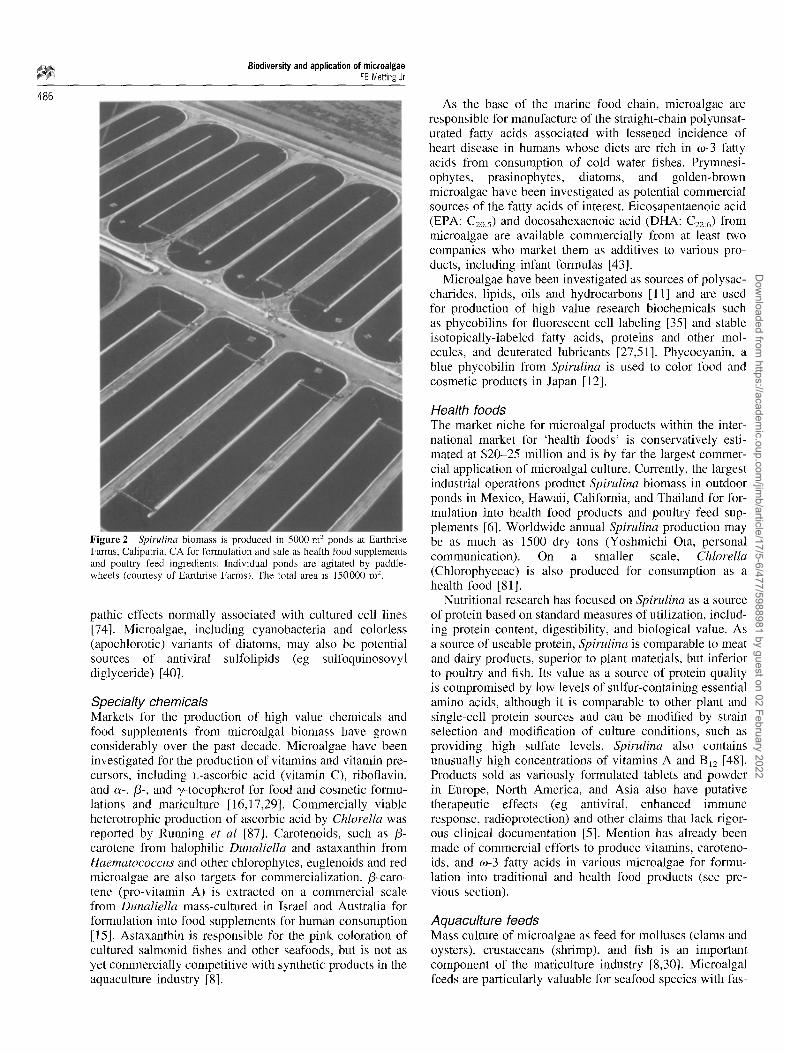
Figure 2 Spirulina biomass is produced in 5000 m 2 ponds at Earthrise Farms, Calipatria, CA for formulation and sale as health food supplements and poultry feed ingredients. Individual ponds are agitated by paddle- wheels (courtesy of Earthrise Farms). The total area is 150000 m 2.
pathic effects normally associated with cultured cell lines [74]. Microalgae, including cyanobacteria and colorless (apochlorotic) variants of diatoms, may also be potential sources of antiviral sulfolipids (eg sulfoquinosovyl diglyceride) [40].
Specialty chemicals Markets for the production of high value chemicals and food supplements from microalgal biomass have grown considerably over the past decade. Microalgae have been investigated for the production of vitamins and vitamin pre- cursors, including L-ascorbic acid (vitamin C), riboflavin, and c~-,/3-, and y-tocopherol for food and cosmetic formu- lations and mariculture [16,17,29]. Commercially viable heterotrophic production of ascorbic acid by Chlorella was reported by Running et al [87]. Carotenoids, such as /3- carotene from halophilic Dunaliella and astaxanthin from Haematococcus and other chlorophytes, euglenoids and red microalgae are also targets for commercialization. /3-caro- tene (pro-vitamin A) is extracted on a commercial scale from Dunaliella mass-cultured in israel and Australia for formulation into food supplements for human consumption [15]. Astaxanthin is responsible for the pink coloration of cultured salmonid fishes and other seafoods, but is not as yet commercially competitive with synthetic products in the aquaculture industry [8].
As the base of the marine food chain, microalgae are responsible for manufacture of the straight-chain polyunsat- urated fatty acids associated with lessened incidence of heart disease in humans whose diets are rich in w-3 fatty acids from consumption of cold water fishes. Prymnesi- ophytes, prasinophytes, diatoms, and golden-brown microalgae have been investigated as potential commercial sources of the fatty acids of interest. Eicosapentaenoic acid (EPA: C2o:5) and docosahexaenoic acid (DHA: C22:6) from microalgae are available commercially from at least two companies who market them as additives to various pro- ducts, including infant formulas [43].
Microalgae have been investigated as sources of polysac- charides, lipids, oils and hydrocarbons [11] and are used for production of high value research biochemicals such as phycobilins for fluorescent cell labeling [35] and stable isotopically-labeled fatty acids, proteins and other mol- ecules, and deuterated lubricants [27,51]. Phycocyanin, a blue phycobilin from Spirulina is used to color food and cosmetic products in Japan [12].
Health foods The market niche for microalgal products within the inter- national market for 'health foods' is conservatively esti- mated at $20-25 million and is by far the largest commer- cial application of microalgal culture. Currently, the largest industrial operations product Spirulina biomass in outdoor ponds in Mexico, Hawaii, California, and Thailand for for- mulation into health food products and poultry feed sup- plements [6]. Worldwide annual Spirulina production may be as much as 1500 dry tons (Yoshmichi Ota, personal communication). On a smaller scale, Chlorella (Chlorophyceae) is also produced for consumption as a health food [81].
Nutritional research has focused on Spirulina as a source of protein based on standard measures of utilization, includ- ing protein content, digestibility, and biological value. As a source of useable protein, Spirulina is comparable to meat and dairy products, superior to plant materials, but inferior to poultry and fish. Its value as a source of protein quality is compromised by low levels of sulfur-containing essential amino acids, although it is comparable to other plant and single-cell protein sources and can be modified by strain selection and modification of culture conditions, such as providing high sulfate levels. Spirulina also contains unusually high concentrations of vitamins A and B12 [48]. Products sold as variously formulated tablets and powder in Europe, North America, and Asia also have putative therapeutic effects (eg antiviral, enhanced immune response, radioprotection) and other claims that lack rigor- ous clinical documentation [5]. Mention has already been made of commercial efforts to produce vitamins, caroteno- ids, and (o-3 fatty acids in various microalgae for formu- lation into traditional and health food products (see pre- vious section).
Aquaculture feeds Mass culture of microalgae as feed for molluscs (clams and oysters), crustaceans (shrimp), and fish is an important component of the mariculture industry [8,30]. Microalgal feeds are particularly valuable for seafood species with fas-
Dow
nloaded from https://academ
ic.oup.com/jim
b/article/17/5-6/477/5988981 by guest on 02 February 2022
Biodiversity and application of microalgae FB Metting Jr
tidious dietary requirements that cannot be met by formu- lation with traditional agricultural commodity products, such as corn, soybeans, and fish and food processing by- products. In these cases, microalgae commonly provide essential amino acids, fatty acids or other unidentified growth factor requirements or are used to provide caroten- oids for coloration of the final product. In nearly all cases, the microalgae are produced at the aquaculture facility, fre- quently by simple fertilization of incoming seawater, and fed directly to the animals as dilute, living cultures [8].
Waste treatment Microalgae are principal or otherwise important biological components of various systems for treating municipal and industrial effluents. Oxygen from photosynthesis is the pri- mary microalgal contribution to treatment of municipal wastewaters in open ponds or impoundments. The most common designs include facultative ponds, which are deeper and support surface growth of microalgae, and high- rate oxidation ponds which are specifically engineered to encourage maximum oxygenation by microalgae. High-rate ponds are shallow and depend on mechanical mixing to maximize algal production and removal of biochemical oxygen demand [71]. In both configurations, microalgal communities are commonly dominated by green microalgae (eg Ankistrodesmus, Chlorella, Scenedesmus). Biomass from high-rate oxidation ponds can be harvested for use as animal feed and the concept has been demonstrated as a component of integrated approaches to recycling of live- stock wastes [57]. Non-living microalgal biomass has also been subject to investigation and early-stage commercializ- ation for its potential in recovery of metals and radio- nuclides from aqueous wastestreams as an alternative to ion exchange resins [39]. Additionally, research has been targeted at the use of microalgal systems for mitigation of waste heat [96] and CO= [52] from industrial processes.
Biofertilizers Microalgae are employed in agriculture as biofertilizers and soil conditioners [64]. Biofertilizers consist of N=-fixing cyanobacteria in lowland tropical rice cultivation. Filamen- tous species of Anabaena, Nostoc, Aulosira, Tolypothrix and Scytonema are common in China, India and elsewhere in Asia, where they can provide 20 or more kg N ha -1 year -I, sufficient for one third or more of the requirements of traditional rice cultivars [84]. In China, Vietnam and Africa, Anabaena azollae, symbiotic in leaf cavities of the water fern Azolla is used as a green manure or companion crop in rice production [66]. On a small scale, palmelloid (mucilage-producing) microalgae in the genus Chlamydo- monas (Chlorophyceae) have been used as soil-condition- ing agents for erosion control of pivot-irrigated soils in North America [64].
A c k n o w l e d g e m e n t s
Thanks are due to Robert Andersen, Center for Culture of Marine Phytoplankton, Bigelow Laboratory for Marine Sciences, West Boothbay Harbor, ME, for sharing preprints and references to the literature on algal diversity and for providing a thorough review of the preliminary manuscript;
to Gerald Cysewski, Cyanotech Corporation, Kailua-Kona, HI and Yoshimichi Ota, Earthrise Farms, Calipatria, CA for insights regarding commercial Spirulina and Dunaliella markets: to Mr Ota for Figure 2; and to Sharon Lepel and Sue Carver (Pacific Northwest National Laboratory), respectively, for secretarial and library assistance.
R e f e r e n c e s
1 Akatsuka I (ed). 1990. Introduction to Applied Phycology. SPB Aca- demic Publishing, The Hague.
2 Andersen RA. 1992. Diversity of eukaryotic algae. Biodiversity and Conservation 1: 267-292.
3 Andersen RA. 1996. Algae. In: Maintaining Cultures for Biotechnol- ogy and Industry (Hunter-Cevera JC and A Belt, eds), pp 29-64, Aca- demic Press, San Diego.
4 Bar-Or Y and M Shilo. 1988. Unique characteristics and exopolysac- charides of benthic cyanobacteria of potential importance for their mass cultivation. In: Polysaccharides from Microalgae: A New Agro- industry (Ramus J and MC Jones, eds), pp 26-32, International Work- shop Proceedings, Duke University Marine Laboratory, Beaufort.
5 Belay A, Y Ota, K Miyakawa and H Shimamatsu. 1993. Current knowledge on potential health benefits of Spirulina. J Appl Phycol 5: 235-241.
6 Belay A, Y Ota, K Miyakawa and H Shimamatsu. 1994. Production of high quality Spirulina at Earthrise Farms. In: Algal Biotechnology in the Asia-Pacific Region (Phang SM et al, eds), pp 92-102, Univer- sity of Malaya, Kuala Lumpur.
7 Ben-Amotz A and M Avron. 1980. Glycerol,/3-carotene and dry algal meal production by commercial cultivation of Dunaliella. In: Algae Biomass. Production and Use (Shelef G and CJ Soeder, eds), pp 603- 610, Elsevier, Amsterdam.
8 Benemann JR. 1992, Microalgae aquaculture feeds. J Appl Phycol 4: 233-245.
9 Bjornland T and S Liaanen-Jensen. 1989. Distribution pattern of carot- enoids in relation to chromophyte phylogeny and systematics. In: The Chromophyte Algae: Problems and Perspectives (Green JC, BSC Leadbetter and WL Diver, eds), pp 37-60, Clarendon Press, Oxford.
10 Bold HC and MJ Wynne. 1985. Introduction to the Algae. 2nd edn. Prentice-Hall, Englewood Cliffs.
11 Borowitzka MA. 1988. Fats, oils and hydrocarbons. In: Micro-algal Biotechnology (MA and LJ Borowitzka, eds), pp 257-287, Cambridge University Press, Cambridge, UK.
12 Borowitzka MA. 1988. Vitamins and fine chemicals from micro-algae. In: Micro-algal Biotechnology (MA and LJ Borowitzka, eds), pp 153- 196, Cambridge University Press, Cambridge, UK.
13 Borowitzka MA. 1988. Microalgae as sources of pharmaceuticals and other biologically active compounds. J Appl Phycol 7: 3-15.
14 Borowitzka MA and LJ Borowitzka (eds). 1988. Micro-algal Biotech- nology. Cambridge University Press, Cambridge.
15 Borowitzka MA and LJ Borowitzka. 1988, Dunaliella. In: Micro-algal Biotechnology (MA and LJ Borowitzka, eds), pp 27-58, Cambridge University Press, Cambridge, UK.
16 Brown MR and CL Farmer. 1994. Riboflavin content of six species of microalgae in mariculture. J Appl Phycol 6: 61-65.
17 Brown Mr and KA Miller. 1992. The ascorbic acid content of eleven species of microalgae used in maricnlture, J Appl Phycol 4: 205-212.
18 Burlew JS (ed). 1953. Algal Culture from Laboratory to Pilot Plant. Carnegie Institute, Washington, DC.
19 Carmichael WW. 1992. Cyanobacteria secondary metabolites--the cyanotoxins. J Appl Bacteriol 72: 445-459.
20 Castenholz RW. 1969. Thermophilic blue-green algae and the thermal environment. Bacteriol Rev 33: 476-504.
21 Castenholz RW. 1992. Species useage, concept, and evolution in the cyanobacteria (blue-green algae). J Phycol 28: 737-745.
22 Castenholz RW and JB Waterbury. 1989. Oxygenic photosynthetic bacteria. Group I. Cyanobacteria. In: Bergey's Manual of Systematic Bacteriology, Vol 3 (Staley JT et al, eds), pp 1710-1798, Williams and Wilkens, Baltimore, Maryland.
23 Cavalier-Smith T. 1989. The kingdom Chromista. In: The Chromo- phyte Algae: Problems and Perspectives (Green JC, BSC Leadbetter and WL Diver, eds), pp 381-407, Clarendon Press, Oxford.
487
Dow
nloaded from https://academ
ic.oup.com/jim
b/article/17/5-6/477/5988981 by guest on 02 February 2022
~ Biodiversity and application of microalgae FB Metting Jr
488 24 Chen H, SE Bingham, V Chantler, B Pritchard and DJ Kyle. 1990. ~3C-labeled fatty acids from microalgae. Dev lnd Microbiol 31:57 64.
25 Chishom SW, SL Frankel, R Goericke, RJ Olson, B Palenik, JB Water- bury, L West-Johnsrud and ER Zettler. 1992. Prochlorococcus mar- inus nov gen sp: an oxyphototrophic marine prokaryote containing divinyl chlorophyll a and b. Archive fur Mikrobiologie 157: 297-300.
26 Cox ER (ed). 1980. Phytoflagellates. Developments in Marine Biology 2: Elsevier/North Holland, New York.
27 Cox J, H Chen, C Kabacoff, J Singer, S Hoeksema and D Kyle. 1989. The production of 2H-, 13C-, and ~SN-labelled biochemicals using microalgae. In: Stable Isotopes in Pediatric, Nutritional, and Metabolic Research (Chapman T , ed), Intercept Ltd Press, Andover.
28 Darley WM. 1982. Algal Biology: a Physiological Approach. Blackwell Scientific Publications, Oxford.
29 De Roeck-Holtzman Y, I Quere and C Claire. 1991. Vitamin analysis of five planktonic microalgae and one macroalga. J Appl Phycol 3: 259-264.
30 DePauw N and G Persoone. 1988. Micro-algae for aquaculture. In: Micro-algal Biotechnology (MA and LA Borowitzka, eds), pp 197 221, Cambridge University Press, Cambridge.
31 Fogg GE. 1987. Marine planktonic cyanobacteria. In: The Cyanobac- teria (Fay P and C Van Baalen, eds), pp 393-414, Elsevier, Amster- dam.
32 Geitler L. 1932. Cyanophyceae. In: L. Rabenhorst's Kryptogammen- flora. Reprinted in 1985 by Koeltz Scientific Books, Koenigstein.
33 Giovannoni SJ, S Turner, GJ Olsen, S Barns, DJ Lane and NR Pace. 1988. Evolutionary relationships among cyanobacteria and green chlo- roplasts. J Bacteriol 170: 3584-3592.
34 Glazer AN. 1987. Phycobilisomes: assembly and attachment, in: The Cyanobacteria (Fay P and C Van Baalen, eds), pp 69-94, Elsevier, Amsterdam.
35 Glazer AN. 1994. Phycobiliproteins--a family of valuable, widely used fluorophores. J Appl Phycol 6: 105-112.
36 Gleason FK, J Porwoll, JL Flippen-Anderson and C George. 1986. X- ray structure determination of the naturally occurring isomer of cyano- bacterin. J Org Chem 51:1615 1616.
37 Goodwin TW. 1974. Sterols. In: Algal Physiology and Biochemistry (Stewart WDP ed), pp 266-280, Botanical Monographs 10, University of California Press, Berkeley.
38 Gorham PR and WW Carmichael. 1988. Hazards of freshwater blue- green algae. In: Algae and Human Affairs (Lembi CA and JR Waa- land, eds), pp 403-432, Cambridge University Press, Cambridge.
39 Greene B and GW Bedell. 1990. Algal gels or immobilized algae for metal recovery, in: Introduction to Applied Phycology (Akatsuka I, ed), pp 137-150, SPB Academic Publishing, The Hague.
40 Gustafson KR, JH Cardellina, RW Fuller, OW Weislow, RF Kiser, KM Snader, GML Patterson and MR Boyd. 1989. AIDS-antiviral sup folipids from cyanobacteria (blue-green algae). J Nail Canc Inst 81: 1252-1258.
41 Harlin MM and WM Darley. 1988. The algae: an overview. In: Algae and Human Affairs (Lembi CA and RA Waaland, eds), pp 3-27, Cam- bridge University Press, Cambridge.
42 Hellebust JA. 1988. Polysaccharides produced by chromophyte microalgae, lu: Polysaccharides from Microalgae: A New Agroindus- try (Ramus J and MC Jones, eds), pp 13-19, International Workshop Proceedings, Duke University Marine Laboratory, Beaufort, North Carolina.
43 Hodgson J. 1996. Heliosynthese takes on Martek infant formula mar- ket. Nature Biotechnol 14: 700.
44 Hoeksema SD, PW Behrens, R Gladue, KL Arnett, MS Cole, JM Rutten and DJ Kyle. 1989. An EPA-containing oil from microalgae in culture, in: Health Effects of Fish and Fish Oils (Chandra RK, ed), pp 337-347, ARTS Biomedical Publishers, St John's, Newfoundland.
45 Hoppe HA, T Levring and Y Tanaka (eds). 1979. Marine Algae in Pharmaceutical Sciences, Vol 1. de Gruyter, Berlin.
46 Hoppe HA and T Levring (eds). Marine Algae in Pharmaceutical Sciences, Vol 2. de Gruyter, Berlin.
47 Ikawa M and JJ Sasner. 1990. The chemistry and physiology of algal toxins. In: Introduction to Applied Phycology. I. (Akatsuka 1, ed), pp 27 65, SPB Academic Publishing, The Hague.
48 Jassby A. t988. Spirulina: a model for microalgae as human food. In: Algae and Human Affairs (Lembi CA and JR Waaland, eds), pp 149- 179, Cambridge University Press, Cambridge.
49 John DM. 1994. Biodiversity and conservation: an algal perspective. The Phycologist 38: 3-15.
50 Kates M. 1987. Lipids of diatoms and of halophilic species, in: The Metabolism, Structure, and Function of Plant Lipids (Stumpf PK, JB Mudd and WD Nes, eds), pp 613-621, Plenum Press, New York.
51 Kyle DJ. 1989. Biodeuteration: a novel method for the production of deuterated lipids. Lubrication Engineering 45:355 359.
52 Laws EA and JL Beming. 1991. A study of the energetics and econo- mics of microalgal mass culture with the marine chlorophyte Tetra- selmis suecica. Implications for use of power stack gases. Biotechnol Bioeng 37: 936-947.
53 Leavitt R. 1986. Osmotic regulation in Chlorella sp 580 as a mech- anism for the production of iJproline. Beihefte zur Nova Hedwigia. Heft 83: 139-141.
54 Lembi CA, SW O'Neil and DF Spencer. 1988. Algae as weeds: econ- omic impact, ecology, and management alternatives. In: Algae and Human Affairs (Lembi CA and JR Waaland, eds), pp 455M81, Cam- bridge University Press, Cambridge.
55 Lembi CA and JR Waaland (eds). Algae and Human Affairs. Cam- bridge University Press, Cambridge.
56 Lewin RA. 1989. Group II. Order Prochlorales. In: Bergey's Manual of Systematic Bacteriology, Vol 3 (Staley JT et al, eds), pp 1799- 1806, Williams and Wilkens, Baltimore.
57 Lincoln EP and JFK Earle. 1990. Wastewater treatment with microal- gae. In: Introduction to Applied Phycology. (Akatsuka I, ed), pp 429 446, SPB Academic Publishing, The Hague.
58 Manhart JR and RM McCourt. 1992. Molecular data and species con- cepts in the algae. J Phycol 28: 730-737.
59 Margulis L, JO Corliss, M Melkonian and DJ Chapman (eds). 1990. Handbook of Protoctista. Jones and Bartlett, Boston.
60 Mattox KR and KD Stewart. 1984. Classification of the green algae: a concept based on comparative cytology. In: Systematics of the Green Algae (Irvine DEG and DM John, eds), pp 27-72, Academic Press, London.
61 Meeks JC. 1974. Chlorophylls. In: Algal Physiology and Biochemistry (Stewart SDP, ed), pp 161-175, Botanical Monographs 10, University of California Press, Berkeley.
62 Metting FB. 1981. The systematics and ecology of soil algae. Botan Rev 47: I95-312.
63 Metting FB. 1991. Biological surface features of semi-arid lands and deserts. In: Semi-Arid Lands and Deserts. Soil Resource and Recla- mation (Skujins J, ed), pp 257-293, Marcel Dekker, New York.
64 Metting FB. 1990. Microalgae applications in agriculture. Dev lnd Microbioi 31: 265-270.
65 Metting FB and JW Pyne. 1986. Biologically active compounds from microalgae. Enzyme Microb Technol 8: 386-394.
66 Metting FB, WR Rayburn and PA Reynaud. 1988. Algae and agricul- ture. In: Algae and Human Affairs (Lembi CA and JR Waaland, eds), pp 335-370, Cambridge University Press, Cambridge.
67 Murray RGE. 1989. The higher taxa, or, a place for everything.. .? In: Bergey's Manual of Systematic Bacteriology, Vol 3 (Staley JT et al, eds), pp 1631-I634, Williams and Wilkens, Baltimore.
68 Norton TA, RA Andersen and M Melkonian. 1996. Algal biodiversity. Phycologia 35:308 326.
69 Okada S, M Murakami and Y Yamaguchi. 1995. Hydrocarbon compo- sition of newly isolated strains of Botryococcus braunii. J Appl Phycol 7: 555-559.
70 O'Kelley CJ. 1993. Relationships of eukaryotic algal groups to other protists. In: Ultrastructure of Microalgae (Beruer T, ed), pp 269-293, CRC Press, Boca Raton.
71 Oswald WJ. 1988. Micro-algae and waste-water treatment. In: Micro- algal Biotechnology (MA and LJ Borowitzka, eds), pp 305-328, Cam- bridge University Press, Cambridge.
72 Palenik B and R Haselkorn. 1992. Multiple evolutionary origins of prochlorophytes, the chlorophyll-b containing prokaryotes. Nature 355: 265-267.
73 Patterson DL. Stramenopiles: cfiromophytes from a protistan perspec- tive. In: The Chromophyte Algae: Problems and Perspectives (Green, JC, BSC Leadbetter and WL Diver, eds), pp 357-379, Clarendon Press, Oxford.
74 Patterson GML, KK Baker, CL Baldwin, CM Bolis, FR Caplan, LK Larsen, IA Levine, RE Moore, CS Nelson, KD Tschappat, GD Tuang, MR Boyd, JH Cardellina II, RP Collins, KR Gustafson, KM Snader,
Dow
nloaded from https://academ
ic.oup.com/jim
b/article/17/5-6/477/5988981 by guest on 02 February 2022

Biodiversity and application of rnicroalgae FB Metting Jr
OW Weislow and RA Lewin. 1993. Antiviral activity of cultured blue- green algae (Cyanophyta). J Phycol 29: 125-130.
75 Patterson GW. 1987. Sterol synthesis and distribution and algal phy- logeny. In: The Metabolism, Structure, and Function of Plant Lipids (Stumpf PK, JB Mudd and WD Nes, eds), pp 631-636, Plenmn Press, New York.
76 Pesando D. 1990. Antibacterial and antifungal activities of marine algae. In: Introduction to Applied Phycology (Akatsuka l, ed), pp 3 - 26, SPB Academic Publishing, The Hague.
77 Radmer RJ. 1996. Algal diversity and commercial algal products. BioScience 46: 263-270.
78 Radmer RJ and BC Parker. 1994. Commercial applications of algae: opportunities and constraints. J Appl Phycol 6: 93-98.
79 Rayburn WR. 1988. Polysaccharides of chloropbytes. In: Polysac- charides from Microalgae: A New Agroindustry (Ramus J and MC Jones, eds), pp 20-25, International Workshop Proceedings, Duke Uni- versity Marine Laboratory, Beaufort.
80 Reith M. 1995. Molecular biology of rhodophyte and chromophyte plastids. Annu Rev Plant Physiol Plant Mol Biol 46: 549-575.
81 Richmond A. 1986. Microalgae of economic potential. In: Handbook of Microalgal Mass Culture (Richmond A, ed), pp 199-243, CRC Press, Boca Raton.
82 Richmond A. 1986, Cell response to environmental factors. In: Hand- book of Microalgal Mass Culture (Richmond A, ed), pp 69-99, CRC Press, Boca Raton.
83 Richmond A and EW Becker. 1986. Technological aspects of mass cultivation--a general outline. In: Handbook of Microalgal Mass Cul- ture (Richmond A, ed), pp 245-263, CRC Press, Boca Raton.
84 Roger PA and SA Kulasooriya. 1980. Blue-Green Algae and Rice. International Rice Research Institute, Los Bafios, The Philippines.
85 Rogers LJ and JR Gallon (eds). 1988. Biochemistry of the Algae and Cyanobacteria. Oxford Scientific Publications, Clarendon Press, Oxford.
86 Round FE, RM Crawford and DG Mann. 1990. The Diatoms. Cam- bridge University Press, Cambridge.
87 Running JA, RH Huss and PT Olsen. 1994. Heterotrophic production of ascorbic acid by microalgae. J Appl Phycol 6: 99-104.
88 Saunders GW, D Potter, MP Paskind and RA Andersen. 1995. Cladis- tic analyses of combined traditional and molecular data sets reveal an algal lineage. Proc Natl Acad Sci USA 92: 244-248.
89 Shapiro LP and RRL Guillard. 1986. Physiology and ecology of the marine eukaryotic ultraplankton. Can Bull Fish Aquat Sci 214: 371- 389.
90 Sieburth J McN, V Smetacek and J Lang. 1978. Pelagic ecosystem structure: heterotrophic compartments of the plankton and their relation to plankton size fractions. Limnol Oceanogr 23: 1256-1263.
91 Stewart WDP (ed). 1974. Algal Physiology and Biochemistry. Botan- ical Monographs 10. University of California Press, Berkeley.
92 Thomsen HA. 1986. A survey of the smallest eukaryotic organisms of the marine phytoplaukton. Can Bull Fish Aquat Sci 214: 121-158.
93 van den Hoek C, D Mann and HM Jahns. 1994. An Introduction to Phycology. Cambridge University Press, Cambridge.
94 Vonshak A, 1988. Porpbyridium. In: Micro-algal Biotechnology (MA and LJ Borowitzka, eds), pp 85-134, Cambridge University Press, Cambridge.
95 Waterbury JB, SW Watson, RRL Guillard and LE Brand. 1979. Wide- spread occurrence of a unicellular marine, planktonic cyanobacterium. Nature 277: 293-294.
96 Wilde EA, JR Benemann, JC Weissman and DW Tillett. 1991. Culti- vation of algae and nutrient removal in a waste heat utilization process. J Appl Phycol 3: 159-167.
97 Wilmotte A and S Golubic. 1991. Morphological and genetic criteria in the taxonomy of Cyanophyta/Cyanobacteria. Algol Stud 64: 1-24.
98 Winter A, RW Jordan and PH Roth. 1994. Biogeography of living coccolithophores in ocean waters. In: Coccolithophores (Winter A and WG Siesser, eds), pp 161-176, Cambridge University Press, Cam- bridge.
99 Woese CR, O Kandler and ML Wheelis. 1990. Towards a natural sys- tem of organisms: proposal for the domains Archaea, Bacteria, and Eukarya. Proc Natl Acad Sci USA 87: 4576-4579.
489
Dow
nloaded from https://academ
ic.oup.com/jim
b/article/17/5-6/477/5988981 by guest on 02 February 2022