Isolation and screening of marine microalgae from Kendari ...
Upload
khangminh22Category
view
0download
0
Research Article
Isolation of Several Indigenous Microalgae from Kallar Kahar Lake, Chakwal PakistanNaila Ghani1, Nargis Shahzadi1, Sana Sadaf 2*, Inam Ullah3, Ehsan Ali2, Javed Iqbal1,2, Tanzila Rafique4, Munazza Maqbool1
1 Department of Chemistry, University of Agriculture Faisalabad, Pakistan2 Punjab Bio-Energy Institute, University of Agriculture, Faisalabad, Pakistan3 Department of Chemistry, University of Okara, Okara, Pakistan4 Department of Botany, Government College Women University, Faisalabad, Pakistan
*Corresponding author: Sana Sadaf, Punjab Bio-Energy Institute, University of Agriculture, Faisalabad, Pakistan. Tel: +92-3215605440;E-mail: [email protected]; [email protected]
Copyright © 2019 The Author(s); Published by National Institute of Genetic Engineering and Biotechnology. This is an open access article, distributed under the terms of the Creative Commons Attribution-NonCommercial 4.0 International License (http://creativecommons.org/licenses /by-nc/4.0/) which permits others to copy and redistribute material just in noncommercial usages, provided the original work is properly cited.
Background: Kallar Kahar lake, Punjab, Pakistan is a rich source of phytoplankton which can be used for biofuel production.Objective: This study was conducted to investigate the presence of different microalgae species present in this lake and their possible utilization for bioenergy production. Materials and Methods: The crude culture was examined under microscope. Isolation of the identified species was carried out by using serial dilution and colony picking methods. Isolated strains were evaluated by investigating their biomass productivity, salinity resistance and auto-flocculation ability.Results: Four different microalgae species (Chlorella, Scenedesmus, Oscillatoria and Spirulina) were identified in the crude sample. The experimental results indicated that, among the four isolated strains, the Oscillatoria species showed highest biomass productivity (4.2 gL-1) and Scenedesmus showed comparatively higher salt resistance. Scenedesmus also showed great potential of auto-flocculation as around 70 % of its cells sediment within 5 h without addition of any external flocculating agent. The lipid content in the isolated strains has also been carried out using Soxhlet extraction. Conclusion: Four different microalgae strains have been found in Kallar Kahar lake that reflected good biomass productivity and are capable of auto-flocculation.Key words: Auto-flocculation; Lipid content estimation; Microalgae; Salinity resistance
Iranian J Biotech. 2020 July;18(3): e2214 DOI: 10.30498/IJB.2020.122025.2214
1. BackgroundMicroalgae are unicellular photoautotrophic microorganisms (2-200 µm) in which both eukaryotic and prokaryotic species are included. Cyanobacteria are prokaryotic microalgae. Species of phyla prochlorophyta and cyanophyta are prokaryotic whereas species of phyla Chlorophyta, Euglenophyta, Glaucophyta, Cryptophyta, Chlorarachniophyta, Haptophyta, Heterokontophyta, Rhodophyta and Dinophyta are eukaryotic (1, 2). Microalgae has large diversity of habitat such as rivers, dams, lacustrine, hyper saline, freshwater, brackish wastewater maturation ponds, coastal and marine areas (3). For the growth of microalgae, appropriate pH, macronutrients (nitrates and phosphates), light, vitamins, suitable salinity, CO2 and trace elements are required (4).Development of microalgal technology began in the middle of last century. Now, there are many commercial application of microalgae (5). Ecologically, microalgae tremendously influences the universal biogeochemical
cycle because about 30 % worldwide CO2 fixation per year is based on microalgae and it is also important part of marine food web (6). Microalgae also plays vital role in aquaculture (5). It was recognized that 50-60 species of microalgae out of 80,000 species can be used commercially as a good source for animal and human food due to the presence of high quality protein supplement (7). Economically, microalgae as nutraceuticals are potentially used for the production of cosmetics (8), pharmaceuticals, biofuel and for bioremediation (9, 10). In this era of energy scarcity, microalgae evolved as an important source for biofuel production because natural sources of energy such as fossil fuels are limiting and depleting with the passage of time. Moreover, combustion of fossil fuels causes air pollution and global warming due to the emission of SOX and COX (11). Microalgae is recognized as a good source of biodiesel production because it has rapid growing

71Iran J Biotech. 2020 July;18(3): e2214
Ghani N et al.
ability as well as high amount of lipids are stored in it in the form of triglycerides. Microalgae require little amount of water for its growth and can be grown on unfertile land (12).
2. Objective The objective of this study was to isolate and identify different species of microalgae collected from Kallar Kahar Lake Chakwal for their possible utilization in biofuel production. Different parameters such as pH, resistance towards salinity and auto-flocculation ability of microalgae were also optimized.
3. Materials and Methods
3.1 Sample Collection Microalgal samples were collected from the water of Kallar Kahar Lake, Chakwal Pakistan. Monsoon climate is favorable for the growth of microalgae therefore microalgae samples were collected in August, 2017 when the rate of rainfall was sufficient. The samples were kept at 4 ͦC. The crude culture was enriched with BG-11 medium which contains NaNO3, MgSO4.7H2O, K2HPO4.3H2O, CaCl2.2H2O, EDTA dinitrium-salt, citric acid, ferric chloride, NH4Cl, Na2CO3, NaCl, the micronutrients and traces elements which include H3BO3, ZnSO4·7H2O, MnCl4·H2O, CuSO4·5H2O, NaMoO4·2H2O and Co (NO3)2·6H2O. The pH was adjusted to neutral by using HCl or NaOH. Aeration was also provided to the crude culture.
3.2. Microscopic Analysis of Crude Culture The enriched crude culture was examined under microscope to determine the number of strains present in crude culture. Isolation and identification of crude microalgal sample was monitored by using Euromex ISCOPE series (Holland). Microscopic images were taken with the help of Euromex Microscope Holland DC 5000 C CMEX 5 CAMERA.
3.3. Isolation of Strains The isolation of microalgae strains from crude culture has been carried out by using two different methods which include colony picking and serial dilution methods. In colony picking method, petriplates were prepared by pouring agarized BG-11 medium under sterilized conditions. These petiplates were inoculated with a small colony of microalgae from crude culture with the help of sterilized inoculation loop (7). The inoculated petriplates were kept near window under sunlight. After the growth of microalgae culture in petriplate, a small colony was again picked up and transferred to freshly
prepare agarized BG-11 medium containing petriplates. This method was repeated many times until the single strains of microalgae were obtained. The second method used for isolation of microalgae strains from crude culture was serial dilution method in which liquid form of crude culture was transferred to BG-11 medium. The diltution of crude culture for isolation of microalgae strains was carried out in two different ways. In the first method, the crude culture was diluted ten times and this diluted culture was used for inoculation in petriplates containing liquid BG-11 media (without agar). This process was repeated many times for the isolation of single strain of microalgae (13). In second method, the enriched culture was diluted many times (> 20 times) until to observe a single cell under microscope. The single cell containing slide was then shifted to the petriplate containing liquid BG-11 medium (14). The petriplate was placed near window until to achieve growth in petriplate.
3.4. Microscopic Identification of Isolated StrainsIsolated species were identified based on their morphology and cell size measured through microscope.
3.5. Molecular Identification of Isolated StrainsMolecular identification of different microscopically identified microalgal strains was carried out. For this purpose, the microalgae strains were provided to Plant Molecular Virology and Gene silencing Lab, Agricultural Biotechnology Division (ABD), National Institute for Biotechnology & Genetic Engineering (NIBGE), Faisalabad, Pakistan. Isolated microalgae strains were stored at -80℃ before DNA extraction. Sampling, DNA extraction, amplification, sequencing and classification of microscopically identified microalgae strains was carried out by following the procedure described by Alonso et al., 2012 (15). In DNA extraction, total genomic DNA was extracted by taking batch of cells of 20µL from respective microalgae strains using Nucleo Spin plant II (Macherey-Nagel). The corresponding microalgae samples were amplified by using 16s rRNA and 18s rRNA molecular markers. Later on, sequencing of obtained products was carried out in order to get the results (15).
3.6. Different Properties of Isolated MicroalgaepH of growth media plays an important role for microalgae cultivation; therefore, experiment was designed to evaluate the effect of pH the on growth of isolated strains by varying the pH of BG-11 growth media from 3-11. For the optimization of flocculent dose to harvest

72 Iran J Biotech. 2020 July;18(3): e2214
Ghani N et al.
microalgae from the liquid culture, Potassium Aluminum Sulfate was used as flocculent in different concentrations ranging from 0.01-1 %. Biomass productivity of the isolated strains was estimated by measuring the weight of the dried biomass obtained from 1 L liquid culture through centrifugation.Resistance of isolated strains against salinity was determined by using different concentrations of NaCl in BG-11 medium (1 %, 4 %, 7 % and 10 %). 200 mL of each of these solutions was added to conical flasks and in one conical flask BG-11 medium was added without NaCl for the use as control. All these flasks were inoculated with 1 mL culture of isolated strains. Growth of isolated strains was monitored in each flask to determine the ability of strains to grow in saline environment.Auto-flocculating ability of isolated strains was determined by putting specific volume of liquid culture of each isolated strain in conical flasks. The conical flasks containing isolated strains of microalgae were allowed to settle. Further, settling time was noted to determine the auto-flocculating ability of isolated strains.Lipid content in microalgae strains was determined
using solvent extraction which was carried out in an In-line extraction unit from Behr Labor-Technik, Germany.
4. Results
4.1. Isolation of Microalgae Microscopic study revealed that five different strains were present in the crude culture which can be seen in Figure 1. These strains showed resemblance with Chlorella, Scenedesmus, Oscillatoria and Spirulina species. For the isolation of strains, the serial dilution method was found to be most efficient as compared to the colony picking method. One strain (Oscillatoria sp.) was successfully isolated through colony picking method. As dilution process of isolation was carried out in two different ways, the Chlorella was successfully isolated through dilution method in which the culture was diluted many times until a single cell on slide while Scenedesmus and Spirulina were isolated through serial dilution of crude culture in which the culture was diluted ten times and the process was repeated for five times.
Oscillatoria Scenedesmus acutus Chlorella Spirulina Scenedesmus obliquus
Figure 1. Microscopic images of crude sample showing the presence of different microalgae strains. All these four panels are showing the crude cultures of microalgae collected randomly from different locations of kallar kahar lake Chakwal.

73Iran J Biotech. 2020 July;18(3): e2214
Ghani N et al.
4.2. Identification of Isolated StrainsThe isolated microalgae strains were identified based on their morphology (cell structure and cell size). The first isolated strain was round in shape (Fig. 2A). The average cell size was found to be 2.7 µm. Overall, the cell size of randomly selected cells was observed to vary from 2 µm to 3 µm. The cell size measurement and cell shape showed that this isolated strain belongs to Chlorella sp. This cell size also is also similar to the cell size reported in literature (16). The second isolated strain was oval in shape (Fig. 2B). The average cell length was found to be 6 µm and average width was 3.9 µm. Minimum cell length was observed to be 5 µm and maximum cell length was 7 µm. The width was found to be in the range of 3 µm to 6 µm. Oval shape of cells and cell size of this strain show its close resemblance with Scenedesmus sp. This
measured cell size resembles to the cell size of the Scenedesmus sp. reported in the literature (17). The third isolated strain consists of small cells stacked on each other to form a long thread-like structure which has close resemblance with Oscillatoria sp (Fig. 2C). Average cell length was found to be 2 µm and 1 µm was the average width. Cell length varied from 1 µm to 3 µm. This cell length resembles the cell length given in literature (18). The forth isolated strain was in the form of long spiral thread which has close resemblance with Spirulina sp. (Fig. 2D). Average width of these filaments is 1.8 µm which varied from 1 µm to 2 µm which is similar to the filament width given in literature (19). Distance between two turn of spiral filamentous structure was also measured. In this regard, 1.5 µm was the average distance between two turn which varied from 1 µm to 2 µm.
B
D
A
C
Figure 2. Microscopic images of isolated strain (A) Chlorella (B) Scenedesmus (C) Oscillatoria (D) Spirulina
4.3 Molecular Identification of Isolated StrainsMicroscopically identified microalgae strains such as Chlorella, Scenedesmus, Oscillatoria and Spirulina were subjected to DNA extraction. Molecular
characterization of microalgae samples was carried out by using molecular markers i.e. 16s rRNA and 18s rRNA. The 16s rRNA and 18s rRNA were amplified from algae samples and amplification size was adjusted
74 Iran J Biotech. 2020 July;18(3): e2214
Ghani N et al.
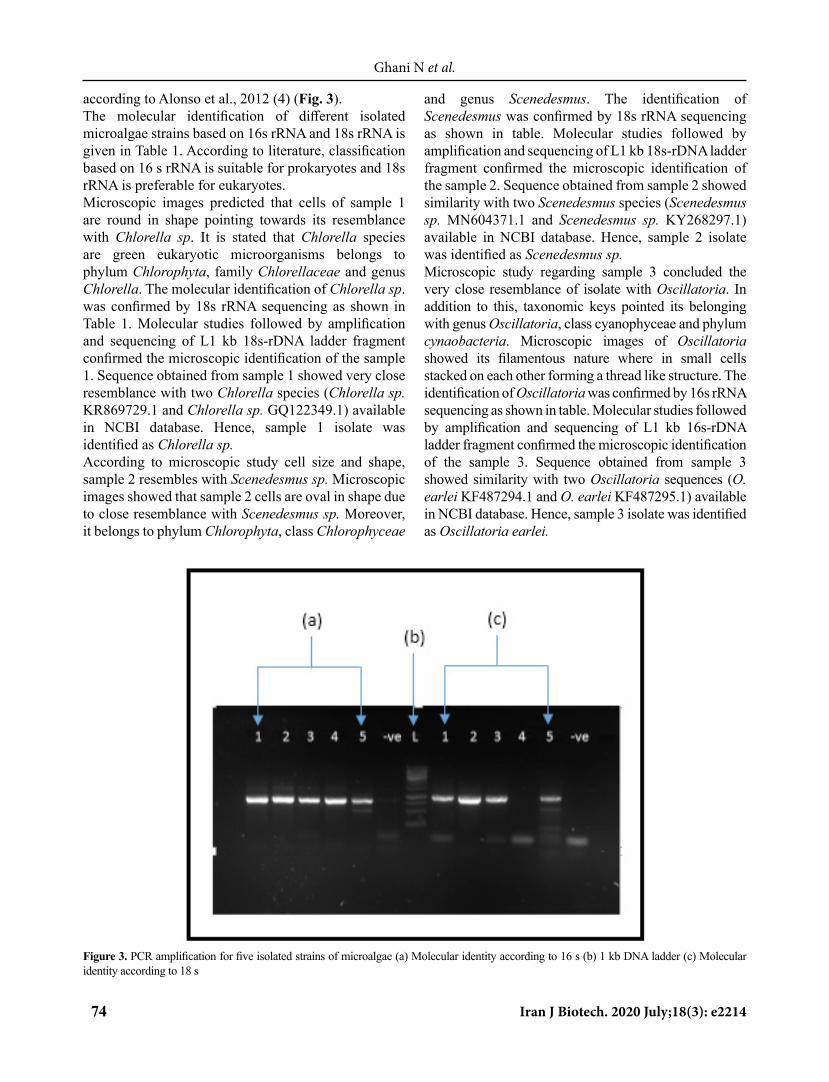
according to Alonso et al., 2012 (4) (Fig. 3). The molecular identification of different isolated microalgae strains based on 16s rRNA and 18s rRNA is given in Table 1. According to literature, classification based on 16 s rRNA is suitable for prokaryotes and 18s rRNA is preferable for eukaryotes. Microscopic images predicted that cells of sample 1 are round in shape pointing towards its resemblance with Chlorella sp. It is stated that Chlorella species are green eukaryotic microorganisms belongs to phylum Chlorophyta, family Chlorellaceae and genus Chlorella. The molecular identification of Chlorella sp. was confirmed by 18s rRNA sequencing as shown in Table 1. Molecular studies followed by amplification and sequencing of L1 kb 18s-rDNA ladder fragment confirmed the microscopic identification of the sample 1. Sequence obtained from sample 1 showed very close resemblance with two Chlorella species (Chlorella sp. KR869729.1 and Chlorella sp. GQ122349.1) available in NCBI database. Hence, sample 1 isolate was identified as Chlorella sp. According to microscopic study cell size and shape, sample 2 resembles with Scenedesmus sp. Microscopic images showed that sample 2 cells are oval in shape due to close resemblance with Scenedesmus sp. Moreover, it belongs to phylum Chlorophyta, class Chlorophyceae
and genus Scenedesmus. The identification of Scenedesmus was confirmed by 18s rRNA sequencing as shown in table. Molecular studies followed by amplification and sequencing of L1 kb 18s-rDNA ladder fragment confirmed the microscopic identification of the sample 2. Sequence obtained from sample 2 showed similarity with two Scenedesmus species (Scenedesmus sp. MN604371.1 and Scenedesmus sp. KY268297.1) available in NCBI database. Hence, sample 2 isolate was identified as Scenedesmus sp. Microscopic study regarding sample 3 concluded the very close resemblance of isolate with Oscillatoria. In addition to this, taxonomic keys pointed its belonging with genus Oscillatoria, class cyanophyceae and phylum cynaobacteria. Microscopic images of Oscillatoria showed its filamentous nature where in small cells stacked on each other forming a thread like structure. The identification of Oscillatoria was confirmed by 16s rRNA sequencing as shown in table. Molecular studies followed by amplification and sequencing of L1 kb 16s-rDNA ladder fragment confirmed the microscopic identification of the sample 3. Sequence obtained from sample 3 showed similarity with two Oscillatoria sequences (O. earlei KF487294.1 and O. earlei KF487295.1) available in NCBI database. Hence, sample 3 isolate was identified as Oscillatoria earlei.
Figure 3. PCR amplification for five isolated strains of microalgae (a) Molecular identity according to 16 s (b) 1 kb DNA ladder (c) Molecular identity according to 18 s

75Iran J Biotech. 2020 July;18(3): e2214
Ghani N et al.
Morphological features of sample 4 analyzed at microscopic level revealed that it has resemblance with spirulina. Furthermore, taxonomic keys investigated that it belongs to the genus spirulina, class cyanophyceae and phylum cyanobacteria. Microscopic images of spirulina subsalsa showed filamentous coiled and spiral appearance (16). The identification of spirulina subsalsa was confirmed by 16s rRNA sequencing as shown in table. Molecular studies followed by amplification and sequencing of L1 kb 16s-rDNA ladder fragment confirmed the microscopic identification of the sample 4. Sequence obtained from sample 4 showed similarity with two spirulina sequences (S. subsalsa AY575935.1 and S. subsalsa LC215280.1) available in NCBI database. Hence, sample 4 isolate was identified as Spirulina subsalsa.
4.4. Optimization of pH and Flocculent DoseGrowth of microalgae can be affected by change in pH in different ways. pH can alter the availability of
carbon, essential nutrients and trace metals. For the optimization of pH, experiment was conducted by varying pH of growth media from 3-12. It was found that 7 pH is more suitable for the growth of Chlorella, 7.5 for Scenedesmus, 10 for Oscillatoria and 9 for Spirulina (Fig. 4).High energy input for the harvesting of microalgae is one of the bottlenecks for its commercialization. This is because of their colloidal stability of suspension, lower cell density (less than 1 g.L) and small cell size (2-20 µm) in culture medium. In this study, potassium aluminum sulphate (alum) was used as flocculent for rapid harvesting of microalgal biomass. Dose of flocculent fluctuated from 0.01-1 % for its optimization. It was observed that 0.1 % flocculent is sufficient for the harvesting of Chlorella in 5 min and 0.05 % dose is enough for the harvesting of Scenedesmus, Oscillatoria and Spirulina but Scenedesmus takes 5 min for its complete flocculation while Oscillatoria and Spirulina take 15 min for their complete harvesting.
Figure 4. Optimization of pH for growth of (A) Chlorella sp. (B) Scenedesmus sp. (C) Oscillatoria sp. (D) Spirulina sp. (Growth media volume= 200mL; Inoculation size=1mL)
4.5. Different Properties of Isolated MicroalgaeBiomass productivity of isolated strains was found to investigate their possible utilization for different purposes. It was found that the biomass productivity of Chlorella, Scenedesmus, Oscillatoria and Spirulina is
0.94 g, 0.58 g, 4.2 g and 3.45 g per liter respectively. Oscillatoria has maximum biomass productivity.
4.5.1. Salinity ResistanceResistance of these strains against salinity was also
76 Iran J Biotech. 2020 July;18(3): e2214
Ghani N et al.
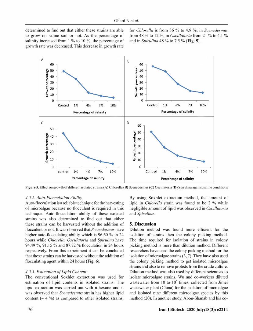
determined to find out that either these strains are able to grow on saline soil or not. As the percentage of salinity increased from 1 % to 10 %, the percentage of growth rate was decreased. This decrease in growth rate
for Chlorella is from 36 % to 4.9 %, in Scenedesmus from 48 % to 12 %, in Oscillatoria from 21 % to 4.1 % and in Spirulina 48 % to 7.5 % (Fig. 5).
Figure 5. Effect on growth of different isolated strains (A) Chlorella (B) Scenedesmus (C) Oscillatoria (D) Spirulina against saline conditions
4.5.2. Auto-Flocculation Ability Auto-flocculation is a reliable technique for the harvesting of microalgae because no flocculent is required in this technique. Auto-flocculation ability of these isolated strains was also determined to find out that either these strains can be harvested without the addition of flocculent or not. It was observed that Scenedesmus have higher auto-flocculating ability which is 96.60 % in 24 hours while Chlorella, Oscillatoria and Spirulina have 94.49 %, 91.15 % and 87.72 % flocculation in 24 hours respectively. From this experiment it can be concluded that these strains can be harvested without the addition of flocculating agent within 24 hours (Fig. 6).
4.5.3. Estimation of Lipid ContentThe conventional Soxhlet extraction was used for estimation of lipid contents in isolated strains. The lipid extraction was carried out with n-hexane and it was observed that Scenedesmus strain has higher lipid content (~ 4 %) as compared to other isolated strains.
By using Soxhlet extraction method, the amount of lipid in Chlorella strain was found to be 2 % while negligible amount of lipid was observed in Oscillatoria and Spirulina.
5. DiscussionDilution method was found more efficient for the isolation of strains then the colony picking method. The time required for isolation of strains in colony picking method is more than dilution method. Different researchers have used the colony picking method for the isolation of microalgae strains (3, 7). They have also used the colony picking method to get isolated microalgae strains and also to remove protists from the crude culture. Dilution method was also used by different scientists to isolate microalgae strains. Wu and co-workers diluted wastewater from 10 to 105 times, collected from Jimei wastewater plant (China) for the isolation of microalgae and isolated nine different microalgae species by this method (20). In another study, Abou-Shanab and his co-

77Iran J Biotech. 2020 July;18(3): e2214
Ghani N et al.
Autoflocculation of Chlorella sp.
Autoflocculation of Scedesmus sp.
Autoflocculation of Oscillatoria sp
Autoflocculation of Spirullina sp
Figure 6. Auto-flocculation of different isolated strains (Fully grown culture volume=200 mL)

78 Iran J Biotech. 2020 July;18(3): e2214
Ghani N et al.
worker isolated 45 algal species from freshwater lake by using dilution method (21).Microscopy is a simple and quick method for identification of unknown strains. The first isolated strain showed resemblance with Chlorella sp. (22). The cell size measurement and cell shape show that this isolated strain belongs to Chlorella sp. The reported cell size in literature for Chlorella is 2-4 µm (23). Illman et al., 2000 reported that diameter of Chlorella vulgaris ranges from 2-10 µm (17). Oval shape of cells and cell size of this strain show its close resemblance with Scenedesmus sp. (18). The third isolated strain consists of small cells stacked on each other to form long thread like structure which has close resemblance with Oscillatoria sp . Oscillatoria has been observed in sea waters by different researchers and it exists in the form of dense bloom (24, 25). The forth isolated strain was Spirulina sp. Spirulina sp. belonging to the family of oscillatoriaecae. Helical (spiral) shape of filaments is the characteristic of this genus, but the parameters such as dimensions and pitch length of this helix vary from species to species (26). Growth parameters such as temperature also affect the dimension of this helix. In solid media helical shape changed to true spirals (27). It is essential to determine the optimum pH for the growth of microalgae to enhance its growth rate and lipid content (28). Physiological effects potentially occur at extreme pH (29). Different researchers worked on the optimization of media pH for these microalgae species and our results are in accordance with their findings (30-32). In microalgae, there is no aggregation due to the negatively charged surface (33). This negative charge can be neutralized by adding different flocculants such as alum, ferric chloride and ferric sulfate (34). These flocculants increase particle size by the aggregation of microalgal cells which assist the process of flocculation. Flocculent should be less toxic, inexpensive and effective at lower concentration (35). The study results revealed that very little amount of alum is required for harvesting these strains which show their good self-flocculation ability. Chemical flocculent such as aluminum affect the composition of fatty acid methyl ester, use of its biomass for animal feed and cells of microalgae also damaged by aluminum salts. Moreover, these flocculent also remain present in the lipid extracted from microalgal biomass and interfere with the process of trans-esterification. The isolated strains depicted good biomass productivity especially Oscillatoria which is an important nitrogen fixer, good food source for zooplanktons and also meet oxygen demand of fish in ponds with their high biomass productivity (36, 37). The amount of lipid in isolated strains was not found so high for use in biofuel production. In this study, lipid
content was estimated using Soxhlet extraction with n-hexane. As microalgae cell was is rigid so some other cell disruption techniques might increase the chance of lipid release from microalgae cells for their possible use in bioenergy (38, 39).
6. ConclusionKallar Kahar lake Chakwal is the potential source of different useful microalgae strains. Four species of microalgae were successfully isolated from crude culture and identified on the basis of their morphology and molecular identification. Serial dilution method proved an efficient method for the isolation of different microalgae strains. It was found that from these species Oscillatoria and Spirulina require alkaline pH for their maximum growth. Maximum biomass can be obtained from Oscillatoria sp. due to its high biomass productivity and Scenedesmus sp. have high resistance against salinity as well as auto-flocculation ability as compared to other isolated strains.
AcknowledgementThe authors acknowledge the Government of Punjab Pakistan for providing financial assistance to Punjab Bio-Energy Institute, University of Agriculture Faisalabad. Authors are also thankful to Mr. Abdul Qayyum, Assistant Professor (English) Postgraduate College Attock for his help in improving the quality of this manuscript.
References1. Greenwell HC, Laurens L, Shields R, Lovitt R, Flynn K. Placing
Microalgae on the Biofuels Priority List: A Review of the Technological Challenges. J R Soc Interface. 2010;7(46):703-26. https://doi.org/10.1098/rsif.2009.0322
2. Duong VT, Li Y, Nowak E, Schenk PM. Microalgae Isolation and Selection for Prospective Biodiesel Production. Energies. 2012;5(6):1835-49. https://doi.org/10.3390/en5061835
3. Mutanda T, Ramesh D, Karthikeyan S, Kumari S, Anandraj A, Bux F. Bioprospecting for Hyper-Lipid Producing Microalgal Strains for Sustainable Biofuel Production. Bioresour technol. 2011;102(1):57-70. https://doi.org/10.1016/j.biortech.2010.06.077
4. Brennan L, Owende P. Biofuels from Microalgae—A Review of Technologies for Production, Processing, and Extractions of Biofuels and Co-products. Renew Sust Energ Rev. 2010;14(2):557-77. https://doi.org/10.1016/j.rser.2009.10.009
5. Spolaore P, Joannis-Cassan C, Duran E, Isambert A. Commercial Applications of Microalgae. J Biosci Bioeng. 2006;101(2):87-96. https://doi.org/10.1263/jbb.101.87
6. Platt T, Fuentes-Yaco C, Frank KT. Spring Algal Bloom and Larval Fish Survival. Nature. 2003;423(6938):398-9. https://doi.org/10.1038/423398b
7. Parvin M, Zannat M, Habib M. Two Important Techniques for Isolation of Microalgae. Asian Fish. Sci. 2007;20(1/2):117.
8. Kim S-K, Ravichandran YD, Khan SB, Kim YT. Prospective of the Cosmeceuticals Derived from Marine Organisms. Biotechnol Bioproc E. 2008;13(5):511-23. https://doi.org/10.1007/s12257-008-0113-5

79Iran J Biotech. 2020 July;18(3): e2214
Ghani N et al.
9. Cardinale BJ. Biodiversity Improves Water Quality Through Niche Partitioning. Nature. 2011;472(7341):86-9. https://doi.org/10.1038/nature09904
10. Jahn MT, Schmidt K, Mock T. A Novel Cost Effective and High-Throughput Isolation and Identification Method for Marine Microalgae. Plant Methods. 2014;10(1):26. https://doi.org/10.1186/1746-4811-10-26
11. Yahaya MF, Demshemino I, Nwadike I, Sylvester O, Okoro LN. A Review on the Chromatographic Analysis of Biodiesel. Int. J. Educ. Res. 2013;1(8).
12. Janaun J, Ellis N. Perspectives on Biodiesel as a Sustainable Fuel. Renew Sust Energ Rev. 2010;14(4):1312-20. https://doi.org/10.1016/j.rser.2009.12.011
13. Ponnuswamy I, Soundararajan Madhavan D, Shabudeen S, Shoba U. Isolation and Identification of Green Microalgae for Carbon Sequestration & Waste Water Treatment by Using PCR Studies. Int J Innov Res Sci Eng Technol. 2008.
14. Hutagalung RA, Sukoco AE, Soedharma D, Goreti LM, Andrean I, Elshaddai B, et al. Isolation, Identification and Growth Optimization of Microalgae Derived from Soft Coral Dendronephthya sp. Apcbee Procedia. 2014;10:205-310. https://doi.org/10.1016/j.apcbee.2014.10.057
15. Alonso M, Lago FC, Vieites JM, Espiñeira M. Molecular Characterization of Microalgae Used in Aquaculture with Biotechnology Potential. Aquac Int. 2012;20(5):847-57. https://doi.org/10.1007/s10499-012-9506-8
16. Dar RA, Arora M, Phutela UG. Optimization of Cultural Factors of Newly Isolated Microalga Spirulina Subsalsa and its Co-digestion with Paddy Straw for Enhanced Biogas Production. Bioresour. Technol Reports. 2019;5:185-98. https://doi.org/10.1016/j.biteb.2019.01.009
17. Illman A, Scragg A, Shales S. Increase in Chlorella Strains Calorific Values when Grown in Low Nitrogen Medium. Enzyme Microb. Technol. 2000;27(8):631-5. https://doi.org/10.1016/s0141-0229(00)00266-0
18. Olenina I. Biovolumes and Size-Classes of Phytoplankton in the Baltic Sea. 2006.
19. Mohan N, Sivasubramanian V. Mass Cultivation of _Chroococcus Turgidus_ and _Oscillatoria sp. _ and Effective Harvesting of Biomass by Low-cost methods. Nature Precedings. 2010:1-. https://doi.org/10.1038/npre.2010.4331.1
20. Wu Y, Guan K, Wang Z, Xu B, Zhao F. Isolation, Identification and Characterization of an Electrogenic Microalgae Strain. PloS one. 2013;8(9). https://doi.org/10.1371/journal.pone.0073442
21. Abou-Shanab RA, Matter IA, Kim S-N, Oh Y-K, Choi J, Jeon B-H. Characterization and Identification of Lipid-Producing Microalgae Species Isolated from a Freshwater Lake. Biomass Bioenerg. 2011;35(7):3079-85. https://doi.org/10.1016/j.biombioe.2011.04.021
22. Held P, Raymond K. Determination of Algal Cell Lipids Using Nile Red-Using Microplates to Monitor Neutral Lipids in Chlorella Vulgaris. Biofuel Res J. 2011;8:1-5.
23. Converti A, Casazza AA, Ortiz EY, Perego P, Del Borghi M. Effect of Temperature and Nitrogen Concentration on the Growth and Lipid Content of Nannochloropsis Oculata and Chlorella Vulgaris for Biodiesel Production. Chem Eng Process. 2009;48(6):1146-51. https://doi.org/10.1016/j.cep.2009.03.006
24. Rani VU, Perumal UE, Palanivel S. Morphology and Taxonomy of Oscillatoria Princeps Vaucher ex Gomont (OSCILLATORIALES, OSCILLATORIACEAE). Indian J Edu Inf Manage. 2016;5:1.
25. Shamina M, Ram AT. Occurrence of Oscillatoria Perornata Skuja f. Attenuata Skuja (Oscillatoriacae): New Report to India from Marine Habitat. Int J Curr Microbiol App Sci. 2014;3(7):648-50.
26. Usharani G, Saranraj P, Kanchana D. Spirulina Cultivation: A Review. Int J Pharm Biol Arch. 2012;3(6).
27. Ali SK, Saleh AM. Spirulina-An Overview. Int J Pharm. 2012;4(3):9-15.
28. Cabello J, Toledo-Cervantes A, Sánchez L, Revah S, Morales M. Effect of the Temperature, pH and Irradiance on the Photosynthetic Activity by Scenedesmus Obtusiusculus under Nitrogen Replete and Deplete Conditions. Bioresour Technol. 2015;181:128-35. https://doi.org/10.1016/j.biortech.2015.01.034
29. Sutherland DL, Howard-Williams C, Turnbull MH, Broady PA, Craggs RJ. The Effects of CO2 Addition along a pH Gradient on Wastewater Microalgal Photo-physiology, Biomass Production and Nutrient Removal. Water Res. 2015;70:9-26. https://doi.org/10.1016/j.watres.2014.10.064
30. Kim W, Park JM, Gim GH, Jeong S-H, Kang CM, Kim D-J, et al. Optimization of Culture Conditions and Comparison of Biomass Productivity of Three Green Algae. Bioproc Biosyst Eng. 2012;35(1-2):19-27. https://doi.org/10.1007/s00449-011-0612-1
31. Buayam N, Davey MP, Smith AG, Pumas C. Effects of Copper and pH on the Growth and Physiology of Desmodesmus sp. AARLG074. Metabolites. 2019;9(5):84. https://doi.org/10.3390/metabo9050084
32. Ray S, Bagchi S. Nutrients and pH Regulate Algicide Accumulation in Cultures of The Cyanobacterium Oscillatoria Laetevirens. New Phytol. 2001;149(3):455-60. https://doi.org/10.1046/j.1469-8137.2001.00061.x
33. Wan C, Alam MA, Zhao X-Q, Zhang X-Y, Guo S-L, Ho S-H, et al. Current Progress and Future Prospect of Microalgal Biomass Harvest Using Various Flocculation Technologies. Bioresour Technol. 2015;184:251-7. https://doi.org/10.1016/j.biortech.2014.11.081
34. Grima EM, Belarbi E-H, Fernández FA, Medina AR, Chisti Y. Recovery of Microalgal Biomass and Metabolites: Process Options and Economics. Biotechnol Adv. 2003;20(7-8):491-515. https://doi.org/10.1016/s0734-9750(02)00050-2
35. Lal A, Das D. Biomass Production and Identification of Suitable Harvesting Technique for Chlorella sp. MJ 11/11 and Synechocystis PCC 6803. 3 Biotech. 2016;6(1):41. https://doi.org/10.1007/s13205-015-0360-z
36. Tõnno I, Agasild H, Kõiv T, Freiberg R, Nõges P, Nõges T. Algal Diet of Small-Bodied Crustacean Zooplankton in a Cyanobacteria-Dominated Eutrophic Lake. PloS one. 2016;11(4). https://doi.org/10.1371/journal.pone.0154526
37. Zimba PV, Grimm CC, Dionigi CP, Weirich CR. Phytoplankton Community Structure, Biomass, and Off‐Flavor: Pond Size Relationships in Louisiana Catfish Ponds. J World Aquacult Soc. 2001;32(1):96-104. https://doi.org/10.1111/j.1749-7345.2001.tb00927.x
38. Ramzan H, Sadaf S, Iqbal J, Ullah I, Mahr S, Ali E, et al. Solar Assisted Cell Wall Disruption of Indigenously Isolated Microalgae Strains: Process Optimization. Mater Res Express. 2019;6(6):065506. https://doi.org/10.1088/2053-1591/ab0cb6
39. Akram S, Iqbal J, Ullah I, Rafique T, Bhatti IA, Sadaf S. Bismuth Vanadate: An Efficient Photocatalyst for Rupturing of Microalgae Cell Wall. Mater Res Express. 2019;6(8):085502. https://doi.org/10.1088/2053-1591/ab1aed
Copyright © 2022 FDOKUMEN