Isolation and screening of marine microalgae from Kendari ...
11
AACL Bioflux, 2018, Volume 11, Issue 5. http://www.bioflux.com.ro/aacl 1445 Isolation and screening of marine microalgae from Kendari waters, Southeast Sulawesi, Indonesia suitable for outdoor mass cultivation in hypersaline media Indrayani, Haslianti, Asriyana Department of Aquatic Resources Management, Faculty of Fisheries and Marine Sciences, Halu Oleo University, Kampus Bumi Tridharma Anduonohu, Kendari 93232, Southeast Sulawesi, Indonesia. Corresponding author: Indrayani, [email protected] Abstract. Screening of local microalgae species for any commercial application has a competitive advantage as they are well adapted to the local climatic environment. Microalgae capable to grow in hypersaline media are particularly interesting as they are more sustainable and economical to grow and less prone to contaminations. The aim of this study was to isolate and screen local microalgae species from Kendari waters, Southeast Sulawesi, Indonesia suitable for mass cultivation in outdoor open pond systems in hypersaline media. The water samples for isolation of microalgae were collected from several coastal areas in Kendari, Southeast Sulawesi including Tanjung Tiram Beach, Nambo Beach, Batu Gong Beach, Toronipa Beach and Bokori Island. The isolation of microalgae was done using agar plating technique (1% agar in f/2 medium). The water samples were plated on agar medium and incubated under dim light, 12:12 hours light and dark cycle and at ambient room temperature. There are hundreds of isolates generated after repeated streaking on agar medium. The isolates are dominated by diatom and cyanobacteria. Three isolates (IND-UHO-002, IND-UHO-003 and IND-UHO-029) showed fast growth at high salinity (5% NaCl) and hence are promising strains for mass cultivation in outdoor open pond system in hypersaline media. Key Words: diatom, cyanobacteria, local microalgae species, high salinity. Introduction. Microalgae are prokaryotic or eukaryotic photosynthetic microorganisms that can be found in all ecosystems both aquatic and terrestrial (Richmond 2004; Mata et al 2010). They are an extremely heterogeneous group of microorganisms which are potentially rich source of important chemicals with potential application in the feed, food, nutritional, cosmetics, pharmaceuticals and even in fuel industries (Olaizola 2003). Although, there are over thousands or even millions of microalgae species existing in nature (Hannon et al 2010), only a few of them have been successfully produced commercially for the production of high value products (i.e. Dunaliella salina for β- carotene, Haematococcus pluvialis for astaxanthin and Crypthecodinium cohnii for EPA production) (Saha & Murray 2018; Khan et al 2018). Hence, bioprospecting of microalgae species with commercial potential is thus an important and challenging task. Species or strain selection is the first and critical step in bioprospecting of microalgae for any commercial application (Barclay & Apt 2013; Borowitzka 2013a). Screening of microalgae species involves a series of steps including sample collection, isolation, purification, identification, maintenance and characterization of potential products (Gong & Jiang 2011). There are two possible sources of selecting/screening microalgae; from microalgae culture collections and from natural environments. Species selection through microalgae culture collections can be accessed easily although the number of microalgae species kept in the culture collection is only a small fraction of microalgae species that exist in nature (Borowitzka 2013a). On the other hand, untapped resources of microalgae species can be isolated from natural environments. Isolating and selecting of local microalgae species/strains has a competitive advantage especially for
-
Upload
khangminh22 -
Category
Documents
-
view
1 -
download
0
Transcript of Isolation and screening of marine microalgae from Kendari ...

AACL Bioflux, 2018, Volume 11, Issue 5. http://www.bioflux.com.ro/aacl 1445
Isolation and screening of marine microalgae from Kendari waters, Southeast Sulawesi, Indonesia suitable for outdoor mass cultivation in hypersaline media Indrayani, Haslianti, Asriyana
Department of Aquatic Resources Management, Faculty of Fisheries and Marine Sciences,
Halu Oleo University, Kampus Bumi Tridharma Anduonohu, Kendari 93232, Southeast Sulawesi, Indonesia. Corresponding author: Indrayani, [email protected]
Abstract. Screening of local microalgae species for any commercial application has a competitive advantage as they are well adapted to the local climatic environment. Microalgae capable to grow in hypersaline media are particularly interesting as they are more sustainable and economical to grow and less prone to contaminations. The aim of this study was to isolate and screen local microalgae species from Kendari waters, Southeast Sulawesi, Indonesia suitable for mass cultivation in outdoor open pond systems in hypersaline media. The water samples for isolation of microalgae were collected from several coastal areas in Kendari, Southeast Sulawesi including Tanjung Tiram Beach, Nambo Beach, Batu Gong Beach, Toronipa Beach and Bokori Island. The isolation of microalgae was done using agar plating technique (1% agar in f/2 medium). The water samples were plated on agar medium and incubated under dim light, 12:12 hours light and dark cycle and at ambient room temperature. There are hundreds of isolates generated after repeated streaking on agar medium. The isolates are dominated by diatom and cyanobacteria. Three isolates (IND-UHO-002, IND-UHO-003 and IND-UHO-029) showed fast growth at high salinity (5% NaCl) and hence are promising strains for mass cultivation in outdoor open pond system in hypersaline media. Key Words: diatom, cyanobacteria, local microalgae species, high salinity.
Introduction. Microalgae are prokaryotic or eukaryotic photosynthetic microorganisms that can be found in all ecosystems both aquatic and terrestrial (Richmond 2004; Mata et al 2010). They are an extremely heterogeneous group of microorganisms which are potentially rich source of important chemicals with potential application in the feed, food, nutritional, cosmetics, pharmaceuticals and even in fuel industries (Olaizola 2003). Although, there are over thousands or even millions of microalgae species existing in nature (Hannon et al 2010), only a few of them have been successfully produced commercially for the production of high value products (i.e. Dunaliella salina for β-carotene, Haematococcus pluvialis for astaxanthin and Crypthecodinium cohnii for EPA production) (Saha & Murray 2018; Khan et al 2018). Hence, bioprospecting of microalgae species with commercial potential is thus an important and challenging task.
Species or strain selection is the first and critical step in bioprospecting of microalgae for any commercial application (Barclay & Apt 2013; Borowitzka 2013a). Screening of microalgae species involves a series of steps including sample collection, isolation, purification, identification, maintenance and characterization of potential products (Gong & Jiang 2011). There are two possible sources of selecting/screening microalgae; from microalgae culture collections and from natural environments. Species selection through microalgae culture collections can be accessed easily although the number of microalgae species kept in the culture collection is only a small fraction of microalgae species that exist in nature (Borowitzka 2013a). On the other hand, untapped resources of microalgae species can be isolated from natural environments. Isolating and selecting of local microalgae species/strains has a competitive advantage especially for

AACL Bioflux, 2018, Volume 11, Issue 5. http://www.bioflux.com.ro/aacl 1446
microalgae species intending to be mass produced in outdoor as they are well adapted to the local climatic environment (Larkum et al 2012).
Marine microalgae that can grow in saline-hypersaline media are particularly interesting because they are more sustainable and economical to grow compared to freshwater microalgae. Marine microalgae use seawater that can be accessed easily and freely by just pumping the seawater from coastal areas whereas freshwater microalgae require limited and expensive freshwater. Marine microalgae can be grown in non-arable lands and therefore will not compete for agriculturals (Borowitzka & Moheimani 2013b). Moreover, hypersaline microalgae are less prone to contaminations by other microorganisms (i.e microalgae, protozoas and bacteria) as not many organisms could withstand high salt concentration. Therefore, the aim of this study was to isolate local microalgae species from Kendari waters, Southeast Sulawesi, Indonesia and to screen them for potential mass cultivation in outdoor open pond systems in saline-hypersaline media for any commercial applications. This study is the first to isolate local microalgae species from several coastal areas in Kendari and to initiate the establisment of the microalgae culture collection in Kendari, Southeast Sulawesi, Indonesia.
Material and Method Sampling sites. Sampling of microalgae was conducted from March to May 2017 at several coastal areas in Kendari, Southeast Sulawesi, Indonesia including Tiram Cape (4o2’24”BT-122o40’1”LS), Nambo Beach (4o0’5”BT-122o36’58”LS), Batu Gong Beach (3o52’43”BT-122o30’45”LS), Toronipa Beach (3o54’0”BT-122o39’36”LS) and Bokori Island (3o55’34”BT-122o40’0”LS) (Figure 1).
Figure 1. Sampling sites.
Sample collection. Water samples were collected manually using plastic water bottles volume 1.5 L and also using a plankton net mesh size 25 μm. For the plankton net, about 50 litres of seawater at each location were filtered through the plankton net and collected in a 50 mL tube. In the lab, the water samples from each location were transferred into small glass bottles (100 mL) and enriched with nutrients (nitrate, phosphate, silicate and trace elements) based on f/2 medium stock solution (Guillard & Ryther 1962) and then incubated in a culture shelve with illumination of a 20 watt of white fluorescent tubes under 12:12 hour light and dark cycles and at ambient room temperature whereas the sample from plankton net was prepared for isolation straight away.

AACL Bioflux, 2018, Volume 11, Issue 5. http://www.bioflux.com.ro/aacl 1447
Isolation and identification of microalgae. Isolation of the microalgae was carried out using agar plating technique (Andersen & Kawachi 2005). The medium used was f/2 medium (Guillard & Ryther 1962). Agar medium was prepared by adding 1% of agar into the liquid medium prior to autoclaving.
Sample from plankton net (0.1 mL) was spotted on the middle of the agar plates and spread evenly on the surface using a glass spreader. The plates were then sealed with parafilm to avoid drying out and incubated in the culture shelve under dim light (a 20 watt of white fluorescent tube light) with 12 h light and 12 h dark cycle and at room temperature. The same protocols were applied for the enrichment samples.
Colonies emerged on the agar plates were picked up and re-streaked onto fresh agar medium using standard microbiology agar streaking technique. Pure unialgal colonies were obtained after repeated streaking on fresh agar media and confirmed by microscopic observation. The pure colonies were then transferred into liquid medium by inoculating a single colony into each well of the 24 and 96-wells microtiter plate containing 1 mL and 200 μL of f/2 medium, respectively.
Identification of the microlagal isolates was based on the morphological characteristics using a microscope and phytoplankton identification books (Yamaji 1976; Newell & Newell 1977; Tomas 1997; Barsanti & Gualtieri 2006). The isolates were identified up to genus level. Screening of microalgae suitable for mass cultivation in hypersaline media in outdoors. For screening, new isolates were grown in microtiter well plates and then gradually scale-up from 0.2, 2, 10, 50 and 100 mL at ambient room temperature (28-34oC), 12h:12h light and dark cycles and under light intensity of 2 x 20 watt of white florescent tube light (about 70-80 μmol.photon.m-2.s-2). The cultures were then observed for their ability to grow in liquid medium. The cultures that grew well in liquid medium were then selected for further screening test. About 20 mL aliquots of the cultures were then transferred to flasks containing 50 mL of f/2 medium at three different salinities (3, 4 and 5% NaCl) and cultured for one week. The growth of the cultures was monitored every day by measuring the optical density of the cultures at OD750 nm using UV-Spectrometer (Varian, Cary 50 UV Visible). The specific growth rate (μ) was then calculated using the following equation (Moheimani et al 2013):
μ = Ln (N2/N1)/t2-t1 where: N1 and N2 are the OD reading at time 1 (t1) and 2 (t2) within the exponential phase. Results Isolation of microalgae. Hundreds of microalgal isolates (pure colonies) have been succesfully established after repeated streaking on agar f/2 medium (Figure 1a) and after growing on 24 and 96 microtiter well-plates (Figure 1b). Out of hundreds of isolates on agar, about 67 isolates successfully grown in liquid medium (Figure 1c).
Figure 2. Isolates grown on agar plate (a), 24 and 96 microtiter well-plates (b) and small
containers and flasks (c).
c b a

AACL Bioflux, 2018, Volume 11, Issue 5. http://www.bioflux.com.ro/aacl 1448
Identification of the microalgal species was only performed on the species that can grow in liquid medium. The isolates grown in liquid medium are dominated by diatoms/ Bacillariophyceae including Diatoma sp., Navicula sp., Melosira sp., Synura sp., Surirella sp.), filamentous cyanobacteria (Oscillatoria sp. and Anabaena sp.) and green algae/Chlorophyte including Chlorella sp. and Chlamydomonas sp. (Table 1). Most of the diatom species isolated are from Bokori Island and Tanjung Tiram Beach whereas the filamentous cyanobacteria and the green algae are from Batu Gong, Toronia Beach and Nambo Beach.
Table 1 Identification of newly isolated local species microalgae in Coastal areas in Kendari Southeast
Sulawesi Indonesia
Isolate code Scientific name Place of collection IND-UHO 001 Diatoma sp. Bokori Island IND-UHO 002 Diatoma sp. Bokori Island IND-UHO 003 Chlorella sp. Bokori Island IND-UHO 004 Diatoma sp. Bokori Island IND-UHO 005 Navicula sp. Tanjung Tiram Beach IND-UHO 006 Navicula sp. Tanjung Tiram Beach IND-UHO 007 Navicula sp. Tanjung Tiram Beach IND-UHO 008 Diatoma sp. Tanjung Tiram Beach IND-UHO 009 Navicula sp. Tanjung Tiram Beach IND-UHO 010 Navicula sp. Tanjung Tiram Beach IND-UHO 011 Diatoma sp. Bokori Island IND-UHO 012 Synura sp. Bokori Island IND-UHO 013 Synura sp. Bokori Island IND-UHO 014 Synura sp. Bokori Island IND-UHO 015 Navicula sp. Bokori Island IND-UHO 016 Navicula sp. Bokori Island IND-UHO 017 Navicula sp. Bokori Island IND-UHO 018 Nitzschia sp. Bokori Island IND-UHO 019 Aphanocapsa sp. Bokori Island IND-UHO 020 Tabellaria sp. Bokori Island IND-UHO 021 Aphanocapsa sp. Bokori Island IND-UHO-022 Porphyridium sp. Bokori Island IND-UHO-023 Tetraselmis sp. Bokori Island IND-UHO 025 Surirella sp. Bokori Island IND-UHO 028 Diatoma sp. Bokori Island IND-UHO 029 Melosira sp. Nambo Beach IND-UHO 030 Melosira sp. Nambo Beach IND-UHO 031 Oscillatoria sp. Nambo Beach IND-UHO 033 Chlorella sp. Bokori Island IND-UHO 034 Oscillatoria sp. Batu Gong Beach IND-UHO 035 Oscillatoria sp. Bato Gong Beach IND-UHO 036 Oscillatoria sp. Toronipa Beach IND-UHO 037 Oscillatoria sp. Toronipa Beach IND-UHO 038 Oscillatoria sp. Nambo Beach IND-UHO 039 Dinobryon sp. Nambo Beach IND-UHO 040 Melosira sp. Nambo Beach IND-UHO 041 Oscillatoria sp. Nambo Beach IND-UHO 042 Oscillatoria sp. Nambo Beach IND-UHO 043 Oscillatoria sp. Nambo Beach IND-UHO 044 Oscillatoria sp. Nambo Beach IND-UHO 045 Oscillatoria sp. Nambo Beach IND-UHO 046 Oscillatoria sp. Nambo Beach IND-UHO 051 Oscillatoria sp. Toronipa Beach IND-UHO 052 Oscillatoria sp. Toronipa Beach IND-UHO 053 Oscillatoria sp. Toronipa Beach IND-UHO 054 Oscillatoria sp. Toronipa beach IND-UHO 055 Oscillatoria sp. Toronipa Beach IND-UHO 056 Oscillatoria sp. Toronipa Beach IND-UHO 057 Oscillatoria sp. Batu Gong Beach

AACL Bioflux, 2018, Volume 11, Issue 5. http://www.bioflux.com.ro/aacl 1449
IND-UHO 058 Navicula sp. Batu Gong Beach IND-UHO 059 Anabaena sp. Batu Gong Beach IND-UHO 060 Oscillatoria sp. Batu Gong Beach IND-UHO 061 Oscillatoria sp. Batu Gong Beach IND-UHO 062 Oscillatoria sp. Batu Gong Beach IND-UHO 063 Navicula sp. Batu Gong Beach IND-UHO 064 Melosira sp. Tanjung Tiram Beach IND-UHO 065 Oscillatoria sp. Tanjung Tiram Beach IND-UHO 066 Oscillatoria sp. Tanjung Tiram Beach IND-UHO 067 Diatoma sp. Nambo Beach IND-UHO 068 Navicula sp. Nambo Beach IND-UHO 069 Coscinodiscus sp. Toronipa Beach IND-UHO 070 Oscillatoria sp. Batu Gong Beach IND-UHO 072 Chlamydomonas sp. Tanjung Tiram Beach IND-UHO 073 Diatoma sp. Nambo Beach IND-UHO 075 Oscillatoria sp. Nambo Beach IND-UHO 076 Chlorella sp. Nambo Beach IND-UHO 077 Chlorella sp. Nambo Beach
Screening of newly isolated microalgae suitable for mass cultivation in outdoors in hypersaline media. Out of 67 isolates grown in liquid medium, there were about 8 isolates that showed good growth and did not stick to the culture vessels so that they remain in suspension to get expose to light and nutrients for optimum growth. These isolates were then screened for their ability to grow at high salinity (Figure 3).
Figure 3. Photomicrograph (at 100x magnification) of newly isolated microalgae potential for mass cultivation in outdoors: A. isolate Diatoma sp. IND-UHO-002, B. isolate Chlorella sp. IND-UHO-003, C. isolate Navicula sp. IND-UHO-017, D. isolate Navicula sp. IND-UHO-018, E. isolate Aphanocapsa sp. IND-UHO-019, F. isolate Porphyridium sp. IND-UHO-022, G. isolate Melosira sp. IND-UHO-029,
H. Isolate Chlamydomonas sp. IND-UHO-072.
A B C
D E F
H G

AACL Bioflux, 2018, Volume 11, Issue 5. http://www.bioflux.com.ro/aacl 1450
Out of eight isolates, three isolates namely isolate Melosira sp. IND-UHO-029, Chlorella sp. IND-UHO-003 and Diatoma sp. IND-UHO-002 showed very good growth over wide ranges of salinity tested (3-5% NaCl) and showed no lag phase following inoculation indicating that they can adapt well at high salinity (Figure 4).
Figure 4. Growth curves of the newly isolated microalgae from Kendari waters at different
salinities.
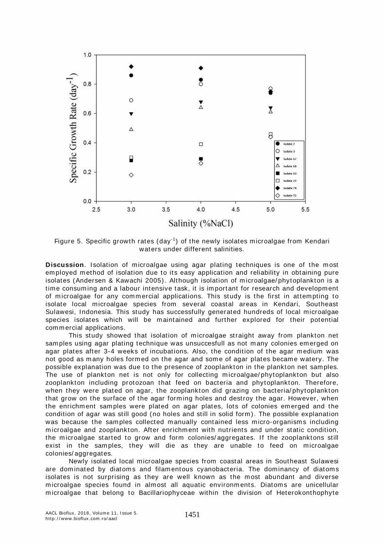
The specific growth rates of eight isolates under different salinity are shown in the Figure 5. The isolate IND-UHO-029 had the higest specific growth rate at salinity 3 and 4% NaCl followed by isolates IND-UHO-002 and IND-UHO-003. However, at 5% NaCl, the three isolate had about the same specific growth rate of about 0.74-0.77 d-1. Isolates IND-UHO-017 and IND-UHO-018 showed similar pattern in which the specific growth rate increased and reached maksimum at 4% salinity of about 0.68 and 0.64 d-1, respectively before decreasing at 5% to about 0.64 and 0.61 d-1, respectively. It is interestingly to note that the specific growth rates of isolate IND-UHO 019, IND-UHO 022 and IND-UHO 72 increased as the salinity increase.
AACL Bioflux, 2018, Volume 11, Issue 5. http://www.bioflux.com.ro/aacl 1451
Figure 5. Specific growth rates (day-1) of the newly isolates microalgae from Kendari
waters under different salinities. Discussion. Isolation of microalgae using agar plating techniques is one of the most employed method of isolation due to its easy application and reliability in obtaining pure isolates (Andersen & Kawachi 2005). Although isolation of microalgae/phytoplankton is a time consuming and a labour intensive task, it is important for research and development of microalgae for any commercial applications. This study is the first in attempting to isolate local microalgae species from several coastal areas in Kendari, Southeast Sulawesi, Indonesia. This study has successfully generated hundreds of local microalgae species isolates which will be maintained and further explored for their potential commercial applications.
This study showed that isolation of microalgae straight away from plankton net samples using agar plating technique was unsuccesfull as not many colonies emerged on agar plates after 3-4 weeks of incubations. Also, the condition of the agar medium was not good as many holes formed on the agar and some of agar plates became watery. The possible explanation was due to the presence of zooplankton in the plankton net samples. The use of plankton net is not only for collecting microalgae/phytoplankton but also zooplankton including protozoan that feed on bacteria and phytoplankton. Therefore, when they were plated on agar, the zooplankton did grazing on bacteria/phytoplankton that grow on the surface of the agar forming holes and destroy the agar. However, when the enrichment samples were plated on agar plates, lots of colonies emerged and the condition of agar was still good (no holes and still in solid form). The possible explanation was because the samples collected manually contained less micro-organisms including microalgae and zooplankton. After enrichment with nutrients and under static condition, the microalgae started to grow and form colonies/aggregates. If the zooplanktons still exist in the samples, they will die as they are unable to feed on microalgae colonies/aggregates.
Newly isolated local microalgae species from coastal areas in Southeast Sulawesi are dominated by diatoms and filamentous cyanobacteria. The dominancy of diatoms isolates is not surprising as they are well known as the most abundant and diverse microalgae species found in almost all aquatic environments. Diatoms are unicellular microalgae that belong to Bacillariophyceae within the division of Heterokonthophyte

AACL Bioflux, 2018, Volume 11, Issue 5. http://www.bioflux.com.ro/aacl 1452
(Falciatore & Bowler 2002) and considered to be the most important group of phytoplankton responsible for almost 40% of marine primary productivity (Falkowski et al 1998). According to Scala & Bowler (2001), there are over 250 genera of diatoms with perhaps as many as 100,000 species which are more than three orders of magnitude in size of land plants and exhibit a remarkable variety of shapes. The diatoms can be easily distinguished based on their morphological characteristics especially their ability to generate highly ornamented silicious cell walls (frustules) constructed of two almost equal valves like a petri dish (Falciatore & Bowler 2002). Besides diatoms, another group of microalgae dominated the isolates is cyanobacteria. Cyanobacteria which is also known as blue-green algae are a diverse group of prokaryotic photosynthetic microorganisms that can grow rapidly due to their simple structures and growth requirements as well as efficient use of light, CO2 and other inorganic nutrients (Parmar et al 2011).
For commercial production of microalgae for any applications, microalgae should be able to grow at a large scale production system. Outdoor open pond systems are the most employed commercial production system of microalgae in the world (Borowitzka & Moheimani 2013a). For example, commercial production of Dunaliella salina for β-carotene production at Hutt Lagoon Australia (Borowitzka 2013b), Arthrospira by Earthrise Nutritionals, LLC (California, USA) and Hainan DIC Microalgae (China) and to produce astaxanthin from Haematococcus pluvialis by Cyanotech Co. (Hawaii, USA) and Parry Agro Industries Ltd (India) (Zittelli et al 2013). However, not many microalgae are suitable for mass cultivation in outdoor open pond systems. There are several key criteria of microalgae suitable for large scale cultivation in outdoors including the ability of the microalgae to tolerate wide range of temperature and salinity (Brennan & Owende 2010; Fon-Sing et al 2013; Indrayani 2017). In this study, we have successfully isolated and screened several microalgal strains that can grow well over a wide range of salinities. There are three strains that show the highest growth rate at high salinity up to 5% NaCl (note that the seawater salinity is ranging from 3 to 3.4% NaCl) namely Diatoma sp. IND-UHO-002, Chlorella sp. IND-UHO-003 and Melosira sp. IND-UHO-029. Unlike freshwater algae that would be very stressfull and could possibly death when expose to seawater salinity, marine algae are more tolerant to salinity changes where the optimal salinity for growth is normally near seawater salinity (Borowitzka 2018).
The ability of microalgae to tolerate wide range of salinity and temperature are the most important criteria for the success of microalgal culture under outdoor conditions because outdoor cultures will be exposed to varying environmental conditions (Khatoon et al 2010). The salinity of the culture could decrease due to dilution of heavy rains or it could increase due to extensive evaporation of hot sunny days. Therefore, the microalgal species capable of growing well over wide range of salinity will not be affected by variation of the outdoor conditions and could sustain high productivy throughout the year. Microalgal species capable of growing at hypersaline environment will also be less prone to contamination as not many organisms could withstand high salt concentration and therefore monoalgal cultures could be maintained for long periods in outdoors (Preetha et al 2012; Barclay & Apt 2013; Borowitzka 2013a; Fon-Sing & Borowitzka 2016). Most important, microalgae capable to grow at high salinity are more sustainable and economical to grow as they can utilize seawater which is free and plenty instead of use of expensive and limited fresh water (Borowitzka & Moheimani 2013b; Resurreccion et al 2012; Yang et al 2011). Similarly, cultures will be exposed to daily and seasonal variation of temperatures. The microalgal strains were isolated from local environment and therefore the temperature optima for these algae generally reflect the temperature conditions found in their natural environments. For a tropical country like Indonesia, temperature is relatively constant with little fluctuation. The daily air temperatures range between 22 and 34oC and that is the reason why we tested to grow the isolates at ambient room temperature (28-34oC) and then selected the species that can withstand ambient temperature. As pointed out by Payer et al (1980), it is important to select microalgal strains that have optimum temperatures within the range of temperatures that will be encountered in outdoor.

AACL Bioflux, 2018, Volume 11, Issue 5. http://www.bioflux.com.ro/aacl 1453
Conclusions and future direction. Hundreds of newly isolated local microalgae species from coastal areas of Kendari, Southeast Sulawesi, Indonesia have been successfully established which are dominated by diatoms and filamentous cyanobacteria. Microalgae strains Melosira sp. IND-UHO-029, Chlorella sp. IND-UHO-003 and Diatoma sp. IND-UHO-002 are the most promising strains for mass cultivation in outdoor raceway ponds in saline-hypersaline media for commercial applications due to their ability to grow well in liquid medium, ability to grow at ambient temperature, ability to grow at high salinity and ability to remain in suspension. The next step of studies will be to determine their optimum growth conditions, to characterize their biochemical compositions (i.e lipid, protein, carbohydrate and carotenoid pigments) and to grow them under real conditions in outdoor open pond systems. In the long run, the isolated species will be developed for commercial applications (i.e biodiesel and high value products). Acknowledgements. The authors wish to acknowledge the financial support provided by the Ministry of Research and Higher Education of Republic of Indonesia through the National Strategic Research Grant 2018 (Contract no. 437/UN29.20/PPM/2018) for the project entitled “Isolation and Screening of Oleaginous Marine Microalgae in Kendari Waters Potential for Mass Cultivation in Hypersaline Media for Biodiesel Feedstock”. References Andersen R. A., Kawachi M., 2005 Traditional microalgae isolation techniques. In: Algal
culturing techniques. Andersen R. A. (ed), Elseviser Academic Press, London, pp. 83-100.
Barclay W., Apt K., 2013 Strategies for bioprospecting microalgae for potential commercial applications. In: Handbook of microalgal culture: applied phycology and biotechnology. Richmond A., Hu Q. (eds), Wiley-Blackwell, UK, pp. 69-79.
Barsanti L., Gualtieri P., 2006 Algae: anatomy, biochemistry, and biotechnology. Taylor & Francis, Boca Raton, FL, 301 pp.
Borowitzka M. A., 2013a Dunaliella: biology, production, and markets. In: Handbook of microalgal culture: applied phycology and biotechnology. Richmond A., Hu Q. (eds), Wiley-Blackwell, UK, pp. 359-368.
Borowitzka M. A., 2013b Species and strain selection. In: Algae for biofuels and energy. Borowitzka M. A., Moheimani N. R. (eds), Springer, Dordrecht, pp. 77-89.
Borowitzka M. A., 2018 The ‘stress’ concept in microalgal biology - homeostatis, acclimation and adaptation. Journal of Applied Phycology. doi.org/10.1007/s10811-018-1399-0.
Borowitzka M. A., Moheimani N. R., 2013a Open pond culture systems. In: Algae for biofuels and energy. Borowitzka M. A., Moheimani N. R. (eds), Springer, Dordrecht, pp. 133-152.
Borowitzka M. A., Moheimani N. R., 2013b Sustainable biofuels from algae. Mitigation and Adaptation Strategies for Global Change 18(1):13-25.
Brennan L., Owende P., 2010 Biofuels from microalgae - a review of technologies for production, processing, and extractions of biofuels and co-products. Renewable and Sustainable Energy Reviews 14(2):557-577.
Falciatore A., Bowler C., 2002 Revealing the molecular secrets of marine diatoms. Annual Review of Plant Biology 53:109-130.
Falkowski P. G., Barber R. T., Smetacek V., 1998 Biogeochemical controls and feedbacks on ocean primary production. Science 281(5374):200-206.
Fon-Sing S., Borowitzka M. A., 2016 Isolation and screening of euryhaline Tetraselmis spp. suitable for large-scale outdoor culture in hypersaline media for biofuels. Journal of Applied Phycology 28:1-14.
Fon-Sing S., Isdepsky A., Borowitzka M. A., Moheimani N. R., 2013 Production of biofuels from microalgae. Mitigation and Adaptation Strategies for Global Change 18:47-72.
Gong Y., Jiang M., 2011 Biodiesel production with microalgae as feedstock: from strains to biodiesel. Biotechnology Letters 33:1269-1284.

AACL Bioflux, 2018, Volume 11, Issue 5. http://www.bioflux.com.ro/aacl 1454
Guillard R. R. L., Ryther J. H., 1962 Studies of marine planktonic diatoms. I. Cyclotella nana Hustedt and Detonula confervacea (Cleve) Gran. Canadian Journal of Microbiology 8:229-239.
Hannon M., Gimpel J., Tran M., Rasala B., Mayfield S., 2010 Biofuels from algae: challenges and potential. Biofuels 1(5):763-784.
Indrayani, 2017 Isolation and characterization of microalgae with commercial potential. PhD Dissertation, Murdoch University, Perth, Australia, 214 pp.
Khan M. I., Shin J. H., Kim J. D., 2018 The promising future of microalgae: current status, challenges, and optimization of a sustainable and renewable industry for biofuels, feed, and other products. Microbial Cell Factories 17:36.
Khatoon H., Banerjee S., Yusoff F. M., Shariff M., 2010 Effects of salinity on the growth and proximate composition of selected tropical marine periphytic diatoms and cyanobacteria. Aquaculture Research 41:1348-1355.
Larkum A. W. D., Ross I. L., Kruse O., Hankamer B., 2012 Selection, breeding and engineering of microalgae for bioenergy and biofuel production. Trends in Biotechnology 30(4):198-205.
Mata T. M., Martins A. A., Caetano N. S., 2010 Microalgae for biodiesel production and other applications: a review. Renewable and Sustainable Energy Reviews 14(1):217-232.
Moheimani N. R., Borowitzka M. A., Isdepsky A., Fon Sing S., 2013 Standard methods for measuring growth of algae and their composition. In: Algae for biofuels and energy. Borowitzka M. A., Moheimani N. R. (eds), Springer, Dordrecht, pp. 265-284.
Newell G. E., Newell R. C., 1977 Marine plankton. A practical guide. 5th edition, Hutchinson of London, 244 pp.
Olaizola M., 2003 Commercial development of microalgal biotechnology: from test tube to the marketplace. Biomolecular Engineering 20:459-466.
Parmar A., Singh N. K., Pandey A., Gnansounou E., Madamwar D., 2011 Cyanobacteria and microalgae: a positive prospect for biofuels. Bioresource Technology 102:10163-10172.
Payer H. D., Chiemvichak Y., Hosakul K., Kongpanichkul C., Kraidej L., Nguitragul M., Reungmanipytoon S., Buri P., 1980 Temperature as an important climatic factor during mass production of microscopic algae. In: Algae biomass. Production and use. Shelef G., Soeder C. J. (eds), Elsevier/North Holland Biomedical Press, Amsterdam, pp. 389-399.
Preetha K., John L., Subin C. S., Vijayan K. K., 2012 Phenotypic and genetic characterization of Dunaliella (Chlorophyta) from Indian salinas and their diversity. Aquatic Biosystems 8:27.
Resurreccion E. P., Colosi L. M., White M. A., Clarens A. F., 2012 Comparison of algae cultivation methods for bioenergy production using a combined life cycle assessment and life cycle costing approach. Bioresource Technology 126:298-306.
Richmond A., 2004 Principles for attaining maximal microalgal productivity in photobioreactors: an overview. Hydrobiologia 512(1-3):33-37.
Saha S. K., Murray P., 2018 Exploitation of microalgae species for nutraceutical purposes: cultivation aspects. Fermentation 4(46):1-17.
Scala S., Bowler C., 2001 Molecular insights into the novel aspects of diatom biology. Cellular and Molecular Life Sciences 58:1666-1673.
Tomas C. R., 1997 Identifying marine phytoplankton. Academic Press, London, 858 pp. Yamaji I., 1976 Illustration of the marine plankton of Japan. Hoikusha Publishing Co.
Ltd., Osaka, Japan, 369 pp. Yang J., Xu M., Zhang X., Hu Q., Sommerfeld M., Chen Y., 2011 Life-cycle analysis on
biodiesel production from microalgae: water footprint and nutrients balance. Bioresource Technology 102:159-165.
Zittelli G. C., Biondi N., Rodolfi L., Tredici M. R., 2013 Photobioreactors for mass production of microalgae. In: Handbook of microalgal culture: applied phycology and biotechnology. Richmond A., Hu Q. (eds), Wiley-Blackwell, UK, pp. 225-266.

AACL Bioflux, 2018, Volume 11, Issue 5. http://www.bioflux.com.ro/aacl 1455
Received: 06 June 2018. Accepted: 28 August 2018. Published online: 28 September 2018. Authors: Indrayani, Department of Aquatic Resources Management, Faculty of Fisheries and Marine Sciences, Halu Oleo University, Kampus Bumi Tridharma Anduonohu, Kendari 93232, Southeast Sulawesi, Indonesia, e-mail: [email protected] Haslianti, Department of Aquatic Resources Management, Faculty of Fisheries and Marine Sciences, Halu Oleo University, Kampus Bumi Tridharma Anduonohu, Kendari 93232, Southeast Sulawesi, Indonesia, e-mail: [email protected] Asriyana, Department of Aquatic Resources Management, Faculty of Fisheries and Marine Sciences, Halu Oleo University, Kampus Bumi Tridharma Anduonohu, Kendari 93232, Southeast Sulawesi, Indonesia, e-mail: [email protected] This is an open-access article distributed under the terms of the Creative Commons Attribution License, which permits unrestricted use, distribution and reproduction in any medium, provided the original author and source are credited. How to cite this article: Indrayani, Haslianti, Asriyana, 2018 Isolation and screening of marine microalgae from Kendari waters, Southeast Sulawesi, Indonesia suitable for outdoor mass cultivation in hypersaline media. AACL Bioflux 11(5):1445-1455.