Activation of diacylglycerol kinase α is required for VEGF-induced angiogenic signaling in vitro
11
Activation of diacylglycerol kinase a is required for VEGF-induced angiogenic signaling in vitro Gianluca Baldanzi 1,4 , Stefania Mitola 2,4 , Santina Cutrupi 1 , Nicoletta Filigheddu 1 , Wim J van Blitterswijk 3 , Fabiola Sinigaglia 1 , Federico Bussolino 2,4 and Andrea Graziani* ,1,4 1 Department of Medical Sciences, University Amedeo Avogadro of Piemonte Orientale, v. Solaroli 17, 28100, Novara, Italy; 2 Institute for Cancer Research and Treatment (IRCC) and Department of Oncological Sciences, University of Torino, Italy; 3 Division of Cellular Biochemistry at the Netherlands Cancer Institute, 1066 CX Amsterdam, The Netherlands Vascular endothelial growth factor-A (VEGF-A) pro- motes angiogenesis by stimulating migration, proliferation and organization of endothelium, through the activation of signaling pathways involving Src tyrosine kinase. As we had previously shown that Src-mediated activation of diacylglycerol kinase-a (Dgk-a) is required for hepato- cytes growth factor-stimulated cell migration, we asked whether Dgk-a is involved in the transduction of angiogenic signaling. In PAE-KDR cells, an endothelial- derived cell line expressing VEGFR-2, VEGF-A 165 , stimulates the enzymatic activity of Dgk-a: activation is inhibited by R59949, an isoform-specific Dgk inhibitor, and is dependent on Src tyrosine kinase, with which Dgk-a forms a complex. Conversely in HUVEC, VEGF-A 165 - induced activation of Dgk is only partially sensitive to R59949, suggesting that also other isoforms may be activated, albeit still dependent on Src tyrosine kinase. Specific inhibition of Dgk-a, obtained in both cells by R59949 and in PAE-KDR by expression of Dgk-a dominant-negative mutant, impairs VEGF-A 165 -depen- dent chemotaxis, proliferation and in vitro angiogenesis. In addition, in HUVEC, specific downregulation of Dgk-a by siRNA impairs in vitro angiogenesis on matrigel, further suggesting the requirement for Dgk-a in angio- genic signaling in HUVEC. Thus, we propose that activation of Dgk-a generates a signal essential for both proliferative and migratory response to VEGF-A 165 , suggesting that it may constitute a novel pharmacological target for angiogenesis control. Oncogene (2004) 23, 4828–4838. doi:10.1038/sj.onc.1207633 Published online 3 May 2004 Keywords: diacylglycerol kinase; VEGF; phosphatidic acid; Src; angiogenesis Introduction Tumor growth and metastasis to distant organs, as well as revascularization of ischemic tissues and female cycle are dependent on angiogenesis, the formation of new blood vessels by branching from pre-existing ones (Folkman, 1972; Carmeliet and Jain, 2000). Angiogen- esis is promoted by angiogenic factors secreted by hypoxic tissues, such as vascular endothelial growth factor (VEGF), basic fibroblast growth factor (bFGF) and hepatocytes growth factor (HGF) (Bussolino et al., 1992; Matsumoto and Claesson-Welsh, 2001). In particular, VEGF-A, whose receptors are mostly ex- pressed on endothelial cells, plays a crucial role in the angiogenesis in vivo (Folkman, 1972; Carmeliet and Jain, 2000; Matsumoto and Claesson-Welsh, 2001). Because antiangiogenic therapy is currently believed to be a promising approach to cancer treatment (Folkman, 1972; Carmeliet and Jain, 2000; Matsumoto and Claesson-Welsh, 2001), the identification of new pro- teins involved in the biochemical mechanisms transdu- cing angiogenic extracellular signals would provide novel biochemical targets for pharmacological interven- tion. VEGF-A stimulates angiogenesis by activating several signaling pathways in a spatially and timely coordinate manner, leading to endothelial cells migra- tion, proliferation and organization in tubular structures (reviewed in Matsumoto and Claesson-Welsh, 2001). Among the numerous pathways activated, the genera- tion of phosphatidylinositol(4,5)bis-phosphate (PI(4,5)P 2 )-derived second messengers plays a crucial role in VEGF-A angiogenic signaling. Indeed, VEGF-A-induced angiogenic signaling requires both phosphorylation and hydrolysis of PI(4,5)P 2 , mediated, respectively, by phosphatidylinositol 3-kinase (PI 3- kinase) and phospholipase C-g (PLC-g to generate phosphatidylinositol(3,4,5)tris-phosphate (PI(3,4,5)P 3 ) and diacylglycerol (DG). While PI(3,4,5)P 3 is required for activation of Akt, which mediates VEGF-A-induced survival signaling (Gerber et al., 1998), DG is required for activation of PKC-a, which mediates VEGF-A- induced proliferative and chemotactic signaling (Wellner et al., 1999; Matsumoto and Claesson-Welsh, 2001). Diacylglycerol kinase (Dgk) enzymes phosphorylate Received 2 July 2003; revised 10 February 2004; accepted 12 February 2004; Published online 3 May 2004 *Correspondence: A Graziani; E-mail: [email protected] 4 These authors contributed equally to this work Oncogene (2004) 23, 4828–4838 & 2004 Nature Publishing Group All rights reserved 0950-9232/04 $30.00 www.nature.com/onc
-
Upload
independent -
Category
Documents
-
view
5 -
download
0
Transcript of Activation of diacylglycerol kinase α is required for VEGF-induced angiogenic signaling in vitro
Activation of diacylglycerol kinase a is required for VEGF-induced
angiogenic signaling in vitro
Gianluca Baldanzi1,4, Stefania Mitola2,4, Santina Cutrupi1, Nicoletta Filigheddu1,Wim J van Blitterswijk3, Fabiola Sinigaglia1, Federico Bussolino2,4
and Andrea Graziani*,1,4
1Department of Medical Sciences, University Amedeo Avogadro of Piemonte Orientale, v. Solaroli 17, 28100, Novara, Italy; 2Institutefor Cancer Research and Treatment (IRCC) and Department of Oncological Sciences, University of Torino, Italy; 3Division ofCellular Biochemistry at the Netherlands Cancer Institute, 1066 CX Amsterdam, The Netherlands
Vascular endothelial growth factor-A (VEGF-A) pro-motes angiogenesis by stimulating migration, proliferationand organization of endothelium, through the activation ofsignaling pathways involving Src tyrosine kinase. As wehad previously shown that Src-mediated activation ofdiacylglycerol kinase-a (Dgk-a) is required for hepato-cytes growth factor-stimulated cell migration, we askedwhether Dgk-a is involved in the transduction ofangiogenic signaling. In PAE-KDR cells, an endothelial-derived cell line expressing VEGFR-2, VEGF-A165,stimulates the enzymatic activity of Dgk-a: activation isinhibited by R59949, an isoform-specific Dgk inhibitor,and is dependent on Src tyrosine kinase, with which Dgk-aforms a complex. Conversely in HUVEC, VEGF-A165-induced activation of Dgk is only partially sensitive toR59949, suggesting that also other isoforms may beactivated, albeit still dependent on Src tyrosine kinase.Specific inhibition of Dgk-a, obtained in both cells byR59949 and in PAE-KDR by expression of Dgk-adominant-negative mutant, impairs VEGF-A165-depen-dent chemotaxis, proliferation and in vitro angiogenesis.In addition, in HUVEC, specific downregulation of Dgk-aby siRNA impairs in vitro angiogenesis on matrigel,further suggesting the requirement for Dgk-a in angio-genic signaling in HUVEC. Thus, we propose thatactivation of Dgk-a generates a signal essential for bothproliferative and migratory response to VEGF-A165,suggesting that it may constitute a novel pharmacologicaltarget for angiogenesis control.Oncogene (2004) 23, 4828–4838. doi:10.1038/sj.onc.1207633Published online 3 May 2004
Keywords: diacylglycerol kinase; VEGF; phosphatidicacid; Src; angiogenesis
Introduction
Tumor growth and metastasis to distant organs, as wellas revascularization of ischemic tissues and female cycleare dependent on angiogenesis, the formation of newblood vessels by branching from pre-existing ones(Folkman, 1972; Carmeliet and Jain, 2000). Angiogen-esis is promoted by angiogenic factors secreted byhypoxic tissues, such as vascular endothelial growthfactor (VEGF), basic fibroblast growth factor (bFGF)and hepatocytes growth factor (HGF) (Bussolino et al.,1992; Matsumoto and Claesson-Welsh, 2001). Inparticular, VEGF-A, whose receptors are mostly ex-pressed on endothelial cells, plays a crucial role in theangiogenesis in vivo (Folkman, 1972; Carmeliet andJain, 2000; Matsumoto and Claesson-Welsh, 2001).Because antiangiogenic therapy is currently believed tobe a promising approach to cancer treatment (Folkman,1972; Carmeliet and Jain, 2000; Matsumoto andClaesson-Welsh, 2001), the identification of new pro-teins involved in the biochemical mechanisms transdu-cing angiogenic extracellular signals would providenovel biochemical targets for pharmacological interven-tion. VEGF-A stimulates angiogenesis by activatingseveral signaling pathways in a spatially and timelycoordinate manner, leading to endothelial cells migra-tion, proliferation and organization in tubular structures(reviewed in Matsumoto and Claesson-Welsh, 2001).Among the numerous pathways activated, the genera-tion of phosphatidylinositol(4,5)bis-phosphate(PI(4,5)P2)-derived second messengers plays a crucialrole in VEGF-A angiogenic signaling. Indeed,VEGF-A-induced angiogenic signaling requires bothphosphorylation and hydrolysis of PI(4,5)P2, mediated,respectively, by phosphatidylinositol 3-kinase (PI 3-kinase) and phospholipase C-g (PLC-g to generatephosphatidylinositol(3,4,5)tris-phosphate (PI(3,4,5)P3)and diacylglycerol (DG). While PI(3,4,5)P3 is requiredfor activation of Akt, which mediates VEGF-A-inducedsurvival signaling (Gerber et al., 1998), DG is requiredfor activation of PKC-a, which mediates VEGF-A-induced proliferative and chemotactic signaling (Wellneret al., 1999; Matsumoto and Claesson-Welsh, 2001).Diacylglycerol kinase (Dgk) enzymes phosphorylate
Received 2 July 2003; revised 10 February 2004; accepted 12 February2004; Published online 3 May 2004
*Correspondence: A Graziani; E-mail: [email protected] authors contributed equally to this work
Oncogene (2004) 23, 4828–4838& 2004 Nature Publishing Group All rights reserved 0950-9232/04 $30.00
www.nature.com/onc
diacylglycerol to generate phosphatidic acid (PA),regulating in a reciprocal manner the level of two lipidsecond messengers (Topham and Prescott, 1999). ThusDgk enzymes may act both as terminators of DG-mediated signaling and as activators of PA-mediatedsignals. Indeed, activation of Dgk enzymes has beenreported to downregulate DG-regulated enzymes suchas PKCs and Ras GTP releasing proteins (RasGRP),respectively, in platelets and T cells (De Chaffoy deCourcelles et al., 1989; Jones et al., 2002; Zhong et al.,2002). On the other hand, activation of Dgk-a has beenreported to convey proliferative and motility signals,respectively, in IL-2-stimulated T lymphocytes and inHGF-stimulated endothelial cells (Flores et al., 1996;Cutrupi et al., 2000). In addition, generation of PAthrough phospholipase D (PLD)-mediated hydrolysis ofphospholipid is required for vescicular trafficking,lamellipodia, cell migration and proliferation in differ-ent cellular systems (English, 1996; Honda et al., 1999;O’Luanaigh et al., 2002).
To date, nine distinct Dgk isoforms have been clonedin mammalians, encoding soluble proteins that rever-sibly associate to the membrane or to the nucleus(reviewed in Topham and Prescott, 1999). All isoformsshare a highly conserved catalytic domain, preceded byat least two Zn fingers, homologous to the C1 domain ofPKCs. Five different families have been defined accord-ing to the different N-terminal regulatory domains.Class I Dgk, comprising a-, b- and g-isoforms, shares acommon N-terminal autoinhibitory motif and a pair ofEF-hand calcium-binding domains preceding the Znfingers. Dgk-a is abundant in T lymphocytes, but is alsoexpressed in endothelial and epithelial cells, fibroblastsand oligodendrocytes (Schaap et al. 1990; Cutrupi et al.,2000; data not shown). The biological functions andbiochemical regulation of Dgk enzymes are currentlyunder investigation (reviewed in van Blitterswijk andHoussa, 2000). We have previously shown that uponHGF stimulation of a porcine aortic cell line (PAE),Dgk-a is activated and associates in a complex with Src.In addition, we have also shown that Dgk-a is regulatedby tyrosine phosphorylation and that its activationrequires Src tyrosine kinase activity (Cutrupi et al.,2000). Specific inhibition of Dgk-a, either throughexpression of a dominant-negative mutant or by celltreatment with R59949, a pharmacological inhibitor,impairs HGF-induced chemotaxis of endothelial cells(Cutrupi et al., 2000) and IL-2-induced proliferation ofT lymphocytes (Flores et al., 1996).
Thus, based on these data and on the absoluterequirement for Src tyrosine kinase activity in VEGF-A-induced proliferation and motility of endothelial cells(Abu-Ghazaleh et al., 2001), we set to investigate therole of Dgk-a in VEGF-A signaling in PAE cellsexpressing VEGFR-2 (PAE-KDR) and in humanumbilical vein endothelial cells (HUVEC). Herein wereport that (i) VEGF-A165 stimulates Dgk-a activity inan Src-dependent manner and, at least in PAE-KDR,induces the formation of a Dgk-a/Src complex; (ii) bothpharmacological inhibition of Dgk-a in both cells andexpression of Dgk-a dominant-negative mutant in PAE-
KDR impair VEGF-A165-induced chemotaxis and pro-liferation, as well as in vitro formation of tubulestructures in matrigel; (iii) specific downregulation ofDgk-a by siRNA in HUVEC impairs in vitro formationof tubule structures in matrigel. These are the firstevidences indicating the involvement of Dgk-a inVEGF-A signal transduction, providing the first de-monstration that Dgk-a activation is necessary for theVEGF-A-triggered angiogenic program. Furthermore,these data indicate Dgk-a as a promising novelpharmacological target for angiogenesis control.
Results
VEGF-A165 activates Dgk-a
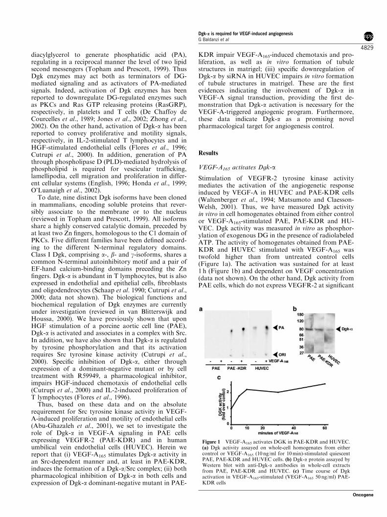
Stimulation of VEGFR-2 tyrosine kinase activitymediates the activation of the angiogenetic responseinduced by VEGF-A in HUVEC and PAE-KDR cells(Waltenberger et al., 1994; Matsumoto and Claesson-Welsh, 2001). Thus, we have measured Dgk activityin vitro in cell homogenates obtained from either controlor VEGF-A165-stimulated PAE, PAE-KDR and HU-VEC. Dgk activity was measured in vitro as phosphor-ylation of exogenous DG in the presence of radiolabeledATP. The activity of homogenates obtained from PAE-KDR and HUVEC stimulated with VEGF-A165 wastwofold higher than from untreated control cells(Figure 1a). The activation was sustained for at least1 h (Figure 1b) and dependent on VEGF concentration(data not shown). On the other hand, Dgk activity fromPAE cells, which do not express VEGFR-2 at significant
Figure 1 VEGF-A165 activates DGK in PAE-KDR and HUVEC.(a) Dgk activity assayed on whole-cell homogenates from eithercontrol or VEGF-A165 (10 ng/ml for 10min)-stimulated quiescentPAE, PAE-KDR and HUVEC cells. (b) Dgk-a protein assayed byWestern blot with anti-Dgk-a antibodies in whole-cell extractsfrom PAE, PAE-KDR and HUVEC. (c) Time course of Dgkactivation in VEGF-A165-stimulated (VEGF-A165 50 ng/ml) PAE-KDR cells
Dgk-a is required for VEGF-induced angiogenesisG Baldanzi et al
4829
Oncogene
levels, was not increased upon VEGF-A165 stimulation(Figure 1a). Intriguingly, even in the absence ofexogenous VEGF-A165, basal Dgk activity from PAE-KDR was much higher than from PAE cells, suggestingthat the sole expression of VEGFR-2, putativelythrough an autocrine loop, is sufficient to generate asignal that upregulates Dgk activity. Indeed, PAE andPAE-KDR express VEGF, as measured by RT–PCR,and both basal ERK-1/2 and Src activities are upregu-lated in unstimulated PAE-KDR cells compared withPAE cells (data not shown).
However, these experiments, which clearly indicatethat VEGF-A165 stimulates a Dgk activity, do not allowto establish which of the different Dgk isoforms areactivated upon VEGF-A165 stimulation of these cells.Dgk-a is expressed at similar levels in PAE and PAE-KDR cells, as measured by Western blot with Dgk-aantibodies, while in HUVEC its expression is signifi-cantly lower (Figure 1c). Thus in order to verify whetherDgk-a is regulated by VEGF-A165, we assayed in vitrothe Dgk activity in anti-Dgk-a immunoprecipitates fromlysates of PAE-KDR cells, either control or VEGF-A165
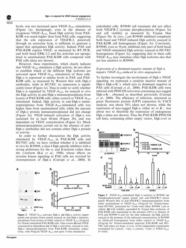
stimulated. Indeed, Dgk activity in anti-Dgk-a immu-noprecipitates from VEGF-A165-stimulated cells washigher than from unstimulated cells, while the amountof Dgk-a protein immunoprecipitated did not change(Figure 2a). VEGF-induced activation of Dgk-a wassustained for at least 60min (Figure 2b), and wasdependent on VEGF concentration (Figure 2c). Mockimmunoprecipitates carried out in the absence of anti-Dgk-a antibodies did not contain either Dgk-a proteinor activity.
In order to further characterize the Dgk activitystimulated by VEGF-A165 in PAE-KDR cells andHUVEC cells, we have verified whether it is inhibitedin vitro by R59949, a class I Dgk-specific inhibitor with astrong preference for the a- and b-isoform rather thanthe g-isoform (Kai et al., 1994), whose effects ontyrosine kinase signaling in PAE cells are reverted byoverexpression of Dgk-a (Cutrupi et al., 2000). In
endothelial cells, R59949 cell treatment did not affectboth VEGFR-2 tyrosine phosphorylation (Figure 3a)and cell viability as measured by Trypan blue(Figure 3b). In vitro, 1 mM R59949 inhibited completelyboth basal and VEGF-induced Dgk activity assayed inPAE-KDR cell homogenates (Figure 3c). Conversely,R59949, even at 10 mM, inhibited only part of both basaland VEGF-stimulated Dgk activity assayed in HUVEChomogenates (Figure 3c), suggesting that in these cellsVEGF-A165 may stimulate other Dgk isoforms also thatare less sensitive to R59949.
Expression of a dominant-negative mutant of Dgk-aimpairs VEGF-A165-induced in vitro angiogenesis
To further investigate the involvement of Dgk-a VEGFsignaling, we expressed a catalytic inactive mutant ofDgk-a Dgk-a-K�), which acts as dominant negative inPAE cells (Cutrupi et al., 2000). PAE-KDR cells wereinfected with PINCOS retrovirus containing myc-taggedDgk-a-K�, obtained as described previously (Cutrupiet al., 2000). The efficiency of infection, measured asgreen fluorescent protein (GFP) expression by FACSanalysis, was about 70% (data not shown), while theexpression of myc-tagged Dgk-a, either wt or K�, wasabout two to threefold the expression of endogenousDgk-a (data not shown). Thus the PAE-KDR-PINCOScell lines, containing either empty vector, Dgk-a-wt or
Figure 2 VEGF-A165 activates Dgk-a. (a) Dgk-a activity (upperpanel) and protein (lower panel) assayed on anti-Dgk-a immuno-precipitates from lysates of either control or VEGF-A165 (10 ng/mlfor 10min)-stimulated PAE-KDR cells. Time course (b) and VEGFconcentration dependence (c) of Dgk-a activation, assayed in anti-Dgk-a immunoprecipitates from PAE-KDR stimulated, respec-tively, with 50 ng/ml VEGF-A165 and upon 15min stimulation
Figure 3 VEGF-A165-stimulated Dgk is sensitive to R59949. (a)Antiphosphotyrosine (upper panel) and anti-VEGFR-2 (lowerpanel) Western blot of anti-VEGFR-2 immunoprecipitates fromeither unstimulated or VEGF-A165 (10 ng/ml for 10min)-stimu-lated HUVEC, pretreated for 15min with either R59949 1mM orvehicle. (b) Cell viability, quantified by Trypan blue exclusion, ofPAE-KDR cells maintained in the presence (10%) or absence ofFCS and R59949 (1mM) for the time indicated. (c) Dgk activityassayed in the presence of the indicated concentrations of R59949on whole-cell homogenates from either control or VEGF-A165
(10 ng/ml for 10min)-stimulated quiescent PAE-KDR and HU-VEC cells (data are mean 7s.e.m. of five independent experimentsnormalized for control, 1t-test vs control, *t-test vs VEGF-A165,Po0.05)
Dgk-a is required for VEGF-induced angiogenesisG Baldanzi et al
4830
Oncogene
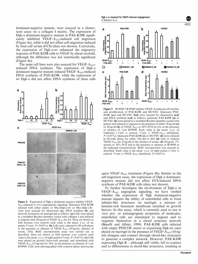
dominant-negative mutant, were assayed in a chemo-taxis assay on a collagen I matrix. The expression ofDgk-a dominant-negative mutant in PAE-KDR signifi-cantly inhibited VEGF-A165-induced cell migration(Figure 4a), while it did not affect cell migration inducedby fetal calf serum (FCS) (data not shown). Conversely,the expression of Dgk-a-wt enhanced the migratoryresponse of PAE-KDR cells to VEGF by about twofold,although the difference was not statistically significant(Figure 4a).
The same cell lines were also assayed for VEGF-A165-induced DNA synthesis. The expression of Dgk-adominant-negative mutant reduced VEGF-A165-inducedDNA synthesis of PAE-KDR, while the expression ofwt Dgk-a did not affect DNA synthesis of these cells
upon VEGF-A165 treatment (Figure 4b). Similar to thecell migration assay, the expression of Dgk-a dominant-negative mutant did not affect FCS-induced DNAsynthesis of PAE-KDR cells (data not shown).
To further investigate the involvement of Dgk-a inVEGF-A165 angiogenic signaling, we have verifiedwhether the expression of Dgk dominant-negativemutant impairs the ability of endothelial cells to formtubular-like structures on matrigel, a mixture oflaminin-rich basement membrane enriched in growthfactors. In this assay, which is currently used to test invitro pro- or antiangiogenic properties of molecules,endothelial cells are stimulated to migrate and toorganize themselves in a chord structure network(Benelli and Albini, 1999). PAE-KDR cells infectedwith empty PINCOS vector or expressing Dgk-wt onceplated on matrigel in the presence of VEGF-A165 (10 ng/ml) elongate and connect through chord-like structuresto generate a complex network. However, PAE-KDRexpressing Dgk-K�, although still viable, fail to connectand to differentiate in chord-like structures, resulting in
Figure 4 Expression of Dgk-a dominant negative inhibits VEGF-A165-induced in vitro angiogenetic signaling. Quiescent PAE-KDRinfected with either empty or Myc-Dgk-a-wt or Myc-Dgk-a-K�
virus were assayed for chemotaxis (a), DNA synthesis (b) andnetwork formation on matrigel (c) as follows. (a) Cells were platedin a modified Boyden chamber coated with collagen I and inducedto migrate with 20 ng/ml of VEGF-A165 for 6 h. Data are shown asfold increase over control; each value is the mean 7s.e. of sixpoints (*t-test, Po0.01%). (b) Cells were cultured in 96-well platesin the presence or absence of VEGF-A165 (10 ng/ml, absence ofserum, 24 h). BrdU incorporation assay was carried out asdescribed. Data are shown as fold increase over control; eachvalue is the mean 7s.e. of eight points (*t-test, Po0.01%). (c) Cellswere plated on growth factor-rich matrigel, and stimulated withVEGF-A165 (25 ng/ml) for 24 h, in the presence or absence of 1 mM
R59949. Cells were photographed with contrast phase microscopy
Figure 5 R59949 5 R59949 inhibits VEGF-A-induced cell motilityand proliferation of PAE-KDR and HUVEC. Quiescent PAE-KDR (a,c) and HUVEC (b,d) were assayed for chemotaxis (a,b)
and DNA synthesis (c,d) as follows: quiescent PAE-KDR (a) orHUVEC (b) were plated in a modified Boyden chamber coated withgelatin and induced to migrate in the presence of either 10 ng/ml (a)
or 20 ng/ml (b) of VEGF-A165 or 20% FCS for 6 h, in the presenceor absence of 1 mM R59949. Each value is the mean 7s.e. oftriplicates (1t-test vs control, *t-test vs VEGF-A165 stimulated,Po0.01%). Quiescent PAE-KDR (c) or HUVEC (d) were culturedin 96-wells plates for either 24 h (c) or 48 h (d) with or withoutVEGF-A165 (c) 25 ng/ml in the absence of serum, (d) 10 ng/ml, 2%serum) or 10% FCS and in the presence or absence of R59949 atthe indicated concentrations. BrdU incorporation was assayed asdescribed. Each value is the mean 7s.e. of eight points (1t-test vscontrol, *t-test vs VEGF-A165 stimulated, Po0.01%)
Dgk-a is required for VEGF-induced angiogenesisG Baldanzi et al
4831
Oncogene
a severe impairment of the formation of the character-istic network (Figure 4c).
Pharmacological inhibition of Dgk-a impairs in vitroVEGF-A165-induced angiogenesis
Based on the sensitivity of Dgk-a to the inhibition bymicromolar concentrations of R59949, we assayed itsability to inhibit VEGF-A165-induced biological re-sponses in both HUVEC and PAE-KDR cells. Indeedwe had previously shown that R59949 inhibition ofHGF-induced chemotaxis was completely reverted byoverexpression of Dgk-a, providing a strong indicationfor the specificity of R59949-induced inhibition (Cutrupiet al., 2000).
In the presence of 1 mM R59949, VEGF-A165-stimu-lated cell migration was inhibited by about 50 and 60%,respectively, in PAE-KDR and HUVEC, while basalcell motility of unstimulated cells was not significantlyaffected. However, in both PAE-KDR and HUVEC,R59949 did not inhibit cell migration induced by serum,suggesting that Dgk-a stands in the signaling pathwayconveying signals specifically from the VEGFR-2 to thecell motility machinery (Figure 5a and b).
We then investigated the ability of R59949 to inhibitVEGF-A165-induced DNA synthesis in both PAE-KDRand HUVEC. In serum-starved PAE-KDR, VEGF-A165
induced a twofold increase of DNA synthesis, which wascompletely abolished in the presence of 1 mM R59949(Figure 5c). In HUVEC cultured in 2% FCS, VEGF-A165 stimulates DNA synthesis, which was inhibited byR59949 at concentrations equal or higher than 5 mM
(Figure 5d). The reduced ability of R59949 to inhibitDNA synthesis in HUVEC may depend on serumsequestration of R59949 (De Chaffoy de Courcelleset al., 1989), although the involvement of other Dgkisoforms, less sensitive to the inhibitor (Jiang et al.,2000), could not be ruled out.
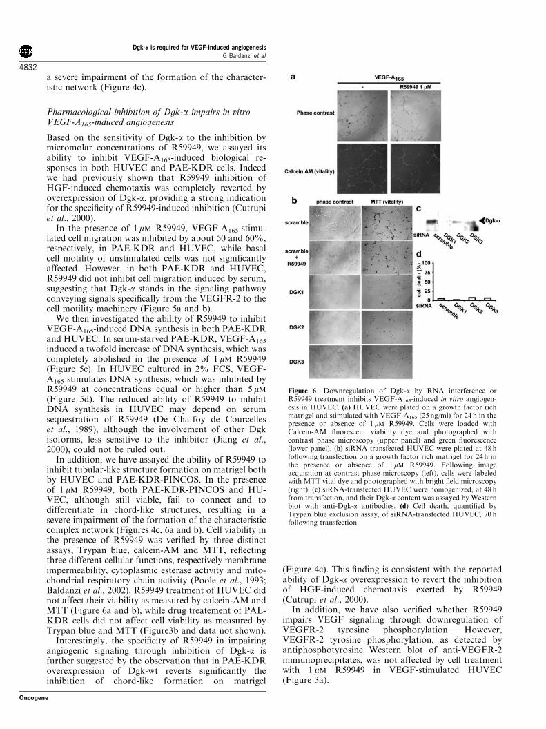
In addition, we have assayed the ability of R59949 toinhibit tubular-like structure formation on matrigel bothby HUVEC and PAE-KDR-PINCOS. In the presenceof 1 mM R59949, both PAE-KDR-PINCOS and HU-VEC, although still viable, fail to connect and todifferentiate in chord-like structures, resulting in asevere impairment of the formation of the characteristiccomplex network (Figures 4c, 6a and b). Cell viability inthe presence of R59949 was verified by three distinctassays, Trypan blue, calcein-AM and MTT, reflectingthree different cellular functions, respectively membraneimpermeability, cytoplasmic esterase activity and mito-chondrial respiratory chain activity (Poole et al., 1993;Baldanzi et al., 2002). R59949 treatment of HUVEC didnot affect their viability as measured by calcein-AM andMTT (Figure 6a and b), while drug treatement of PAE-KDR cells did not affect cell viability as measured byTrypan blue and MTT (Figure3b and data not shown).
Interestingly, the specificity of R59949 in impairingangiogenic signaling through inhibition of Dgk-a isfurther suggested by the observation that in PAE-KDRoverexpression of Dgk-wt reverts significantly theinhibition of chord-like formation on matrigel
(Figure 4c). This finding is consistent with the reportedability of Dgk-a overexpression to revert the inhibitionof HGF-induced chemotaxis exerted by R59949(Cutrupi et al., 2000).
In addition, we have also verified whether R59949impairs VEGF signaling through downregulation ofVEGFR-2 tyrosine phosphorylation. However,VEGFR-2 tyrosine phosphorylation, as detected byantiphosphotyrosine Western blot of anti-VEGFR-2immunoprecipitates, was not affected by cell treatmentwith 1 mM R59949 in VEGF-stimulated HUVEC(Figure 3a).
Figure 6 Downregulation of Dgk-a by RNA interference orR59949 treatment inhibits VEGF-A165-induced in vitro angiogen-esis in HUVEC. (a) HUVEC were plated on a growth factor richmatrigel and stimulated with VEGF-A165 (25 ng/ml) for 24 h in thepresence or absence of 1 mM R59949. Cells were loaded withCalcein-AM fluorescent viability dye and photographed withcontrast phase microscopy (upper panel) and green fluorescence(lower panel). (b) siRNA-transfected HUVEC were plated at 48 hfollowing transfection on a growth factor rich matrigel for 24 h inthe presence or absence of 1 mM R59949. Following imageacquisition at contrast phase microscopy (left), cells were labeledwith MTT vital dye and photographed with bright field microscopy(right). (c) siRNA-transfected HUVEC were homogenized, at 48 hfrom transfection, and their Dgk-a content was assayed by Westernblot with anti-Dgk-a antibodies. (d) Cell death, quantified byTrypan blue exclusion assay, of siRNA-transfected HUVEC, 70 hfollowing transfection
Dgk-a is required for VEGF-induced angiogenesisG Baldanzi et al
4832
Oncogene
Downregulation of Dgk-a expression by RNAinterference impairs HUVEC endothelial cellorganization on matrigel
In order to provide further evidence that Dgk-a isinvolved in VEGF angiogenic signaling in HUVEC, wesynthesized three single interfering RNAs for the Dgk-atranscript designed with Cenix algorithm, which did notoverlap with any other known transcript from thehuman genome. Transfection of HUVEC cells with anyof the three interfering RNAs lowers but does notabolish the expression of Dgk-a, as detected by Westernblot on whole-cell lysates (Figure 6c). Transfection withthe three interfering RNAs impairs the formation ofchord-like structures on matrigel with efficacy similar tothat obtained with 1 mM R59949 cell treatment. Con-versely, transfection with a scrambled siRNA, whichdoes not overlap with any known transcript, did notaffect chord-like formation. In addition, downregula-tion of Dgk-a expression did not affect cell viability ofHUVEC, as measured by Trypan blue exclusion(Figure 6d) and MTT (Figure 6b).
In summary, these data indicate that either inhibitionof Dgk-a catalytic activity by both expression ofdominant-negative mutant or cell treatment with aspecific inhibitor, either downregulation of its expres-sion by interfering RNA, severely impair the ability ofendothelial cells to migrate, proliferate and organize aschord-like networks, providing further support to theclaim that catalytic function of Dgk-a is required for thetransduction of VEGF-A165 angiogenic signal.
VEGF-A165-induced activation of Dgk-a is mediatedby Src
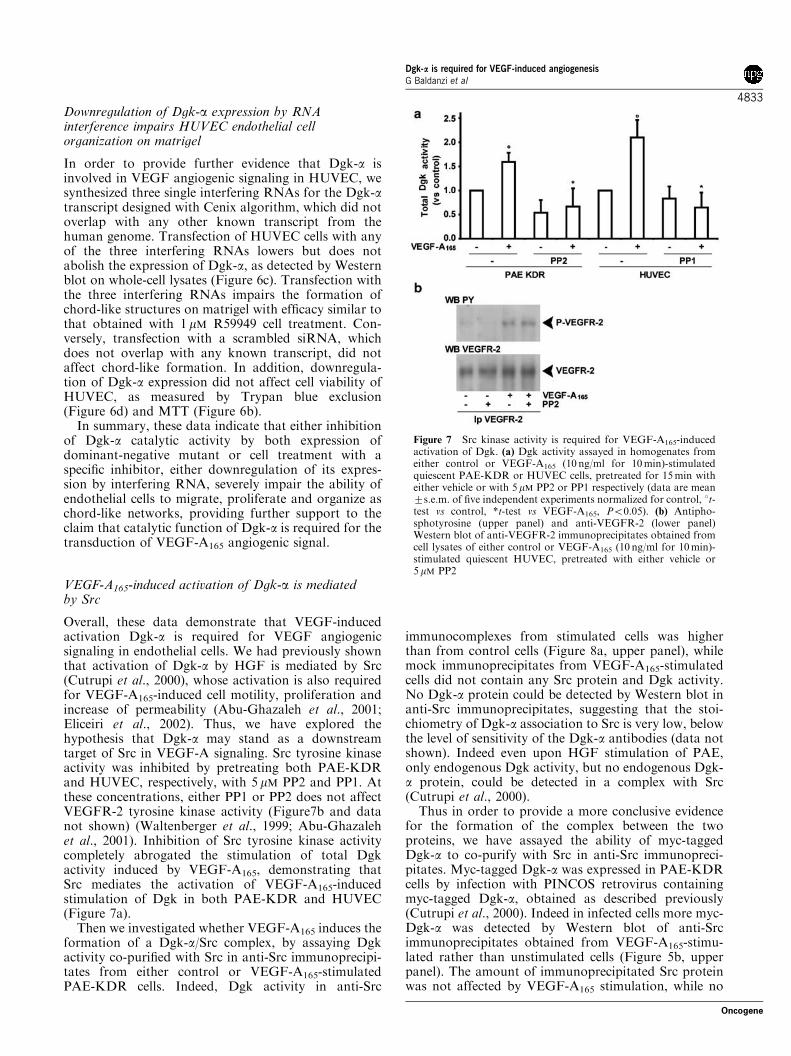
Overall, these data demonstrate that VEGF-inducedactivation Dgk-a is required for VEGF angiogenicsignaling in endothelial cells. We had previously shownthat activation of Dgk-a by HGF is mediated by Src(Cutrupi et al., 2000), whose activation is also requiredfor VEGF-A165-induced cell motility, proliferation andincrease of permeability (Abu-Ghazaleh et al., 2001;Eliceiri et al., 2002). Thus, we have explored thehypothesis that Dgk-a may stand as a downstreamtarget of Src in VEGF-A signaling. Src tyrosine kinaseactivity was inhibited by pretreating both PAE-KDRand HUVEC, respectively, with 5 mM PP2 and PP1. Atthese concentrations, either PP1 or PP2 does not affectVEGFR-2 tyrosine kinase activity (Figure7b and datanot shown) (Waltenberger et al., 1999; Abu-Ghazalehet al., 2001). Inhibition of Src tyrosine kinase activitycompletely abrogated the stimulation of total Dgkactivity induced by VEGF-A165, demonstrating thatSrc mediates the activation of VEGF-A165-inducedstimulation of Dgk in both PAE-KDR and HUVEC(Figure 7a).
Then we investigated whether VEGF-A165 induces theformation of a Dgk-a/Src complex, by assaying Dgkactivity co-purified with Src in anti-Src immunoprecipi-tates from either control or VEGF-A165-stimulatedPAE-KDR cells. Indeed, Dgk activity in anti-Src
immunocomplexes from stimulated cells was higherthan from control cells (Figure 8a, upper panel), whilemock immunoprecipitates from VEGF-A165-stimulatedcells did not contain any Src protein and Dgk activity.No Dgk-a protein could be detected by Western blot inanti-Src immunoprecipitates, suggesting that the stoi-chiometry of Dgk-a association to Src is very low, belowthe level of sensitivity of the Dgk-a antibodies (data notshown). Indeed even upon HGF stimulation of PAE,only endogenous Dgk activity, but no endogenous Dgk-a protein, could be detected in a complex with Src(Cutrupi et al., 2000).
Thus in order to provide a more conclusive evidencefor the formation of the complex between the twoproteins, we have assayed the ability of myc-taggedDgk-a to co-purify with Src in anti-Src immunopreci-pitates. Myc-tagged Dgk-a was expressed in PAE-KDRcells by infection with PINCOS retrovirus containingmyc-tagged Dgk-a, obtained as described previously(Cutrupi et al., 2000). Indeed in infected cells more myc-Dgk-a was detected by Western blot of anti-Srcimmunoprecipitates obtained from VEGF-A165-stimu-lated rather than unstimulated cells (Figure 5b, upperpanel). The amount of immunoprecipitated Src proteinwas not affected by VEGF-A165 stimulation, while no
Figure 7 Src kinase activity is required for VEGF-A165-inducedactivation of Dgk. (a) Dgk activity assayed in homogenates fromeither control or VEGF-A165 (10 ng/ml for 10min)-stimulatedquiescent PAE-KDR or HUVEC cells, pretreated for 15min witheither vehicle or with 5 mM PP2 or PP1 respectively (data are mean7s.e.m. of five independent experiments normalized for control, 1t-test vs control, *t-test vs VEGF-A165, Po0.05). (b) Antipho-sphotyrosine (upper panel) and anti-VEGFR-2 (lower panel)Western blot of anti-VEGFR-2 immunoprecipitates obtained fromcell lysates of either control or VEGF-A165 (10 ng/ml for 10min)-stimulated quiescent HUVEC, pretreated with either vehicle or5mM PP2
Dgk-a is required for VEGF-induced angiogenesisG Baldanzi et al
4833
Oncogene
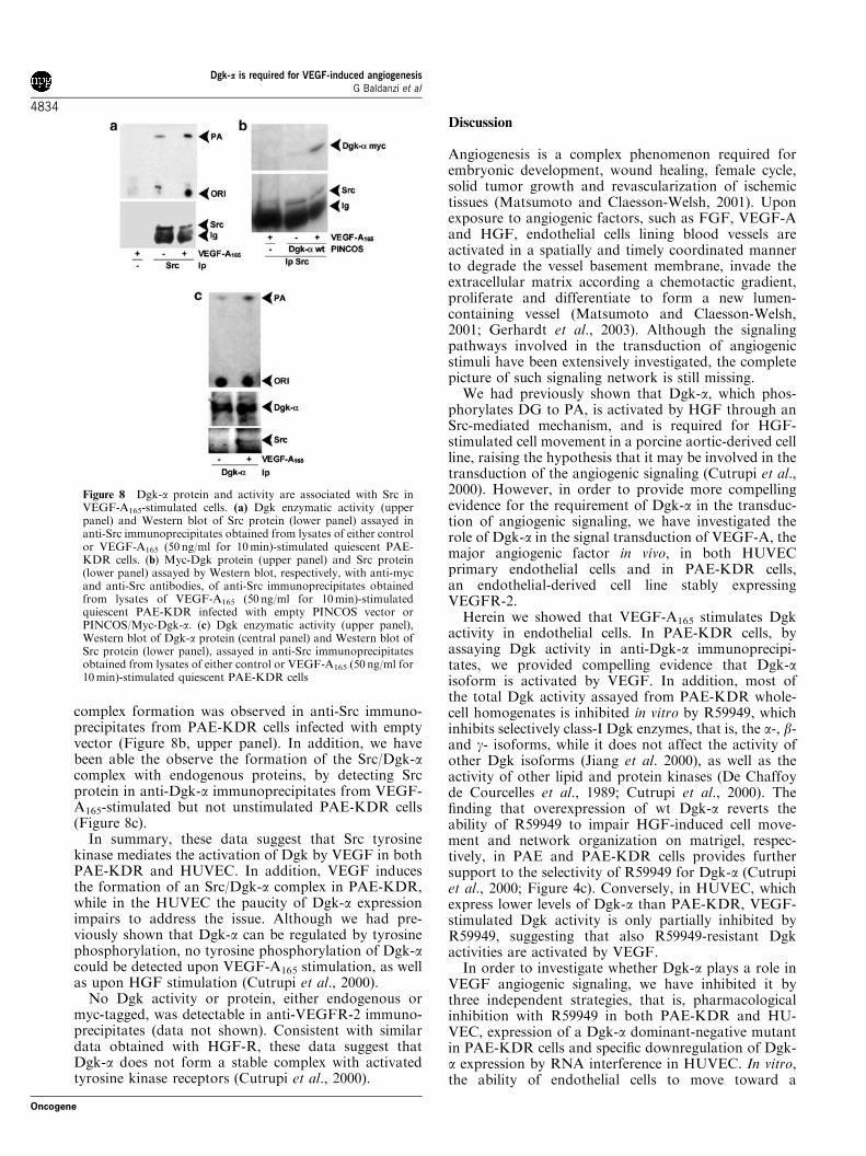
complex formation was observed in anti-Src immuno-precipitates from PAE-KDR cells infected with emptyvector (Figure 8b, upper panel). In addition, we havebeen able the observe the formation of the Src/Dgk-acomplex with endogenous proteins, by detecting Srcprotein in anti-Dgk-a immunoprecipitates from VEGF-A165-stimulated but not unstimulated PAE-KDR cells(Figure 8c).
In summary, these data suggest that Src tyrosinekinase mediates the activation of Dgk by VEGF in bothPAE-KDR and HUVEC. In addition, VEGF inducesthe formation of an Src/Dgk-a complex in PAE-KDR,while in the HUVEC the paucity of Dgk-a expressionimpairs to address the issue. Although we had pre-viously shown that Dgk-a can be regulated by tyrosinephosphorylation, no tyrosine phosphorylation of Dgk-acould be detected upon VEGF-A165 stimulation, as wellas upon HGF stimulation (Cutrupi et al., 2000).
No Dgk activity or protein, either endogenous ormyc-tagged, was detectable in anti-VEGFR-2 immuno-precipitates (data not shown). Consistent with similardata obtained with HGF-R, these data suggest thatDgk-a does not form a stable complex with activatedtyrosine kinase receptors (Cutrupi et al., 2000).
Discussion
Angiogenesis is a complex phenomenon required forembryonic development, wound healing, female cycle,solid tumor growth and revascularization of ischemictissues (Matsumoto and Claesson-Welsh, 2001). Uponexposure to angiogenic factors, such as FGF, VEGF-Aand HGF, endothelial cells lining blood vessels areactivated in a spatially and timely coordinated mannerto degrade the vessel basement membrane, invade theextracellular matrix according a chemotactic gradient,proliferate and differentiate to form a new lumen-containing vessel (Matsumoto and Claesson-Welsh,2001; Gerhardt et al., 2003). Although the signalingpathways involved in the transduction of angiogenicstimuli have been extensively investigated, the completepicture of such signaling network is still missing.
We had previously shown that Dgk-a, which phos-phorylates DG to PA, is activated by HGF through anSrc-mediated mechanism, and is required for HGF-stimulated cell movement in a porcine aortic-derived cellline, raising the hypothesis that it may be involved in thetransduction of the angiogenic signaling (Cutrupi et al.,2000). However, in order to provide more compellingevidence for the requirement of Dgk-a in the transduc-tion of angiogenic signaling, we have investigated therole of Dgk-a in the signal transduction of VEGF-A, themajor angiogenic factor in vivo, in both HUVECprimary endothelial cells and in PAE-KDR cells,an endothelial-derived cell line stably expressingVEGFR-2.
Herein we showed that VEGF-A165 stimulates Dgkactivity in endothelial cells. In PAE-KDR cells, byassaying Dgk activity in anti-Dgk-a immunoprecipi-tates, we provided compelling evidence that Dgk-aisoform is activated by VEGF. In addition, most ofthe total Dgk activity assayed from PAE-KDR whole-cell homogenates is inhibited in vitro by R59949, whichinhibits selectively class-I Dgk enzymes, that is, the a-, b-and g- isoforms, while it does not affect the activity ofother Dgk isoforms (Jiang et al. 2000), as well as theactivity of other lipid and protein kinases (De Chaffoyde Courcelles et al., 1989; Cutrupi et al., 2000). Thefinding that overexpression of wt Dgk-a reverts theability of R59949 to impair HGF-induced cell move-ment and network organization on matrigel, respec-tively, in PAE and PAE-KDR cells provides furthersupport to the selectivity of R59949 for Dgk-a (Cutrupiet al., 2000; Figure 4c). Conversely, in HUVEC, whichexpress lower levels of Dgk-a than PAE-KDR, VEGF-stimulated Dgk activity is only partially inhibited byR59949, suggesting that also R59949-resistant Dgkactivities are activated by VEGF.
In order to investigate whether Dgk-a plays a role inVEGF angiogenic signaling, we have inhibited it bythree independent strategies, that is, pharmacologicalinhibition with R59949 in both PAE-KDR and HU-VEC, expression of a Dgk-a dominant-negative mutantin PAE-KDR cells and specific downregulation of Dgk-a expression by RNA interference in HUVEC. In vitro,the ability of endothelial cells to move toward a
Figure 8 Dgk-a protein and activity are associated with Src inVEGF-A165-stimulated cells. (a) Dgk enzymatic activity (upperpanel) and Western blot of Src protein (lower panel) assayed inanti-Src immunoprecipitates obtained from lysates of either controlor VEGF-A165 (50 ng/ml for 10min)-stimulated quiescent PAE-KDR cells. (b) Myc-Dgk protein (upper panel) and Src protein(lower panel) assayed by Western blot, respectively, with anti-mycand anti-Src antibodies, of anti-Src immunoprecipitates obtainedfrom lysates of VEGF-A165 (50 ng/ml for 10min)-stimulatedquiescent PAE-KDR infected with empty PINCOS vector orPINCOS/Myc-Dgk-a. (c) Dgk enzymatic activity (upper panel),Western blot of Dgk-a protein (central panel) and Western blot ofSrc protein (lower panel), assayed in anti-Src immunoprecipitatesobtained from lysates of either control or VEGF-A165 (50 ng/ml for10min)-stimulated quiescent PAE-KDR cells
Dgk-a is required for VEGF-induced angiogenesisG Baldanzi et al
4834
Oncogene
chemotactic gradient, to proliferate and to organize in anetwork of chord-like structures on matrigel reflectstheir behavior during the angiogenic process in vivo.
In PAE-KDR cells, the inhibition by both expressionof Dgk-a dominant-negative mutant, and cell treatmentwith 1 mM R59949 severely impairs VEGF-inducedchemotaxis and DNA synthesis, as well as networkformation on matrigel, without affecting cell viability.Thus, the data clearly demonstrate that in PAE-KDRcells Dgk-a is activated by VEGF and that its catalyticfunction is required for VEGF-induced angiogenicsignaling in vitro.
In HUVEC, R59949, which inhibits part of theVEGF-stimulated Dgk activity, impairs VEGF-inducedchemotaxis and DNA synthesis, as well as networkorganization on matrigel. The higher concentration ofthe drug required to impair DNA synthesis may furthersuggest that other R59949-resistant Dgk isoforms maybe involved in the transduction of VEGF proliferativesignaling in HUVEC. Alternatively, R59949 may besequestered by the serum required to obtain HUVECproliferation in cultures (De Chaffoy de Courcelles et al.,1989). However, more direct evidence that Dgk-a isrequired for in vitro angiogenesis in HUVEC primaryendothelial cells comes from the demonstration thatspecific downregulation of Dgk-a expression by RNAinterference totally impairs the ability of HUVEC toform networks on matrigel, without affecting cellviability. The same results were obtained with threedifferent dsRNA, which target respectively two se-quences at the N-terminal of the Dgk-a and a sequencenext to the ATP-binding site. The three sequences donot feature any homology with other Dgk isoforms orother proteins.
In summary, these experiments strongly suggest thatactivation of Dgk-a is required for angiogenic signalingin both PAE and HUVEC endothelial cells, althoughthe possibility that in HUVEC, which express a low levelof the a-isoform, other Dgk isoform may also beinvolved in VEGF signaling should be taken intoaccount.
Dgk-a does not contain any domain suggesting adirect interaction with tyrosine kinase receptors, andindeed does not associate with either VEGFR-2 orHGF-R (data not shown). However, a Dgk activity isupregulated upon expression of v-Src in fibroblasts(Sugimoto et al., 1984); we and others have recentlyshown that Src stimulates Dgk-a activity in vitro andmediates HGF- and IL-2-stimulated Dgk-a activation inintact cells (Cutrupi et al., 2000; Cipres et al., 2003).Here, we reported that in both PAE-KDR andHUVEC, the VEGF-induced stimulation of Dgkactivity is blunted by cell treatment, respectively, withPP2 and PP1, two Src family-specific inhibitors that donot affect Dgk activity in vitro (data not shown). Thesedata imply that even if other Dgk isoforms are alsostimulated by VEGF in these cells, their activation iscompletely Src family kinase dependent. Indeed, veryrecent data indicate that Dgk-a associates with Src uponGnRH stimulation of pituitary cells (Davidson et al.,2004). In addition, in PAE-KDR cells, we were able to
show that VEGF stimulation induces the formation of acomplex between Dgk-a and Src, but not between Dgk-aand the activated receptor, as Dgk-a does not co-purifyin antireceptor immunoprecipitates (data not shown).This may depend on the low stoichiometry of the Src/Dgk-a complex, that is, only a small fraction ofreceptor-associated Src forms a ternary complex withDgk-a. Alternatively, receptor-activated Src may comeoff the complex with the receptor and may associatewith Dgk-a. Indeed according to current models of itsfunction, Src is recruited to the active receptor throughits SH2 domain, switches to the active open conforma-tion, allowing complex formation with its substrates, forinstance p130Cas and FAK, their processive phosphor-ylation and establishing a more stable complex withthem through its SH2 domain, excluding the receptor(Nakamoto et al., 1996; Thomas et al., 1998; Scott andMiller, 2000; Pellicena and Miller, 2001).
Although these data indicate Dgk-a as a putativesubstrate of Src tyrosine kinase activity, no tyrosinephosphorylation of Dgk-a could be detected uponVEGF-A stimulation of either PAE-KDR or HUVEC.However, tyrosine phosphorylation of Dgk-a waspreviously shown in PAE cells upon treatment withsodium pervanadate, and in COS cells upon coexpres-sion of both Src and Dgk-a (Cutrupi et al., 2000). Inboth cases, tyrosine phosphorylation correlates with anincrease of Dgk-a enzymatic activity. We speculate thateither tyrosine phosphorylation of Dgk-a occurs at verylow stoichiometry or is a transient event leading to theswitch of Dgk-a to an active conformation. Alterna-tively, Src may induce tyrosine phosphorylationof a regulator of Dgk-a. Indeed recent findings indicatethat PI(3,4,5)P3, the lipid product of PI 3-kinase,binds and stimulates Dgk-a activity and that activeLck stimulates Dgk-a in a PI 3-kinase-dependentmanner (Cipres et al., 2003). Therefore, it is likely thatmultiple mechanisms may participate in the regulationof Dgk-a. The identification of the molecular determi-nants of the physical and functional interactionsbetween the two proteins will shed light on suchmechanisms.
VEGF-A-induced angiogenesis, both in vitro andin vivo, requires Src function: Src is required forVEGF-A-induced chemotaxis and proliferation of en-dothelial cells in vitro (Abu-Ghazaleh et al., 2001), whilegenetic evidences in vivo indicate that deletion of eitherSrc or Yes gene in mice impairs VEGF-dependentincrease of endothelial permeability, that is, edemaformation (Eliceiri et al., 2002). Furthermore, retroviral-mediated overexpression of Src dominant negative in asubcutaneous model of angiogenesis severely impairsneo-vascularization in vivo (Eliceiri et al., 1999). Thus,the demonstration that VEGF-induced stimulation ofDgk activity depends on Src activity, and that Dgk-a isrequired for VEGF-A-induced angiogenetic responsesin vitro suggest that it may act as a crucial Src effectorin VEGF angiogenic signaling in endothelial cells. Inaddition, at least in HUVEC, the involvement of otherSrc-regulated Dgk isoforms in VEGF signaling is notruled out.
Dgk-a is required for VEGF-induced angiogenesisG Baldanzi et al
4835
Oncogene
The signaling pathways downstream to Dgk-a havenot been extensively investigated yet. Dgk enzymesphosphorylate DG to generate PA. Indeed VEGF-Astimulates PLC-g-mediated hydrolysis of PI(4,5)P2 togenerate DG, which then leads to activation of PKC(Matsumoto and Claesson-Welsh, 2001). PKC-a and -bisoforms are required for VEGF-stimulated cell prolif-eration and migration in vitro, as well as for neovascu-larization in vivo (Wellner et al., 1999; Wang et al.,2002).
In some cell signaling system, activation of Dgk-a,which removes DG to generate PA, contributes toterminate DG-mediated signaling, while its inhibitionresults in the accumulation of DG, leading to sustainedDG-mediated signaling. However, in endothelial cells,the increased activity of PKCa, which indeed is activatedby VEGF-A in a DG-mediated manner, is expected toenhance rather than inhibit VEGF-A-induced angio-genic response (Wellner et al., 1999; Matsumoto andClaesson-Welsh, 2001; Wang et al., 2002). Conversely,the role of activation of Dgk-a in VEGF-A angiogenicsignaling may consist in the generation of PA-mediatedsignals. The role of PA in VEGF-A signaling stillremains to be investigated. In vitro, PA is a regulator ofseveral signaling proteins, some of which are involved inVEGF-A signaling (English, 1996; Gomez-Cambroneroand Keire, 1998; Topham and Prescott, 1999). Forinstance, VEGF-A activates PKCz, which is positivelyregulated by PA and Src tyrosine phosphorylation(Limatola et al., 1994; Seibenhener et al., 1999).Similarly, VEGF-A activates Raf, which is regulatedby PA in vitro and in intact cells (Andresen et al., 2002).Activation of both PKCz and Raf is required forVEGF-induced angiogenic signaling (Matsumoto andClaesson-Welsh, 2001). Among other putative targets ofPA in vitro, Rho-GDI and PI(4)P 5-kinase might play arole in VEGF-A signaling, respectively, by mediatingRac activation (Del Pozo et al., 2002; Zeng et al., 2002)and by participating in the dynamic recruitment of focaladhesion proteins required for cell movement (Jenkinset al., 1994; Di Paolo et al., 2002). In addition, both invitro and in intact cells, PA also regulates activation ofmTor, a serine kinase involved in the transduction ofgrowth factor-stimulated proliferative signaling(Fang et al., 2001). However, the role of mTor inVEGF-A signaling has not been investigated. Alterna-tively, PA generated through activation of Dgk-amay be further metabolized to lysosphosphatidicacid (LPA), through a phospholipase A2. Then LPA,through an autocrine loop, may stimulate its receptor(s),edg.2 and -4, whose expression has been reportedin endothelial cells (Panetti, 2002). Indeed, exogenouslyadded LPA stimulates endothelial cell proliferationand cell matrix-adhesion dependent cell migration(Panetti, 2002). Interestingly, in prostate carcinoma,bombesin, a growth factor for these cells, hasbeen shown to activate an LPA autocrine circuit,which is required for bombesin signaling (Xie et al.,2002). However, no evidence of growth factor-stimu-lated synthesis of LPA has been reported in endo-thelial cells.
Finally we provided evidence indicating that Dgk-a isan essential component of VEGF-A signal transduction,which is required to convey both chemotactic andproliferative signals downstream from Src. Becausetumor cells produce a wide array of angiogenic factors,it has been suggested that successful strategies to blockangiogenesis in cancer patients may repress the ability ofendothelial cells to participate in the angiogenic process.As Dgk-a activity is required for angiogenesis and quitespecific inhibitors are available, Dgk-a may represent anew and promising target for pharmaceutical control ofangiogenesis.
Materials and methods
Cells culture, reagents and antibodies
Porcine aortic endothelial cell line (PAE), a cell line derivedfrom porcine aortic endothelium, and PAE-KDR, a PAE-derived cell line stably expressing VEGFR-2, were a kind giftof Waltenberger (Waltenberger et al., 1994), and were culturedin Ham’s F12 supplemented with 10% FCS, 2mM glutamine,100U/ml penicillin and 50 mg/ml streptomycin (Life Technol-ogies). Human endothelial cells from umbilical cord veins(HUVEC) were prepared and characterized as previouslydescribed, and were grown in M199 medium (BioWhittaker,Walkersville, MD, USA) supplemented with 20% FCS (LifeTechnologies), endothelial cell growth factor (100 mg/ml)(Sigma Chemical Co., St Louis, MO, USA) and porcineheparin (Sigma) (100 mg/ml). HUVEC were used at secondpassage and grown on a plastic surface coated with porcinegelatin (Sigma), otherwise specified. Recombinant VEGF-A165
was purchased from R&D systems, and R59949, PP1 and PP2were from Alexis. Anti-Src antibodies (N-16 for immunopre-cipitation and SRC-2 for Western blotting) were from SantaCruz Biotechnology, and 4G10 antiphosphotyrosine and 9E10anti-myc tag antibodies were from UBI. Anti-Dgk-a anti-bodies were a mix of monoclonal obtained as described(Schaap et al., 1993). Anti-VEGFR-2 antibodies were fromSanta Cruz Biotechnologies. ECL and secondary antibodieswere from Perkin-Elmer. PINCOS, PINCOS/Myc-Dgk-a,PINCOS/Myc-Dgk-a-K� and the PHOENIX retroviral systemwere previously described (Cutrupi et al., 2000). Unlessspecified, all other reagents were from Sigma-Aldrich.
Infection with retroviral vectors
Phoenix cells obtained by G Nolan (Swift et al., 1999) weretransiently transfected by calcium phosphate (Cell PhectTransfection kit, Amersham-Pharmacia) with 20mg PINCOS,PINCOS/Myc-Dgk-a or 20mg PINCOS/Myc-Dgk-a-K� ingrowth media containing chloroquin (25 mM). Cells wereincubated for 8 h at 371C (5% CO2) and the precipitate wasremoved by washing with PBS. Then, cells were incubated withDMEM containing 10% FCS for 72 h; the retroviral super-natant was collected, the debris removed by 1500 g centrifuga-tion, filtered by a 0.8 pore filter, and supplemented withPolybrene (8 mg/ml). Log-phase growing PAE-KDR cells wereinfected by addition of 5ml retroviral supernatant. Following16 h from infection, cells were placed into 10% FCS mediumfor 48 h and serum starved overnight in 0.5% FCS prior tofurther experimental manipulations. PINCOS-infected cellsexpress both GFP and the protein of interest. Efficiency ofinfection was about 70% as measured by FACS analysis ofGFP expression.
Dgk-a is required for VEGF-induced angiogenesisG Baldanzi et al
4836
Oncogene
Preparation of cell lysate, homogenates, immunoprecipitationand Western blotting
Subconfluent PAE-KDR cultures were made quiescent by 16 hserum starvation. Subconfluent HUVEC cultures were madequiescent by incubating for 16 h in 5% FCS. Following cellstimulation, the cell lysates were prepared in buffer A (25mM
Hepes pH 8, 10% glycerol, 150mM NaCl, 5mM EDTA, 2mM
EGTA, 1mM ZnCl2, 50mM ammonium molibdate, suppliedbefore use with 1mM sodium orthovanadate, 10mM NaF,0.5mM dithiothreitol, 0.2mM phenylmethylsulfonyl fluoride,1mg/ml leupeptin, 1mg/ml aprotinin and 1mg/ml pepstatin,1mg/ml soybean trypsin inhibitor) supplemented with 1% NP-40, as described previously (Cutrupi et al., 2000). Whereindicated, cell extracts were prepared by collecting the cellswith a rubber scraper in buffer A and homogenizing them insyringe–needle in buffer A. Protein concentration was deter-mined by the BCA method (Pierce). Immunoprecipitation,SDS–PAGE and Western blots were performed as describedpreviously (Cutrupi et al., 2000).
Dgk-a assay
Dgk-a was assayed in the immunoprecipitates essentially asdescribed (Schaap et al., 1993): immunocomplexes wereincubated for 5min at 301C with 1mg/ml diolein (Fluka),1mM ATP, 30 mCi-[g32P]ATP (Amersham), 10mM MgCl2,1mM ZnCl2, 1mM EGTA in 25mM Hepes pH 8, and thereaction stopped with 50 ml of 1M HCl and 100 ml ofchloroform :methanol 1 : 1. Lipids were then extracted, andseparated by TLC in chloroform :methanol :water : 25% ammo-nium hydroxide (60 : 47 : 11 : 4). TLC was exposed on aautoradiographic film (Kodak Biomax), [32P]PA was identifiedby co-migration with nonradioactive PA standards stained byincubation in an iodine chamber. PA spots were quantified usingphosphor imager (Biorad). Dgk activity in homogenates (1–5ml)was assayed as described above, except that ATP was 5mM.
Cell migration assay
For migration on gelatin, endothelial cells were seeded (1� 105
cells in 50 ml suspension) in the upper chamber of a modifiedBoyden chamber. The undersurface of PVDF filter (8 mmpores, Nucleopore) was coated with 0.1% gelatin. The lowerchamber was filled with serum-free medium with or withoutVEGF-A165 and incubated at 371C in air with 5% CO2 for 6h.Where indicated, 1mM R59949 was added to the upper chamberwith the cells. Filters were then removed, stained with Diff-Quik(Baxter Diagnostic AG) and the cells of five fields were countedat the inverted microscope with a high-power oil immersionobjective (Zeiss). For migration on collagen I, endothelial cellswere seeded (1� 105 cells in 50ml suspension) in the upperchamber of a chemotox chamber (Nuroprobe), coated with20ng/ml collagen I. The lower chamber was filled with serum-free medium with or without VEGF-A165 and incubated at 371Cin air with 5% CO2 for 6h. Filters were then removed, stainedwith crystal violet and the cells of five fields were counted at theinverted microscope (Zeiss axiovert).
Cell proliferation assay
PAE KDR cells were plated and after 6 h treated as indicatedfor 24 h. HUVEC cells were plated and after 24 h treated as
indicated for additional 48 h. BrdU incorporation wasmeasured with ‘Cell Proliferation ELISA Biotrak System’from Amersham-Pharmacia as suggested by the manufacturer.
Matrigel morphogenetic assay
Matrigel (Collaborative Biomedical Products, Becton Dick-inson, Milan, Italy; not purified from contaminant growthfactors; 0.2ml). was added to each well of a 48-well plate andincubated at 371C for 30min to allow gel formation.Endothelial cells (2� 104/well) were plated onto Matrigel.After 48 h incubation in the presence or absence of theindicated treatments in 5% CO2 humidified atmosphere at371C, the three-dimensional organization of the cells wasexamined under an inverted phase contrast photomicroscope(DM-IBM model; Leica Microsystems, Wetzlar, Germany)and then photographed. Not purified matrigel allows sponta-neous in vitro angiogenesis (Marconcini et al., 1999).
RNA interference
HUVEC were transfected with siPORT lipid transfectionagent (Ambion) as suggested by the manufacturer. Celltransfection procedures must be optimized for each HUVECpreparation using Silencer siRNA Transfection KIT (Am-bion). siRNA used were chemically synthesized as double-strand RNA (Ambion). The sequence was designed usingCenix bioscience algorithm, which considers Tm, nucleotidecontent of the 30 overhangs, nucleotide distribution over thelength of the siRNA, and presence and location of mismatchesto off target genes. A key step in the algorithm is a stringentanalysis of each siRNA sequence to maximize the targetspecificity. Sequences were as follows: DGK1 sense GGUCAGUGAUGUCCUAAAGTT, antisense CUUUAGGACAUCACUGACCTT (target AAGGTCAGTGATGTCCTAAAG);DGK2 sense GGAUGGCGAGAUGGCUAAATT, antisenseUUUAGCCAUCUCGCCAUCCTC (target AAGGATGGCGAGATGGCTAAA); and DGK3 sense GGAUUUAGA-GAUGAGUAAATT, antisense UUUACUCAUCUCUAAAUCCTT (target AAGGATTTAGAGATGAGTAAA). A GAPDH scramble siRNA was used as negative control (providedwith Silencer siRNA Transfection KIT from Ambion).
Reproducibility and data analysis
All the experiments shown are representative of three or moreindependent experiments with similar results. In Figures 3cand 7a, data are mean 7s.e.m. of five independent experimentsnormalized for control.
AcknowledgementsThis study was supported by grants from University AAvogadro of Piemonte Orientale, Regione Piemonte, theItalian Ministry for University and Research (PRIN 2002-03University research program and FIRB post-genomic pro-gram), Fondazione Cariplo (to AG), Associazione Italiana perla Ricerca sul Cancro (to AG and FB) and Istituto Superioredi Sanita (IV Programma Nazionale di Ricerca sull’AIDS-2001 to FB). GB and SC were supported by a fellowship fromFIRC.
References
Abu-Ghazaleh R, Kabir J, Jia H, Lobo M and Zachary I.(2001). Biochem. J., 360, 255–264.
Andresen BT, Rizzo MA, Shome K and Romero G. (2002).FEBS Lett., 531, 65–68.
Dgk-a is required for VEGF-induced angiogenesisG Baldanzi et al
4837
Oncogene

Baldanzi G, Filigheddu N, Cutrupi S, Catapano F, BonissoniS, Fubini A, Malan D, Baj G, Granata R, Broglio F, PapottiM, Surico N, Bussolino F, Isgaard J, Deghenghi R,Sinigaglia F, Prat M, Muccidli G, Ghigo E and GrazianiA. (2002). J. Cell Biol., 759, 1029–1037.
Benelli R and Albini A. (1999). Int. J. Biol. Markers, 14,
243–246.Bussolino F, Di Renzo MF, Ziche M, Bocchietto E, Olivero
M, Naldini L, Gaudino G, Tamagnone L, Coffer A andComoglio PM. (1992). J. Cell Biol., 119, 629–641.
Carmeliet P and Jain RK. (2000). Nature, 407, 249–257.Cipres A, Carrasco S, Merino E, Diaz E, Krishna UM, Falck
JR, Martinez AC and Merida I. (2003). J. Biol. Chem., 278,35629–35635.
Cutrupi S, Baldanzi G, Gramaglia D, Maffe A, Schaap D,Giraudo E, van Blitterswijk W, Bussolino F, Comoglio PMand Graziani A. (2000). EMBO J., 19, 4614–4622.
Davidson L, Pawson AJ, Maturana RL, Freestone SH, BarranP, Millar RP and Maudsley S. (2004). J. Biol. Chem., inpress, M310784200.
De Chaffoy de Courcelles D, Roevens P, Van Belle H, KennisL, Somers Y and De Clerck FJ. (1989). Biol. Chem., 264,
3274–3285.Del Pozo MA, Kiosses WB, Alderson NB, Meller N, Hahn
KM and Schwartz MA. (2002). Nat. Cell Biol., 4, 232–239.Di Paolo G, Pellegrini L, Letinic K, Cestra G, Zoncu R,
Voronov S, Chang S, Guo J, Wenk MR and De Camilli P.(2002). Nature, 420, 85–89.
Eliceiri BP, Paul R, Schwartzberg PL, Hood JD, Zeng J andCheresh D. (1999). Mol. Cell, 4, 915–924.
Eliceiri BP, Puente XS, Hood JD, Stupack DG, SchlaepferDD, Huang XZ, Sheppard D and Cheresh DA. (2002).J. Cell Biol., 157, 149–160.
English D. (1996). Cell Signal., 8, 341–347.Fang Y, Vilella-Bach M, Bachmann R, Flanigan A and Chen
J. (2001). Science, 294, 1942–1945.Flores I, Casaseca T, Martinez AC, Kanoh H and Merida I.
(1996). J. Biol. Chem., 271, 10334–10340.Folkman J. (1972). Ann. Surg., 175, 409–416.Gerber HP, McMurtrey A, Kowalski J, Yan M, Keyt BA, Dixit
V and Ferrara N. (1998). J. Biol. Chem., 273, 30336–30343.Gerhardt H, Golding M, Fruttiger M, Ruhrberg C, Lundkvist
A, Abramsson A, Jeltsch M, Mitchell C, Alitalo K, Shima Dand Betsholtz C. (2003). J. Cell Biol., 161, 1163–1177.
Gomez-Cambronero J and Keire P. (1998). Cell Signal., 10,
387–397.Honda A, Nogami M, Yokozeki T, Yamazaki M, Nakamura
H, Watanabe H, Kawamoto K, Nakayama K, Morris A,Frohman MA and Kanaho Y. (1999). Cell, 99, 521–532.
Jenkins GH, Fisette PL and Anderson RA. (1994). J. Biol.Chem., 269, 11547–11554.
Jiang Y, Sakane F, Kanoh H and Walsh JP. (2000). Biochem.Pharmacol., 59, 763–772.
Jones DR, Sanjuan MA, Stone JC and Merida I. (2002).FASEB J., 16, 595–597.
Kai M, Sakane F, Imai S, Wada I and Kanoh HJ. (1994). Biol.Chem., 269, 18492–18498.
Limatola C, Schaap D, Moolenaar WH and van BlitterswijkWJ. (1994). Biochem. J., 304, 1001–1008.
Marconcini L, Marchio S, Morbidelli L, Cartocci E, Albini A,Ziche M, Bussolino F and Oliviero S. (1999). Proc. Natl.Acad. Sci. USA, 96, 9671–9676.
Matsumoto T and Claesson-Welsh L. (2001). Sci. STKE, 112,
RE21.Nakamoto T, Sakai R, Ozawa K, Yazaki Y and Hirai H.
(1996). J. Biol. Chem., 271, 8959–8965.O’Luanaigh N, Pardo R, Fensome A, Allen-Baume V, Jones
D, Holt MR and Cockcroft S. (2002). Mol. Biol. Cell, 13,3730–3746.
Panetti TS. (2002). Biochim. Biophys. Acta., 23, 190–196.Pellicena P and Miller WT. (2001). J. Biol. Chem., 276,
28190–28196.Poole CA, Brookes NH and Clover GM. (1993). J. Cell Sci.,
106, 685–692.Schaap D, de Widt J, van der Wal J, Vandekerckhove J, van
Damme J, Gussow D, Ploegh HL, van Blitterswijk WJ andvan der Bend RL. (1990). FEBS Lett., 275, 151–158.
Schaap D, van der Wal J, van Blitterswijk WJ, van der BendRL and Ploegh HL. (1993). Biochem. J., 289, 875–881.
Scott MP and Miller WT. (2000). Biochemistry, 39,
14531–14537.Seibenhener ML, Roehm J, White WO, Neidigh KB,
Vandenplas ML and Wooten MW. (1999). Mol. Cell Biol.Res. Commun., 2, 28–31.
Sugimoto Y, Whitman M, Cantley LC and Erikson RL.(1984). Proc. Natl. Acad. Sci. USA, 81, 2117–2121.
Swift S, Lorens J, Achacoso P and Nolan GP. (1999). Curr.Protocols Immunol., 10.17.14–10.17.29.
Thomas JW, Ellis B, Boerner RJ, Knight WB, White II GCand Schaller MD. (1998). J. Biol. Chem., 273, 577–583.
Topham MK and Prescott SM. (1999). J. Biol. Chem., 274,
11447–11450.van Blitterswijk WJ and Houssa B. (2000). Cell Signal., 12,
595–605.Waltenberger J, Claesson-Welsh L, Siegbahn A, Shibuya M
and Heldin CH. (1994). J. Biol. Chem., 269, 26988–29695.Waltenberger J, Uecker A, Kroll J, Frank H, Mayr U, Bjorge
JD, Fujita D, Gazit A, Hombach V, Levitzki A and BohmerFD. (1999). Circ. Res., 85, 12–22.
Wang A, Nomura M, Patan S and Ware JA. (2002). Circ. Res.,90, 609–616.
Wellner M, Maasch C, Kupprion C, Lindschau C, Luft FCand Haller H. (1999). Arterioscler. Thromb. Vasc. Biol., 19,
178–185.Xie Y, Gibbs TC, Mukhin YV and Meier KE. (2002). J. Biol.
Chem., 277, 32516–32526.Zeng H, Zhao D and Mukhopadhyay D. (2002). J. Biol.
Chem., 277, 46791–46798.Zhong XP, Hainey EA, Olenchock BA, Zhao H, Topham MK
and Koretzky GA. (2002). J. Biol. Chem., 277, 31089–31098.
Dgk-a is required for VEGF-induced angiogenesisG Baldanzi et al
4838
Oncogene