HGF Mediates Cell Proliferation of Human Mesothelioma Cells through a PI3K/MEK5/Fra1 Pathway
Upload
independentCategory
view
1download
0
Diacylglycerol kinase-α mediates HGF-induced epithelial cell scatter by regulating Rac
activation and membrane ruffling
Federica Chianale*†, Santina Cutrupi*‡†, Elena Rainero*, Gianluca Baldanzi*§, Paolo E. Porporato*,
Sara Traini*, Nicoletta Filigheddu║, Viola F. Gnocchi*, Massimo M. Santoro¶, Ornella Parolini§,
Wim J. van Blitterswijk#, Fabiola Sinigaglia*, and Andrea Graziani*
*Department of Medical Sciences, ║Department of Clinical and Experimental Medicine and
¶DISAV, University of Piemonte Orientale “A. Avogadro”, Novara 28100, Italy. ‡Department of
Animal and Human Biology, University of Torino, Italy. §Centro Ricerche “E. Menni” Ospedale
Poliambulanza Brescia, Italy. #The Netherlands Cancer Institute, Amsterdam, The Netherlands. †These authors contributed equally to this work.
RUNNING TITLE: Dgkα regulates Rac and membrane ruffling
KEY WORDS: Diacylglycerol kinase, HGF, scatter, migration, Rac.
CORRESPONDING AUTHOR: Andrea Graziani. Mailing Address: Dept. of Medical Sciences,
University of Piemonte Orientale “A. Avogadro”, via Solaroli 17, 28100 Novara, Italy; Tel: +39
0321660676; FAX: +39 0321620421; e-mail: [email protected].

ABSTRACT
Diacylglycerol kinases (Dgk) phosphorylate diacylglycerol (DG) to phosphatidic acid (PA),
thus turning off and on, respectively, DG-mediated and PA-mediated signalling pathways. We
previously showed that HGF, VEGF and ALK activate Dgkα in endothelial and leukaemia cells
through a Src-mediated mechanism, and that activation of Dgkα is required for chemotactic,
proliferative and angiogenic signalling in vitro. Inhere we investigate the downstream events and
signalling pathways regulated by Dgkα, leading to cell scatter and migration upon HGF treatment
and v-Src expression in epithelial cells. We report that specific inhibition of Dgkα, obtained either
pharmacologically by R59949 treatment, or by expression of Dgkα dominant-negative mutant, or
by siRNA-mediated downregulation of endogenous Dgkα, impairs i) HGF- and v-Src-induced cell
scatter and migration, without affecting the loss of intercellular adhesions; ii) HGF-induced cell
spreading, lamellipodia formation, membrane ruffling and focal adhesions remodelling; iii) HGF-
induced Rac activation and membrane targeting. In summary, we provide evidence that
Dgkα, activated downstream of tyrosine kinase receptors and Src, regulates crucial steps directing
Rac activation and Rac-dependent remodelling of actin cytoskeleton and focal contacts in migrating
epithelial cells.

INTRODUCTION
Epithelial tissues are characterized by monolayers of highly polarized cells, while in vitro
epithelial cells grow to form discrete colonies. During embryonic development and tissue repair, as
well as through cancer progression, epithelial cells acquire a highly motile and invasive phenotype
in a process commonly known as epithelial-mesenchymal transition (EMT) (Thiery, 2002; Thiery et
al., 2006). In vitro, the scattering of epithelial cells, i.e. the dispersal of colonies due to loss of
intercellular adhesion and acquisition of cell motility, is triggered by growth factors stimulation and
by oncogenes activation, recapitulating the early phases of EMT (Avizienyte and Frame, 2005).
HGF and oncogenic Src induce in vitro cell scatter of several epithelial cells, while in vivo
their inappropriate activation is associated to progression and acquisition of a metastatic phenotype
in several epithelial-derived cancer (Irby and Yeatman, 2000; Danilkovitch and Zbar, 2002). Within
hours from stimulation of their tyrosine kinase activities, both HGF and v-Src induce scattering of
epithelial cell colonies through loss of cadherin-mediated cell-cell adhesions and increase of their
motility, due to formation of lamellipodia and remodelling of cortical actin and focal adhesions
(Beherens et al., 1993; Lamorte et al., 2002). The signalling pathways by which HGF and v-Src
stimulate EMT, cell scattering and invasiveness have been extensively investigated in several
epithelial cells (Thiery, 2002). Recruitment of Gab-1, along with activation of PI 3-kinase, PLCγ,
Ras and Rac are required (Lamorte et al., 2002 and refs herein). Src plays a crucial role in HGF
signalling as its activity is required for HGF-mediated cell motility, anchorage-independent growth
and tumorigenesis. Indeed Src mediates HFG-induced tyrosine phosphorylation of catenins, leading
to downregulation of cadherin-mediated cell-cell adhesions, and of several focal adhesion proteins
required for cell motilitiy and invasiveness, such as FAK, Paxillin and p130Cas (Beherens et al.,
1993; Rahimi et al., 1998; Nakaigawa et al., 2000).
Diacylglycerol kinases, which phosphorylate diacylglycerol (DG) to phosphatidic acid (PA),
comprise a family of ten distinct enzymes, grouped in five classes each featuring distinct regulatory
domains and a highly conserved catalytic domain preceded by two cysteine-rich atypical C1
domains (Topham and Prescott, 1999; Imai et al., 2005). DG is an established activator of several
typical C1 domain-containing proteins, such as PKCs, RasGRPs and chimaerins. Similarly, several
signalling proteins have been reported to be regulated by PA, including serine kinases, such as
mTor, Raf and atypical PKCs, small GTPase regulating proteins, such as SOS, RhoGDI, Ras- and
Rho-GAPs, and signalling lipid metabolizing enzymes, such as PI(4)P 5-kinase and PLC-
γ (Topham, 2006; Zhao et al., 2007). However, a common specific PA binding domain has not been
identified yet. Thus, by regulating in a reciprocal manner the level of both DG and PA lipid second

messengers, Dgk enzymes may act as terminators of DG-mediated signals as well as activators of
PA-mediated ones.
Recent evidences showed that α, ζ and θ Dgk isoforms are regulated by extracellular ligands
and play a role in signal transduction (van Blitterswijk and Houssa, 2000; Luo et al., 2003). T cells
derived by Dgkα -/- mice feature enhanced DG-mediated RasGRP activity upon TCR activation,
leading to over-activation of the Ras pathway and a defect in anergy, while overexpression of Dgkα
in T cells impairs TCR signalling (Olenchock et al., 2006). Several evidence in T cells indicate that
Dgkα and ζ, by interacting respectively with RasGRP and PKC, up-regulate cell sensitivity to TCR
activation by negatively modulating the intensity and the kinetic of DG-mediated signalling (Luo et
al., 2003; Sanjuan et al., 2003; Zhong et al., 2003). Conversely, mast cells derived from Dgkζ -/-
mice feature a diminished FcεRI-mediated degranulation, correlating with impaired PLCγ activation
and calcium response, both likely dependent on PA production (Olenchock et al., 2006).
We have previously shown that in endothelial and leukaemia cells, activation of Dgkα
downstream from tyrosine kinase receptors, such as HGF-R, VEGFR-2, and ALK, is required for
either chemotactic or proliferative signalling induced by their respective ligands, as well as for cell
proliferation upon IL-2 stimulation of T cells (Cutrupi et al., 2000; Baldanzi et al., 2004;
Bacchiocchi et al., 2005). Growth factors stimulate Dgkα through a mechanism requiring complex
formation with Src and phosphorylation of Dgkα on Tyr335 by Src itself (Cutrupi et al., 2000;
Baldanzi et al., 2007). The specific signalling pathways regulated by activation of Dgkα still await
elucidation.
Herein we investigate the role of Dgkα in HGF-induced cell migration of epithelial cells.
We show that Dgkα activation is required for HGF- and v-Src-induced scattering of MDCK cells,
and particularly in those mechanisms leading to cell spreading and F-actin cytoskeleton and focal
adhesions remodelling. By further investigating the role of Dgkα in HGF early signalling, we show
that upon 15 minutes from HGF stimulation, Dgkα activity is necessary for membrane targeting and
activation of Rac, and for Rac-regulated formation of membrane ruffles.
These data, by indicating Dgkα as a key signal transducer of motility signals downstream
HGF and v-Src, strongly suggest that it may represent a key regulator in the processes of invasion
and metastasis.
MATERIALS AND METHODS
Cell culture
MDCK (Madin-Darby canine kidney cells) and MDCK-ts-v-Src (Baldanzi et al., 2004) are a
kind gift of W. Birchmeier (Berlin). Cells were cultured in high glucose DMEM GlutaMAXTM
medium (Gibco), supplemented with 10% fetal bovine serum (Gibco) and antibiotic-antimicotic
solution (Sigma), in humidified atmosphere with 5% CO2. MDCK cells were cultured at 37°C,
while MDCK-ts-v-Src were normally grown at 40.5°C (inactive v-Src) and shifted to 35°C to
achieve v-Src activation.
Reagents
Recombinant human HGF was purchased from Peprotech; R59949 (Diacylglycerol Kinase
Inhibitor II) from Sigma. DMSO, vehicle for R59949, was always used in control samples at the
same dilution as R59949. Anti-Myc and anti-Rac1 were from Upstate, anti-Paxillin from BD
Transduction Laboratories, anti-Paxillin pTyr31 and pTyr118 and anti-Akt pSer473 from Biosource,
anti-Akt from Cell Signaling, anti-α-Tubulin from Sigma, anti-Vinculin from Novus Biological,
anti-FAK from Calbiochem, Alexa Flour 546/633 Phalloidin from Molecular Probes. Anti-Dgkα
was kindly provided by W.J. van Blitterswijk (the Netherlands Cancer Institute, Amsterdam).
Secondary HRP-conjugated antibodies were purchased from PerkinElmer Life Sciences; secondary
FITC- and TRITC-conjugated antibodies were purchased from DAKO.
Expression vectors, transfections and infections with retroviral vectors
Myc-Dgkα cDNA cloned into pMT2 expression vector has been previously described
(Cutrupi et al., 2000). GFP-Dgkα-WT (wild type) was obtained by cloning Dgkα in pcDNA-
DEST53 (Invitrogen) using Gateway kit (Invitrogen) according to manufacturer’s instructions.
Briefly, Dgkα cDNA was inserted in pDONOR 2.11 vector by PCR and BP recombination. LR
recombination was performed to transfer Dgkα in pcDNA-DEST53 for N-terminal GFP fusion;
detailed information and protocols are available on www.invitrogen.com. G434D point mutation on
Dgkα to obtain the kinase-defective dominant negative mutant (GFP-Dgkα-DN) was performed
using QuikChange Site-Directed Mutagenesis Kit 22 (Stratagene) as previously described (Cutrupi
et al., 2000). PINCOS retroviral vector, PINCOS/Dgkα-DN and PINCOS/Dgkα-WT, expressing
both GFP and the inserted gene, have already been described (Cutrupi et al., 2000). Transient
transfections were performed using Lipofectamine2000 Reagent (Invitrogen) according to the
manufacturer’s instructions.

MDCK cells stably expressing PINCOS/empty vector or PINCOS/Dgkα-DN or
PINCOS/Dgkα-WT were obtained by infection. Briefly, GP2-293 packaging cell line (Clontech,
kindly provided by R. Piva, University of Torino) was transiently co-transfected, by
Lipofectamine2000 Reagent (Invitrogen) according to the manufacturer’s instructions, with the
envelope vector pVSV-G (Clontech) together with PINCOS or PINCOS/Dgkα-DN or
PINCOS/Dgkα-WT.. The next day the medium was changed to normal growth medium. Forty-eight
hours after infection, the retroviral supernatant was collected, the debris removed by centrifugation
at 1500g, and the supernatant was filtered by a 0.45 μm pore filter and added with Polybrene (8
μg/ml). MDCK cells, plated in a six-well plate, were infected by adding 2 ml of retroviral
supernatant and 1 ml of growth medium. The day after the first infection cells were re-infected as
previously described. Sixteen hours later, cells were placed and maintained in growth medium.
Efficiency of infection was about 80%, as measured by FACS analysis and/or observation with
fluorescence microscope of GFP expressing cells .
The murine Dgkα, resistant to canine Dgkα siRNAs, was cloned in the lentiviral vector
pLenti4V5 (Invitrogen). Lentiviruses were produced following the manufacturer’s instructions and
used to infect MDCK cells, which were then selected in Zeocin-containing medium to obtain a
stably-expressing cell line.
RNA interference
siRNAs against canine Dgkα were chemically synthesized as double-strand RNA (Ambion).
Sequences were as follows: C1 sense GCUCAGAAGUGGACAGGAUtt antisense
AUUCUGUCCACUUCUGAGCtg; C2 sense CCCAGACAUCCUGAAAACCtt antisense
GGUUUUCAGGAUGUCUGGGtc; C3 sense CCUUCCACACCACAAAAACtt antisense
GUUUUUGUGGUGUGGAAGGtg. A GAPDH scramble siRNA (Ambion) was used as negative
control.
The BLOCK-iTTM Fluorescent Oligo (Invitrogen) is a fluorescein-labelled dsRNA oligomer
and was used to obtain indication of the transfection efficiency with siRNAs.
Dgk assay
Dgkα activity was assayed in anti-Myc immunoprecipitates as described (Cutrupi et al.,
2000). Briefly, after immunoprecipitation and extensive washing in Lysis Buffer (25 mM HEPES
pH 8, 150 mM NaCl, 1% NP-40, 5 mM EDTA, 2 mM EGTA, 1 mM ZnCl2, 50 mM NaF, 10%
glycerol supplemented with protease inhibitors [Protease Inhibitors Cocktail, Sigma]), Litium
Cloride Buffer (500 mM LiCl, 25 mM Tris-HCl pH 8) and TNE (Tris 25 mM pH 8, NaCl 150 mM,

EDTA 1 mM), all supplemented with fresh 1 mM Na3VO4, the immunocomplexes were assayed at
room temperature for 10 minutes by incubation with 1 mg/ml diolein (Fluka, dried in nitrogen
atmosphere, resuspended and sonicated in 1 mM EGTA, 25 mM Hepes pH 8), 5 mM ATP, 10
μCi/sample [γ-32P]ATP (Amersham), 10 mM MgCl2, 1 mM ZnCl2. Lipids were then extracted as
described (Graziani et al., 1991), and PA was separated by TLC in chloroform: methanol: water:
32% ammonium hydroxide (60:47:10:3). TLC plates had been previously coated with (potassium
oxalate 1.3%, EDTA 5 mM): methanol 3:2. [32P]-PA was identified by co-migration with non-
radioactive PA standards (Fluka) stained by incubation in a iodine chamber. Radioactive signals
were detected and quantified with GS-250 Molecular Imager and its Phosphor Analyst Software
(Biorad). One half of immunoprecipitated lysates was assayed for Dgk activity, while the other half
was heat-denatured in Laemmli Buffer, separated in SDS-PAGE, blotted and probed with anti-Myc
antibody.
Scatter, chemotaxis and wound healing
For HGF-induced cell scatter, MDCK cells were plated at low density in 24-well plates and
allowed to growth in small colonies. Cells were stimulated in serum-free medium with 2 ng/ml
HGF for 24 hours, in presence or absence of 1 μM R59949, fixed with 3% paraformaldehyde, 4%
sucrose in PBS, and then photographed with phase-contrast optics with a 20x objective (Zeiss). For
v-Src induced cell scatter, MDCK-ts-v-Src cells were shifted to the permissive temperature of 35°C
in 0% FBS medium for 24 hours, in presence or absence of 1 μM R59949.
Chemotaxis assay was performed in a Neuro Probe Standard 48 Well Chemotaxis Chamber
according to manufacturer’s instructions. Briefly, the bottom chamber was filled with serum-free
DMEM containing 50 ng/ml HGF as chemoattractant, in presence or absence of 1μM R59949. 105
cells were seeded in the upper chamber and let migrate overnight through a polycarbonate filter
coated with 0.1% gelatine. Migrated cells were fixed and stained with Diff-Quick (Dade Behring
Inc.) before counting.
In wound healing assay, cells grown to confluency were scratched using a pipette tip. Cells
were then allowed to migrate into the wound for 7 hours in serum-free medium containing 2.5
ng/ml HGF, in presence or in absence of 1 μM R59949, and then photographed with phase-contrast
optics with a 20x objective (Zeiss). Migration was quantified by calculating the area of wound at
time points t0 (time of wound) and 7h (7 hours after wound). Normalization was obtained by the
formula: [area(t0)-area(7h)]/area(t0).
Invasion
Invasion assays were performed in serum-free medium in 6.5 mm Transwells with 8 μm-
pore size membranes. The Transwell membrane was pre-coated with 10 μg Matrigel (BD
Biosciences) in 50 μl of cold serum-free medium and dried overnight at room temperature. 105 cells
were seeded in the upper chamber of the Transwell apparatus. The lower chamber was filled with
DMEM 2% FBS in presence or absence of 100 ng/ml HGF and cells were allowed to migrate for 48
hours. After washing with PBS, the cells on the upper surface of the Transwell membrane were
removed using a cotton-tipped swab, while those cells onto the lower face were fixed in
glutaraldehyde and stained with crystal-violet. Fixed cells were then photographed and invasion
was quantified by optical densitometry.
Immunofluorescence
MDCK cells were seeded in small colonies on glass coverslips (Marienfeld) in 24-well cell
culture plates. Cells were overnight starved and then stimulated with 10 ng/ml HGF for the
indicated times. 1μM R59949 was given as pre-treatment in short-time HGF experiments (15
minutes), while in long-time experiments (from 4 hours onward) was given together with stimulus.
After stimulation, cells were washed twice in PBS and fixed by incubation with PBS 3%
paraformaldheyde-4% sucrose. Cells were then permeabilized in cold HEPES-Triton Buffer (20
mM HEPES pH 7.4, 300 mM sucrose, 50 mM NaCl, 3 mM MgCl2, 0.5% Triton X-100), washed
with PBS containing 0.2% BSA and incubated for 15 minutes with PBS containing 2% BSA. 15 μl
of primary antibody (1:100 in PBS/2% BSA) was added directly onto each glass coverslip in a
humidified chamber for 30 minutes and the excess of antibody was washed away with PBS/0.2%
BSA. Cells were then incubated for additional 15 minutes with PBS/2% BSA and FITC-/TRITC-
conjugated secondary antibodies and/or Alexa Fluor 546/633 Phalloidin (1:30 and 1:200 in PBS/2%
BSA respectively) were added for 30 minutes in the humidified chamber. After washes, each glass
coverslip was washed briefly in water and blocked onto a glass microscope slide by Mowiol (20%
Mowiol 4-88, 2.5% DABCO in PBS pH 7.4) and let polymerize. Confocal images were acquired
with the Leica confocal microscopy TSP2 and LCS Leica confocal software. Basal planes are
shown.
Western blotting and cell fractionation.
Cell lysates were prepared after cold PBS washing by scraping on ice in Lysis Buffer (25
mM HEPES pH 8, 150 mM NaCl, 1% NP-40, 5 mM EDTA, 2 mM EGTA, 1 mM ZnCl2, 50 mM
NaF, 10% glycerol supplemented with fresh 1 mM Na3VO4 and protease inhibitors [Protease
Inhibitors Cocktail, Sigma]). Clarified lysates were denatured by boiling in Laemmli Buffer for
direct western blotting.
Detergent-soluble and insoluble fractions were obtained according to Potempa and Ridley
(Mol. Biol. Cell, 1998). Briefly, cells were lysed in NP-40 buffer (25 mM HEPES/NaOH pH 7.4,
150 mM NaCl, 1% NP-40, 4 mM EDTA, 25 mM NaF, 10% glycerol supplemented with fresh 1
mM Na3VO4 and protease inhibitors) for 30 minutes on a rotating wheel at 4°C. The lysates were
centrifuged at 10000 g for 30 minutes, and the supernatant was collected as the NP-40-soluble
fraction (S). The pellet was resuspended in 100 μl of 25 mM HEPES pH 7.5, 4 mM EDTA, 25 mM
NaF, 1% SDS and 1 mM Na3VO4. After addition of 900 μl of the NP-40 buffer, the homogenate
was passed 10 times through a 27-gauge needle and left for 30 minutes on a rotating wheel at 4°C.
The lysates were then centrifuged at 10000 g for 30 minutes and the supernatant was collected as
the NP-40-insoluble fraction (I). Equal sample volumes were loaded for SDS-PAGE.
RacGTP pull-down assay
RacGTP pull-down assays were performed according to Zondag et al. (J. Cell. Biol., 2000).
Briefly, MDCK cells were seeded in 15 cm-diameter cell culture plates and overnight starved in 0%
FBS medium before stimulation with 100 ng/ml HGF for 15 minutes. 1μM R59949, when used,
was added with a 30 minute pre-treatment and maintained during the following HGF stimulation.
Cells were then washed in ice-cold PBS and lysed with GST-Fish Buffer (50 mM Tris-HCl pH 7.5,
1 mM EDTA, 100 mM NaCl, 5% glycerol, 0.1% Triton X-100 supplemented with fresh 1 mM
Na3VO4, protease inhibitors and 1 mM DTT) and harvested by scraping. The clarified lysates were
incubated for 45 minutes with purified GST-PAK-BD at 4°C, pre-coupled to GSH-Sepharose beads
(GE Healthcare). After 3 washes with GST-Fish Buffer, samples were resuspended in Laemmli
Buffer, heat-denatured and separated by SDS-PAGE in a 12% polyacrylammide gel. A small
amount of each sample was directly denatured in Laemmli Buffer for whole cell lysate proteins
analysis.
Statistical analysis
At least triplicates were analysed when quantification was performed. Couples of conditions
were compared using Student t-test. Histograms represent means ± standard errors.

RESULTS
Dgkα activation mediates HGF-induced scatter and migration of MDCK cells.
We previously showed that activation of Dgkα in endothelial cells is required for VEGF and
HGF-induced chemotaxis (Cutrupi et al., 2000; Baldanzi et al., 2004). However the role of Dgkα in
epithelial cell scattering has never been investigated, as well as the signalling pathways involved.
MDCK cells express endogenous Dgkα and feature a R59949-sensitive Dgk activity
associated to anti-phosphotyrosine immunoprecipitates upon HGF stimulation (data not shown).
Upon v-Src activation, obtained by shifting MDCK-ts-v-Src cells to the permissive temperature,
Dgkα is activated in a time-dependent manner reaching a maximum activity after 1h (Figure 1).
Activation of Dgkα by v-Src was evaluated by assaying Dgk activity in anti-Myc
immunoprecipitates of MDCK-ts-v-Src cells transiently transfected with Myc-Dgkα. Similarly,
Myc-Dgkα was also activated by HGF in MDCK cells (data not shown), as previously reported in
endothelial cells (Cutrupi et al., 2000).
MDCK cells form discrete compact colonies that, upon either HGF stimulation or v-Src
activation, undergo scatter, which involves cell spreading, dissolution of inter-cellular adhesions
and migration of cells away from one another (Beherens et al., 1993; Weidner et al., 1993; Palacios
and D’Souza-Schorey, 2003).
In order to investigate the role of Dgkα in cell scattering and migration, Dgkα activity was
inhibited in MDCK cells by R59949, a pharmacological isoform-specific Dgk inhibitor. Cell
treatment with 1μM R59949 (Figure 2A), severely impair HGF-induced cell scatter. The specificity
of Dgkα inhibition by R59949 cell treatment was verified in a wound healing assay. Indeed,
overexpression of Dgk in MDCK cells fully re-establishes HGF-induced cell migration even in
presence of 1μM R59949 (Figure 2B).
Moreover, HGF-induced cell scatter was also impaired by stable expression of Dgkα kinase-
defective mutant, acting as dominant-negative (Dgkα-DN) (Figure 2C). About 80% of cells were
infected with PINCOS/Dgkα-DN, as measured by FACS analysis (data not shown) and as shown in
GFP panels; global overexpression of Dgkα-DN is shown by western blot (Figure 2C).
In order to further verify the specificity of Dgkα requirement in HGF-induced cell scatter,
the endogenous protein was downregulated by transient transfection of specific siRNAs. Three
siRNAs were designed (C1, C2, C3), transiently transfected in MDCK cells, and proved to be
effective in knocking down canine Dgkα, as verified by western blot; negative control siRNA does
not affect Dgkα expression (Figure 2D). Transfection of MDCK cells, with the same conditions,

with BLOCK-iT Fluorescent Oligo demonstrates that the efficiency of siRNA internalization into
MDCK cells is near to 100% (Figure 2D). Similarly to R59949 treatment and expression of Dgkα-
DN, C3 siRNA-mediated downregulation of endogenous Dgkα inhibits HGF-induced MDCK cell
scatter (Figure 2E). Similar results were obtained with C1 and C2 (data not shown). In order to
provide further evidence of the specificity of Dgkα requirement for cell scatter, we generated
MDCK cells stably expressing murine Dgkα, whose expression is not affected by any of three
siRNAs directed against the canine hortologue (MDCK/Mus-Dgkα). Indeed, transient transfection
of C3 (Figure 2E), C1 or C2 (data not shown) in these cells does not affect HGF-induced cell
scatter.
We further verified that Dgkα is required for HGF-induced cell migration in a quantitative
chemotaxis assay. Indeed 1μM R59949 completely abolishes HGF-induced chemotaxis of MDCK
cells toward the HGF-filled lower chamber (Figure 2F), while it does not affect cell basal migration.
A motile phenotype is essential also for the acquired ability of scattering MDCK cells to
invade the extracellular matrix, a typical feature of metastatic carcinoma. Thus we verified the role
of Dgkα in HGF-induced invasion of MDCK cells through a Matrigel barrier, a common assay to
investigate the signalling pathways leading to metastatic progression (Birchmeier et al., 2003).
Indeed inhibition of Dgkα by expression of Dgkα-DN, strongly impairs HGF-induced in vitro
invasiveness of MDCK cells (Figure 2G).
Similarly to HGF-induced cell scatter, inhibition of Dgkα, either by R59949 treatment or
downregulation of the endogenous protein by C3 siRNA, strongly impairs MDCK cell scattering
induced upon ts-v-Src activation (Figure 2H). Similar results were obtained with C1 and C2
siRNAs (data not shown).
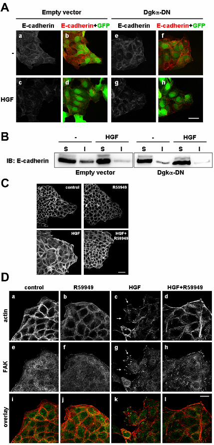
Dgkα inhibition uncouples spreading, cytoskeletal remodelling and lamellipodia
formation from downregulation of E-cadherin-mediated intercellular adhesions.
In HGF-induced cell scattering, loss of cell-cell contacts is preceded by internalization of E-
cadherins at 4-6 hours from HGF stimulation (Beherens et al., 1993; Potempa et al., 1998; Kimura
et al., 2006), which occurs concomitantly to colony spreading, so that the area covered by each
colony increases two to three fold. At the same time, cells at the colony outer edge undergo
dramatic morphological changes, featuring extended lamellipodia, where focal adhesion proteins,
such as Paxillin and Focal Adhesion Kinase (FAK), are recruited at new sites of adhesion and at the
tips of stress fibres (Weidner et al., 1993; Ridley et al., 1995; Palacios and D’Souza-Schorey,
2003).
We observed that inhibition of Dgkα, either by 1μM R59949 treatment (data not shown) or

by expression of Dgkα-DN, does not affect the HGF-induced internalization and removal of E-
cadherins from cell-cell contacts (Figure 3A, panel g), occurring upon 6 hours of cell stimulation. In
addition, we performed fractionation of MDCK cell lysates in NP-40-soluble and NP-40-insoluble
fractions. Upon 6 hours of treatment, HGF induces a decrease in the amount of E-cadherin in the
insoluble fraction, independently from Dgkα-DN expression (Figure 3B).
Conversely, inhibition of Dgkα by 1μM R59949 results in a remarkable reduction of HGF-
induced colony spreading upon 4 hours of cell stimulation (Figure 3C). Moreover, staining for F-
actin clearly shows that Dgkα inhibition strongly affects HGF-dependent morphological changes
such as lamellipodia formation (Figure 3D, panels a,b,c,d). Consistently with inhibition of
lamellipodia formation, R59949 treatment severely affects HGF-induced remodeling of focal
adhesions spatial organization, as, visualized by staining for FAK (Figure 3D, panels e,f,g,h).
Inhibition of Dgkα in unstimulated MDCK cells does not affect their morphology concerning all of
the analyzed aspects.
These data strongly suggest that Dgkα is not involved in the mechanisms by which HGF
downregulates E-cadherin-mediated intercellular adhesions, and that its inhibition uncouples HGF-
induced events leading to loss of intercellular adhesions from the signalling pathways mediating
cell spreading, F-actin remodelling, lamellipodia formation and eventually cell migration.
Dgkα is required for HGF-induced membrane ruffles formation and focal adhesions
remodelling.
Upon few minutes of HGF stimulation, MDCK cells at the outer edge of colonies undergo
intense ruffling. They eject small membrane protrusions, whose formation relies on regulated
recruitment of molecular scaffolds to growing focal complexes at new adhesion sites, coupled to the
coordinated organization of actin filaments into lamella network and bundled arrays. Eventually
membrane ruffles evolve in wider lamellipodia driving and providing direction to cell migration
(Small et al., 2002). Thus we verified whether the effects of Dgkα inhibition observed after hours
of HGF stimulation derived from impairment of events occurring at earlier time points, such as
formation of membrane ruffles and new focal complexes.
We ascertained Dgkα localization in resting or HGF-treated MDCK cell by transiently
transfecting a GFP-Dgkα fusion protein. In untreated cells Dgkα displays cytoplasmic localization,
but upon 15 minutes of HGF treatment it accumulates at the cell periphery, in correspondence of the
protruding plasma membrane (Figure 4A). This observation suggests that Dgkα may play a role in
HGF-induced earlier events leading to membrane ruffle formation.

Thus we set to investigate earlier changes in F-actin cytoskeleton organization in response to
HGF. Upon 15 minutes of HGF treatment, small membrane ruffles develop on the outer membranes
of cells at colony edge (Figure 4B, arrows). The percentage of cells featuring membrane ruffles
raises from less than 20% in control cells (vehicle- and R59949-treated cells) to about 50% in HGF-
treated cells. In presence of 1μM R59949, the percentage of membrane ruffle displaying cells upon
HGF stimulation is reduced to almost control value (Figure 4B). In order to further verify the
specificity of Dgkα requirement in HGF-induced ruffle formation, we showed that transient
transfection of either C1, C2 or C3 siRNA impairs HGF-induced membrane ruffling and that this
inhibition is completely overridden by the expression of the Dgkα murine hortologue, which is not
affected by any of three siRNA (Figure 4C). Consistently, HGF fails to induce membrane ruffles in
cells expressing Dgkα-DN compared to cells expressing the vector alone (Figure 4D). In
conclusion, these data demonstrate that the formation of membrane ruffles occurring upon 15
minutes of HGF treatment depends on stimulation of Dgkα activity.
Membrane ruffles formation implies the recruitment of focal adhesion proteins at new
adhesion sites within the ruffle itself. In epithelial cells, Paxillin recruitment to newly-formed focal
complexes, where it acts as a scaffold for signalling molecules, is required for HGF-induced
signalling leading to cell migration (Lamorte et al., 2003; Ishibe et al., 2004; Chen et al., 2005).
In resting MDCK cells, Paxillin is partially diffuse in the cytoplasm, while in cells at colony
edge it is also localized in focal adhesions along the outer plasma membrane (Figure 5A and B,
panel a). Upon 15 minutes of HGF stimulation, Paxillin condensates to the newly-formed focal
complexes in correspondence of membrane ruffles (Figure 5A, panel c and Figure 5B, panel b).
Upon inhibition of Dgkα by either 1μM R59949 (Figure 5A, panel d), or by expression of Dgkα-
DN (Figure 5B, panel f), Paxillin accumulates along the outer plasma membrane instead of being
recruited in the area of ruffling, while ruffles formation is impaired. Inhibition of Dgkα in
unstimulated cells does not significantly affect Paxillin localization either in the cytoplasm or at
focal adhesions along the outer plasma membrane.
In order to verify that Paxillin indeed accumulates in structures identifiable as focal
complexes, we analyzed its co-localization with Vinculin, a resident protein whose function is to
stabilize them (Ziegler et al., 2006). In unstimulated cells Vinculin and Paxillin co-localize at focal
complexes along the outer plasma membrane of colony-edge cells and upon HGF stimulation they
are both recruited to newly-formed focal complexes in the area of ruffling, in a manner fully
dependent on Dgkα activity. In fact, inhibition of Dgkα, while impairing HGF-induced neo-
formation of ruffles and focal complexes at membrane ruffles, does not affect Vinculin and Paxillin
co-localization (Figure 5C).

Upon growth factor stimulation Src- and FAK-mediated phosphorylation of Paxillin is
required to recruit and coordinate multiple signalling complexes, regulating events at the leading
edge of migrating cells (reviewed in Brown and Turner, 2004). Phosphorylation of Paxillin on
tyrosine 31 and 118 mediates its association with Crk and is required for growth factors-induced
Paxillin-mediated migratory signals (Nakamura et al., 2000; Petit et al., 2000). Thus we verified
whether inhibition of Dgkα affects HGF-induced phosphorylation of Paxillin Tyr31 and Tyr118,
identified by anti-phosphotyrosine specific antibodies. Western blot analysis of Paxillin tyrosine
phosphorylation reveals that HGF induces Paxillin phosphorylation of both Tyr31 and Tyr118 in
control MDCK cells (Figure 5D). Surprisingly basal phosphorylation of Paxillin in both residues is
enhanced in cells expressing Dgkα-DN, and is not further affected by HGF stimulation (Figure 5D).
In summary these data demonstrate that upon minutes of HGF stimulation, activation of
Dgkα is required for the formation of membrane ruffles and for the succeeding remodelling of
Paxillin- and Vinculin-containing focal complexes.
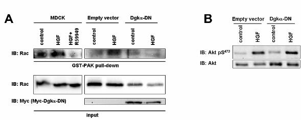
Dgkα is required for HGF-induced Rac activation and membrane targeting.
The data presented above strongly suggest that activation of Dgkα is involved in the
signalling mechanisms leading from HGF-receptor activation to ruffle formation.
Membrane ruffle formation is dependent on the activation of Rac small GTPase, which acts
upstream of the recruitment of WAVE and Arp2/3 complexes at new adhesion sites promoting F-
actin polymerization (Takenawa et al., 2007). In migrating cells active Rac localization at leading
edge is enhanced and allows the coupling with its downstream effectors (Kurokawa and Matsuda,
2005). In MDCK cells HGF activates Rac, whose function is required for HGF-induced cell scatter,
spreading and for ruffles and lamellipodia formation (Ridley et al., 1995; Royale et al., 2000).
Activation of endogenous Rac was assayed by GST-PAK pull-down to purify active GTP-
bound Rac from lysates of either control or HGF-stimulated MDCK cells. HGF treatment results in
activation of endogenous Rac. Inhibition of Dgkα, by either 1μM R59949 or by Dgkα-DN
expression, severely impairs HGF-induced Rac activation, without affecting Rac basal state of
activation (Figure 6A). Rac activation requires the coordinated activity of its direct upstream
regulators, which are recruited in multi-molecular complexes at the cell leading edge. As several
Rac GEFs are regulated through their PH domain by D-3 phosphoinositides (Welch et al., 2003),
we verified whether inhibition of Dgkα affects the PI 3-kinase pathway, as measured by Akt
phosphorylation. Indeed HGF induces Akt phosphorylation in both control and Dgkα-DN
expressing MDCK cells (Figure 6B), demonstrating that Dgkα does not mediate Rac activation by
regulating PI 3-kinase.

Rac activation is tightly coupled to its targeting to specific cholesterol-enriched membrane
microdomains, defined by ligand-activated integrin signalling (Grande-Garcìa et al., 2005). Thus
we verified whether inhibition of Dgkα may interfere with HGF-induced targeting of Rac to the
plasma membrane. By confocal microscopy, we observed the localization of endogenous Rac in
MDCK cells (Figure 7A). In most unstimulated cells, Rac is both cytoplasmic and at intercellular
contacts, while only about 20% of colony-edge cells feature Rac at the outer plasma membrane
(Figure 7A, panel a). Following 15 minutes of HGF stimulation, the percentage of colony-edge
cells featuring Rac at the outer plasma membrane raises to more than 40% (Figure 7A, panel c),
while localization of Rac at cell-cell contacts is not affected. Inhibition of Dgkα by 1μM R59949
treatment completely abolishes HGF-induced Rac membrane targeting (Figure 7A, panel d), while
it does not significantly affect Rac localization in unstimulated cells (Figure 7A, panel b), nor Rac
localization at cell-cell contacts. Similar results were obtained when Dgkα was inhibited upon
expression of Dgkα-DN (Figure 7B). Upon HGF stimulation Rac is properly membrane localized in
cell infected with the empty vector (Figure 7B, panel b), while it remains predominantly
cytoplasmic in Dgkα-DN-expressing cells (Fig. 7B, panel f).
In summary these data demonstrate that Dgkα is required for HGF-induced activation and
targeting of Rac to the plasma membrane and for the following formation of membrane ruffles, thus
strongly suggesting that Dgkα is involved in the signalling pathways regulating Rac function and
targeting upon activation of HGF receptor. Thoroughly, this data demonstrate that Dgkα plays a
pivotal role in the migratory signalling downstream HGF, being involved in early molecular events
such as Rac activation, membrane ruffles protrusion, and formation and organization of new focal
adhesions, and that it consequently regulates the acquisition of a migratory phenotype in epithelial
cells.

DISCUSSION
In this study we investigated the role of Dgkα in HGF- and v-Src-induced cell migration.
We show that Dgkα specific inhibition, obtained either pharmacologically, or by expression of a
kinase-defective dominant-negative mutant, or by siRNA-mediated downregulation of the
endogenous protein, impairs both HGF- and/or v-Src-induced cell scatter and migration. This
finding is consistent with previous demonstrations from our laboratory that Dgkα is activated by
growth factors through a mechanism requiring its tyrosine phosphorylation mediated by Src family
tyrosine kinases, and that its function is required for migration of endothelial cells (Cutrupi et al.,
2000; Baldanzi et al., 2004; Bacchiocchi et al., 2005; Baldanzi et al., 2007). Moreover these data
suggest that Dgkα represents a crucial node in the signalling network downstream Src regulating
epithelial cell scattering and switching to a motile mesenchymal phenotype.
Although both HGF stimulation and v-Src activation promote epithelial cell dispersion by
coordinating loss of intercellular adhesions and migration of cells away from one another, the two
events are regulated through distinct signalling pathways (Palacios et al., 2001). Intriguingly, Dgkα
inhibition uncouples the down-regulation of E-cadherin-mediated intercellular adhesions from cell
migration, strongly suggesting that Dgkα may regulate specifically those signalling events required
for HGF- and v-Src-stimulated epithelial cell motility. Thus we investigated the role of Dgkα in
well characterized HGF-induced morphological and molecular events leading to cell migration.
Spreading and lamellipodia protrusion with formation of new focal adhesions at the leading
edge are mandatory steps in cell migration (Ridley et al. 1995; Small et al., 2002). We show inhere
that upon Dgkα inhibition, no cell spreading, lamellipodia extension and remodelling of focal
adhesions are observed upon HGF treatment, suggesting that activation of Dgkα is likely to be
required for an earlier event. Rapid formation of membrane ruffles, upon minutes from growth
factors stimulation, preludes to establishment of extended lamellipodia at the leading edge of
migrating cells (Royale et al., 2000). Indeed, upon inhibition of Dgkα, MDCK cells fail to extend
membrane ruffles after HGF stimulation. Intriguingly, while recent findings indicate that Dgkα is
enriched in the pseudopodia of spontaneously invasive epithelial MSV-MDCK-INV cells (Jia et al.,
2005), we show that Dgkα is recruited to membrane ruffles upon HGF treatment. Altogether these
data provide the first circumstantial evidence that Dgkα may act in growth factors signalling at the
leading edge of migrating cells.
Ruffles formation, cell spreading and lamellipodia protrusion are dependent on Rac small
GTPase activation, occurring through its targeting to newly-formed focal complexes (Ridley et al.,

1995; Burridge and Wennerberg, 2004; Rossman et al., 2005). Rac targeting and GTP loading are
regulated by a complex signalling network involving the recruitment of distinct Rac-regulating
proteins to multiple molecular complexes at the leading edge of migrating cells.
An increasing body of evidence suggests that Dgks regulate small GTPases, including Rac,
through multiple mechanisms, whose complexity still awaits elucidation. In T cells Dgkα and ζ
negatively regulate Ras pathway, by finely tuning the access of RasGRP1, a C1 domain-containing
Ras GEF, to its activator DG (Jones et al., 2002; Olenchock et al., 2006, Zha et al., 2006).
However, in epithelial cells, neither the overexpression nor the downregulation of Dgkα affects the
Ras pathway, as detected by ERK-1/2 phosphorylation (our unpublished results). In addition Dgkγ,
but not Dgkα, upon its recruitment to the plasma membrane, negatively regulates PDGF- and EGF-
induced Rac activation and membrane ruffling, by enhancing the activity of β2-chimaerin, a Rac
GAP containing a C1 and a SH2 domain (Tsushima et al., 2004, Yasuda et al., 2007). These
observations provide further support to the previous finding that DG-dependent membrane
recruitment of β2-chiamerin determines the extent and the kinetic of EGF-induced Rac activation.
(Wang et al., 2006). Conversely, in neurons and skeletal myoblasts Dgkζ acts in a complex with
Rac at specific sites of the plasma membrane and control the remodeling of F-actin cytoskeleton
leading to neurite extension and membrane ruffle protrusion, possibly by facilitating Rac1 activation
and/or localization to the cell surface (Abramovici et al., 2003; Yakubchyk et al., 2005).
Furthermore Dgkζ and PI(4)P 5-kinase co-localize with F-actin at lamellipodia protrusions in
epithelial cells (Luo et al., 2004), where Dgk-generated PA is required for full activation of PI(4)P
5-kinase activity, consistently with a role of both lipid kinases in positive regulation of Rac
function. Interestingly a Dgk and a PI(4)P 5-kinase activities were found to associate in a complex
with Rac and RhoGDI (Tolias et al., 1998). RhoGDI forms a complex with Rac, keeping it in a
cytosolic inactive GDP-bound form, and upon Rac activation it contributes to Rac targeting to
specific sites at the plasma membrane (Moissoglu et al., 2006). As Rac targeting implies the
displacement of the interaction between Rac and RhoGDI, the finding that in vitro PA and PI(4,5)P2
impair RhoGDI affinity for Rac (Chuang et al., 1993; Ugolev et al., 2006), raises the hypothesis
that activation of the RhoGDI-associated Dgk may allow the release of Rac from RhoGDI, and
leads to speculate that also Dgkα may regulate Rac activation through this mechanism. Altogether,
these data strongly indicate that distinct Dgk isoforms act as regulators of Rac membrane targeting
and activation through multiple mechanisms, whose complexity still awaits to be elucidated.
Several Rac GEFs, such as Vav2, DOCK180/Elmo, βPIX and Tiam1, are regulated either
directly or indirectly through Src-dependent tyrosine phosphorylation (Lamorte et al., 2002;
Servitja et al., 2003; Santy et al., 2005), and/or interaction with PI(3,4,5)P3 (Welch et al., 2003).
Although there is no direct evidence for a role of any Dgk isoforms in the regulation of any Rac
GEFs, based on the observations reported inhere, we may discuss several hypothesis, providing a
framework for further investigation.
Several data indicate that, upon growth factors and v-Src stimulation, rapid Rac-mediated
membrane ruffling occurs through the recruitment of βPIX to Paxillin-containing focal complexes
(Cotton et al., 2007). Indeed βPIX mediates rapid ruffles formation upon PDGF, EGF and FGF
treatment in different cell types (Lee et al., 2001; Park et al., 2004; Shin et al., 2006), and the
interaction between βPIX and Rac is necessary and sufficient for Rac recruitment to membrane
ruffles and focal adhesions (ten Klooster et al., 2006). Crk recruitment to tyrosine-phosphorylated
Paxillin contributes to βPIX localization to focal complexes (Lamorte et al., 2003). Indeed we show
that HGF stimulates phosphorylation of Paxillin on both Tyr31 and Tyr118, the two major
determinants for Crk association. Surprisingly, the inhibition of Dgkα enhances basal
phosphorylation of Paxillin on both residues, but does not affect their phosphorylation upon HGF
stimulation, suggesting that Dgkα may affect βPIX function in a complex manner. Moreover βPIX
and Dgkα do not associate in a complex, not even upon HGF stimulation (data not shown).
Upon minutes of growth factors stimulation, βPIX recruitment and Rac activation are
promoted by rapid GTP/GDP cycling of Arf6, suggesting that Arf6 plays a pivotal role in Rac-
mediated membrane ruffling (ten Klooster et al., 2006; Cotton et al., 2007). Furthermore, upon
hours of HGF stimulation, ARF6 has been recently shown to regulate Rac targeting at tubule tips of
MDCK cells grown in 3D collagen (Tushir and D’Souza-Schorey, 2007). Interestingly, several Arf
GAPs are regulated by phospholipids, including PA (Randazzo et al., 2000). Moreover, PLD-
induced production of PA downstream of Arf6 is required for Arf6-dependent epithelial cell
ruffling and migration (Santy and Casanova, 2001). Thus we may speculate that also Dgkα may
contribute to regulate Arf6 function in coordinating Rac activation, focal adhesions remodelling and
membrane ruffles formation.
Several Rac and Arf GEFs are regulated by PI(3,4,5)P3, the product of PI 3-kinase. However
we can rule out that Dgkα mediates Rac activation by regulating PIP3 synthesis, as inhibition of
Dgkα does not affect HGF-induced activation of Akt, a major PIP3 target. Conversely, the finding
that PIP3 might contribute to recruit and activate Dgkα (Ciprés et al., 2003) allow to speculate that
Dgkα might contribute to couple PIP3 generation to the activation of one of the PIP3-dependent Rac
GEFs, such as Vav2 and Tiam1. However, the expression of a either wild-type or kinase-defective
Dgkα in fibroblasts does not affect PDGF-induced Rac activation and ruffles formation (Tsushima
et al., 2004), both mediated by Vav2 (Liu and Burridge, 2000). Moreover, the Rac GEF Tiam1 is

mainly involved in maintaining E-cadherin-mediated epithelial cell-cell adhesions (Mertens et al.,
2003), event which we showed to not be regulated by Dgkα, making Tiam1 an unlike target of
Dgkα activity.
In conclusion, inhere we clearly demonstrate that activation of Dgkα is required for HGF-
and v-Src-induced cell migration. By exploring some significant molecular events affected by Dgkα
inhibition, we raise the hypothesis that Dgkα may act in growth factors migratory signalling by
mediating Rac targeting and activation, thus revealing a novel signalling pathway linking tyrosine-
kinase receptors and Src to small GTPases in the context of cell migration.

AKNOWLEDGEMENTS
This study was supported by grants from the Italian Ministry for University and Research
(PRIN 2004-05 and FIRB 2001 post-genomic program to AG; FIRB 2001 and RBNE019J9W_003
to OP), from the Regione Piemonte (Ricerca Sanitaria and CIPE), Fondazione Cariplo, AIRC
(Italian Association for Cancer Research) and AICR (International Agency for Cancer Research,
Glasgow) to AG. NF was supported by FIRB. We thank Cecilia Deantonio, Miriam Gaggianesi,
Rosanna Panico-Guercia and Marianna Notario for their helpful work throughout the project.
REFERENCES
Avizienyte, E. and Frame, M.C. (2005). Src and FAK signalling controls adhesion fate and the
epithelial-to-mesenchymal transition. Curr. Opin. Cell Biol. 17, 542-547.
Abramovici, H., Hogan, A.B., Obagi, C., Topham, M.K., and Gee, S.H. (2003). Diacylglycerol
kinase-zeta localization in skeletal muscle is regulated by phosphorylation and interaction with
syntrophins. Mol Biol Cell. 14, 4499-4511.
Bacchiocchi, R., Baldanzi, G., Carbonari, D., Capomagi, C., Colombo, E., van Blitterswijk, W.J.,
Graziani, A., and Fazioli F. (2005). Activation of α-Diacylglycerol kinase is critical for the
mitogenic properties of anaplastic lymphoma kinase. Blood 106, 2175-2182.
Baldanzi, G., Mitola, S., Cutrupi, S., Filigheddu, N., van Blitterswijk, W.J., Sinigaglia, F.,
Bussolino, F., and Graziani, A. (2004). Activation of diacylglycerol kinase alpha is required for
VEGF-induced angiogenic signaling in vitro. Oncogene 23, 4828-4838.
Baldanzi, G., Cutrupi, S., Chianale, F., Gnocchi, V., Rainero, E., Porporato, P., Filigheddu, N., van
Blitterswijk, W.J., Parolini, O., Bussolino, F., Sinigaglia, F., and Graziani, A. (2007).
Diacylglycerol kinase-a phosphorylation by Src on Y335 is required for activation, membrane
recruitment and Hgf induced cell motility. Oncogene in press.
Beherens, J., Vakaet, L., Friis, R., Winterhager, E., Van Roy, F., Mareel, M. M., and Birchmeier,
W. (1993). Loss of epithelial differentiation and gain of invasiveness correlates with tyrosine
phosphorylation of the E-cadherin/β-catenin complex in cells transformed with a temperature-
sensitive v-src gene. J. Cell Biol. 120, 757-766.
Birchmeier, C., Birchmeier, W., Gherardi, E., Vande Woude, G.F. (2003). Met, metastasis, motility
and more. Nat. Rev. Mol. Cell. Biol. 4, 915-925.
Brown, M.C., and Turner, C.E. (2004). Paxillin: adapting to change. Physiol. Rev. 4, 1315-1339.
Burridge, K., and Wennerberg, K. (2004). Rho and Rac take the center stage. Cell 116, 167-179.

Caloca M.J., Wang H., and Kazanietz M.G. (2003). Characterization of the Rac-GAP (Rac-GTPase-
activating protein) activity of beta2-chimaerin, a 'non-protein kinase C' phorbol ester receptor.
Biochem J. 375, 313-21.
Canagarajah B., Leskow F.C., Ho J.Y., Mischak H., Saidi L.F., Kazanietz M.G., and Hurley J.H.
(2004). Structural mechanism for lipid activation of the Rac-specific GAP, beta2-chimaerin.
Cell 119, 407-418.
Chem, H.-Y., Shen, C.-H., Tsai, Y.-T., Lin, F.-C., Huang, Y.-P., and Chen, R.-H. (2004). Brk
activates Rac1 and promotes cell migration and invasion by phosphorylating Paxillin. Mol. Cell.
Biol. 24, 10558-10572.
Chen, G.C., Turano, B., Ruest. P.J., Hagel, M., Settleman, J., and Thomas, S.M. (2005). Regulation
of Rho and Rac signaling to the actin cytoskeleton by paxillin during Drosophila development. Mol.
Cell Biol. 25, 979-987.
Chen, H.C., Chan, P.C., Tang, M.J., Cheng, C.H., and Chang, T.J. (1998). Tyrosine
phosphorylation of focal adhesion kinase stimulated by hepatocyte growth factor leads to mitogen-
activated protein kinase activation. J. Biol. Chem. 273, 25777-25782.
Chuang, T.-H., Bohl, B.P., and Bokoch, G.M. (1993). Biologically active lipids are regulator of
Rac-GDI complexation. J. Biol. Chem. 268, 26206-26211.
Ciprés, A., Carrasco, S., Martinez-A, C., and Mérida, I. (2003). Regulation of Diacylglycerol
Kinase α by Phosphoinositide 3-Kinase lipid products. J. Biol. Chem. 278, 35629-35635.
Cotton, M., Boulay, P.-L., Houndolo, T., Vitale, N., Pitcher, J.A., and Claing, A. (2007).
Endogenous ARF6 interacts with Rac1 upon Angiotensin II stimulation to regulate membrane
ruffling and cell migration. Mol. Biol. Cell 18, 501-511.
Cutrupi, S., Baldanzi, G., Gramaglia, D., Maffè, A., Schaap, D., Giraudo, E., van Blitterswijk, W.,
Bussolino, F., Comoglio, P.M., and Graziani, A. (2000). Src-mediated activation of alpha-

diacylglycerol kinase is required for hepatocyte growth factor-induced cell motility. EMBO J. 19,
4614-4622.
Danilkovitch-Miagkova, A., and Zbar B (2002). Dysregulation of Met receptor tyrosine kinase
activity in invasive tumors J. Clin. Invest. 109, 863-867.
Grande-Garcìa, A., Echarri, A., and Del Pozo, M.A. (2005) Integrin regulation of membrane
domain trafficking and Rac targeting. Biochem. Soc. Trans. 33, 609-613.
Graziani, A., Gramaglia, D., Cantley, L.C., and Comoglio, P.M. (1991) The tyrosine-
phosphorylated hepatocyte growth factor/scatter factor receptor associates with phosphatidylinositol
3-kinase. J. Biol. Chem. 266, 22087-22090
Imai, S., Kai, M., Yasuda, S., Kanoh, H., and Sakane, F. (2005). Identification and characterization
of a novel human type II diacylglycerol kinase, DGK kappa. J. Biol. Chem. 280, 39870-39881.
Irby, R.B., and Yeatman, T.J. (2000). Role of Src expression and activation in human cancer.
Oncogene 19, 5636-5642.
Ishibe, S., Joly, D., Lium Z.-X., and Cantley, L.G. (2004). Paxillin serves as an ERK-regulated
scaffold for coordinating FAK and RAC activation in epithelial morphogenesis. Mol. Cell 16, 257-
267.
Jones, D.R., Sanjuan, M.A., Stone, J.C., and Merida, I. (2002). Expression of a catalytically inactive
form of diacylglycerol kinase alpha induces sustained signaling through RasGRP.
FASEB J. 16, 595-7.
Kimura, T., Sakisaka, T., Baba, T., Yamada, T., and Takai, Y. (2006). Involvement of the Ras-Ras-
activated Rab5 guanine nucleotide exchange factor RIN2-Rab5 pathway in Hepatocyte Growth
Factor-induced endocytosis of E-cadherin. The J. Biol. Chem. 281, 10598-10609.
Kurokawa, K., and Matsuda, M. (2005). Localized RhoA activation as a requirement for the
induction of membrane ruffling. Mol. Biol. Cell 16, 4294-4303.

Lamorte, L., Rodrigues, S., Sangwan, V., Turner, C.E., and Park, M. (2003). Crk associates with a
multimolecular Paxillin/GIT2/β-PIX complex and promotes Rac-dependent relocalization of
Paxillin to focal contacts. Mol. Biol. Cell 14, 2818-2831.
Lamorte, L., Royal, I., Naujokas, M., and Park, M. (2002). Crk Adapter Proteins Promote an
Epithelial– Mesenchymal-like Transition and Are Required for HGF-mediated Cell Spreading and
Breakdown of Epithelial Adherens Junctions. Mol. Biol. Cell 13, 1449-1461.
Lee S.-H., Eom, M., Lee, S.J., Kim, S., Park, H.-J., and Park, D. (2001). β Pix-enhanced p38
activation by Cdc42/Rac/PAK/MKK3/6-mediated pathaway. Implication in the regulation of
membrane ruffling. J. Biol. Chem. 276, 25066-25072.
Liu, B.P., and Burridge, K. (2000). Vav2 activates Rac1, Cdc42, and RhoA downstream from
growth factor receptors but not beta1 integrins. Mol. Cell. Biol. 20, 7160-7169.
Luo, B., Prescott, S.M., and Topham, M.K. (2003). Association of diacylglycerol kinase zeta with
protein kinase C alpha: spatial regulation of diacylglycerol signaling. J. Cell Biol. 160, 929-37.
Luo, B., Prescott, S.M., and Topham, M.K. (2004). Diacylglycerol Kinase ζ regulates
phosphatidylinositol 4-phosphate 5-kinase Iα by a novel mechanism. Cell Signall. 16, 891-897.
Meng, W., Numazaki, M., Takeuchi, K., Uchibori, Y., Ando-Akatsuka, Y., Tominaga, M., and
Tominaga, T. (2004). DIP (mDia interacting protein) is a key molecule regulating Rho and Rac in a
Src-dependent manner. EMBO J. 23, 760-71.
Mertens, A.E., Roovers, R.C., and Collard, J.G. (2003). Regulation of Tiam1-Rac signalling.
FEBS Lett. 546, 11-16.
Moissoglu, K., Slepchenko, B.M., Meller, N., Horwitz, A.F., and Schwartz, M.A. (2006). In vivo
dynamics of Rac-membrane interactions. Mol. Biol. Cell 17, 2770-2779.
Nakaigawa, N., Weirich, G., Schmidt, L., and Zbar, B. (2000). Tumorigenesis mediated by Met
mutant M1268T is inhibited by dominat-negative Src. Oncogene 19, 2296-3002.

Nakamura, K., Yano, H., Uchida, H., Hashimoto, S., Schaefer, E., and Sabe, H. (2000). Tyrosine
phosphorylation of paxillin {alpha} is involved in temporospatial regulation of paxillin-containing
focal adhesions and F-actin organization in motile cells. J. Biol. Chem. 275, 21155-21164.
Olenchock, B.A., Guo, R., Carpenter, J.H., Jordan, M., Topham, M.K., Koretzky, G.A., and Zhong,
X.-P. (2006). Disruption of diacylglycerol metabolism impairs induction of T cell anergy. Nat.
Immunol. 7, 1174-1181.
Olenchock, B.A., Guo, R., Silverman, MA, Wu, J.N., Carpenter, J.H., Koretzky, G.A., and Zhong,
X.-P. (2006). Impaired degranulation but enhanced cytokine production after Fc epsilonRI
stimulation of diacylglycerol kinase zeta-deficient mast cells. J. Exp. Med. 203, 1471-1480.
Palacios, F., and D'Souza-Schorey, C. (2003). Modulation of Rac1 and ARF6 activation during
epithelial cell scattering. J. Biol. Chem. 278, 17395-17400.
Palacios, F., Price, L., Schweitzer, J., Collard, J.G., and D'Souza-Schorey, C. (2001). An essential
role for ARF6-regulated membrane traffic in adherens junction turnover and epithelial cell
migration. EMBO J. 20, 4973-86.
Park, H.S., Lee, S.H., Park, D., Lee, J.S., Ryu, S.H., Lee, W.J., Rhee, S.G., and Bae, Y.S. (2004).
Sequential activation of Phosphatidylinositol 3-Kinase, βPix, Rac1 and Nox1 in Growth Factor-
induced production of H2O2. Mol. Cell. Biol. 24, 4384-4394.
Petit, V., Boyer, B., Lentz, D., Turner, C.E., Thiery, J.P., and Vallés, A.M. (2000). Phosphorylation
of tyrosine residues 31 and 118 on Paxillin regulates cell migration through an association with
CRK in NTB-II cells. J. Cell Biol. 148, 958-969.
Potempa, S., and Ridley, A.J. (1998). Activation of both MAP Kinase and Phosphatidylinositide 3-
Kinase by Ras is required for Hepatocyte Growth Factor/Scatter Factor-induced adherens junction
disassembly. Mol. Biol. Cell 9, 2185-2200.
Rahimi, N., Hung, W., Tremblay, E., Saulnier, R., and Elliott, B. (1998). c-Src kinase activity is
required for hepatocyte growth fator-induced cell motility and anchorage-indipendent growth of
mammary carcinoma cells. J. Biol. Chem. 273, 33714-33721.
Randazzo, P.A., Nie, Z., Miura, K., and Hsu, V.W. (2000). Molecular aspects of the cellular
activities of ADP-ribosylation factors. Sci. STKE. 2000 re1.
Ridley, A., Comoglio, P.M., and Hall, A. (1995). Regulation of Scatter Factor/Hepatocyte Growth
Factor responses by Ras, Rac, and Rho in MDCK cells. Mol. Cell. Biol. 15, 1110-1122.
Royale, I., Lamarche-Vane, N., Lamorte, L., Kaibuchi, K., and Park M., (2000). Activation of
Cdc42, Rac, PAK, and Rho-Kinase in response to Hepatocyte Growth Factor differentially
regulates epithelial cell colony spreading and dissociation. Molecular Biology of the Cell 11, 1709-
1725.
Sanjuan, M.A., Pradet-Balade, B., Jones, D.R., Martinez-A, C., Stone, J.C., Garcia-Sanz, J.A., and
Merida, I. (2003). T cell activation in vivo targets diacylglycerol kinase alpha to the membrane: a
novel mechanism for Ras attenuation. J. Immunol. 170, 2877-28783.
Santy, L.C., and Casanova, J.E. (2001). Activation of ARF6 by ARNO stimulates epithelial cell
migration through downstream activation of both Rac1 and phospholipase D.
J. Cell Biol. 154, 599-610.
Santy, L.C., Ravichandran, K.S., and Casanova, J.E. (2005). The DOCK180/Elmo complex couples
ARNO-mediated Arf6 activation to the downstream activation of Rac1. Curr. Biol. 15, 1749-1754.
Servitja, J.M., Marinissen, M.J., Sodhi, A., Bustelo, X.R., and Gutkind, J.S. (2003). Rac1 function
is required for Src-induced transformation. Evidence of a role for Tiam1 and Vav2 in Rac activation
by Src. J. Biol. Chem. 278, 34339-34346.
Shin, E.-Y., Lee, C.-S., Cho, T.G., Kim, Y,G., Song, S., Juhnn, Y.-S., Park, S.C., Manser, E., and
Kimn E.-G. (2006). β Pak-interacting Exchange Factor-mediated Rac1 activation requires smgGDS
guanine nucleotide exchange factor in Basic Fibroblast Growth Factor-induced neurite outgrowth. J.
Biol. Chem. 281, 35954-35964.
Small, J.V., Stradal, T., Vignal, E., and Rottner, K. (2002). The lamellipodium: where motility
begins. TRENDS Cell Biol. 12, 112-120.

Takenawa ,T., and Suetsugu, S. (2007). The WASP-WAVE protein network: connecting the
membrane to the cytoskeleton. Nat. Rev. Mol. Cell. Biol. 8, 37-48.
ten Klooster J.P., Jaffer, Z.M., Chernoff, J., and Hordijk P.L. (2006). Targeting and activation of
Rac1 are mediated by exchange factor β-Pix. J. Cell. Biol. 172, 759-769.
Thiery, J.P. (2002). Epithelial-mesenchymal transitions in tumour progression. Nat. Rev. Cancer 2,
442-454.
Thiery, J.P., and Sleeman, J.P. (2006). Complex networks orchestrate epithelial-mesenchymal
transitions. Nat. Rev. Mol. Cell. Biol. 7, 131-142.
Tolias, K.F., Couvillon, A.D., Canley, L.C., and Carpenter, C.L. (1998). Characterization of a
Rac1- and RhoGDI-associated lipid kinase signaling complex. Mol. Cell. Biol. 18, 762-770.
Topham, M.K. (2006) Signaling roles of diacylglycerol kinases. J. Cell. Biochem. 97, 474-484.
Topham, M.K., and Prescott, S.M. (1999). Mammalian diacylglycerol kinases, a family of lipid
kinases with signaling functions. J. Biol. Chem. 274, 11447-11450.
Topham, M.K., and Prescott, S.M. (2001). Diacylglycerol kinase zeta regulates Ras activation by a
novel mechanism. J. Cell Biol. 152, 1135-1143.
Tsushima, S., Kai, M., Yamada, K., Imai, S., Houkin, K., Kanoh, H., and Sakane, F. (2004).
Diacylglycerol kinase gamma serves as an upstream suppressor of Rac1 and lamellipodium
formation. J. Biol. Chem. 279, 28603-28613.
Turner, C.E., Brown, M.C., Perrotta, J.A., Riedy, M.C., Nikolopoulos, S.N., McDonald, A.R.,
Bagrodia, S., Thomas, S., and Leventhal, P.S. (1999). Paxillin LD4 Motif Binds PAK and PIX
through a novel 95-kD Ankyrin repeat, ARF-GAP protein: a role in cytoskeletal remodelling. J.
Cell Biol. 145, 851-863.

Tushir, J.S., and D’Souza-Schorey, C. (2007). ARF6-dependent activation of ERK and Rac1
modulates epithelial tubule development. EMBO J. 26, 1806-1819.
Ugolev, Y., Molshanski-Mor, S., Weinbaum, C., and Pick, E. (2006). Liposomes comprising
anionic but not neutral phospholipids cause dissociation of [Rac(1 or 2)-RhoGDI] complexes and
support amphiphile-independent NADPH oxidase activation by such complexes. J. Biol. Chem.
281, 19204-19219.
Vallés, A.M., Beuvin, M., and Boyer, B. (2004). Activation of Rac1 by Paxillin-Crk-DOCK180
signaling complex is antagonized by Rap1 in migrating NTB-II cells. J. Biol. Chem. 279, 44490-
44496.
van Blitterswijk, W.J., and Houssa, B. (2000). Properties and functions of diacylglycerol kinases.
Cell. Signal. 12, 10595-10605.
Wang, H., Yang, C., Leskow, F.C., Sun, J., Canagarajah, B., Hurley, J.H., and Kazanietz, M.G.
(2006). Phospholipase C gamma/diacylglycerol-dependent activation of beta2-chimaerin restricts
EGF-induced Rac signaling. EMBO J. 25, 2062-2074.
Weidner, K.M., Sachs, M., and Birchmeier, W. (1993) The Met receptor tyrosine kinase transduces
motility, proliferation, and morphogenic signals of scatter factor/hepatocyte growth factor in
epithelial cells. J. Cell Biol. 121, 145-154.
Welch, H.C.E., Coadwell, W.J., Stephens, L.R., and Hawkins, P.T. (2003). Phosphoinositide 3-
kinase dependent activation of Rac. FEBS Lett. 546, 93-97.
Yakubchyk, Y., Abramovici, H., Maillet, J.C., Daher, E., Obagi, C., Parks, R.J., Topham, M.K., and
Gee, S.H. (2005). Regulation of neurite outgrowth in N1E-115 cells through PDZ-mediated
recruitment of diacylglycerol kinase zeta. Mol. Cell. Biol. 25, 7289-302.
Yasuda, S., Kai, M., Imai, S., Kanoh, H., and Sakane, F. (2007). Diacylglycerol kinase γ interacts
with and activates b2-chimaerin, a Rac-specific GAP, in response to epidermal growth factor. FEBS
Lett. 581, 551-557.

Zha, Y., Marks, R., Ho, A.W., Peterson, A.C., Janardhan, S., Brown, I., Praveen, K., Stang, S.,
Stone, J.C., and Gajewski, T.F. (2006). T cell anergy is reversed by active Ras and is regulated by
diacylglycerol kinase-α. Nat. Immunol. 7, 1166-1173.
Zhao, C., Du, G., Skowronek, K., Frohman, M.A., and Bar-Sagi D. (2007). Phospholipase D2-
generated phosphatidic acid couples EGFR stimulation to Ras activation by Sos. Nat. Cell Biol. 9,
706-712.
Zhong, X.P., Hainey, E.A., Olenchock, B.A., Jordan, M.S., Maltzman, J.S., Nichols, K.E., Shen, H.,
and Koretzky, G.A. (2003). Enhanced T cell responses due to diacylglycerol kinase zeta deficiency.
Nat. Immunol. 4, 882-890.
Ziegler, W.H., Liddington, R.C., Critchley, D.R. (2006). The structure and regulation of vinculin.
Trends Cell Biol. 16, 453-460.
Zondang, G.C.M., Evers, E.E., ten Klooster, J.P., Janssen, L., van der Kammen,R.A., Collard, J.G.
(2000) Oncogenic Ras downregulates Rac activity, which leads to increased Rho activity and
epithelial-mesenchimal transition. J. Cell Biol. 149, 775-781

FIGURE LEGENDS
Figure 1. v-Src activates Dgkα. MDCK-ts-v-Src maintained at the non-permissive
temperature of 40.5°C were transiently transfected with Myc-Dgkα, starved overnight in 0% FBS
medium and shifted to the permissive temperature of 35°C for the times indicated. Cell lysates were
immunoprecipitated with an anti-Myc antibody. Half of each immunoprecipitate was separated by
SDS-PAGE and after blotting was probed with anti-Myc; the other half was assayed for Dgk
activity as described in Materials and Methods.
Figure 2. Dgkα is required for HGF-induced cell scatter and migration of MDCK cells (A)
MDCK cell colonies were treated, in 0% FBS medium, with HGF 2 ng/ml in presence or absence of
1μM R59949 for 24 hours. Representative fields are shown. (B) Control or MDCK/Dgkα-WT cells
were allowed to migrate into the wounded area in 0% FBS medium with 2.5 ng/ml HGF in presence
or absence of 1μM R59949 for 7 hours. Quantification was performed as described in Materials and
Methods. Means of at least 4 experiments with standard errors are shown. ** = p<0.005. The
western blot shows the level of Myc-Dgkα-WT expression. (C) MDCK/empty vector or
MDCK/Dgkα-DN were treated, in 0% FBS medium, with HGF 2 ng/ml for 24 hours.
Representative fields are shown. The western blot shows the level of Myc-Dgkα-DN expression.
(D) Lysates of MDCK cells transiently transfected with negative control siRNA or canine Dgkα
siRNAs C1, C2 and C3 were separated by SDS-PAGE and after blotting were probed for Dgkα and
tubulin. MDCK cell colonies were transfected with Block-IT fluorescent siRNA to evaluate the
efficiency of transfection. (E) MDCK and MDCK/Mus-Dkgα cell colonies were transiently
transfected with negative control siRNA or canine Dgkα siRNAs and treated, in 0% FBS medium,
with HGF 2 ng/ml for 24 hours. Representative fields are shown. (F) MDCK cells were seeded in
the upper part of a chemotaxis chamber and induced to migrate in presence of 50 ng/ml of HGF in
the bottom part, in presence or absence of 1μM R59949. The histograms represent the number of
migrated cells, means of 8 different wells with standard errors. ** = p<0.005. A representative
experiment is shown. (G) MDCK/empty vector or MDCK/Dgkα-DN cells were seeded in the upper
chamber of a Transwell apparatus. Invasion through a Matrigel-covered porous membrane was
induced in 48 hours in 2% FBS medium by the presence of 100 ng/ml HGF in the lower chamber.
Fixed cells on the Transwells lower face were stained with crystal-violet, photographed and
quantified by optical densitometry. Means of three experiments are shown, with standard errors; * =
p ≤ 0.05. (H) MDCK-ts-v-Src cell colonies, maintained at the non-permissive temperature of

40.5°C, were transiently transfected with negative control siRNA or canine Dgkα siRNAs, as
indicated, placed in 0% FBS medium and shifted to the permissive temperature of 35°C for 24
hours. The same experiment was performed with untransfected cells, in presence or absence of 1μM
R59949, as indicated. Representative pictures are shown.
Figure 3. Dgkα is required for HGF-induced cell spreading and lamellipodia formation, but
not for down-regulation of E-cadherin-mediated intercellular adhesions. (A) MDCK/empty vector
or MDCK/Dgkα-DN cells were treated with HGF 10 ng/ml for 6 hours, fixed and stained for E-
cadherin. Representative pictures are shown. Scale bar = 20 μm. (B) MDCK/empty vector or
MDCK/Dgkα-DN cells were treated with HGF 10 ng/ml for 6 hours. Cell lysates were fractionated
into a NP-40-soluble (S) and a NP-40-insoluble (I) fraction. Equal sample volumes were loaded,
separated by SDS-PAGE and probed for E-cadherin. (C) MDCK cell colonies were starved
overnight in a 2% FBS medium and treated with HGF 10 ng/ml for 4 hours, in presence or absence
of 1μM R59949. Fixed cells were stained for actin filaments with phalloidin. Scale bar = 40 μm.
(D) MDCK cells treated as described in (C) were fixed and stained for actin (red, panels a,b,c,d)
and FAK (green, panel e,f,g,h). Scale bar = 16 μm. Representative pictures are shown.
Figure 4. Dgkα is required for HGF-induced membrane ruffling of MDCK cells. (A)
MDCK cells were transiently transfected with GFP-Dgkα, starved overnight in 0% FBS medium,
stimulated with HGF 10 ng/ml for 15 minutes, fixed and stained for actin. Scale bar = 8 μm (B)
MDCK cell colonies were starved overnight in 0% FBS medium, treated with HGF 10 ng/ml for 15
minutes in presence or absence of 1μM R59949, fixed and stained for actin. Scale bar = 16 μm.
Confocal acquired images were observed and cells at the edge of colonies were scored for presence
of membrane ruffles (arrows). The percentage of cells with membrane ruffles was calculated.
Means of three experiments with standard errors are shown. ** = p<0.005. (C) MDCK and
MDCK/Mus-Dgkα were transiently transfected with negative control siRNA or canine Dgkα
siRNAs C1, C2 or C3, starved overnight in 0% FBS medium and treated with HGF 10 ng/ml for 15
minutes. Confocal acquired images were observed and cells at the edge of colonies were scored for
presence of membrane ruffles. The percentage of cells with membrane ruffles was calculated.
Means of three experiments with standard errors are shown. * = p<0.05, ** = p<0.005. (D)
MDCK/empty vector or MDCK/Dgkα-DN cells were starved overnight in 0% FBS medium, treated
with HGF 10 ng/ml for 15 minutes, fixed and immunostained for actin (red, panels a,b,c,d). The
arrows indicate membrane ruffles in empty vector-infected cells. Scale bar = 16 μm. Representative

pictures are shown. Confocal acquired images were observed and cells at the edge of colonies were
scored for presence of membrane ruffles. Means of three experiments with standard errors are
presented. * = p<0.05.
Figure 5. Dgkα is required for HGF-induced Paxillin localization to newly-formed focal
complexes. (A) MDCK cell colonies were starved overnight in 0% FBS medium, treated with HGF
10 ng/ml for 15 minutes in presence or absence of 1μM R59949, fixed and stained for Paxillin
(green, panels a,b,c,d) and actin (red, panels e,f,g,h). Representative pictures are shown. Scale bar =
16 μm. (B) MDCK/empty vector or MDCK/Dgkα-DN cells were starved overnight in 0% FBS
medium, treated with HGF 10 ng/ml for 15 minutes, fixed and immunostained for Paxillin (red,
panels a,b,e,f). Thick arrows indicate Paxillin localization at focal adhesions in the areas of
membrane ruffling, while the slim arrow indicates Paxillin localization at cell periphery in a Dgkα-
DN-infected cell, without membrane ruffles. Scale bar = 16 μm. Representative pictures are shown.
(C) MDCK cell colonies were treated as described in (A), fixed and stained for Paxillin (green),
Vinculin (red) and actin (blue). Representative pictures are shown. Scale bar = 16 μm. (D)
MDCK/empty vector or MDCK/Dgkα-DN cells were starved overnight in 0% FBS medium and
treated with HGF 50 ng/ml for 15 minutes. Whole cell lysates were separated by SDS-PAGE and
probed with anti-Paxillin pTyr31 and pTyr118 and anti-Paxillin.
Figure 6. Dgkα is required for HGF-induced Rac activation. (A) MDCK cells were starved
overnight in 0% FBS medium, treated with HGF 100 ng/ml for 15 minutes in presence or absence
of 1μM R59949 and lysed. GTP-bound active Rac was purified in each sample by pull-down with
GST-fused PAK CD domain. MDCK/empty vector and MDCK/Dgkα-DN cells were starved
overnight in 0% FBS medium, treated with HGF 100 ng/ml for 15 minutes and pull-down assays
were performed as described before. (B). MDCK/empty vector and MDCK/Dgkα-DN cells were
starved overnight in 0% FBS medium, treated with HGF 50 ng/ml for 15 minutes and lysed. Whole
cell lysates were separated by SDS-PAGE and probed with anti-Akt pSer473 and Akt.
Figure 7. Dgkα is required for HGF-induced Rac localization to the plasma membrane. (A)
MDCK cell colonies were starved overnight in 0% FBS medium, treated with HGF 10 ng/ml for 15
minutes in presence or absence of 1μM R59949, fixed and stained for Rac (green, panels a,b,c,d)
and actin (red). Representative pictures are shown. Scale bar = 16 μm. Confocal acquired images
were observed and the percentage of cells at the edge of colonies featuring Rac at the outer plasma

membrane was calculated. Means of three experiments with standard errors are shown. * = p<0.05.
(B) MDCK/empty vector or MDCK/Dgkα-DN cells were starved overnight in 0% FBS medium,
treated with HGF 10 ng/ml for 15 minutes, fixed and immunostained for Rac (red, panels a,b,e,f).
Representative pictures are shown. Scale bar = 20 μm. Confocal acquired images were observed
and the percentage of edge-of-colony cells featuring Rac at the outer plasma membrane was
calculated. Means of three experiments with standard errors are presented. ** = p<0.005.


Copyright © 2022 FDOKUMEN