PGC-1 mediates exercise-induced skeletal muscle VEGF expression in mice
13
doi:10.1152/ajpendo.00076.2009 297:92-103, 2009. First published Apr 28, 2009; Am J Physiol Endocrinol Metab Wojtaszewski, Juan Hidalgo and Henriette Pilegaard Lotte Leick, Ylva Hellsten, Joachim Fentz, Stine S. Lyngby, Jørgen F. P. You might find this additional information useful... 59 articles, 39 of which you can access free at: This article cites http://ajpendo.physiology.org/cgi/content/full/297/1/E92#BIBL including high-resolution figures, can be found at: Updated information and services http://ajpendo.physiology.org/cgi/content/full/297/1/E92 can be found at: AJP - Endocrinology and Metabolism about Additional material and information http://www.the-aps.org/publications/ajpendo This information is current as of June 26, 2009 . http://www.the-aps.org/. 20814-3991. Copyright © 2005 by the American Physiological Society. ISSN: 0193-1849, ESSN: 1522-1555. Visit our website at organization. It is published 12 times a year (monthly) by the American Physiological Society, 9650 Rockville Pike, Bethesda MD publishes results of original studies about endocrine and metabolic systems on any level of AJP - Endocrinology and Metabolism on June 26, 2009 ajpendo.physiology.org Downloaded from
-
Upload
independent -
Category
Documents
-
view
0 -
download
0
Transcript of PGC-1 mediates exercise-induced skeletal muscle VEGF expression in mice

doi:10.1152/ajpendo.00076.2009 297:92-103, 2009. First published Apr 28, 2009;Am J Physiol Endocrinol Metab
Wojtaszewski, Juan Hidalgo and Henriette Pilegaard Lotte Leick, Ylva Hellsten, Joachim Fentz, Stine S. Lyngby, Jørgen F. P.
You might find this additional information useful...
59 articles, 39 of which you can access free at: This article cites http://ajpendo.physiology.org/cgi/content/full/297/1/E92#BIBL
including high-resolution figures, can be found at: Updated information and services http://ajpendo.physiology.org/cgi/content/full/297/1/E92
can be found at: AJP - Endocrinology and Metabolismabout Additional material and information http://www.the-aps.org/publications/ajpendo
This information is current as of June 26, 2009 .
http://www.the-aps.org/.20814-3991. Copyright © 2005 by the American Physiological Society. ISSN: 0193-1849, ESSN: 1522-1555. Visit our website at organization. It is published 12 times a year (monthly) by the American Physiological Society, 9650 Rockville Pike, Bethesda MD
publishes results of original studies about endocrine and metabolic systems on any level ofAJP - Endocrinology and Metabolism
on June 26, 2009 ajpendo.physiology.org
Dow
nloaded from

PGC-1� mediates exercise-induced skeletal muscle VEGF expression in mice
Lotte Leick,1 Ylva Hellsten,2 Joachim Fentz,2 Stine S. Lyngby,1 Jørgen F. P. Wojtaszewski,2
Juan Hidalgo,3 and Henriette Pilegaard1
1Centre of Inflammation and Metabolism and Copenhagen Muscle Research Centre, Department of Biology, Sectionof Molecular, Cellular, and Integrative Physiology; 2Copenhagen Muscle Research Centre, Molecular Physiology Group,Section of Human Physiology, Department of Exercise and Sport Sciences, University of Copenhagen, Copenhagen, Denmark;and 3Institute of Neurosciences and Department of Cellular Biology, Physiology, and Immunology, Autonomous Universityof Barcelona, Barcelona, Spain
Submitted 6 February 2009; accepted in final form 22 April 2009
Leick L, Hellsten Y, Fentz J, Lyngby SS, Wojtaszewski JF, HidalgoJ, Pilegaard H. PGC-1� mediates exercise-induced skeletal muscle VEGFexpression in mice. Am J Physiol Endocrinol Metab 297: E92–E103, 2009.First published April 28, 2009; doi:10.1152/ajpendo.00076.2009.—Theaim of the present study was to test the hypothesis that PGC-1� isrequired for exercise-induced VEGF expression in both young and oldmice and that AMPK activation leads to increased VEGF expressionthrough a PGC-1�-dependent mechanism. Whole body PGC-1�knockout (KO) and littermate wild-type (WT) mice were submitted toeither 1) 5 wk of exercise training, 2) lifelong (from 2 to 13 mo of age)exercise training in activity wheel, 3) a single exercise bout, or 4) 4wk of daily subcutaneous AICAR or saline injections. In skeletalmuscle of PGC-1� KO mice, VEGF protein expression was �60–80% lower and the capillary-to-fiber ratio �20% lower than in WT.Basal VEGF mRNA expression was similar in WT and PGC-1� KOmice, but acute exercise and AICAR treatment increased the VEGFmRNA content in WT mice only. Exercise training of young miceincreased skeletal muscle VEGF protein expression �50% in WTmice but with no effect in PGC-1� KO mice. Furthermore, a training-induced prevention of an age-associated decline in VEGF proteincontent was observed in WT but not in PGC-1� KO muscles. Inaddition, repeated AICAR treatments increased skeletal muscle VEGFprotein expression �15% in WT but not in PGC-1� KO mice. Thisstudy shows that PGC-1� is essential for exercise-induced upregula-tion of skeletal muscle VEGF expression and for a training-inducedprevention of an age-associated decline in VEGF protein content.Furthermore, the findings suggest an AMPK-mediated regulation ofVEGF expression through PGC-1�.
exercise training; angiogenesis; adenosine 5�-monophosphate-acti-vated protein kinase; peroxisome proliferator-activated receptor-�coactivator-1�; vascular endothelial growth factor
THE CAPILLARY BED IS ESSENTIAL for optimal oxygen and substrateexchange in skeletal muscle (28), and a training-induced in-crease in skeletal muscle capillarization is a well-known phe-nomenon and is associated with an improved oxidative capac-ity of the muscle (9). An age-dependent impairment of angio-genesis in response to ischemia has been demonstrated inrabbits (43, 44). However, the age-related impairment of an-giogenesis is reversible by exercise (31).
Vascular endothelial growth factor (VEGF) is widely ac-cepted as a critical factor in the angiogenic process, withVEGF stimulating vascular endothelial cell growth, survival,and proliferation (12). Thus, enhanced capillarization withexercise training correlates with increased expression of VEGF
(2, 23). In accordance, decreased capillarization with advancedage is associated with lower VEGF expression and lowerVEGF promoter activity, indicating a defect in transcriptionalregulation of VEGF (11, 43, 44). In addition, inhibition ofendogeneous VEGF production dramatically reduces basalskeletal muscle capillarization (49), and angiogenesis inducedby treadmill running in rodents has been demonstrated to bedependent on the availability of VEGF (2, 52). A single bout ofdynamic exercise has been reported to be sufficient to induceVEGF mRNA and protein expression in skeletal muscle (10,17, 18, 42), but the factors involved in regulating this responsehave not been clarified.
Peroxisome proliferator-activated receptor-� coactivator-1�(PGC-1�) is also robustly induced by exercise (6, 41) and isknown to be a key regulator of mitochondrial biogenesis (32).PGC-1� is a transcriptional coactivator activating a broadrange of transcription factors and thereby regulating transcrip-tion of their target genes (32). Recently, it was shown thatPGC-1� effectively regulates VEGF possibly through the tran-scription factor estrogen-related receptor-� (ERR�) to elicitformation of new capillaries in rodent skeletal muscle (5). Thisnovel PGC-1�/ERR� pathway was reported to be independentof hypoxia-inducible factor-1� (HIF-1�), which is a wellknown hypoxia-induced transcription factor that regulatesVEGF expression (4, 5).
Furthermore, it was demonstrated that ablation of the fem-oral artery induced an accelerated formation of new capillariesin mice overexpressing PGC-1� in skeletal muscle, whereasPGC-1� knockout (KO) mice had a markedly reduced abilityto form new capillaries in the limb (5). These findings empha-size PGC-1� as a novel and important regulator of angiogen-esis in skeletal muscle, but whether PGC-1� plays a role inregulating exercise-induced VEGF expression in both youngand aged mice is unknown.
Exercise imposes a major metabolic challenge to the muscle,which increases the AMP/ATP ratio and activates the energysensor 5�-AMP-activated protein kinase (AMPK) (54). Acti-vation of AMPK by injection of mice with the AMPK activator5-aminoimidazole-4-carboxamide-1-�-D-ribofuranoside (AICAR)induces PGC-1� mRNA expression, and this effect is totallyabolished in AMPK�2 KO mice, suggesting that the PGC-1�induction is mediated by AMPK�2 (27). Furthermore, repeatedAICAR-induced activation of AMPK in mice increasesPGC-1� expression, mitochondrial biogenesis (21, 26, 48, 55),VEGF protein expression, and angiogenesis in mouse skeletalmuscle (38). Some of the effects of AMPK activation inskeletal muscle have been suggested to occur through PGC-1�
Address for reprint requests and other correspondence: L. Leick, Dept. ofBiology, Univ. of Copenhagen, Universitetsparken 13, 2100 Copenhagen,Denmark (e-mail: [email protected]).
Am J Physiol Endocrinol Metab 297: E92–E103, 2009.First published April 28, 2009; doi:10.1152/ajpendo.00076.2009.
0193-1849/09 $8.00 Copyright © 2009 the American Physiological Society http://www.ajpendo.orgE92
on June 26, 2009 ajpendo.physiology.org
Dow
nloaded from

(24), but whether AMPK-induced VEGF expression occursthrough PGC-1� is unknown.
We hypothesized that PGC-1� is obligatory for exercise-induced upregulation of VEGF expression in both young andold mice. Furthermore, we hypothesized that AMPK activationby AICAR induces VEGF expression by a PGC-1�-dependentmechanism.
METHODS
Mice
PGC-1� KO on a C57BL/6 mouse strain was used in the experi-ments. Their generation and phenotype have been described elsewhere(30, 33), and this strain of PGC-1� KO mice has normal skeletalmuscle fiber type composition compared with littermate wild-type(WT) mice. All mice were kept at an 11:13-h light-dark cycle andreceived standard rodent chow (Altromin no. 1324; Chr. Pedersen,Ringsted, Denmark), and the mice were studied at 4–5 mo of age orin the lifelong exercise training experiment at �13 mo of age.Experiments were approved by the Danish Animal ExperimentalInspectorate and complied with the “European Convention for theProtection of Vertebrate Animals Used for Experiments and OtherScientific Purposes” (Council of Europe, no. 123, Strasbourg, France,1985).
Exercise Training
The muscle samples used in the exercise training part in the presentstudy have previously been used in an other study (30). In short,PGC-1� KO and littermate WT mice were divided into either atraining group (8 males and 8 females of each genotype) or a controlgroup (8 males and 8 females of each genotype). The training groupscompleted 5 wk of treadmill exercise training consisting of 60 min at14 m/min at a 10% slope 5 times/wk (Exer 4 treadmill; ColumbusInstruments, Columbus, OH). Also, the mice in the training groupshad access to an activity wheel in the cage (Minimitter activity cage).Time spent in activity wheel per day was measured by a computer(Sigma Sport, Neustadt, Germany). To ensure similar running vol-umes in WT and PGC-1� KO mice, the activity wheel of some WTmice was blocked 4 h into the dark period every evening during thelast 4 wk. Thus the wheel running duration per day was similar in WTand PGC-1� KO mice (30). Wheel running duration and body weightof WT and PGC-1� KO mice are shown in Table 1. All runningwheels were blocked 36–37 h before mice were euthanized bycervical dislocation. White gastrocnemius (WG) and quadriceps mus-cles were removed and quickly frozen in liquid nitrogen.
Aging and Lifelong Exercise Training
Female PGC-1� KO and littermate WT mice were divided intocontrol (n � 8) and training groups (n � 8). Mice in the training grouphad access to an activity wheel (Minimitter activity cage) in the cagefrom 2 until 13 mo of age, whereas control mice were placed insimilar cages but without running wheel. Time spent in activity wheelwas measured as described above. To ensure similar running volumesin WT and PGC-1� KO mice, the activity wheel was blockedperiodically for WT mice, and therefore, the wheel running durationper day was directed by the PGC-1� KO mice. Wheel runningduration and body weight of WT and PGC-1� KO mice are shown inTable 2. The mice were �13 mo old when euthanized. All runningwheels were blocked 36–37 h before mice were euthanized bycervical dislocation. WG and quadriceps muscles were removed andquickly frozen in liquid nitrogen.
Table 1. Training study
Wheel running/day, min
Body weight, g
Control Trained
FemalesWT 92�8 23.4�0.4 23.8�0.5PGC-1� KO 88�13 21.6�0.7 22.3�0.4
MalesWT 106�10 30.3�0.9* 28.3�1.2*PGC-1� KO 98�13 24.6�1.3 26.0�0.6
Values are means � SE; n � 8 for each group. WT, wild type; PGC-1� KO,peroxisome proliferator-activated receptor-� coactivator-1� knockout. Wheelrunning duration/day and body weight at 4–5 mo of age of female and maleWT and PGC-1� KO mice in the training study (30). *Significantly differentfrom PGC-1� KO mice within sex and group, P � 0.05.
Table 2. Age study
Age in mo (females) Wheel running/day, min
�4WT 106�17PGC-1� KO 93�13
�9WT 29�6PGC-1� KO 31�7
�13WT 16�3PGC-1� KO 14�4
Females
Body weight, g
Control Trained
WT 26.9�0.7† 24.1�0.1*PGC-1� KO 22.0�0.7 23.0�0.4
Values are means � SE; n � 8 for each group. Wheel running duration/dayat �4, �9, and �13 mo of age for female WT and PGC-1� KO mice in theage study. Body weight of female WT and PGC-1� KO mice at �13 mo ofage. *Significantly different from control mice within given genotype; †sig-nificantly different from PGC-1� KO mice within given group, P � 0.05.
Table 3. Single exercise study
Body Weight, g
FemalesWT 21.2�0.4*PGC-1� KO 19.8�0.4
MalesWT 27.6�0.7*PGC-1� KO 23.7�0.5
Body weight of female and male WT and PGC-1� KO mice in the singleexercise study. Values are means � SE; n � 12–16 for each group. *Signif-icantly different from PGC-1� KO mice within sex, P � 0.05.
Table 4. Single AICAR treatment study
Body weight, g
FemalesWT 21.3�0.5*PGC-1� KO 18.9�0.3
MalesWT 26.7�0.6*PGC-1� KO 24.1�0.6
Values are means � SE; n � 12–13 for each group. Body weight of femaleand male WT and PGC-1� KO mice in the single 5-aminoimidazole-4-carboxamide-1-�-D-ribofuranoside (AICAR) study. *Significantly differentfrom PGC-1� KO mice within sex, P � 0.05.
E93PGC-1� MEDIATES EXERCISE-INDUCED VEGF EXPRESSION
AJP-Endocrinol Metab • VOL 297 • JULY 2009 • www.ajpendo.org
on June 26, 2009 ajpendo.physiology.org
Dow
nloaded from

Single Exercise Bout
Male and female PGC-1� KO and littermate WT mice either restedor performed a single 1-h exercise bout on a treadmill (14 m/min, 10%slope, Exer 4 treadmill; Columbus Instruments). Body weight of WTand PGC-1� KO mice is shown in Table 3. Female and male micewere divided equally between experimental groups and were eutha-nized by cervical dislocation either immediately after exercise or at 2or 6 h after exercise, as described previously (30), and soleus, WG,and quadriceps muscles were removed and quickly frozen in liquidnitrogen.
Single AICAR Treatment
Male and female PGC-1� KO and littermate WT mice were givena single subcutaneous injection of AICAR (500 mg/kg body wt;Toronto Research Chemicals, Toronto, ON, Canada) dissolved in0.9% NaCl or saline (0.9% NaCl), as described previously (26). Bodyweight of WT and PGC-1� KO mice is shown in Table 4. Female andmale mice were divided equally between experimental groups, andmice were euthanized by cervical dislocation at 30 min or at 4 h afterthe saline or AICAR injection. The quadriceps muscles were removedand quickly frozen in liquid nitrogen.
Repeated AICAR Treatment Protocol
Female PGC-1� KO and WT littermate mice were given dailysubcutaneous injections of AICAR (500 mg/kg body wt; TorontoResearch Chemicals) dissolved in 0.9% NaCl or saline (0.9% NaCl),as described previously (26). Body weight of WT and PGC-1� KOmice is shown in Table 5. AICAR was given over a 27-day period,with injection every 2nd day during the first 6 days and every dayduring the rest of the period.
Mice were anesthetized 24 h after the last AICAR injection by anintraperitoneal injection of pentobarbital sodium (6 mg of pentobar-bital sodium/100 g body wt). The WG and quadriceps muscles wereremoved and quickly frozen in liquid nitrogen. All samples were keptat 80°C until further analysis.
RNA Isolation and Reverse Transcription
Soleus muscles from both legs were used, and portions of WG andquadriceps muscles (crushed in liquid nitrogen to ensure homogene-ity) were used for RNA isolation. RNA isolation was performed on15–20 mg of muscle tissue using a guanidinium thiocyanate-phenol-choloroform method, as described previously (40).
Reverse transcription (RT) was performed using the superscript IIRNase H system (Invitrogen, Carlsbad, CA), as described previously(40), and diluted in nuclease-free H2O.
PCR
Real-time PCR was performed using the ABI 7900 sequencedetection system (Applied Biosystems, Foster City, CA). Primers andTaqman probes for amplifying gene-specific fragments were designedusing mouse-specific database (Ensemble) and Primer Express (Ap-plied Biosystems). Primers and probe were obtained from TAGCopenhagen (Copenhagen, Denmark), with VEGF probe being 5�-FAM and 3�-TAMRA labeled and GAPDH (Applied Biosystems)being 5�-VIC and 3�-TAMRA labeled. Primers and probe used forreal-time PCR were, for VEGF, forward primer 5�-ACCCTGGCTT-TACTGCTGTACCT-3�, probe 5�-AAGTGGTCCCAGGCTGCAC-CCAC-3�, and reverse primer 5�-TCATGGGACTTCTGCTCTC-CTT-3�. Real-time PCR was performed as described previously (40).
Muscle Lysate Preparation
From soleus, WG, and quadriceps, muscle lysates were prepared byhomogenization using a polytron (PT 1200; Kinematica). The homog-enates were prepared as described previously (26). Homogenatesrotated end over end at 4°C for 1 h. Lysates were prepared from thehomogenates by centrifugation for 20 min at 16,000 g and 4°C. Totalprotein content was determined by the bicinchoninic acid method(Pierce Chemical). Unless stated specifically, all chemicals were ofanalytic grade from Sigma-Aldrich.
SDS-PAGE and Western Blotting
Muscle lysate proteins were separated using Tris �HCl gels (Bio-Rad) and transferred (semidry) to polyvinylidene difluoride mem-branes (Immobilion Transfer Membrane; Millipore). Standard West-ern blotting procedures were used for detection of specific proteins, asdescribed previously (8). Following detection and quantification usinga charge-coupled device image sensor and 1D software (Kodak ImageStation, 2000 MM; Kodak), the protein content was expressed inarbitrary units relative to standard samples loaded in duplicates oneach separate gel.
Primary antibodies used for Western blotting were phospho-AMPK� Thr172 no. 2535 (Cell Signaling Technology, Boston, MA)
Table 5. Repeated AICAR treatment study
Genotype (females)
Body Weight, g
Saline AICAR
WT 22.8�0.1 22.8�0.1PGC-1� KO 20.0�0.1 20.0�0.1
Body weight of female WT and PGC-1� KO mice in the repeated AICARtreatment study. Values are means � SE; n � 10 for each group.
Fig. 1. Representative capillary stainings of a cross sectionof extensor digitorum longus (EDL) of wild-type (WT) andperoxisome proliferator-activated receptor-� coactivator-1�(PGC-1�) knockout (KO) mice.
E94 PGC-1� MEDIATES EXERCISE-INDUCED VEGF EXPRESSION
AJP-Endocrinol Metab • VOL 297 • JULY 2009 • www.ajpendo.org
on June 26, 2009 ajpendo.physiology.org
Dow
nloaded from

and AMPK�2 (kind donation from Prof. G. Hardie, Dundee, UK)detecting phospho-AMPK� Thr172 and AMPK�2 at �63 kDa; phos-pho-p38 MAPK Thr180/Tyr182 no. 9211 and p38 MAPK no. 9212(Cell Signaling Technology) detecting phospho-p38 MAPK Thr180/Tyr182 and p38 MAPK at �42 kDa; hexokinase II (HKII) no. 2867(Cell Signaling Technology) detecting HKII at �100 kDA; VEGF(A-20), sc-152, no. J806 (Santa Cruz Biotechnology, Santa Cruz,CA), detecting VEGF protein at �23 kDA; and CD31 (PCAM1,M-20), sc-1506, no. LO208 (Santa Cruz Biotechnology), detectingCD31 at �130 kDA. Secondary antibodies used were anti-rabbithorseradish peroxidase-conjugated immunoglobulins (DakoCytoma-tion, Glostrup, Denmark). AMPK and p38 MAPK phosphorylations
were normalized to total AMPK�2 and total p38 MAPK proteincontent, respectively.
Histochemical Staining of Capillaries
The number of capillaries in muscle was determined on 8-mtransverse sections of frozen samples of the extensor digitorum longus(EDL) muscle. The sections were air-dried for 20 min and then fixedin 20°C acetone for 30 s. The sections were then again air-dried,FITC-conjugated Griffonia simplifolica (RL-1102, 1:100 vol/vol;Vector) was added, and the sections were incubated for 1 h. Afteraddition of the lectin, all procedures were conducted in the dark. Thesections were carefully rinsed with phosphate-buffered saline fol-lowed by distilled water, and the sections were mounted. Stainedcapillaries were examined and photographed in a Zeiss Axioplanmicroscope.
For each muscle sample, all analyses were performed on a mini-mum of three different areas. The origin of all pictures was blinded forthe observer before quantification of fibers and capillaries. A countingframe was placed over each picture to allow quantification of capillarydensities (CD) and capillary-to-fiber ratios (C:F), and the number ofmuscle fibers and capillaries was counted according to Gundersen’srule A (16), whereby all structures of interest intersected by the upperand right edges of each particular square border were included andthose intersected by the lower and left edges were excluded. C:F wasdefined as the total number of capillaries per total number of fibers,and CD was defined as the number of capillaries per mm2. Mean fiber
Table 6. Capillary density, FCSA, capillary/fiber ratio,VEGF, and CD31 protein content in EDL muscle in WTand PGC-1� KO mice
WT PGC-1� KO
Capillary density (capillaries, mm2) 850�18.9 858�18FCSA, mm2 1.53�0.05* 1.15�0.02Capillary/fiber ratio 1.27�0.03* 0.98�0.01VEGF protein, AU 2.42�0.6* 0.81�0.2CD31 protein, AU 0.53�0.05 0.52�0.05
Values are means � SE; n � 7–10. FCSA, fiber cross-sectional area; EDL,extensor digitorum longus. Protein content is expressed as arbitrary units (AU)normalized to a control sample. *Significantly different from PGC-1� KOmice within given group, P � 0.05.
Fig. 2. VEGF protein content in white gastrocnemius(WG; A) and quadriceps (B) and CD31 protein con-tent in WG (C) and quadriceps (D) in control (CO)and trained (TR) WT and PGC-1� KO mice. Proteincontent is expressed as arbitrary units (AU) normal-ized to a control sample. Values are means � SE; n �15–16. *Significantly different from control micewithin given genotype, P � 0.05; #significantly dif-ferent from PGC-1� KO mice within given group,P � 0.05. Representative blots are shown at A–D, top.
E95PGC-1� MEDIATES EXERCISE-INDUCED VEGF EXPRESSION
AJP-Endocrinol Metab • VOL 297 • JULY 2009 • www.ajpendo.org
on June 26, 2009 ajpendo.physiology.org
Dow
nloaded from

cross-sectional area (FCSA) was estimated as total area countedrelative to the total number of fibers counted.
Statistics
All data are presented as means � SE. Two-way analysis ofvariance was applied to evaluate the effect of genotype and exercise/AICAR treatment on body weight, mRNA, and protein content.Student-Newman-Keuls post hoc test was used to locate differences.A t-test was applied to evaluate differences in body weight betweengenotypes within each sex in the single AICAR and single exerciseexperiment. Before statistical analysis, mRNA data were log trans-formed to ensure homogeneity of variances. Differences are consid-ered significant at P � 0.05. Statistical calculations were performedusing Sigma Stat statistical software (version 2.03; Sigma Stat, Chi-cago, IL).
RESULTS
Basal Skeletal Muscle Capillarization
Figure 1 shows a capillary staining of white muscle EDL ofWT and PGC-1� KO mice. The C:F in EDL was 20% lower(P � 0.05), and the FCSA was 25% lower (P � 0.05) in
PGC-1� KO mice than in WT mice (table 6). Thus thecapillary density (cap/mm2) was similar in PGC-1� KO andWT animals. The VEGF protein content in EDL was 70%lower (P � 0.05) in PGC-1� KO mice than in WT mice;however, in accordance with the CD data, no genotype differ-ences were observed in the protein content of the endothelialmarker protein CD31 (Table 6).
Exercise Training
No differences between males and females in skeletal mus-cle VEGF mRNA expression or VEGF, CD31, or HKII proteinexpression were observed, and therefore, male and femalesamples were analyzed together.
VEGF mRNA expression. Five weeks of exercise traininginduced in WT quadriceps an �60% increase (P � 0.05) inVEGF mRNA expression relative to control mice. However, inPGC-1� KO mice, no training-induced changes were detectedin VEGF mRNA. In untrained mice, no genotype differencewas detected in quadriceps VEGF mRNA content, but in
Fig. 3. VEGF protein content in WG (A) andquadriceps (B) and CD31 protein content inWG (C) and quadriceps (D) of young (YC;4–5 mo of age) as well as aged (�13 mo)controls (AC) and aged lifelong-trained (AT)WT and PGC-1� KO mice. Protein content isexpressed as AU normalized to a control sam-ple. Values are means � SE; n � 8. *Signif-icantly different from YC mice within thatgenotype, P � 0.05; †significantly differentfrom old control mice within that genotype,P � 0.05; #significantly different from PGC-1�KO mice within given group, P � 0.05. Repre-sentative Western blots are shown at A–D, top.
E96 PGC-1� MEDIATES EXERCISE-INDUCED VEGF EXPRESSION
AJP-Endocrinol Metab • VOL 297 • JULY 2009 • www.ajpendo.org
on June 26, 2009 ajpendo.physiology.org
Dow
nloaded from

trained mice the VEGF mRNA expression was 50% higher(P � 0.05) in WT mice than in KO mice (data not shown).
VEGF protein expression. In WT mice, 5 wk of exercisetraining increased (P � 0.05) VEGF protein content �50–60% in WG and quadriceps (Fig. 2, A and B). However, nochanges were observed in VEGF protein content in PGC-1�KO mice in either of the muscles with training, implying thatPGC-1� is crucial for an exercise-induced regulation of VEGFexpression. The VEGF protein content in WG and quadricepswas �60–80% lower (P � 0.05) in PGC-1� KO mice than inWT both before and after the exercise training regime (Fig. 2,A and B). This finding further strengthens a role of PGC-1� inregulating VEGF protein expression in skeletal muscles.
CD31 protein expression. Exercise training was associatedwith an �25% increase (P � 0.05) in CD31 protein content inquadriceps of both WT and PGC-1� KO animals (Fig. 2, C andD), indicating that PGC-1� is not required for a training-induced elevation in endothelial marker protein content, andthus CD is likely not required either. No significant training-induced changes were observed in WG in either genotype.Furthermore, no genotype differences were observed in CD31protein content either in WG or in quadriceps (Fig. 2, C and D).
HKII protein expression. HKII protein content was used asa marker of training-induced skeletal muscle adaptations notregulated by PGC-1� and thus an example of a protein equallyexpressed in PGC-1� KO and WT muscles. HKII data from theexercise training study have been published in a previous work(30). In short, exercise training induced similar increases inHKII protein content in the two genotypes, with �45% in WGand �130% in quadriceps, and the HKII protein content wassimilar in KO and WT mice in both untrained and trainedanimals (30).
Aging and Lifelong Exercise Training
VEGF protein expression. Aging was associated with an�50–60% decrease (P � 0.05) in VEGF protein expression inWG and quadriceps in both genotypes compared with youngmice (Fig. 3, A and B).
In WT mice, lifelong exercise training prevented this age-associated decline in VEGF protein expression in both of theinvestigated muscles, and lifelong exercise-trained WT mice
had �40% higher (P � 0.05) VEGF protein expression thanage-matched controls.
However, in PGC-1� KO mice, lifelong exercise did notprevent this age-associated drop in VEGF protein expression.Thus, VEGF protein expression in quadriceps of lifelong-trained PGC-1� KO mice was �50% lower (P � 0.05) than inyoung PGC-1� KO mice (Fig. 3, A and B), and lifelongexercise-trained PGC-1� KO mice had VEGF protein contentsimilar to that of age-matched PGC-1� controls (Fig. 3, A andB). These findings indicate that PGC-1� is required for thetraining-induced prevention of the age-induced decline inVEGF expression.
CD31 protein expression. Aging had no effect on CD31expression in any of the muscles in either genotype (Fig. 3, Cand D). Furthermore, no significant changes with lifelongexercise training were observed in CD31 protein expression inany of the muscles in either genotype (Fig. 3, C and D).However, it may be noted that a nonsignificant increase inCD31 protein was apparent in lifelong exercise-trained agedWT mice, suggesting that PGC-1� may have some impact ontraining-induced increased endothelial marker protein expres-sion in muscles of aged mice.
HKII protein expression. Aging did not change HKII ex-pression in any of the muscles in either genotype (Fig. 4, A andB), and, as expected, no genotype differences were observed inHKII protein content either in WG or in quadriceps. HKIIprotein expression in quadriceps of lifelong-trained WT andPGC-1� KO mice was 25% higher (P � 0.05) than bothaged-matched and young controls (Fig. 4B). In WG, however,exercise did not induce any significant changes in HKII proteinexpression in either WT or PGC-1� KO mice (Fig. 4A).
Single Exercise Bout
Endurance exercise training consists of repeated single ex-ercise bouts, and to elucidate the underlying mechanism for aVEGF protein upregulation with endurance exercise training,the effects of a single exercise bout on VEGF mRNA andVEGF protein expression as well as on potential signalingmolecules regulating VEGF expression through PGC-1� wereexamined.
Fig. 4. Hexokinase II (HKII) protein content inWG (A) and quadriceps (B) of YC (4–5 mo ofage) as well as AC (�13 mo) and AT WT andPGC-1� KO mice. Protein content is expressedas AU normalized to a control sample. Valuesare means � SE; n � 8. *Significantly differ-ent from YC mice within that genotype, P �0.05; †significantly different from old controlmice within that genotype, P � 0.05. Represen-tative Western blots are shown at A and B, top.
E97PGC-1� MEDIATES EXERCISE-INDUCED VEGF EXPRESSION
AJP-Endocrinol Metab • VOL 297 • JULY 2009 • www.ajpendo.org
on June 26, 2009 ajpendo.physiology.org
Dow
nloaded from

VEGF mRNA and protein expression. A single exercise boutinduced a nonsignificant, 20% increase in VEGF mRNA inquadriceps muscle at 6 h of recovery only in WT mice (Fig. 5).No changes were detected in WG VEGF mRNA content at theinvestigated time points after exercise in either genotype.However, in soleus muscle a single exercise bout induced a50% increase (P � 0.05) in VEGF mRNA content in WT mice,but with no change in PGC-1� KO mice (data not shown).These findings suggest that PGC-1� may be required forexercise-induced VEGF mRNA expression. A single exercisebout did not affect VEGF protein expression in quadricepsmuscle at 2 or 6 h of recovery in either genotype (data notshown). This finding indicates that a single exercise bout is notsufficient to elicit detectable changes at the protein level within6 h of recovery.
AMPK Thr172 and p38 MAPK Thr180/Tyr182 phosphoryla-tion. A single exercise bout induced an 80% increase (P �0.05) in phospho (p)-AMPK Thr172 in WT mice and a 300%increase (P � 0.05) in PGC-1� KO mice (Fig. 6A). Nogenotype difference (P � 0.05) in p-AMPK Thr172 was ob-served before exercise, but p-AMPK Thr172 was �100%
higher (P � 0.05) in PGC-1� KO than in WT animals afterexercise (Fig. 6A). A single exercise bout increased (P � 0.05)p-p38 MAPK Thr180/Tyr182 �100% in WT mice and PGC-1�KO mice. No genotype difference was observed in p-p38MAPK Thr180/Tyr182 either before or after exercise (Fig. 6B).
Thus, exercise induces both AMPK Thr172 and p38 MAPKThr180/Tyr182 phosphorylation in both genotypes. To be able tofocus on just the potential role of AMPK Thr172 phosphoryla-tion in PGC-1�-mediated regulation of VEGF expression, anadditional experiment with AICAR treatment was conducted.
Single AICAR Treatment
AMPK Thr172 and p38 MAPK Thr180/Tyr182 phosphoryla-tion. Thirty minutes after a single injection of AICAR, p-AMPK Thr172 increased (P � 0.05) 60% in WT mice and160% in PGC-1� KO mice, leading to a 100% higher (P �0.05) p-AMPK Thr172 in PGC-1� KO animals than in WTanimals. No genotype difference was observed in p-AMPKThr172 30 min after a single saline injection (Fig. 7A). A singleinjection of AICAR did not induce any changes in p-p38MAPK Thr180/Tyr182 in either genotype (Fig. 7B). Thus, asexpected, the AICAR treatment induced p-AMPK Thr172 andnot p-p38 MAPK Thr180/Tyr182.
VEGF mRNA expression. Four hours after a single injectionof AICAR, quadriceps VEGF mRNA expression increased(P � 0.05) �100% in WT mice, but with no significant changein PGC-1� KO mice (Fig. 8). No genotype difference wasobserved 4 h after a single AICAR/saline injection. However,these findings support a role of PGC-1� in AICAR-mediatedregulation of VEGF mRNA expression.
Repeated AICAR Treatment
VEGF mRNA expression. Repeated AICAR/saline treatmentdid not induce a significant change in quadriceps VEGFmRNA expression in either genotype. VEGF mRNA expres-sion was �60% higher (P � 0.05) in WT mice after bothrepeated saline and AICAR treatments (data not shown). Theseresults reveal no detectable cumulative effect on VEGF mRNAin either genotype after repeated AICAR treatment.
VEGF protein expression. As in the other experimentalparts, the VEGF protein content in muscles of PGC-1� KO
Fig. 5. VEGF mRNA in quadriceps muscle from WT and PGC-1� KO miceat rest (pre) as well as at 2 and 6 h of recovery from exercise. VEGF mRNAcontent is normalized to the GAPDH mRNA content. Values are means � SE;n � 5–7.
Fig. 6. AMPK Thr172 phosphorylation (p-AMPK Thr172; A) and p38 MAPK Thr180/Tyr182 phosphorylation (p-p38 MAPK; B) inquadriceps before (pre) and immediately afterexercise (EX). Protein phosphorylations arenormalized to total AMPK�2 and total p38MAPK protein content, respectively, and ex-pressed as AU. Values are means � SE; n �6–8. *Significantly different from pre withinthat genotype, P � 0.05; #significantly differ-ent from PGC-1� KO mice within givengroup, P � 0.05. Representative Westernblots are shown at A and B, top.
E98 PGC-1� MEDIATES EXERCISE-INDUCED VEGF EXPRESSION
AJP-Endocrinol Metab • VOL 297 • JULY 2009 • www.ajpendo.org
on June 26, 2009 ajpendo.physiology.org
Dow
nloaded from

mice was lower (P � 0.05) than in WT mice (Fig. 9, A and B).Repeated AICAR treatment in WT mice increased (P � 0.05)VEGF protein expression �20% in quadriceps (Fig. 9B) andinduced a nonsignificant increase in WG VEGF protein ex-pression (Fig. 9A) relative to saline treatment. Because nochanges were observed in VEGF protein expression afterAICAR treatment relative to saline in PGC-1� KO-mice, thesefindings suggest an AICAR/PGC-1�-mediated regulation ofVEGF expression in quadriceps. Together, the VEGF mRNAand protein adaptations to single and repeated AICAR treat-ments indicate that PGC-1� is required for the transient VEGFmRNA induction to each treatment, leading to cumulativeeffects at the protein level, whereas the acute VEGF mRNAresponses to AICAR treatment do not lead to an accumulationof VEGF mRNA in the present study.
CD31 protein expression. In accordance with the results inthe other experimental parts, no genotype-specific differencewas observed in CD31 protein expression in either muscle(Fig. 9, C and D). Repeated AICAR treatments had no effecton CD31 protein expression either in WT or in PGC-1� KOanimals, implying that the protocol with repeated AMPKactivation was not a sufficient stimulus to increase the expres-sion of this endothelial marker protein.
HKII protein expression. No genotype-specific differencewas observed in HKII protein expression in either muscle (Fig. 10,A and B). Repeated AICAR treatment of WT and PGC-1�mice increased (P � 0.05) HKII protein expression �55% inWG and �110% in quadriceps (Fig. 10, A and B). These datashow that AICAR induced similar HKII protein adaptations inPGC-1� KO and WT mice.
DISCUSSION
The main findings of the present study are that skeletalmuscles of PGC-1� KO mice have a reduced expression ofVEGF protein and that PGC-1� is a regulatory component inexercise-induced increases in skeletal muscle VEGF expres-sion in both young and aged mice. Furthermore, the resultssuggest that AICAR-induced regulation of skeletal muscleVEGF expression is mediated by AMPK/PGC-1� signaling.
PGC-1� is Required for Exercise-Induced Increasesin VEGF Expression
Previous studies using cultured muscle cells (58) and trans-genic mice (32) have demonstrated that PGC-1� plays a keyrole in regulating mitochondrial biogenesis, and a recent studyprovides evidence that PGC-1� also affects angiogenesis afteran ischemic insult through regulation of VEGF expression (5).Thus, it is possible that PGC-1� is involved in regulating bothexercise-induced mitochondrial biogenesis and angiogenesis inskeletal muscle, thereby coordinating the adaptations in musclecapillarization and oxidative capacity with training (19, 22,45). In line with this suggestion are the present findings that anexercise-induced upregulation of VEGF mRNA in response toa single exercise bout is PGC-1� dependent and that repeatedexercise bouts (training) increase skeletal muscle VEGF pro-tein expression in WT mice but not in PGC-1� KO mice. Sucha role of PGC-1� in training-induced VEGF expression isfurther emphasized by the current finding that PGC-1� isrequired for the training-induced prevention of a reduced basalVEGF protein expression in mouse skeletal muscle with age.Thus, lifelong training could prevent the age-associated declinein VEGF expression (31) in skeletal muscle of WT but notPGC-1� KO mice in the present study. Together, these obser-vations are in agreement with results from experiments onmuscle cells in culture showing that PGC-1� is required for the
Fig. 8. VEGF mRNA in quadriceps muscle from WT and PGC-1� KO mice 4 hafter a saline or AICAR injection in WT and PGC-1� KO mice. Target mRNAcontent is normalized to the GAPDH mRNA content. Values are means � SE;n � 6. *Significantly different from saline within given genotype, P � 0.05.
Fig. 7. p-AMPK Thr172 (A) and p-p38MAPK (B) in quadriceps 30 min after a salineor 5-aminoimidazole-4-carboxamide-1-�-D-ribofuranoside (AICAR) injection in WT andPGC-1� KO mice. Protein phosphorylationswere normalized to total AMPK�2 and totalp38 MAPK protein content and are expressedas AU. Values are means � SE; n � 6.*Significantly different from saline withinthat genotype, P � 0.05; #significantly dif-ferent from PGC-1� KO mice within givengroup, P � 0.05. Representative Westernblots are shown at A and B, top.
E99PGC-1� MEDIATES EXERCISE-INDUCED VEGF EXPRESSION
AJP-Endocrinol Metab • VOL 297 • JULY 2009 • www.ajpendo.org
on June 26, 2009 ajpendo.physiology.org
Dow
nloaded from

induction of VEGF mRNA expression in response to bothnutrient and oxygen deprivation (5). Recent work from ourlaboratory demonstrated that PGC-1� is redundant in training-induced increases in expression of several mitochondrial pro-teins (30). Thus, the present observation that PGC-1� is oblig-atory for the exercise-induced increase in VEGF expressionunderlines the fact that PGC-1� has a particularly prominentrole in regulating training-induced VEGF expression, with noapparent compensatory mechanisms that can take over whenPGC-1� is absent.
The present observation that PGC-1� seemed to be requiredfor the mRNA induction of VEGF mRNA after a singleexercise bout indicates that PGC-1� mediates exercise-inducedtransient increases in VEGF mRNA content, leading to anaccumulation of VEGF mRNA and protein with training.However, whereas lack of PGC-1� was associated with low-ered basal VEGF protein, the basal VEGF mRNA expressionwas generally similar in PGC-1� KO and WT mice. Thus,PGC-1� does not seem to be necessary for maintaining basalVEGF mRNA levels, and therefore, PGC-1� may also affectVEGF protein expression by mechanisms other than transcrip-
tional regulation of VEGF either directly or indirectly. Apossibility for such regulation may be an insulin-mediatedeffect, because VEGF expression has been shown to be in-creased by insulin (25), and PGC-1� whole body KO micehave reduced serum insulin levels in the fed state (33).
PGC-1� is Required for AMPK-Mediated Increasesin VEGF Expression
Exercise provides a large stimulus, and multiple signalingpathways are most likely involved in regulating exercise-induced VEGF expression. Thus, AMPK phosphorylation in-creased in both genotypes in response to the acute exercisebout, in accordance with previous studies (54). PGC-1� KOmice exhibited a larger exercise-induced increase in AMPKphosphorylation. This finding may be related to the fact thatPGC-1� KO mice have lower exercise capacity and thus, aremore metabolically stressed by a single exercise bout. Further-more, this finding may be related to the lower muscle glycogencontent previously shown in PGC-1� KO mice at rest andafter a single bout of exercise (30), because exercise-induced
Fig. 9. VEGF protein expression in WG (A)and quadriceps (B) and CD31 protein contentin WG (C) and quadriceps (D) of WT andPGC-1� KO mice after a 27-day program ofsaline (SA) or AICAR (AI) injections. Proteincontent is expressed as AU normalized to acontrol sample. Values are means � SE; n �10. *Significantly different from saline micewithin that genotype, P � 0.05; #significantlydifferent from PGC-1� KO mice within givengroup, P � 0.05. Representative blots areshown at A–D, top.
E100 PGC-1� MEDIATES EXERCISE-INDUCED VEGF EXPRESSION
AJP-Endocrinol Metab • VOL 297 • JULY 2009 • www.ajpendo.org
on June 26, 2009 ajpendo.physiology.org
Dow
nloaded from

AMPK phosphorylation is enhanced when muscle glycogen isreduced (57). AMPK activation has been shown to directproangiogenic effects in endothelial cells (36, 37, 46). How-ever, it was recently reported that inactivation of AMPK didnot alter the induction of VEGF protein expression or theangiogenic response to exercise (59), implying that additionalexercise-induced intracellular signaling pathways other thanAMPK-mediated activation of PGC-1� regulate VEGF expres-sion in response to exercise. One such pathway could be anexercise-induced activation of p38 MAPK (15) that has beendemonstrated to powerfully phosphorylate and activatePGC-1� (1) and participate in the stabilization of VEGFmRNA (39, 56). Thus, because p38 MAPK is activated byexercise in the present study, it is possible that a p38 MAPKactivation of PGC-1� has contributed to the training-inducedincrease in VEGF protein expression in WT. Furthermore,high-intensity exercise has been shown to increase HIF-1�content and activity (3, 13, 50), and because HIF-1� is a keytranscriptional regulator of the VEGF gene, this representsanother signaling pathway to increase VEGF expression byexercise training.
To focus on the potential role of an AMPK/PGC-1�-medi-ated VEGF regulation, an AICAR treatment experiment wasconducted. Because a single AICAR injection does not induceHIF-1� in normoxic conditions (29, 38) and a single AICARinjection induced AMPK phosphorylation and not p38 MAPKin the present study (in accordance with previous findings) (1,20, 54), the AICAR injection protocol applies a model morespecific than an exercise protocol to investigate the potentialrole of AMPK/PGC-1� signaling in regulation of VEGF ex-pression. The present findings that acute activation of AMPKby AICAR increases VEGF mRNA expression and that re-peated activation of AMPK by daily injections with AICARleads to increased VEGF protein expression in mouse skeletalmuscle are in agreement with a previous study (38) and suggesta role of AMPK in regulating VEGF expression. In accor-dance, repeated AICAR injections have been found to upregu-late VEGF protein expression in WT but not AMPK�2 KOmice (Jørgensen SB, Richter EA, and Wojtaszewski JFP,unpublished observations). A novel observation in the current
study is that the AICAR-induced VEGF expression was abol-ished in the PGC-1� KO mice, which suggests that AICAR-mediated increases in VEGF expression are in part throughAMPK/PGC-1� signaling. Together, these findings put for-ward that exercise-induced regulation of VEGF expressionmay involve an AMPK/PGC-1�-dependent signaling pathway,but additional pathways also seem to play a role.
Basal Capillarization
Previous studies have suggested that VEGF protein expres-sion is correlated with capillary-to-fiber ratio (59). Further-more, inactivation of the VEGF gene has been shown toabolish two-thirds of basal skeletal muscle capillarization,measured as capillary-to-fiber ratio and capillary density (49).In accordance, the VEGF protein level that was lower in thePGC-1� KO mice than in WT mice in the present study wasassociated with a 20% lower capillary-to-fiber ratio, providingevidence for a functional significance of the demonstratedPGC-1�-mediated regulation of VEGF expression. The findingof similar capillary density and protein content of the endothe-lial cell marker CD31 in PGC-1� KO and WT mice is inaccordance with the observed smaller fiber size in the PGC-1�KO mice.
Capillarization and Exercise Training
We acknowledge that, to fully understand the vascularadaptations to different interventions, a description of FCSA,capillary density, and capillary-to-fiber ratio is necessary.However, in the exercise training studies, muscle samples forhistochemical analysis were not obtained. Instead, as an indi-cation of vascularization, the expression of the endothelialmarker CD31 was determined. The present finding that CD31protein expression was augmented after exercise training inquadriceps of young WT mice is in accordance with previousstudies in rodents (2, 7, 35, 47, 53) that show increasedcapillary density after training. CD31 protein expression wasalso increased after exercise training in the quadriceps muscleof young PGC-1� KO mice despite the lack of an increase inVEGF protein expression. In accordance, a previous study
Fig. 10. HKII protein expression in WG (A)and quadriceps (B) of WT and PGC-1� KOmice after a 27-day program of SA or AIinjections. Protein content is expressed as AUnormalized to a control sample. Values aremeans � SE; n � 10. *Significantly differentfrom saline mice within that genotype, P �0.05. Representative blots are shown at A andB, top.
E101PGC-1� MEDIATES EXERCISE-INDUCED VEGF EXPRESSION
AJP-Endocrinol Metab • VOL 297 • JULY 2009 • www.ajpendo.org
on June 26, 2009 ajpendo.physiology.org
Dow
nloaded from

showing that VEGF receptor antagonist treatment decreases,but does not eliminate, training-induced muscle capillarizationin rats (34) indicates that capillarization is not simply a func-tion of VEGF availability. Thus, this may explain such appar-ent dissociation between increases in CD31 and VEGF proteinexpression in PGC-1� KO mice.
Capillarization and Age
The age-associated loss of muscle capillaries present inhumans does not appear to occur in rodents, and the presentfinding of no changes in CD31 protein with aging in mouseskeletal muscle is in line with previous studies showing nodifference in capillary density in old relative to young mice(14, 51).
Previous studies have demonstrated an age-dependent im-pairment of angiogenesis in response to ischemia (43, 44).However, high-intensity exercise training of old rats has beenshown to restore the ability to form new capillaries in responseto hypoxia (31). Because exercise training increased VEGFand induced an apparent increase in CD31 protein content inthe old WT mice but not in old mice lacking PGC-1�, it isspeculated that PGC-1� may play a role in exercise-inducedcapillary growth, where one potential pathway involves VEGFsignaling.
In conclusion, lack of PGC-1� in skeletal muscle is associ-ated with a reduced basal level of VEGF protein as well as alower capillary-to-fiber ratio in skeletal muscle. Furthermore,PGC-1� is obligatory for exercise-induced upregulation ofVEGF expression in skeletal muscle of young mice and for thetraining-induced prevention of an age-associated decline inVEGF protein expression, emphasizing that PGC-1� plays amajor role in regulating exercise-induced VEGF expression. Inaddition, AMPK activation by AICAR enhanced the expres-sion of VEGF in WT mice only, which could indicate that theexercise-induced increase in VEGF expression may depend onboth AMPK and PGC-1�.
ACKNOWLEDGMENTS
We acknowledge the skillfull technical assistance of Karina Olsen. Wethank Prof. B. Spiegelman for kindly providing PGC-1��/ mice initially tostart breeding in house.
GRANTS
The study was supported by the Lundbeck Foundation, the Novo NordiskFoundation, the Danish Medical Research Council, and a grant from EuropeanCommission FP6 Integrated Project Exgenesis (Ref. no. LSHM-CT-2004-005272). The Centre of Inflammation and Metabolism is supported by theDanish National Research Foundation (Grant no. 02-512-555). The Copenha-gen Muscle Research Centre is supported by grants from The University ofCopenhagen and Rigshospitalet.
REFERENCES
1. Akimoto T, Pohnert SC, Li P, Zhang M, Gumbs C, Rosenberg PB,Williams RS, Yan Z. Exercise stimulates Pgc-1alpha transcription inskeletal muscle through activation of the p38 MAPK pathway. J BiolChem 280: 19587–19593, 2005.
2. Amaral SL, Papanek PE, Greene AS. Angiotensin II and VEGF areinvolved in angiogenesis induced by short-term exercise training. Am JPhysiol Heart Circ Physiol 281: H1163–H1169, 2001.
3. Ameln H, Gustafsson T, Sundberg CJ, Okamoto K, Jansson E,Poellinger L, Makino Y. Physiological activation of hypoxia induciblefactor-1 in human skeletal muscle. FASEB J 19: 1009–1011, 2005.
4. Arany Z. PGC-1 coactivators and skeletal muscle adaptations in healthand disease. Curr Opin Genet Dev 18: 426–434, 2008.
5. Arany Z, Foo SY, Ma Y, Ruas JL, Bommi-Reddy A, Girnun G,Cooper M, Laznik D, Chinsomboon J, Rangwala SM, Baek KH,Rosenzweig A, Spiegelman BM. HIF-independent regulation of VEGFand angiogenesis by the transcriptional coactivator PGC-1alpha. Nature451: 1008–1012, 2008.
6. Baar K, Wende AR, Jones TE, Marison M, Nolte LA, Chen M, KellyDP, Holloszy JO. Adaptations of skeletal muscle to exercise: rapidincrease in the transcriptional coactivator PGC-1. FASEB J 16: 1879–1886, 2002.
7. Bigard AX, Brunet A, Guezennec CY, Monod H. Effects of chronichypoxia and endurance training on muscle capillarity in rats. Pflugers Arch419: 225–229, 1991.
8. Birk JB, Wojtaszewski JF. Predominant alpha2/beta2/gamma3 AMPKactivation during exercise in human skeletal muscle. J Physiol 577:1021–1032, 2006.
9. Booth FW, Tseng BS, Fluck M, Carson JA. Molecular and cellularadaptation of muscle in response to physical training. Acta Physiol Scand162: 343–350, 1998.
10. Breen EC, Johnson EC, Wagner H, Tseng HM, Sung LA, Wagner PD.Angiogenic growth factor mRNA responses in muscle to a single bout ofexercise. J Appl Physiol 81: 355–361, 1996.
11. Croley AN, Zwetsloot KA, Westerkamp LM, Ryan NA, PendergastAM, Hickner RC, Pofahl WE, Gavin TP. Lower capillarization, VEGFprotein, and VEGF mRNA response to acute exercise in the vastuslateralis muscle of aged vs. young women. J Appl Physiol 99: 1872–1879,2005.
12. Ferrara N. Vascular endothelial growth factor: molecular and biologicalaspects. Curr Top Microbiol Immunol 237: 1–30, 1999.
13. Gavin TP, Wagner PD. Effect of short-term exercise training on angio-genic growth factor gene responses in rats. J Appl Physiol 90: 1219–1226,2001.
14. Gavin TP, Westerkamp LM, Zwetsloot KA. Soleus, plantaris andgastrocnemius VEGF mRNA responses to hypoxia and exercise arepreserved in aged compared with young female C57BL/6 mice. ActaPhysiol (Oxf) 188: 113–121, 2006.
15. Gibala MJ, McGee SL, Garnham AP, Howlett KF, Snow RJ, Har-greaves M. Brief intense interval exercise activates AMPK and p38MAPK signaling and increases the expression of PGC-1� in humanskeletal muscle. J Appl Physiol 106: 929–934, 2009.
16. Gundersen HJ. Notes on the estimation of the numerical density ofarbitrary profiles: the edge effect. J Microsc 111: 219–223, 1977.
17. Gustafsson T, Knutsson A, Puntschart A, Kaijser L, Nordqvist AC,Sundberg CJ, Jansson E. Increased expression of vascular endothelialgrowth factor in human skeletal muscle in response to short-term one-legged exercise training. Pflugers Arch 444: 752–759, 2002.
18. Gustafsson T, Puntschart A, Kaijser L, Jansson E, Sundberg CJ.Exercise-induced expression of angiogenesis-related transcription andgrowth factors in human skeletal muscle. Am J Physiol Heart Circ Physiol276: H679–H685, 1999.
19. Henriksson J, Reitman JS. Time course of changes in human skeletalmuscle succinate dehydrogenase and cytochrome oxidase activities andmaximal oxygen uptake with physical activity and inactivity. Acta PhysiolScand 99: 91–97, 1977.
20. Ho RC, Fujii N, Witters LA, Hirshman MF, Goodyear LJ. Dissocia-tion of AMP-activated protein kinase and p38 mitogen-activated proteinkinase signaling in skeletal muscle. Biochem Biophys Res Commun 362:354–359, 2007.
21. Holmes BF, Kurth-Kraczek EJ, Winder WW. Chronic activation of5�-AMP-activated protein kinase increases GLUT-4, hexokinase, andglycogen in muscle. J Appl Physiol 87: 1990–1995, 1999.
22. Hudlicka O. Growth of capillaries in skeletal and cardiac muscle. CircRes 50: 451–461, 1982.
23. Hudlicka O, Brown M, Egginton S. Angiogenesis in skeletal and cardiacmuscle. Physiol Rev 72: 369–417, 1992.
24. Jager S, Handschin C, St-Pierre J, Spiegelman BM. AMP-activatedprotein kinase (AMPK) action in skeletal muscle via direct phosphoryla-tion of PGC-1alpha. Proc Natl Acad Sci USA 104: 12017–12022, 2007.
25. Jiang ZY, He Z, King BL, Kuroki T, Opland DM, Suzuma K, SuzumaI, Ueki K, Kulkarni RN, Kahn CR, King GL. Characterization ofmultiple signaling pathways of insulin in the regulation of vascularendothelial growth factor expression in vascular cells and angiogenesis.J Biol Chem 278: 31964–31971, 2003.
26. Jørgensen SB, Treebak JT, Viollet B, Schjerling P, Vaulont S,Wojtaszewski JF, Richter EA. Role of AMPK�2 in basal, training-, and
E102 PGC-1� MEDIATES EXERCISE-INDUCED VEGF EXPRESSION
AJP-Endocrinol Metab • VOL 297 • JULY 2009 • www.ajpendo.org
on June 26, 2009 ajpendo.physiology.org
Dow
nloaded from
AICAR-induced GLUT4, hexokinase II, and mitochondrial protein ex-pression in mouse muscle. Am J Physiol Endocrinol Metab 292: E331–E339, 2007.
27. Jørgensen SB, Wojtaszewski JF, Viollet B, Andreelli F, Birk JB,Hellsten Y, Schjerling P, Vaulont S, Neufer PD, Richter EA, PilegaardH. Effects of �-AMPK knockout on exercise-induced gene activation inmouse skeletal muscle. FASEB J 19: 1146–1148, 2005.
28. Krogh A. The supply of oxygen to the tissues and the regulation of thecapillary circulation. J Physiol 52: 457–474, 1919.
29. Lee M, Hwang JT, Lee HJ, Jung SN, Kang I, Chi SG, Kim SS, Ha J.AMP-activated protein kinase activity is critical for hypoxia-induciblefactor-1 transcriptional activity and its target gene expression underhypoxic conditions in DU145 cells. J Biol Chem 278: 39653–39661, 2003.
30. Leick L, Wojtaszewski JF, Johansen ST, Kiilerich K, Comes G,Hellsten Y, Hidalgo J, Pilegaard H. PGC-1� is not mandatory forexercise- and training-induced adaptive gene responses in mouse skeletalmuscle. Am J Physiol Endocrinol Metab 294: E463–E474, 2008.
31. Leosco D, Rengo G, Iaccarino G, Sanzari E, Golino L, De Lisa G,Zincarelli C, Fortunato F, Ciccarelli M, Cimini V, Altobelli GG,Piscione F, Galasso G, Trimarco B, Koch WJ, Rengo F. Prior exerciseimproves age-dependent vascular endothelial growth factor downregula-tion and angiogenesis responses to hind-limb ischemia in old rats. JGerontol A Biol Sci Med Sci 62: 471–480, 2007.
32. Lin J, Wu H, Tarr PT, Zhang CY, Wu Z, Boss O, Michael LF,Puigserver P, Isotani E, Olson EN, Lowell BB, Bassel-Duby R,Spiegelman BM. Transcriptional co-activator PGC-1 alpha drives theformation of slow-twitch muscle fibres. Nature 418: 797–801, 2002.
33. Lin J, Wu PH, Tarr PT, Lindenberg KS, St-Pierre J, Zhang CY,Mootha VK, Jager S, Vianna CR, Reznick RM, Cui L, Manieri M,Donovan MX, Wu Z, Cooper MP, Fan MC, Rohas LM, Zavacki AM,Cinti S, Shulman GI, Lowell BB, Krainc D, Spiegelman BM. Defectsin adaptive energy metabolism with CNS-linked hyperactivity in PGC-1alpha null mice. Cell 119: 121–135, 2004.
34. Lloyd PG, Prior BM, Li H, Yang HT, Terjung RL. VEGF receptorantagonism blocks arteriogenesis, but only partially inhibits angiogenesis,in skeletal muscle of exercise-trained rats. Am J Physiol Heart CircPhysiol 288: H759–H768, 2005.
35. Lloyd PG, Prior BM, Yang HT, Terjung RL. Angiogenic growth factorexpression in rat skeletal muscle in response to exercise training. Am JPhysiol Heart Circ Physiol 284: H1668–H1678, 2003.
36. Nagata D, Mogi M, Walsh K. AMP-activated protein kinase (AMPK)signaling in endothelial cells is essential for angiogenesis in response tohypoxic stress. J Biol Chem 278: 31000–31006, 2003.
37. Ouchi N, Kobayashi H, Kihara S, Kumada M, Sato K, Inoue T,Funahashi T, Walsh K. Adiponectin stimulates angiogenesis by promot-ing cross-talk between AMP-activated protein kinase and Akt signaling inendothelial cells. J Biol Chem 279: 1304–1309, 2004.
38. Ouchi N, Shibata R, Walsh K. AMP-activated protein kinase signalingstimulates VEGF expression and angiogenesis in skeletal muscle. Circ Res96: 838–846, 2005.
39. Pages G, Berra E, Milanini J, Levy AP, Pouyssegur J. Stress-activatedprotein kinases (JNK and p38/HOG) are essential for vascular endothelialgrowth factor mRNA stability. J Biol Chem 275: 26484–26491, 2000.
40. Pilegaard H, Ordway GA, Saltin B, Neufer PD. Transcriptional regu-lation of gene expression in human skeletal muscle during recovery fromexercise. Am J Physiol Endocrinol Metab 279: E806–E814, 2000.
41. Pilegaard H, Saltin B, Neufer PD. Exercise induces transient transcrip-tional activation of the PGC-1alpha gene in human skeletal muscle.J Physiol 546: 851–858, 2003.
42. Richardson RS, Wagner H, Mudaliar SR, Henry R, Noyszewski EA,Wagner PD. Human VEGF gene expression in skeletal muscle: effect ofacute normoxic and hypoxic exercise. Am J Physiol Heart Circ Physiol277: H2247–H2252, 1999.
43. Rivard A, Berthou-Soulie L, Principe N, Kearney M, Curry C,Branellec D, Semenza GL, Isner JM. Age-dependent defect in vascularendothelial growth factor expression is associated with reduced hypoxia-inducible factor 1 activity. J Biol Chem 275: 29643–29647, 2000.
44. Rivard A, Fabre JE, Silver M, Chen D, Murohara T, Kearney M,Magner M, Asahara T, Isner JM. Age-dependent impairment of angio-genesis. Circulation 99: 111–120, 1999.
45. Saltin B, Rowell LB. Functional adaptations to physical activity andinactivity. Fed Proc 39: 1506–1513, 1980.
46. Shibata R, Ouchi N, Kihara S, Sato K, Funahashi T, Walsh K.Adiponectin stimulates angiogenesis in response to tissue ischemiathrough stimulation of amp-activated protein kinase signaling. J BiolChem 279: 28670–28674, 2004.
47. Soares JM. Effects of training on muscle capillary pattern: intermittent vscontinuous exercise. J Sports Med Phys Fitness 32: 123–127, 1992.
48. Suwa M, Nakano H, Kumagai S. Effects of chronic AICAR treatment onfiber composition, enzyme activity, UCP3, and PGC-1 in rat muscles.J Appl Physiol 95: 960–968, 2003.
49. Tang K, Breen EC, Gerber HP, Ferrara NM, Wagner PD. Capillaryregression in vascular endothelial growth factor-deficient skeletal muscle.Physiol Genomics 18: 63–69, 2004.
50. Vogt M, Puntschart A, Geiser J, Zuleger C, Billeter R, Hoppeler H.Molecular adaptations in human skeletal muscle to endurance trainingunder simulated hypoxic conditions. J Appl Physiol 91: 173–182, 2001.
51. Wagatsuma A. Effect of aging on expression of angiogenesis-relatedfactors in mouse skeletal muscle. Exp Gerontol 41: 49–54, 2006.
52. Wagner PD, Olfert IM, Tang K, Breen EC. Muscle-targeted deletion ofVEGF and exercise capacity in mice. Respir Physiol Neurobiol 151:159–166, 2006.
53. Waters RE, Rotevatn S, Li P, Annex BH, Yan Z. Voluntary runninginduces fiber type-specific angiogenesis in mouse skeletal muscle. Am JPhysiol Cell Physiol 287: C1342–C1348, 2004.
54. Winder WW, Hardie DG. Inactivation of acetyl-CoA carboxylase andactivation of AMP-activated protein kinase in muscle during exercise.Am J Physiol Endocrinol Metab 270: E299–E304, 1996.
55. Winder WW, Holmes BF, Rubink DS, Jensen EB, Chen M, HolloszyJO. Activation of AMP-activated protein kinase increases mitochondrialenzymes in skeletal muscle. J Appl Physiol 88: 2219–2226, 2000.
56. Winzen R, Kracht M, Ritter B, Wilhelm A, Chen CY, Shyu AB,Muller M, Gaestel M, Resch K, Holtmann H. The p38 MAP kinasepathway signals for cytokine-induced mRNA stabilization via MAP ki-nase-activated protein kinase 2 and an AU-rich region-targeted mecha-nism. EMBO J 18: 4969–4980, 1999.
57. Wojtaszewski JF, Jørgensen SB, Hellsten Y, Hardie DG, Richter EA.Glycogen-dependent effects of 5-aminoimidazole-4-carboxamide (AICA)-riboside on AMP-activated protein kinase and glycogen synthase activities inrat skeletal muscle. Diabetes 51: 284–292, 2002.
58. Wu Z, Puigserver P, Andersson U, Zhang C, Adelmant G, Mootha V,Troy A, Cinti S, Lowell B, Scarpulla RC, Spiegelman BM. Mechanismscontrolling mitochondrial biogenesis and respiration through the thermo-genic coactivator PGC-1. Cell 98: 115–124, 1999.
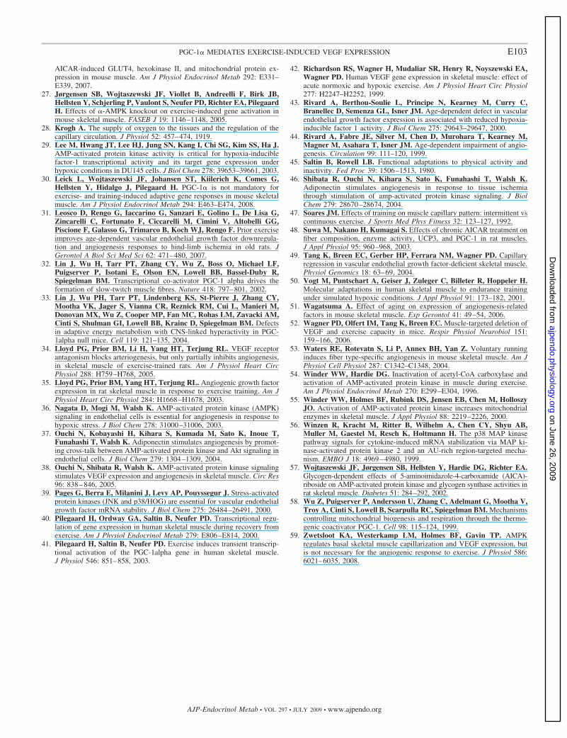
59. Zwetsloot KA, Westerkamp LM, Holmes BF, Gavin TP. AMPKregulates basal skeletal muscle capillarization and VEGF expression, butis not necessary for the angiogenic response to exercise. J Physiol 586:6021–6035, 2008.
E103PGC-1� MEDIATES EXERCISE-INDUCED VEGF EXPRESSION
AJP-Endocrinol Metab • VOL 297 • JULY 2009 • www.ajpendo.org
on June 26, 2009 ajpendo.physiology.org
Dow
nloaded from