ACE2 X-RAY STRUCTURES REVEAL A LARGE HINGE-BENDING MOTION IMPORTANT FOR INHIBITOR BINDING AND...
13
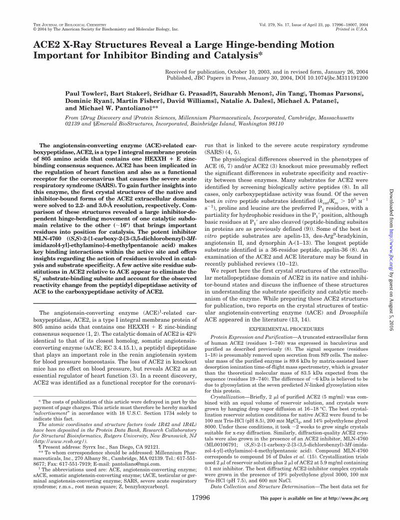
ACE2 X-Ray Structures Reveal a Large Hinge-bending Motion Important for Inhibitor Binding and Catalysis* Received for publication, October 10, 2003, and in revised form, January 26, 2004 Published, JBC Papers in Press, January 30, 2004, DOI 10.1074/jbc.M311191200 Paul Towler‡, Bart Staker§, Sridhar G. Prasad§¶, Saurabh Menon‡, Jin Tang, Thomas Parsons, Dominic Ryan‡, Martin Fisher‡, David Williams‡, Natalie A. Dales‡, Michael A. Patane‡, and Michael W. Pantoliano‡** From ‡Drug Discovery and Protein Sciences, Millennium Pharmaceuticals, Incorporated, Cambridge, Massachusetts 02139 and §Emerald BioStructures, Incorporated, Bainbridge Island, Washington 98110 The angiotensin-converting enzyme (ACE)-related car- boxypeptidase, ACE2, is a type I integral membrane protein of 805 amino acids that contains one HEXXH E zinc- binding consensus sequence. ACE2 has been implicated in the regulation of heart function and also as a functional receptor for the coronavirus that causes the severe acute respiratory syndrome (SARS). To gain further insights into this enzyme, the first crystal structures of the native and inhibitor-bound forms of the ACE2 extracellular domains were solved to 2.2- and 3.0-Å resolution, respectively. Com- parison of these structures revealed a large inhibitor-de- pendent hinge-bending movement of one catalytic subdo- main relative to the other (16°) that brings important residues into position for catalysis. The potent inhibitor MLN-4760 ((S,S)-2-{1-carboxy-2-[3-(3,5-dichlorobenzyl)-3H- imidazol4-yl]-ethylamino}-4-methylpentanoic acid) makes key binding interactions within the active site and offers insights regarding the action of residues involved in catal- ysis and substrate specificity. A few active site residue sub- stitutions in ACE2 relative to ACE appear to eliminate the S 2 substrate-binding subsite and account for the observed reactivity change from the peptidyl dipeptidase activity of ACE to the carboxypeptidase activity of ACE2. The angiotensin-converting enzyme (ACE) 1 -related car- boxypeptidase, ACE2, is a type I integral membrane protein of 805 amino acids that contains one HEXXH E zinc-binding consensus sequence (1, 2). The catalytic domain of ACE2 is 42% identical to that of its closest homolog, somatic angiotensin- converting enzyme (sACE; EC 3.4.15.1), a peptidyl dipeptidase that plays an important role in the renin angiotensin system for blood pressure homeostasis. The loss of ACE2 in knockout mice has no effect on blood pressure, but reveals ACE2 as an essential regulator of heart function (3). In a recent discovery, ACE2 was identified as a functional receptor for the coronavi- rus that is linked to the severe acute respiratory syndrome (SARS) (4, 5). The physiological differences observed in the phenotypes of ACE (6, 7) and/or ACE2 (3) knockout mice presumably reflect the significant differences in substrate specificity and reactiv- ity between these enzymes. Many substrates for ACE2 were identified by screening biologically active peptides (8). In all cases, only carboxypeptidase activity was found. Of the seven best in vitro peptide substrates identified (k cat /K m 10 5 M 1 s 1 ), proline and leucine are the preferred P 1 residues, with a partiality for hydrophobic residues in the P 1 position, although basic residues at P 1 are also cleaved (peptide-binding subsites in proteins are as previously defined (9)). Some of the best in vitro peptide substrates are apelin-13, des-Arg 9 -bradykinin, angiotensin II, and dynorphin A-(1–13). The longest peptide substrate identified is a 36-residue peptide, apelin-36 (8). An examination of the ACE2 and ACE literature may be found in recently published reviews (10 –12). We report here the first crystal structures of the extracellu- lar metallopeptidase domain of ACE2 in its native and inhibi- tor-bound states and discuss the influence of these structures in understanding the substrate specificity and catalytic mech- anism of the enzyme. While preparing these ACE2 structures for publication, two reports on the crystal structures of testic- ular angiotensin-converting enzyme (tACE) and Drosophila ACE appeared in the literature (13, 14). EXPERIMENTAL PROCEDURES Protein Expression and Purification—A truncated extracellular form of human ACE2 (residues 1–740) was expressed in baculovirus and purified as described previously (8). The signal sequence (residues 1–18) is presumably removed upon secretion from Sf9 cells. The molec- ular mass of the purified enzyme is 89.6 kDa by matrix-assisted laser desorption ionization time-of-flight mass spectrometry, which is greater than the theoretical molecular mass of 83.5 kDa expected from the sequence (residues 19 –740). The difference of 6 kDa is believed to be due to glycosylation at the seven predicted N-linked glycosylation sites for this protein. Crystallization—Briefly, 2 l of purified ACE2 (5 mg/ml) was com- bined with an equal volume of reservoir solution, and crystals were grown by hanging drop vapor diffusion at 16 –18 °C. The best crystal- lization reservoir solution conditions for native ACE2 were found to be 100 mM Tris-HCl (pH 8.5), 200 mM MgCl 2 , and 14% polyethylene glycol 8000. Under these conditions, it took 2 weeks to grow single crystals suitable for x-ray diffraction. Similarly, diffraction-quality ACE2 crys- tals were also grown in the presence of an ACE2 inhibitor, MLN-4760 (ML00106791; (S,S)-2-{1-carboxy-2-[3-(3,5-dichlorobenzyl)-3H-imida- zol-4-yl]-ethylamino}-4-methylpentanoic acid). Compound MLN-4760 corresponds to compound 16 of Dales et al. (15). Crystallization trials used 2 l of reservoir solution plus 2 l of ACE2 at 5.9 mg/ml containing 0.1 mM inhibitor. The best diffracting ACE2-inhibitor complex crystals were grown in the presence of 19% polyethylene glycol 3000, 100 mM Tris-HCl (pH 7.5), and 600 mM NaCl. Data Collection and Structure Determination—The best data set for * The costs of publication of this article were defrayed in part by the payment of page charges. This article must therefore be hereby marked “advertisement” in accordance with 18 U.S.C. Section 1734 solely to indicate this fact. The atomic coordinates and structure factors (code 1R42 and 1R4L) have been deposited in the Protein Data Bank, Research Collaboratory for Structural Bioinformatics, Rutgers University, New Brunswick, NJ (http://www.rcsb.org/). ¶ Present address: Syrrx Inc., San Diego, CA 92121. ** To whom correspondence should be addressed: Millennium Phar- maceuticals, Inc., 270 Albany St., Cambridge, MA 02139. Tel.: 617-551- 8677; Fax: 617-551-7919; E-mail: [email protected]. 1 The abbreviations used are: ACE, angiotensin-converting enzyme; sACE, somatic angiotensin-converting enzyme; tACE, testicular or ger- minal angiotensin-converting enzyme; SARS, severe acute respiratory syndrome; r.m.s., root mean square; Z, benzyloxycarbonyl. THE JOURNAL OF BIOLOGICAL CHEMISTRY Vol. 279, No. 17, Issue of April 23, pp. 17996 –18007, 2004 © 2004 by The American Society for Biochemistry and Molecular Biology, Inc. Printed in U.S.A. This paper is available on line at http://www.jbc.org 17996 by guest on August 5, 2016 http://www.jbc.org/ Downloaded from
-
Upload
independent -
Category
Documents
-
view
3 -
download
0
Transcript of ACE2 X-RAY STRUCTURES REVEAL A LARGE HINGE-BENDING MOTION IMPORTANT FOR INHIBITOR BINDING AND...
ACE2 X-Ray Structures Reveal a Large Hinge-bending MotionImportant for Inhibitor Binding and Catalysis*
Received for publication, October 10, 2003, and in revised form, January 26, 2004Published, JBC Papers in Press, January 30, 2004, DOI 10.1074/jbc.M311191200
Paul Towler‡, Bart Staker§, Sridhar G. Prasad§¶, Saurabh Menon‡, Jin Tang�, Thomas Parsons�,Dominic Ryan‡, Martin Fisher‡, David Williams‡, Natalie A. Dales‡, Michael A. Patane‡,and Michael W. Pantoliano‡**
From ‡Drug Discovery and �Protein Sciences, Millennium Pharmaceuticals, Incorporated, Cambridge, Massachusetts02139 and §Emerald BioStructures, Incorporated, Bainbridge Island, Washington 98110
The angiotensin-converting enzyme (ACE)-related car-boxypeptidase, ACE2, is a type I integral membrane proteinof 805 amino acids that contains one HEXXH � E zinc-binding consensus sequence. ACE2 has been implicated inthe regulation of heart function and also as a functionalreceptor for the coronavirus that causes the severe acuterespiratory syndrome (SARS). To gain further insights intothis enzyme, the first crystal structures of the native andinhibitor-bound forms of the ACE2 extracellular domainswere solved to 2.2- and 3.0-Å resolution, respectively. Com-parison of these structures revealed a large inhibitor-de-pendent hinge-bending movement of one catalytic subdo-main relative to the other (�16°) that brings importantresidues into position for catalysis. The potent inhibitorMLN-4760 ((S,S)-2-{1-carboxy-2-[3-(3,5-dichlorobenzyl)-3H-imidazol4-yl]-ethylamino}-4-methylpentanoic acid) makeskey binding interactions within the active site and offersinsights regarding the action of residues involved in catal-ysis and substrate specificity. A few active site residue sub-stitutions in ACE2 relative to ACE appear to eliminate theS2� substrate-binding subsite and account for the observedreactivity change from the peptidyl dipeptidase activity ofACE to the carboxypeptidase activity of ACE2.
The angiotensin-converting enzyme (ACE)1-related car-boxypeptidase, ACE2, is a type I integral membrane protein of805 amino acids that contains one HEXXH � E zinc-bindingconsensus sequence (1, 2). The catalytic domain of ACE2 is 42%identical to that of its closest homolog, somatic angiotensin-converting enzyme (sACE; EC 3.4.15.1), a peptidyl dipeptidasethat plays an important role in the renin angiotensin systemfor blood pressure homeostasis. The loss of ACE2 in knockoutmice has no effect on blood pressure, but reveals ACE2 as anessential regulator of heart function (3). In a recent discovery,ACE2 was identified as a functional receptor for the coronavi-
rus that is linked to the severe acute respiratory syndrome(SARS) (4, 5).
The physiological differences observed in the phenotypes ofACE (6, 7) and/or ACE2 (3) knockout mice presumably reflectthe significant differences in substrate specificity and reactiv-ity between these enzymes. Many substrates for ACE2 wereidentified by screening biologically active peptides (8). In allcases, only carboxypeptidase activity was found. Of the sevenbest in vitro peptide substrates identified (kcat/Km � 105 M�1
s�1), proline and leucine are the preferred P1 residues, with apartiality for hydrophobic residues in the P1� position, althoughbasic residues at P1� are also cleaved (peptide-binding subsitesin proteins are as previously defined (9)). Some of the best invitro peptide substrates are apelin-13, des-Arg9-bradykinin,angiotensin II, and dynorphin A-(1–13). The longest peptidesubstrate identified is a 36-residue peptide, apelin-36 (8). Anexamination of the ACE2 and ACE literature may be found inrecently published reviews (10–12).
We report here the first crystal structures of the extracellu-lar metallopeptidase domain of ACE2 in its native and inhibi-tor-bound states and discuss the influence of these structuresin understanding the substrate specificity and catalytic mech-anism of the enzyme. While preparing these ACE2 structuresfor publication, two reports on the crystal structures of testic-ular angiotensin-converting enzyme (tACE) and DrosophilaACE appeared in the literature (13, 14).
EXPERIMENTAL PROCEDURES
Protein Expression and Purification—A truncated extracellular formof human ACE2 (residues 1–740) was expressed in baculovirus andpurified as described previously (8). The signal sequence (residues1–18) is presumably removed upon secretion from Sf9 cells. The molec-ular mass of the purified enzyme is 89.6 kDa by matrix-assisted laserdesorption ionization time-of-flight mass spectrometry, which is greaterthan the theoretical molecular mass of 83.5 kDa expected from thesequence (residues 19–740). The difference of �6 kDa is believed to bedue to glycosylation at the seven predicted N-linked glycosylation sitesfor this protein.
Crystallization—Briefly, 2 �l of purified ACE2 (5 mg/ml) was com-bined with an equal volume of reservoir solution, and crystals weregrown by hanging drop vapor diffusion at 16–18 °C. The best crystal-lization reservoir solution conditions for native ACE2 were found to be100 mM Tris-HCl (pH 8.5), 200 mM MgCl2, and 14% polyethylene glycol8000. Under these conditions, it took �2 weeks to grow single crystalssuitable for x-ray diffraction. Similarly, diffraction-quality ACE2 crys-tals were also grown in the presence of an ACE2 inhibitor, MLN-4760(ML00106791; (S,S)-2-{1-carboxy-2-[3-(3,5-dichlorobenzyl)-3H-imida-zol-4-yl]-ethylamino}-4-methylpentanoic acid). Compound MLN-4760corresponds to compound 16 of Dales et al. (15). Crystallization trialsused 2 �l of reservoir solution plus 2 �l of ACE2 at 5.9 mg/ml containing0.1 mM inhibitor. The best diffracting ACE2-inhibitor complex crystalswere grown in the presence of 19% polyethylene glycol 3000, 100 mM
Tris-HCl (pH 7.5), and 600 mM NaCl.Data Collection and Structure Determination—The best data set for
* The costs of publication of this article were defrayed in part by thepayment of page charges. This article must therefore be hereby marked“advertisement” in accordance with 18 U.S.C. Section 1734 solely toindicate this fact.
The atomic coordinates and structure factors (code 1R42 and 1R4L)have been deposited in the Protein Data Bank, Research Collaboratoryfor Structural Bioinformatics, Rutgers University, New Brunswick, NJ(http://www.rcsb.org/).
¶ Present address: Syrrx Inc., San Diego, CA 92121.** To whom correspondence should be addressed: Millennium Phar-
maceuticals, Inc., 270 Albany St., Cambridge, MA 02139. Tel.: 617-551-8677; Fax: 617-551-7919; E-mail: [email protected].
1 The abbreviations used are: ACE, angiotensin-converting enzyme;sACE, somatic angiotensin-converting enzyme; tACE, testicular or ger-minal angiotensin-converting enzyme; SARS, severe acute respiratorysyndrome; r.m.s., root mean square; Z, benzyloxycarbonyl.
THE JOURNAL OF BIOLOGICAL CHEMISTRY Vol. 279, No. 17, Issue of April 23, pp. 17996–18007, 2004© 2004 by The American Society for Biochemistry and Molecular Biology, Inc. Printed in U.S.A.
This paper is available on line at http://www.jbc.org17996
by guest on August 5, 2016
http://ww
w.jbc.org/
Dow
nloaded from

native ACE2 was at 2.2-Å resolution and was collected at the AdvancedPhoton Source (Argonne National Laboratory). A total of 44 x-ray datasets were collected for native ACE2, including a large number of heavyatom soaks of atoms that had good anomalous signals. The data sets foreach derivative were collected at different wavelengths to maximize theanomalous signals for the bound heavy atoms. Native ACE2 data werecollected to 2.2-Å resolution at � � 1.28 Å to maximize the anomaloussignal at the zinc absorption edge.
The heavy atom positions were determined and confirmed by a com-bination of visual inspection of Patterson maps and automatic searchprocedures, which included SHAKE ‘N BAKE (16) and SHELXD (17).The heavy atom parameters were refined and optimized using thecomputer programs SHARP (18), MLPHARE (19), and XHEAVY (20).The experimental phases were improved by solvent flattening andhistogram matching.
Once the native ACE2 structure was determined, it was used to solve
the inhibitor-bound structure of ACE2 to 3.0-Å resolution using molec-ular replacement methods that employed the program AMoRe in theCCP4 software suite (21). The native structure was split into twosubdomains: subdomains I and II (see Fig. 3 for definition). SubdomainII was used for molecular replacement and refined in REFMAC5, whichresulted in the appearance of electron density for subdomain I. Subdo-main I was then fitted into the density by hand, and the structure wasrefined as a whole. Final refinement was accomplished using the soft-ware suite CNX (22).
RESULTS AND DISCUSSION
Native ACE2 Structure—The three-dimensional structure ofthe extracellular region of native ACE2 was determined bymultiple isomorphous replacement with anomalous scatteringand refined to a crystallographic R-factor of 23.5% (Rfree �
TABLE IHeavy atom data statistics for human native ACE2
pCMB, p-chloromercuribenzoate; PIP, di-�-iodobis(ethylenediamine) diplatinum(II) nitrate; NA, not applicable.
Derivative Native (Zn) pCMB HgCl2 PIP K2PtCl4
Heavy atom Zn Hg Hg Pt PtMolarity (mM) NA 1 1 1 1Length of soak (day(s)) NA 3.5 30 1 30No. sites/asymmetric unita 1 1 1 2 2Wavelength (Å)b 1.2824 1.009 1.009 1.072 1.072Unique reflections 49,286c 21,652c 17,421 13,152 14,087Resolution (Å) 40–2.2 30–2.9 30–3.0 30–3.4 30–3.3Completeness (%) 96.3 96.6 90.6 95.4 94.2Rsym (%)d 5.7 10.5 10.4 9.7 11.6Rmerge
e NA 21.3 37.6 20.6 21.8Rcullis
f 0.94 0.73 0.93 0.96 0.97Phasing powerg 1.57 1.51 0.66 0.45 0.39
a Each asymmetric unit contains one human ACE2 protein.b Data were collected at the Brookhaven National Laboratory (NSLS, beam line X25) or at the Argonne National Laboratory (APS, beam line
sector 32, COM-CAT). The wavelength for the native (zinc) data set was 1.2824 Å to maximize the anomalous signal at the zinc absorption edge.c Values do not include Bijvoet pairs. Inclusion of Bijvoet pairs increases the number of reflections to 91,550 for native (zinc) ACE2 and 41,716
for the p-chloromercuribenzoate derivative.d Rsym � ��Ii � Im�/�Im, where Ii is the intensity of the measured reflection and Im is the mean intensity of all symmetry-related reflections.e Rmerge � ��FPH � FP�/��FPH�.f Rcullis � ��(FPH � FP) � FH(calc)�/��FPH � FP�.g Phasing power � FH/ERMS, where ERMS is the residual lack of closure.
TABLE IIRefinement statistics for native and inhibitor-bound ACE2 structures
Numbers in parentheses represent final shell of data. PDB, Protein Data Bank.
Native ACE2 (PDB code1R42)
Inhibitor-bound ACE2 (PDB code1R4L)
Resolution (Å) 46.7–2.20 (2.34–2.20) 43.3–3.0 (3.19–3.00)No. reflections 47,465 (5982) 17,228 (2250)Rsym (%)a 5.7 (40.8) 7.0 (20.4)Completeness (%) 96.3 (81.8) 96.8 (85.1)Space group C2 C2a 103.64 100.53b 89.46 86.51c 112.40 105.86� 109.15 103.65Unit cell volume (Å3) 986,854 894,383Solvent content (%)b 53 53Molecules/asymmetric unit 1 1Reflections used in Rfree 4798 1723No. protein atoms 5165 5147No. solvent atoms 302 13No. zinc atoms 1 1No. chloride atoms 1 1No. sugar atoms 42 28R-factor (%) 23.5 (37.9) 25.3 (37.9)Rfree (%) 28.7 (39.8) 33.7 (46.0)r.m.s. deviations from ideal stereochemistry
Bond lengths (Å) 0.008 0.008Bond angles 1.4° 1.5°Dihedrals 21.7° 22.2°Impropers 0.92° 0.97°
Mean B-factor (all atoms; Å2) 59.9 74.5a See Footnote d of Table I.b Vsolvent � 1–1.23/Vm, where Vm is the volume of protein in the unit cell/volume of unit cell, assuming one molecule/asymmetric unit and four
asymmetric units in the monoclinic unit cell.
Native and Inhibitor-bound ACE2 Crystal Structures 17997
by guest on August 5, 2016
http://ww
w.jbc.org/
Dow
nloaded from
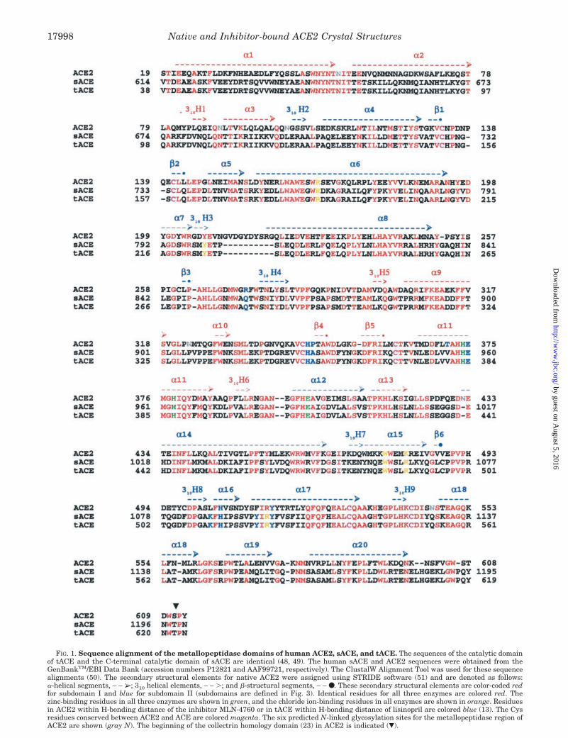
FIG. 1. Sequence alignment of the metallopeptidase domains of human ACE2, sACE, and tACE. The sequences of the catalytic domainof tACE and the C-terminal catalytic domain of sACE are identical (48, 49). The human sACE and ACE2 sequences were obtained from theGenBankTM/EBI Data Bank (accession numbers P12821 and AAF99721, respectively). The ClustalW Alignment Tool was used for these sequencealignments (50). The secondary structural elements for native ACE2 were assigned using STRIDE software (51) and are denoted as follows:�-helical segments, – – ➢; 3 10 helical elements, – – �; and �-structural segments, – – ●. These secondary structural elements are color-coded redfor subdomain I and blue for subdomain II (subdomains are defined in Fig. 3). Identical residues for all three enzymes are colored red. Thezinc-binding residues in all three enzymes are shown in green, and the chloride ion-binding residues in all enzymes are shown in orange. Residuesin ACE2 within H-bonding distance of the inhibitor MLN-4760 or in tACE within H-bonding distance of lisinopril are colored blue (13). The Cysresidues conserved between ACE2 and ACE are colored magenta. The six predicted N-linked glycosylation sites for the metallopeptidase region ofACE2 are shown (gray N). The beginning of the collectrin homology domain (23) in ACE2 is indicated (�).
Native and Inhibitor-bound ACE2 Crystal Structures17998
by guest on August 5, 2016
http://ww
w.jbc.org/
Dow
nloaded from
28.7%) at 2.2-Å resolution. The heavy atom data statistics aresummarized in Table I, and the refinement statistics for nativeACE2 are summarized in Table II.
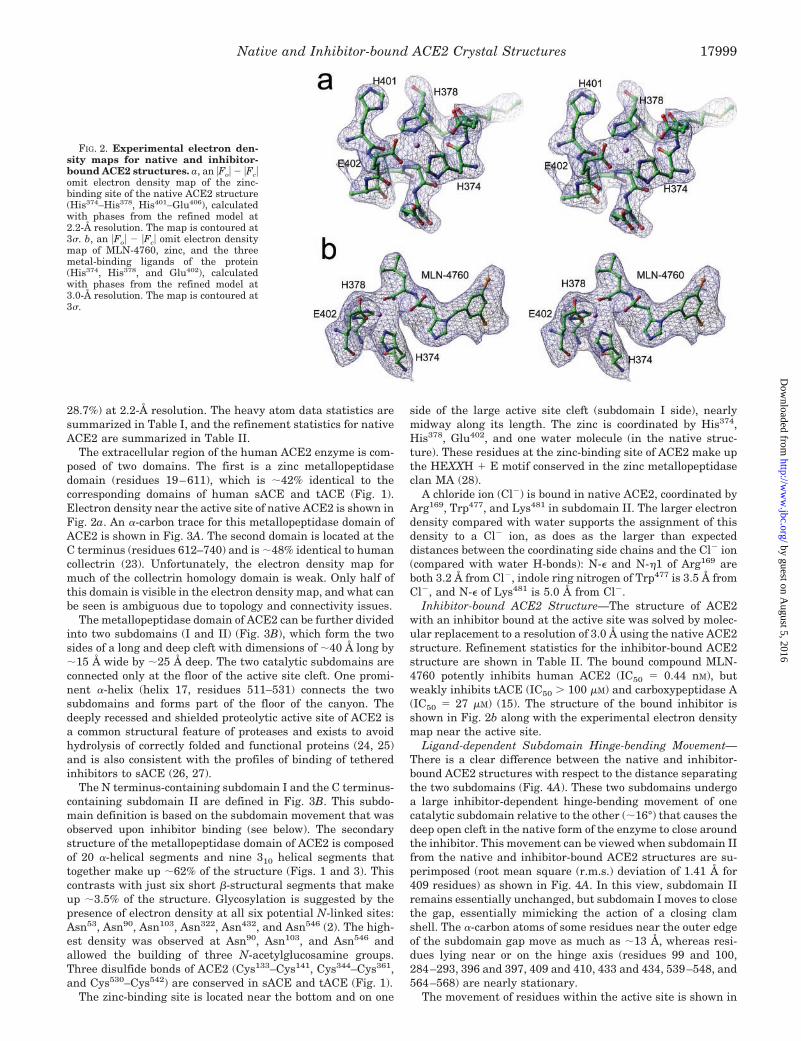
The extracellular region of the human ACE2 enzyme is com-posed of two domains. The first is a zinc metallopeptidasedomain (residues 19–611), which is �42% identical to thecorresponding domains of human sACE and tACE (Fig. 1).Electron density near the active site of native ACE2 is shown inFig. 2a. An �-carbon trace for this metallopeptidase domain ofACE2 is shown in Fig. 3A. The second domain is located at theC terminus (residues 612–740) and is �48% identical to humancollectrin (23). Unfortunately, the electron density map formuch of the collectrin homology domain is weak. Only half ofthis domain is visible in the electron density map, and what canbe seen is ambiguous due to topology and connectivity issues.
The metallopeptidase domain of ACE2 can be further dividedinto two subdomains (I and II) (Fig. 3B), which form the twosides of a long and deep cleft with dimensions of �40 Å long by�15 Å wide by �25 Å deep. The two catalytic subdomains areconnected only at the floor of the active site cleft. One promi-nent �-helix (helix 17, residues 511–531) connects the twosubdomains and forms part of the floor of the canyon. Thedeeply recessed and shielded proteolytic active site of ACE2 isa common structural feature of proteases and exists to avoidhydrolysis of correctly folded and functional proteins (24, 25)and is also consistent with the profiles of binding of tetheredinhibitors to sACE (26, 27).
The N terminus-containing subdomain I and the C terminus-containing subdomain II are defined in Fig. 3B. This subdo-main definition is based on the subdomain movement that wasobserved upon inhibitor binding (see below). The secondarystructure of the metallopeptidase domain of ACE2 is composedof 20 �-helical segments and nine 310 helical segments thattogether make up �62% of the structure (Figs. 1 and 3). Thiscontrasts with just six short �-structural segments that makeup �3.5% of the structure. Glycosylation is suggested by thepresence of electron density at all six potential N-linked sites:Asn53, Asn90, Asn103, Asn322, Asn432, and Asn546 (2). The high-est density was observed at Asn90, Asn103, and Asn546 andallowed the building of three N-acetylglucosamine groups.Three disulfide bonds of ACE2 (Cys133–Cys141, Cys344–Cys361,and Cys530–Cys542) are conserved in sACE and tACE (Fig. 1).
The zinc-binding site is located near the bottom and on one
side of the large active site cleft (subdomain I side), nearlymidway along its length. The zinc is coordinated by His374,His378, Glu402, and one water molecule (in the native struc-ture). These residues at the zinc-binding site of ACE2 make upthe HEXXH � E motif conserved in the zinc metallopeptidaseclan MA (28).
A chloride ion (Cl�) is bound in native ACE2, coordinated byArg169, Trp477, and Lys481 in subdomain II. The larger electrondensity compared with water supports the assignment of thisdensity to a Cl� ion, as does as the larger than expecteddistances between the coordinating side chains and the Cl� ion(compared with water H-bonds): N-� and N-�1 of Arg169 areboth 3.2 Å from Cl�, indole ring nitrogen of Trp477 is 3.5 Å fromCl�, and N-� of Lys481 is 5.0 Å from Cl�.
Inhibitor-bound ACE2 Structure—The structure of ACE2with an inhibitor bound at the active site was solved by molec-ular replacement to a resolution of 3.0 Å using the native ACE2structure. Refinement statistics for the inhibitor-bound ACE2structure are shown in Table II. The bound compound MLN-4760 potently inhibits human ACE2 (IC50 � 0.44 nM), butweakly inhibits tACE (IC50 � 100 �M) and carboxypeptidase A(IC50 � 27 �M) (15). The structure of the bound inhibitor isshown in Fig. 2b along with the experimental electron densitymap near the active site.
Ligand-dependent Subdomain Hinge-bending Movement—There is a clear difference between the native and inhibitor-bound ACE2 structures with respect to the distance separatingthe two subdomains (Fig. 4A). These two subdomains undergoa large inhibitor-dependent hinge-bending movement of onecatalytic subdomain relative to the other (�16°) that causes thedeep open cleft in the native form of the enzyme to close aroundthe inhibitor. This movement can be viewed when subdomain IIfrom the native and inhibitor-bound ACE2 structures are su-perimposed (root mean square (r.m.s.) deviation of 1.41 Å for409 residues) as shown in Fig. 4A. In this view, subdomain IIremains essentially unchanged, but subdomain I moves to closethe gap, essentially mimicking the action of a closing clamshell. The �-carbon atoms of some residues near the outer edgeof the subdomain gap move as much as �13 Å, whereas resi-dues lying near or on the hinge axis (residues 99 and 100,284–293, 396 and 397, 409 and 410, 433 and 434, 539–548, and564–568) are nearly stationary.
The movement of residues within the active site is shown in
FIG. 2. Experimental electron den-sity maps for native and inhibitor-bound ACE2 structures. a, an �Fo� � �Fc�omit electron density map of the zinc-binding site of the native ACE2 structure(His374–His378, His401–Glu406), calculatedwith phases from the refined model at2.2-Å resolution. The map is contoured at3�. b, an �Fo� � �Fc� omit electron densitymap of MLN-4760, zinc, and the threemetal-binding ligands of the protein(His374, His378, and Glu402), calculatedwith phases from the refined model at3.0-Å resolution. The map is contoured at3�.
Native and Inhibitor-bound ACE2 Crystal Structures 17999
by guest on August 5, 2016
http://ww
w.jbc.org/
Dow
nloaded from

Fig. 4B. This view is a close-up of Fig. 4A with subdomain II ofthe native and inhibitor-bound ACE2 structures superimposed.Many of these residues move as much as 6–9 Å after binding ofthe inhibitor. Similar subdomain hinge-bending motions havealso been observed for other zinc metalloproteases (29, 30). Thelargest previously observed ligand-dependent movement formetalloproteases is a 14° hinge-bending subdomain motiondemonstrated for Pseudomonas aeruginosa elastase (open ver-sus closed) that resulted in an �2-Å movement to close theN-terminal/C-terminal subdomain gap (29). Domain closuremovements in proteins are a common mechanism for the posi-tioning of critical groups around substrates and inhibitors(31–33) and also for the trapping of substrate and reactionintermediates (34).
Inhibitor Binding Interactions and Implications for Sub-strate Specificity and Catalysis—Both metallopeptidase subdo-mains of ACE2 are nearly equally involved in binding of theinhibitor MLN-4760. Inspection of the interactions betweenMLN-4760 and ACE2 revealed important residues responsiblefor inhibitor binding and presumably for substrate binding andcatalysis (Fig. 5). The inhibitor MLN-4760 has two carboxylategroups, one of which binds to the zinc atom by displacing thebound water molecule present in the native ACE2 structure.The zinc coordination sphere (His374, His378, and Glu402) is asubset of the 21 residues of ACE2 that are located within 4.5 Åof the bound inhibitor and that make up the greater part of theactive site. Six of the most important of these residues contrib-ute specific H-bonding interactions with MLN-4760 (Fig. 5).The side chains of Arg273, His505, and His345 are H-bonded tothe terminal carboxylate of the inhibitor. The carbonyl oxygenatom of Pro346 and the N-� atom of His345 are within H-bondingdistance of the secondary amine group of the inhibitor. Thr371
is within H-bonding distance of the imidazole ring of the 3,5-dichlorobenzylimidazole group of MLN-4760. The phenolicgroup of Tyr515 donates an H-bond to the zinc-bound carboxy-late group of the inhibitor. The carboxyl group of Glu375 iswithin H-bonding distance of the other oxygen atom of the
zinc-bound carboxylate of MLN-4760, but is presumably notprotonated until peptide hydrolysis occurs (see below).
The zinc-bound carboxylate of MLN-4760 appears to mimicthe zinc-bound tetrahedral intermediate characteristic of nu-cleophilic attack of the scissile bond by the zinc-bound waterduring peptide hydrolysis (35). This transition state structureis usually stabilized by H-bonds donated by imidazole, pheno-lic, or guanidino functional groups of neighboring amino acidside chains in other zinc metalloproteases (30, 35). For ACE2,this carboxyl anion stabilization most likely occurs through thephenolic group of Tyr515 (Fig. 5).
Besides the zinc coordination sphere and potential H-bond-contributing residues, there are an additional 11 residues ofACE2 that make close contacts (4.5 Å) with MLN-4760, manyof which are shown in Fig. 5. These residues do not contributedirect H-bonding interactions with the inhibitor, but provideimportant electrostatic and van der Waals interactions withMLN-4760. These residues define the S1 and S1� peptide-bind-ing subsites, thereby providing the inhibitor, as well as sub-strate, binding specificity.
The isobutyl group of MLN-4760 packs nicely into the S1
subsite of ACE2 (Fig. 5A), suggesting a similar preferred fit fora leucyl side chain at the P1 position of peptide substrates. Thetopology and chemical environment of the S1 subsite are dic-tated by four residues (Tyr510, Arg514, Phe504, and Thr347) thatare expected to restrict the size of substrate P1 side chains.Large substrate P1 residues such as Trp, Tyr, Phe, Arg, and Lyswould require significant movement of the phenolic side chainof Tyr510, which forms a lid over the top of the S1 subsite. Thisobservation is consistent with the reported substrate specificitydata showing only medium sized residues (Pro and Leu) at theP1 position for preferred substrates with kcat/Km � 105 M�1 s�1
(8). Peptides with Phe and Tyr at the P1 position, such asangiotensin-(1–9) (DRVYIHPFH), bradykinin (RPPGFSPFR),Leu-enkephalin (YGGFL), Met-enkephalin (YGGFM), andangiotensin-(1–5) (DRVYI), are not substrates for ACE2 de-spite the presence of preferred hydrophobic or basic P1� resi-
FIG. 3. Overview of the native ACE2crystal structure. A, �-carbon trace ofthe native ACE2 structure looking downinto the metallopeptidase active site cleft.The metallopeptidase catalytic domain iscolored red. The active site zinc ion isshown as a yellow sphere, and the singlebound chloride ion is shown as a greensphere. The S1� subsite for inhibitor andsubstrate binding is to the right of thezinc ion, and the S1 subsite is to the left.The collectrin homology domain at the Cterminus is disordered and denoted by thegreen dotted line. B, ribbon diagram ofnative ACE2 showing the secondarystructure and also the two subdomains (Iand II) that form the two sides of theactive site cleft. The two subdomains aredefined as follows: the N terminus- andzinc-containing subdomain I (red), com-posed of residues 19–102, 290–397, and417–430; and the C terminus-containingsubdomain II (blue), composed of residues103–289, 398–416, and 431–615. Thisdefinition is based on motion observedupon inhibitor binding (see Fig. 4). Zincand chloride ions are denoted as describedfor A.
Native and Inhibitor-bound ACE2 Crystal Structures18000
by guest on August 5, 2016
http://ww
w.jbc.org/
Dow
nloaded from

dues. Some ACE2-catalyzed hydrolysis was observed forpeptides with a slightly larger residue (His) at the P1 site(i.e. angiotensin I), but the catalytic efficiency was orders ofmagnitude lower (kcat/Km � 4.9 103 M�1 s�1) despite goodbinding.
The limited size of the S1 subsite may also account for theobserved orientation of binding of MLN-4760. The design prin-ciples leading to the identification of MLN-4760 were based onassumptions from substrate screening studies and informationregarding known metallopeptidase inhibitors (36). Conse-quently, MLN-4760 was designed as a His-Leu mimetic, inwhich the isobutyl side chain was predicted to mimic the Leuside chain (P1�), and the substituted His was expected to bindin the S1-S2 subsites (15). Furthermore, the Leu carboxylatewas envisioned to mimic the substrate’s C-terminal carboxy-late, and the His carboxylate was expected to coordinate thezinc atom. Although the inhibitor potency optimization wassupported by this design hypothesis (several selective inhibi-tors at 10 nM), experimentally, the inhibitor-bound crystalstructure reveals that MNL-4760 binds in the opposite orien-tation: the two carboxylates are reversed, the isobutyl sidechain occupies the S1 subsite, and the 3,5-dichlorobenzylimida-zole group binds in the S1� subsite. This alternative bindingmode may be the result of the small S1 subsite, which cannotaccommodate the 3,5-dichlorobenzyl group. Models of MLN-4760 bound to ACE2 that were based on the tACE structure(37) did not predict the observed binding mode shown in Fig. 5.Retrospective comparison of the inhibitor structure-activity re-lationship with the crystal structure supports the unantici-pated binding orientation. During the potency optimization
studies, it appeared that the His portion of the inhibitors bettertolerated changes in size and physicochemical properties com-pared with the Leu portion, which is consistent with the newexperimental evidence (Fig. 5).
The S1� subsite of ACE2 is much larger than the S1 subsiteand is formed by the lengthwise channel between the twosubdomains. In addition to the aforementioned residues thatcontribute specific H-bonding interactions with MLN-4760(Arg273, His345, His505, Thr371, and the carbonyl group ofPro346), there are a number of other residues that make closecontacts with the 3,5-dichlorobenzylimidazole group of the in-hibitor. The side chains of Phe274, Pro346, Thr371, Met360 andthe disulfide linkage of Cys344 and Cys361 provide a hydropho-bic environment for the S1� subsite. Additional close contactsare observed for the side chains of Asp368, Glu145, Asn149, andLys363 and the carbonyl groups of Cys344 and Cys361, whichaccommodate the far end of the dichlorobenzyl ring as depictedin Fig. 5B. The large size of the this S1� subsite easily accom-modates the 3,5-dichlorobenzylimidazole group of MLN-4760and mirrors the observed preference for large hydrophobic orbasic residues (Arg and Lys) at the P1� position of peptidesubstrates (8).
Comparison with Structural Homologs—There are four pro-teins in the Protein Data Bank (38) that have overall architec-tural similarity to ACE2. They include the following: 1) therecently solved human tACE (code 1O86) (13), an enzyme of theM2 metallopeptidase family (EC 3.4.15.1); 2) Drosophila ACE(code 1J36), with which ACE2 shares �35% sequence identity(14); 3) rat neurolysin (code 1I1I) (39), an M3 metallopeptidasefamily member (EC 3.4.24.16) with which ACE2 shares just
FIG. 4. Superposition of the nativeand inhibitor-bound ACE2 struc-tures. A, the 409 �-carbon atoms corre-sponding to subdomain II of the nativeand inhibitor-bound ACE2 structureswere superimposed with an r.m.s. devia-tion of 1.41 Å. Native ACE2 is colored red,and inhibitor-bound ACE2 is coloredgreen. The zinc ion is shown as a yellowsphere, and the inhibitor MLN-4760 isshown in a ball-and-stick rendering withdefault atom coloring: gray, carbon; blue,nitrogen; red, oxygen; green, chlorine.This view is looking down the length ofthe active site cleft and is rotated 90° fromthat shown in Fig. 3. This perspective il-lustrates the �16° hinge-bending move-ment of subdomain I relative to subdo-main II that occurs upon inhibitorbinding to ACE2. B, shown is a close-upview of the active sites of the superim-posed native (red) and inhibitor-bound(green) ACE2 structures. This is the samesuperposition of subdomain II for bothstructures as described for A. In this per-spective, the residues of subdomain Iwithin the active site are shown to moveupon inhibitor binding relative to those insubdomain II. The inhibitor MLN-4760 isshown in stick rendering with the sameatom color code as described for A. Theaverage movement for residues near theactive site is 6–9 Å. The yellow spheresare the two positions of the zinc atom inthe native and inhibitor-bound struc-tures. This figure was prepared usingMOE 2003.02 software (Chemical Com-puting Group, Inc.).
Native and Inhibitor-bound ACE2 Crystal Structures 18001
by guest on August 5, 2016
http://ww
w.jbc.org/
Dow
nloaded from

�17% sequence identity; and 4) Pyrococcus furiosus car-boxypeptidase (code 1KA2) (40), a member of the M32 car-boxypeptidase family.
Comparison with tACE—The �-carbon atoms of lisinopril-bound tACE were superimposed onto the equivalent atoms ofinhibitor-bound ACE2 (588 residues) with an r.m.s. deviation of1.80 Å (Fig. 6A), suggesting that these two protein structuresare very similar. The largest difference between the ACE2 andtACE �-carbon traces is the insertion of a loop extension of 10residues (Gly211–Gly220) between �-helices 7 and 8 in sub-domain II of ACE2.
An improved superposition of the inhibitor-bound ACE2 andtACE structures (r.m.s. deviation of 0.53 Å) was obtained by
considering just the equivalent �-carbon atoms of the afore-mentioned 21 residues of ACE2 that come within 4.5 Å of thebound MLN-4760 (Fig. 6B). Thirteen of these active site resi-dues are conserved in the tACE and sACE enzymes: Glu145,Cys344, His345, Cys361, Asp368, His374, Glu375, His378, Glu402,Phe504, His505, Arg514, and Tyr515. These structural similaritiesreflect the nearly identical way that MLN-4760 and lisinoprilsit in the superimposed active sites (Fig. 6B).
The remaining eight active site ACE2 residues are substitutedin tACE as follows: Asn149 3 Thr, Arg273 3 Gln, Pro346 3 Ala,Thr347 3 Ser, Met360 3 Gln, Lys363 3 Thr, Thr371 3 Val, andTyr510 3 Val. These changes at the active sites of ACE2, tACE,and sACE presumably play a significant role in the observed
FIG. 5. Binding interactions of theinhibitor MLN-4760 at the active siteof ACE2. A, the residues of ACE2 thatcontribute direct binding interactionswith the inhibitor MLN-4760 are shown.MLN-4760 is shown in stick renderingwith the same atom color code as de-scribed in the legend to Fig. 4A, exceptcarbon is orange. The �-helix 11 segmentderived from subdomain I has the �-car-bon wire colored red, and turns and �-el-ements derived from subdomain II havethe �-carbon wire colored blue. ProbableH-bonding interactions are shown asblack dashed lines. The zinc ion is shownas a yellow sphere. ACE2 residues coordi-nating the zinc ion are shown in stickrendering. B, shown is a schematic viewof MLN-4760 binding interactions. MLN-4760 is shown in black. Residues derivedfrom subdomain I are red, and residuesderived from subdomain II are blue. Theequivalent residues in tACE are in givenin parentheses. Distances are measured inangstroms.
Native and Inhibitor-bound ACE2 Crystal Structures18002
by guest on August 5, 2016
http://ww
w.jbc.org/
Dow
nloaded from

differences in substrate specificity and inhibitor binding profilesfor these homologous enzymes. As was discussed previously,Tyr510 and Thr347 line the S1 subsite of ACE2 and confine thissubsite for the accommodation of only small and medium sizedside chains such as leucyl and prolyl, which is consistent with theknown substrate preferences (8). The smaller side chains at theS1 subsite of tACE make this subsite somewhat larger in tACEthan observed for ACE2. In a superposition of the inhibitor-bound ACE2 and lisinopril-bound tACE structures, the Tyr510
phenolic group of ACE2 occupies the space where the P1 phenyl-propyl group of the lisinopril resides (Fig. 6B).
Another important difference between ACE2 and tACE is theArg273 3 Gln substitution. The guanidino group of Arg273
makes a bidentate H-bond with the terminal carboxylate ofMLN-4760 (Fig. 5). Not only does the guanidino group of Arg273
stabilize the terminal carboxylate of the inhibitors and peptidesubstrates, but its larger size (compared with Gln in tACE)causes steric crowding at the potential S2� binding subsite. Thesuperposition of lisinopril-bound tACE onto MLN-4760-boundACE2 reveals that the guanidino group of Arg273 nearly super-imposes on the terminal carboxylate of the P2� Pro residue oflisinopril, thereby severely limiting the size of the S2� subsite inACE2 compared with tACE (Fig. 6B). This single residue dif-ference in ACE2 relative to tACE essentially eliminates the S2�subsite in ACE2 and offers an explanation for the observedswitch in the peptidyl dipeptidase activity of tACE to the ob-served carboxypeptidase activity of ACE2. This difference atthe S1� and S2� subsites for the homologous enzymes also helps
to clarify why the potent ACE inhibitors lisinopril, enalaprilat,and captopril are inactive against ACE2 (2).
The native tACE structure was reported to be nearly identi-cal to the lisinopril-bound tACE structure (13), suggesting thatno ligand-dependent conformational change occurs for tACE orat least under the conditions used to obtain these crystals (pH4.7, 50 mM sodium acetate with an unspecified [Cl�]) (41).However, inspection of the reported native tACE structure(Protein Data Bank code 1O8A) reveals that two ligands arebound to the active site: an acetate molecule bound to the zincatom and an unknown compound that presumably originatedduring protein expression and purification. This compound wasmodeled as an N-carboxyalanine and is isosteric with 2-meth-ylsuccinic acid according to notes deposited with the nativetACE coordinates in the Protein Data Bank file. This 2-meth-ylsuccinate-like ligand is bound to tACE in a manner thatclosely resembles that observed for lisinopril. Five of the sameH-bonds present in the lisinopril-bound tACE structure arealso present for the 2-methylsuccinate-like ligand. Therefore,the reported native tACE structure would appear to be anotherligand-bound structure, and its closed conformational state isconsistent with other ligand-bound tACE and ACE2 structures.
Structural Homology for the Catalytic Motif—ACE2 and re-lated ACE enzymes are related to other HEXXH clan membersthrough divergent evolution with an overall primary sequenceidentity that is 15% (Fig. 7A). The two largest metallopepti-dase clans are clan MA (gluzincin family) and clan MB, whichinclude the many families of zinc metallopeptidases that have
FIG. 6. Superposition of the ACE2 and tACE structures. A, the �-carbon atoms in lisinopril-bound tACE (13) were superimposed onto theequivalent atoms in inhibitor-bound ACE2 (588 residues) with an r.m.s. deviation of 1.80 Å. MLN-4760-bound ACE2 is magenta, and lisinopril-bound tACE is green. MLN-4760 is shown bound to ACE2 with the same color code described in the legend to Fig. 4A. Similarly, the zinc andchloride ions are shown as described in the legend to Fig. 3. The orientation is the same as that shown for native ACE2 in Fig. 3. Structures weresuperimposed using MOE 2003.02 software. B, the 21 �-carbon atoms at the inhibitor-bound active site of ACE2 (residues 4.5 Å from the inhibitor)were superimposed onto the equivalent atoms of lisinopril-bound tACE (Protein Data Bank code 1O86) with an r.m.s. deviation of 0.53 Å. The activesite of ACE2 and MLN-4760 are shown in default colors, with the inhibitor displayed in stick rendering. Labels are for ACE2 residues only. Theactive site residues of tACE are shown in orange, with the inhibitor lisinopril colored purple in stick rendering. The zinc ion is shown as a yellowsphere, and the second chloride ion of tACE (CL2) is shown as an orange sphere. This chloride ion site does not exist in ACE2 due to the Glu398
substitution for Pro407 (see “Results and Discussion”). Other important differences between ACE2 and tACE are as follows: Arg273 versus Gln281,Phe274 versus Thr282, and Tyr510 versus Val518, respectively.
Native and Inhibitor-bound ACE2 Crystal Structures 18003
by guest on August 5, 2016
http://ww
w.jbc.org/
Dow
nloaded from
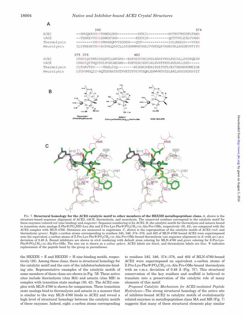
the HEXXH � E and HEXXH � H zinc-binding motifs, respec-tively (28). Among these clans, there is structural homology forthe catalytic motif and the core of the inhibitor/substrate-bind-ing site. Representative examples of the catalytic motifs ofsome members of these clans are shown in Fig. 7B. These activesites include thermolysin (clan MA) and astacin (clan MB) incomplex with transition state analogs (30, 42). The ACE2 com-plex with MLN-4760 is shown for comparison. These transitionstate analogs bind to thermolysin and astacin in a manner thatis similar to the way MLN-4760 binds to ACE2 and reveal ahigh level of structural homology between the catalytic motifsof these enzymes. Indeed, eight �-carbon atoms corresponding
to residues 345, 346, 374–378, and 402 of MLN-4760-boundACE2 were superimposed on equivalent �-carbon atoms ofZ-Pro-Lys-Phe�(PO2CH2)-DL-Ala-Pro-OMe-bound thermolysinwith an r.m.s. deviation of 0.49 Å (Fig. 7C). This structuralconservation of the key residues and scaffold is believed totranslate into a preservation of the catalytic role of manyelements of this motif.
Proposed Catalytic Mechanism for ACE2-mediated PeptideHydrolysis—The strong structural homology of the active siteof inhibitor-bound ACE2 to catalytic motifs of evolutionarilyrelated enzymes in metallopeptidase clans MA and MB (Fig. 7)suggests that many of these structural elements play similar
FIG. 7. Structural homology for the ACE2 catalytic motif to other members of the HEXXH metallopeptidase clans. A, shown is thestructure-based sequence alignment of ACE2, tACE, thermolysin, and neurolysin. The conserved residues correspond to the catalytic motif forthese enzymes (colored red (zinc binding) and magenta). Sequence numbering is for ACE2. B, the catalytic motifs for thermolysin and astacin boundto transition state analogs Z-Phe�(PO2NH)-Leu-Ala and Z-Pro-Lys-Phe�(PO2CH2)-DL-Ala-Pro-OMe, respectively (30, 42), are compared with theACE2 complex with MLN-4760. Distances are measured in angstroms. C, shown is the superposition of the catalytic motifs of ACE2 (red) andthermolysin (green). Eight �-carbon atoms corresponding to residues 345, 346, 374–378, and 402 of MLN-4760 bound ACE2 were superimposedonto the equivalent �-carbon atoms of Z-Pro-Lys-Phe�(PO2CH2)-DL-Ala-Pro-OMe-bound thermolysin (see sequence alignment in A) with an r.m.s.deviation of 0.49 Å. Bound inhibitors are shown in stick rendering with default atom coloring for MLN-4760 and green coloring for Z-Pro-Lys-Phe�(PO2CH2)-DL-Ala-Pro-OMe. The zinc ion is shown as a yellow sphere. ACE2 labels are black, and thermolysin labels are blue. � indicatesreplacement of the peptide bond by the group in parentheses.
Native and Inhibitor-bound ACE2 Crystal Structures18004
by guest on August 5, 2016
http://ww
w.jbc.org/
Dow
nloaded from

catalytic roles in ACE2. These similarities have led to a pro-posed catalytic mechanism for ACE2 (Fig. 8) that has manyfeatures in common with mechanisms proposed for otherHEXXH zinc metallopeptidase clan members (30, 35, 40). Themechanism reveals a probable initial ES complex formationand generation of the tetrahedral intermediate. The formationof this ES complex causes an �16° subdomain hinge-bendingmovement of subdomain I toward subdomain II, bringing allthe catalytic components into a functional orientation. Thesesubstrate and inhibitor-dependent subdomain movements areconsistent with induced fit and transition state theories ofcatalysis (43).
Other metallopeptidases exhibit hinge-bending motionsupon ligand binding (see above), but the average ligand-de-pendent displacements of 6–9 Å for residues at the active siteof ACE2 (Fig. 4B) are much larger than previously reporteddisplacements (�2 Å) (29). These relative movements at theactive site of metallopeptidases are a function of the hinge-bending angle and also the size of the opposing subdomains.Another distinguishing feature of this mechanism is the protontransfer from His505 to the leaving nitrogen atom of the P1�
residue. A protonated histidine is supported by the pH-rateprofile that has a maximum at 6.5 (8). Additional evidence forthe role for His505 comes from site-directed mutagenesis insACE, where mutation of the analogous His1089 to Ala resultsin a 100-fold drop in kcat and 4.5-fold drop in Km (44). Similarly,the His1089 3 Leu mutation results in a 671-fold drop in kcat
and a 6.2-fold drop in Km. The much larger impact on kcat
compared with Km supports a catalytic role for this histidinerather than an important binding function (44). His505 appearsto be too far from the zinc-bound carboxylate (6.3 Å) to bedirectly involved in H-bonding stabilization of the carbonyltetrahedral intermediate, as was expected from sequence ho-mology (44). Protonation and stabilization of the sp3 hybridizednitrogen of the peptide intermediate as depicted in Fig. 8 maybetter explain the site-directed mutagenesis data. Many ele-ments of this mechanism are believed to be relevant to tACEand sACE as well, except with release of dipeptide products dueto the larger, more accommodating S2� subsite in ACE.
Anion-binding Sites—An interesting aspect of the ACE2-mediated substrate hydrolysis is the activation by chloride ionsand other anions (8) in much the same way that anions activate
FIG. 8. Proposed mechanism forACE2-catalyzed hydrolysis of peptidesubstrates. A, shown is the ES complexand progression to the tetrahedral inter-mediate. Substrate binding to one subdo-main induces a subdomain hinge-bendingmovement (indicated by large arrows) toclose the active site cleft and to bring im-portant residues into position for cataly-sis. This movement is followed by attackof a zinc-bound water at the carbonylgroup of the scissile amide bond to form atetrahedral intermediate, resulting intransfer of a proton from the attackingwater to Glu375 (35). Simultaneously, aproton is transferred from His505 to theleaving nitrogen atom of the P1� residue.This sp3 hybridized nitrogen is stabilizedby H-bonds from Pro346, His505, and/orHis345. B, collapse of the tetrahedral in-termediate to the products occurs bybreaking of the amide C–N bond togetherwith abstraction of a proton from Glu375
by the emerging free nitrogen of the prod-uct amino acid. The new emerging prod-uct carboxyl group can then transfer aproton back to His505 either directly byexchange between carboxyl oxygen atomsor by exchange with solvent.
Native and Inhibitor-bound ACE2 Crystal Structures 18005
by guest on August 5, 2016
http://ww
w.jbc.org/
Dow
nloaded from
substrate hydrolysis and inhibitor binding for sACE and tACE(45, 46). A single bound chloride ion was identified in the highresolution native ACE2 structure (Fig. 3). The location of thisanion-binding site in subdomain II is nearly identical to theposition of a chloride ion in the tACE structure, which wasdesignated CL1 (13). The chloride-coordinating ligands Arg169,Trp477, and Lys481 correspond to Arg186, Trp485, and Arg489,respectively, in tACE. In ACE2, this anion-binding site is �21Å away from the active site zinc ion and �16 Å away from thedichlorobenzyl group of bound MLN-4760. A second chloride-binding site was identified for tACE (designated CL2) andfound to be composed of coordinating side chains from Arg522
and Tyr224 and bracketed by Pro407 and Pro519 on two sides.Arg522 and Tyr227 are conserved in ACE2 as Arg514 and Tyr207,but the two bracketing proline residues are replaced in ACE2with Glu398 and Ser511, respectively. This double substitutionhas the effect of eliminating the CL2 anion-binding site inACE2 because it causes the projection of Glu and Ser sidechains into the location of where CL2 binds to tACE (Fig. 6B).
With only a single common anion-binding site observed forACE2 and tACE (CL1), it is possible to imagine that this site isresponsible for the anion activation effect in both homologousenzymes. On the other hand, site-directed mutagenesis studiessuggest that the Cl�-coordinating ligand Arg1098 of sACE(Arg522 of tACE) at the second chloride ion site in tACE (CL2)plays a role in anion activation in sACE (47). The equivalentresidue in ACE2 (Arg514) cannot play the same role becausethis chloride-binding site is replaced in ACE2 with the Glu398/Ser511 double substitution.
A simple explanation for the experimentally observed anionactivation in ACE2 is that a second chloride ion site exists insubstrate- or inhibitor-bound ACE2, but at a different locationthan observed for tACE. This second anion-binding site, spe-cific to the inhibitor-bound form of the enzyme, may help toshift the open/closed conformational equilibrium in favor of theclosed or catalytic competent closed conformer, thus ensuring atighter grasp on the transition state for peptide hydrolysis.Kinetic models for the effect of anions on the activity andinhibition of sACE (45, 46) are consistent with chloride ionsinfluencing catalysis through shifts in conformer equilibria.Unfortunately, the lower resolution of the MLN-4760-boundACE2 structure (3.0 Å) limits the distinction between chlorideions and water molecules in this model. One or more of the 13water molecules modeled into the inhibitor-bound ACE2 struc-ture could be additional bound chloride ions, but higher reso-lution data are needed to confirm. Similarly, it is not knownwhether the second chloride ion of tACE (CL2) is unique toinhibitor-bound tACE and not bound in native tACE becauseno true native tACE structure is currently available.
ACE2 as the SARS Coronavirus Receptor—The recent iden-tification of ACE2 as a functional receptor for the SARS coro-navirus (4, 5) has raised questions about what binding deter-minants on ACE2 interact with the virus. Mutations of theACE2 zinc-coordinating residues His374 and His378 to asparag-ines were found to have no effect on syncytial formation (4),suggesting that interfering with the active site has no effect onviral spike protein binding to ACE2. However, the large con-formational change observed upon MLN-4760 binding to ACE2could prove to be unfavorable for viral binding to its receptorand/or syncytial formation. Thus, metallopeptidase inhibitorssuch as MLN-4760 may still prove useful for prevention of viralbinding to ACE2 and blockage of infection. The ACE2 struc-tures reported here are expected to be useful in aiding efforts toidentify the binding site for the SARS coronavirus spike pro-tein. Moreover, the x-ray structures should enable the futuredesign of more potent and specific inhibitors of ACE2, at either
the metallopeptidase site or any separate viral spike protein-binding exosite that may be discovered.
Acknowledgments—We thank Rebecca Matthew and Geoff Stamper forhelp with the characterization of early ACE2 crystals and Larry Dick, PaulHales, Chad Vickers, Peter Tummino, Mary Donoghue, and Susan Acton formany discussions regarding the characterization of ACE2. We are gratefulto Kazumi Shiosaki, Julian Adams, Andrew Nichols, and Michael Kurandafor support.
REFERENCES
1. Donoghue, M., Hsieh, F., Baronas, E., Godbout, K., Gosselin, M., Stagliano, N.,Donovan, M., Woolf, B., Robison, K., Jeyaseelan, R., Breitbart, R. E., andActon, S. (2000) Circ. Res. 87, E1–E9
2. Tipnis, S. R., Hooper, N. M., Hyde, R., Karran, E., Christie, G., and Turner,A. J. (2000) J. Biol. Chem. 275, 33238–33243
3. Crackower, M. A., Sarao, R., Oudit, G. Y., Yagil, C., Kozieradzki, I., Scanga,S. E., Oliveira-dos-Santos, A. J., da Costa, J., Zhang, L., Pei, Y., Scholey, J.,Ferrario, C. M., Manoukian, A. S., Chappell, M. C., Backx, P. H., Yagil, Y.,and Penninger, J. M. (2002) Nature 417, 822–828
4. Li, W., Moore, M. J., Vasilieva, N., Sui, J., Wong, S. K., Berne, M. A., Soma-sundaran, M., Sullivan, J. L., Luzuriaga, K., Greenough, T. C., Choe, H.,and Farzan, M. (2003) Nature 426, 450–454
5. Xiao, X., Chakraborti, S., Dimitrov, A. S., Gramatikoff, K., and Dimitrov, D. S.(2003) Biochem. Biophys. Res. Commun. 312, 1159–1164
6. Krege, J. H., John, S. W., Langenbach, L. L., Hodgin, J. B., Hagaman, J. R.,Bachman, E. S., Jennette, J. C., O’Brien, D. A., and Smithies, O. (1995)Nature 375, 146–148
7. Esther, C. R., Jr., Howard, T. E., Marino, E. M., Goddard, J. M., Capecchi,M. R., and Bernstein, K. E. (1996) Lab. Investig. 74, 953–965
8. Vickers, C., Hales, P., Kaushik, V., Dick, L., Gavin, J., Tang, J., Godbout, K.,Parsons, T., Baronas, E., Hsieh, F., Acton, S., Patane, M., Nichols, A., andTummino, P. (2002) J. Biol. Chem. 277, 14838–14843
9. Schechter, I., and Berger, A. (1967) Biochem. Biophys. Res. Commun. 27,157–162
10. Turner, A. J., and Hooper, N. M. (2002) Trends Pharmacol. Sci. 23, 177–18311. Danilczyk, U., Eriksson, U., Crackower, M. A., and Penninger, J. M. (2003) J.
Mol. Med. 81, 227–23412. Oudit, G. Y., Crackower, M. A., Backx, P. H., and Penninger, J. M. (2003)
Trends Cardiovasc. Med. 13, 93–10113. Natesh, R., Schwager, S. L., Sturrock, E. D., and Acharya, K. R. (2003) Nature
421, 551–55414. Kim, H. M., Shin, D. R., Yoo, O. J., Lee, H., and Lee, J. O. (2003) FEBS Lett.
538, 65–7015. Dales, N. A., Gould, A. E., Brown, J. A., Calderwood, E. F., Guan, B., Minor,
C. A., Gavin, J. M., Hales, P., Kaushik, V. K., Stewart, M., Tummino, P. J.,Vickers, C. S., Ocain, T. D., and Patane, M. A. (2002) J. Am. Chem. Soc. 124,11852–11853
16. Hauptman, H. A. (1997) Methods Enzymol. 277, 3–1317. Abrahams, J. P., and de Graaff, R. A. (1998) Curr. Opin. Struct. Biol. 8,
601–60518. Bricogne, G., Vonrhein, C., Flensburg, C., Schiltz, M., and Paciorek, W. (2003)
Acta Crystallogr. Sect. D Biol. Crystallogr. 59, 2023–203019. Otwinowski, Z. (1991) in Isomorphous Replacement and Anomalous Scatter-
ing: Proceedings of the CCP4 Study Weekend, (Wolf, W., Evans, P. R., andLeslie, A. G. W., eds) pp. 80–86, Daresbury Laboratory, Warrington, UnitedKingdom
20. McRee D. E. (1999) Practical Protein Crystallography, 2nd Ed., AcademicPress, Inc., San Diego, CA
21. Navaza, J., and Saludjian, P. (1997) Methods Enzymol. 276, 581–59422. Brunger, A. T., Adams, P. D., Clore, G. M., DeLano, W. L., Gros, P., Grosse-
Kunstleve, R. W., Jiang, J. S., Kuszewski, J., Nilges, M., Pannu, N. S., Read,R. J., Rice, L. M., Simonson, T., and Warren, G. L. (1998) Acta Crystallogr.Sect. D Biol. Crystallogr. 54, 905–921
23. Zhang, H., Wada, J., Hida, K., Tsuchiyama, Y., Hiragushi, K., Shikata, K.,Wang, H., Lin, S., Kanwar, Y. S., and Makino, H. (2001) J. Biol. Chem. 276,17132–17139
24. Fulop, V., Bocskei, Z., and Polgar, L. (1998) Cell 94, 161–17025. Rockel, B., Peters, J., Kuhlmorgen, B., Glaeser, R. M., and Baumeister, W.
(2002) EMBO J. 21, 5979–598426. Pantoliano, M. W., Holmquist, B., and Riordan, J. F. (1984) Biochemistry 23,
1037–104227. Bernstein, K. E., Welsh, S. L., and Inman, J. K. (1990) Biochem. Biophys. Res.
Commun. 167, 310–31628. Rawlings, N. D., and Barrett, A. J. (1995) Methods Enzymol. 248, 183–22829. Holland, D. R., Tronrud, D. E., Pley, H. W., Flaherty, K. M., Stark, W.,
Jansonius, J. N., McKay, D. B., and Matthews, B. W. (1992) Biochemistry31, 11310–11316
30. Grams, F., Dive, V., Yiotakis, A., Yiallouros, I., Vassiliou, S., Zwilling, R., Bode,W., and Stocker, W. (1996) Nat. Struct. Biol. 3, 671–675
31. Gerstein, M., Lesk, A. M., and Chothia, C. (1994) Biochemistry 33, 6739–674932. Gerstein, M., and Krebs, W. (1998) Nucleic Acids Res. 26, 4280–429033. Teague, S. J. (2003) Nat. Rev. Drug Discov. 2, 527–54134. Knowles, J. R. (1991) Philos. Trans. R. Soc. Lond. Ser. B Biol. Sci. 332,
115–12135. Matthews, B. W. (1988) Acc. Chem. Res. 21, 333–34036. Patchett, A. A., Harris, E., Tristram, E. W., Wyvratt, M. J., Wu, M. T., Taub,
D., Peterson, E. R., Ikeler, T. J., ten Broeke, J., Payne, L. G., Ondeyka,D. L., Thorsett, E. D., Greenlee, W. J., Lohr, N. S., Hoffsommer, R. D.,Joshua, H., Ruyle, W. V., Rothrock, J. W., Aster, S. D., Maycock, A. L.,
Native and Inhibitor-bound ACE2 Crystal Structures18006
by guest on August 5, 2016
http://ww
w.jbc.org/
Dow
nloaded from
Robinson, F. M., Hirschmann, R., Sweet, C. S., Ulm, E. H., Gross, D. M.,Vassil, T. C., and Stone, C. A. (1980) Nature 288, 280–283
37. Guy, J. L., Jackson, R. M., Acharya, K. R., Sturrock, E. D., Hooper, N. M., andTurner, A. J. (2003) Biochemistry 42, 13185–13192
38. Berman, H. M., Westbrook, J., Feng, Z., Gilliland, G., Bhat, T. N., Weissig, H.,Shindyalov, I. N., and Bourne, P. E. (2000) Nucleic Acids Res. 28, 235–242
39. Brown, C. K., Madauss, K., Lian, W., Beck, M. R., Tolbert, W. D., and Rodgers,D. W. (2001) Proc. Natl. Acad. Sci. U. S. A. 98, 3127–3132
40. Arndt, J. W., Hao, B., Ramakrishnan, V., Cheng, T., Chan, S. I., and Chan,M. K. (2002) Structure 10, 215–224
41. Gordon, K., Redelinghuys, P., Schwager, S. L., Ehlers, M. R., Papageorgiou,A. C., Natesh, R., Acharya, K. R., and Sturrock, E. D. (2003) Biochem. J.371, 437–442
42. Holden, H. M., Tronrud, D. E., Monzingo, A. F., Weaver, L. H., and Matthews,B. W. (1987) Biochemistry 26, 8542–8553
43. Kraut, J. (1988) Science 242, 533–54044. Fernandez, M., Liu, X., Wouters, M. A., Heyberger, S., and Husain, A. (2001)
J. Biol. Chem. 276, 4998–500445. Shapiro, R., Holmquist, B., and Riordan, J. F. (1983) Biochemistry 22,
3850–385746. Shapiro, R., and Riordan, J. F. (1984) Biochemistry 23, 5234–524047. Liu, X., Fernandez, M., Wouters, M. A., Heyberger, S., and Husain, A. (2001)
J. Biol. Chem. 276, 33518–3352548. Soubrier, F., Alhenc-Gelas, F., Hubert, C., Allegrini, J., John, M., Tregear, G.,
and Corvol, P. (1988) Proc. Natl. Acad. Sci. U. S. A. 85, 9386–939049. Ehlers, M. R., Fox, E. A., Strydom, D. J., and Riordan, J. F. (1989) Proc. Natl.
Acad. Sci. U. S. A. 86, 7741–774550. Higgins, D. G., Thompson, J. D., and Gibson, T. J. (1996) Methods Enzymol.
266, 383–40251. Frishman, D., and Argos, P. (1995) Proteins 23, 566–579
Native and Inhibitor-bound ACE2 Crystal Structures 18007
by guest on August 5, 2016
http://ww
w.jbc.org/
Dow
nloaded from
Michael W. PantolianoDominic Ryan, Martin Fisher, David Williams, Natalie A. Dales, Michael A. Patane and Paul Towler, Bart Staker, Sridhar G. Prasad, Saurabh Menon, Jin Tang, Thomas Parsons,
Inhibitor Binding and CatalysisACE2 X-Ray Structures Reveal a Large Hinge-bending Motion Important for
doi: 10.1074/jbc.M311191200 originally published online January 30, 20042004, 279:17996-18007.J. Biol. Chem.
10.1074/jbc.M311191200Access the most updated version of this article at doi:
Alerts:
When a correction for this article is posted•
When this article is cited•
to choose from all of JBC's e-mail alertsClick here
http://www.jbc.org/content/279/17/17996.full.html#ref-list-1
This article cites 49 references, 12 of which can be accessed free at
by guest on August 5, 2016
http://ww
w.jbc.org/
Dow
nloaded from