Access of proteinase K to partially translocated nascent polypeptides in intact and...
9
Access of Proteinase K to Partially Translocated Nascent Polypeptides in Intact and Detergent-solubilized Membranes Timothy Connolly, Paula Collins, and Reid Gilmore Department of Biochemistry, University of Massachusetts Medical School, Worcester, Massachusetts 01655 Abstract. We have used proteinase K as a probe to detect cytoplasmically and lumenally exposed segments of nascent polypeptides undergoing transport across mammalian microsomal membranes. A series of trans- location intermediates consisting of discrete-sized na- scent chains was prepared by including microsomal membranes in cell-free translations of mRNAs lacking termination codons. The truncated mRNAs were de- rived from preprolactin and the G protein of vesicular stomatitis virus and encoded nascent chains ranging between 64 and 200 amino acid residues long. Par- tially translocated nascent chains of 100 amino acid residues or less were insensitive to protease digestion from the external surface of the membrane while longer nascent chains were susceptible to digestion by externally added protease. We conclude that the in- creased protease sensitivity of larger nascent chains is due to the exposure of a segment of the nascent poly- peptide on the cytoplasmic face of the membrane. In contrast, low molecular weight nascent chains were re- markably resistant to protease digestion even after de- tergent solubilization of the membrane. The protease resistant behavior of detergent solubilized nascent chains could be abolished by release of the polypep- tide from the ribosome or by the addition of protein denaturants. We propose that the protease resistance of partially translocated nascent chains can be ascribed to components of the translocation apparatus that remain bound to the nascent chain after detergent solubiliza- tion of the membrane. T RANSPORT of nascent secretory proteins across the RER is mediated by multiple proteinaceous compo- nents which function in a defined sequence during each translocation event. In higher eukaryotes, the 54-kD subunit of the signal recognition particle (SRP) ~ binds to the amino-terminal signal sequence of the polypeptide upon emergence of the nascent chain from the large ribosomal subunit (17, 18, 37). In so doing, ribosomes synthesizing pro- teins destined for transport across the ER are selected for recognition by the SRP receptor (12) (or docking protein [20]). Displacement of SRP from the ribosome occurs upon interaction of the SRP-ribosome complex with the SRP receptor (11) in a reaction that is tightly coupled to the GTP- dependent insertion of the signal sequence into the mem- brane (6). Events which mediate the subsequent transport of the polypeptide across the membrane are less well defined. Partially translocated nascent secretory polypeptides cannot be extracted from the microsomal membrane by high salt so- lutions or EDTA, but are extracted by protein denaturants (11), suggesting that both nascent chain attachment to the membrane and transport of the polypeptide across the bilayer may be mediated by integral membrane proteins. Integral membrane proteins of 35 (38) and 42 kD (26) have been de- tected by photoatiinity labeling with nascent secretory chains 1. Abbreviations used in this paper: K-RM, salt-extracted microsomal mem- branes; SRP, signal recognition particle. and synthetic signal sequences respectively. These polypep- tides are proposed to function as signal sequence receptors during transport of the nascent chain across the microsomal membrane (26, 38). Several experimental approaches demonstrated that the mammalian translocation apparatus can accommodate a wider variety of translocation substrates and function under less restrictive conditions than had been initially believed. Both the amino-terminal and carboxyl-terminal domains flanking an internal signal sequence in an in vitro-constructed fusion protein can be translocated across microsomal mem- branes in vitro (23). Transport of nascent polypeptides can be initiated late during synthesis in a cotranslational assay (2, 32) or after synthesis in a posttranslational assay provided that the nascent chain remains bound to the ribosome via tRNA (22, 24). Although posttranslational membrane inser- tion of nascent secretory or membrane proteins can proceed by a guanine ribonucleotide-dependent reaction when the length of the nascent chain is relatively short (6, 15, 39), in- tegration or translocation of larger nascent polypeptides re- quires ribonucleotide hydrolysis (21, 24). These observations have raised crucial questions concerning the mechanism of nascent chain transport across the mammalian endoplasmic reticulum. Are nascent polypeptides transported in a linear conformation when microsomal membranes are present dur- ing translation of the polypeptide, or are partially folded pro- tein domains cotranslationally transported in a discontinuous © The Rockefeller University Press, 0021-9525/89/02/299/9 $2.00 The Journal of Cell Biology, Volume 108, February 1989 299-307 299 on November 12, 2013 jcb.rupress.org Downloaded from Published February 1, 1989
Transcript of Access of proteinase K to partially translocated nascent polypeptides in intact and...
Access of Proteinase K to Partially Translocated Nascent Polypeptides in Intact and Detergent-solubilized Membranes T i m o t h y Connol ly , P a u l a Col l ins , a n d Reid G i l mo r e
Department of Biochemistry, University of Massachusetts Medical School, Worcester, Massachusetts 01655
Abstract. We have used proteinase K as a probe to detect cytoplasmically and lumenally exposed segments of nascent polypeptides undergoing transport across mammalian microsomal membranes. A series of trans- location intermediates consisting of discrete-sized na- scent chains was prepared by including microsomal membranes in cell-free translations of mRNAs lacking termination codons. The truncated mRNAs were de- rived from preprolactin and the G protein of vesicular stomatitis virus and encoded nascent chains ranging between 64 and 200 amino acid residues long. Par- tially translocated nascent chains of 100 amino acid residues or less were insensitive to protease digestion from the external surface of the membrane while longer nascent chains were susceptible to digestion by
externally added protease. We conclude that the in- creased protease sensitivity of larger nascent chains is due to the exposure of a segment of the nascent poly- peptide on the cytoplasmic face of the membrane. In contrast, low molecular weight nascent chains were re- markably resistant to protease digestion even after de- tergent solubilization of the membrane. The protease resistant behavior of detergent solubilized nascent chains could be abolished by release of the polypep- tide from the ribosome or by the addition of protein denaturants. We propose that the protease resistance of partially translocated nascent chains can be ascribed to components of the translocation apparatus that remain bound to the nascent chain after detergent solubiliza- tion of the membrane.
T RANSPORT of nascent secretory proteins across the RER is mediated by multiple proteinaceous compo- nents which function in a defined sequence during
each translocation event. In higher eukaryotes, the 54-kD subunit of the signal recognition particle (SRP) ~ binds to the amino-terminal signal sequence of the polypeptide upon emergence of the nascent chain from the large ribosomal subunit (17, 18, 37). In so doing, ribosomes synthesizing pro- teins destined for transport across the ER are selected for recognition by the SRP receptor (12) (or docking protein [20]). Displacement of SRP from the ribosome occurs upon interaction of the SRP-ribosome complex with the SRP receptor (11) in a reaction that is tightly coupled to the GTP- dependent insertion of the signal sequence into the mem- brane (6). Events which mediate the subsequent transport of the polypeptide across the membrane are less well defined. Partially translocated nascent secretory polypeptides cannot be extracted from the microsomal membrane by high salt so- lutions or EDTA, but are extracted by protein denaturants (11), suggesting that both nascent chain attachment to the membrane and transport of the polypeptide across the bilayer may be mediated by integral membrane proteins. Integral membrane proteins of 35 (38) and 42 kD (26) have been de- tected by photoatiinity labeling with nascent secretory chains
1. Abbreviations used in this paper: K-RM, salt-extracted microsomal mem- branes; SRP, signal recognition particle.
and synthetic signal sequences respectively. These polypep- tides are proposed to function as signal sequence receptors during transport of the nascent chain across the microsomal membrane (26, 38).
Several experimental approaches demonstrated that the mammalian translocation apparatus can accommodate a wider variety of translocation substrates and function under less restrictive conditions than had been initially believed. Both the amino-terminal and carboxyl-terminal domains flanking an internal signal sequence in an in vitro-constructed fusion protein can be translocated across microsomal mem- branes in vitro (23). Transport of nascent polypeptides can be initiated late during synthesis in a cotranslational assay (2, 32) or after synthesis in a posttranslational assay provided that the nascent chain remains bound to the ribosome via tRNA (22, 24). Although posttranslational membrane inser- tion of nascent secretory or membrane proteins can proceed by a guanine ribonucleotide-dependent reaction when the length of the nascent chain is relatively short (6, 15, 39), in- tegration or translocation of larger nascent polypeptides re- quires ribonucleotide hydrolysis (21, 24). These observations have raised crucial questions concerning the mechanism of nascent chain transport across the mammalian endoplasmic reticulum. Are nascent polypeptides transported in a linear conformation when microsomal membranes are present dur- ing translation of the polypeptide, or are partially folded pro- tein domains cotranslationally transported in a discontinuous
© The Rockefeller University Press, 0021-9525/89/02/299/9 $2.00 The Journal of Cell Biology, Volume 108, February 1989 299-307 299
on Novem
ber 12, 2013jcb.rupress.org
Dow
nloaded from
Published February 1, 1989
or segmental manner (33)? Is there direct contact between the ribosome and the membrane surface, or is the ribosome merely tethered to the membrane by the nascent polypeptide?
In an effort to address some of these questions, we have taken advantage of the fact that nascent polypeptides encoded by mRNAs that lack termination codons remain associated with the ribosome via peptidyl-tRNA (6, 21, 24). By translat- ing a series of truncated mRNAs derived from a single pro- tein in the presence of SRP and microsomal membranes, our laboratory has generated partially translocated nascent chains that correspond to sequential intermediates in the transport of the protein. Proteinase K was used as a probe to detect cytoplasmically and lumenally exposed segments of nascent polypeptides undergoing transport across microsomal mem- branes. We show here that intermediates in protein transport can be detected and characterized on the basis of the protease sensitivity of the nascent polypeptide.
Materials and Methods
Cell-free Transcription and Translation
The plasmid pDM9G containing a cDNA insert for the G protein of vesicu- lar stomatitis virus (VSV) was constructed by insertion of an Eco RI frag- ment from the plasmid pSVGL (27) into the Eco R1 site of pSP65. The plas- mid pDM9G was linearized within the protein coding region before SP6 RNA polymerase transcription with Hinf I, Ava II, or Mbo II to obtain mRNA transcripts that encode 64, 90, and 200 residues, respectively, of the VSV G protein. Restriction endonucleases were obtained from New En- gland Biolabs (Beverly, MA) and SP6 RNA polymerase was from Promega Biotech (Madison, WI). The plasmid pGEMBPI (6) containing a eDNA in- sert for bovine preprolactin downstream from the T7 RNA polymerase pro- moter was linearized within the protein coding region with Pvu II, Mbo II, or Rsa I before transcription with T7 RNA polymerase to obtain transcripts which encode 86, 100, and 131 residues, respectively, of preprolactin. T7 RNA polymerase was purified from the bacterial strain BL21/pARI219. The purification protocol and BL21/pAR1219 were generously provided by Dr. William Studier (Brookhavcn National Laboratories, Upton, NY). The lin- earized DNA templates were transcribed at a concentration of 0.1 mg/ml in 40 mM Tris-Cl, pH 7.5, 12.5 mM NaC1, 6 mM MgCI2, 2 mM spermi- dine, 10 mM dithiothreitol, 0.5 mM each of ATP, GTP, CTP, and UTP, 0.5 U/#I of placental RNase inhibitor (RNasin; Promega Biotech), and either 15/~g/ml of T7 RNA polymerase or 400 U/ml of SP6 RNA polymerase (16). The mRNA transcripts were purified after transcription as described (6).
A 100 #1 cell-free translation system contained 30 #1 of staphylococcal nuclease digested wheat germ $23 (9), 100 #Ci of [3SSlmethionine, and 1 U/#I of human placental RNase inhibitor. Cell-free translations were ad- justed to 100 mM KOAc, 2.5 mM Mg(OAc)2, and supplemented with 0.002 % Nikkol (octaethyleneglycol-mono-N-dodecyl ether; Nikko Chemi- cal Co., Ltd, Tokyo, Japan) to stabilize SRP activity (35). The cell-free translation products of the truncated mRNAs are referred to with a nomencla- ture where pPL-86 and pG-90 correspond to the amino terminal 86 residues of preprolactin and 90 residues of the VSV G protein, respectively. SRP and salt-extracted microsomal membranes (K-RM) were prepared from canine pancreas microsomal membranes as described previously (35, 36).
Protease Digestion of Translation Products
Cell-free translation products (12.5-25 #1) were chilled on ice and adjusted to a total volume of 50 #1 by dilution with protease digestion buffer (50 mM triethanolamine [TEA], 150 mM KOAc, 2.5 mM Mg(OAc)2). Where noted, the protease digestions were supplemented with Triton X-100 by the addition of a concentrated detergent stock solution. A freshly prepared solution of proteinase K (Boehringer Mannbeim Biochemicals, Indianapolis, IN) in protease digestion buffer was added to the samples to obtain final protease concentrations ranging between 0.02 and 1.0 mg/ml. Protease digestions were allowed to proceed for 1 h on ice before adjustment of the sample to 2 mM PMSF to inactivate the proteinase K.
General Methods
Translation products derived from VSV G protein were concentrated by
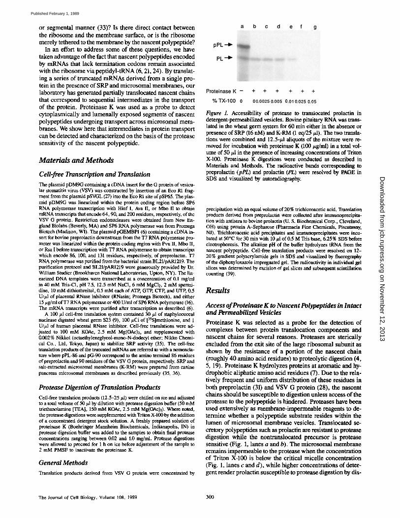
Figure 1. Accessibility of protease to translocated prolactin in detergent-permeabilized vesicles. Bovine pituitary RNA was trans- lated in the wheat germ system for 60 min either in the absence or presence of SRP (16 nM) and K-RM (1 eq/25 #1). The two transla- tions were combined and 12.5-#1 aliquots of the mixture were re- moved for incubation with proteinase K (100 #g/nil) in a total vol- ume of 50 #1 in the presence of increasing concentrations of Triton X-100. Proteinase K digestions were conducted as described in Materials and Methods. The radioactive bands corresponding to preprolactin (pPL) and prolactin (PL) were resolved by PAGE in SDS and visualized by autoradiography.
precipitation with an equal volume of 20% trichloroacetic acid. Translation products derived from preprolactin were collected after immunoprecipita- tion with antisera to bovine prolactin (U. S. Biochemical Corp., Cleveland, OH) using protein A-Sepharose (Pharmacia Fine Chemicals, Piscataway, N J). Trichloroacetic acid precipitates and immunoprecipitates were incu- bated at 50°C for 30 rain with 10 #1 of 0.5 M Tris base, 6.25% SDS before electrophoresis. The alkaline pH of the buffer hydrolyzes tRNA from the nascent polypeptide. Cell-free translation products were resolved on 12- 20% gradient polyacrylamide gels in SDS and visualized by fluorography of the diphenyloxazole impregnated gel. The radioactivity in individual gel slices was determined by excision of gel slices and subsequent scintillation counting (39).
Results
Access of Proteinase K to Nascent Polypeptides in Intact and Permeabilized Vesicles
Proteinase K was selected as a probe for the detection of complexes between protein translocation components and nascent chains for several reasons. Proteases are gterically excluded from the exit site of the large ribosomal subunit as shown by the resistance of a portion of the nascent chain (roughly 40 amino acid residues) to proteolytic digestion (4, 5, 19). Proteinase K hydrolyze8 proteins at aromatic and hy- drophobic aliphatic amino acid residues (7). Due to the rela- tively frequent and uniform distribution of these residues in both preprolactin (31) and VSV G protein (28), the nascent chains should be susceptible to digestion unless access of the protease to the polypeptide is hindered. Proteases have been used extensively as membrane-impermeable reagents to de- termine whether a polypeptide substrate resides within the lumen of microsomal membrane vesicles. Translocated se- cretory polypeptides such as prolactin are resistant to protease digestion while the nontranslocated precursor is protease sensitive (Fig. 1, lanes a and b). The microsomal membrane remains impermeable to the protease when the concentration of Triton Xd00 is below the critical micelle concentration (Fig. 1, lanes c and d), while higher concentrations of deter- gent render prolactin susceptible to protease digestion by dis-
The Journal of Cell Biology, Volume 108, 1989 300
on Novem
ber 12, 2013jcb.rupress.org
Dow
nloaded from
Published February 1, 1989
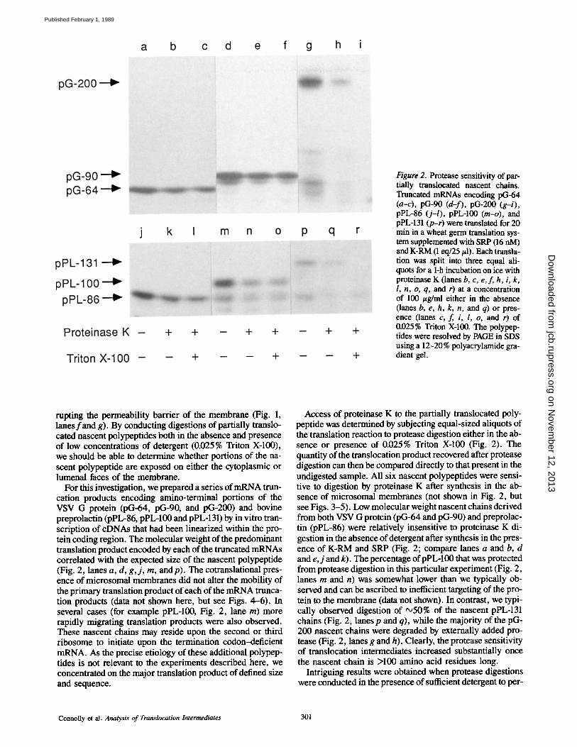
Figure 2. Protease sensitivity of par- tially translocated nascent chains. Truncated mRNAs encoding pG-64 (a-c), pG-90 (d-f), pG-200 (g-i), pPL-86 (j-l), pPL-100 (m-o), and pPL-131 (p-r) were translated for 20 min in a wheat germ translation sys- tem supplemented with SRP (16 nM) and K-RM (1 eq/25/A). Each transla- tion was split into three equal ali- quots for a 1-h incubation on ice with proteinase K (lanes b, c, e, f, h, i, k, 1, n, o, q, and r) at a concentration of 100 #g/ml either in the absence (lanes b, e, h, k, n, and q) or pres- ence (lanes c, f, i, l, o, and r) of 0.025% Triton X-100. The polypep- tides were resolved by PAGE in SDS using a 12-20% polyacrylamide gra- dient gel.
rupting the permeability barrier of the membrane (Fig. 1, lanesfand g). By conducting digestions of partially translo- cated nascent polypeptides both in the absence and presence of low concentrations of detergent (0.025% Triton X-100), we should be able to determine whether portions of the na- scent polypeptide are exposed on either the cytoplasmic or lumenal faces of the membrane.
For this investigation, we prepared a series of mRNA trun- cation products encoding amino-terminal portions of the VSV G protein (pG-64, pG-90, and pG-200) and bovine preprolactin (pPL-86, pPL-100 and pPL-131) by in vitro tran- scription of cDNAs that had been linearized within the pro- tein coding region. The molecular weight of the predominant translation product encoded by each of the truncated mRNAs correlated with the expected size of the nascent polypeptide (Fig. 2, lanes a, d, g, j , m, and p). The cotranslational pres- ence of microsomal membranes did not alter the mobility of the primary translation product of each of the mRNA trunca- tion products (data not shown here, but see Figs. 4-6). In several cases (for example pPL-100, Fig. 2, lane m) more rapidly migrating translation products were also observed. These nascent chains may reside upon the second or third ribosome to initiate upon the termination codon-deficient mRNA. As the precise etiology of these additional polypep- tides is not relevant to the experiments described here, we concentrated on the major translation product of defined size and sequence.
Access of proteinase K to the partially translocated poly- peptide was determined by subjecting equal-sized aliquots of the translation reaction to protease digestion either in the ab- sence or presence of 0.025% Triton X-100 (Fig. 2). The quantity of the translocation product recovered after protease digestion can then be compared directly to that present in the undigested sample. All six nascent polypeptides were sensi- tive to digestion by proteinase K after synthesis in the ab- sence of microsomal membranes (not shown in Fig. 2, but see Figs. 3-5). Low molecular weight nascent chains derived from both VSV G protein (pG-64 and pG-90) and preprolac- tin (pPL-86) were relatively insensitive to proteinase K di- gestion in the absence of detergent after synthesis in the pres- ence of K-RM and SRP (Fig. 2; compare lanes a and b, d and e, j and k). The percentage of pPL-100 that was protected from protease digestion in this particular experiment (Fig. 2, lanes rn and n) was somewhat lower than we typically ob- served and can be ascribed to inefficient targeting of the pro- tein to the membrane (data not shown). In contrast, we typi- cally observed digestion of '~50% of the nascent pPL-131 chains (Fig. 2, lanes p and q), while the majority of the pG- 200 nascent chains were degraded by externally added pro- tease (Fig. 2, lanes g and h). Clearly, the protease sensitivity of translocation intermediates increased substantially once the nascent chain is >100 amino acid residues long.
Intriguing results were obtained when protease digestions were conducted in the presence of sufficient detergent to per-
Connolly et al. Analysis of Translocation Intermediates 301
on Novem
ber 12, 2013jcb.rupress.org
Dow
nloaded from
Published February 1, 1989

Figure 3. Protection of pG-64 by SRP and microsomal membranes. The truncated mRNA encoding pG-64 was translated in a wheat germ system in the absence of SRP and K-RM (a), with 16 nM SRP (b) or with 16 nM SRP and K-RM (c). After 20 min of synthesis, the three translations were subaliquotted for a l-h digestion on ice with the following concentrations of proteinase K in a total volume of 50 #1: (lane a) 0.0 mg/ml, (lane b) 0.02 mg/ml, (lane c) 0.1 mg/ml, (lane d) 0.2 mg/ml, (lane e) 0.5 mg/ml, and (lane f ) 1.0 mg/ml. The radioactive polypeptide corresponding to pG-64 was resolved by PAGE in SDS. The quantity of [35S]methionine- labeled pG-64 was determined as described in Materials and Methods and plotted in panel d as percent of pG-64 digested in translations containing no added SRP (..), translations containing SRP alone (o), and translations containing SRP and K-RM (A).
meabilize the vesicles. While the larger two nascent chains (pG-200 and pPL-131) were completely digested by protein- ase K when sufficient detergent was present to permeabilize the membrane (Fig. 2, lanes i and r), the shorter nascent chains derived from both preprolactin (pPL-86, pPL-100) and VSV G protein (pG-64 and pG-100) were not more sen- sitive to protease digestion after the addition of detergent (Fig. 2, lanes c, f, l, and o). The enhanced protease sensitiv- ity of pPL-131 and pG-200 is readily explained by access of the protease to a lumenally exposed portion of the nascent polypeptide. In contrast, shorter nascent polypeptides de- rived from both proteins appear not to have lumenally ex- posed segments.
Protease Resistance of Short Nascent Chains
The 64-residue G protein nascent chain (pG-64) contains 48
residues from the mature protein in addition to the 16-resi- due signal sequence. Between 20 and 30 residues of the poly- peptide including the signal sequence should be exposed on the surface of the ribosome for interaction with the transloca- tion apparatus. After synthesis in the absence of SRP and K-RM, pG-64 was readily digested upon incubation with a low concentration (20/~g/ml) of proteinase K (Fig. 3 a; for quantitation see Fig. 3 d). The protease digestion product visible in Fig. 3, lanes b-e corresponds to a fragment of the nascent chain which resists further protease digestion due to its location within a protease inaccessible space in the large ribosomal subunit (4, 5, 19). The majority of pG-64 was in- sensitive to digestion by low concentrations of protease when SRP was present during translation (Fig. 3 b). The most rea- sonable explanation for this observation is that proteinase K is denied access to the signal sequence of pG-64 by SRP. Complete digestion of the SRP bound form of pG-64 re- quired a significantly higher concentration of proteinase K (Fig. 3 b). A biphasic protease sensitivity curve was obtained after quantitation of the data in Fig. 3 b. Approximately 25 % of the nascent chains were extremely sensitive to protease digestion and presumably correspond to a subpopulation of pG-64 chains not bound by SRE
The protease insensitivity of pG-64 observed in Fig. 2 can- not be explained by protection of the signal sequence by SRP alone. Although high concentrations of externally added pro- tease (1 mg/ml) can completely digest the SRP bound form of the polypeptide (Fig. 3 b), only 50% of the pG-64 that was synthesized in the presence of both K-RM and SRP was digested under these conditions (Fig. 3 c). The resistance of pG-64 to externally added protease is most readily explained by close contact between the membrane surface and the ribo- some, thereby sterically excluding proteinase K from the polypeptide chain.
Cytoplasmic Exposure of Larger Nascent Polypeptides
We conducted the following experiment to determine whether the protease sensitivity of pG-200 observed in Fig. 2 may have been due to inefficient targeting of the protein to the membrane. The truncated mRNA encoding pG-200 was translated in the presence of SRP and K-RM to prepare a translocation intermediate. Aliquots of the translation reac- tion were then incubated with puromycin at 25°C to induce termination of the polypeptide and allow subsequent trans- port of bonafide translocation intermediates across the mem- brane bilayer. Translocation of pG-200 can be detected by the addition of high-mannose oligosaccharide to asparagine-162 of the mature G protein to produce the glycosylated form of the polypeptide (g-G-200). Because asparagine 162 is lo- cated only 12 residues from the last amino acid encoded by the truncated mRNA, glycosylation of the protein cannot oc- cur while the polypeptide remains bound to the ribosome as a peptidyl-tRNA. Due to the lumenal location of the oligosaccharyl transferase (34), glycosylation of g-G-200 is indicative of transport of the entire polypeptide into the lu- men. The apparent molecular weight of pG-200 is not altered by synthesis in the presence of K-RM and SRP (Fig. 4 a, lanes a and c). Puromycin termination of pG-200 was ac- companied by conversion of 73% of the precursor into the glycosylated and translocated g-G-200 (Fig. 4 a, compare lanes c and e). As expected >95 % of the translocated g-G- 200 was protected from protease digestion by the membrane
The Journal of Cell Biology, Volume 108, 1989 302
on Novem
ber 12, 2013jcb.rupress.org
Dow
nloaded from
Published February 1, 1989
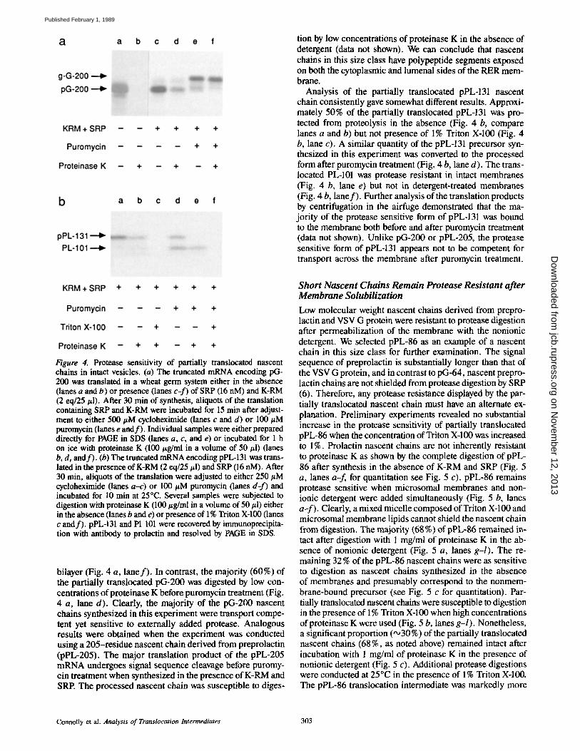
Figure 4. Protease sensitivity of partially translocated nascent chains in intact vesicles. (a) The truncated mRNA encoding pG- 200 was translated in a wheat germ system either in the absence (lanes a and b) or presence (lanes c-f) of SRP (16 nM) and K-RM (2 eq/25 #1). After 30 min of synthesis, aliquots of the translation containing SRP and K-RM were incubated for 15 rain after adjust- ment to either 500 #M cycloheximide (lanes c and d) or 100 #M puromycin (lanes e and f) . Individual samples were either prepared directly for PAGE in SDS (lanes a, c, and e) or incubated for 1 h on ice with proteinase K (100 #g/ml in a volume of 50 #1) (lanes b, d, and f) . (b) The truncated mRNA encoding pPL-131 was trans- lated in the presence of K-RM (2 eq/25 #1) and SRP (16 nM). After 30 min, aliquots of the translation were adjusted to either 250 #M cycloheximide (lanes a-c) or 100 #M puromycin (lanes d-f) and incubated for 10 min at 25°C. Several samples were subjected to digestion with proteinase K (100 #g/ml in a volume of 50 #1) either in the absence (lanes b and e) or presence of 1% Triton X-100 (lanes c and f) . pPL-131 and Pl 101 were recovered by immunoprecipita- tion with antibody to prolactin and resolved by PAGE in SDS.
bilayer (Fig. 4 a, lane f ) . In contrast, the majority (60%) of the partially translocated pG-200 was digested by low con- centrations of proteinase K before puromycin treatment (Fig. 4 a, lane d). Clearly, the majority of the pG-200 nascent chains synthesized in this experiment were transport compe- tent yet sensitive to externally added protease. Analogous results were obtained when the experiment was conducted using a 205-residue nascent chain derived from preprolactin (pPL-205). The major translation product of the pPL-205 mRNA undergoes signal sequence cleavage before puromy- cin treatment when synthesized in the presence of K-RM and SRP. The processed nascent chain was susceptible to diges-
tion by low concentrations of proteinase K in the absence of detergent (data not shown). We can conclude that nascent chains in this size class have polypeptide segments exposed on both the cytoplasmic and lumenal sides of the RER mem- brane.
Analysis of the partially translocated pPL-131 nascent chain consistently gave somewhat different results. Approxi- mately 50% of the partially translocated pPL-131 was pro- tected from proteolysis in the absence (Fig. 4 b, compare lanes a and b) but not presence of 1% Triton X-100 (Fig. 4 b, lane c). A similar quantity of the pPL-131 precursor syn- thesized in this experiment was converted to the processed form after puromycin treatment (Fig. 4 b, lane d). The trans- located PL-101 was protease resistant in intact membranes (Fig. 4 b, lane e) but not in detergent-treated membranes (Fig. 4 b, lane f ) . Further analysis of the translation products by centrifugation in the airfuge demonstrated that the ma- jority of the protease sensitive form of pPL-131 was bound to the membrane both before and after puromycin treatment (data not shown). Unlike pG-200 or pPL-205, the protease sensitive form of pPL-131 appears not to be competent for transport across the membrane after puromycin treatment.
Short Nascent Chains Remain Protease Resistant after Membrane Solubilization
Low molecular weight nascent chains derived from prepro- lactin and VSV G protein were resistant to protease digestion after permeabilization of the membrane with the nonionic detergent. We selected pPL-86 as an example of a nascent chain in this size class for further examination. The signal sequence of preprolactin is substantially longer than that of the VSV G protein, and in contrast to pG-64, nascent prepro- lactin chains are not shielded from protease digestion by SRP (6). Therefore, any protease resistance displayed by the par- tially translocated nascent chain must have an alternate ex- planation. Preliminary experiments revealed no substantial increase in the protease sensitivity of partially translocated pPL-86 when the concentration of Triton X-100 was increased to 1%. Prolactin nascent chains are not inherently resistant to proteinase K as shown by the complete digestion of pPL- 86 after synthesis in the absence of K-RM and SRP (Fig. 5 a, lanes a-f, for quantitation see Fig. 5 c). pPL-86 remains protease sensitive when microsomal membranes and non- ionic detergent were added simultaneously (Fig. 5 b, lanes a-f). Clearly, a mixed micelle composed of Triton X-100 and microsomal membrane lipids cannot shield the nascent chain from digestion. The majority (68%) of pPL-86 remained in- tact after digestion with 1 mg/ml of proteinase K in the ab- sence of nonionic detergent (Fig. 5 a, lanes g-l). The re- maining 32 % of the pPL-86 nascent chains were as sensitive to digestion as nascent chains synthesized in the absence of membranes and presumably correspond to the nonmem- brahe-bound precursor (see Fig. 5 c for quantitation). Par- tially translocated nascent chains were susceptible to digestion in the presence of 1% Triton X-100 when high concentrations ofproteinase K were used (Fig. 5 b, lanes g-l). Nonetheless, a significant proportion ("o30 %) of the partially translocated nascent chains (68 %, as noted above) remained intact after incubation with 1 mg/ml of proteinase K in the presence of nonionic detergent (Fig. 5 c). Additional protease digestions were conducted at 25°C in the presence of 1% Triton X-100. The pPL-86 translocation intermediate was markedly more
Connolly et al. Analysis of Translocation Intermediates 303
on Novem
ber 12, 2013jcb.rupress.org
Dow
nloaded from
Published February 1, 1989
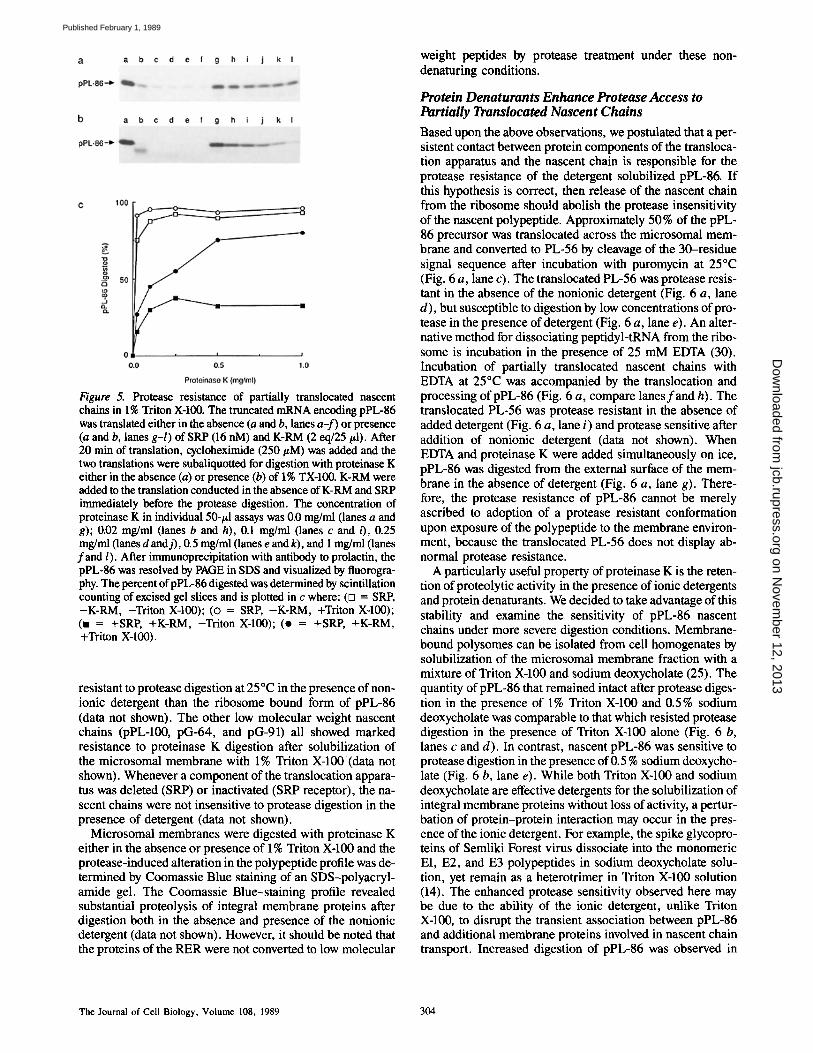
Figure 5. Protease resistance of partially translocated nascent chains in 1% Triton X-100. The truncated mRNA encoding pPL-86 was translated either in the absence (a and b, lanes a-f) or presence (a and b, lanes g-l) of SRP (16 nM) and K-RM (2 eq/25/~1). After 20 min of translation, cycloheximide (250/zM) was added and the two translations were subaliquotted for digestion with proteinase K either in the absence (a) or presence (b) of 1% TX-100. K-RM were added to the translation conducted in the absence of K-RM and SRP immediately before the protease digestion. The concentration of proteinase K in individual 50-#1 assays was 0.0 mg/ml (lanes a and g); 0.02 mg/ml (lanes b and h), 0.1 mg/ml (lanes c and 0, 0.25 mg/ml (lanes d and j), 0.5 mg/ml (lanes e and k), and 1 mg/ml (lanes fand l). After immunoprecipitation with antibody to prolactin, the pPL-86 was resolved by PAGE in SDS and visualized by fluorogra- phy. The percent ofpPL-86 digested was determined by scintillation counting of excised gel slices and is plotted in c where: (D = SRP, -K-RM, -Triton X-100); (o = SRP, -K-RM, +Triton X-100); (n = +SRP, +K-RM, -Triton X-100); (o = +SRP, +K-RM, +Triton X-100).
resistant to protease digestion at 25°C in the presence of non- ionic detergent than the ribosome bound form of pPL-86 (data not shown). The other low molecular weight nascent chains (pPL-100, pG-64, and pG-91) all showed marked resistance to proteinase K digestion after solubilization of the microsomal membrane with 1% Triton X-100 (data not shown). Whenever a component of the translocation appara- tus was deleted (SRP) or inactivated (SRP receptor), the na- scent chains were not insensitive to protease digestion in the presence of detergent (data not shown).
Microsomal membranes were digested with proteinase K either in the absence or presence of 1% Triton X-100 and the protease-induced alteration in the polypeptide profile was de- termined by Coomassie Blue staining of an SDS-polyacryl- amide gel. The Coomassie Blue-staining profile revealed substantial proteolysis of integral membrane proteins after digestion both in the absence and presence of the nonionic detergent (data not shown). However, it should be noted that the proteins of the RER were not converted to low molecular
weight peptides by protease treatment under these non- denaturing conditions.
Protein Denaturants Enhance Protease Access to Partially Translocated Nascent Chains
Based upon the above observations, we postulated that a per- sistent contact between protein components of the transloca- tion apparatus and the nascent chain is responsible for the protease resistance of the detergent solubilized pPL-86. If this hypothesis is correct, then release of the nascent chain from the ribosome should abolish the protease insensitivity of the nascent polypeptide. Approximately 50 % of the pPL- 86 precursor was translocated across the microsomal mem- brane and converted to PL-56 by cleavage of the 30-residue signal sequence after incubation with puromycin at 25°C (Fig. 6 a, lane c). The translocated PL-56 was protease resis- tant in the absence of the nonionic detergent (Fig. 6 a, lane d), but susceptible to digestion by low concentrations of pro- tease in the presence of detergent (Fig. 6 a, lane e). An alter- native method for dissociating peptidyl-tRNA from the ribo- some is incubation in the presence of 25 mM EDTA (30). Incubation of partially translocated nascent chains with EDTA at 25°C was accompanied by the translocation and processing of pPL-86 (Fig. 6 a, compare lanesfand h). The translocated PL-56 was protease resistant in the absence of added detergent (Fig. 6 a, lane i) and protease sensitive after addition of nonionic detergent (data not shown). When EDTA and proteinase K were added simultaneously on ice, pPL-86 was digested from the external surface of the mem- brane in the absence of detergent (Fig. 6 a, lane g). There- fore, the protease resistance of pPL-86 cannot be merely ascribed to adoption of a protease resistant conformation upon exposure of the polypeptide to the membrane environ- ment, because the translocated PL-56 does not display ab- normal protease resistance.
A particularly useful property of proteinase K is the reten- tion of proteolytic activity in the presence of ionic detergents and protein denaturants. We decided to take advantage of this stability and examine the sensitivity of pPL-86 nascent chains under more severe digestion conditions. Membrane- bound polysomes can be isolated from cell homogenates by solubilization of the microsomal membrane fraction with a mixture of Triton X-100 and sodium deoxycholate (25). The quantity of pPL-86 that remained intact after protease diges- tion in the presence of 1% Triton X-100 and 0.5% sodium deoxycholate was comparable to that which resisted protease digestion in the presence of Triton X-100 alone (Fig. 6 b, lanes c and d). In contrast, nascent pPL-86 was sensitive to protease digestion in the presence of 0.5 % sodium deoxycho- late (Fig. 6 b, lane e). While both Triton X-100 and sodium deoxycholate are effective detergents for the solubilization of integral membrane proteins without loss of activity, a pertur- bation of protein-protein interaction may occur in the pres- ence of the ionic detergent. For example, the spike glycopro- teins of Semliki Forest virus dissociate into the monomeric El, E2, and E3 polypeptides in sodium deoxycholate solu- tion, yet remain as a heterotrimer in Triton X-100 solution (14). The enhanced protease sensitivity observed here may be due to the ability of the ionic detergent, unlike Triton X-100, to disrupt the transient association between pPL-86 and additional membrane proteins involved in nascent chain transport. Increased digestion of pPL-86 was observed in
The Journal of Cell Biology, Volume 108, 1989 304
on Novem
ber 12, 2013jcb.rupress.org
Dow
nloaded from
Published February 1, 1989
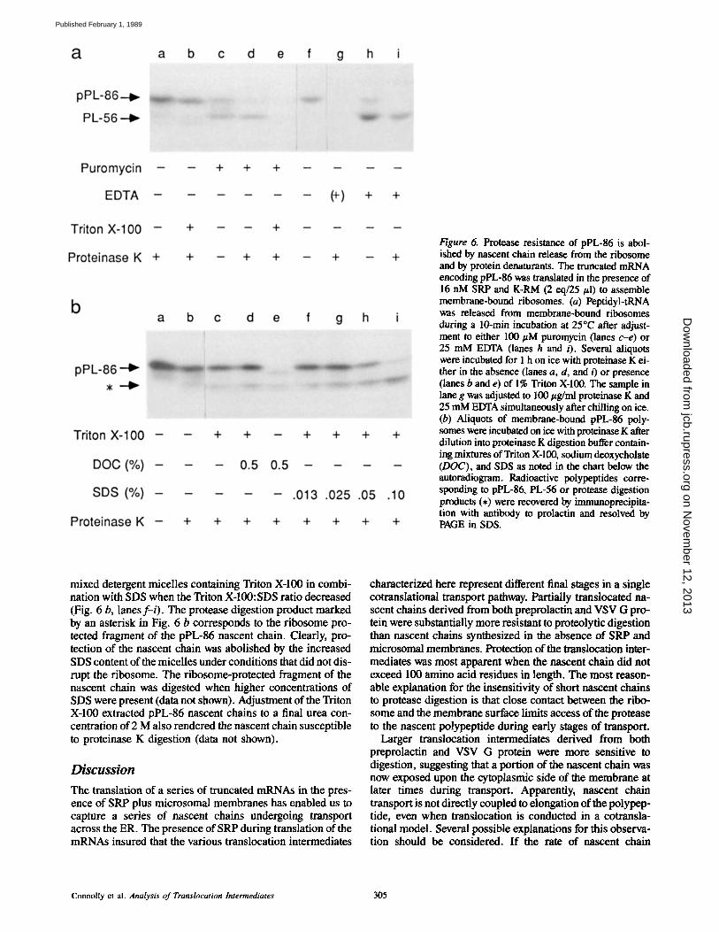
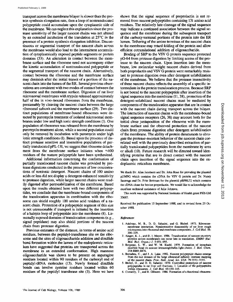
Figure 6. Protease resistance of pPL-86 is abol- ished by nascent chain release from the ribosome and by protein denaturants. The truncated mRNA encoding pPL-86 was translated in the presence of 16 nM SRP and K-RM (2 eq/25 #1) to assemble membrane-bound ribosomes. (a) Peptidyl-tRNA was released from membrane-bound ribosomes during a 10-min incubation at 25"C after adjust- ment to either 100 #M puromycin (lanes c-e) or 25 mM EDTA (lanes h and i). Several aliquots were incubated for 1 h on ice with proteinase K ei- ther in the absence (lanes a, d, and i) or presence 0anes b and e) of 1% Triton X-100. The sample in lane g was adjusted to 100 #g/ml proteinase K and 25 mM EDTA simultaneously after chilling on ice. (b) Aliquots of membrane-bound pPL-86 poly- somes were incubated on ice with proteinase K after dilution into proteinase K digestion buffer contain- ing mixtures of Triton X-100, sodium deoxycholate (DOC), and SDS as noted in the chart below the autoradiogram. Radioactive polypeptides corre- sponding to pPL-86, PL-56 or protease digestion products (,) were recovered by immunoprecipita- tion with antibody to prolactin and resolved by PAGE in SDS.
mixed detergent micelles containing Triton X-100 in combi- nation with SDS when the Triton X-100:SDS ratio decreased (Fig. 6 b, lanes f- i) . The protease digestion product marked by an asterisk in Fig. 6 b corresponds to the ribosome pro- tected fragment of the pPL-86 nascent chain. Clearly, pro- tection of the nascent chain was abolished by the increased SDS content of the micelles under conditions that did not dis- rupt the ribosome. The ribosome-protected fragment of the nascent chain was digested when higher concentrations of SDS were present (data not shown). Adjustment of the Triton X-100 extracted pPL-86 nascent chains to a final urea con- centration of 2 M also rendered the nascent chain susceptible to proteinase K digestion (data not shown).
Discussion
The translation of a series of truncated mRNAs in the pres- ence of SRP plus microsomal membranes has enabled us to capture a series of nascent chains undergoing transport across the ER. The presence of SRP during translation of the mRNAs insured that the various translocation intermediates
characterized here represent different final stages in a single cotranslational transport pathway. Partially translocated na- scent chains derived from both preprolactin and VSV G pro- tein were substantially more resistant to proteolytic digestion than nascent chains synthesized in the absence of SRP and microsomal membranes. Protection of the translocation inter- mediates was most apparent when the nascent chain did not exceed 100 amino acid residues in length. The most reason- able explanation for the insensitivity of short nascent chains to protease digestion is that close contact between the ribo- some and the membrane surface limits access of the protease to the nascent polypeptide during early stages of transport.
Larger translocation intermediates derived from both preprolactin and VSV G protein were more sensitive to digestion, suggesting that a portion of the nascent chain was now exposed upon the cytoplasmic side of the membrane at later times during transport. Apparently, nascent chain transport is not directly coupled to elongation of the polypep- tide, even when translocation is conducted in a cotransla- tional model. Several possible explanations for this observa- tion should be considered. I f the rate of nascent chain
Connolly et al. Analysis of Translocation Intermediates 305
on Novem
ber 12, 2013jcb.rupress.org
Dow
nloaded from
Published February 1, 1989
transport across the membrane bilayer is slower than the pro- tein synthesis elongation rate, then a loop of nontranslocated polypeptide could accumulate upon the cytoplasmic side of the membrane. We can neglect this explanation since the pro- tease sensitivity of the larger nascent chains was not altered by an extended incubation of the translation at 25°C in the presence of a protein synthesis elongation inhibitor. Discon- tinuous or segmental transport of the nascent chain across the membrane would also lead to the intermittent accumula- tion of cytoplasmically exposed peptide segments or protein domains (33). An alteration in contact between the mem- brane surface and the ribosome need not accompany either the kinetic accumulation of nontranslocated polypeptide or the segmental transport of the nascent chain. Alternatively, contact between the ribosome and the membrane surface may diminish after the initial transit of a portion of the na- scent chain into the lumen of the ER. Several previous obser- vations are consistent with two modes of contact between the ribosome and the membrane surface. Digestion of rat liver microsomal membranes with trypsin released approximately half of the in vivo-bound ribosomes from the membrane, presumably by cleaving the nascent chain between the large ribosomal subunit and the membrane surface (29). Two dis- tinct populations of membrane-bound ribosomes were de- tected by puromycin treatment of isolated microsomal mem- branes under low and high ionic strength conditions (1). One population of ribosomes was released from the membrane by puromycin treatment alone, while a second population could only be removed by incubation with puromycin under high ionic strength conditions (1). Based upon the presence of dis- tinct protease sensitive and insensitive populations of par- tially translocated pPL-131, we suggest that ribosome detach- ment from the membrane surface initiates when ~100 residues of the mature polypeptide have been synthesized.
Additional information concerning the conformation of partially translocated nascent chains was provided by pro- tease digestions conducted in the presence of low concentra- tions of nonionic detergent. Nascent chains of 100 amino acids or less did not display a detergent-enhanced sensitivity to protease digestion, while larger nascent chains were read- ily digested after permeabilization of the membrane. Based upon the results obtained here with two different polypep- tides, we conclude that the membrane-bound components of the translocation apparatus in combination with the ribo- some can shield roughly 100 amino acid residues of a na- scent chain. Protection of a polypeptide segment of this size is not unreasonable if transport is initiated by the insertion of a hairpin loop of polypeptide into the membrane (8). Lu- menally exposed domains of translocation components (e.g., signal peptidase) may also shield portions of the nascent chain from protease digestion.
Previous estimates of the distance, in terms of amino acid residues, between the peptidyl-transferase site on the ribo- some and the sites of oligosaccharide addition and disulfide bond formation within the lumen of the endoplasmic reticu- lum have suggested that proteins are transported across the membrane in an extended conformation. High mannose oligosaccharide was shown to be present on asparagine residues located within 90 residues of the carboxyl end of peptidyl-tRNA molecules (13). Newly formed disulfide bonds can involve cysteine residues located within 60 residues of the peptidyl transferase site (3). Here we have
shown that the signal sequence of preprolactin is not re- moved from nascent polypeptides containing 131 amino acid residues. The relatively late cleavage of the signal sequence may indicate a continued association between the signal se- quence and the membrane during the subsequent transport of the carboxy-terminal portions of the protein into the ER lumen. Tethering of the amino terminus of the nascent chain to the membrane may retard folding of the protein and allow efficient cotranslational addition of oligosaccharide.
Binding of SRP to the VSV G protein sequence protected pG-64 from protease digestion by limiting access of the pro- tease to the nascent chain. Upon insertion into the mem- brane, low molecular weight nascent chains derived from both preprolactin and VSV G protein were remarkably resis- tant to protease digestion even after detergent solubilization of the membrane. We believe that the protease insensitivity of these nascent chains reflects the assembly of a specific in- termediate in the protein translocation process. Because SRP is not bound to the nascent polypeptide after insertion of the signal sequence into the membrane (11, 38), protection of the detergent-solubilized nascent chains must be mediated by components of the translocation apparatus that are in contact with the nascent chain during transport across the bilayer. The interaction of nascent chains with the membrane-bound signal sequence receptors (26, 38) may account both for the initial close juxtaposition of the ribosome with the mem- brane surface and the observed protection of the nascent chain from protease digestion after detergent solubilization of the membrane. The ability of protein denaturants to abro- gate the protease-resistant behavior of the nascent chain cor- related well with the previously described extraction of par- tially translocated polypeptides from the membrane by urea or alkali (10). Future research will be directed toward iden- tifying proteins that are in direct contact with the nascent chain upon insertion of the signal sequence into the en- doplasmic reticulum membrane.
We thank Dr. Alan Jacobson and Dr. John Rose for providing the plasmid
pDM9G which contains the eDNA for VSV G protein and Dr. Nancy
Sasavage and Dr. Fritz Rottman for the plasmid pBPRL72 which contains
the eDNA clone for bovine preprolaetin. We would like to acknowledge the
excellent technical assistance of Julie Johnson.
This work was supported by National Institutes of Health grant PHS GM
35687.
Received for publication 15 September 1988, and in revised form 25 Oc-
tober 1988,
Rcf~renc~
1. Adelman, M. B., D. D. Sabatini, and G. Blobel. 1973. Ribosome- membrane interaction. Nondestructive disassembly of rat liver rough microsomes into ribosomal and membrane components. J. Cell Biol. 56: 206-229.
2. Ainger, K. J., and D. I. Meyer. 1986. Translocation of nascent secretory proteins across membranes can occur late in translation. EMBO (Eur. Mol. Biol. Organ.)J. 5:951-955.
3. Bergman, L, W., and W. M. Kuehl. 1979. Formation of intrachain disulfide bond on nascent immunoglobulin light chains. J. Biol. Chem. 254:8869-8876.
4. Bemabeu, C., and J. A. Lake. 1982. Nascent polypeptide chains emerge from the exit domain of the large ribosomal subanit: immune mapping of the nascent chain. Proc. Natl. Acad. Sci. USA. 79:3111-3115.
5. Blobel, G., and D. D. Sabatini. 1970. Controlled proteolysis of nascent polypeptides in rat liver cell fractions. I. Location of the polypeptides within ribosomes. J. Cell Biol. 45:130-145.
6. Connolly, T., and R. Gilmore. 1986. Formation of a functional ribosome-
The Journal of Cell Biology, Volume 108, 1989 306
on Novem
ber 12, 2013jcb.rupress.org
Dow
nloaded from
Published February 1, 1989
membrane junction during translocation requires the participation of a GTP-binding protein. J. Cell Biol. 103:2253-2261.
7. Ebeling, W., N. Hennrich, M. Klockow, H. Metz, H. D. Orth, and H. Lang. 1974. Proteinase K from Tritirachium album Limber. Eur. J. Bio- chem. 47:91-97.
8. Engelman, D. M., and T. A. Steitz. 1981. The spontaneous insertion of proteins into and across membranes: the helical hairpin hypothesis. Cell. 23:411-422.
9. Erickson, A. H., and G. Blobel. 1983. Cell-free translation of messenger RNA in a wheat germ system. Methods Enzymol, 96:38-50.
10. Gilmore, R., and G. Blobel. 1985. Translocation of secretory proteins across the microsomal membrane occurs through an environment accessi- ble to aqueous perturbants. Cell. 42:497-505.
11. Gilmore, R., and G. Blobel. 1983. Transient involvement of the signal rec- ognition particle and its receptor in the microsomal membrane prior to protein translocation. Cell. 35:677-685.
12. Gilmore, R., P. Walter, and G. Blobel. 1982. Protein translocation across the endoplasmic retieulum. II. Isolation and characterization of the signal recognition particle receptor. J. Cell Biol. 95:470--477.
13. Glabe, C. G., J. A. Hanover, and W. J. Lennarz. 1980. Glycosylation of ovalbumin nascent chains. J. Biol. Chem. 255:9236-9242.
14. Helenius, A., E. Fries, H. Garoff, and K. Simons. 1975. Solubilization of the Semliki Forest virus membrane with sodium deoxycholate. Biochim. Biophys. Acta. 436:319-334.
15. Hoffman, K., and R. Gilmore. 1988. Guanosine triphosphate promotes the posttranslational integration of opsin into the endoplasmie reticulum membrane. J. Biol. Chem. 263:4381--4385.
16. Krieg, P. A., and D. A. Melton. 1984. Functional messenger RNAs are produced by SP6 in vitro transcription of cloned cDNAs. Nucleic Acids. Res. 12:7057-7070.
17. Krieg, U. C., P. Walter, and A. E. Johnson. 1986. Photocrosslinking oftbe signal sequence of nascent preprolactin to the 54kD polypeptide of the signal recognition particle. Proc. Natl. Acad. Sci. USA. 83:8604-8608.
18. Kurzchalia, T. V., M. Wiedmann, A. S. Girshovich, E. S. Bochkareva, H. Bielka, and T. A. Rapoport. 1986. The signal sequence of nascent preprolactin interacts with the 54 kD polypeptide of the signal recognition particle. Nature (Lond.). 320:634-636.
19. Malkin, L. I., and A. Rich. 1967. Partial resistance of nascent polypeptide chains to proteolytic digestion due to ribosomal shielding. J. Mol. Biol. 26:329-346.
20. Meyer, D. I., E. Krause, and B. Dobberstein. 1982. Secretory protein translocation across membranes: the role of the 'docking protein'. Nature (Lond.). 297:647-650.
21. Mueckier, M., and H. F. Lodish. 1986. Posttranslational insertion of a fragment of the glucose transporter into microsomes requires phosphoan- hydride bond cleavage. Nature (Lond). 322:549-552.
22. Mueckler, M., and H. F. Lodish. 1986. The human glucose transporter can insert posttranslationally into microsomes. Cell. 44:629-637.
23. Perara, E., and V. R. Lingappa. 1985. A former amino terminal signal se- quence engineered to an internal location directs translocation of both
flanking protein domains. Z Cell Biol. 101:2292-2301. 24. Perara, E., R. E. Rothman, and V. R. Lingappa. 1986. Uncoupling translo-
cation from translation: implications for transport of proteins across membranes. Science (Wash. DC). 232:348-352.
25. Ramsey, J. C., and W. J. Steele. 1976, A procedure for the quantitative recovery of homogenous populations of undegraded free and bound poly- somes from rat liver. Biochemistry. 15:1704-1712.
26. Robinson, A., M.A. Kaderbhai, and B. M. Austen. 1987. Identification of signal sequence binding proteins integrated into the rough endoplasmic reticulum membrane. Biochem. J. 242:767-777.
27. Rose, J. K., and J. E. Bergmann. 1982. Expression from cloned eDNA of cell-surface and secreted forms of the glyeoprotein of vesicular stomatitis virus. Cell. 30:753-762.
28. Rose, J. K., and C. Gallione. 1981. Nueleotide sequences of the mRNAs encoding the VSV G and M proteins as determined from eDNA clones containing the complete coding regions. J. Virol. 39:519-528.
29. Sabatini, D. D., and G. Blobel. 1970. Controlled proteolysis of nascent polypeptides in rat liver cell fractions. H. Location of the polypeptides in rough microsomes. J. Cell Biol. 45:146-157.
30. Sabatini, D. D., Y. Tashiro, G. E. Palade. 1966. On the attachment of ribo- somes to microsomal membranes. J. Mol. Biol. 19:503-524.
31. Sasavage, N. L., J. H. Nilson, S. Horowitz, and F. M. Rottman. 1982. Nucleotide sequence of bovine prolactin messenger RNA. J. Biol. Chem. 257:678-681.
32. Siegel, V., and P. Walter. 1988. The affinity of signal recognition particle for presecretory proteins is dependent on nascent chain length. EMBO (Eur. Mol. Biol. Organ.)J. 7:1769-1775.
33. Singer, S. J., P. A. Maher, and M. P. Yaffe. 1987. On the translocation of proteins across membranes. Proc. Natl. Acad. Sci. USA. 84:1015- 1019.
34. Snider, M. D., and P. W. Robbins. 1982. Transmembrane organization of protein glycosylation. Mature oligosaecharide-lipid is located on the lu- menal side of microsomes from Chinese hamster ovary cells. J. Biol. Chem. 257:6796-6801.
35. Walter, P., and G. Blobel. 1983. Preparation of microsomal membranes for cotranslational protein translocation. Methods Enzymol. 96:84-93.
36. Walter, P., and G. Blobel. 1981. Translocation of proteins across the en- doplasmic reticulum. III. Signal recognition protein (SRP) causes signal sequence-dependent and site-specific arrest of chain elongation that is re- leased by microsomal membranes. J. Cell Biol. 91:557-561.
37. Walter, P., I. Ibrahimi, and G. Blobel. 1981. Translocation of proteins across the endoplasmic retieulum. I. Signal recognition protein (SRP) binds to in vitro-assembled polysomes synthesizing secretory protein. J. Cell Biol. 91:545-550.
38. Wiedmann, M.,T. V. Kurzchalia, E. Hartmann, and T. A. Rapoport. 1987. A signal sequence receptor in the endoplasmic reticulum membrane. Na- ture (Lond). 328:830-833.
39. Wilson, C., T. Connolly, T. Morrison, and R. Gilmore. 1988. Integration of membrane proteins into the endoplasmic reticulum requires GTP. J. Cell Biol. 107:69-77.
Connolly et al. Analysis of Translocation Intermediates 307
on Novem
ber 12, 2013jcb.rupress.org
Dow
nloaded from
Published February 1, 1989