Accessibility and distribution of intraerythrocytic antigens of Plasmodium -infected erythrocytes...
8
Parasitol Res (1993) 79:579-586 Parasitelngy Research Springer-Verlag1993 Accessibility and distribution of intraerythrocytic antigens of Plasmodium-infected erythrocytes following mild glutaraldehyde fixation and detergent extraction Mark F. Wiser 1,2 Lynn V.V. Faur a, H. Norbert Lanners 2,3 Mary Kelly 2, Russell B. Wilson 2,4 1 Department of Tropical Medicine,Tulane UniversitySchool of Public Health and Tropical Medicine,New Orleans, LA 701t 2, USA z Interdisciplinary Program in Molecularand Cell Biology,TulaneUniversity,New Orleans, LA 70112, USA 3 Tulane RegionalPrimate ResearchCenter, Covington,LA 70433, USA Department of Pathologyand Laboratory Medicine,TulaneUniversitySchoolof Medicine,New Orleans, LA 70112, USA Received: 16 March 1993 / Accepted: 2 June 1993 Abstract. Malarial antigens on the surface of infected erythrocytes have been described by many investigators. However, few of these antigens have been unambiguous- ly demonstrated to be exposed on the surface of erythro- cytes. This study demonstrates that mild glutaraldehyde fixation results in the cytoplasmic face of the host mem- brane becoming accessible to antibody under conditions that normally do not expose the cytoplasmic face of uninfected erythrocytes. These results indicate that cau- tion should be used in interpreting data on the mem- brane disposition of malarial antigens. Detergent extrac- tion of the glutaraldehyde-fixed erythrocytes results in an increased permeabilization such that malarial anti- gens on the parasite surface and within the cytoplasm of the infected erythrocyte are accessible to antibody. The accessibility of these antigens was demonstrated by both immunofluorescence and two-color flow cyto- metry. The antigens within the host cytoplasm were not diffuse but associated with patchy aggregates. Analysis of the antigens associated with the cytoplasmic aggre- gates by immunoelectron microscopy indicated that they were not associated with membrane-bound compart- ments. The fixation and permeabilization protocol de- scribed herein will have useful applications for the char- acterization and analysis of malarial antigens. The malarial parasite alters the host erythrocyte. These alterations include changes in the overall metabolism of the erythrocyte along with structural changes in the host erythrocyte membrane and cytoplasm. For exam- ple, the erythrocyte membrane of infected cells exhibits an increase in permeability to small-molecular-weight Abbreviations: Ag, antigen; FITC, fluorescein isothiocyanate; HBSS, Hanks' balanced salt solution; mAb, monoclonalantibody; MSP1, merozoite surface protein 1; PBS, phosphate-bufferedsa- line; PfHRPII, Plasmodium falciparum histidine-rich protein II; RESA, ring-stageerythrocyte surfaceantigen Correspondence to: M. Wiser metabolites (Ginsberg 1990), reflecting the needs of an actively growing and replicating parasite. In addition, structural alterations such as knobs, caveolae, vesicles, and clefts are associated with the erythrocyte membrane and cytoplasm during Plasmodium infections (Atkinson and Aikawa 1990). There is an intense interest in identifying parasite an- tigens expressed on the erythrocyte surface as potential vaccine candidates (Miller etal. 1986). However, the demonstration of malarial antigens on the surface of the erythrocyte has been problematic. One of the few definitive demonstrations of a Plasmodiurn protein ex- pressed on the erythrocyte surface is a strain-specific high-molecular-weight protein, referred to as PfEMP1, that is believed to be involved in cytoadherence (Leech et al. 1984; Howard et al. 1988; Biggs et al. 1992). Most other claims of surface-exposed antigens either are not well documented or are controversial. For example, Pf155/RESA, initially believed to be exposed on the erythrocyte surface (Coppel et al. 1984), is now generally accepted to be localized to the cytoplasmic face of the erythrocyte membrane (Berzins 1991). The experiments reported herein further emphasize the complications in interpreting data concerned with the localization of Plasmodium antigens. A mild glutaral- dehyde fixation results in the exposure of the submem- brane cytoskeleton in infected erythrocytes, whereas un- infected erythrocytes are for the most part unaffected. Extraction of the fixed erythrocytes with nonionic deter- gen exposes additional intraerythrocytic antigens of the parasitized cell. The fixation and permeabilization pro- cedures described herein will be useful in the application of techniques such as immunofluorescence and flow cy- tometry to the study of how the malarial parasite affects and alters the host erythrocyte. The other aspect of this study concerns a further char- acterization of malarial antigens associated with cyto- plasmic inclusions within the infected erythrocyte (Wiser 1989). Analysis by immunofluorescence clearly demon- strates these antigens within the host cell cytoplasm. However, they are not associated with membrane-bound
-
Upload
independent -
Category
Documents
-
view
1 -
download
0
Transcript of Accessibility and distribution of intraerythrocytic antigens of Plasmodium -infected erythrocytes...

Parasitol Res (1993) 79:579-586
Parasitelngy Research �9 Springer-Verlag 1993
Accessibility and distribution of intraerythrocytic antigens of Plasmodium-infected erythrocytes following mild glutaraldehyde fixation and detergent extraction Mark F. Wiser 1,2 Lynn V.V. Faur a, H. Norbert Lanners 2,3 Mary Kelly 2, Russell B. Wilson 2,4
1 Department of Tropical Medicine, Tulane University School of Public Health and Tropical Medicine, New Orleans, LA 701 t 2, USA z Interdisciplinary Program in Molecular and Cell Biology, Tulane University, New Orleans, LA 70112, USA 3 Tulane Regional Primate Research Center, Covington, LA 70433, USA
Department of Pathology and Laboratory Medicine, Tulane University School of Medicine, New Orleans, LA 70112, USA
Received: 16 March 1993 / Accepted: 2 June 1993
Abstract. Malarial antigens on the surface of infected erythrocytes have been described by many investigators. However, few of these antigens have been unambiguous- ly demonstrated to be exposed on the surface of erythro- cytes. This study demonstrates that mild glutaraldehyde fixation results in the cytoplasmic face of the host mem- brane becoming accessible to antibody under conditions that normally do not expose the cytoplasmic face of uninfected erythrocytes. These results indicate that cau- tion should be used in interpreting data on the mem- brane disposition of malarial antigens. Detergent extrac- tion of the glutaraldehyde-fixed erythrocytes results in an increased permeabilization such that malarial anti- gens on the parasite surface and within the cytoplasm of the infected erythrocyte are accessible to antibody. The accessibility of these antigens was demonstrated by both immunofluorescence and two-color flow cyto- metry. The antigens within the host cytoplasm were not diffuse but associated with patchy aggregates. Analysis of the antigens associated with the cytoplasmic aggre- gates by immunoelectron microscopy indicated that they were not associated with membrane-bound compart- ments. The fixation and permeabilization protocol de- scribed herein will have useful applications for the char- acterization and analysis of malarial antigens.
The malarial parasite alters the host erythrocyte. These alterations include changes in the overall metabolism of the erythrocyte along with structural changes in the host erythrocyte membrane and cytoplasm. For exam- ple, the erythrocyte membrane of infected cells exhibits an increase in permeability to small-molecular-weight
Abbreviations: Ag, antigen; FITC, fluorescein isothiocyanate; HBSS, Hanks' balanced salt solution; mAb, monoclonal antibody; MSP1, merozoite surface protein 1; PBS, phosphate-buffered sa- line; PfHRPII, Plasmodium falciparum histidine-rich protein II; RESA, ring-stage erythrocyte surface antigen
Correspondence to: M. Wiser
metabolites (Ginsberg 1990), reflecting the needs of an actively growing and replicating parasite. In addition, structural alterations such as knobs, caveolae, vesicles, and clefts are associated with the erythrocyte membrane and cytoplasm during Plasmodium infections (Atkinson and Aikawa 1990).
There is an intense interest in identifying parasite an- tigens expressed on the erythrocyte surface as potential vaccine candidates (Miller etal. 1986). However, the demonstration of malarial antigens on the surface of the erythrocyte has been problematic. One of the few definitive demonstrations of a Plasmodiurn protein ex- pressed on the erythrocyte surface is a strain-specific high-molecular-weight protein, referred to as PfEMP1, that is believed to be involved in cytoadherence (Leech et al. 1984; Howard et al. 1988; Biggs et al. 1992). Most other claims of surface-exposed antigens either are not well documented or are controversial. For example, Pf155/RESA, initially believed to be exposed on the erythrocyte surface (Coppel et al. 1984), is now generally accepted to be localized to the cytoplasmic face of the erythrocyte membrane (Berzins 1991).
The experiments reported herein further emphasize the complications in interpreting data concerned with the localization of Plasmodium antigens. A mild glutaral- dehyde fixation results in the exposure of the submem- brane cytoskeleton in infected erythrocytes, whereas un- infected erythrocytes are for the most part unaffected. Extraction of the fixed erythrocytes with nonionic deter- gen exposes additional intraerythrocytic antigens of the parasitized cell. The fixation and permeabilization pro- cedures described herein will be useful in the application of techniques such as immunofluorescence and flow cy- tometry to the study of how the malarial parasite affects and alters the host erythrocyte.
The other aspect of this study concerns a further char- acterization of malarial antigens associated with cyto- plasmic inclusions within the infected erythrocyte (Wiser 1989). Analysis by immunofluorescence clearly demon- strates these antigens within the host cell cytoplasm. However, they are not associated with membrane-bound
580
clef ts o r ves ic les as i n d i c a t e d by i m m u n o e l e c t r o n m i c r o s - copy .
Materials and methods
Parasites
The general procedures for maintenance of parasites and the isola- tion of infected erythrocytes have been described previously (Wiser and Schweiger 1985; Wiser etal . 1988). Briefly, mice (CD-1, Charles Rivers) were infected with either Plasmodiurn berghei (Bayer strain) or P. chabaudi. Both strains produce a rapidly ful- minating infection and neither demonstrates a preference for reticu- locytes. Anesthetized mice were exsanguinated following axillary incision and the infected blood was collected with a heparinized Pasteur pipet into a heparinized tube. Blood was passed over a CF-11 powdered cellulose (Whatman) column equilibrated with Hanks' balanced salt solution (HBSS) and then washed four times in HBSS.
Immunofluorescence
Infected erythrocytes, isolated and washed as described above, were suspended at 5% hematocrit in HBSS, and glutaraldehyde was added at a final concentration of 0.025%. Following a 10-min incubation (all operations were carried out at room temperature unless noted otherwise), 2 vols. of 0.1 M glycine in phosphate- buffered saline (PBS; 20raM phosphate, pH 7.2, containing 0.15 M NaC1) were added and followed by another 10-rain incuba- tion. The fixed erythrocytes were recovered by centrifugation at 500 g for 5 min, washed twice in HBSS containing 1% bovine se- rum albumin (HBSS-BSA), and resuspended at half the original volume (i.e., approximately 10% hematocrit). In some cases the fixed erythrocytes were extracted with 0.1% Triton X-100 for 20 min.
The fixed erythrocytes were incubated for 1 h with the primary antibodies described above, washed three times in HBSS-BSA, in- cubated for 1 h with either FITC-conjugated anti-mouse or anti- rabbit IgG, and washed three additional times. If desired, the para- sites were counterstained with ethidium bromide (10 ~tg/ml). The samples were examined for epifluorescence under UV illumination.
Antibody reagents
The monoclonal antibodies (mAbs) used in this study have been described previously. MAb-13.5 recognizes a 93-kDa protein of P. chabaudi associated with the host erythrocyte membrane, and mAb-16.3 recognizes a similar 65-kDa protein of P. berghei (Wiser et al. 1988). MAb-16.8 recognizes a different epitope on the same 65-kDa P. berghei protein, and mAb-17.4 recognizes a 45-kDa P. berghei protein associated with the erythrocyte membrane (Wiser et al. 1988). MAb-T4.3, mAb-I2.6, and mAb-W3.5 recognize P. berghei antigens with molecular weights of 120, 31, and 13 kDa, respectively, that are associated with inclusion bodies within the host erythrocyte cytoplasm (Wiser 1986). MAb-F4.4 recognizes a 230-kDa antigen, commonly referred to as MSP1, found on the surface of schizonts and merozoites (Wiser 1986). A rabbit poly- clonal antisera against spectrin was obtained from Sigma (S1515), and rabbit antisera against ankyrin and band 4.1 were generously supplied by Dr. J. Morrow (Yale University). Culture supernatant medium from unfused myeloma cells was used as a negative con- trol. Fluorescein isothiocyanate (FITC)-conjugated anti-mouse or anti-rabbit IgG served as the secondary antibodies. Table 1 sum- marizes the characteristics of the antibodies used in this study and identifies the figures demonstrating the immunofluorescence re- sults.
Flow cytometry
Infected erythrocytes were fixed, extracted with detergent, incubat- ed with primary and FITC-conjugated secondary antibodies, and counterstained with ethidium bromide in the manner described above for the immunofluorescence samples. The samples were ana- lyzed for FITC and ethidium bromide fluorescence using logarith- mic amplifiers on a Coulter Elite flow cytometer equipped with an argon laser (Hialeah, Fla.).
Electron microscopy
P. berghei-infected erythrocytes were collected and washed in HBSS as described above. Infected erythrocytes were fixed for 10 min at room temperature (RT) with 1% paraformaldehyde and 0.2% glutaraldehyde in 0.1 M sodium phosphate buffer (pH 7.4; Aikawa and Atkinson 1990), Without prior osmication or uranyl acetate treatment, the samples were embedded in London Resin (LR) White, sectioned, and mounted on either gold or nickel grids. To expose more antigenic sites, sections were H2Oz-etched and then blocked for 30 rain with PBS containing 5% nonfat dried milk and 0.01% Tween-20 followed by a washing with PBS con- taining 1% BSA (fraction V) and 0.01% Tween-20. The washed
Table 1. Antibodies used in the immunofluorescence studies
Antibody Antigen Species Cellular Figure a Reference b mol. wt. location (kDa)
mAb-13.5 93 Plasmodium chabaudi Erythrocyte membrane 1 c, d Wiser et al. (1988) mAb-16.3 65 P, berghei Erythrocyte membrane I a, b Wiser et al. (1988) mAb-16.8 65 P. berghei Erythrocyte membrane -~ Wiser et al. (1988) mAb-17.4 45 P. berghei Erythrocyte membrane -c Wiser et al. (1988) mAb-I2.6 31 P. berghei Erythrocyte cytoplasm 2 ~ d Wiser (1986) mAb-W3.5 13 P. berghei Erythrocyte cytoplasm Not shown a Wiser (1986) mAb-T4.3 120 P. berghei Erythrocyte cytoplasm -c Wiser (1986) mAb-F4.4 230 P. berghei Parasite membrane 2e, f Wiser (1986) Anti-spectrin e 220/240 Mammalian Erythrocyte membrane 1 e-h Sigma
" The figures in this paper demonstrating the immunofluorescence pattern b The reference describing the initial characterization of the mAbs c These mAbs were tested but resulted in no immunofluorescence d The results obtained with mAb-W3.5 are not shown but are qualitatively the same as those obtained with mAb-I2.6 e Antibodies against ankyrin and band 4.1 also exhibited the same results as the anti-spectrin antibodies
581
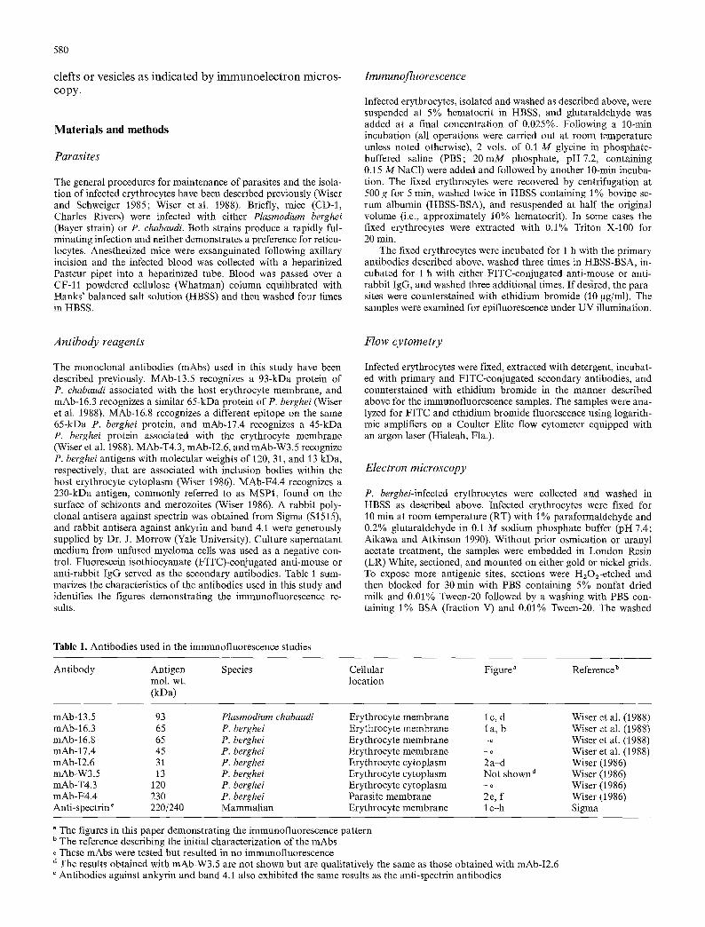
Fig. 1 a-h. Erythrocyte membrane immunofiuorescence. Plasmodium berghei-infected (a, b, el l) or P. chabaudi-infected (c, d) erythro- cytes were fixed with 0.025% glu- taraldehyde, incubated with either mAb-16.3 (a, b), mAb-13.5 (e, d), or anti-spectrin (e--h) and ana- lyzed by immunofluorescence as described in Materials and meth- ods (also see Table 1). Shown are the immunofluorescence as viewed under UV illumination (a, c, e) and the corresponding fields as viewed under direct light (b, d, f). Arrowheads indicate the eryth- rocytes that are fluorescing. In the case of staining with anti- spectrin, it was not always clear that all of the fluorescing cells were infected; therefore, the para- sites were counterstained with ethidium bromide (g, h) and the pictures were developed such that either the immunofluorescence (green in the original) was most apparent (g) or the ethidium bro- mide staining (orange in the orig- inal) was most apparent (h). Bar = 10 gm
grids were incubated for 2 h with either mAb-I2.6 or mAb-W3.5 (undiluted conditioned media from hybridoma cultures), thorough- ly washed, incubated for 1 h with 5-nm protein A-colloidal gold (Janssen Biotech), and washed again. Supernatant media from un- fused myelomas were used as negative controls.
To confirm the results obtained with protein A-gold, immuno- staining was repeated using ferritin-conjugated goat anti-mouse IgG (Cappel Laboratories) diluted 1 : 10 (v/v) with PBS as the sec- ondary antibody. Gold and ferritin markers were stabilized by fixa- tion with 2.5% glutaraldehyde in wash buffer. Thin sections on copper grids were stained with uranyl acetate and lead citrate and examined with a JEOL JEM-1200EX II electron microscope.
The gold particles distributed over the different subcellular frac- tions were quantitated by counting the numbers of gold particles found over uninfected erythrocytes, the cytoplasm of infected erythrocytes, the parasite, and the area between erythrocytes (i.e., plastic) and dividing these counts by the total square inches making up these four fractions. The area of the fractions was determined
by cutting out these subcetlular fractions and weighing the photo- graphs as compared with a known area of developed photographic paper.
Results
M A b - 16.3 and mAb-13.5 specifically recognize P. bergh- ei and P. chabaudi acidic phosphopro te ins associated with the erythrocyte m e m b r a n e (Wiser et al. 1988). Both of these mAbs recognize the erythrocyte surface of in- fected cells fixed with 0.025% glu tara ldehyde as deter- mined by indirect immunof luorescence (Fig. l a-d) . MAb-16.8 and mAb-17.4 did no t react in this assay (Ta- ble 1). This f luorescence is no t observed if the infected erythrocytes are unfixed or fixed with less t han 0.025%

582
Fig. 2a-f. Immunofluorescence following detergent extraction. P. bergheMnfected erythrocytes were fixed with 0.025% glutaralde- hyde and analyzed for indirect immunofluorescence using mAb- I2.6 (a, b). Alternatively, the infected erythrocytes were extracted with 0.1% Triton X-100 following glutaraldehyde fixation and ana- lyzed for immunofluorescence with mAb-I2.6 (e, d) or mAb-F4.4 (e, t). Immunofluorescent images as viewed under UV illumination are shown in the left-hand panels and the corresponding images as viewed under direct light are shown on the right. Bar = 10 gm
glutaraldehyde. At increasing glutaraldehyde concentra- tions, detection of FITC fluorescence is greatly dimin- ished due to the increasing levels of glutaraldehyde-in- duced autofluorescence. At concentrations of glutaralde- hyde exceeding 0.25%, no specific FITC fluorescence is detectable above the autofluorescence (data not shown). Paraformaldehyde was also tried as a fixative, but this resulted in lysis of the erythrocytes (data not shown).
The acidic phosphoproteins are localized to the cyto- plasmic face of the erythrocyte membrane (Wiser et al. 1988), indicating that the mild glutaraldehyde fixation is making the internal face of the erythrocyte membrane accessible to antibody. To confirm this, antibodies against proteins that are definitely localized exclusively to the cytoplasmic face of the erythrocyte membrane were also tested in this liquid-phase immunofluorescence assay. Antibodies against spectrin (Fig. 1 e-h), ankyrin and band 4.1 (data not shown) all result in a strong membrane-associated fluorescence of infected erythro- cytes, confirming that the fixation method is exposing the internal face of the erythrocyte membrane. Interest- ingly, the fluorescence due to anti-spectrin (and anti-
ankyrin and anti-band 4.1) antibodies is almost exclu- sively associated with infected erythrocytes, suggesting that the accessibility to the cytoplasmic face is a result of both the fixation procedure and parasite infection.
However, as confirmed by- counterstaining with ethi- dium bromide, a few uninfected erythrocytes do exhibit a membrane fluorescence with anti-spectrin antibodies. In the data presented (Fig. 1 g, h), two of three fluoresc- ing erythrocytes exhibit both a parasite-associated ethi- dium bromide fluorescence and an erythrocyte mem- brane-associated FITC fluorescence. The third erythro- cyte exhibits only an FITC fluorescence. The number of uninfected erythrocytes that fluoresced varied from experiment to experiment, but generally, 1%-2% of the uninfected erythrocytes fluoresce and the fluorescence associated with uninfected erythrocytes is generally less intense than that of infected erythrocytes as demon- strated in Fig. 1 g. All of the infected erythrocytes react with anti-spectrin, and no apparent difference in the in- tensity of labeling is observed between ring-, tropho- zoite-, or schizont-infected erythrocytes.
MAb-I2.6, recognizing a 31-kDa P. berghei antigen associated with undefined structures within the cyto- plasm of the infected erythrocyte (Wiser 1986), was also tested in this liquid-phase immunofluorescence assay. A barely detectable fluorescence is observed with mAb-I2.6 (Fig. 2a, b). Attempts to enhance the permeability of the fixed erythrocytes to antibody were carried out by extracting them in detergent. Triton X-100, saponin, and sodium docecyl sulfate (SDS) were all tested at concen- trations ranging from 0.01% to 1.0%. The reactivity of mAb-I2.6 was greatly enhanced by extracting the fixed erythrocytes with all of the detergents, but 0.1% Triton X-100 was optimal (data not shown). Brightly fluoresc- ing aggregates within the cytoplasm of the infected erythrocyte are readily detected after permeabilization of the samples with detergent (Fig. 2c, d). This pattern of fluorescence is similar to that previously reported for acetone-fixed thin blood smears (Wiser 1986). MAb- W3.5, recognizing a 13-kDa antigen, gave results identi- cal to those obtained with mAb-I2.6 (data not shown). MAb-T4.3 did not react in the immunofluorescence as- say (Table 1).
Similarly mAb-F4.4, recognizing MSP1 on the sur- face of schizonts, is reactive only with the glutaralde- hyde-fixed erythrocytes after Triton X-100 permeabiliza- tion (Fig. 2e, f). The pattern of immunofluorescence is typical for this antigen (Holder 1988), with the surface of the budding merozoites being clearly visible. An im- mature parasite not expressing MSP1 is also apparent in the same field. Extraction with Triton X-100 also en- hances the reactivity of mAb-16.3, but the increase in fluorescence is not nearly as great as in the cases of mAb-I2.6 and mAb-F4.4 (data not shown). Detergent extraction resulted in all erythrocytes fluorescing when anti-spectrin was used (data not shown).
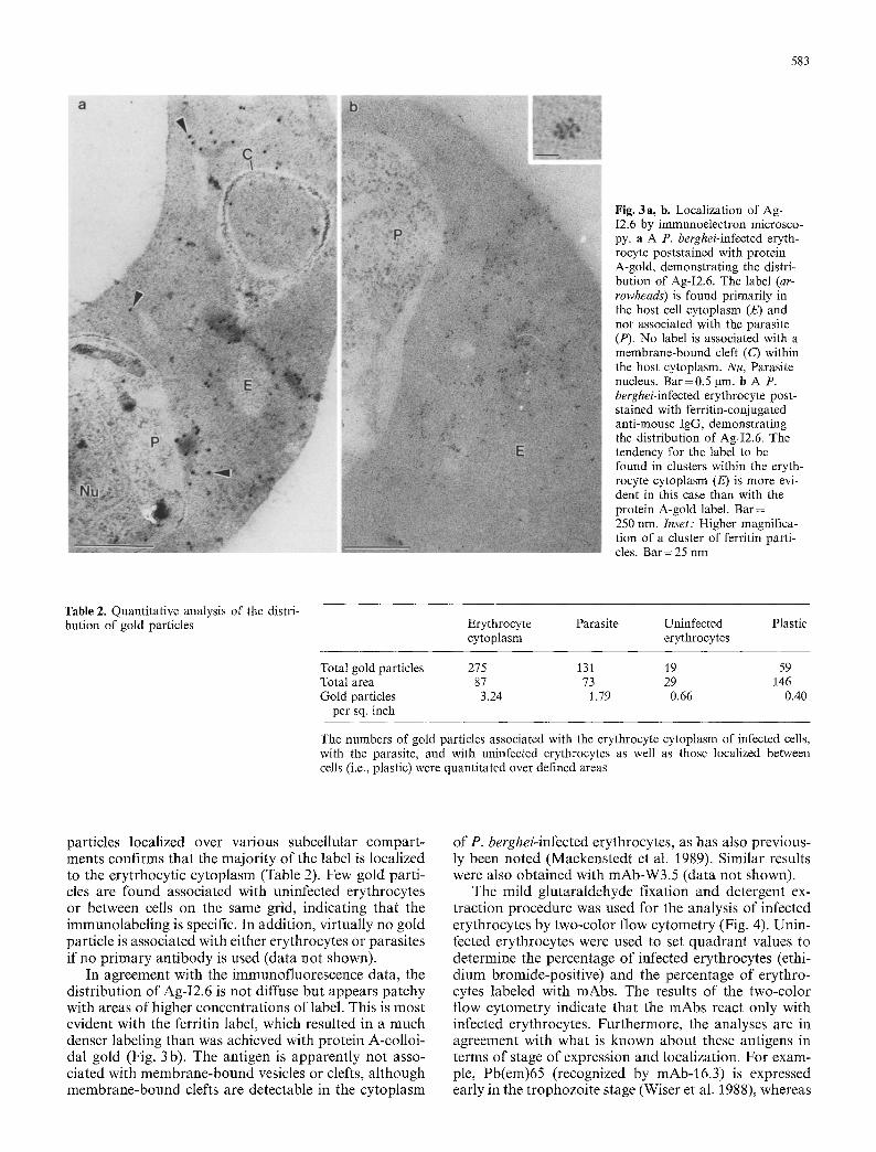
The nature of the aggregates, or inclusion bodies, recognized by mAb-I2.6 and mAb-W3.5 was further in- vestigated by immunoelectron microscopy. The antigen recognized by mAb-I2.6 is localized to the erythrocyte cytoplasm (Fig. 3). Quantitating the numbers of gold
583
Fig. 3a, b. Localization of Ag- I2.6 by immunoelectron microsco- py. a A P. berghei-infected eryth- rocyte poststained with protein A-gold, demonstrating the distri- bution of Ag-I2.6. The label (ar- rowheads) is found primarily in the host cell cytoplasm (E) and not associated with the parasite (P). No label is associated with a membrane-bound cleft (C) within the host cytoplasm. Nu, Parasite nucleus. Bar=0.5 gm. b A P. berghei-infected erythrocyte post- stained with ferritin-conjugated anti-mouse IgG, demonstrating the distribution of Ag-I2.6. The tendency for the label to be found in clusters within the eryth- rocyte cytoplasm (E) is more evi- dent in this case than with the protein A-gold label. Bar = 250 nm. Inset: Higher magnifica- tion of a cluster of ferritin parti- cles. Bar=25 nm
Table 2. Quantitative analysis of the distri- bution of gold particles Erythrocyte Parasite Uninfected Plastic
cytoplasm erythrocytes
Total gold particles 275 131 19 59 Total area 87 73 29 146 Gold particles 3.24 1.79 0.66 0.40
per sq. inch
The numbers of gold particles associated with the erythrocyte cytoplasm of infected cells, with the parasite, and with uninfected erythrocytes as well as those localized between cells (i.e., plastic) were quantitated over defined areas
particles localized over various subcellular compar t - ments confirms that the majori ty of the label is localized to the erytrhocytic cytoplasm (Table 2). Few gold parti- cles are found associated with uninfected erythrocytes or between cells on the same grid, indicating that the immunolabeling is specific. In addition, virtually no gold particle is associated with either erythrocytes or parasites if no pr imary ant ibody is used (data not shown).
In agreement with the immunofluorescence data, the distribution of Ag-I2.6 is not diffuse but appears patchy with areas of higher concentrations of label. This is most evident with the ferritin label, which resulted in a much denser labeling than was achieved with protein A-colloi- dal gold (Fig. 3 b). The antigen is apparent ly not asso- ciated with membrane-bound vesicles or clefts, al though membrane-bound clefts are detectable in the cytoplasm
of P. berghei-infected erythrocytes, as has also previous- ly been noted (Mackenstedt et al. 1989). Similar results were also obtained with mAb-W3.5 (data not shown).
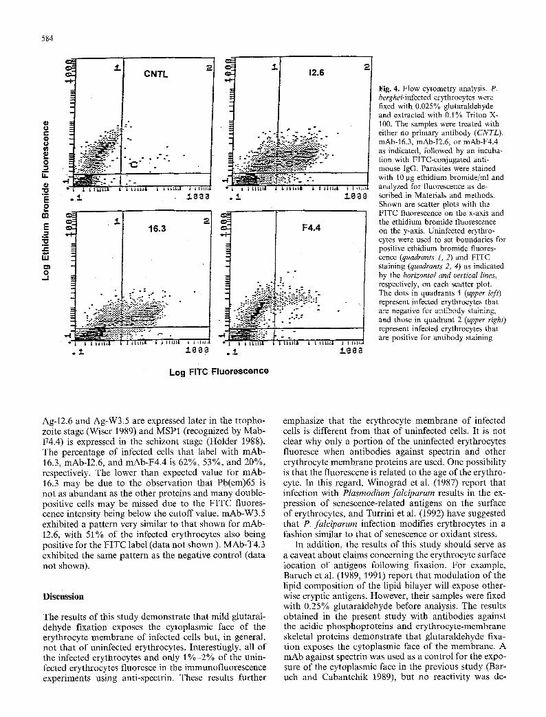
The mild glutaraldehyde fixation and detergent ex- traction procedure was used for the analysis of infected erythrocytes by two-color flow cytometry (Fig. 4). Unin- fected erythrocytes were used to set quadrant values to determine the percentage of infected erythrocytes (ethi- dium bromide-positive) and the percentage of erythro- cytes labeled with mAbs. The results of the two-color flow cytometry indicate that the mAbs react only with infected erythrocytes. Furthermore, the analyses are in agreement with what is known about these antigens in terms of stage of expression and localization. For exam- ple, Pb(em)65 (recognized by mAb-16.3) is expressed early in the trophozoite stage (Wiser et al. 1988), whereas
584
r e-
.= o
m u.
E o
m
E .2
ul
o _1
e s 3.
CNTL
~ 1 7 6
- : : N ; - : ..
- : . . . . : ~ . - ~ : _- : - - . -
3 . 2 16.3
- . :
; - : . �9 "-:.: &:-.: :;-,_: - . ":::'--.":-:; :'::"--':--'::-:~Z2~',:
. ~ : .%.::-~-.-..':-~ -...-.:~.~:~-~..,.~-.~.--:
1 12.6 2
.:::..:,;:::_ .: -
" . ' .
o: : . "-;.:"
1 I L t l L | ~
F4.4
"! LtL~|A " L i L I l l l l i t t ~
Fig. 4. Flow cytometry analysis. P. berghei-infected erythrocytes were fixed with 0.025% glutaraldehyde and extracted with 0.1% Triton X- 100. The samples were treated with either no primary antibody (CNTL). mAb-16.3, mAb-I2.6, or mAb-F4.4 as indicated, followed by an incuba- tion with FITC-conjugated anti- mouse IgG. Parasites were stained with 10 lag ethidium bromide/ml and analyzed for fluorescence as de- scribed in Materials and methods. Shown are scatter plots with the FITC fluorescence on the x-axis and the ethidium bromide fluorescence on the y-axis. Uninfected erythro- cytes were used to set boundaries for positive ethidium bromide fluores- cence (quadrants 1, 2) and FITC staining (quadrants 2, 4) as indicated by the horizontal and vertical lines, respectively, on each scatter plot. The dots in quadrants 1 (upper left) represent infected erythrocytes that are negative for antibody staining, and those in quadrant 2 (upper right) represent infected erythrocytes that are positive for antibody staining
Log FITC Fluorescence
Ag-I2.6 and Ag-W3.5 are expressed later in the tropho- zoite stage (Wiser 1989) and MSP1 (recognized by Mab- F4.4) is expressed in the schizont stage (Holder 1988). The percentage of infected cells that label with mAb- 16.3, mAb-I2.6, and mAb-F4.4 is 62%, 53%, and 20%, respectively. The lower than expected value for mAb- 16.3 may be due to the observation that Pb(em)65 is not as abundant as the other proteins and many double- positive cells may be missed due to the F ITC fluores- cence intensity being below the cutoff value, mAb-W3.5 exhibited a pattern very similar to that shown for mAb- I2.6, with 51% of the infected erythrocytes also being positive for the FITC label (data not shown ). MAb-T4.3 exhibited the same pattern as the negative control (data not shown).
D i s c u s s i o n
The results of this study demonstrate that mild glutaral- dehyde fixation exposes the cytoplasmic face of the erythrocyte membrane of infected cells but, in general, not that of uninfected erythrocytes. Interestingly, all of the infected erythrocytes and only 1 % - 2 % of the unin- fected erythrocytes fluoresce in the immunofluorescence experiments using anti-spectrin. These results further
emphasize that the erythrocyte membrane of infected cells is different from that of uninfected cells. It is not clear why only a port ion of the uninfected erythrocytes fluoresce when antibodies against spectrin and other erythrocyte membrane proteins are used. One possibility is that the fluorescene is related to the age of the erythro- cyte. In this regard, Winograd et al. (1987) report that infection with Plasmodiurn falciparum results in the ex- pression of senescence-related antigens on the surface of erythrocytes, and Turrini et al. (1992) have suggested that P. falciparum infection modifies erythrocytes in a fashion similar to that of senescence or oxidant stress.
In addition, the results of this study should serve as a caveat about claims concerning the erythrocyte surface location of antigens following fixation. For example, Baruch et al. (1989, 1991) report that modulation of the lipid composition of the lipid bilayer will expose other- wise cryptic antigens. However, their samples were fixed with 0.25% glutaraldehyde before analysis. The results obtained in the present study with antibodies against the acidic phosphoproteins and erythrocyte-membrane skeletal proteins demonstrate that glutaraldehyde fixa- tion exposes the cytoplasmic face of the membrane. A mAb against spectrin was used as a control for the expo- sure of the cytoplasmic face in the previous study (Bar- uch and Cabantchik 1989), but no reactivity was de-
585
tected. One possible explanation for this lack of reactivi- ty is that the epitope recognized by this particular anti- spectrin mAb is sensitive to glutaraldehyde fixation. Al- ternatively, the differences between the study reported herein and the previous investigation (Baruch and Ca- bantchik 1989) could be explained by mouse erythro- cytes behaving differently from human erythrocytes dur- ing the fixation procedure. Regardless of the explana- tion, extreme care should be taken in the interpretation of data describing parasite antigens on the erythrocyte surface.
Problems with other methods for the identification of surface antigens such as iodination with lactoperoxi- dase have also been reported. For example, despite its cytoplasmic disposition, Pf155/RESA (Berzins 1991) has been heavily labelled with 1251 by the lactoperoxidase method (Saul et al. 1988). Infected erythrocytes tend to be more fragile and the amount of radioisotope incorpo- rated into hemoglobin and spectrin needs to be carefully monitored (Howard et al. 1982). In addition, the concen- tration of erythrocytes, iodine, and other components can also result in labeling of intraerythrocytic proteins (Howard et al. 1979). In regard to this report, the 93- kDa P. chabaudi protein is not iodinated with lactoper- oxidase (Wunderlich etal. 1988). Treatment of intact erythrocytes with proteases is another method to identify surface proteins. However, Vernot-Hernandez and Heid- rich (1985) have observed that treatment of intact in- fected erythrocytes results in some loss of spectrin along with a 92-kDa P. falciparum protein associated with knob formation. In summary, nmltiple methods and ad- equate controls are necessary before malarial antigens can be unambiguously localized to the surface of the infected erythrocyte.
The fixation/permeabilization protocol described her- ein is useful for the analysis and characterization of ma- larial antigens by immunofluorescence and flow cyto- metry. The use of 0.025% glutaraldehyde eliminates the autofluorescence that interferes with immunofluores- cence and flow cytometry analyses. In addition, the abili- ty to carry out the immunofluorescence in the fluid phase as opposed to thin smears fixed on microsocpe slides has some advantages. For example, in the cases of Ag- I2.6 and Ag-W3.5, the ability to view the immunofluor- escence patterns in three dimensions confirmed the ear- lier conclusions that these antigens were associated with inclusions within the erythrocyte cytoplasm (Wiser 1986). One cannot unambiguously conclude that these antigens are not associated with the erythrocyte mem- brane solely on the basis of the immunofluorescence pat- terns previously reported for acetone-fixed thin smears (Wiser 1986). Similar patterns of immunofluorescence have in the past been attributed to malarial antigens associated with the host erythrocyte membrane (Koenen et al. 1984), only to be shown later to be associated with cytoplasmic granules adjacent to the parasitophorous vacuolar membrane (Scherf et al. 1992). Similarly, the immunofluorescence obtained with mAb-F4.4 in this fluid-phase assay gives a clearer picture of the three- dimensional morphology of the segmenting schizont than does the use of acetone-fixed thin smears (Wiser 1986).
The nature of the structures that are revealed by mAbs-I2.6 and -W3.5 is not completely understood at this time. Immunofluorescence indicates that the anti- gens are associated with aggregates within the cytoplasm of the host erythrocyte, and electron microscopy indi- cates that they are not associated with membrane-bound vesicles or clefts. Ag-I2.6 and Ag-W3.5 exhibit a strong surface hydrophobicity as evidenced by the inability to elute them from hydrophobic columns (Wiser, unpub- lished results). One speculation is that the surface hydro- phobicity of these proteins promotes an aggregation and assembly into these undefined structures within the host cytoplasm. The acidic phosphoproteins have recently been proposed to be secreted from the parasite as soluble proteins (Wiser and Lanners 1992). It is possible that Ag-I2.6 and Ag-W3.5 are also secreted from the parasite as soluble proteins and then assemble into the non-mem- brane-bound aggregates.
PfHRPII has also been described to be localized to non-membrane-bound aggregates within the host cyto- plasm (Howard et al. 1986). The function of PfHRPII is unknown at this time, but the protein is secreted from the infected erythrocyte, The aggregate structures to which PfHRPII localizes have been proposed to be in- volved in the extraparasite transport of this protein through the host cytoplasm. Ag-I2.6 and Ag-W.35 are also released from the infected erythrocyte into the cul- ture medium (Wiser 1989) and may also be transported through the host cytoplasm by these aggregate struc- tures. These non-membrane-bound structures probably represent a different compartment and, possibly, a dif- ferent extraparasite transport mechanism than do the vesicles and membrane-bound clefts (Stenzel and Kara 1989; Barnwell 1990). In agreement with this, Gormley et al. (1992) have recently demonstrated that several P. falciparum antigens are associated with protein aggre- gates within the host cytoplasm but are definitely not associated with lipids.
In addition to providing a more qualitative analysis of malarial antigens in terms of immunofluorescene, the fixation/permeabilization protocol can also be used in quantitative analyses such as two-color flow cytometry. These studies extend those of Pattanapanyasat et al. (1992), in which antibodies against decay-accelerating factor were used to show that two-color analysis of Plas- modium-infected erythrocytes is possible. In addition, permeabilization of the infected erythrocytes with low concentrations of detergent allows for the detection and characterization of other malarial antigens besides those localized to the erythrocyte membrane.
Acknowledgements. These experiments were supported in part by Biomedical Research Support Grant Funds from Tulane University and from the Louisiana Board of Regents through the Louisiana Education Quality Support Fund, contract number LEQSF(1990- 91)-RD-A-23 (to M.F.W.). The electron microscopy segment of this work was supported by NIH grants RR 00164~ Division of Research Resources, and R22 AI26189, NIAID (to H.N.L.). The skillful technical help of Maryjane Dodd with the electron micros- copy and of Ed Benes with the flow cytometry is also gratefully acknowledged.

586
References
Aikawa M, Atkinson CT (1990) Immunoelectron microscopy of parasites. Adv Parasitol 29:151-214
Atkinson CT, Aikawa M (1990) Ultrastructure of malaria-infected erythrocytes. Blood Cells 16:351-368
Barnwell JW (1990) Vesicle-mediated transport of membrane and proteins in malaria-infected erythroeytes. Blood Cells 16:379- 395
Baruch D, Cabantchik ZI (1989) Passive modulation of antigenic expression in the surface of normal and malaria-infected eryth- rocytes. Mol Biochem Parasitol 36:127-138
Baruch D, Glickstein H, Cabantchik ZI (1991) Plasmodiumfalci- parurn: modulation of surface antigenic expression of infected erythrocytes as revealed by cell fluorescence ELISA. Exp Par- asitol 73 : 440-450
Berzins K (1991) Pf155/RESA is not a surface antigen of Plasmo- diumfalciparum-infected erythrocytes. Parasitol Today 7:193- 194
Biggs BA, Anders RF, Dillon HE, Davern KM, Martin M, Peter- sen C, Brown GV (1992) Adherence of infected erythrocytes to venular endothelium selects for antigenic variants of Plasmo- diumfalciparum. J Immunol 149:2047-2054
Coppel RL, Cowman AF, Anders RF, Bianeo AE, Saint RB, Lingelbach KR, Kemp DJ, Brown GV (1984) Immune sera recognize on erythrocytes a Plasmodium falciparum antigen composed of repeated amino acid sequences. Nature 310:789- 792
Ginsberg H (1990) Alterations caused by the intraerythrocytic ma- laria parasite in the permeability of its host cell membrane. Comp Biochem Physiol [A] 95:31 39
Gormley JA, Howard R J, Taraschi TF (1992) Trafficking of malar- ial proteins to the host cell cytoplasm and erythroeyte surface membrane involves multiple pathways. J Cell Biol 119 : 1481 1495
Holder AA (1988) The precursor to major merozoite surface anti- gens: structure and role in immunity. Prog Allergy 41 : 72-97
Howard RJ, Smith PM, Mitchell GF (1979) Optimal conditions for lactoperoxidase catalyzed radioiodination of external pro- teins on mouse erythrocytes. Aust J Exp Biol Med Sci 57:355- 368
Howard RJ, Barnwell JW, Kao V, Daniel WA, Aley SB (1982) Radioiodination of new protein antigens on the surface of Plas- modium knowlesi schizont-infected erythrocytes. Mol Biochem Parasitol 6: 343-367
Howard RJ, Uni S, Aikawa A, Aley SB, Leech JH, Lew AM, Wellems TE, Rener J, Taylor DW (1986) Secretion of a malarial histidine-rich protein (PfHRPII) from Plasmodium falciparum- infected erythrocytes. J Cell Biol 103 : 1269-1277
Howard RJ, Barnwell JW, Rock EP, Neequaye J, Ofori-Adjei D, Maloy WL, Lyon JA, Saul A (1988) Two approximately 300 kilodalton Plasmodiumfalciparum proteins at the surface mem- brane of infected erythrocytes. Mol Bioehem Parasitol 27:207- 224
Koenen M, Scherf A, Mercereau O, Langsley G, Sibilli L, Dubois P, Silva LP da, Miiller-Hill B (1984) Human antisera detect a Plasmodium falciparum genomic clone encoding a nonapep- tide repeat. Nature 311:382-385
Leech JH, Barnwell JW, Miller LH, Howard RJ (1984) Identifica- tion of a strain-specific malarial antigen exposed on the surface of Plasmodium faleiparum-infected erythrocytes. J Exp Med 159:15621575
Mackenstedt U, Brockelman CR, Mehlhorn H, Raether W (1989) Comparative morphology of human and animal malaria para- sites. 1. Host-parasite interface. Parasitol Res 75:528-535
Miller LH, Howard RJ, Carter R, Good M, Nussenzweig RS, Nussenzweig V (1986) Research towards malarial vaccines. Sci- ence 284:1349-1356
Pattanapanyasat K, Webster HK, Udomsangpetch R, Wanachiw- anawin W, Yongvanitehit K (1992) Flow cytometric two-color staining technique for simultaneous determination of human erythrocyte membrane antigen and intracellular DNA. Cyto- metry 13:182-187
Pattanapanyasat K, Udomsangpetch R, Webster HK (1993) Two- color flow cytometric analysis of intraerythrocytic malaria par- asite DNA and surface membrane-associated antigen in eryth- rocytes infected with Plasmodium falciparum. Cytometry 14: 449M54
Saul A, Maloy WL, Rock EP, Howard RJ (1988) A portion of the Pf155/RESA antigen of Plasmodiumfaleiparum is accessible on the surface of infected erythrocytes. Immunol Cell Biol 66: 269-276
Scherf A, Carter R, Petersen C, Alano P, Nelson R, Aikawa M, Mattei D, Silva LP da, Leech J (1992) Gene activation of Pf11-1 of Plasmodium falciparurn by chromosome breakage and heal- ing: identification of a gametocyte-specific protein with a po- tential role in gametogenesis. EMBO J 11:2293-2301
Stenzel DJ, Kara UAK (1989) Sorting of malarial antigens into vesicular compartments within the host cell cytoplasm as dem- onstrated by immunoelectron microscopy. Eur J Cell Biol 49:311-318
Turrini T, Ginsburg H, Bussolino F, Pescarmona GP, Serra MV, Arese P (1992) Phagocytosis of Plasmodiumfalciparum-infected human red blood cells by human monocytes: involvement of immune and non-immune determinants and dependence on parasite developmental stage. Blood 80 : 801-808
Vernot-Hernandez JP, Heidrich HG (1985) The relationship to knobs of the 92,000 D protein specific for knobby strains of Plasmodiurnfaleiparum. Z Parasitenkd 71:41-51
Winograd E, Greenan JRT, Sherman IW (1987) Expression of senescent antigen on erythrocytes infected with a knobby vari- ant of the human malaria parasite Plasmodium faleiparum. Proc Natl Acad Sci USA 84:1931 1935
Wiser MF (1986) Characterization of monoclonal antibodies di- rected against erythrocytic stage antigens of Plasmodium bergh- ei. Eur J Cell Biol 42:45-51
Wiser MF (1989) Plasmodium antigens external to the parasite but within the infected erythrocyte. Parasitol Res 75 : 206211
Wiser MF, Lanners HN (1992) Rapid transport of the acidic phosphoproteins of Plasmodiurn berghei and P. chabaudi from the intraerythrocytic parasite to the host membrane using a miniaturized fractionation procedure. Parasitol Res 78:193-200
Wiser MF, Schweiger HG (1985) Cytosolic protein kinase activity associated with the maturation of the malaria parasite Plasmo- dium berghei. Mol Biochem Parasitol 17:179-189
Wiser MF, Leible MB, Plitt B (1988) Acidic phosphoproteins asso- ciated with the host erythrocyte membrane of erythrocytes in- fected with Plasmodium berghei and P. chabaudi. Mol Biochem Parasitol 27:11-22
Wunderlich F, Helwig M, Schillinger G, Speth V, Wiser ME (1988) Expression of the parasite protein Pc90 in plasma membranes of erythrocytes infected with Plasmodium chabaudi. Eur J Cell Biol 47:157-164
Note added in proof
Pattanapanyasat et al. (1993) have recently described two-color flow cytometry of Plasmodiumfalciparum-infected erythrocytes fol- lowing fixation with 0.025% glutaraldehyde and permeabilization with saponin.