Bio-functionalization of electro-synthesized polypyrrole surface by heme enzyme using a mixture of...
10
Applied Surface Science 257 (2011) 10926–10935 Contents lists available at ScienceDirect Applied Surface Science jou rn al h om epa g e: www.elsevier.com/locate/apsusc Bio-functionalization of electro-synthesized polypyrrole surface by heme enzyme using a mixture of Nafion and glutaraldehyde as synergetic immobilization matrix: Conformational characterization and electrocatalytic studies Mohammed ElKaoutit a,∗ , Ignacio Naranjo-Rodriguez a , Manuel Domínguez b , José Luis Hidalgo-Hidalgo-de-Cisneros a a Departamento de Química Analítica, Facultad de Ciencias, Universidad de Cádiz, 11510 Puerto Real, Cádiz, Spain b Departamento de Física de la Materia Condensada, Facultad de Ciencias, Universidad de Cádiz, 11510 Puerto Real, Cádiz, Spain a r t i c l e i n f o Article history: Received 2 May 2011 Received in revised form 26 July 2011 Accepted 1 August 2011 Available online 9 August 2011 Keywords: Polypyrrole–glutaraldehyde–Nafion Conformational changes Horseradish peroxidase Direct bioelectrocatalysis Bio-interface characterization a b s t r a c t Use of a mixture of Nafion and glutaraldehyde as new immobilization matrix was described. The per- centage of Nafion was optimized to prevent denaturation of horseradish peroxidase enzyme after its crosslinkage with glutaraldehyde on electro-synthesized polypyrrole surface. Topographic study by Atomic Force Microscopy (AFM) shows that the enzyme seems to have been introduced inside the ionic cluster of Nafion. The characterization of the resulting bio-interfaces by UV–vis and FT-IR shows that the intra-crosslinkage phenomena caused by the use of glutaraldehyde can be eliminated by the optimiza- tion of the concentration of Nafion additive. The secondary structure contents of native and immobilized enzyme were analyzed by a Gaussian curve fitting of the respective FT-IR spectra in the amide I region. Immobilized enzyme presented notable increasing percentages of globular and short helical structure compared with native enzyme. This indicates that immobilized enzyme was folded which is in accordance with AFM studies and supports the enzyme entrance inside ionic clutter of Nafion. Thanks to synergic effects of the polypyrrole conducting polymer and the perfluorosulfonic acid poly- mer Nafion, HRP enzyme was immobilized in its “native” state, the resulting biosensor was able to sense peroxide without any chemical mediator and can be categorized as third generation. © 2011 Elsevier B.V. All rights reserved. 1. Introduction In the last two decades, applications of biosensors based on redox enzymes have seen an almost exponential increase in phar- maceutical, biotechnology, food, and other diverse areas such as green chemistry and bio-energy. Furthermore, since the birth of the first biosensor thank to the revolutionary work of Clark and Lyons [1], several groups have dedicated their efforts to achieve biosensors able to face the actual analytical challenges. One of the interesting lines in these efforts is the modification of the signal transfer mode between redox enzymes and electrodes. Thus, to reduce the well-known limitation of the first-generation biosen- sors (based on sensing of products or substrate with slow redox activities, i.e. oxygen, peroxide . . .), other designs to sense the bio- process were proposed. Second generation biosensors based on artificial electron mediators instead of the natural co-substrates, and finally, the third generation biosensors, in which the signal is attributed to a direct electron transfer between the enzyme ∗ Corresponding author. Tel.: +34 956016361; fax: +34 956016460. E-mail address: [email protected] (M. ElKaoutit). cofactors and the electrode (in the absence of any artificial medi- ator), have been proposed [2]. One of the main requirements for the third generation biosensors is an especial immobilization matrix. Thus after immobilization step the redox enzymes must conserve their stability and activity, and the used biomaterials in this matrix must promote the direct electron transfer between the enzymes and the electrode as well as protect the bioprocess from the possible fouling and interferences. Conducting poly- mers, defined as intrinsically conducting polymer or electroactive conjugated polymers, doted by a -conjugation along the poly- mer backbone, have received extensive interest over the last two decades in biosensor field thanks to their ability to efficiently trans- fer electric charge produced by the redox enzymes to transducer components [3–5]. Bio-functionalization of these polymer/electrode interfaces can be achieved by conventional methods such as adsorption, cross-linking, covalent linkage, and physical or electrochemical entrapment. However, from a practical point of view, the use of additive-protective resins is primordial to prevent several prob- lems such as the effect of interferences, the deterioration of the conducting polymers and the adsorption of isolating fouling [6–12]. On the other hand the use of glutaraldehyde (Glut) and support 0169-4332/$ – see front matter © 2011 Elsevier B.V. All rights reserved. doi:10.1016/j.apsusc.2011.08.009
Transcript of Bio-functionalization of electro-synthesized polypyrrole surface by heme enzyme using a mixture of...

Bum
MJa
b
a
ARRAA
KPCHDB
1
rmgtLbitrsapaai
0d
Applied Surface Science 257 (2011) 10926– 10935
Contents lists available at ScienceDirect
Applied Surface Science
jou rn al h om epa g e: www.elsev ier .com/ locate /apsusc
io-functionalization of electro-synthesized polypyrrole surface by heme enzymesing a mixture of Nafion and glutaraldehyde as synergetic immobilizationatrix: Conformational characterization and electrocatalytic studies
ohammed ElKaoutit a,∗, Ignacio Naranjo-Rodrigueza, Manuel Domínguezb,osé Luis Hidalgo-Hidalgo-de-Cisnerosa
Departamento de Química Analítica, Facultad de Ciencias, Universidad de Cádiz, 11510 Puerto Real, Cádiz, SpainDepartamento de Física de la Materia Condensada, Facultad de Ciencias, Universidad de Cádiz, 11510 Puerto Real, Cádiz, Spain
r t i c l e i n f o
rticle history:eceived 2 May 2011eceived in revised form 26 July 2011ccepted 1 August 2011vailable online 9 August 2011
eywords:olypyrrole–glutaraldehyde–Nafiononformational changes
a b s t r a c t
Use of a mixture of Nafion and glutaraldehyde as new immobilization matrix was described. The per-centage of Nafion was optimized to prevent denaturation of horseradish peroxidase enzyme after itscrosslinkage with glutaraldehyde on electro-synthesized polypyrrole surface. Topographic study byAtomic Force Microscopy (AFM) shows that the enzyme seems to have been introduced inside the ioniccluster of Nafion. The characterization of the resulting bio-interfaces by UV–vis and FT-IR shows that theintra-crosslinkage phenomena caused by the use of glutaraldehyde can be eliminated by the optimiza-tion of the concentration of Nafion additive. The secondary structure contents of native and immobilizedenzyme were analyzed by a Gaussian curve fitting of the respective FT-IR spectra in the amide I region.
orseradish peroxidaseirect bioelectrocatalysisio-interface characterization
Immobilized enzyme presented notable increasing percentages of globular and short helical structurecompared with native enzyme. This indicates that immobilized enzyme was folded which is in accordancewith AFM studies and supports the enzyme entrance inside ionic clutter of Nafion.
Thanks to synergic effects of the polypyrrole conducting polymer and the perfluorosulfonic acid poly-mer Nafion, HRP enzyme was immobilized in its “native” state, the resulting biosensor was able to senseperoxide without any chemical mediator and can be categorized as third generation.
. Introduction
In the last two decades, applications of biosensors based onedox enzymes have seen an almost exponential increase in phar-aceutical, biotechnology, food, and other diverse areas such as
reen chemistry and bio-energy. Furthermore, since the birth ofhe first biosensor thank to the revolutionary work of Clark andyons [1], several groups have dedicated their efforts to achieveiosensors able to face the actual analytical challenges. One of the
nteresting lines in these efforts is the modification of the signalransfer mode between redox enzymes and electrodes. Thus, toeduce the well-known limitation of the first-generation biosen-ors (based on sensing of products or substrate with slow redoxctivities, i.e. oxygen, peroxide . . .), other designs to sense the bio-rocess were proposed. Second generation biosensors based on
rtificial electron mediators instead of the natural co-substrates,nd finally, the third generation biosensors, in which the signals attributed to a direct electron transfer between the enzyme∗ Corresponding author. Tel.: +34 956016361; fax: +34 956016460.E-mail address: [email protected] (M. ElKaoutit).
169-4332/$ – see front matter © 2011 Elsevier B.V. All rights reserved.oi:10.1016/j.apsusc.2011.08.009
© 2011 Elsevier B.V. All rights reserved.
cofactors and the electrode (in the absence of any artificial medi-ator), have been proposed [2]. One of the main requirementsfor the third generation biosensors is an especial immobilizationmatrix. Thus after immobilization step the redox enzymes mustconserve their stability and activity, and the used biomaterials inthis matrix must promote the direct electron transfer betweenthe enzymes and the electrode as well as protect the bioprocessfrom the possible fouling and interferences. Conducting poly-mers, defined as intrinsically conducting polymer or electroactiveconjugated polymers, doted by a �-conjugation along the poly-mer backbone, have received extensive interest over the last twodecades in biosensor field thanks to their ability to efficiently trans-fer electric charge produced by the redox enzymes to transducercomponents [3–5].
Bio-functionalization of these polymer/electrode interfacescan be achieved by conventional methods such as adsorption,cross-linking, covalent linkage, and physical or electrochemicalentrapment. However, from a practical point of view, the use of
additive-protective resins is primordial to prevent several prob-lems such as the effect of interferences, the deterioration of theconducting polymers and the adsorption of isolating fouling [6–12].On the other hand the use of glutaraldehyde (Glut) and support
M. ElKaoutit et al. / Applied Surface Science 257 (2011) 10926– 10935 10927
afion
cotwftaotqtsehtph
m[PtHaee
aboveskmseian
Scheme 1. Chemical structure of N
ontaining amine groups, like polypyrrole (PPy) (Scheme 1), is onef the most frequently used techniques for enzyme immobiliza-ion. Thus, because glutaraldehyde is a small molecule (Scheme 1)ith high reactivity acting as cross-linker between these sur-
ace’s amine groups and amine residue of proteins. Neverthelesshis method entails inevitable inter-crosslinking between somemine groups of proteins and consequently causes a denaturationf enzymes. To prevent this, several efforts were carried out, andhe use of other inactive proteins such as serum albumin was fre-uently reported. Our group has presented an approach based onhe combination of glutaraldehyde and Nafion resin to assembleecond and third generation biosensors [13–17]. Nafion is a cationxchange polymer containing three different phases (Scheme 1):ydrophilic cluster of sulfonic acid groups with diameters from 30o 50 nm, hydrophobic semi crystalline region which is Teflon-likeolymer backbone and interfacial domain which is intermediate inydrophobicity.
The advantages of using the additive-protective Nafion agentay lie in the electrostatic rejection of the interferences and fouling
17,18], in increasing the stability and mechanical strength of thePy film [19,20], and in promoting the direct electron transfer fromhe enzyme to the PPy film and also to the electrode [17,21–23].owever, in spite of the usual use of this material to immobilizend protect biomolecules, few researchers have dedicated theirfforts to elucidate the highest biocompatibility of this resin withntrapped biomolecules.
In this context, we present here for the first time, as wells we are informed, an investigation of new third generationiosensor based on horseradish peroxidase (HRP) immobilizedn electrochemically synthesized polypyrrole conducting filmia crosslinking by a mixture of glutaraldehyde and Nafion ionxchange resin. In this work we are trying essentially to investigateeveral new aspects. So, bordering the operational and electro-inetic studies of the resulted biosensor, special attempts have beenade on the investigation of the change of the enzyme secondary
tructure in its new environment, on the study of the protective
ffect of Nafion over possible denaturation of the enzyme, detect-ng a possible conformational changes in the heme catalytic site,nd on the characterization of the new biosensor surface at theano-scale., polypyrrole and glutaraldehyde.
2. Experimental
2.1. Apparatus and reagents
Electrochemical measurements were performed with anAutolab PGSTAT20 (Ecochemie, Ultrecht, The Netherlands) poten-tiostat/galvanostat interfaced with a personal computer, usingthe AutoLab software GPES for waveform generation and dataacquisition and elaboration. A single-compartment three-electrodecell, with a platinum auxiliary electrode and an Ag/AgCl, 3 MKCl reference electrode was employed. Surface topological studieswere performed using an Atomic Force Microscope (AFM, VeecoNanoscope IIIa) in tapping mode. Phosphorus (n) doped siliconcantilevers, with spring constants in the range 20–80 N m−1, wereused. Calibration of the microscope was achieved by imaging cal-ibration gratings supplied by the manufacturer. AFM images wereexamined for artifacts, and reproducibility was checked in theusual way, i.e. by changing the AFM cantilever and by eithermoving (during the experiment) the sample in the x or y direc-tions or by varying the scanning angle and frequency. HRP (E. C.1. 11. 1. 7, 269 U mg−1) was purchased from Sigma (Steinheim,Germany), KH2PO4, K2HPO4, acetic acid, sodium acetate, Tris andHCl for phosphate, acetate or Tris–HCl buffer were from Fluka(Buchs, Switzerland), Merck (Darmstad, Germany), and Sigma,respectively. Nafion-perfluorinated ion-exchange resin (Cat. No. 27,470-4) 5% (w/v) in a mixture of lower aliphatic alcohols and water,and glutaraldehyde, 25 wt.% solution in water were obtained fromAldrich (Steinheim, Germany); peroxide 30% H2O2, Perhydrol, wasfrom Merck. Pure water was obtained by passing twice-distilledwater through a Milli-Q system (18 M� cm, Millipore, Bedford,MA).
2.2. Preparation and functionalization of PPy film
Polymer films were electrochemically prepared by electrode-position from an aqueous 5 × 10⁻⁵ M solution of pyrrole monomer
in 0.1 M LiC1O4 as supporting electrolyte. The polymerization wascarried out using glassy carbon (GC) as the working electrode, pre-viously cleaned as described elsewhere [24]. Voltammetric modewas employed for film growth [25–27]. Typical black film of PPy can
1 face Sc
biqpt3Ftteoses
2
cctaAemasabtb
eAts
d(faS
SfmeSfs
3
3
3
tHtadtii
0928 M. ElKaoutit et al. / Applied Sur
e then observed on the electrode surface. The resulted conduct-ng polymeric film was bio-functionalized as follows: an adequateuantity of enzyme HRP was dissolved in 30 �L of 0.2 M, pH 7hosphate buffer solution. To this enzyme solution, 1.25 �L of glu-araldehyde was added, set to polymerize in ultrasonic bath for
min, and modified by adequate volume of 5% Nafion solution.rom the resulting solution, an adequate quantity was deposited onhe surface of the PPy-GC electrode with a �-syringe and allowedo dry under ambient conditions. The resultant biosensor has 150nzyme units, 0.9% of glutaraldehyde and different percentagesf Nafion. Before its use, the enzyme electrode was dipped intirred buffer solution for 15 min, to eliminate the excess of nonntrapped enzyme, rinsed with the same buffered solution andtored immersed in buffer at 4 ◦C when it was not in use.
.3. Measurements
Electrochemical experiments were carried out in an electro-hemical cell with 25 mL of an aerated buffer saline solutionontaining 0.05 M of KCl and 0.05 M of the adequate buffer, at roomemperature (22 ± 2 ◦C). The three-electrode system consisted ofn enzyme-modified PPy-GC electrode as working electrode, ang/AgCl (3 M KCl) and a platinum wire as reference and auxiliarylectrodes, respectively. For the chronoamperometric measure-ents, a selected potential was applied to the working electrode
nd the background current was registered until reaching theteady state. Calculated amounts of H2O2 were added to the cell,nd the corresponding current–time curves were recorded. Theiosensor response was measured as the difference between theotal and the background current. A magnetic stirrer and stirringar were used to provide continuous convective transport.
Tapping mode AFM measurements were performed over differ-nt regions of all samples, to check sample surface homogeneity.ll AFM images selected to be shown here are representative of
he samples surface topology. To compare the registered data, thecanned area is always 5 �m × 5 �m.
Solid phase UV–vis spectra were recorded, at normal inci-ence, using a double-beam UV/visible/NIR spectrophotometerPerkinElmer, Model Lambda-19). Layers of Nafion–HRP–Glut–PPyor Fig. 1A, and HRP with and without Glut or Nafion, were manu-lly deposited onto 1-mm-thick borosilicate glass substrates (BDHuper premium).
Fourier transform infrared (FT-IR) spectra were obtained on ahimadzu FT-IR 8400S with a resolution of 4 cm−1 and scanningrom 4000 to 400 cm−1. Powder of Nafion–HRP–Glut and HRP were
easured as KBr pellets at 1.5–2%. For the first sample, a solution ofnzyme, glutaraldehyde, and Nafion was prepared as described inection 2.2, deposited on glass substrate and removed after dryingor one night, to homogenize it in a mortar for pellet making. Theecond sample was prepared directly from commercial HRP.
. Results and discussion
.1. Study of conformational change in HRP after immobilization
.1.1. Absorbance measurementOptical measurements were made to determine the effect of
he immobilization step on the structure of the HRP enzyme.eme absorption provides a very useful conformational test for
he study of heme proteins. The spectrum of HRP entrapped into solid Nafion–Glut–PPy film, made by the immobilization proce-
ure described in Section 2.2, is shown in Fig. 1A. It can be seen thathe characteristic bands of the native HRP have been preserved ints new environment. Thus, the asymmetric Soret band at approx-mately 407 nm, a shoulder at approximately 380 nm in the lowerience 257 (2011) 10926– 10935
wavelength range, and the two weak absorption bands (charge-transfer) at approximately 505 and 640 nm, were observed, givinga relatively good agreement with the spectra for a native hemeenzyme registered in aqueous media [28–30]. In addition, the samespectrum shows other bands, characteristic of PPy film. The first isaround 474 and it is assigned to the �–�* absorption. The secondis the manifested increase in the background up to 690 nm, whichcan be considered as the beginning of the bipolaron charge trans-fer band normally observed at 900 nm. These two bands indicatethe high conductivity of PPy doped film, and give good agreementwith the reported works on the absorption activity of PPy films[8,11]. On the other hand, to investigate the effect of Nafion additionon the conformational changes of the enzyme after its crosslink-age, UV–vis spectroscopy was performed for several films, usingthe same quantity of enzyme and Glut but different Nafion con-centrations. The results are presented in Fig. 1B and C. SpectrumB-b, corresponding to the crosslinked enzyme without Nafion, ischaracterized by the presence of a peak with broad summit anda modest blue shift of the charge transfer in the visible regions(Fig. 1C-c). The change in the shape of the UV peaks can be indica-tive of an important denaturation of the heme site attributed to anintracrosslinkage of the enzyme. When 0.5% of Nafion was added,the corresponding spectrum (i.e. Fig. 1B-c) gets a shape similar tothe native enzyme shown in Fig. 1B-a, indicating the protectiveeffect of Nafion over enzyme denaturation. This can be possiblydue to the hybrid character (hydrophobic–hydrophilic) of this resinwhich can offer additional functional group to decrease glutaralde-hyde super reactivity and another biocompatible site (ionic cluster)to enzyme imbibing. However, when the concentrations of Nafionare up to the optimum value of 0.5%, the corresponding spectra (i.e.Fig. 1B-d and B-e) show the same shape of denatured enzyme as inspectrum (B-b). This can be attributed to an excess of methanolincluded in the increase of Nafion addition. Finally, it is importantto note that, comparing the absorbance intensity in the spectra ofFig. 1B-c and that of Fig. 1B-a, we observe an important decrease. Ithas been reported that a variation in absorbance intensity of hemegroup of same concentration can be attributed to two factors: avariation in anionic electrolyte nature or concentration [31,32] ora simple folding of proteins structure [33,34]. Nafion is a highlyanionic repulsive resin, from this fact we can imagine an eradica-tion of the anionic phosphate from the heme site of the enzymeand consequently a decrease in the absorbance. However an expla-nation based on enzyme folding can be also anticipated taking intoaccount a possible introduction of enzyme in ionic cluster of Nafionstructure and the high compatibility of this region compared to thatof fluorocarbon hydrophobic semi-crystalline region.
3.1.2. FT-IR measurementThe effect of the immobilization step was also studied by FT-IR
spectroscopy. This technique is a sensitive tool to probe the sec-ondary structure of proteins. Changes in the shape of the infraredadsorption in the amide I band region near 1640 cm−1 (primaryC O stretch), and amide II near 1550 cm−1 (N–H band, C–N stretch)can provide detailed information on the secondary structure of theimmobilized enzyme. As shown in Fig. 2A, spectrum of amide I andamide II of Nafion–HRP–Glut has a similar shape to that of nativeHRP. The similarities of the two spectra suggest that HRP retainsits native structure after crosslinkage with Glut in the presence ofadequate quantity of Nafion. However, the change in the stronglyintense bands showed in the region from 3000 to 3800 cm−1, char-acteristic of O–H stretching vibration, can be indicative on theinduction of hydrogen bonding, and also on the new environment
of the enzyme. Native HRP spectrum shows an intense band inthis region indicating important hydrogen bonding force, which isresponsible of the helical form of the enzyme molecule. The inten-sity of this band decreases in the spectrum corresponding to HRP
M. ElKaoutit et al. / Applied Surface Science 257 (2011) 10926– 10935 10929
300 400 500 600 7001.350
1.375
1.400
1.425
1.450
407
A
465 510 640
Abs
orba
nce
Wavelength (nm)
300 400 50 0 60 0 700 80 0
B
e/HRP-Glut -2%Naf ion
d/HRP-Glut -1%Naf ion
c/HR P-Glut-0.5%Nafion
b/HR P-Glut
a/HRP
Wavelenght (nm )
7006506005505004500.0
0.5C
e
d
c
b
a
F , and
B
csaarpb
ig. 1. Solid phase UV–vis spectra of Nafion–HRP–Glut–PPy (A), HRP (B-a and C-a)-b (C-b), B-c (C-c), B-d (C-d), and B-e (C-e), respectively.
rosslinked and modified by Nafion. This polymer includes in itstructure a hydrophobic organic fluorocarbon region (the bandsround 1237 and 1150 cm−1 are attributed to C–F symmetric and
symmetric stretching, respectively) and a hydrophilic ion-clusteregion (the band at 1056 cm−1 attributed to SO3− was not shown,ossibly because of its feeble adsorption and the existence of a largeand of amide III from 1430 to 1050 cm−1) [35–37]. The apparitionNafion–Glut (B-c-d-e and C-c-d-e). Nafion concentrations were 0, 0.5, 1, and 2% in
of the band around 1380 cm−1 attributed to SO3H and character-istic of hydrated Nafion can explain the intensity decreases in theregion of hydrogen bonding induction, and provides an initial indi-
cation of a possible entrance of the enzyme in the ionic clusterregion of Nafion. So, we can imagine a high interaction betweenSO3− group of Nafion and usual proton of hydrogen bond in theenzyme molecule, which is the consequence of the apparition of
10930 M. ElKaoutit et al. / Applied Surface Science 257 (2011) 10926– 10935
F owdeG
tasidIaWt
TS
ig. 2. (A) FT-IR spectra of the two samples: powder of commercial HRP (A-a), and paussian curve fitting of corresponding amide I region (R2 = 1 for the two fitting).
he SO3H group. To investigate these facts and also to find an ideabout where the enzyme can be introduced in the Nafion regions,econdary structure contents of enzyme in the two states (i.e.mmobilized by Nafion–Glut matrix and in its native state) wereetermined by Gaussian curve fitting of the spectra in the amide
region (Fig. 2B). This method, Gaussian curve fitting, is reason-bly accurate and depends greatly on the precision of the calculus.e can found a great difference between fits from the same spec-
ra if a few differences in precision parameters were observed. So
able 1econdary structure of HRP under different conditions.
Sample Gaussian curves-fitting
Band position (cm−1)
HRP powder in KBr 1681.6
1673.3
1667.7
1661.6
1653.3
1645.6
1638.3
1628.7
HRP–Glut–Nafion powder in KBr 1685.3
1667.3
1653.1
1640.6
1626.4
r of HRP crosslinked with glutaraldehyde and modified by 0.5% of Nafion (A-b). (B)
to avoid this drawback and acquire convincing data we selectedjust the fits with same ideal regression; equal to one. The num-ber of components and their peaks position were determined bysecond derivation according to the literature [34]. The resultedpeaks were assigned to the corresponding secondary structure as
described in previous works [34,38–42] and the major secondarystructures were listed in Table 1. Native HRP presents around 34%of it structure as helical with an important percentage of the longone of it, and 24% as planar or extended structure. In contrast, theArea (%) Assignment
22.6 �-Turn4.7 �-Turn5.3 �-Turn
11.5 �-Helix (stable and long)14.6 �-Helix (stable and short)
7.1 �-Helix8.1 Sheet
24.2 Sheet
18.4 �-Turn19.3 �-Turn15.4 �-Helix16.1 �-Sheet parallel22.7 �-Sheet
M. ElKaoutit et al. / Applied Surface Science 257 (2011) 10926– 10935 10931
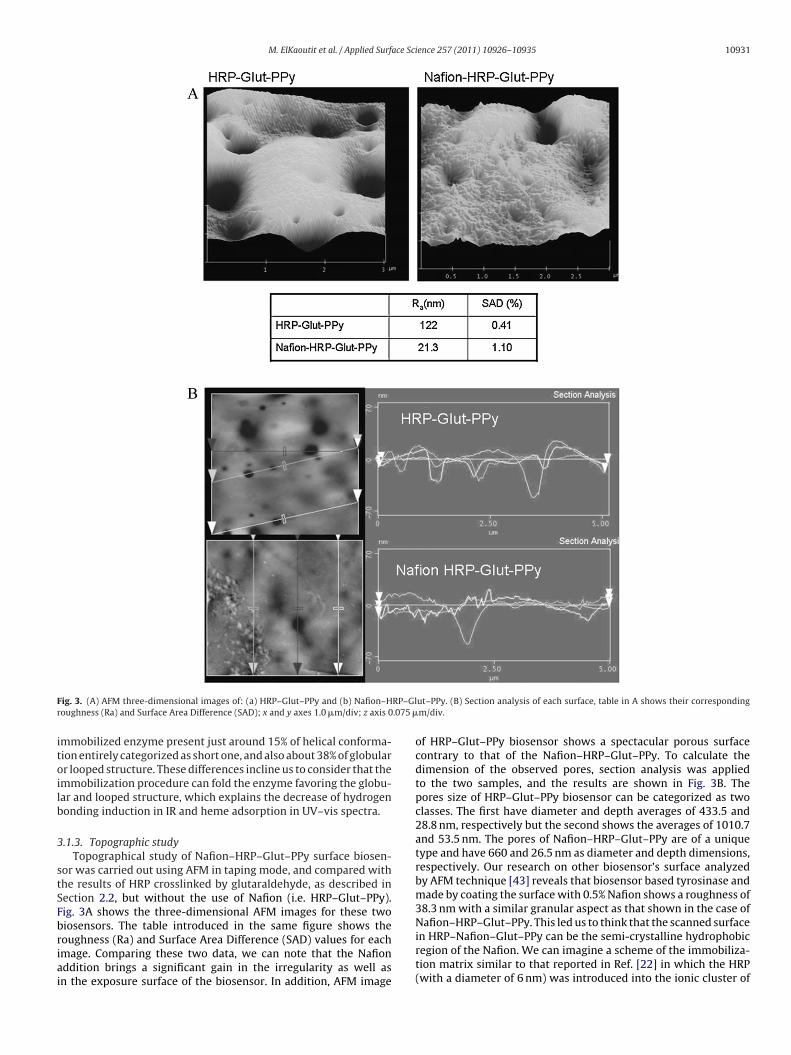
F RP–Glr .075 �
itoilb
3
stSFbriai
ig. 3. (A) AFM three-dimensional images of: (a) HRP–Glut–PPy and (b) Nafion–Houghness (Ra) and Surface Area Difference (SAD); x and y axes 1.0 �m/div; z axis 0
mmobilized enzyme present just around 15% of helical conforma-ion entirely categorized as short one, and also about 38% of globularr looped structure. These differences incline us to consider that themmobilization procedure can fold the enzyme favoring the globu-ar and looped structure, which explains the decrease of hydrogenonding induction in IR and heme adsorption in UV–vis spectra.
.1.3. Topographic studyTopographical study of Nafion–HRP–Glut–PPy surface biosen-
or was carried out using AFM in taping mode, and compared withhe results of HRP crosslinked by glutaraldehyde, as described inection 2.2, but without the use of Nafion (i.e. HRP–Glut–PPy).ig. 3A shows the three-dimensional AFM images for these twoiosensors. The table introduced in the same figure shows the
oughness (Ra) and Surface Area Difference (SAD) values for eachmage. Comparing these two data, we can note that the Nafionddition brings a significant gain in the irregularity as well asn the exposure surface of the biosensor. In addition, AFM imageut–PPy. (B) Section analysis of each surface, table in A shows their correspondingm/div.
of HRP–Glut–PPy biosensor shows a spectacular porous surfacecontrary to that of the Nafion–HRP–Glut–PPy. To calculate thedimension of the observed pores, section analysis was appliedto the two samples, and the results are shown in Fig. 3B. Thepores size of HRP–Glut–PPy biosensor can be categorized as twoclasses. The first have diameter and depth averages of 433.5 and28.8 nm, respectively but the second shows the averages of 1010.7and 53.5 nm. The pores of Nafion–HRP–Glut–PPy are of a uniquetype and have 660 and 26.5 nm as diameter and depth dimensions,respectively. Our research on other biosensor’s surface analyzedby AFM technique [43] reveals that biosensor based tyrosinase andmade by coating the surface with 0.5% Nafion shows a roughness of38.3 nm with a similar granular aspect as that shown in the case ofNafion–HRP–Glut–PPy. This led us to think that the scanned surface
in HRP–Nafion–Glut–PPy can be the semi-crystalline hydrophobicregion of the Nafion. We can imagine a scheme of the immobiliza-tion matrix similar to that reported in Ref. [22] in which the HRP(with a diameter of 6 nm) was introduced into the ionic cluster of
1 face Science 257 (2011) 10926– 10935
NUhf
3
a
H
C
ebadnis
3e
1tptro2ccrdaeice
3a
btccti[rT
tTadNtTd[o
every day, constructing two calibrations curves of H2O2 under thesame working conditions; when the biosensor was not in use it wasstored immersed in buffered saline solution at 4 ◦C. The biosensor
0932 M. ElKaoutit et al. / Applied Sur
afion (30–50 nm). In our case this assumption was supported byV–vis and FT-IR studies which pointed to a folding of protein andigh interaction between sulfonated group of Nafion and protein’s
unctional group.
.2. Bio-catalysis and biosensor response
Direct bio-catalysis of peroxide with HRP enzyme was reporteds follows [2–17]:
RP–Fe(III) + H2O2 → Compound I + H2O
ompound I + H2O2 → HRP–Fe(III) + O2
Therefore the bio-electrocatalytic activity of immobilizednzyme would be manifested in amperometric assays when theiosensors were polarized at negative potential and successivemounts of peroxide are added to the cell. However the strongependence of this response on parameters such as polymers thick-ess, pH, and potential require a detailed study of these parameters
n order to achieve optimal responses regarding stability and sen-itivity.
.2.1. Optimization of the number of cycles in PPylectrosynthesis
The responses of four different enzyme electrodes, made by 5,0, 20, and 40 entire cycles and functionalized as described in Sec-ion 2.2, but without Nafion, were compared in the presence oferoxide for several days. The best sensitivity was obtained forhe biosensor elaborated with 20 cycles, and the worse one (nonepetitive response and unstable film) was observed for those elab-rated with 5 and 10 cycles. The sensitivity of the biosensor for0 cycles was 40% larger than that of the one elaborated with 40ycles, and the relative standard deviations (mean of 6 calibrationurves) of the two biosensors were 5.3 and 8.1%, respectively. Withespect to the life time, biosensors elaborated with 5 or 10 cycleso not showed electrochemical response after one week, with dis-ppearance of the black film in the electrodes surfaces, but thoselaborated with 20 and 40 cycles maintained 65% and 50% of theirnitial response after ten days. Therefore, a polarization with 20ycles was adopted for PPy electrosynthesis on GC in all followingxperiments.
.2.2. Effect of potential and pH on direct electrocatalysis of HRPt Nafion–Glut–PPy film
The effect of potential on the response of Nafion–HRP–Glut–PPyiosensor is shown in Fig. 4. Significant steady-state current relatedo the reduction of compound I, and proportional to H2O2 con-entration is already observed at −50 mV. The slope of calibrationurve or sensitivity increases considerably with the applied poten-ial decreasing from −50 to −350 mV, and can be attributed to thencreasing activation energy for the fast reduction of compound I44,45]. The sensitivity approaches a maximum at −350 mV, andeaches a plateau with RSD of 3.3% from −250 mV to this value.hus, the value of −350 mV was selected as the working potential.
The effect of pH of the medium on the biosensor sensitivityowards H2O2, in phosphate buffer solution, is shown in Fig. 5.he current value and also the sensitivity achieved a maximumt a pH value close to 6. The optimum pH value for soluble peroxi-ase was reported as around 7 [46]. It has been demonstrated thatafion ion exchange material produces acidic environments, due to
he sulfonic acid groups located within its micellar structure [47].
his effect is not observed in our experiments, which are in accor-ance with the optimum pH for HRP electro-entrapped in PPy film48]. This fact can be probably due to the access of cationic ions inur medium thanks to the choice of the saline buffer solution as aFig. 4. Effect of potential on the response of Nafion–HRP–Glut–PPy, H2O2 biosensor.The showed sensitivities are deduced from study-state current registered in 0.05 Mphosphate at pH 6.
working medium. Displacing the pH of the medium towardsbasic values the sensitivity of the biosensor decreases drastically,because the decreases in the proton concentration necessary for thereduction of compound I [44,45]. In addition, when the nature of thebuffer is changed from acetate to phosphate and from phosphate toH-Tris, it can be observed two discontinuities in the sensitivity–pHcurve evolution. This phenomenon can be attributed to the differ-ence in electrolytic strength in the three cases, and to its effect onthe conductivity of PPy film. Finally, a phosphate saline buffer atpH 6 was selected for all further experiments.
3.2.3. Stability and reproducibility of the biosensorThe stability of the biosensor response constitutes a primordial
requirement in elaboration of biosensors. Many different parame-ters regarding this aspect were considered. The repeatability of HRPbased biosensors was calculated by six repetitive calibration curvesof H2O2 performed in the same day, and using the same electrode inthe optimal working conditions previously established. A relativestandard deviation (RSD) of 2.7% was obtained for sensitivity, cal-culated as the slope of the six calibration curves. The useful lifetimeof the biosensors was checked by performing repetitive measures
Fig. 5. Effect of pH on the response of Nafion–HRP–Glut–PPy, H2O2 biosensor. Theshowed sensitivities are deduced from study-state current registered at a potentialof −350 mV in 0.05 M phosphate buffer.
M. ElKaoutit et al. / Applied Surface Science 257 (2011) 10926– 10935 10933
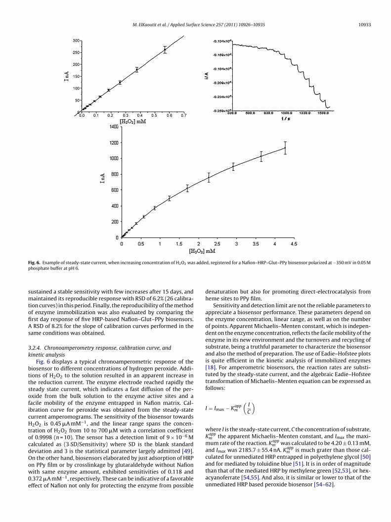
Fig. 6. Example of steady-state current, when increasing concentration of H O was added, registered for a Nafion–HRP–Glut–PPy biosensor polarized at −350 mV in 0.05 Mp
smtofiAs
3k
bttsoficHtocdOow0e
2 2
hosphate buffer at pH 6.
ustained a stable sensitivity with few increases after 15 days, andaintained its reproducible response with RSD of 6.2% (26 calibra-
ion curves) in this period. Finally, the reproducibility of the methodf enzyme immobilization was also evaluated by comparing therst day response of five HRP-based Nafion–Glut–PPy biosensors.
RSD of 8.2% for the slope of calibration curves performed in theame conditions was obtained.
.2.4. Chronoamperometry response, calibration curve, andinetic analysis
Fig. 6 displays a typical chronoamperometric response of theiosensor to different concentrations of hydrogen peroxide. Addi-ions of H2O2 to the solution resulted in an apparent increase inhe reduction current. The enzyme electrode reached rapidly theteady state current, which indicates a fast diffusion of the per-xide from the bulk solution to the enzyme active sites and aacile mobility of the enzyme entrapped in Nafion matrix. Cal-bration curve for peroxide was obtained from the steady-stateurrent amperomograms. The sensitivity of the biosensor towards2O2 is 0.45 �A mM−1, and the linear range spans the concen-
ration of H2O2 from 10 to 700 �M with a correlation coefficientf 0.9998 (n = 10). The sensor has a detection limit of 9 × 10⁻⁶ Malculated as (3·SD/Sensitivity) where SD is the blank standardeviation and 3 is the statistical parameter largely admitted [49].n the other hand, biosensors elaborated by just adsorption of HRP
n PPy film or by crosslinkage by glutaraldehyde without Nafionith same enzyme amount, exhibited sensitivities of 0.118 and.372 �A mM−1, respectively. These can be indicative of a favorableffect of Nafion not only for protecting the enzyme from possible
denaturation but also for promoting direct-electrocatalysis fromheme sites to PPy film.
Sensitivity and detection limit are not the reliable parameters toappreciate a biosensor performance. These parameters depend onthe enzyme concentration, linear range, as well as on the numberof points. Apparent Michaelis–Menten constant, which is indepen-dent on the enzyme concentration, reflects the facile mobility of theenzyme in its new environment and the turnovers and recycling ofsubstrate, being a truthful parameter to characterize the biosensorand also the method of preparation. The use of Eadie–Hofstee plotsis quite efficient in the kinetic analysis of immobilized enzymes[18]. For amperometric biosensors, the reaction rates are substi-tuted by the steady-state current, and the algebraic Eadie–Hofsteetransformation of Michaelis–Menten equation can be expressed asfollows:
I = Imax − Kappm
(I
C
)
where I is the steady-state current, C the concentration of substrate,Kapp
m the apparent Michaelis–Menten constant, and Imax the maxi-mum rate of the reaction. Kapp
m was calculated to be 4.20 ± 0.13 mM,and Imax was 2185.7 ± 55.4 nA. Kapp
m is much grater than those cal-culated for unmediated HRP entrapped in polyethylene glycol [50]
and for mediated by toluidine blue [51]. It is in order of magnitudethan that of the mediated HRP by methylene green [52,53], or hex-acyanoferrate [54,55]. And also, it is similar or lower to that of theunmediated HRP based peroxide biosensor [54–62].
1 face Sc
3
rbustibia
4
vlUihaftoiSosN
bf
A
0(
R
[
[
[
[
[
[
[
[
[
[
[
[
[
[
[
[
[
[
[
[
[
[
[
[
[
[
[
[
0934 M. ElKaoutit et al. / Applied Sur
.2.5. Influence of interferenceThe effect of substances that possibly interfere with the
esponse of the enzyme electrode has been also studied. Cali-ration curves were constructed in the presence of ascorbic andric acids at the concentration of 0.4 mM in the cell. The corre-ponding sensitivities were compared with that obtained withouthese interferences. The two acids did not cause any observablenterference with the response of Nafion–HRP–Glut–PPy peroxideiosensor. Thus, the proposed biosensor shows a good selectiv-
ty thanks to the negatively charged interface created by Nafionddition.
. Conclusion
In this paper, we successfully immobilized HRP on PPy carbonitreous functionalized electrode by the combination of cross-inked glutaraldehyde and additive-protective Nafion. Solid phaseV–vis study of the immobilized enzyme proves the availabil-
ty of the proposed procedure to preserve the native state of theeme catalytic group in HRP. FT-IR spectra comparison of nativend immobilized enzyme suggests that HRP retains the essentialeatures of its original structure. Results obtained by analyzinghe protein spectra in the amide I region shows that the processf immobilization has the effect of folding the enzyme, enhanc-ng the globular secondary structure. Furthermore, apparition ofO3H vibration in the spectrum of immobilized enzyme, decreasef heme UV absorbance and AFM study, are in good agreement, andhow that the HRP enzyme is probably introduced in the ion clusterafion region.
Finally, we studied parameters affecting the response of theiosensor, and constructed a simple and satisfactory sensitive toolor the determination of H2O2 with very high stability.
cknowledgements
We thank to the Junta de Andalucía/FEDER (FQM249/P08-FQM-4006) and Ministerio de Ciencia e Innovación of Spain/FEDERCTQ2010-19058) for their financial support.
eferences
[1] L.C. Clark, C. Lyons, Electrode systems for continuous monitoring in cardiovas-cular surgery, Ann. N. Y. Acad. Sci. 102 (1962) 29–45.
[2] U. Wollenberger, Third generation biosensors-integrating recognition andtransduction in electrochemical sensors, in: L. Gorton (Ed.), Biosensors andModern Biospecific Analytical Techniques, 1st edition, Wilson & Wilson’s, Ams-terdam, 2005, pp. 65–130.
[3] T. Ahuja, I.A. Mir, D. Kumar, Rajesh, Biomolecular immobilization on conductingpolymers for biosensing applications, Biomaterials 28 (2007) 791–805.
[4] S. Cosnier, Biomolecule immobilization on electrode surfaces by entrapmentor attachment to electrochemically polymerized films, Biosens. Bioelectron. 14(1999) 443–456.
[5] M. Gerard, A. Chauby, B.D. Malhotra, Application of conducting polymers tobiosensors, Biosens. Bioelectron. 17 (2002) 345–359.
[6] N. Hamdi, J. Wang, H.G. Monbouquette, Polymer films as permselective coatingsfor H2O2-sensing electrodes, J. Electroanal. Chem. 581 (2005) 258–264.
[7] E.W. Kristensen, W.G. Kuhr, R.M. Wightman, Temporal characterization of per-fluorinated ion exchange coated microvoltammetric electrodes for in vivo use,Anal. Chem. 59 (1987) 1752–1757.
[8] C. Chen, Y. Jiang, J. Kan, A noninterference polypyrrole glucose biosensor,Biosens. Bioelectron. 22 (2006) 639–643.
[9] D.J. Harrison, R.F.B. Turner, H.P. Baltes, Characterization of perfluorosulfonicacid polymer coated enzyme electrodes and a miniaturized integrated poten-tiostat for glucose analysis in whole blood, Anal. Chem. 60 (1988) 2002–2007.
10] M. Umana, J. Waller, Protein-modified electrodes. The glucose oxi-dase/polypyrrole system, Anal. Chem. 58 (1986) 2979–2983.
11] V.K. Gade, D.J. Shirale, P.D. Gaikwad, P.A. Savale, K.P. Kakde, H.J. Kharat, M.Shirsat, Immobilization of GOD on electrochemically synthesized PPy–PVS
composite film by cross-linking via glutaricdialdehyde for determination ofglucose, React. Funct. Polym. 66 (2006) 1420–1426.12] Y.T. Kong, M. Boopthi, Y-B. Shim, Direct electrochemistry of horseradish per-oxidase bonded on a conducting polymer modified glassy carbon electrode,Biosens. Bioelectron. 19 (2003) 227–232.
[
ience 257 (2011) 10926– 10935
13] J.L. Hidalgo-Hidalgo de Cisneros, I. Naranjo-Rodríguez, M. ElKaoutit, PatentP2000702165, Spain, August 2007.
14] M. ElKaoutit, I. Naranjo-Rodriguez, K.R. Temsamani, M.D. De la Vega, J.L.Hidalgo-Hidalgo de Cisneros, Dual laccase–tyrosinase based sonogel-carbonbiosensor for monitoring polyphenols in beers, J. Agric. Food Chem. 55 (2007)8011–8018.
15] M. ElKaoutit, I. Naranjo-Rodriguez, K.R. Temsamani, M.P. Hernández-Artiga, D.Bellido-Milla, J.L. Hidalgo-Hidalgo de Cisneros, A comparison of three amper-ometric phenoloxidase-sonogel-carbon based biosensors for determination ofpolyphenols in beers, Food Chem. 110 (2008) 1019–1024.
16] M. ElKaoutit, I. Naranjo-Rodriguez, K.R. Temsamani, M. Domínguez, J.L.Hidalgo-Hidalgo de Cisneros, Investigation of biosensor signal bioamplifica-tion: comparison of direct electrochemistry phenomena of individual laccaseand dual laccase–tyrosinase copper enzymes at a sonogel-carbon electrode,Talanta 75 (2008) 1348–1355.
17] M. ElKaoutit, I. Naranjo-Rodriguez, M. Domínguez, M.P. Hernández-Artiga, D.Bellido-Milla, J.L. Hidalgo-Hidalgo de Cisneros, A third-generation hydrogenperoxide biosensor based on HRP enzyme immobilized in Nafion–sonogel-carbon composite, Electrochim. Acta 53 (2008) 7131–7137.
18] M. El Kaoutit, I. Naranjo-Rodriguez, K.R. Temsamani, J.L. Hidalgo-Hidalgo deCisneros, The sonogel-carbon materials as basis for development of enzymebiosensors for phenols and polyphenols monitoring: a detailed compara-tive study of three immobilization matrixes, Biosens. Bioelectron. 22 (2007)2958–2966.
19] P. Auderbert, P. Alderbet, M. Pineri, New electrochemical synthesis of electronicand ionic conductive polymer composites, Synth. Met. 32 (1989) 1–14.
20] F. Endres, G. Schwitzgebel, Morphological and electrochemical studies ofpolypyrrole/Nafion® , Synth. Met. 88 (1997) 73–78.
21] J. Hong, H. Ghourchian, A.A. Moosavi-Movahedi, Direct electron transfer ofredox proteins on a Nafion-cysteine modified gold electrode, Electrochem.Commun. 8 (2006) 1572–1576.
22] J. Hong, A.A. Moosavi-Movahedi, H. Ghourchian, A. Molaei, S. Razaei-Zaechi,Direct electron transfer of horseradish peroxidase on Nafion-cysteine modifiedgold electrode, Electrochim. Acta 52 (2007) 6261–6267.
23] Q. Huang, Z. Lu, J.F. Rusling, Composite films of surfactants, nafion, andproteins with electrochemical and enzyme activity, Langmuir 12 (1996)5472–5480.
24] M. El Kaoutit, D. Bouchta, H. Zejli, N. Izaoumen, K.R. Temsamani, A simple con-ducting polymer-based biosensor for the detection of atrazine, Anal. Lett. 37(2004) 1671–1681.
25] N. Izaoumen, D. Bouchta, H. Zejli, M. El Kaoutit, A.M. Stalcup, K.R. Temsamani,Electrosynthesis and analytical performances of functionalized poly(pyrrole/�-cyclodextrin) films, Talanta 66 (2005) 111–117.
26] K.R. Temsamani, H.B. Mark Jr., W. Kutner, A.M. Stalcup, A simple one-stepelectrosynthesis of poly(pyrrole-sulfated-�-cyclodextrin) film, J. Solid StateElectrochem. 6 (2002) 391–395.
27] K.R. Temsamani, Ö. Ceylan, S. Ö ztemiz, T.P. Gbatu, A.M. Stalcup, H.B. Mark Jr., W.Kutner, Electrochemically aided solid phase microextraction: conducting poly-mer film material applicable for cationic analytes, J. Solid State Electrochem. 6(2002) 494–497.
28] R. Santucci, F. Laurenti, F. Sinibaldi, R.P. Ferrari, Effect of dimethyl sulfoxideon the structure and the functional properties of horseradish peroxidase asobserved by spectroscopy and cyclic voltammetry, Biochim. Biophys. Acta 1596(2002) 225–233.
29] E. Stellwangen, The reversible unfolding of horse heart ferricytochrome c, Bio-chemistry 7 (1968) 2893–2898.
30] T.T. Herskovits, B. Gadegbeku, H. Jaillet, On the structural stability and solventdenaturation of proteins. I. Denaturation by the alcohols and glycols, J. Biol.Chem. 245 (1970) 2588–2898.
31] G.H. Carlson, P. Nicholls, D. Svistunenko, G.I. Berglund, J. Hajdu, Complexes ofhorseradish peroxidase with formate, acetate, and carbon monoxide, Biochem-istry 44 (2005) 635–642.
32] G.I. Berglund, G.H. Carlsson, A.T. Smith, H. Szöke, A. Henriksen, J. Hajdu, Thecatalytic pathway of horseradish peroxidase at high resolution, Nature 417(2002) 463–468.
33] G. Smulevich, M. Paoli, G. De Sanctis, A.R. Mantini, F. Ascoli, M. Coletta, Spec-troscopic evidence for conformational transition in horseradish peroxidase atvery low pH, Biochemistry 36 (1997) 640–649.
34] W. Al-Azzam, E.A. Pastrana, Y. Ferrer, Q. Huang, R. Schweitzer-Stenner, K.Griebenow, Structure of poly(ethylene glycol)-modified horseradish perox-idase in organic solvents: infrared amide I spectral changes upon proteindehydration are largely caused by protein structural changes and not by waterremoval per se, Biophys. J. 83 (2002) 3637–3651.
35] J. Zhang, D.P. Wilkinson, H. Wang, Z-H. Liu, FTIR and electrochemicalobservation of water content reduction in a thin Nafion film induced byan impregnation of metal complex cations, Electrochim. Acta 50 (2005)4082–4088.
36] C-Y. Chen, J-I. Garnica-Rodriguez, M.C. Duke, R.F. Dalla Costa, A.L. Dicks,J.C. Diniz da Costa, Nafion/polyaniline/silica composite membranes for directmethanol fuel cell application, J. Power Sources 166 (2007) 324–330.
37] U. Sen, S.Ü. C elik, A. Ata, A. Bozkurt, Anhydrous proton conducting membranes
for PEM fuel cells based on Nafion/Azole composites, Int. J. Hydrogen Energy33 (2008) 2808–2815.38] Q. Huang, Q. Huang, R.A. Pinto, K. Griebenow, R. Schweitzer-Stenner, W.J. WeberJr., Inactivation of horseradish peroxidase by phenoxyl radical attack, J. Am.Chem. Soc. 127 (2005) 1431–1437.
ace Sci
[
[
[
[
[
[
[
[
[
[
[
[
[
[
[
[
[
[
[
[
[
[
[
Acta 273 (1993) 59–70.
M. ElKaoutit et al. / Applied Surf
39] I.E. Holzbaur, A.M. English, A.A. Ismail, FTIR study of the thermal denaturationof horseradish and cytochrome c peroxidases in D2O, Biochemistry 35 (1996)5488–5494.
40] K. Griebenow, A.M. Klibanov, On protein denaturation in aqueous–organicmixtures but not in pure organic solvents, J. Am. Chem. Soc. 118 (1996)11695–11700.
41] D. Aichun, P. Huang, W.S. Caughey, Protein secondary structures in water fromsecond-derivative amide I infrared spectra, Biochemistry 29 (1990) 3303–3308.
42] R. Khurana, L.F. Anthony, Do parallel �-helix proteins have a unique Fouriertransform infrared spectrum? Biophys. J. 78 (2000) 994–1000.
43] M. ElKaoutit, I. Naranjo-Rodríguez, K.R. Temsamani, M. Dominguez, J.L.Hidalgo-Hidalgo de Cisneros, Structural characteristics and cyclic voltammet-ric behaviour of Sonogel-Carbon tyrosinase biosensors. A detailed comparativestudy of three immobilization matrixes, J. Sol–Gel Sci. Technol. 45 (2008)157–163.
44] A. Lindgren, M. Tanaka, T. Ruzgas, L. Gorton, I. Gazaryan, K. Ishimori, I. Mor-ishima, Direct electron transfer catalysed by recombinant forms of horseradishperoxidase: insight into the mechanism, Electrochem. Commun. 1 (1999)171–175.
45] L. Gorton, A. Lindgren, T. Larson, F.D. Munteanu, T. Ruzgas, I. Gazaryan, Directelectron transfer between heme-containing enzymes and electrodes as basisfor third generation biosensors, Anal. Chim. Acta 400 (1999) 91–108.
46] H.A. Harbury, Oxidation–reduction potentials of horseradish peroxidase, J. Biol.Chem. 255 (1957) 1009–1024.
47] C. Nistor, J. Emnéus, L. Gorton, A. Ciucu, Improved stability and alteredselectivity of tyrosinase based graphite electrodes for detection of phenoliccompounds, Anal. Chim. Acta 387 (1999) 309–326.
48] S.S. Razola, B.L. Ruiz, N.M. Diez, H.B. Mark, J.M. Kauffmann, Hydrogen peroxidesensitive amperometric biosensor based on horseradish peroxidase entrappedin a polypyrrole electrode, Biosens. Bioelectron. 17 (2002) 921–928.
49] J.C. Millar, J.N. Millar, Estadistica para Quimica Analitica, Addison-WesleyIberoamericana, Wilmington, USA, 1993.
50] Y. Xu, W. Peng, X. Liu, G. Li, A new film for the fabrication of an unmediatedH2O2 biosensor, Biosens. Bioelectron. 20 (2004) 533–537.
51] Y. Liu, J. Lei, H. Ju, Amperometric sensor for hydrogen peroxide based on elec-tric wire composed of horseradish peroxidase and toluidine blue-multiwalledcarbon nanotubes nanocomposite, Talanta 74 (2008) 965–970.
[
ence 257 (2011) 10926– 10935 10935
52] B. Wang, S. Dong, Sol–gel-derived amperometric biosensor for hydrogen per-oxide based on methylene green incorporated in Nafion film, Talanta 51 (2000)565–572.
53] C. Lei, J. Deng, Hydrogen peroxide sensor based on coimmobilized methy-lene green and horseradish peroxidase in the same montmorillonite-modifiedbovine serum albumin-glutaricdialdehyde matrix on a glassy carbon electrodesurface, Anal. Chem. 68 (1996) 3344–3349.
54] J. Li, S.N. Tan, H. Ge, Silica sol–gel immobilized amperometric biosensor forhydrogen peroxide, Anal. Chim. Acta 335 (1996) 137.
55] B. Wang, J. Zhang, G. Cheng, S. Dong, Amperometric enzyme electrode for thedetermination of hydrogen peroxide based on sol-gel/hydrogel composite film,Anal. Chim. Acta 407 (2000) 111–118.
56] S-Q. Liu, H-X. Ju, Renewable reagentless hydrogen peroxide sensorbased on direct electron transfer of horseradish peroxidase immobilizedon colloidal gold-modified electrode, Anal. Biochem. 307 (2002) 110–116.
57] T. Ferri, A. Poscia, R. Santucci, Direct electrochemistry of membrane-entrappedhorseradish peroxidase. Part II: amperometric detection of hydrogen peroxide,Bioelectrochem. Bioengerg. 45 (1998) 221–226.
58] X. Yi, J. Huang-Xian, C. Hong-Yuan, Direct electrochemistry of horseradish per-oxidase immobilized on a colloid/cysteamine-modified gold electrode, Anal.Biochem. 278 (2000) 22–28.
59] J. Wang, M. Gu, J. Di, Y. Gao, Y. Wu, Y. Tu, A carbon nanotube/silicasol–gel architecture for immobilization of horseradish peroxidase forelectrochemical biosensor, Bioprocess. Biosyst. Eng. 30 (2007) 289–296.
60] H. Chen, S. Dong, Direct electrochemistry and electrocatalysis of horseradishperoxidase immobilized in sol–gel-derived ceramic–carbon nanotubenanocomposite film, Biosens. Bioelectron. 22 (2007) 1811–1815.
61] E. Csoregi, L. Gorton, G. Marko-Varga, Carbon fibres as electrode materials forthe construction of peroxidase-modified amperometric biosensors, Anal. Chim.
62] Z. Tong, R. Yuan, Y. Chai, X. Xie, S. Chen, A novel and simple biomoleculesimmobilization method: electro-deposition ZrO2 doped with HRP for fab-rication of hydrogen peroxide biosensor, J. Biotechnol. 128 (2007) 567–575.