A Study of MECT1-MAML2 in Mucoepidermoid Carcinoma and Warthin's Tumor of Salivary Glands
6
A Study of MECT1-MAML2 in Mucoepidermoid Carcinoma and Warthin’s Tumor of Salivary Glands Carmo Martins,* † Branca Cavaco, † Giovanni Tonon, ‡ Frederic J. Kaye, ‡ Jorge Soares,* and Isabel Fonseca* From the Departamento de Patologia Morfolo ´gica * and Centro de Investigac ¸a ˜o de Patobiologia Molecular (CIPM), † Instituto Portugue ˆs de Oncologia de Francisco Gentil, Lisboa, Portugal; and the Genetics Branch, ‡ National Cancer Institute and Naval Hospital, Bethesda, Maryland The t(11;19)(q21;p13) chromosomal translocation has been described in two distinct types of salivary gland neoplasms: mucoepidermoid carcinoma (MEC) and Warthin’s tumor (WT). Since this translocation has been recently shown to generate a MECT1-MAML2 fusion gene, we evaluated 10 primary MEC and seven primary WT to further define the molecular associa- tion of these two entities using cytogenetic , as well as in situ hybridization (ISH) and reverse transcriptase- polymerase chain reaction (RT-PCR) analyses di- rected against the fusion gene. A karyotype was estab- lished in all neoplasms except for two MEC cases. Of the eight karyotyped MECs, five showed the t(11; 19)(q21;p13), two had a normal karyotype, and one case presented a Y and X. Three of the WT revealed a normal karyotype and four had several abnormali- ties which did not involve chromosomes 11 and 19. ISH analysis performed in cytogenetic suspension and/or in tumor paraffin sections demonstrated MAML2 rearrangement in 7 of 10 cases of MEC: all five cases with t(11;19), one case with normal karyotype, and one unkaryotyped case. RT-PCR analysis con- firmed the expression of the MECT1-MAML2 gene in all MEC cases that were positive by ISH analysis. Nei- ther the t(11;19) nor MECT1-MAML2 was detected in any case of WT, nor in control samples from poly- morphous low-grade adenocarcinoma , acinic cell car- cinoma, or normal parotid gland tissue. We have demonstrated that ISH and RT-PCR are sensitive meth- ods for detecting MECT1-MAML2 in MEC. In contrast, we did not detect the t(11;19) nor MECT1-MAML2 ex- pression in seven cases of WT. (J Mol Diagn 2004, 6:205–210) Mucoepidermoid carcinoma (MEC) of the salivary glands represents 5% of all salivary gland tumors and 20% of the malignant forms. 1 To date, the karyotypic profile in this tumor type has only been described in 26 cases: 2 in seven cases there were rearrangements of 11q14 –21 and 19p12–13, mainly as a chromosomal translocation t(11;19)(q21;p12–13) 3–7 and it was the sole abnormality in two cases. 4,7 The remaining five cases showed either a more complex translocation involving other chromo- somes or other rearrangements. 3,5,6 The same abnormal- ity has also been described in mucoepidermoid carcino- mas originated in bronchial glands of the lung. 8,9 Interestingly, the t(11;19)(q13–21;p12–13) was also re- ported in Warthin’s tumor (WT), 10,11 a benign salivary gland neoplasm, which suggested an unexpected cyto- genetic association between two otherwise unrelated sal- ivary gland tumors. However, it is known that WT can arise and/or co-exist with MEC, 12–14 warranting the reap- praisal of this association. The t(11;19)(q21;p13) found in MEC has recently been cloned. 15 Two genes are involved: mucoepidermoid car- cinoma translocated 1 ( MECT1) gene and a member of the mastermind-like gene family ( MAML2) located at 19p13 and 11q21, respectively. 15 This translocation gen- erates a chimeric gene MECT1-MAML2 that fuses exon 1 of MECT1 with exons 2–5 of MAML2. MECT1-MAML2 fusion product disrupts the normal mechanism of the Notch signaling pathway, activating Notch-target genes independently of exogenous signals, therefore represent- ing a novel mechanism for altered Notch function in tu- morigenesis. 15 In addition, the recent identification of the MECT1 gene product as a potent co-activator for genes that are regulated by cyclic AMP responsive elements suggests that MECT1-MAML2 may be disrupting both Notch and CREB signaling pathways to induce tumori- genesis. 16,17 To further substantiate the importance of the MECT1- MAML2 fusion gene in MEC tumorigenesis and to inves- tigate a common molecular pathway with WT, we studied a series of 10 primary MEC and seven primary WT sali- vary gland tumors using conventional cytogenetics, in situ hybridization (ISH), and reverse transcriptase-poly- merase chain reaction (RT-PCR). Accepted for publication March 30, 2004. Address reprint requests to Prof. Isabel Fonseca, Departamento de Patologia Morfolo ´ gica, Instituto Portugue ˆs de Oncologia, Rua Prof. Lima Basto, 1099 – 023 Lisboa, Portugal. E-mail: [email protected] saude.pt. Journal of Molecular Diagnostics, Vol. 6, No. 3, August 2004 Copyright © American Society for Investigative Pathology and the Association for Molecular Pathology 205
-
Upload
independent -
Category
Documents
-
view
1 -
download
0
Transcript of A Study of MECT1-MAML2 in Mucoepidermoid Carcinoma and Warthin's Tumor of Salivary Glands
A Study of MECT1-MAML2 in MucoepidermoidCarcinoma and Warthin’s Tumor of Salivary Glands
Carmo Martins,*† Branca Cavaco,†
Giovanni Tonon,‡ Frederic J. Kaye,‡
Jorge Soares,* and Isabel Fonseca*From the Departamento de Patologia Morfologica * and Centro
de Investigacao de Patobiologia Molecular (CIPM),† Instituto
Portugues de Oncologia de Francisco Gentil, Lisboa, Portugal;
and the Genetics Branch,‡ National Cancer Institute and Naval
Hospital, Bethesda, Maryland
The t(11;19)(q21;p13) chromosomal translocationhas been described in two distinct types of salivarygland neoplasms: mucoepidermoid carcinoma (MEC)and Warthin’s tumor (WT). Since this translocationhas been recently shown to generate a MECT1-MAML2fusion gene, we evaluated 10 primary MEC and sevenprimary WT to further define the molecular associa-tion of these two entities using cytogenetic, as well asin situ hybridization (ISH) and reverse transcriptase-polymerase chain reaction (RT-PCR) analyses di-rected against the fusion gene. A karyotype was estab-lished in all neoplasms except for two MEC cases. Ofthe eight karyotyped MECs, five showed the t(11;19)(q21;p13), two had a normal karyotype, and onecase presented a �Y and �X. Three of the WT revealeda normal karyotype and four had several abnormali-ties which did not involve chromosomes 11 and 19.ISH analysis performed in cytogenetic suspensionand/or in tumor paraffin sections demonstratedMAML2 rearrangement in 7 of 10 cases of MEC: all fivecases with t(11;19), one case with normal karyotype,and one unkaryotyped case. RT-PCR analysis con-firmed the expression of the MECT1-MAML2 gene inall MEC cases that were positive by ISH analysis. Nei-ther the t(11;19) nor MECT1-MAML2 was detected inany case of WT, nor in control samples from poly-morphous low-grade adenocarcinoma, acinic cell car-cinoma, or normal parotid gland tissue. We havedemonstrated that ISH and RT-PCR are sensitive meth-ods for detecting MECT1-MAML2 in MEC. In contrast,we did not detect the t(11;19) nor MECT1-MAML2 ex-pression in seven cases of WT. (J Mol Diagn 2004,6:205–210)
Mucoepidermoid carcinoma (MEC) of the salivary glandsrepresents 5% of all salivary gland tumors and 20% of themalignant forms.1 To date, the karyotypic profile in thistumor type has only been described in 26 cases:2 in
seven cases there were rearrangements of 11q14–21and 19p12–13, mainly as a chromosomal translocationt(11;19)(q21;p12–13)3–7 and it was the sole abnormalityin two cases.4,7 The remaining five cases showed eithera more complex translocation involving other chromo-somes or other rearrangements.3,5,6 The same abnormal-ity has also been described in mucoepidermoid carcino-mas originated in bronchial glands of the lung.8,9
Interestingly, the t(11;19)(q13–21;p12–13) was also re-ported in Warthin’s tumor (WT),10,11 a benign salivarygland neoplasm, which suggested an unexpected cyto-genetic association between two otherwise unrelated sal-ivary gland tumors. However, it is known that WT canarise and/or co-exist with MEC,12–14 warranting the reap-praisal of this association.
The t(11;19)(q21;p13) found in MEC has recently beencloned.15 Two genes are involved: mucoepidermoid car-cinoma translocated 1 (MECT1) gene and a member ofthe mastermind-like gene family (MAML2) located at19p13 and 11q21, respectively.15 This translocation gen-erates a chimeric gene MECT1-MAML2 that fuses exon 1of MECT1 with exons 2–5 of MAML2. MECT1-MAML2fusion product disrupts the normal mechanism of theNotch signaling pathway, activating Notch-target genesindependently of exogenous signals, therefore represent-ing a novel mechanism for altered Notch function in tu-morigenesis.15 In addition, the recent identification of theMECT1 gene product as a potent co-activator for genesthat are regulated by cyclic AMP responsive elementssuggests that MECT1-MAML2 may be disrupting bothNotch and CREB signaling pathways to induce tumori-genesis.16,17
To further substantiate the importance of the MECT1-MAML2 fusion gene in MEC tumorigenesis and to inves-tigate a common molecular pathway with WT, we studieda series of 10 primary MEC and seven primary WT sali-vary gland tumors using conventional cytogenetics, insitu hybridization (ISH), and reverse transcriptase-poly-merase chain reaction (RT-PCR).
Accepted for publication March 30, 2004.
Address reprint requests to Prof. Isabel Fonseca, Departamento dePatologia Morfologica, Instituto Portugues de Oncologia, Rua Prof. LimaBasto, 1099–023 Lisboa, Portugal. E-mail: [email protected].
Journal of Molecular Diagnostics, Vol. 6, No. 3, August 2004
Copyright © American Society for Investigative Pathology
and the Association for Molecular Pathology
205
Materials and Methods
Case Selection
Case selection was based on the availability of karyotypicinformation and/or frozen tissue from the surgical speci-men. All cases with the diagnosis of mucoepidermoidcarcinoma and Warthin’s tumor were reclassified accord-ing to the World Health Organization’s criteria,18 and MECcases were graded according to Batsakis and Luna’s cri-teria.12,19 The clinical, histopathological, and karyotypicdata are shown in Table 1.
Conventional Cytogenetic Analysis
Chromosome metaphases of tumor cells were obtainedfrom short-term primary cultures as previously de-scribed.20 Chromosomes were GTG-banded, and thekaryotypes were defined according to International Sys-tem of Human Cytogenetic Nomenclature (ISCN).21 Adetailed description of the tumor karyotypes is reportedin Table 1.
ISH Analysis
ISH analysis was performed on cell suspension (meta-phase and/or interphase nuclei) left from conventionalcytogenetic analysis and on 4-�m formalin-fixed, paraf-fin-embedded tumor tissue sections. For the cell suspen-sion, a standard ISH protocol with fluorescence detection(FISH) was applied.22 For the tumor tissue sections, theISH analysis followed the method of Alers et al,23 usingeither fluorescence (FISH) or chromogenic (CISH) de-tection.
Two BAC clones probes named RP11–16K5 andRP11–676L3 that, together, cover the entire chromosome
region of MAML2 gene as described by Tonon et al15
were used. The BAC clones DNA was isolated followingthe protocol of the distributor (Children’s Hospital Oak-land Research Institute, Oakland, CA, USA). The probeswere labeled by random octamer priming (Bioprime DNALabeling, Invitrogen SA, Barcelona, Spain). For FISH pro-cedures, the BAC clones were labeled differently withbiotin and digoxigenin and detected, respectively, byCy3-avidin (Jackson Immunoresearch Lab, West Grove,PA, USA) and anti-digoxigenin-FITC (Roche DiagnosticsGmbH, Mannheim, Germany). For CISH, both BACclones were labeled with fluorescein (FITC) using horse-radish peroxidase anti-fluorescein (Roche DiagnosticsGmbH) and H2O2-diaminobenzidine for visualization.
In FISH preparations, the metaphases and nuclei ma-terial were counterstained with DAPI-Vectashield mount-ing solution (Vector, Burlingame, CA, USA). The CISHtissue sections were counter-stained with Meyer’s hema-toxylin and mounted with Entellan (Merck, WhitehouseStation, NJ, USA). The FISH and CISH signals were ana-lyzed and recorded using a Cytovision System (AppliedImaging, Newcastle-on-Tyne, UK).
In each case, at least 200 interphase nuclei from par-affin-embedded tumor sections were evaluated. Whenpresent, normal salivary gland tissue was also analyzed.The presence of three dots (CISH preparations) or of splitsignals from BAC probes (FISH preparations) in morethan 10% of the nuclei represent rearrangement ofMAML2 gene, following the criteria of Jenkins et al.24
RT-PCR Analysis
mRNA was extracted from tissue that was frozen in liquidnitrogen immediately after surgery, using the QuickPrepMicro mRNA purification Kit (Amersham Pharmacia Bio-
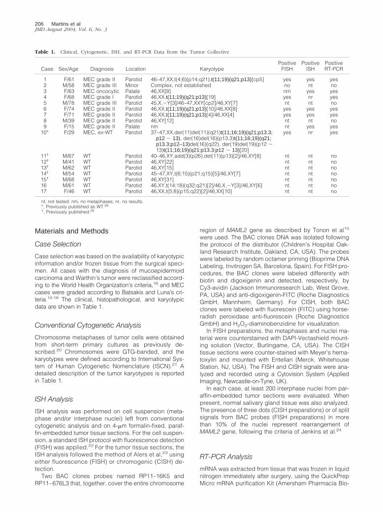
Table 1. Clinical, Cytogenetic, ISH, and RT-PCR Data from the Tumor Collective
Case Sex/Age Diagnosis Location KaryotypePositive
FISHPositive
ISHPositiveRT-PCR
1 F/61 MEC grade II Parotid 46–47,XX,t(4;6)(p14;q21),t(11;19)(q21;p13)[cp5] yes yes yes2 M/58 MEC grade III Minor Complex, not established no nt no3 F/63 MEC oncocytic Palate 46,XX[8] nm yes yes4 F/68 MEC grade I Parotid 46,XX,t(11;19)(q21;p13)[19] yes nr yes5 M/78 MEC grade III Parotid 45,X,�Y[3]/46–47,XXY[cp2]/46,XY[7] nt nt no6 F/74 MEC grade II Parotid 46,XX,t(11;19)(q21;p13)[10]/46,XX[8] yes yes yes7 F/71 MEC grade II Parotid 46,XX,t(11;19)(q21;p13)[4]/46,XX[4] yes yes yes8 M/39 MEC grade II Parotid 46,XY[12] nt nt no9 F/15 MEC grade II Palate nm nt yes yes
10* F/29 MEC, ex-WT Parotid 37–47,XX,der(11)del(11)(q21)t(11;16;19)(q21;p13.3;p12 � 13), der(16)del(16)(p13.3)t(11;16;19)(q21;p13.3;p12–13)del(16)(q22), der(19)del(19)(p12 �13)t(11;16;19)(q21;p13.3;p12 � 13)[20]
yes nr yes
11† M/67 WT Parotid 40–46,XY,add(3)(p26),del(11)(p13)[2]/46,XY[8] nt nt no12† M/41 WT Parotid 46,XY[22] nt nt no13† M/62 WT Parotid 46,XY[15] nt nt no14† M/54 WT Parotid 45–47,XY,t(6;15)(p21;q15)[5]/46,XY[7] nt nt no15† M/68 WT Parotid 46,XY[31] nt nt no16 M/61 WT Parotid 46,XY,t(14;18)(q32;q21)[2]/46,X,�Y[3]/46,XY[6] nt nt no17 F/46 WT Parotid 46,XX,t(5;8)(p15;q22)[2]/46,XX[10] nt nt no
nt, not tested; nm, no metaphases; nr, no results.*, Previously published as WT.26
†, Previously published.26
206 Martins et alJMD August 2004, Vol. 6, No. 3

tech, Piscataway, NJ, USA), according to the manufac-turer’s protocol. As controls, one case of polymorphouslow-grade adenocarcinoma (PLGA), one case of aciniccell carcinoma (ACC), and tissue from one normal parotidgland were used. Synthesis of cDNA was performed withapproximately 0.3 �g of mRNA, at 37°C for 90 minutes,using random primers p(dN)6 (Roche DiagnosticsGmbH) and reverse transcriptase (SuperScript II RNaseH�, Invitrogen SA), following the manufacturer’s instruc-tions. Amplifications by PCR were carried out using 5 �lof cDNA, forward primer (5�-atggcgacttcgaacaatccgcg-gaa-3�) and reverse primer (5�-aacaggccattgccag-gagaatgtgtatggg-3�). The oligonucleotide primers weredesigned to amplify a segment from the MECT1-MAML2cDNA, starting in exon 1 from the MECT1 gene andfinishing in exon 2 from the MAML2 gene, with an ex-pected size of 386 bp. One �l of each PCR reaction wasthen used as template for a second amplification usingnested forward (5�-ggaaattcagcgagaagatcg-3�) and re-verse (5�-tatgggatggcagagtgttagtc-3�) primers. Theseoligonucleotide primers amplify a segment of the MECT1-MAML2 chimeric cDNA, starting in exon 1 from theMECT1 gene and finishing in exon 2 from the MAML2gene, with an expected size of 339 bp. First round andnested RT-PCRs were performed over 35 cycles of am-plification, using the following conditions: 95°C for 45seconds, 56°C for 45 seconds, and 72°C for 1 minute.Amplification reactions, with a total volume of 50 �l, con-tained final concentrations of 20 mmol/L Tris-HCl (pH8.4), 50 mmol/L KCl, 200 �mol/L dNTPs (Invitrogen SA),1.5 mmol/L MgCl2, 0.6 �mol/L of each primer, and 2U ofTaqDNA polymerase (Invitrogen SA). Negative controlsfor cDNA synthesis and PCRs, in which the template wasreplaced by sterile water, were included in each experi-ment. RNA integrity and efficiency of cDNA synthesiswere confirmed in each sample by RT-PCR for the house-keeping gene phosphoglycerate kinase 1 (PGK1), usingpreviously published oligonucleotide primers.25 Amplifi-cation with these primers yielded a 247-bp PCR product.PCR products were analyzed and purified by electro-phoresis in a 2% agarose gel stained with ethidium bro-mide. All of the results were repeated, at least twice, fromdifferent batches of mRNA extracted at different times,from the same tumor. In the cases that presented theMECT1-MAML2 rearrangement, the PCR product wassequenced (ABI Prism 310 Genetic Analyzer, using theABI Prism Big Dye Terminator Cycle Sequencing ReadyReaction Kit version 2.0; Applied Biosystems, Foster City,USA).
Results
Cytogenetic Analysis
Cytogenetic information was obtained from all 17 casesexcept for case 9 due to unsuccessful cell culture and forcase 2 which showed aneuploidy and abnormal met-aphases, however a karyotype could not be defined dueto unsatisfactory quality (Table 1). Five of the eight fullykaryotyped MEC revealed a t(11;19)(q21;p13). This was
the sole abnormality found in cases 4, 6, and 7 and thetranslocation was present together with other aberrationsin cases 1 and 10. Figure 1a illustrates the usual histo-logical features of MEC (case 6) and Figure 1b depictsthe respective karyotype containing the t(11;19)(q21;p13) as the sole karyotypic aberration. Case 10 carried amore complex translocation t(11;16;19) that involved16p13.3 in addition to 11q21 and 19q13. This case hadbeen initially diagnosed as a WT,26 and was reclassifiedas a MEC ex-WT (Figure 2a, case 10). Figure 2b illus-trates the karyotype of case 10. Case 5 demonstrated twosmall clones with either loss of Y or gain of X and theremaining two MEC cases 3 and 8 had a normal karyo-type. The series of WT contained three cases (12, 13, and15) with normal karyotype and four with several abnor-malities. None involved rearrangements of chromosome11 and 19.
ISH Analysis
FISH Analysis on Cell Suspension
The five MEC cases with t(11;19) and case 2 wereprobed with the BAC clones probes RP11–16K5 andRP11–676L3 using FISH (Table 1). Rearrangement ofMAML2 gene in the t(11;19) positive cases was con-firmed by a split signal from the two BAC clones. Onmetaphases, the signal from clone RP11–16K5, whichcontains the exon 1 of MAML2 gene15 was detected onder19 and the signal from clone RP11–676L3 on der11(Figure 1c). In case 10, the signals from MAML2 BACclones were found on der11 and der16 (Figure 2c). Case2, without karyotypic evidence of 11q21 and 19p13 rear-rangements, showed no split signal.
ISH Analysis on Paraffin Sections
ISH analysis was performed in paraffin tumor tissuesections to verify MAML2 rearrangements in unculturedtumor cells, in situ, and to compare the results obtainedwith cytogenetic and RT-PCR analysis. ISH was appliedto seven MEC cases: this included all of the t(11;19)G-band positive cases, as well as case 3 with a normalkaryotype and case 9 which could not be karyotyped(Table 1). Except for cases 4 and 10, where the signalscould not be evaluated due to poor hybridization, allremaining cases confirmed MAML2 rearrangementshown in FISH preparations by a split signal from BACprobes (Figure 1d) and in CISH by three brown dots(Figure 1e) in more than 10% of the cells. No MAML2rearrangement was observed in normal tissue (Figure 1f).
RT-PCR Analysis
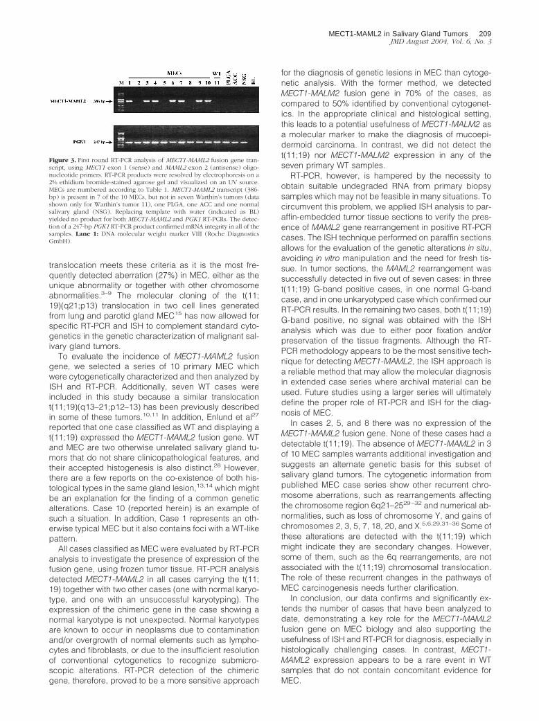
To investigate the expression of the MECT1-MAML2 chi-meric gene, we carried out RT-PCR using tumor mRNA(Table 1). This analysis revealed the expression of therearrangement between MECT1 and MAML2 genes inseven of the 10 (70%) mucoepidermoid carcinomas, butin none of the seven WT (Figure 3). Sequencing analysis
MECT1-MAML2 in Salivary Gland Tumors 207JMD August 2004, Vol. 6, No. 3

of the positive RT-PCR products confirmed the presenceof the same rearrangement in the seven MECs, with thefusion product between MECT1 exon 1 and MAML2 exon2. There was no expression of this rearrangement eitherin the two salivary glands carcinomas (ACC and PLGA)or in the normal salivary gland used as controls (Figure3). We also carried out nested RT-PCR, which confirmedthe results obtained in the first PCR (data not shown).The 247-bp RT-PCR product for the PGK1 housekeep-ing gene was detected in all 20 samples, indicating
adequate sampling and confirming mRNA integrity(Figure 3).
Discussion
A chromosomal translocation may be considered char-acteristic for a given cancer if it is detected frequentlyand, most importantly, if it occurs as the sole cytogeneticabnormality. The t(11;19)(q21;p12–13) chromosomal
Figure 1. Mucoepidermoid carcinoma case 6 with a t(11;19)(q21;p13). a: Case 6 is a typical MEC, with mucin producing cells (ii), intermediate (iii), and squamous(i) cells (H&E). b: Representative karyotype showing the t(11;19) as the sole karyotypic abnormality. c: FISH analysis on a metaphase spread with the BAC clonesprobes RP11–16K5 (green) and RP11–676L3 (red) for MAML2 gene showing the intragenic rearrangement of the gene: the hybridization probes are either locatedimmediately adjacent in normal chromosome 11, or separated, one (green) from RP11–16k5 on der19 and the other (red) from RP11–676L3 on der11. d: FISHanalysis on a paraffin tumor section with the BAC probes RP11–16K5 (red) and RP11–676L3 (green) for MAML2 gene showing the intragenic rearrangement ofthe gene in interphase nuclei: the hybridization signals from both BAC probes are either located in close proximity indicating the normal gene MAML2 or separatedindicating rearrangement of the gene. e: CISH analysis of a tumor section from paraffin-embedded tissue. Both BAC clones probes RP11–16K5 and RP11–676L3for MAML2 gene were labeled with FITC and detected with DAB-chromogen (brown). There is an intragenic rearrangement of the gene in interphase nuclei: threebrown spots could be observed representing one of them the normal copy of MAML2 gene and two smaller signals the split of the gene. f: CISH analysis of normalsalivary gland tissue, of the same section (e), using the same BAC probes, showing two spots per nucleus.
Figure 2. Mucoepidermoid carcinoma case 10 with a t(11;16;19)(q21;p13.3;p13). a: In low-power view the tumor presents cystic spaces, lined by eosinophyliccells and has a prominent lymphoid infiltration with follicules with germinal centers. Higher power demonstrates that the lining of the cysts is composed of thethree cell populations that are diagnostic of MEC (H&E). b: Representative karyotype showing the t(11;16;19)(q21;p13.3;p13). c: FISH analysis on a metaphasespread with the BAC clones RP11–16K5 (green) and RP11–676L38 (red) for MAML2 gene showing the intragenic rearrangement of the gene: both hybridizationsignals are immediately adjacent in normal chromosome 11, the hybridization signal from RP11–16k5 on der16 and the hybridization signal from RP11–676L3 onder11.
208 Martins et alJMD August 2004, Vol. 6, No. 3
translocation meets these criteria as it is the most fre-quently detected aberration (27%) in MEC, either as theunique abnormality or together with other chromosomeabnormalities.3–9 The molecular cloning of the t(11;19)(q21;p13) translocation in two cell lines generatedfrom lung and parotid gland MEC15 has now allowed forspecific RT-PCR and ISH to complement standard cyto-genetics in the genetic characterization of malignant sal-ivary gland tumors.
To evaluate the incidence of MECT1-MAML2 fusiongene, we selected a series of 10 primary MEC whichwere cytogenetically characterized and then analyzed byISH and RT-PCR. Additionally, seven WT cases wereincluded in this study because a similar translocationt(11;19)(q13–21;p12–13) has been previously describedin some of these tumors.10,11 In addition, Enlund et al27
reported that one case classified as WT and displaying at(11;19) expressed the MECT1-MAML2 fusion gene. WTand MEC are two otherwise unrelated salivary gland tu-mors that do not share clinicopathological features, andtheir accepted histogenesis is also distinct.28 However,there are a few reports on the co-existence of both his-tological types in the same gland lesion,13,14 which mightbe an explanation for the finding of a common geneticalterations. Case 10 (reported herein) is an example ofsuch a situation. In addition, Case 1 represents an oth-erwise typical MEC but it also contains foci with a WT-likepattern.
All cases classified as MEC were evaluated by RT-PCRanalysis to investigate the presence of expression of thefusion gene, using frozen tumor tissue. RT-PCR analysisdetected MECT1-MAML2 in all cases carrying the t(11;19) together with two other cases (one with normal karyo-type, and one with an unsuccessful karyotyping). Theexpression of the chimeric gene in the case showing anormal karyotype is not unexpected. Normal karyotypesare known to occur in neoplasms due to contaminationand/or overgrowth of normal elements such as lympho-cytes and fibroblasts, or due to the insufficient resolutionof conventional cytogenetics to recognize submicro-scopic alterations. RT-PCR detection of the chimericgene, therefore, proved to be a more sensitive approach
for the diagnosis of genetic lesions in MEC than cytoge-netic analysis. With the former method, we detectedMECT1-MALM2 fusion gene in 70% of the cases, ascompared to 50% identified by conventional cytogenet-ics. In the appropriate clinical and histological setting,this leads to a potential usefulness of MECT1-MALM2 asa molecular marker to make the diagnosis of mucoepi-dermoid carcinoma. In contrast, we did not detect thet(11;19) nor MECT1-MALM2 expression in any of theseven primary WT samples.
RT-PCR, however, is hampered by the necessity toobtain suitable undegraded RNA from primary biopsysamples which may not be feasible in many situations. Tocircumvent this problem, we applied ISH analysis to par-affin-embedded tumor tissue sections to verify the pres-ence of MAML2 gene rearrangement in positive RT-PCRcases. The ISH technique performed on paraffin sectionsallows for the evaluation of the genetic alterations in situ,avoiding in vitro manipulation and the need for fresh tis-sue. In tumor sections, the MAML2 rearrangement wassuccessfully detected in five out of seven cases: in threet(11;19) G-band positive cases, in one normal G-bandcase, and in one unkaryotyped case which confirmed ourRT-PCR results. In the remaining two cases, both t(11;19)G-band positive, no signal was obtained with the ISHanalysis which was due to either poor fixation and/orpreservation of the tissue fragments. Although the RT-PCR methodology appears to be the most sensitive tech-nique for detecting MECT1-MAML2, the ISH approach isa reliable method that may allow the molecular diagnosisin extended case series where archival material can beused. Future studies using a larger series will ultimatelydefine the proper role of RT-PCR and ISH for the diag-nosis of MEC.
In cases 2, 5, and 8 there was no expression of theMECT1-MAML2 fusion gene. None of these cases had adetectable t(11;19). The absence of MECT1-MAML2 in 3of 10 MEC samples warrants additional investigation andsuggests an alternate genetic basis for this subset ofsalivary gland tumors. The cytogenetic information frompublished MEC case series show other recurrent chro-mosome aberrations, such as rearrangements affectingthe chromosome region 6q21–2529–32 and numerical ab-normalities, such as loss of chromosome Y, and gains ofchromosomes 2, 3, 5, 7, 18, 20, and X.5,6,29,31–36 Some ofthese alterations are detected with the t(11;19) whichmight indicate they are secondary changes. However,some of them, such as the 6q rearrangements, are notassociated with the t(11;19) chromosomal translocation.The role of these recurrent changes in the pathways ofMEC carcinogenesis needs further clarification.
In conclusion, our data confirms and significantly ex-tends the number of cases that have been analyzed todate, demonstrating a key role for the MECT1-MAML2fusion gene on MEC biology and also supporting theusefulness of ISH and RT-PCR for diagnosis, especially inhistologically challenging cases. In contrast, MECT1-MAML2 expression appears to be a rare event in WTsamples that do not contain concomitant evidence forMEC.
Figure 3. First round RT-PCR analysis of MECT1-MAML2 fusion gene tran-script, using MECT1 exon 1 (sense) and MAML2 exon 2 (antisense) oligo-nucleotide primers. RT-PCR products were resolved by electrophoresis on a2% ethidium bromide-stained agarose gel and visualized on an UV source.MECs are numbered according to Table 1. MECT1-MAML2 transcript (386-bp) is present in 7 of the 10 MECs, but not in seven Warthin’s tumors (datashown only for Warthin’s tumor 11), one PLGA, one ACC and one normalsalivary gland (NSG). Replacing template with water (indicated as BL)yielded no product for both MECT1-MAML2 and PGK1 RT-PCRs. The detec-tion of a 247-bp PGK1 RT-PCR product confirmed mRNA integrity in all of thesamples. Lane 1: DNA molecular weight marker VIII (Roche DiagnosticsGmbH).
MECT1-MAML2 in Salivary Gland Tumors 209JMD August 2004, Vol. 6, No. 3
Acknowledgments
We thank Lucia Roque, B.Sc., Ph.D. and Valeriano Leite,M.D., Ph.D. for scientific advice and critical reading of themanuscript.
References
1. Ellis GL, Auclair PL: Tumors of the salivary glands. Atlas of TumorPathology. Edited by Rosai J, Sobin LH. Washington DC, ArmedForces Institute of Pathology, 1996, p 17
2. Mitelman F, Johansson B, Mertens F: Mitelman Database of Chromo-some Aberrations in Cancer. http://cgap.nci.nih.gov/Chromosomes/Mitelman.2003
3. Dahlenfors R, Lindahl L, Mark J: A fourth minor salivary gland muco-epidermoid carcinoma with 11q14–21 and 19p12 rearrangements.Hereditas 1994, 120:287–288
4. Dahlenfors R, Wedell B, Rundcrantz H, Mark J: Translocation (11;19)(q14–21;p12) in a parotid mucoepidermoid carcinoma of a child.Cancer Genet Cytogenet 1995, 79:188
5. Hrynchack M, White V, Berean K, Horsman D: Cytogenetic findings inseven lachrymal gland neoplasms. Cancer Genet Cytogenet 1994,75:133–138
6. Nordkvist A, Gustafsson H, Juberg-Ode M, Stenman G: Recurrentrearrangements of 11q14–22 in mucoepidermoid carcinoma. CancerGenet Cytogenet 1994, 74:77–83
7. El-Naggar AK, Lovell M, Killary A, Clayman G, Batsakis J: A muco-epidermoid carcinoma of the minor salivary gland with t(11;19)(q21;p13.1) as the sole karyotypic abnormality. Cancer Genet Cytogenet1996, 87:29–33
8. Johansson M, Mandahl N, Johansson L, Hambraeus G, Mitelman F,Heim S: Translocation 11;19 in a mucoepidermoid tumor of the lung.Cancer Genet Cytogenet 1995, 80:85–86
9. Stenman G, Petursdottir V, Mellgren G, Mark J: A child with a t(11;19)(q14–21;p12) in a pulmonary mucoepidermoid carcinoma. Vir-chows Arch 1998, 433:579–581
10. Bullerdiek J, Haubrich J, Meyer K, Bartnizke S: Translocation t(11;19)(q21;p13.1) as the sole chromosome abnormality in a cystadenolym-phoma (Warthin’s tumor) of the parotid gland. Cancer Genet Cyto-genet 1988, 35:129–132
11. Mark J, Dahlenfors R, Stenman G, Nordquist A: A human adenolym-phoma showing the chromosomal aberrations del(7)(p12;p14–15)and t(11;19)(q21;p12–13). Anticancer Res 1989, 9:1565–1566
12. Luna MA: Salivary mucoepidermoid carcinoma: revisited. Proceed-ings of the Joint Meeting of the Working Group on Head and NeckPathology of the European Society of Pathology and the North Amer-ican Society of Head and Neck Pathology of the United States andCanadian Society of Pathology. Ed. Gale N, Luzar B. Ljubljana, 2003,pp 99–105
13. Seifert G: Mucoepidermoid carcinoma in a salivary duct cyst of theparotid gland: contribution to the development of tumours in salivarygland cysts. Pathol Res Pract 1996, 192:1211–1217
14. Williamson JD, Simmons BH, El-Naggar AK, Medeiros J: Mucoepi-dermoid carcinoma involving Warthin tumor. Am J Clin Pathol 2000,114:564–570
15. Tonon G, Mody S, Wu L, Kubo A, Coxon AB, Komiya T, O’Neil K,Stover K, El-Naggar AK, Griffin JD, Kirsch IR, Kaye FJ: t(11;19)(q21;p13) translocation in mucoepidermoid carcinoma creates a novelfusion product that disrupts a Notch signaling pathway. Nat Genet2003, 33:208–213
16. Conkright MD, Canettieri G, Screaton R, Guzman E, Miraglia L, Ho-genesch JB, Montminy M: TORCs: transducers of regulated CREBactivity. Mol Cell 2003, 12:413–423
17. Iourgenko V, Zhang W, Mickanin C, Daly I, Jiang C, Hexham JM, OrthAP, Miraglia L, Meltzer J, Garza D, Chirn GW, McWhinnie E, Cohen D,Skelton J, Terry R, Yu Y, Bodian D, Buxton FP, Zhu J, Song C, Labow
MA: Identification of a family of cAMP response element-bindingprotein co-activators by genome-scale functional analysis in mam-malian cells. Proc Natl Acad Sci USA 2003, 100:12147–12152
18. Seifert G, Sobin LH. Histological typing of salivary gland tumours.World Health Organization International Histological Classification ofTumours, ed 2. New York, Springer-Verlag, 1991
19. Batsakis JG, Luna MA: Histopathologic grading of salivary glandneoplasms: i. Mucoepidermoid carcinoma. Ann Otol Rhinol Laryngol1990, 99:835–838
20. Jin Y, Mertens F, Jin C: Non-random chromosome abnormalities inshort-term cultured primary squamous cell carcinomas of the headand neck. Cancer Res 1995, 55:3204–3210
21. ISCN: An International System for Human Cytogenetic Nomenclature.Edited by Mitelman F. Basel, Karger, 1995
22. Hoglund M, Johansson B, Pedersen-Bjergaard J, Marynen P, Mitel-man F: Molecular characterization of 12p abnormalities in hemato-logic malignancies: deletion of KIP1, rearrangement of TEL, andamplification of CCND2. Blood 1996, 87:324–330
23. Alers JC, Krijtenburg PJ, Vissers KJ, Krishnadath SK, Bosman FT,Van Dekken H: Interphase in situ hybridization to disaggregated andintact tissue specimens of prostatic adenocarcinomas. HistochemCell Biol 1995, 104:479–486
24. Jenkins B, Quian J, Lee HK, Huang H, Hirasawa K, Bostwick DG,Proffitt J, Wilber K, Lieber MM, Liu W, Smith DI: A molecular cytoge-netic analysis of 7q31 in prostate cancer. Cancer Res 1998, 58:759–766
25. Sugg SL, Zheng L, Rosen IB, Freeman JL, Ezzat S, Asa SL: ret/PTC-1,-2, and -3 oncogene rearrangements in human thyroid carcinomas:implications for metastatic potential? J Clin Endocrinol Metab 1996,81:3360–3365
26. Martins C, Fonseca I, Roque L, Soares J: Cytogenetic characteriza-tion of Warthin’s tumour. Oral Oncol Eur J Cancer 1997, 33:334–347
27. Enlund F, Behboudi A, Andren Y, Oberg C, Lendahl U, Mark J,Stenman G: Altered Notch signaling resulting from expression of aWANTP1-MAML2 gene fusion in mucoepidermoid carcinomas andbenign Warthin’s tumors. Exp Cell Res 2004, 292:21–28
28. Seifert G, Brocheriou C, Cardesa A, Eveson JW: WHO InternationalHistological Classification of Tumors: tentative histological classifica-tion of salivary gland tumors. Pathol Res Pract 1990, 186:555–581
29. Sandros J, Mark J, Happonen R, Stenman G: Specificity of 6q-markers and other recurrent deviations in human salivary gland tu-mors. Anticancer Res 1988, 8:637–644
30. Jin Y, Mertens F, Limon J, Mandhal N, Wennerberg J, Dictor M, HeimS, Mitelman F: Characteristic karyotypic features in lachrymal andsalivary gland carcinomas. Br J Cancer 1994, 70:42–47
31. Jin C, Martins C, Jin Y, Wiegant B, Wennerberg J, Dictor M, D.Gisselsson, B. Strombeck F: Mitelman, Tanke H, Fonseca I, HoglundM, Mertens F: Characterization of chromosome aberrations in salivarygland tumors by fluorescence in situ hybridization (FISH), includingmulticolor COBRA-FISH. Genes Chromosomes Cancer 2001, 30:161–167
32. Martins C, Fonseca I, Roque L, Pinto AE, Soares J: Malignant salivarygland neoplasms: a cytogenetic study of 19 cases. Oral Oncol Eur JCancer 1996, 32B:128–132
33. Horsman DE, Berean K, Durham JS: Translocation t(11;19)(q21;p13.1) in a mucoepidermoid carcinoma of salivary gland. CancerGenet Cytogenet 1995, 80:165–166
34. Nordkvist A, Staffan E, Mark J, Stenman G: Multiple unrelated chro-mosome abnormalities in a metastatic mucoepidermoid carcinoma ofthe parotid gland. Cancer Genet Cytogenet 1992, 61:158–161
35. El-Naggar AK, Lovell M, Killary A, Batsakis J: Trisomy 5 as the solechromosomal abnormality in a primary mucoepidermoid carcinomaof the minor salivary gland. Cancer Genet Cytogenet 1994, 76:96–99
36. El-Naggar AK, Lovell M, Killary A, Batsakis J: Genotypic character-ization of a primary mucoepidermoid carcinoma of the parotid glandby cytogenetic, fluorescence in situ hybridization, and DNA ploidyanalysis. Cancer Genet Cytogenet 1996, 89:38–43
210 Martins et alJMD August 2004, Vol. 6, No. 3