The Fine Structure of the Kasparov Groups II: Topologizing the UCT
Upload
khangminh22Category
view
0download
0
Louisiana State UniversityLSU Digital Commons
LSU Historical Dissertations and Theses Graduate School
1981
A Study of Ascus Developmental Fine Structure inEleutherascus Peruvianus Huang With SpecialReference to Ascospore Ontogeny.Walstine Lon Steffens IIILouisiana State University and Agricultural & Mechanical College
Follow this and additional works at: https://digitalcommons.lsu.edu/gradschool_disstheses
This Dissertation is brought to you for free and open access by the Graduate School at LSU Digital Commons. It has been accepted for inclusion inLSU Historical Dissertations and Theses by an authorized administrator of LSU Digital Commons. For more information, please [email protected].
Recommended CitationSteffens, Walstine Lon III, "A Study of Ascus Developmental Fine Structure in Eleutherascus Peruvianus Huang With SpecialReference to Ascospore Ontogeny." (1981). LSU Historical Dissertations and Theses. 3701.https://digitalcommons.lsu.edu/gradschool_disstheses/3701

INFORMATION TO USERS
This was produced from a copy of a document sent to us for microfilming. While the most advanced technological means to photograph and reproduce this document have been used, the quality is heavily dependent upon the quality of the material submitted.
The following explanation of techniques is provided to help you understand markings or notations which may appear on this reproduction.
1. The sign or “target” for pages apparently lacking from the document photographed is “ Missing Page(s)”. If it was possible to obtain the missing page(s) or section, they are spliced into the film along with adjacent pages. This may have necessitated cutting through an image and duplicating adjacent pages to assure you of complete continuity.
2. When an image on the film is obliterated with a round black mark it is an indication that the film inspector noticed either blurred copy because of movement during exposure, or duplicate copy. Unless we meant to delete copyrighted materials that should not have been filmed, you will find a good image of the page in the adjacent frame. If copyrighted materials were deleted you will find a target note listing the pages in the adjacent frame.
3. When a map, drawing or chart, etc., is part of the material being photographed the photographer has followed a definite method in “sectioning” the material. It is customary to begin filming at the upper left hand corner of a large sheet and to continue from left to right in equal sections with small overlaps. If necessary, sectioning is continued again—beginning below the first row and continuing on until complete.
4. For any illustrations that cannot be reproduced satisfactorily by xerography, photographic prints can be purchased at additional cost and tipped into your xerographic copy. Requests can be made to our Dissertations Customer Services Department.
5. Some pages in any document may have indistinct print. In all cases we have filmed the best available copy.
UniversityMicrofilms
International300 N. ZEEB RD.. ANN ARBOR, Ml 48106

8207840
Steffens, Walstinc Lon, IH
A STUDY OF ASCUS DEVELOPMENTAL FINE STRUCTURE IN ELEUTHERASCUS PERUVIANUS HUANG W ITH SPECIAL REFERENCE TO ASCOSPORE ONTOGENY
The Louisiana State University and Agricultural and Mechanical Col Ph.D. 1981
UniversityMicrofilms
International 300 N. Zeeb Road, Ann Arbor, M I 48106

PLEASE NOTE:
tn all cases this material has been filmed in the best possible way from the available copy. Problems encountered with this document have been identified here with a check mark V .
1. Glossy photographs or pages 1/2. Colored illustrations, paper or print______
3. Photographs with dark background t X
4. Illustrations are poor copy______
5. Pages with black marks, not original copy ______
6. Print shows through as there is text on both sides of page______
7. Indistinct, broken or small print on several pages______
8. Print exceeds margin requirements_____
9. Tightly bound copy with print lost in spine_____
10. Computer printout pages with indistinct print______
11. Page(s)____________lacking when material received, and not available from school orauthor.
12. Page(s)____________seem to be missing in numbering only as text follows.
13. Two pages numbered____________. Text follows.
14. Curling and wrinkled pages
15. Other____________________________________________________________________ _
UniversityMicrofilms
International

A STUDY OF ASCUS DEVELOPMENTAL FINE STRUCTURE IN ELEUTHERASCUS PERUVIANUS HUANG WITH SPECIAL REFERENCE TO ASCOSPORE ONTOGENY
A Dissertation
Submitted to the Graduate Faculty of the Louisiana State University and
Agricultural and Mechanical College in partial fulfillment of the requirements for the degree of
Doctor of Philosophy
in
The Department of Plant Pathology and Crop Physiology
byWalstine Lon Steffens III
B.S., University of Texas at El Paso, 1974 M.S., University of Texas at El Paso, 1976
December, 1981

ACKNOWLEDGEMENTS
At the approaching end of ray graduate career I can now reflect
on the contributions of all who have made it possible and contributed
to its successful course.
My parents encouraged me to seek a college education and sup
ported me entirely through all of ray undergraduate and much of my
graduate career. Dr. J. T. Ellzey of U. T. El Paso introduced me
to science and started ray graduate career. The Department of Zoology
and Physiology at LSU employed me and allowed the use of their
facilities for my research.
My advisor, Dr. John Paul Jones set up my degree plan and
started me on the project which has culminated in this dissertation.
His encouragement and advice throughout these four years is gratefully
acknowledged.
My committee members, Drs. S. C. Tucker, N. L. Horn, K. Derrick,
G. Holcomb, and L. D. Newsom critically reviewed the manuscript and
provided enumerable suggestions for its improvement. Their assistance
in this matter is gratefully acknowledged.
I must especially acknowledge the patience and fortuity of my
wife Nancy in seeing me through ray graduate career. Her encourage
ment and eternal faith in me is the essence of my perseverence.
Jackie Lockwood typed this dissertation.

TABLE OF CONTENTS
Page
ACKNOWLEDGEMENTS i i
LIST OF FIGURES............................................. v
ABSTRACT...................................................... xiii
CHAPTER
I GENERAL INTRODUCTION................................... 1
Materials and Methods................................ 2
II EARLY GROWTH AND DEVELOPMENT............................ 7
Introduction.......................................... 7
Results.............................................. 9
Discussion........................ 56
III ASCOSPORE DELIMITATION................................. 62
Introduction.......................................... 62
Results............... 65
Discussion............................................ 81
IV SPORE WALL DEVELOPMENT.................................. 86
Introduction......................................... 86
Results............................................. 90
Discussion..............................................151
V ASCOSPORE GERMINATION .................................. 169
Introduction............................................ 169
Results........... 170
Discussion..............................................181
LITERATURE CITED................................................188
APPENDIX....................................................... 195
iii

VITA

LIST OF FIGURES
Figure Page
1 A brightfield light micrograph of an Eleutherascus peruvianus squash mount showing vegetative hyphae and several ascocarps containing from one toseveral asci.......................................... 11
2 A Giemsa-stained hypha of _E. peruvianus showing nucleiand a single septum.................................. 11
3 An electron micrograph of a vegetative hypha containing microbodies and numerous mitochondria ................. 13
4 A vegetative hypha of .E. peruvianus containing a pairof haploid nuclei.................................... 13
5 A vegetative hypha showing two nuclei, endoplasmic reticulum, and vacuoles .............................. 15
6 A hyphal septum with a large vacuole filling part of the hyphal lumen, and two Woronin Bodies adjacent tothe septum............................................ 15
7 A scanning electron micrograph of vegetative hyphaewith several septal areas evident ..................... 19
8 A cross section through a hypha parallel to a nearby septum, showing several mitochondria and WoroninBodies............................................... 19
9 A vegetative hyphal septum and pore with a Woronin Body occluding the pore. One Woronin Body has the typical repeating structure .................................. 21
10 A hyphal septum with two Woronin Bodies surrounded bya membranous vesicle.................................. 21
11 A brightfield micrograph of a Giemsa-stained early ascogenous apparatus showing a short ascogenous hypha being derived from the intertwining of two hyphalbranches............................................. 23
12 A phase contrast micrograph of a short ascogenous hypha beginning to bend into a characteristic hook........... 23
13 A Giemsa-stained early dikaryotic crozier with apartially inflated penultimate cell ................... 23
14 The equivalent stage as seen in figure 13 in phasecontrast..............................................- 23
v

Figure Page
15 A scanning electron micrograph of an entire ascogenousapparatus with an expanding crozier at a stage equivalent to that seen in figure 14........................ 25
16 A Giemsa-stain of an expanding crozier showingkaryogamy........................................... 25
17 A crozier soon after karyogamy, showing the largeGiemsa-stained diploid nucleus........................ 25
18 The fully inflated globose diploid ascus of _E. peruvianus showing a Giemsa-stained nucleus inearly prophase I with 5 pairs of chromosomes........... 25
19 The binucleate ascus, Giemsa-stained, followingMeiosis I ............................................ 28
20 The Giemsa-stained tetranucleate ascus followingMeiosis I I .......................................... 28
21 The Giemsa-stained ascus following mitosis andpreceding ascospore delimitation....................... 28
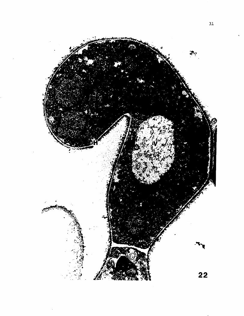
22 An electron micrograph of a non-septate, recurved ascogenous hypha showing 3 of the 4 nuclei and thebasal septum.......................................... 30
23 Part of an ascogenous hypha during the initiation of swelling of the penultimate cell, showing 2 nuclei anda glycogen-rich cytoplasm ............................ 32
24 An entire ascogenous apparatus of El. peruvianus with a partially expanded diploid penultimate cell and singlelarge diploid nucleus................................ 36
25 An electron micrograph of an early crozier showing the haploid tip cell, and swelling diploid penultimate cell with a vacuolate cytoplasm. The septal pore andpore cap of the ascus are also visible. . . . . . . . . . 38
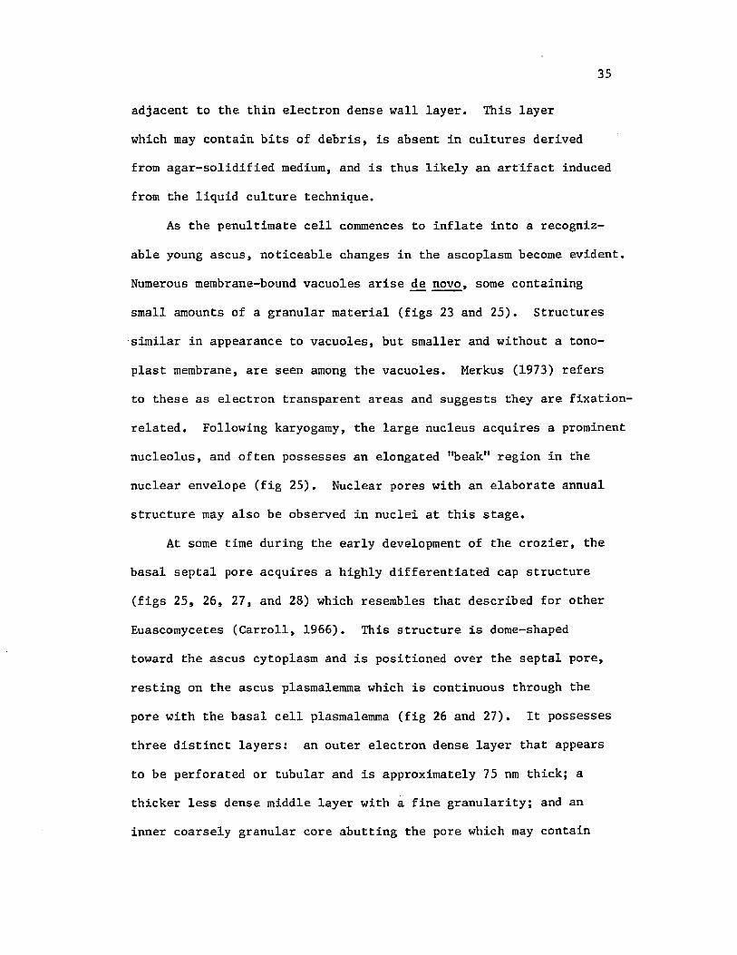
26 A median section through a septal pore cap of Eh peruvianus showing the three layered structure of this organelle overlying the septal pore in the ascoplasm, and some accompanying microbodies and the continuityof the plasmalemma through the septal pore............. 40
27 A median section through a septal pore cap showing some dense tubular material contained within the septal pore.A persistent Woronin Body remains near the septum. . . . 42
vi

Figure Page
28 A septal pore cap of JE. peruvianus..................... 42
29 An expanding ascus of E. peruvianus with the typically homogenous ascoplasm, large central diploid nucleus, and the swelling of an early subopercular ring nearthe apex............................................. 45
30 A section through the subopercular ring (arrow) in the ascus wall at a slightly later stage of developmentthan is seen in figure 2 9 ............................ 47
31 A section through a fully developed subopercular ring in the early ascus of El. peruvianus showing some associated membrane structures and a ribosome-freezone................................................. 47
32 A cross-section through a fully inflated ascus showinga nucleus with a nucleolus containing an electron transparent area and cytoplasm showing an initial predivisional vacuolization .......................... 49
33 An ascus nucleus prior to the first meiotic divisionshowing a prominent nucleus-associated organelle........ 49
34 The detail of a nucleus-associated organelle showingthe typical tripartite appearance ..................... 52
35 A cross-section through an ascus of _E. peruvianus following Meiosis I, and showing 2 nuclei with NAO's,and a vacuolate cytoplasm............................ 52
36 A highly fenestrated ascal nucleus following Meiosis II . 54
37 Four of the 8 nuclei in an ascus following the finaldivision. . ........................................ 54
38 Early formation of the ascus vesicle, showing a doublemembrane system accumulating below the ascus plasma-lemma................................................ 66
39 The formation of a double membrane segment with structure resembling the ascus vesicle by invagination ofthe ascus plasmalemma ................................ 66
40 Invagination of the ascus plasmalemma to form a longdouble membrane segment . . . . ....................... 69
vii

Figure Page
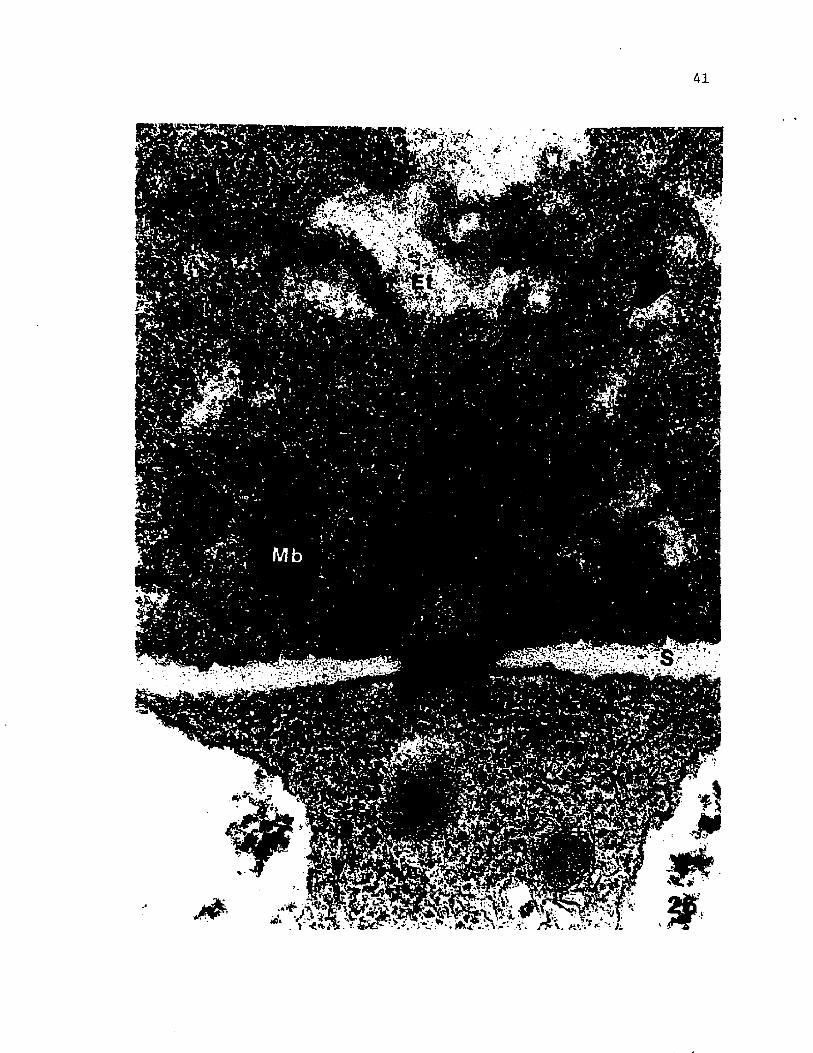
41 An ascus of El. peruvianus during ascus vesicle formation, showing the accumulation of several double-membrane segments in the ascoplasm belowthe ascus plasmalemma ................................ 71
42 A lomasome formed by the invagination and involution of the ascus plasmalemma, and containing vesiclesand membrane sheets.................................. 73
43 An elongate membranous whorl which likely represents a lomasome of the ascus plasmalemma and is closely appressed by the double membrane of the developingascus vesicle........................................ 73
44 Two lomasomes of the ascus plasmalemma with a closely appressed layer of the ascus vesicledouble membrane .............................. 75
45 A phase contrast micrograph of an ascus showing the accumulation of large flattened vesicles in the ascoplasm which will ultimately fuse to form theascus vesicle........................................ 75
46 Part of the ascus vesicle surrounding a singlenucleus near the basal septum......................... 77
47 A Giemsa-stained ascus containing 8 nuclei, each newly delimited in a double membrane by theinvaginated ascus vesicle ............................ 77
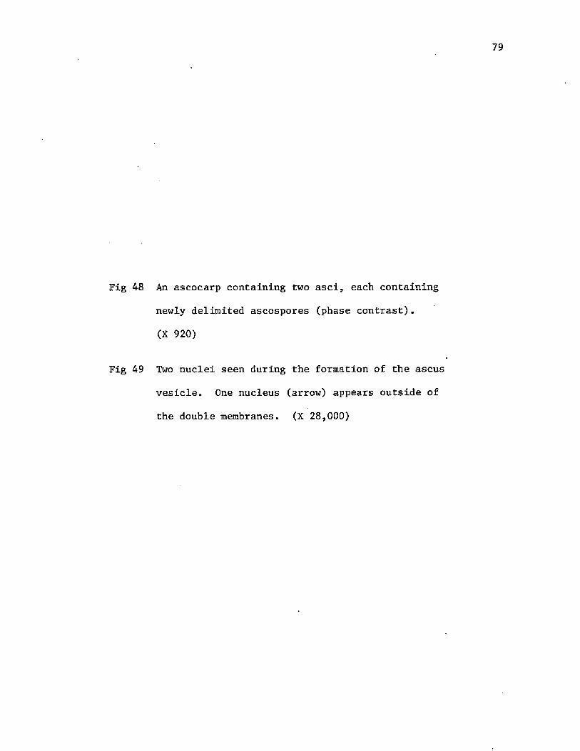
48 A phase contrast micrograph of an ascocarp containing2 asci, each containing newly delimited ascospores. . . . 79
49 Two nuclei seen during the formation of the ascus vesicle. One nucleus appears outside of the double membrane............................................. 79
50 The ascoplasm during ascus vesicle invagination around the nuclei, showing several segments of microtubules in association with the doublemembranes............................................ 82
51 A median section through an ascus showing 4 early ascospores, each with a thin, electron transparent primary wall and similar morphology and compositionin sporoplasm and epiplasm............................ 90
viii

Figure Page
52 An early ascospore with the first appearance of a granular material in the primary wall adjacent tothe plasmalemma...................................... 92
53 An early ascospore with an uneven primary wall, a nucleus with complex annuli, and a vacuolatesporoplasm............................................ 95
54 An ascospore of E. peruvianus with a fully developed primary wall, and sporoplasm differing in densityand morphology from the epiplasm....................... 97
55 An ascospore with a primary wall just prior to secondary wall deposition. The epiplasm is rich in glycogen and contains a hypertrophied tubularstructure........................................... 99
56 The initial appearance of secondary wall on anascospore of _E. peruvianus............................ 99
57 Early secondary wall deposition and an epiplasm with vacuoles containing a dense globular material.A layer of endoplasmic reticulum overlies theinvesting membrane.................................... 102
58 The epiplasm during secondary wall development. An area containing a large amount of glycogen isevident............................................. 102
59 A Giemsa-stained ascus containing 8 ascospores at the time of earliest secondary wall deposition,showing outgrowths of secondary walls ................. 105
60 The early deposition of the secondary wall showing the appearance and accumulation of several dense 65 nm globules in the finely granular matrix ofthe secondary wall.................................... 107
61 An ascospore with early developing secondary wallstained with silver methenamine only................... 110
62 Progressive development of the secondary wall, showing elongation of the dense globules in theareas of greatest deposition, ....................... 112
63 Ascospores and epiplasm during early secondary wall deposition. A large accumulation of epiplasmic glycogen is apparent and its relationship to thedeveloping secondary wall may be noted................. 115
ix

Figure Page
64 A darkfield micrograph of an ascus containing ascospores showing appearance of secondary wall ornamentations........................................ 117
65 Secondary wall formation in an ascospore, indicating the uneven deposition rate which gives rise to the typical pattern of ornamentation................................................... 117
66 An ornamentation produced by differential deposition of the secondary wall. The projection has a triple layer of endoplasmic reticulum overlying the investing membrane, and glycogen outside of these membranes . . 120
67 The developing secondary walls of two ascospores, each with endoplasmic reticulum overlying the areas of greater deposition which form theornamentation ........................................ 122
68 An ascospore during early secondary wall deposition,showing an exaggerated evagination of the investing membrane............................................. 125
69 The evagination of and fusion between the investing membranes of adjacent ascospores. Secondary wallis being deposited between the adjacent spores......... 125
70 An ascospore at the climax of secondary wall deposition. The primary wall is beginning to acquireadditional layers below the secondary wall............. 127
71 Early differentiation of the primary wall. The narrow band underlying the secondary wall is seento increase in density................................ 129
72 An ascospore showing the four layers of the episporeand electron transparent endospore..................... 129
73 A median section through an ascus following secondary wall deposition showing 5 ascospores with typical ornamentation, and the degenerate appearance of theepiplasm............................................. 132
74 A fully ornamented ascospore prior to final maturation with dense sporoplasm and several lipid droplets . . 134
75 Ascospore maturation showing early deposition of the perispore and the apparent action of epiplasmiccomponents in its formation.......................... 137
x

Figure Page
76 The wall of the mature ascospore of Eh peruvianus showing the needle-like ornamentations, developing perispore, and significant density increase in endospore and ascoplasm. Vesicular structuresappear in the endospore.............................. 137
77 The fully mature ascospore wall with parallel microfibrils in the secondary wall, and well-developed perispore.................................. 139
78 A mature ascospore stained with silver methenamine only, showing heavy localization in endospore, the middle epispore layer, secondary wall, and theremaining epiplasm.................................... 142
79 A phase contrast micrograph of 2 Eh peruvianus asci,one of which is becoming pigmented..................... 145
80 Two asci containing fully mature and pigmentedascospores............................................ 145
81 A scanning electron micrograph of an isolated, mature ascospore of E. peruvianus showing the typical ornamentations consisting of spines, short ridges, and a longitudinal brim which spirals aroundthe spore........... 147
82 An isolated ascospore with a polygonal longitudinalbrim which circumscribes the spore..................... 147
83 A face-on view of the longitudinal brim of anisolated ascospore.................................... 149
84 A brightfield micrograph of 2 medially sectioned asci containing mature ascospores and demonstratingthe orderly arrangement of the ascospores ............. 149
85 Detail of a medially sectioned ascus demonstratingthe relationships between brims of adjacent spores. . . . 149
86-88 Stereo pairs of asci containing mature ascospores which have been cut into 5 ym sections, etched, and viewed by SEM. The relationships of the brims between adjacent spores and the continuity of this relationship along the periphery of the brim margins may be observed...................................... 152
89 Detail of a spore mass isolated from an ascus showingfusion between projections on adjacent spores ......... 154
xi

Figure Page
90 An isolated spore mass showing the often tenacious aggregation between spores within an.ascus............... 156
91 The wall of an ascus containing germinating ascospores, showing the increased contrast between the layers . . . . 171
92 A germinating ascospore prior to exit of the germ tube, demonstrating the multinucleate state andaltered morphology of sporoplasm and spore wall ......... 173
93 Detail of the spore wall of a germinating ascospore,showing the decrease in overall thickness of thesecondary wall and the newly appearing electrontransparent areas .................................... 177
94 A germinating ascospore of _E. peruvianus showing the septate germ tube and mitochondria-richnormal appearing sporoplasm .......................... 179
95 A germinating ascospore stained with silvermethenamine and showing sporoplasraic localizationin vacuolar contents and in the germ tube wall......... 182
96 Detail of a silver methenamlne-stained germinating ascospore demonstrating the globular vacuolarcontents............................................. 182
97 Detail of the germ tube wall of a germinating ascospore showing initial deposition within the exit pore, and the dense areas in the endosporeunderlying the ornamentations ........................ 184
98 A silver methenamine-stained germinating ascospore showing the reaction of the morphologically altered ascospore wall........................................ 184
xii
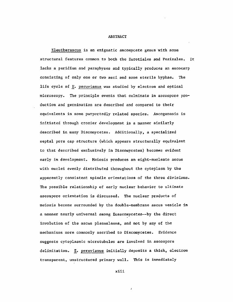
ABSTRACT
Eleutherascus is an enigmatic ascomycete genus with some
structural features common to both the Eurotiales and Pezizales.
lacks a peridium and paraphyses and typically produces an ascocarp
consisting of only one or two asci and some sterile hyphae. The
life cycle of _E. peruvianus was studied by electron and optical
microscopy. The principle events that culminate in ascospore pro
duction and germination are described and compared to their
equivalents in some purportedly related species. Ascogenesis is
initiated through crozier development in a manner similarly
described in many Discomycetes. Additionally, a specialized
septal pore cap structure (which appears structurally equivalent
to that described exclusively in Discomycetes) becomes evident
early in development. Meiosis produces an eight-nucleate ascus
with nuclei evenly distributed throughout the cytoplasm by the
apparently consistent spindle orientations of the three divisions.
The possible relationship of early nuclear behavior to ultimate
ascospore orientation is discussed. The nuclear products of
meiosis become surrounded by the double-membrane ascus vesicle in
a manner nearly universal among Euascomycetes— by the direct
involution of the ascus plasmalemma, and not by any of the
mechanisms more commonly ascribed to Discomycetes. Evidence
suggests cytoplasmic microtubules are involved in ascospore
delimitation. J2. peruvianus initially deposits a thick, electron
transparent, unstructured primary wall. This is immediately

followed by the deposition of a secondary wall at varying rates over
the surface of the primary wall through the activity of the epiplasm.
The appearance of complex membranous connections between adjacent
ascospores, and the deposition of secondary wall material along these
sites is suggested as the mechanism for the formation of the asco
spore brims and the resultant appearance of orderly arrangements of
the spores within the ascus. Germination is by a rupture of the
spore wall and the emergence of a germ tube which is derived de novo
beneath the spore wall layers. Cytological and cytochemical evidence
favor the notion that the spore wall is partially resorbed. by the
germinating sporoplast.
xiv

I. GENERAL INTRODUCTION
Sporogenesis in ascomycetous fungi has been an avidly researched
area in the field of fungal developmental biology. Two basic reasons
for this are the great diversity of form among ascomycetes, and the
amenability of many species to laboratory culture and manipulation.
For these reasons as well as their economic significance, a large
amount of this research has been focused on the Hemiascomycetes,
perhaps to the neglect of some other groups. The Discomycetes have
received possibly more attention than other Euascomycete subclasses,
but are still actually understudied when the actual numbers and
diversity of species are considered.
Eleutherascus is a genus containing 4 simple, highly reduced
species. It is particularly interesting taxonomically, as some of
its members have at some time been placed among the Eurotiales (Korf,
1973), Endomycetales (Batra, 1973), and currently occupy a place in
the Discomycete order Pezizales (von Arx, 1971; Benny and Kimbrough,
1980). Cytological and ultrastructural information on this genus is
currently lacking.
Eleutherascus peruvianus was originally isolated from a soil
sample from Peru and described as a new species of Eleutherascus
(Huang, 1975). It differs from the other species in morphology of
the ascospores, cultural characteristics (Huang, 1975), and lack of
a conidial state (Samson and Luiten, 1975). The asci, being produced
free in or on the substrate without an enclosing peridium j can be
studied dji situ and are well suited for preparation for

2
ultramicroscopy. In many respects, this Discomycete is as well
suited for an extensive ultrastructural investigation as many
Hemiascomycetes.
A study was undertaken with _E. peruvianus to examine it cyto-
logically and to compare the events which culminate in the produc
tion of mature, viable ascospores with those events in some of the
purportedly related species. This investigation will provide
additional information both on developmental processes that are
common to ascomycetes in general and on those which make Eleutherascus
an enigmatic genus.
Materials and Methods
Culture Technique
A pure culture of Eleutherascus peruvianus Huang obtained from
Dr. R. T. Hanlin, University of Georgia was subcultured and maintained
on slants of rabbit food agar (RFA) in 25 ml screw cap apothecary
bottles. The medium was prepared by steeping 30 grams of commercial
rabbit food per liter of deionized water, then filtering through
cheesecloth. This medium stock was then either autoclaved, or mixed
with agar to a final concentration of 1.5% and then autoclaved.
Cultures to be utilized for study were grown on solid RFA or on
rabbit food broth in 10 cm petri plates. The inoculated plates were
incubated 22°C in the dark until the colonies had attained a diameter
of approximately 2 cm, at which time they were thereafter given 8
hours of light and 16 hours dark. Material to be studied by micro
scopy was periodically examined as unfixed, unstained squash mounts
by phase contrast microscopy. Mycelium or agar blocks containing
the desired stages were transferred to vials of the appropriate
fixative, or were examined and photographed as squash mounts in
bright field or phase contrast microscopy.
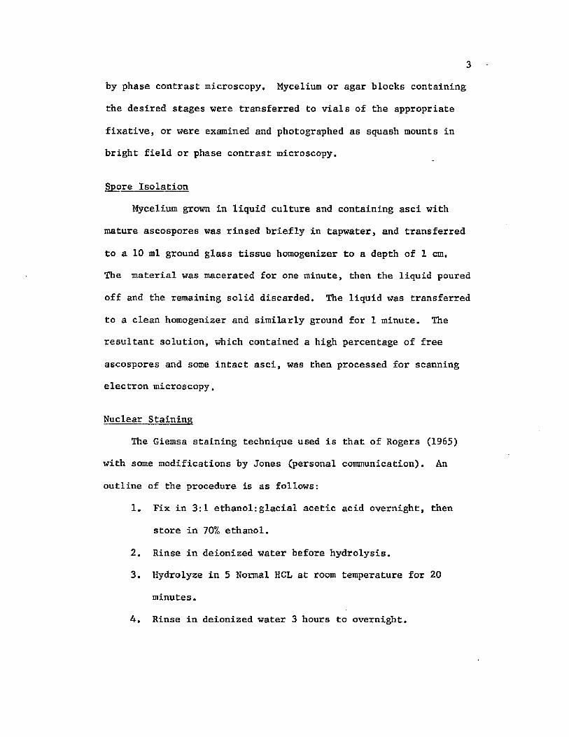
Spore Isolation
Mycelium grown in liquid culture and containing asci with
mature ascospores was rinsed briefly in tapwater, and transferred
to a 10 ml ground glass tissue homogenizer to a depth of 1 cm.
The material was macerated for one minute, then the liquid poured
off and the remaining solid discarded. The liquid was transferred
to a clean homogenizer and similarly ground for 1 minute. The
resultant solution, which contained a high percentage of free
ascospores and some intact asci, was then processed for scanning
electron microscopy.
Nuclear Staining
The Giemsa staining technique used is that of Rogers (1965)
with some modifications by Jones (personal communication). An
outline of the procedure is as follows:
1. Fix in 3:1 ethanol:glacial acetic acid overnight, then
store in 70% ethanol.
2. Rinse in deionized water before hydrolysis.
3. Hydrolyze in 5 Normal HCL at room temperature for 20
minutes.
4. Rinse in deionized water 3 hours to overnight.

5. Place in 0.15 molar phosphate buffer at pH 7.2 for five
minutes.
6. Place material on a slide, blot excess liquid, and add
two drops of buffer and one drop of Giemsa stain (3 grams
Giemsa stain + 250 grams of glycerin + 250 grams of
methanol). Stain for two minutes.
7. Rinse well with water by transferring material to several
drops of water.
8. Add one drop of 1% KOH, apply a coverslip and observe.
Photographs were taken on a Zeiss Standard microscope equipped
with a Nikon Camera using Kodak Type 3646 Ektachrome film.
Transmission Electron Microscopy
Material to be prepared for embedding and thin-sectioning
was fixed for 1-2 hours in 2.57. glutaraldehyde-2.57. acrolein in
0.1 M sodium cacodylate buffer at pH 7.1 and 4°C. The mycelium was
then rinsed in several changes of 0.1 M sodium cacodylate buffer
at pH 7.1. Post-fixation was for three hours at room temperature
in 0.1 M cacodylate-buffered 27. osmium tetroxide. Following three
five minute rinses in deionized water, the material was left over
night in 0.57. aqueous uranyl acetate, then rinsed in deionized
water. Dehydration was through an acetone series followed by rotary
infiltration and embedment in Spurr (1969) resin.
Ultrathin sections (60nm) were cut with a diamond knife on
either a Sorval MT-1 or MT-2B ultramicrotome and mounted on either
copper, nickel, or gold grids. Post-staining was with uranyl
acetate-saturated 507. ethanol and lead citrate. Photographs were

5
taken with either an Hitachi HU11-A operating at 75 Ke V or a
JEOL 100CX operating at 80 Ke V. All electron micrographs were
taken on DuPont Ortho Cronar film.
Silver Methenamine Staining
The method used in this study is as described by Martino
and Zamboni (1967) with some modifications! The technique is
outlined as follows:
1. Ultrathin sections mounted on gold grids are treated with
three five minute changes in 0.5% acetic acid.
2. The sections are rinsed in deionized water, then oxidized
for 20 minutes at room temperature in 1% periodic acid.
3. Following a rinse in deionized water, the sections are
incubated 30 minutes at 60°C in the dark in the silver
methenamine solution [20 ml of 3% methenamine (hexamethylene-
tetraraine) + 1 ml of 5% sodium borate + 1 ml of 5% silver
nitrate].
4. Following a rinse in deionized water, the sections are
immersed in a 1% gold chloride solution for 5 minutes.
5. Following another rinse in deionized water, the sections
are immersed in a 5% sodium thiosulfite solution.
6. The sections are finally rinsed in deionized water and
observed in the TEM.
Scanning Electron Microscopy
Material to be mounted for SEM was fixed in FAA (75 ml ethanol:
20 ml formalin:5 ml glacial acetic acid) for 1-7 days, then rinsed

in deionized water for 15 minutes with 3 changes. It was then
placed in 75 mesh baskets, and dehydrated in an acetone series.
The material in absolute acetone was critical point dried through
CC>2 in a Denton Critical Point Dryer, then mounted on aluminum or copper stubs with silver paint. Ten nanometers of gold-palladium
were applied with an Edwards Sputter Coater, and the specimens
examined with either a Hitachi S-500 Scanning Electron Microscope
operating at 25 Ke V or a JEOL 100CX Transmission Electron
Microscope equipped with an ASID-4 high resolution scanning attach
ment operating at 40 Ke V.

XX. EARLY GROWTH AND DEVELOPMENT
Introduction
In a research project which is to deal with the appearance,
development, and ultimate fate of a specific organ or part of an
organism, it is deemed appropriate to describe adequately the con
ditions and state of the organism prior to the appearance of the
structures in question. Most of the Eumycota produce an extensive,
usually filamentous vegetative thallus prior to the formation of the
reproductive units. Considerable information on the structure and
ontogeny of the vegetative thallus was available even before appli
cation of the electron microscope to fungal development. deBary
(1866), in his famous treatise on thallic organisms described the
ascomycete hypha with its perforate septations and multinucleate
condition. Earlier, Woronin (1864) had noted the relationship
between the septations in ascomycete hyphae and the small granules
which now bear his name. A century later, the electron microscope
revealed the plasmalemraal continuity between hyphal septations,
indicating the actual and functional coenocytic nature of the
ascomycete thallus. Carroll (1966) presented an extensive review
of the septal apparatus literature, and additionally described the
highly specialized septum of the ascogenous system. Bracker (1967)
elaborated on hyphal ultrastructure and attempted to correlate
structure and composition with function and physiology.
7

The anatomy of the ascomycete vegetative thallus can be
generalized, with expected differences in size ranges, specific
inclusions, and abundance of specific organelles among differing
species. Hyphal diameter is usually in the range of 8-12 pm, with
the interseptal distance varying considerably even within a
species. Each septa consists of a plate with a simple central pore
which may be up to 0.5 yim in diameter (Bracker, 1967). The pore is
equipped with at least one Woronin Body on either side, which is
known to act as a plug during cell senescence, and possibly serves
to prevent the migration of large organelles through the septal
pore. The organelles and inclusions composing the hyphal cytoplasm
are detailed extensively elsewhere (Bracker, 1967).
The first account of ascus development through crozier forma
tion with the accompanying nuclear divisions was in the Disco-
mycete Peziza (Dangeard, 1894). Several light microscopic studies
since then in a variety of species have confirmed this as an almost
universal process in the higher ascomycetes, with only a few
notable exceptions (see Reeves, 1967;' Wells, 1970 for a complete
review). For nearly a quarter of a century now, the early forma
tion and development of the undifferentiated ascus has been well
documented in the higher ascomycetes at the fine structural level
(Carroll, 1966; Reeves, 1967; Beckett and Wilson, 1968; Carroll,
1969; Beckett and Crawford, 1970; Wells, 1970, 1972; Blanchard,
1972; Ranalli and Forchiassin, 1976). The importance of
fully understanding the early nuclear and other cytological events
which occur prior to the appearance of the ascospores relates

9
to the taxonomic significance of the ascus, and to the relevance of
these events in a holistic approach to this type of investigation.
In this ultrastructural investigation, there is emphasis on all
aspects of ascosporogenesis and the ultimate state of the ascus.
It is deemed particularly important to describe and bring into
context in the classical sense the events of early morphogenesis,
and concurrently develop a picture of the unique events and
processes that precisely define Eleutherascus.
Results
Cultural Characteristics
Inoculation of fresh rabbit food agar medium with Eleutherascus
peruvianus results in the initiation of visible hyphal growth within
one day. Under the standard culture conditions as outlined pre
viously, the average rate of increase in colonies' less than 3 days
old was about 2-3 mm per day. When the colony reached 1 cm in
width, the growth rate was seen to increase dramatically, with
5-6 cm usually reached in 6 days. Colonies are almost entirely
subsurface on rabbit food agar, with a few sterile aerial hyphae
appearing in young cultures and a slightly more floccose non-
reproductive aerial mycelium in older cultures.
Ascocarps begin to appear within 4 days of inoculation and2rapidly increase to prodigous numbers, with up to 500 per cm not
being uncommon. Eleutherascus peruvianus characteristically pro
duces a highly reduced ascocarp which normally consists only of
1 or 2 globose asci and a few sterile hyphae (fig 1). Neither
paraphyses nor a peridium is present.

10
Hyphal Cytology and Ultrastructure
Six to eight nuclei occupy each hyphal cell in the vegetative
mycelium (fig 2). Nuclei are of variable shape, often occur in
pairs, and generally possess a single large acentric nucleolus
(fig 4). The nuclear envelope is often highly convoluted,
particularly when the nuclear density is high. The annuli appear
to be simple in these somatic nuclei, with no structural complexity
evident at any plane of section or orientation. Mitotic figures
were not observed, nor was any appreciable degree of chromatin
condensation noted. The hyphae of an actively growing vegetative
culture have abundant organelles normally associated with active
growth and development. The hyphal walls have a slight electron
density with the fixation schedule used, have a single undiffer
entiated layer 400 to 500 run in thickness, although this tends to
narrow near the distal ends of the hyphae. The hyphal plasmalemma
appears just inside the wall, and is appressed tightly to it.
Mitochondria with cristae oriented parallel to the long axis of
the organelle occur in prodigous numbers, and are often seen in
close association with microbodies, particularly near septae
(fig 3).
Vacuoles of variable shape and size are consistently evident
and average 1.5 ym in diameter and are often bounded by a highly
convoluted single unit membrane (figs 4 and 5). As the age of the
culture increases, these vacuoles appear to fuse and eventually
form very large vacuoles which appress the cytoplasm against the
hyphal wall (fig 6). Other types of cytoplasmic structures

11
Fig 1 An Eleutherascus peruvianus squash mount showing
vegetative hyphae and several ascocarps containing
from one to several asci (brightfield). (X 230)
Fig 2 A Giemsa-stained hypha of _E. peruvianus showing
nuclei and a single septum. (X 2,300)

12

13
Fig 3
Fig 4
A vegetative hypha containing microbodies and numerous
mitochondria. (X 8,000)
Part of a vegetative hypha of _E. peruvianus containing
a pair of haploid nuclei. (X 16,500)

14
15
Fig 5 A vegetative hypha showing two nuclei, endoplasmic
reticulum, lipid bodies, and vacuoles. (X 11,000)
Fig 6 A hyphal septum with a large vacuole filling part
of the hyphal lumen, and two Woronin Bodies adjacent
to the septum. (X 22,000)

16

17
(lipid globules and endoplasmic reticulum) appear in maturing
cultures, the latter normally appearing in close association with
nuclei (fig 5),
The hyphal septal apparatus observed in _E. peruvianus is
typical of ascoraycetes in general (Bracker, 1967), Hyphae appear
expanded in the areas of the septae, and may be as much as 20%
wider here than in the interseptal regions (fig 7). The septal
pore measures about 1.5 pm in diameter, and is equipped with up
to-10 Woronin bodies on either side of the septal wall (fig 8).
Each Woronin Body measures approximately 2 pm in diameter and has
an internal repeating structure (fig 9). Older cultures or
mechanically damaged cells often have the septal pores plugged by
a Woronin Body (fig 9). Septae near growing tips may have a dense
membrane-bound sheath surrounding the Woronin Bodies and other
cytoplasmic structures (fig 10).
Ascus Ontogeny
Nuclear Cytology
Within 4-6 days after inoculation on rabbit food medium,
ascocarp initials may be observed. This process has been detailed
by Huang (1975) and was reviewed in this study only to determine
the ultimate positional fate of the nuclei from the reduction-
division through spore wall deposition.
Ascocarp initials are most commonly derived from the inter
twining of two short hyphal sidebranches arising from the same or
different main axes (fig 11). Huang (1975) suggests that one fila
ment serves as an antheridium and the other as an ascogonium;

18
however the actual transfer of nuclei from cell to cell has not
been reported. A short ascogenous hypha develops (fig 12) which
becomes septate and develops as a crozier. The inflating penulti
mate cell with its pair of nuclei (fig .13) expands rapidly,
assuming a pyriform shape within a few hours (figs 14 and 15).
At a time which may be just after the initiation of inflation
in the penultimate cell, but before it attains a globose appearance,
karyogamy occurs in the developing crozier (fig 16). The now diploid
crozier has a single large nucleus of about 5 pm and a very prominent
nucleolus (fig 17). The crozier will continue to expand to 30-35 pm
in cultures grown in the liquid medium in preparation for fixation.
When mature and fully inflated, it will have assumed a globose to
subglobose shape.
When expansion of the penultimate cell is complete, the
chromatin begins to condense into distinguishable chromocenters,
and finally into chromosomes (fig 18), The chromosome number
appears to be 2N = 10, consistent with numbers reported for other
purportedly related species. The nucleolus enlarges considerably
and acquires a central vacuole (fig 18), an observation which is
later supported by ultrastructural evidence.
Meiosis I gives rise to the binucleate state, with the products
migrating to opposing sides of the globose ascus (fig 19). There is
no evidence suggesting that this movement is anything more than a
passive spatial accommodation aside from the observation that it
always occurs in a similar fashion. One cannot determine if the
plane of this first division is regular because the asci are
globose and have no reliable reference points.

19
Fig 7
Fig 8
Vegetative hyphae with several septal areas evident
(scanning electron micrograph). (X 2,000)
A hypha parallel to a nearby septum, showing several
mitochondria and Woronin Bodies (transverse section).
‘ (X 17,000)


21
Fig 9
Fig 10
A vegetative hyphal septum and pore with a Woronin
Body occluding the pore. One Woronin Body (arrow)
has the typical repeating structure. (X 8,500)
A hyphal septum with two Woronin Bodies surrounded by
a membranous vesicle. (X 8,500)

22

Fig 11 A Giemsa-stained early ascogenous apparatus showing
a short ascogenous hypha (arrow) being derived from
the intertwining of two hyphal branches (brightfield
micrograph). (X 2,300)
Fig 12 A short ascogenous hypha beginning to bend into a
characteristic hook (phase contrast micrograph).
(X 920)
Fig 13 A giemsa-stained early dikaryotic crozier with a
partially inflated penultimate cell. (X 2,300)
Fig 14 The equivalent stage as seen in figure 13 in phase
contrast. (X 920)

24

25
Fig 15 An entire ascogenous apparatus with an expanding
crozier at a stage equivalent to that seen in
figure 14 (scanning electron micrograph). (X 2,400)
Fig 16 A Giemsa-stained expanding crozier showing karyogamy
(arrow). (X 2,300)
Fig 17 A crozier soon after karyogamy, showing the large
Giemsa-stained diploid nucleus. (X 2,300)
Fig 18 The fully inflated globose diploid ascus of E. peruvianus
showing a Giemsa-stained nucleus in early prophase I with
5 pairs of chromosomes (arrows). (X 2,300)

26
27
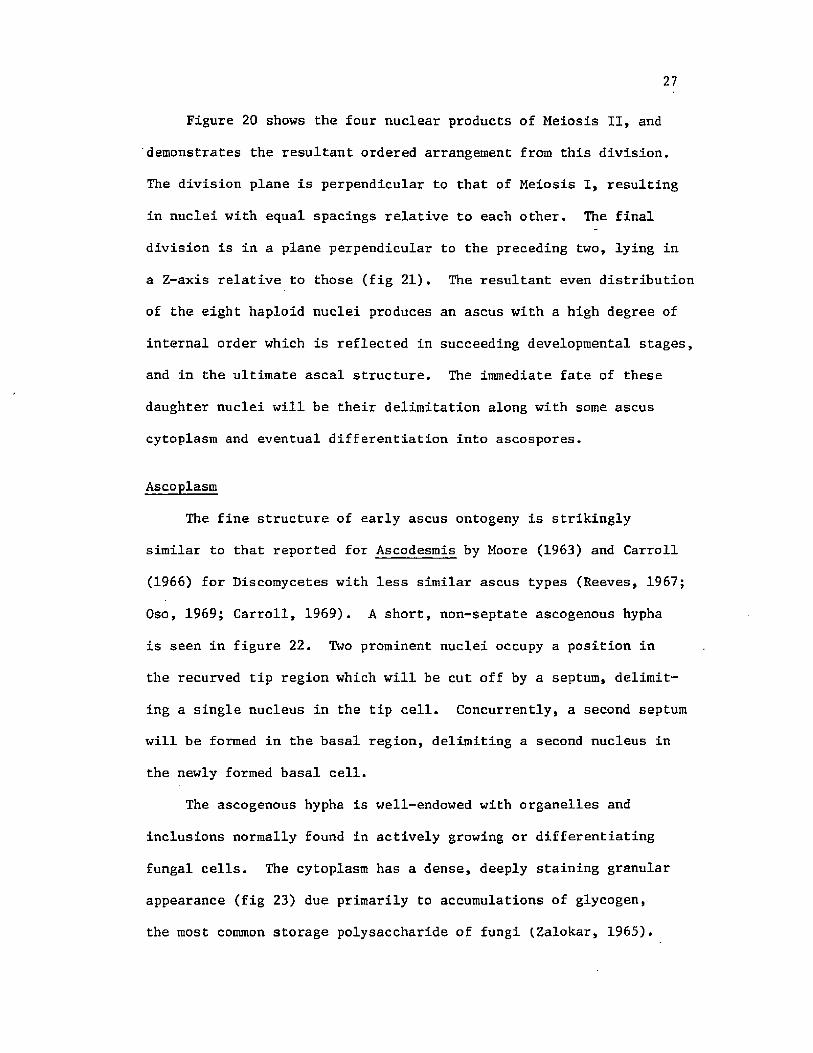
Figure 20 shows the four nuclear products of Meiosis II, and
demonstrates the resultant ordered arrangement from this division.
The division plane is perpendicular to that of Meiosis I, resulting
in nuclei with equal spacings relative to each other. The final
division is in a plane perpendicular to the preceding two, lying in
a Z-axis relative to those (fig 21), The resultant even distribution
of the eight haploid nuclei produces an ascus with a high degree of
internal order which is reflected in succeeding developmental stages,
and in the ultimate ascal structure. The immediate fate of these
daughter nuclei will be their delimitation along with some ascus
cytoplasm and eventual differentiation into ascospores.
Ascoplasm
The fine structure of early ascus ontogeny is strikingly
similar to that reported for Ascodesmis by Moore (1963) and Carroll
(1966) for Discomycetes with less similar ascus types (Reeves, 1967;
Oso, 1969; Carroll, 1969). A short, non-septate ascogenous hypha
is seen in figure 22. Two prominent nuclei occupy a position in
the recurved tip region which will be cut off by a septum, delimit
ing a single nucleus in the tip cell. Concurrently, a second septum
will be formed in the basal region, delimiting a second nucleus in
the newly formed basal cell.
The ascogenous hypha is well-endowed with organelles and
inclusions normally found in actively growing or differentiating
fungal cells. The cytoplasm has a dense, deeply staining granular
appearance (fig 23) due primarily to accumulations of glycogen,
the most common storage polysaccharide of fungi (.Zalokar, 1965).

28
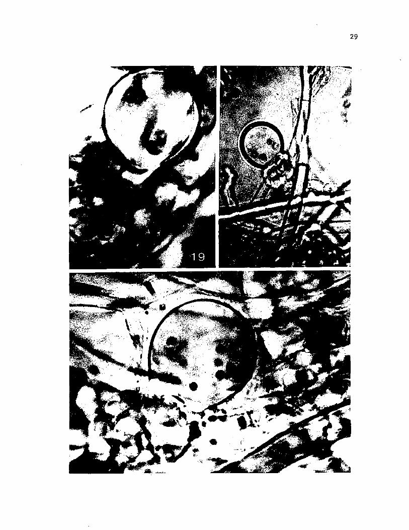
Fig 19 The binucleate ascus, Giemsa-stained, following
Meiosis I. (X 2,300)
Fig 20 The Giemsa-stained tetranucleate ascus following
Meiosis II. (X 920)
Fig 21 The Giemsa-stained ascus following mitosis and
preceding ascospore delimitation. (X 2,300)
29
30
Fig 22 A non-septate, recurved ascogenous hypha showing 3 of
the 4 nuclei and the basal septum (electron micrograph).
(X 10,000)
31

32
Fig 23 Part of an ascogenous hypha during the initiation of
swelling of the penultimate cell, showing 2 nuclei and
glycogen-rich cytoplasm. (X 30,000)

33

Schrantz’s (1968) description of ER-bound glycogen in the immature
ascus of Galactinia does not appear to apply in Eleutherascus.
Lipid droplets also may be found, but have not yet begun to
accumulate to the extent found in much later stages. Mitochondria
with flattened plate-like cristae are abundant, and accumulate
toward the central portion of the ascogenous cell, away from
incipient basal and tip cells. Microbodies with densely staining
contents and occasionally crystalline inclusions are also found,
usually associated with the double-membrane organelles. Membranous
structures are not widespread, and are limited mostly to short
sections of smooth endoplasmic reticulum appearing under the plas-
malemma (figs 22 and 23). Inclusions consisting of membranous
whorls and somewhat resembling lomasomes occur in limited numbers
near the plasmalemma; however they are unlike the true lomasomes
occurring in the mature ascus in Eleutherascus and described in
detail elsewhere (Wilsenach and Kessel, 1965; Bracker, 1967;
Marchant and Robards, 1968).
The wall of the incipient ascus in E. peruvianus is approxi
mately 100 nm In thickness and is seen to contain three layers when
stained conventionally for TEM (fig 23). The inner layer abutting
the plasmalenuna is electron transparent and 50 nm in thickness. A
middle layer averaging 15 nm in thickness is quite electron dense
and is occasionally striated parallel to the inner wall. Together,
these layers constitute an ascus wall similar to that shown by
Merkus (1973) for Ascodesmis and by others. Cultures grown in
liquid medium acquire a loose, fibrillar to floccose outer layer
35
adjacent to the thin electron dense wall layer. This layer
which may contain bits of debris, is absent in cultures derived
from agar-solidified medium, and is thus likely an artifact induced
from the liquid culture technique.
As the penultimate cell commences to inflate into a recogniz
able young ascus, noticeable changes in the ascoplasm become evident.
Numerous membrane-bound vacuoles arise jie novo, some containing
small amounts of a granular material (figs 23 and 25). Structures
similar in appearance to vacuoles, but smaller and without a tono-
plast membrane, are seen among the vacuoles. Merkus (1973) refers
to these as electron transparent areas and suggests they are fixation-
related. Following karyogamy, the large nucleus acquires a prominent
nucleolus, and often possesses an elongated "beak" region in the
nuclear envelope (fig 25). Nuclear pores with an elaborate annual
structure may also be observed in nuclei at this stage.
At some time during the early development of the crozier, the
basal septal pore acquires a highly differentiated cap structure
(figs 25, 26, 27, and 28) which resembles that described for other
Euascomycetes (Carroll, 1966). This structure is dome-shaped
toward the ascus cytoplasm and is positioned over the septal pore,
resting on the ascus plasmalerama which is continuous through the
pore with the basal cell plasmalemma (fig 26 and 27). It possesses
three distinct layers: an outer electron dense layer that appears
to be perforated or tubular and is approximately 75 nm thick; a
thicker less dense middle layer with a fine granularity; and an
inner coarsely granular core abutting the pore which may contain

36
Fig 24 An entire ascogenous apparatus of ji. peruvianus with a
partially expanded diploid penultimate cell and single
large diploid nucleus. (X 5,500)

37
t - - ” (’■vi ~~l

38
Fig 25 An early crozier showing the haploid tip cell (arrow),
and swelling diploid penultimate cell with a vacuolate
cytoplasm. The septal pore and pore cap of the ascus
are also visible (electron micrograph). (X 10,000)

39

40
Fig 26 A median section through a septal pore cap of _E.
peruvianus showing the three-layered structure of this
organelle overlying the septal pore in the ascoplasm,
and some accompanying microbodies and the continuity
of the plasmalemma through the septal pore. (X 83,000)
41
1 -T ‘••cA *-■ ’ • ‘1̂ * * £_iptegm

42
Fig 27 A median section through a septal pore cap showing
some dense tubular material contained within the
septal pore. A persistent Woronin Body remains near
the septum. (X 58,000)
Fig 28 A septal pore cap of _E. peruvianus. (X 54,000)

43

44
some dense material resembling the outer layer (fig 27). The
possible role of this structure during ascus development will be
discussed below.
An unusual feature of Eleutherascus which to my knowledge is
unreported in other Discomycetes is the retention of at least one
Woronin Body in the ascus preceding differentiation of the pore
cap (fig 27). This Woronin Body is apparently displaced from the
immediate vicinity of the pore, but remains at the septum.
Fortuitous sections of developing ascus initials reveal
evidence of an apical apparatus. Figures 29 and 30 show expanding
crozier cells with localized wall thickenings near the apex of the
mature ascus. Careful examination of serial sections through
several asci has failed to show that this structure girdles the
ascus. It appears to be a narrow, localized ingrowth in the ascus
wall. As ascus maturity advances, this ingrowth widens (fig 31),
and becomes surrounded by an electron transparent area and layers
of endoplasmic reticulum. The appearance, development, and subse
quent morphology of this wall ingrowth conforms to Samuelson's
(1978) description of the subopercular ring in Aleuria aurantia.
I have not observed any structural features of the apical ascus
wall which would suggest the presence of an operculum; however I
have not employed the differential staining techniques used by
Samuelson (1975) to examine the ascus wall.
The first visible ultrastructural sign of the impending meiotic
divisions is the appearance of an electron transparent area in the
nucleolus of the diploid nucleus (fig 32). As it lacks the membrane

45
Fig 29 An expanding ascus of E. peruvianus with the typically
homogenous ascoplasm, large central diploid nucleus,
and the swelling of an early subopercular ring near the
apex (arrow). (X 10,000)

46
r'V ^ U Liri A > ^

47
Fig 30 The subopercular ring (arrow) in the ascus wall at a
slightly later stage of development than is seen in
figure 29. (X 20,000)
Fig 31 A fully developed subopercular ring in the early ascus
of _E. peruvianus showing some associated membrane
structures and a ribosome-free zone. (X 68,000)

48
* y i % & . .
v 'mmmm

49
Fig 32 A cross-section through a fully inflated ascus showing
a nucleus with a nucleolus containing an electron
transparent area and cytoplasm showing an initial
predivisional vacuolization. (X 5,200)
Fig 33 An ascus nucleus prior to the first meiotic division
showing a prominent nucleus-associated organelle.
(X 21,000)

50

51
of a true tonoplast, this structure is actually not a vacuole as
it is often described at the light microscope level. The nuclear
envelope becomes highly fenestrated (figs 32 and 33). Light level
observations have indicated that nucleolar disintegration occurs
following chromosomal condensation. It is not certain if forma
tion of the nucleolar transparent area is a prelude to nucleolar
disappearance, because chromosomal condensation has not been
observed ultrastructurally. The space may be an artifact of the
fixation process.
Prior to Meiosis I, the nucleus acquires the typically asco-
mycetous spindle pole body (Aist and Williams, 1972) or nucleus-
associated organelle (NAO) (Heath, 1978). It is suggested that
this structure is present throughout the cell cycle (Girbardt,
1978), however I have only noted its appearance concurrently with
nuclear envelope fenestration (figs 33 and 34). The NAO is seen
to be a complex, osmiophilic tripartite structure lying on the
external nuclear envelope (fig 34), and resembles the structure
described by Wells (1970) in Ascobolus and by others in similar
ascomycetous forms (i.e. Ashton and Moens, 1979; Heath, 1978).
Meiosis I produces two daughter nuclei, each approximately
one-half the diameter of the parent (fig 35). It does not appear
that these nuclei fully reconstruct before entering Meiosis II,
as the NAO remains prominent, the nuclear envelope highly
fenestrated, and the nucleolus absent. With the two subsequent
divisions, the still fenestrated nuclei (figs 36, 37) become more

52
Fig 34 A nucleus-associated organelle of an ascal nucleus
showing the typical tripartite appearance. (X 75,000)
Fig 35 A cross-section through an ascus of Eh peruvianus
following Meiosis I, and showing two nuclei with
NAO's (arrows), and vacuolate cytoplasm. (X 3,500)

53
m

54
Fig 36 A highly fenestrated ascal nucleus following Meiosis II.
(X 27,000)
Fig 37 Four of the 8 nuclei in an ascus following the final
division. (X 14,000)

55

56
rounded and attain positions just below the ascus wall where they
will almost immediately become delimited along with some ascus
cytoplasm.
Discussion
The vegetative thallus of Eh peruvianus is similar to most
other Euascomycetes. The characteristic subsurface growth on
solid medium was initially responsible for some technical prepara
tive problems. It was recently reported (Kimbrough, personal
communication) that _E. peruvianus produces abundant surface
growth and sporulation on a particular medium.
Observations of the fine structure of hyphae in Eh peruvianus
demonstrate no exceptional differences from other Euascomycetes.
One interesting observation which may be typical of many ascomy-
cetes lies in the structure of the nuclear pores of somatic nuclei.
The absence of a complex annular structure could be fixation-
related (Bracker, 1967) or could reflect functional differences
between these and the later nuclei which possess this feature.
It is also conceivable that the number of the annuli decreases at
this time, thus lessening the chance of their being observed. The
role of the annuli is not well understood, although they may
function in the transport of material across the nuclear envelope
(DuPraw, 1972).
It can be concluded that j5. peruvianus initiates ascogenesis
through a crozier-hook formation process in a manner similar to
other members of the subclass Discomycetes. The entire sequence

of nuclear events is easy to follow, as karyogamy occurs at a
time when the expanding crozier is easily recognizable, and the
final division resulting in the nuclear octad is not obscured by
the accumulation of inclusions in the ascoplasm, as is often the
case. Although the entire sequence of nuclear events from fusion
to the final division culminating in the 8-nucleate ascus was not
observed, sufficient information was obtained to be able to
extrapolate the entire sequence of events with certainty.
Astral rays associated with the meiotic nuclei at one or more
of the divisions have been extensively reported by investigators
using light microscopy (Gjurasin, 1893; Harper, 1897; Faull, 1905;
Beckett and Crawford, 1970; Wells, 1970; see Olive, 1965 for a
complete review), Astral rays are not reported in any of the
numerous ultrastructural studies employing permanganate fixatives.
In my observations of _E. peruvianus with the light microscope,
astral rays were never observed in either the Giemsa preparations
or in living cultures using phase contrast or brightfield optics.
Failure to observe them however, does not preclude their existence.
The occasional presence of microtubules outside of the NAO after
Meiosis I possibly indicate either a highly reduced astral ray
complex or insufficient fixation. Wille (personal communication)
recognizes at least 2 classes of microtubules associated with
division. He indicates certain fixation parameters such as
temperature, duration, and the presence of divalent cations that
can induce rapid depolymerization of at least one type.

The ultimate globose shape of the ascus becomes readily
evident, often before karyogamy occurs. In studies of genera with
elongate asci, such as Sporormiella (Blanchard, 1972) and
Podospora (Beckett and Wilson, 1968), croziers form distinct hooks
with the tip cell being appressed to the basal cell early in
development, followed by a rapid elongation of the penultimate
cell. A true hook may not actually be formed in Eleutherascus,
where a generalized swelling of the penultimate cell following
septation of the ascogenous hypha results in a globose ascus. This
pattern of early development is responsible for the tip cell failing
to approach close to the basal cell which is necessary for a second
crozier to be derived in the classical manner: In many cases, X
have observed an ascus bearing mature ascospores which has the tip
still some distance from the basal cell. Since this was never
observed in ascocarps containing more than one ascus, it is tempting
to speculate that the second crozier arises through an ascogenous
hypha derived through the fusion of the tip and basal cells of the
first crozier.
In species with saccate or globose asci, spindle orientation
during meiosis is rather difficult to follow with certainty, which
may account for the tendency to ignore it in these species. Wells
(1970), and Ranalli and Forchiassin (1976) describe spindle orien
tations in 2 Pezizaceous forms (Ascobolus and Saccobolus) as being
vertical-vertical-transverse at Meiosis I, II, and III respectively.
The ultimate arrangement of the ascospores in these species with
elongate asci suggests a relation to spindle orientation. In a

59
less related form, Beckett and Wilson (1968) demonstrate an
additional consequence of consistent spindle orientation, where
the final transverse division in Podospora anserina (Sphaeriales)
results in the close proximity of non-sister + and - mating nuclei
which became included as a binucleate spore.
The pattern of spindle orientation that I think occurs in
Eleutherascus is the only one capable of distributing the nuclei
in an orderly fashion within the globose asci, outside of some
dynamic rearrangement process. No ultrastructural evidence exists
for the latter.
The fine structure of the ascus septum in the higher asco-
mycetes has been described variously (Carroll, 1967a; Bracker,
1967; Kimbrough and Benny, 1978). It is generally agreed that this
dome-shaped structure which develops early in ascus ontogeny is
composed of tightly packed tubules which make it functionally
porous. The role of the pore cap as a "subcellular sieve" which
would isolate the organelles and inclusions of the developing
ascus while still allowing for nutrient and fluid exchanges has
been postulated (Carroll, 1966). Little is known regarding the
assembly of the ascus pore cap; it may arise de novo in the
ascogenous hypha, or may be derived from pre-existing structures.
The occasional retention of Woronin Bodies in the asci of
Eleutherascus is unusual (Carroll, 1966; Bracker, 1967). Either
Woronin Bodies are initially present in the developing ascus and
most later tend to disappear, or their initial suppression is
incomplete.

60
The apparent lack of an operculum or equivalent apical apparatus
led to the inclusion of Eleutherascus in the Plectomycetes (Nicot
and Durand, 1969). Several other species of the genus also lack
this feature (Manoharachary, 1972; Huang and Schmitt, 1973; Batra,
1973; Huang, 1975; van Emden, 1975; Samson and Luiten, 1975).
Huang (1975) carefully examined two species of Eleutherascus and
two species of Ascodesmis, generally accepted as close relatives
(von Arx, 1971). He noted the discharge of ascospores through an
operculum in Ascodesmis, and the lack of this mechanism in
Eleutherascus. Samuelson (1978), in part of his extensive series
of the asci of the Pezizales, described the appearance of a sub-
opercular ring during the development of the apical apparatus in
Aleuria aurantia which resembles a similar structure observed in
Eleutherascus. As in Eleutherascus, the Aleuria structure
develops asymmetrically, is surrounded by a membrane complex,
and disappears after a certain point in development. The ascus
wall in Eleutherascus (unlike Aleuria) remains unchanged in
structure and thickness along its entirety, not forming obvious
operculura-subopercular regions. With the data available thus far,
it must be concluded that the question of ascospore liberation in
Eleutherascus is unresolved.
Several diverse functions have been presumed for the NAO in
developing asci. Wells (1970) likens it to a modified centriole.
Heslot (1958) suggested three functions: the control of division,
ascospore delimitation, and ascospore orientation. Additionally,
it has been implicated in the deposition of a Hemiascomycete

prospore wall in a manner different from the Euascomycetes (Moens,
1971; Davidow and Goetsch, 1978; Ashton and Moens, 1979; Davidow,
Goetsch, and Byers, 1980), and in the association of nuclei of
different mating types (Beckett and Wilson, 1968). Its role in
Euascomycete ascospore ontogeny other than division is associated
with microtubular arrays, such as those occurring in the astral
rays (Wells, 1970; Carroll, 1966). I did not find significant
numbers of microtubules, either nuclear or cytoplasmic in this
investigation; either they are elusive, or fixation was not
suitable.

III. ASCOSPORE DELIMITATION
Introduction
Theories on the mechanism of ascospore delimitation arose
with the earliest microscopic examinations of ascus ontogeny.
Carroll (1966) and Reeves (1967) extensively reviewed the litera
ture on ascospore delimitation and, in agreement with others
(Tyson and Griffiths, 1976) recognized four schools of thought
regarding the process: 1, Fusion of astral rays (Harper, 1897,
1900) based on the observation of astral rays at the beak of each
of the 8 nuclei, and the belief that they folded back over the
nuclei and fused laterally into thin membranes which delimited each
nucleus in a sac-like structure; this process of cell cleavage
from a larger protoplasmic mass was termed "free cell formation";
2, Delimitation by a sheet of material emitted from the central
body of the nucleus and which folded back over and delimited
each nucleus (Faull, 1905); 3, Fusion of vacuoles (Jones, 1926),
where a system of minute vacuoles which fused separated the
future nucleus and sporoplasm of each spore from the epiplasm;
4, Delimitation by the invagination of a membrane vesicle (Mittman,
1932; Chadefaud, 1943) which advocated the delimitation of the
nuclei by a membrane vesicle which initially encircled all 8
nuclei and then invaginated around each nucleus. Early ultra-
structural studies of yeasts (Hashimoto, Gerhardt, Conti, and
Naylor, 1960) conformed the latter hypothesis, and additionally62

indicated that the vesicle is actually constructed from a double
membrane layer. Through numerous ultrastructural investigations,
it is now generally agreed that ascospores of Euascomycetes are
initially delimited by a double membrane ascus vesicle (Moore,
1965; Wilsenach and Kessel, 1965; Beckett, 1966; Delay, 1966;
Reeves, 1966; Bracker, 1967; Carroll, 1967b, 1969; Beckett,
Barton, and Wilson, 1968; Beckett and Wilson, 1968; Greenhalgh
and Evans, 1968; Oso, 1969; Greenhalgh and Griffiths, 1970;
Laane, 1970; Mainwaring, 1971; Wells, 1972; Stiers, 1974, 1976;
Hill, 1975; Hohl and Streit, 1975; Jeng and Hubbes, 1980),
One basic question which has not been satisfactorily
resolved involves the origin of the double-membrane ascus vesicle.
The static image produced by transmission electron microscopy
has not answered all questions regarding the origin and fate of
such structures. Additionally, membranes are particularly prone
to inducible artifact through the preparative procedures. Several
theories for the origin of the double membranes and subsequent
formation of the ascus vesicle have been offered (Greenhalgh and
Griffiths, 1970) and may be grouped into three schools of thought:
1, Endoplasmic reticulum (Wilsenach and Kessell, 1965), based
on the structural similarities between the fragments of double
membrane and the elements of the endoplasmic reticulum, and the
proposition that a fusion of these elements lying below the ascus
plasmalemma and outside of the nuclei result in the enclosure
of the nuclei by the double membrane ascus vesicle; 2, Nuclear
blebbing (Carroll, 1967b, 1969; Oso, 1969); active nuclear

blebbing which produce smaller vesicles that appear to flatten
out and form short double-membrane segments that could fuse to
larger segments suggesting a nuclear origin for the ascus vesicle
membranes. These observations were later supported by Beckett
and Crawford (1970), Wells (1972), and Jeng and Hubbes (1980).
Stiers (1976) believes the ascus vesicle in Ceratocystis fimbriata
to arise from the fusion of numerous small vesicles, but did not
give the source of these vesicles. 3, A plasmalemmal origin, as
seen by Delay (1966) in the morphologies of the ascus vesicle
membranes and the ascus plasmalemma. In examining the fine
structure of the ascus vesicle membranes with high resolution
electron microscopy, Bracker (1967) affirmed the dissimilarity
of this structure to endoplasmic reticulum, and noted its
resemblance to the plasmalemma, suggesting that it arises by an
invagination of this membrane. Wells (1972) In his exhaustive
treatment of Ascobolus stercorarius, though supportive of the
nuclear blebbing hypothesis, admitted that a transformation from
ER-nuclear bleb membrane morphology to ascus vesicle membrane
morphology would be necessary and should be demonstrable if the
hypothesis were correct. Stiers (1974) and Campbell (1973)
affirm the resemblance of the ascus vesicle to the plasmalemma
and present evidence favoring Bracker’s hypothesis.
The mechanism by which the double membrane (or ascus vesicle)
actually delimits the ascospore is unknown, and few references
to it outside of matter-of-course observations have been
made. A method by which the ascus vesicle could invaginate

65
around each of the eight nuclei for the ultimate purpose of
their delimitation was initially proposed by Carroll (1966)
who suggested that astral rays, often very pronounced at this
stage, serve to act as a barrier, thus limiting the invagination
of the ascus vesicle. Bracker (1967) suggests a dynamic role of
astral ray microtubules in directing the vesicle membranes
from the periphery of the ascus. Beckett and Crawford (1970)
noted astral ray microtubule links between the archontosome
(NAO) and the ascus vesicle in Xylosphaera and interpret it as
supportive of Bracker's mechanism. Using current terminology,
the outer membrane of the vesicle is referred to as the
"investing membrane" while the inner one is destined to become
the spore plasmalemma. After the complete envelopment of the
prospore by the double membrane sac, spore wall synthesis may be
initiated.
Results
Formation of the Ascus Vesicle
Subsequent to the third division resulting in eight haploid
daughter nuclei, a series of long, flattened membranous sacs
appear in the ascoplasm, usually in close proximity to the plasma
lemma (fig 38). This double membrane system resembles the endo
plasmic reticulum in its general morphology and extent, but has
structural and staining similarities to the ascus plasmalemma.
Examination of serial sections through an ascus at this stage
generally will show obvious connections between this membrane

66
Fig 38 Early formation of the ascus vesicle, showing a double-
membrane system accumulating below the ascus plasmalemma.
(X 15,000)
Fig 39 The formation of a double membrane segment with structure
resembling the ascus vesicle by invagination of the ascus
plasmalemma (arrow). (X 33,000)

67
sk r.A

68
structure and the plasmalemma (figs 39 and 40), suggesting that
a series of marked focal invaginations of the ascus plasmalemma
form this series of membranes. As development progresses, this
double membrane complex underlies progressively more of the ascus
wall, and may abut one to several lomasomes of the plasmalemma
(fig 41). These structures may be quite complex involutions of the
plasmalemma (figs 42, 43, 44) which could suggest a role in the
synthesis of the double membrane complex; however I have seen no
direct evidence for this. As seen by light microscopy, these
membranes appear as flattened, discoid-shaped vesicles of
varying sizes which progressively increase in size until the
entire cytoplasm below the plasmalemma becomes occupied by them
(fig 45).
Nuclear Delimitation
Ultimately, the elongated sections of membrane fuse and
form the structure termed "ascus vesicle" (Reeves, 1967) or
"ascospore membrane" (Oso, 1969) (fig 46). This ascus vesicle
is seen to break and invaginate around each of the nuclei, thus
delimiting the nucleus and some ascus cytoplasm in a double
membrane sac (figs 47, 48). A few odd circumstances have been
observed where the ascus vesicle apparently forms internal to
one or more nuclei (fig 49). The result could be the elimination
of this nucleus from the spore delimitation process, thus resulting
in fewer than eight ascospores. The significance of this observa
tion will be discussed at a later time.

69
Fig 40 Invagination (arrow) of the ascus plasmalemma to form
a long double membrane segment. (X 37,000)

70
71
Fig 41 An ascus of _E. peruvianus during ascus vesicle formation,
showing the accumulation of several double-membrane
segments in the ascoplasra below the ascus plasmalemma,
(X 14,000)

72
V ■ •'**1

73
Fig 42 A lomasome formed by the invagination and involution of
the ascus plasmalemma, and containing vesicles and
membrane sheets. (X 64,000)
Fig 43 An elongate membranous whorl which likely represents a
lomasome of the ascus plasmalemma and is closely
appressed by the double membrane of the developing ascus
vesicle (arrow). (X 27,000)

74

75
Fig 44 Two lomasomes of the ascus plasmalemma with a closely
appressed layer of the ascus vesicle double membrane.
(X 27,000)
Fig 45 An ascus showing the accumulation of large flattened
vesicles in the ascoplasm which will ultimately fuse to
form the ascus vesicle (phase contrast). (X 920)

76

77
Fig 46 Part of the ascus vesicle (arrows) surrounding a single
nucleus near the basal septum. (X 15,000)
Fig 47 A Giemsa-stained ascus containing 8 nuclei, each newly
delimited in a double membrane by the invaginated ascus
vesicle. (X 2,300)

78
i i . - > : ' y ’ ~ . * *-* * ' r i p w *
79
Fig 48 An ascocarp containing two asci, each containing
newly delimited ascospores (phase contrast).
(X 920)
Fig 49 Two nuclei seen during the formation of the ascus
vesicle. One nucleus (arrow) appears outside of
the double membranes. (X 28,000)

1
81.
During ascus vesicle invagination, ascoplasmic microtubules
appear to radiate out from a local source for the first time in
the ascus and may actually penetrate the membranes of the ascus
vesicle (fig 50). Astral rays are not evident at any time during
development in Eleutherascus.
Discussion
Ascospore delimitation in E!. peruvianus appears similar in
all of its major features to the free cell formation believed
common to all Euascomycetes (Bracker, 1967; Turian, 1974). The
purported role of the ascus plasmalemma in the delimitation of
ascospores has been variously described. Syrop and Beckett
(1976) described ascospore delimitation in Taphrina by a direct
invagination of the plasmalemma. Greenhalgh and Griffiths (1970)
contended that the ascus plasmalemma gives rise to and is often
continuous with the membranes of the ascus vesicle in Hypoxylon.
A similar observation was made by Campbell (1973) in the Phacid-
iaceous Discomycete, Lophodermella. My observations of exagger
ated plasmalemmal invaginations continuous with underlying double
membrane complexes in _E. peruvianus support this hypothesis for
the origin of the ascus vesicle membranes. The prevalence of
ascus plasmalemmal lomasomes during the accumulation of the
double membrane segments and their disappearance with the climax
of this event may suggest a role in the derivation of this
structure. Hill (1975) describes structures of similar morphology
(myelinoid figures) in the Plectomycete Nannizia gypsea, and

82
Fig 50 The ascoplasm during ascus vesicle invagination around
the nuclei, showing several segments of microtubules in
association with the double membranes.
(X 56,000)

83
iH S l
?2&i£fe
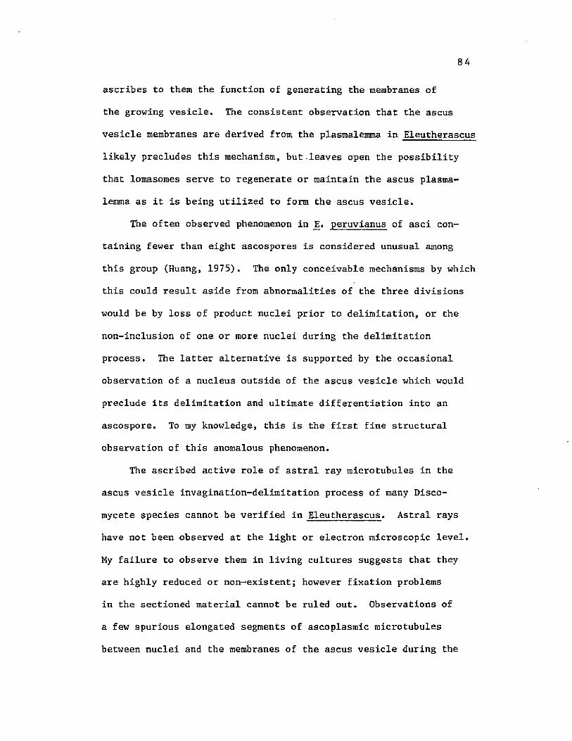
84
ascribes to them the function of generating the membranes of
the growing vesicle. The consistent observation that the ascus
vesicle membranes are derived from the plasmalemma in Eleutherascus
likely precludes this mechanism, but leaves open the possibility
that lomasomes serve to regenerate or maintain the ascus plasma
lemma as it is being utilized to form the ascus vesicle.
The often observed phenomenon in j3. peruvianus of asci con
taining fewer than eight ascospores is considered unusual among
this group (Huang, 1975). The only conceivable mechanisms by which
this could result aside from abnormalities of the three divisions
would be by loss of product nuclei prior to delimitation, or the
non-inclusion of one or more nuclei during the delimitation
process. The latter alternative is supported by the occasional
observation of a nucleus outside of the ascus vesicle which would
preclude its delimitation and ultimate differentiation into an
ascospore. To my knowledge, this is the first fine structural
observation of this anomalous phenomenon.
The ascribed active role of astral ray microtubules in the
ascus vesicle invagination-delimitation process of many Disco-
mycete species cannot be verified in Eleutherascus. Astral rays
have not been observed at the light or electron microscopic level.
My failure to observe them in living cultures suggests that they
are highly reduced or non-existent; however fixation problems
in the sectioned material cannot be ruled out. Observations of
a few spurious elongated segments of ascoplasmic microtubules
between nuclei and the membranes of the ascus vesicle during the

85
delimitation process, may suggest a highly reduced astral ray
complex which functions in the delimitation process in a manner
described earlier (Carroll, 1966; Bracker, 1967).

IV. SPORE WALL DEVELOPMENT
Introduction
A pattern of spore wall development universal in ascomycetes
involves the deposition of at least one wall layer between the
two membranes which delimit the ascospores. In some species,
particularly in the Hemiascomycetes, this may represent the only
deposited ascospore wall layer. Many of the earlier studies con
cluded ascospore wall deposition to be sporoplasmic (via the asco
spore cytoplasm), a conclusion that was probably biased from
observations of oospore and sporangiospore development, neither of
which has epiplasm and both of which obviously have sporoplasmic
centripetal wall deposition. Some elaborate mechanisms have been
proposed by which the various wall layers become deposited as a
single unit (Moore, 1963; Carroll, 1966; Reeves, 1967), many of
which are now questionable in light of modern investigative methods
and interpretation. It was deemed appropriate to investigate and
describe thoroughly the event of ascospore wall deposition and
structure, particularly that of the secondary wall and to compare
the results with earlier work. It is hoped that this information
will be of some use in a more modern holistic approach in determin
ing taxonomic relationships.
The deposition of wall layers has been described in detail by
many investigators in a great multitude of diverse species, which
has ultimately led to much confusion concerning the position of
86

the layers and the terminology applied. Although some earlier work
had been done using yeasts (Hashimoto et al., 1960), Moore (1963)
in his classical description of the spore wall deposition process
in Ascodesmis provided the initial insight into this event as it
occurs in the higher ascomycetes. Numerous additional descriptions
in the Euascomycetes have since been contributed (Wilsenach and
Kessel, 1965; Carroll, 1966; Bracker, 1967; Carroll, 1967b, 1969;
Reeves, 1967; Beckett, Barton, and Wilson, 1968; Beckett and
Wilson, 1968; Greenhalgh and Evans, 1968; Oso, 1969; Laane, 1970;
Mainwaring, 1971; Wells, 1972; Campbell, 1973; Merkus, 1973, 1974,
1975, 1976; Stiers, 1974, 1976; Turian, 1974; Conway, 1975; Hill,
1975; Hohl and Streit, 1975; Tyson and Griffiths, 1976; Jeng and
Hubbes, 1980; Johnson, 1980).
An electron transparent initial or primary wall layer deposited
between the layers of spore-delimiting membrane appears to be the
rule in the Euascomycetes. This is usually the thickest of the wall
layers and may undergo a differentiation into several additional
layers of varying thickness and density either prior to or after
the initiation of the secondary wall layer. The origin of this
wall layer, whether epiplasmic or sporoplasmic, is seldom discussed,
but by general consensus it is the same as that of the secondary
wall. The general lack of consistent, easily recognized structure
in the components of the primary wall has contributed to the lack
of understanding its ontogeny.
The secondary wall conversely is typically dense and often
highly structured, and has afforded numerous opportunities for

investigators to critically examine its appearance and fate. The
deposition of the secondary wall is of particular interest to the
mycologist in that it determines the final morphology of the
ascospores. Merkus (1973, 1974, 1975, 1976) has described the fine
structure of secondary wall deposition in several species of the
representative families of Pezizales. She is thus able to classify
genera as to pattern of secondary wall development and ascospore
type. From her organizational scheme, one can with the proper
information on a particular species acquire insight into probable
taxonomic relationships and possible phylogenic trends.
The morphology of the ascospores in E. peruvianus is one of the
primary features responsible for its inclusion among the Pezizales.
A wealth of information on the patterns of spore wall development
in most of the Discomycete families is now available. Variation
in the ultimate structure and morphology of ascospores has been
attributed to such factors as: glycogen content in the ascoplasm
(Schrantz, 1968; Merkus, 1975); behavior of the investing membrane
during secondary wall deposition (Merkus, 1976); pigment granule
sequestering by the secondary wall (Wells, 1972); and differentia
tion of the secondary wall structure (Merkus, 1976). If Eleuther
ascus does indeed belong in the Pezizales as suggested earlier
(von Arx, 1971; Benny and Kimbrough, 1980), an intensive study of
its method of ascospore morphogenesis would provide evidence
supportive of this contention.

89
Results
Primary Wall Secretion
Following delimitation, the earliest stage of primary wall
deposition observed in Eleutherascus is shown in figure 51. Each
ascospore initial or prospore is surrounded by a thin electron trans
parent wall which has appeared between the delimiting membranes. The
uneven thickness of the early primary wall suggests that its rate of
deposition around the spore is uneven. Merkus (1973) made a similar
observation in the developing ascospores of Ascodesmis.
From its earliest appearance, the primary wall appears to begin
an indistinct differentiation as it is still increasing in thickness.
A band of a very fine granular material appears in the primary wall
adjacent to the sporoplasmalemma (figs 52 and 53). It is of uniform
thickness, and unlike the wall as a whole, appears to be deposited
or derived evenly about the spore. This band will ultimately fill
the entire primary wall, imparting a slightly higher electron density
to it. The origin of this material is unclear, however its behavior
in appearing adjacent to the sporoplasmalemma and moving toward the
investing membrane suggests that it is secreted by the sporoplast,
although strong corresponding cytological evidence for this is lacking.
At this early period in primary wall synthesis, the sporoplasm
and epiplasm share many similarities and few differences. The most
notable difference is a lack of large vacuoles in the sporoplasm
of which the epiplasm has an abundance. A highly osmiophilic
granular material may appear in loose clumps within these vacuoles.
Sporoplasmic vacuoles are much smaller (about 100 nm), and contain a

90
Fig 51 A median section through an ascus showing four early
ascospores, each with a thin, electron transparent
primary wall and similar morphology and composition
in sporoplasm and epiplasm. (X 8,400)

91
'2*rX-
'mM
51

92
Fig 52 An early ascospore with the first appearance of a
granular material in the primary wall adjacent to the
plasmalemma, (X 3,500)

93
m

94
similar material (fig 51). The sporoplasm also appears to lack a
well-developed endoplasmic reticulum which appears in abundance in
the epiplasm as elongate flattened sacs or as tubular cross-sections.
The ascoplasmalemma surrounds the epiplasm as a slightly undulating
10 nm-thick unit membrane which may contain small lomasomes (fig 52).
The sporoplasmalemma may be slightly thicker, more electron dense,
and never contains lomasomes.
During the period of the undifferentiated primary wall in
Eleutherascus, small-electron dense particles averaging 25 nm in
diameter are dispersed evenly throughout the sporoplasm and epiplasm
(fig 53). This material is equivalent to the free glycogen particles
described by Schrantz (1968) in Galactinia and by Merkus (1973, 1974,
1975, 1976). The number of these particles always tends to be
greatest in the epiplasm of Eleutherascus. Other types of storage
inclusions are notably absent in both the epiplasm and sporoplasm at
this time.
The primary wall attains a maximum thickness of approximately
1000 nm. At this time, it is of uniform thickness and the band of
granular material which appeared earlier now occupies its entire
diameter (fig 54).
Immediately prior to the appearance of the secondary wall, some
notable changes occur in the epiplasm and sporoplasms. The amount of
glycogen and the degree of vacuolation in the epiplasm increases
dramatically (fig 55). The glycogen often becomes aggregated into
large clumps near or on the investing membrane. Additionally, a
complex membranous structure consisting of tubules of approximately

95
Fig 53 An early ascospore with an uneven primary wall, a
nucleus with complex annuli (arrows), and a vacuolate
sporoplasm. (X 6,600)

96

97
Fig 54 An ascospore of El. peruvianus with a fully developed
primary wall, and sporoplasm differing in density and
morphology from the epiplasm. (X 3,500)

98

99
Fig 55 An ascospore with a primary wall just prior to secondary
wall deposition. The epiplasm is rich in glycogen and
contains a hypertrophied tubular structure (T). (X 9,000)
Fig 56 The initial appearance of secondary wall on an ascospore
of _E. peruvianus. (X 18,000)

100

101
100 nm in diameter and containing a very densely staining substance
appears in the epiplasm near the center of the ascus (fig 55). This
structure resembles a hypertrophied network of endoplasmic reticulum
or possibly some dictyosomal modification or derivative. By its
centrally located position, it appears to be completely surrounded
by the primary wall-enveloped ascospores. The possible role of
this structure in the deposition of the secondary wall will be
discussed later.
Secondary Wall Secretion
When the primary wall attains a mean thickness of at least
800 nm, a second wall layer is initiated on the surface of the
primary wall and below the investing membrane. This secondary wall
(sensu Merkus, 1973) first appears as a thin (75 nm) finely granular
layer on the primary wall with staining and morphological charac
teristics closely aligned with the granular component of the epiplasm
(figs 56 and 57), A high proportion of the epiplasm appears to autolyze
and be converted into a floccose to granular material which accumu
lates in clumps composed of the 20-30 nm particles characteristic of
glycogen (fig 58). Numerous vacuoles of varying size and containing
inclusions of a densely staining material usually associated with the
tonoplastic membrane typically surround the immature spores.
The sporoplasm remains virtually unchanged except for the
appearance of a few small lipid droplets (figs 56 and 57). The over
all appearance of the sporoplasm suggests quiescence, with no visible
corresponding cytological evidence reflecting the secretion of an
additional wall layer on its surface.

102
Fig 57 Early secondary wall deposition and an epiplasm with
vacuoles containing a dense globular material. A layer
of endoplasmic reticulum overlies the investing
membrane (arrows). (X 41,000)
Fig 58 The epiplasm during secondary wall development. An area
containing a large amount of glycogen is evident.
(X 11,000)

103

104
In contrast, the epiplasm is showing evidence of possible
secretory activity in relation to the developing secondary wall
layer. This is reflected in the appearance and apposition of one
to several layers of membranous sheets resembling the endoplasmic
reticulum adjacent to the investing membrane in the epiplasm
(figs 56, 57 and 58). These thin layers of membrane can easily
be distinguished from the investing membrane which becomes more
dense at the onset of secondary wall development.
Secondary wall deposition occurs at variable rates on the
primary wall surface. Thicker areas appear as asymmetrical bumps
or outgrowths on the spore surface (figs 59, 60). These outgrowths
contain the same finely granular material as the initial wall
secretion and additionally contain from one to several dense
spherical 65 nm globules embedded in the granular matrix. A direct
homologue of these globules is not evident in the sporoplasm, nor
the epiplasm, although the dense material of the vacuole membrane
resembles it more closely than anything else. The fact that these
structures suddenly appear in the secondary wall during its syn
thesis, and are not evident outside the wall suggests that they
are synthesized at least in part in the early secondary wall.
Bodies of similar diameter but more elongated and less dense are
often seen immediately outside of the outgrowths (fig 60). These
bodies seem to be composed of the fine granular substance which
appears to comprise much of the degenerating epiplasm.
Figure 61 shows a silver methenamine stain of a developing
ascospore at an equivalent stage. Aggregations of carbohydrate

Fig 59 A Giemsa-stained ascus containing 8 ascospores at the
time of earliest secondary wall deposition, showing
outgrowths of secondary wall (arrows). (X 2,300)

106

107
Fig 60 The early deposition of the secondary wall showing the
appearance and accumulation of several dense 65 nm
globules (arrows) in the finely granular matrix of the
secondary wall. (X 53,000)

108
« p iimmwkm

109
material are seen as a fine black precipitate. At this time, the
heaviest localization is seen in the ascus wall, an indication of
its complex polysaccharide nature. Likewise, areas of the epiplasm
rich in glycogen show an accumulation of label. A very sparse,
even label appears in the primary wall, indicating the presence
although low concentration of carbohydrate. The thin, newly
deposited secondary wall is similarly labelled, along with its
associated layers of endoplasmic reticulum. The relatively low
level of carbohydrate localization in the secondary wall at this
stage could indicate actual low levels of this substance in a
still highly hydrated primary and secondary wall.
When the areas of secondary wall outgrowth become readily
evident, the accumulation of wall material over the remainder of
the spore surface (the interprojection areas) ceases. The projec
tions continue to increase in size, but the area between them
remains very thin (100 nm) until late in development. The areas
of preferential wall growth continue to increase in size and
in number of the dense globules. Under some condition or
stimulus, the globules begin to elongate (fig 62), possibly due
to fusion between two or more globules to form short cylinders or
rodlets. In some cases, elongation was seen in projections less
than 150 nm in length and perhaps originally containing only ten
or so globules (fig 62). In other cases, projections of over
500 nm in length and containing over 100 unelongated globules
were observed (fig 63). From these observations, it is tempting
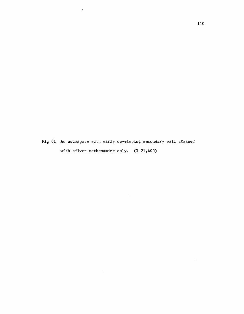
110
Fig 61 An ascospore with early developing secondary wall stained
with silver methenaraine only. (X 21,400)
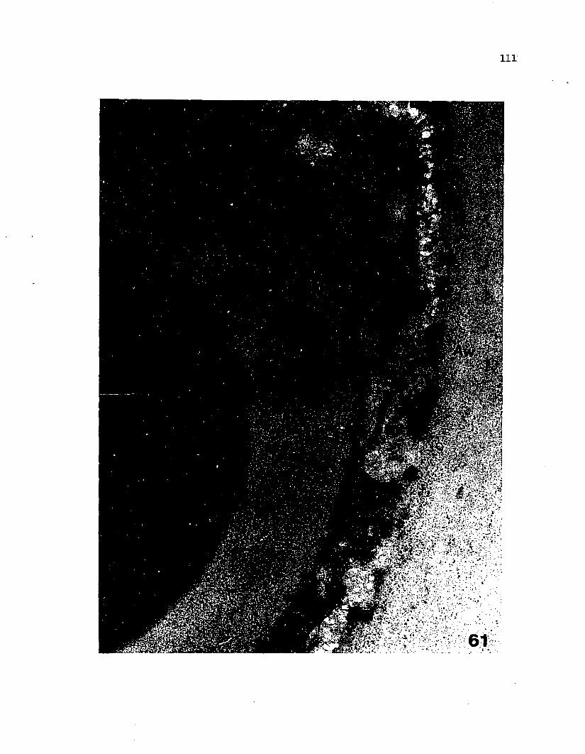
Ill
i wm _ * 1

112
Fig 62 Progressive development of the secondary wall, showing
elongation of the dense globules (arrows) in the areas
of greatest deposition. (X 6,300)

113
swap■C-rff* ■

114
to speculate that the elongation of the globules (by fusion or
otherwise) is not density-related and must be initiated by some
other stimulus, such as position in the epiplasm.
The continual apposition of new material at the sites of
preferential deposition and the intercalation of this material
into the wall as either the dense, elongate globules or the
granular matrix results in a rapid, distal growth of the wall out
growths which appear as highly refractive and slightly birefringent
ornamentations (fig 64). Fine-structural examinations of the wall
at this stage do not reveal any evidence of crystalline or repeating,
structure; thus the birefringence must be only apparent, and is
likely the reflection of a high state of hydration in the newly
synthesized secondary wall, similar to the nacreous wall of some
higher plant cells.
Close to maturity, numerous small developing ornamentations
are distributed randomly over the surface (fig 65). Two large
projections appearing on opposite sides and in a plane parallel
to the closest tangent of the nearby ascus wall are actually the
early signs of an equatorial brim or ridge which circumscribes
the mature spore.
Close examination of a well-progressed site of preferential
wall deposition reveals additional information on the deposition
process. Projections of the secondary wall tend to become overlaid
with a closely appressed layer of endoplasmic reticulum, in a similar
fashion to that observed initially at secondary wall deposition

115
Fig 63 Ascospores and epiplasm during early secondary wall
deposition. A large accumulation of epiplasmic
glycogen is apparent and its relationship to the
developing secondary wall may be noted. (X 29,000)

116

117
Fig 64 A darkfield micrograph of an ascus containing ascospores
showing appearance of secondary wall ornamentations.
(X 920)
Fig 65 Secondary wall formation in an ascospore, indicating the
uneven deposition rate which gives rise to the typical
pattern of ornamentation. (X 7,000)

118

119
(fig 66). The amount of membrane is seen to be greater on
these areas of preferential deposition, with three to five layers
being most common. The distal surface of the projections is always
flat or blunt with a large aggregation of the granular epiplasmic
inclusions consisting of much glycogen, appressed to the layers of
endoplasmic reticulum (figs 66 and 67). This granular material is
clearly evident between the layers of membrane and is seen to collect
below the investing membrane where it is deposited on the surface of
the growing projection of secondary wall (fig 66). As seen at this
level under conventional preparation and staining for electron
microscopy, the smaller epiplasmic inclusions appear to be inter
calated into the wall as the finely granular matrix. The larger,
more clearly definable glycogen granules have not been seen to be
transported across the membranes, and their ultimate fate and role in
secondary wall development is not known.
For a period of time which can actually be up to 24-36 hours, the
structure of the secondary wall changes very little. The projections
continue to grow in length for a while with the tips becoming
noticeably rounded. The wall components become more compacted,
acquiring a reticulate pattern with the fine granular component
becoming increasingly difficult to discern. At the optical microscopy
level, the spores are still quite hyaline, having not yet become
pigmented. The once small lipid droplets have become progressively
larger, and at this time may reach their maximum diameter of about
3 yra.

120
Fig 66 An ornamentation produced by differential deposition
of the secondary wall. The projection has a triple
layer of endoplasmic reticulum (arrows) overlying the
investing membrane, and glycogen outside of these
membranes. (X 25,000)
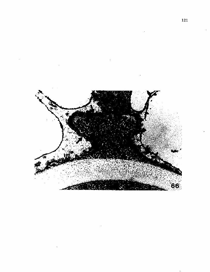
121

122
Fig 67 The developing secondary walls of two ascospores, each
with endoplasmic reticulum overlying the areas of
greater deposition which form the ornamentations.
(X 25,000)

123

124
An unusual morphological event is occasionally observed in
fortuitous sections taken during early secondary wall development.
The investing membrane is observed to evaginate outward in an
exaggerated manner, becoming filled with the granular wall matrix
material (fig 68). This extension of the investing membrane has
been observed to continue outward and fuse with the investing mem
brane of a neighboring ascospore, thus creating a common secretory
apparatus between the two spores (fig 69). The ultimate result is
an area of common secondary wall between the two spores. This
situation will be considered later when mature spore morphology is
discussed.
Primary Wall Differentiation
During the period of development in the secondary wall
involving its compaction and increase in length of the ornamenta
tions, the primary wall undergoes a short period of differentiation.
This is first seen as the appearance of a narrow layer under the
developing secondary wall (fig 70). This layer is only slightly
less dense than the secondary wall, but lacks the reticulate
pattern of the latter (fig 71). This layer becomes evident by
increasing in density.
Further development of this new layer in the primary wall includes
its subdivision into as many as four layers, the outer two being less
dense and thinner than the inner two (fig 72). The entire complex
of layers is about 90 nm in thickness. Additionally, longitudinal
striations appear in this complex of layers (fig 72). These are of

I
125
Fig 68 An ascospore during early secondary wall deposition,
showing an exaggerated evagination of the investing
membrane (arrow). (X 26,000)
Fig 69 The evagination of and fusion between the investing
membranes of adjacent ascospores (arrows). Secondary
wall is being deposited between the adjacent spores.
(X 41,000)

126

127
Fig 70 An ascospore at the climax of secondary wall deposition.
The primary wall is beginning to acquire additional
layers below the secondary wall (arrows). (X, 60,000)

128

Early differentiation of the primary wall. The narrow
band underlying the secondary wall (arrows) has
increased in density. (X 59,000)
An ascospore showing the four layers of the epispore
and electron transparent endospore. (X 65,000)


131
variable size, and have no regularity in spacing or frequency of
occurrence. Using the terminology of Merkus (1973), the newly appear-
inging dense layers underlying the secondary wall are termed "epi-
spore", while the remainder of the old primary wall is "endospore".
Spore Maturation
The ascus containing mature ascospores possessing three
wall layers appears as a highly differentiated structure, far
removed from the ascus containing newly delimited spores. The ascus
wall is unchanged, and still possesses an intact underlying plasma-
lemma (figs 73 and 74). Autolysis of the epiplasm is virtually
complete, with the only recognizable organelles being a few mito
chondria which often lack the outer membrane, some small clumps of
glycogen and other granules underlying the ascus plasmalemma (fig 74),
and many confused masses of unorganized membranes. Structures with
any possible secretory role are no longer evident.
The ascospores have begun to acquire the morphology characteris
tic of their mature state. Each spore now contains several large
lipid globules which often appear to be fusing into still larger
masses (fig 74). The sporoplasm, although quite dense and lacking
vacuoles, has a full complement of organelles and various types of
storage inclusions. The outer secondary wall continues to increase
in density. The projections vary in length from 250 to 2000 nm,
which will be approximately their normal mature size range.
The continual increase in density of the secondary wall through
an apparent compaction of its components ultimately results in a
dense, unstructured homogenous appearance which is particularly

Fig 73 A median section through an ascus following secondary
wall deposition showing 5 ascospores with typical
ornamentation, and the degenerate appearance of the
epiplasm. (X 6,800)

133
S:>

134
Fig 74 A fully ornamented ascospore prior to final maturation
with dense sporoplasm and several lipid droplets.
(X 15,800)

135

136
evident in the ornamentations (fig 75). At this time, a new, very
thin outer wall layer is deposited. This layer first appears as a
loose, floccose deposition covering the surface of the secondary wall
(fig 75). The investing membrane is no longer evident nor is its
fate known. This new layer is interrupted and discontinuous at
first and lacks any observable organization. Loose strands of
residual epiplasm may be closely associated with it (fig 75) and
appear to be somehow involved in its deposition. For the purpose of
this study, this new layer will be termed "perispore".
A reorganization' of the components of the secondary wall is the
terminal stage in its ontogeny. The components of the ornamentations
begin to appear as bundles of fibrils sweeping outward from the base
of the secondary wall and converging at the tips which have now lost
their bluntness or roundness, and are somewhat cone-shaped (figs 76
and 77). The appearance of the wall can now be likened to that
observed in hyphal walls or higher plant walls, with organized
parallel arrays of microfibrils. Close examination reveals sugges
tions of these secondary wall microfibrils penetrating the epispore
and even entering the endospore (fig 77).
The perispore is now well-defined and covers the entire spore
surface as a loosely appressed, undulating layer (figs 76 and 77).
It has a loose, coarsely granular appearance, staining similarly to
proteinaceous compounds. In some areas, it may be composed of at
least three layers of similar thickness.
Other dramatic changes are seen in the remainder of the spore.
With the reorganization of the secondary wall components, the endospore

Fig 75 Ascospore maturation showing early deposition of the
perispore and the apparent action of epiplasmic
components in its formation (arrow). (X 45,000)
Fig 76 The wall of the mature ascospore of JS. peruvianus
showing the needle-like ornamentations, developing
perispore, and significant density increase in
endospore and ascoplasra. Vesicular structures appear
in the endospore (arrows). (X 20,500)

138

Fig 77 The fully mature ascospore wall with parallel micro
fibrils in the secondary wall, and well-developed
perispore. (X 57,000)

140
i*.

141
becomes more electron-dense (fig 76), and contains clumps of
vesicular structures. It is not known if these represent newly
deposited structures or structures simply made apparent by the
increased density of the wall. The sporoplasm additionally has be
come very dense. Only lipid globules are now evident. The overall
appearance of the ascospores suggests sudden dehydration.
The polysaccharide level and distribution in the spore wall has
become altered dramatically from that observed earlier. Silver
methenamine staining now appears heaviest in the newly dense endo
spore and in the electron transparent middle layer of the epispore
(fig 78), The secondary wall which has the greatest electron density
does not react so strongly as the endospore. Except for the peri
spore, which does not appear to react, silver methenamine labelling
in the secondary wall appears to be distributed in a homogenous
fashion. The staining differential between the two layers probably
does not so much reflect differences in total polysaccharide levels
between the layers as It does differences in oxidizability of the
components by periodic acid. In other words, the endospore likely
contains carbohydrates that are simpler in form or are more
accessible and are more completely oxidized to silver-reducing
free aldehydes than the secondary wall. The spores are now sur
rounded by a large vacuole which has been depleted of most reacting
carbohydrates (fig 78); the residual epiplasm still contains large
quantities.
This period of reorganization in the secondary wall parallels
the pigmentation of the spores. Initially hyaline, the spores first

142
Fig 78 A mature ascospore stained with silver methenamine
only, showing heavy localization in endospore, the
middle epispore layer (arrow), secondary wall, and
the remaining epiplasm. (X 74,000)


144
become light amber in color gradually deepening to an ultimate
brownish black. This increase in optical density is seen in figures
79 and 80. As can be seen from observations throughout the wall
development process, pigmentation in Eleutherascus appears to be a
function of a reorganization of the wall components after deposition
has ceased, rather than by the inclusion of pigment granules into a
developing wall,
Morphology and Orientation of the Mature Spores
The distinct morphology of the ascospores in _E. peruvianus is
one important criterion for its placement in the order Pezizales.
The spores are slightly ellipsoidal to spherical, and average 13 urn
in diameter including the ornamentations. These typically include
spines, short ridges, and a longitudinal ridge or brim of variable
shape which can circumscribe the equator of the spore or spiral
around it in a corkscrew fashion (figs 81, 82, 83). The short
ridges, which can travel up to about 30% of the circumference of the
spore appear to be fusions between two or more of the spines. They
have a broad base, tapering to a pointed, spatulate, or bifurcate
apex, and reach a mean length of 8 um.
Several methods of observation have demonstrated convincingly
that the ascospores are not oriented randomly within the ascus,
and that some mechanism produces a certain degree of order.
Median sections through asci containing mature ascospores usually
show the brims of each ascospore to be oriented perpendicular
to the line bisecting that spore through the center of the ascus
(fig 84). The peripheral edge of each brim contacts and often

145
Fig 79 Two _E. peruvianus ascl, one of which Is becoming pigmented
(phase contrast), (X 920)
Fig 80 Two asci containing fully mature and pigmented
ascospores. (X 920)


An isolated, mature ascospore of El. peruvianus showing
the typical ornamentations consisting of spines, short
ridges, and a longitudinal brim which spirals around
the spore (SEM micrograph). (X 8,800)
An isolated ascospore with a polygonal longitudinal
brim which circumscribes the spore. (X 8,500)

148

149
Fig 83 A face-on view of the longitudinal brim of an isolated
ascospore. (X 6,300)
Fig 84 Two medially sectioned asci containing mature ascospores
and demonstrating the orderly arrangement of the asco
spores (bright field). (X 1,800)
Fig 85 Detail of a medially sectioned ascus demonstrating the
relationship between brims of adjacent spores.
(X 7,000)

150

151
seems continuous with the brims of all adjacent spores (figs 84 and
85). This close, continuous, and apparently growth-accommodating
relationship between the spores is made more evident when viewed
three-dimensionally (figs 86, 87 and 88). In cases of relative close
proximity between adjacent spores, the convergent growth between
ornamentations other than the brims has been noted (fig 89). This
accounts for the tenacious aggregation seen between some ascospores
which have been freed from the ascus by mechanical means (fig 90).
Discussion
Great variation is expected in the descriptions of spore wall
ultrastructures by different investigators, primarily as a result
of different fixation and other preparative procedures employed
for electron microscopy. This is especially true of the primary
wall which tends toward homogeneity and is apparently highly
hydrated and quite soft, making it especially vulnerable to
fixation and dehydration artifact and even to deformation from forces
within the ascus. It is conceivable that the uneven thickness
observed in the primary wall of Eleutherascus and in Ascodesmis
(Merkus, 1973) is an artifactual manifestation of this sort. The
observation that spores with initially thin primary walls are often
distorted and have walls of uneven thickness, while spores with thick,
more mature, and presumably stronger walls are nearly always spherical
and of consistent thickness supports this hypothesis. Deposition
of the primary wall apparently does not involve any type of
obvious conventional secretory apparatus as can be seen from this

152
Fig 86-88 Stereo pairs of asci containing mature ascospores
which have been cut into 5 ym sections, etched, and
viewed by SEM. The relationships of the brims
between adjacent spores and the continuity of this
relationship along the periphery of the brim margins
may be observed (arrows). (X 2,250)


154
Fig 89 Detail of a spore mass isolated from an ascus showing
fusion between projections on adjacent spores.
(X 15,200)

155

156
Fig 90 An isolated spore mass showing the often tenacious
aggregation between spores within an ascus. (X 5,400)

157
and previous work. A difficulty in postulating the possible sequence
of events leading to its appearance becomes immediately evident:
nothing is known of the composition or structure of the early undif
ferentiated primary wall. Moore (1963) likens it in structure to
the hyphal wall, a comparison that is now difficult to accept.
Structurally, it resembles a gel matrix as is seen in cell walls, but
without the accompanying microfibrils. This lack of microfibrils is
an obvious departure from the typical cell wall, and could explain
the lack of organellar structures normally associated with the
deposition of this type wall. It is conceivable that the components
(enzymes and substrate) exist in high concentrations in either the
sporoplasm or epiplasm and are transported through active sites on
the spore plasmalemma or investing membrane to condense as the
unstructured primary wall. The accumulation of a fine granular
material adjacent to the plasmalemma in the primary wall prior to
secondary wall formation, and the ultimate accumulation of this
material throughout the primary wall suggests that at least this
material and possibly all of the primary wall is sporoplasmic in
origin. Wilsenach and Kessel (1965) present evidence suggesting
that lomasomes in the sporoplasmalemma are responsible for the
deposition of wall material from the sporoplast in Penicillium
vermiculatum. Moore (1963) did not find lomasomes at this site in
Ascodesmis sphaerospora; however Merkus (1973) did find them in
A. nigricans and A. mlcroscopica. The sporoplasmalemma in
Eleutherascus was always tightly appressed to the developing
wall and never formed lomasomes. This contrasts with
159
Merkus* (1973) observations in Ascodesmis where lomasomes were
reported in the sporoplasmalemma, but never in the ascoplasmalemma.
It is interesting to note that all reports of lomasomes in the
sporoplasmalemma occur after permanganate fixation.
Morphological changes in the structure of the epiplasm and
sporoplasm soon after delimitation reflect the dissimilar roles of
the respective cytoplasms. The increase in vacuolization and
decrease in density of the epiplasm are the first signs of a pro
gressive loss of ultrastructural organization, and apparently reflect
the breakdown and reorganization of some components into new products.
The rate by which this loss of integrity occurs in the epiplasm
roughly parallels the accumulation of glycogen. Merkus (1975)
presents similar observations and discusses the accumulation and roles
of glycogen and other epiplasmic structures as food reserves and as
structural components for the developing spore walls. The rate of
glycogen synthesis and the amounts of glycogen are considered
significant characteristics between differing Discomycete species
(Merkus, 1975).
The appearance of the sporoplasm from the onset of primary wall
development produces the opposite impression from that derived from
the epiplasm. The tendency seems toward organization and accumulation
of stored reserve materials. Structures normally associated with
dynamic activity (ER, dictyosomes, lomasomes, vesicles, microtubules)
are conspicuously absent. The primary ascospore storage product in
Eleutherascus is lipid, which is curiously absent in Ascodesmis
(Merkus, 1973). Silver methenamine staining indicates no significant

160
accumulation of carbohydrate at any stage of the pre-germinating
ascospore.
The deposition of secondary wall material affords an oppor
tunity to observe the secretion of this dense, well-structured
layer and to evaluate the present interpretation with those of
some earlier investigators. In their fine structural studies of
secondary wall deposition in Ascodesmis, Moore (1963) and Carroll
(1966) presented evidence of sporoplasmic secretion. Moore contended
that endoplasmic reticulum occurs in increased amounts in the sporo
plasm, selectively underlying areas of accelerated secondary wall
deposition. Carroll noted the presence of a tubular system in the
sporoplasm and "hints" of tubules penetrating the primary wall. The
net effect of either system would be secondary wall material packaged
in the sporoplast and transported to the surface to be deposited as
the secondary wall. This would necessarily result in a basipetal
growth of the secondary wall projections. It seems clear from this
study and from the extensive survey by Merkus (1973, 1974, 1975,
1976) that secondary wall deposition in the Pezizales is epiplasmic,
and that in at least one group and possibly others, the ornamentations
grow acropetally.
The pattern of early deposition of secondary wall in Eleutherascus
appears to most closely resemble that of Ascodesmis nigricans (Merkus,
1973) with a very thin general initial deposition being followed
shortly by a differential deposition giving rise to the ornamentation.
Unlike in Ascodesmis however, the investing membrane is never
closely appressed to the primary wall. The pattern of secondary

161
wall deposition described by Merkus (1976) into which Eleutherascus
most closely seems to fit is her Group VII, which includes Ascodesmis.
In this group, the secondary wall immediately constitutes the orna
mentation patterns, which do not arise through any breakdown or later
differentiation of the newly deposited secondary wall. In the other
groups, the secondary wall will ultimately disappear altogether with
the epiplasm, or is deposited as a thick undifferentiated layer which
later becomes dense in specified areas, and partially disappears to
reveal the characteristic ornamentation patterns.
Significant differences in applied fixation methods make com
parisons between this and other works difficult if not meaningless.
Following the early deposition of an initially finely granular
secondary wall in localized areas below the investing membrane of
Eleutherascus, a significant departure is seen from the descriptions
of similar events in Ascodesmis (Merkus, 1973), The accumulation
of electron dense granules in the developing secondary wall is
somewhat reminescent of the cases of Trichophaea woolhopei,
Scutellinia, and Sepultaria (Merkus, 1974), although the accumulation
is much greater and more organized in Eleutherascus. Additionally,
the secondary wall in Ascodesmis very early acquires differential
density and some membranous inclusions.
Glycogen has been described as the principle storage poly
saccharide in fungi (Zalokar, 1965). Its abundance in asci, basidia,
sporangia, and other complex fungal structures suggests a
role as a structural precursor or energy source. Merkus (1975)
discusses the accumulation and disappearance of glycogen in several
162
Pezizaceous families and suggested a role of a primary food source
during secondary wall deposition. She observed an increase in
density of the endoplasmic reticulum at the time of increased
glycogen synthesis in Otidea, and suggested a possible role in
glycogen accumulation. Similarly in Eleutherascus, a complex, very
hypertrophied membranous structure appears in the ascoplasm prior
to secondary wall deposition and contains a dense granular material
that is morphologically suggestive of glycogen. This supports the
role of membrane organelles in glycogen accumulation. A decrease
in ascoplasmic glycogen during primary wall deposition, followed by
an increase during early secondary wall formation and its disappear
ance altogether by the end of spore wall deposition are further
supportive of Merkus’ (1975) contentions.
The mechanisms by which epiplasmic material becomes inter
calated within the secondary wall remain unresolved. The fine
granular matrix of the newly deposited wall appears to have an
epiplasmic counterpart, and is observed in what is interpreted as
passage across the ER-investing membrane secretory apparatus. This
interpretation is in agreement with the findings of Merkus (1973,
1976) in Ascodesmis. The larger dense granules which begin to appear
well after the initiation of secondary wall deposition have no epi
plasmic counterpart. Dense granules incorporated into an undifferen
tiated secondary wall are described as pigment granules in Ascobolus
and Saccobolus (Merkus, 1976; Wells, 1972), but are not equivalent
to the material seen in Eleutherascus. In Cheilymenia pulcherrima,
the investing membrane is interrupted and associated with the

163
epiplasm, with dense inclusions and membranous bodies being incor
porated into the secondary wall (Merkus, 1974). Several families
are known in which dense material appears to condense in the develop
ing secondary wall and either ultimately disappears or persists, often
as the ornamentations (Merkus, 1976). It does seem most reasonable
that the early-appearing and persistent dense granules in the
secondary wall of Eleutherascus are the manifestation of a precipita
tion or condensation of pre-existing wall components. Their equiva
lent in other closely related species is not immediately evident.
The observation of the evaginatlon of and fusion between the
investing membranes of adjacent ascospores appears to be without
precedent in the higher ascoraycetes. Close associations between
ascospores which ultimately result in specific morphological features
are known. In Ceratocystis fimbriata (Stiers, 1976), the investing
membranes between two adjacent spores become closely appressed,
resulting in a wall being deposited on each spore which spatially
accommodates the other, forming a pair of brimmed ascospores. The
investing membranes however, do not fuse and the walls between the
adjacent spores retain separate identities. In Saccobolus (Carroll,
1966) secondary wall formation is initiated at points where the
ascospores are tightly appressed. Eventually, the investing membrane
disappears, resulting in all eight ascospores being enclosed in a
common secondary wall. The ultimate result in Eleutherascus is a
thin, broad band of common secondary wall deposited between adjacent
spores, which in single ascospores is seen as the longitudinal brim.

164
The further differentiation of the primary wall occurs in a
pattern similar to most other Pezizaceous Discomycetes (Merkus, 1976).
In Ascodesmis, the epispore acquires its additional layers at a much
earlier time than is seen in Eleutherascus, where they do not appear
until after secondary wall deposition has ended. In observing the
appearance of the epispore and endospore in Ascobolus, Delay (1966)
contends that primary wall material redistributes, possibly by moving
from the inner part of the wall and accumulating or condensing under
the secondary wall. Merkus (1973, 1976) supports this view.
Although this may be the case in Eleutherascus as well, the progres
sive increase in density of the endospore and its accumulation of
carbohydrate materials as revealed by selective staining of mature
ascospores suggests that the endospore-epispore sequesters this
material, possibly from the sporoplast. This could be due to a
source-sink effect where carbohydrates in the outer primary wall
have become tied up in the secondary wall forming a sink to which
material from the inner parts of the wall would flow. The greatly
reduced metabolic state of the quiescent spore could be responsible
for a decrease in plasmalemmal differential permeability allowing
materials from the rich sporoplasmic source to pass into the endo
spore. This hypothesis is supported by selective staining of
mature ascospores which demonstrates practically all of the
ascospore carbohydrate stores to be tied up in the spore wall
layers.
Merkus (1976) recognizes 7 patterns of secondary wall development
in the Pezizales, with distinctions made as to presence or persistence
165
of condensed secondary wall material, presence and persistence of
ornamentations, and origin of the ornamentations, whether derived
from the loss of undifferentiated secondary wall or immediately con
stituted by the secondary wall. Eleutherascus seems to fit best
into Group VII which contains only Ascodesmis microscopica and
A. nigricans. In this group, the secondary wall material immediately
constitutes the ornamentation, which becomes apparent at the earliest
signs of secondary wall deposition.
Although Eleutherascus is similar to Ascodesmis, the two species
differ on a taxonomic point of historic significance. peruvianus
accumulates oil droplets selectively in the sporoplasm, a fact that
excludes it from Merkus' (1976) Group XVIII ascospore type in which
Ascodesmis is the sole representative genus. Additionally, the
ascoplasm is seen to accumulate larger stores of glycogen than does
Ascodesmis, and the endospore become progressively denser which can be
interpreted as a further differentiation. The Eleutherascus
peruvianus-type ascospore is probably best represented in Merkus'
Group X - Lamprospora dictydiola type, which also includes L.
crec1hqueraultii, Aleuria aurantia, Boudiera echinulata, Cheilymenia
pulcherrima, Coprobia granulata, Geopyxls carbonaria, Octospora
musci-muralis, and Iodophanus carneus. In addition to oil droplets
and much glycogen, this group and IJ. peruvianus share the characters
of striation in the epispore, and an electron-dense layer in the
endospore. There are no members of this group that share the
Group VII spore wall deposition pattern shown by Eleutherascus.
This wall deposition pattern however, is most closely approximated

166
by the Group V pattern, which is represented in 6 of the 9 members
of the Type X ascospore group. Ornamentations in this pattern
group are permanent, and arise through the condensation of material
from a thick undifferentiated secondary wall. Eleutherascus appears
to represent a link between the Lamprospora-type and Ascodesmis-
type ascospores, and would best be assigned to an ascospore type
group that combines the two present groups.
The ultimate aspect of the mature ascospores is achieved
following what appears to be a reorganization of the secondary wall
components. The tubular appearance (Carroll, 1966) of the secondary
wall material is replaced by bundles of parallel fibrils oriented
perpendicular to the spore surface. This internal reorganization,
being paralleled by a general change of morphology in the wall
ornamentation involving a slendering or tapering of the ornamenta
tions implies structural compaction, possibly as a result of a
dehydration of the structure or a crystallization of its components.
A similar event is described in several other related forms,
especially Lamprospora crec*hqueraultii (Merkus, 1974).
It is highly probable that the layer which has been designated
"perispore" in this study does not represent a true ascospore wall
layer. It is seen to some extent in most ascomycetes with highly
ornamented spores, and when described is usually attributed to
adhesion of tonoplast membranes (Carroll, 1966) or to remnants of
the epiplasm. It is easily removed from spores which are isolated
by mechanical means.

167
Pigmentation of ascospores in Fezizales has been studied ultra-
structurally in Ascobolus immersus (Delay, 1966), in Saccobolus
glaber (Carroll, 1969) and in Ascobolus furfuraceus (Wells, 1972).
During secondary wall deposition, the wall is penetrated by pigment
granules which ultimately deposit on the epispore. As pigmentation
occurs in Eleutherascus at a time well past the end of secondary
wall deposition, it must be assumed that a different mechanism is
in operation. The transition from the brilliantly hyaline nacreous
wall of the fully ornamented immature spore to the increasingly
brown to near black wall which characterizes the mature spore
parallels the restructuring of the secondary wall components and the
other events related to ascospore maturation. It would seem then
that the factors imparting a generalized increasing electron density
with age to nearly all the ascospore components are additionally
responsible for the characteristic pigmentation of the mature spores.
The arrangement and orientation of mature ascospores within
globose asci is usually described as random or non-ordered (Merkus,
1973). Steffens and Jones (1979) describe the obvious orderly
arrangement of ascospores in the asci of Eleutherascus peruvianus
and discuss the possibility of precise nuclear alignment following
meiosis as being responsible for a consistent alignment in respect
to wall ornamentations. Subsequent investigations have revealed other
mechanisms which may play a significant role in the attainment of this
symmetrical arrangement of ascospores. The basis for the apparent
ordered arrangement lies in the longitudinal brim of each ascospore.
Observations of serially sectioned asci and stereo SEM micrographs

168
of etched embedded asci reveal the brims of each spore to be con
tinuous with the brims of all adjacent spores forming a single thin
layer of continuous wall connecting all adjacent spores in the spore-
ball. It is believed that this interconnecting wall layer which will
ultimately fracture between each spore (forming brimmed ascospores)
is deposited through the interconnection of the investing membrane
between adjacent spores. It is therefore apparent that the "orderly"
arrangement of ascospores is due to epiplasmic developmental events
occurring after initial spore wall deposition. The deposition of
the wall layer which will immediately constitute the ornamentation
pattern is epiplasmic, and likely independent of sporoplasmic
nuclear control. The pattern of ornamentation seen in isolated
spores is infinitely varied, particularly in respect to the brims,
resulting in each spore being unique. This is consistent with the
proposed ontogenetic hypothesis, whereby variation in spacing between
ascospores resulting from abnormal numbers or uneven separation of
nuclei will ultimately result in brims of varying widths and
symmetries among spores. This interpretation does not preclude
nuclear order or ascospore non-randomness but also does not require
it. The question remains unresolved as to how the secondary wall
secretory apparatus consistently becomes modified between adjacent
spores to deposit this continuous layer which ultimately becomes
the brims.

V. ASCOSPORE GERMINATION
Introduction
Ultrastructural studies of mature ascospores are severely
limited by technical restraints. These problems generally reflect
either the hydrophobic nature of some spore components or their
resistence to penetration by the fixatives or embedding media of
electron microscopy. There are extreme reports of ascospores
surviving days of exposure to the chemical fixatives of electron
microscopy (Lowry and Sussman, 1968); these reports serve to
illustrate some of the problems encountered in spore cytology.
Many of the detailed studies, particularly the earlier ones deal
with Hemiascomycetes. The ascogenous yeasts typically produce large
numbers of ascospores which are easily harvested and may be manipu
lated to germinate readily. A disproportionate majority of the
reports describing the ultrastructure of germination in higher
ascomycetes involve conidia, which are easy to obtain in large
quantities and easy to germinate; ascospores are more difficult in
both respects.
The fine-structural changes which immediately precede and then
accompany germination in spores of most fungal classes have been
described to some extent (Akai, Fukutomi, Kunoh, and Shiraishi,
1975; Marchant, 1979). Few however, describe ultrastructure of
germinating ascospores (Lowry and Sussman, 1968; Melendez-Howell,
1969; Hawker, 1971; Beckett, 1976a, 1976b; Millner, Motta, and
Lentz, 1977). In a survey, Bartnicki-Garcia (1968) recognizes
169

170
and describes three types of spore germination, with each type being
characteristic of certain groups of fungi. The first type is
characterized by the extension of one or all of the spore wall
layers to form a germ tube; the second type by the de novo formation
of a nascent wall on a naked protoplast; the third type by jle novo
formation of a vegetative wall under the spore wall. A study of
germinating ascospores of Eleutherascus peruvianus was undertaken
to determine its type of germination and to provide some much
needed information of the ultrastructure of germination in the
Discomycetes.
Results
In cultures of mature ascospores which have been allowed to
grow 7-10 days past maturity, additional changes occur which are
assumed to be involved with the germination process. The morphology
of the ascus wall changes suddenly during this period, although
the exact time is not known. The former relatively electron trans
parent wall, with only a suggestion of two layers, acquires two
very well-defined layers (fig 91). This appears to be primarily
the result of an increase in density or stainability in the outer
layer. Additionally, overall thickness dwindles considerably,
with both layers acquiring a loose, floccose appearance. The means
by which the wall structure may become altered in absence of
accompanying cytoplasm is not known.
The most striking pregermination alteration is seen in the
ascospores. The single haploid nucleus has undergone several

171
Fig 91 The wall of an ascus containing germinating ascospores,
showing the increased contrast between the layers
(arrows), as compared to earlier stages (figs 38, 40,
42, and 52). (X 15,600)

172
i i _ 1/

173
Fig 92 A germinating ascospore prior to exit of the germ tube,
demonstrating the multinucleate state and altered
morphology of sporoplasm and spore wall as compared with
that of the newly mature ascospore (figs 74 and 76).
(X 9,900)

174
wssmam
‘■Wllfew.’v.

175
divisions, resulting in multinucleate ascospores (fig 92) .
Additionally, there is a great decrease in density of the sporo
plasm, with the integrity of all organelles being restored. Large
membrane-bound vacuoles appear, and the once abundant oil droplets
have totally disappeared. With the exception of the multiple nuclei,
the sporoplasts remarkably resemble the newly delimited prospore
stage of their earlier ontogeny.
The ascospore wall has acquired a degenerate appearance, as if
some components are being lost. The broad, conical bases of the
ornamentations have become transparent and contain a loose
fibrillar to granular material (fig 93). The closely packed
parallel arrays of filaments which characterized the mature spore
become increasingly disorganized, disappearing altogether in some
spores. The more distal portions of the ornamentations decrease
in diameter, resulting in needle-like projections (figs 92 and 93).
In the portion of the secondary wall abutting the epispore, loss of
material from the projections may overlap, resulting in breaks or
disruptions in the wall (fig 92). The endospore wall, which had
become more dense earlier, now appears to redistribute some of its
components, losing most of its density and acquiring striations
parallel to the spore surface. A slight increase in its thickness
was also noted. This entire complex of events seen in the asco
spores immediately prior to their germination is termed "ripening."
The event of germination is characterized by an outward
bulging of the ascospore wall, a fracturing of the spore integument,
and the exit of a germ tube from the resulting fissure (fig 94).
176
The germ tube becomes filled with the spore’s cytoplasmic contents
and forms a septum within a few micrometers of the fissure.
Observations of older germinated spores indicate that the entire
cytoplasmic contents of the spore may be utilized (fig 94). All
evidence of germination has been observed only in spores still
contained within the unruptured ascus wall. Likewise, release of
ascospores from the ascus has never been observed.
Silver methenamine staining reveals information on the wall of
the germ tube (figs 95 and 96). This wall averages 350 nm in thick
ness and stains similarly to hyphal walls. The germ tube wall is
seen to be initially deposited de novo within the ruptured spore,
at least 3-5 um inside of the fissure below the endospore wall
(fig 97). In areas where the germ tube has separated from the
endospore, dense areas are revealed in the endospore which underlie
the ornamentations of the secondary wall (fig 97). These dense
areas form a mirror image-like impression of the above ornamentation,
as if some components of the secondary wall have been leached out
into the endospore. Staining with silver methenamine accentuates
this pattern (fig 98). Similar reactions are seen in the leached
out bases of the ornamentations and in the areas underlying them in
the endospore. This could indicate that some components of the
secondary wall, particularly in the wide bases of the ornamentations
and in the inter-ornamentation regions move into the endospore wall
during the ripening process.
Vacuoles containing globules of a dense, highly silver methen-
amine-positive substance now appear in parts of the ascoplasm and

Fig 93 Detail of the spore wall of a germinating ascospore,
showing the decrease in overall thickness of the
secondary wall (arrows) and the newly appearing
electron transparent areas (see figures 74, 76, and 77
for comparison). (X 78,000)
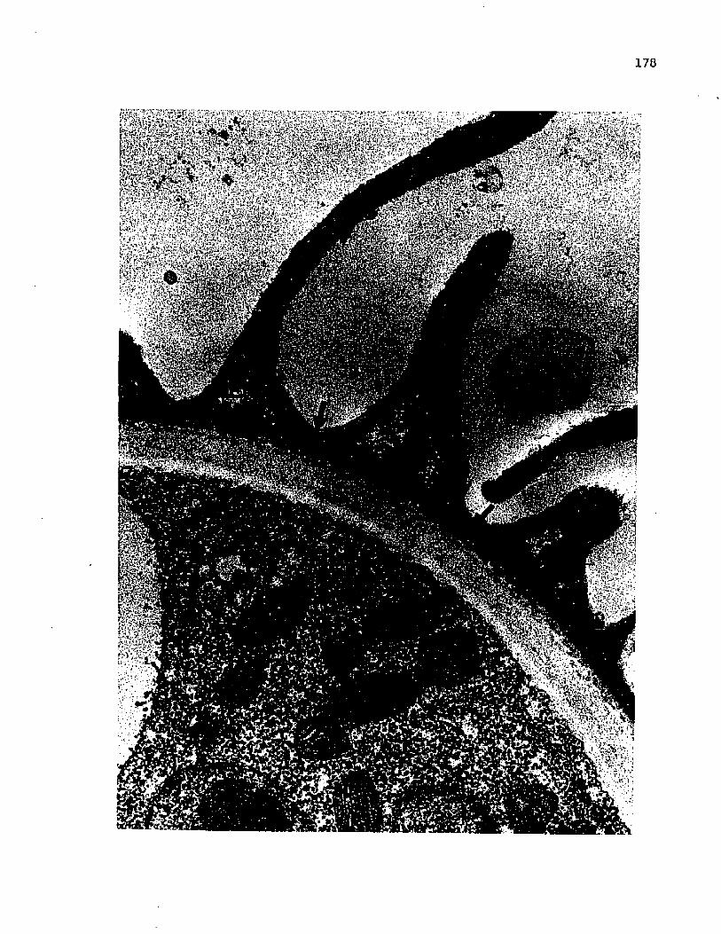
178
* , «',£''?l‘h'- -If <•’'MiHfof'- laMkkr*#*
s^Bafe'^Ssi'irasSapS
JA5-GJ-? •, p \m

179
Fig 94 A germinating ascospore of _E. peruvianus showing the
septate germ tube (arrow) and mitochondria-rich
sporoplasm. (X 8,500)

100
181
germ tube cytoplasm (fig 96). These vacuoles are often closely
associated with the plasmalemma underlying the degenerating ascospore
wall. Inasmuch as the ascoplasm and material closely associated
with the plasmalemma appears to be accrued to the globules in the
vacuoles, it is considered likely that some of the polysaccharide
components of the ascospore wall are resorbed by the sporoplast upon
germination.
Discussion
Morphological changes in the entire wall structure of mature
asci, aside from consistently localized breakdown resulting in
spore dehiscence, are undescribed to my knowledge. The fact that
this alteration of the ascus wall structure seems to coincide
with ascospore ripening and germination makes it tempting to
suggest a relation between the two events. It is reasonable to
assume that such a change in a wall structure could reflect
enzymatic or other chemical activity triggered by the organism.
The only organized cytoplasm competent to respond is contained
within the ascospores and vegetative thallus. In light of the
fact that asci with normal walls and quiescent spores can coexist
with altered-wall asci containing germinating spores, the ascospores
seem to be the most likely source of the trigger for the change in
ascus wall structure, particularly when the timing of germination
and ascus wall alteration is considered.
It is conceivable that alteration of the ascus wall during
germination reflects its partial enzymatic breakdown triggered

182
Fig 95 A germinating ascospore stained with silver methenamine
and showing sporoplasmic localization in vacuolar contents
and in the germ tube wall. (X 7,400)
Fig 96 Detail of a silver methenamine-stained germinating
ascospore demonstrating the globular vacuolar contents.
(X 16,700)

183
' & * . ;’? 0 :- 'f ;-'V. / »■ - v1j~ - » • - *■. . . i ;. t . l

184
Fig 97 Detail of the germ tube wall of a germinating ascospore
showing initial deposition within the exit pore (arrow),
and the dense areas in the endospore underlying the
ornamentations, (X 21,500)
Fig 98 A silver raethenamine-stained germinating ascospore showing
the reaction of the ascospore wall and newly deposited germ
tube wall. (X 33,400)

185

186
directly or indirectly through the release of some factor by the
germinating spores. Release of ascospores by a weakening or
ultimate disintegration of the ascus wall would be one of the few
spore-release mechanisms possible in a species with a resistant
ascus wall and apparently no operculum.
The type of germination seen in the J2. peruvianus ascospore,
in which a vegetative type wall is formed jle novo under the spore
wall, corresponds to Bartnicki-Garcia's (1968) type III germination.
This type of germination appears to be most common in the Mucorales
(Akai et al, 1975) but has been described in the Sphaeriaceous
ascomycete, Daldinia concentrica (Beckett, 1976b). A more common
type of germination in species with chitin-glucan walls is the
type I (Bartnicki-Garcia, 1968).
Gross structural changes in spore wall during germination are
not uncommon. Among the oomycetes, absorption of the inner spore
wall layer, leaving only the thin exospore during germination of
oospores of Phytophthora infestans (Romero and Erwin, 1969) and
P.ythium aphanidermatum (Stanghelleni and Russell, 1973) has been
documented. It seems highly probable that germination in ascospores
of _E. peruvianus is accompanied by a similar resorption of at
least the endospore wall. The appearance of the entire spore
wall during germination is suggestive of dedifferentiation or
degeneration. That material is being removed from all or part
of the ascospore wall during germination is supported by the
concurrent appearance of carbohydrate-containing vacuoles in the
sporoplasm and germ tube cytoplasm; however conversion of some

187
other endogenous storage material to carbohydrate cannot be
entirely ruled out. The simultaneous change in structure seen
in the ascospore and ascus wall at the onset of germination
possibly indicates a common causative mechanism, such as the
release of wall-degradative enzymes from the ascospores upon
their germination. Considerable experimental work will be necessary
to better understand the events associated with ascospore germination
in IS. peruvianus.

LITERATURE CITED
Aist, J. R., and P. H. Williams. 1972. Ultrastructure and time course of mitosis in the fungus Fusariuro oxysporum. J. Cell Biol. 55: 368-89.
Akai, S., H. Fukutomi, H. Kunoh, and M. Shiraishi. 1975. Fine structure of the spore wall and germ tube changes during germination. In The fungus spore: form and function. D. J.Weber and W. M. Hess, Eds., Wiley Interscience, New York.895 pages.
Arx, J. A. von. 1971. On Arachniotus and related genera of the Gymnoascaceae. Persoonia 6(3): 371-80.
Ashton, M. L., and P. B. Moens. 1979. Ultrastructure of sporulation in the Hemiascomycetes Ascoidea corymbosa, A, rubescens, Cephaloascus fragrans, and Saccharomycopsis capsularis.Can. J. Bot. 57(11): 1259-84.
Bartnicki-Garcia, S. 1968. Cell wall chemistry, morphogenesis, and taxonomy of fungi. Ann. Rev. Microbiol. 22: 87-108.
Batra, L. R. 1973. Hemiascosporiaceae (Hemiascomycetidae: Dipodascales), A new family based on Hemiascosporium spinulosum. Mycologia 65: 795-802.
Beckett, A. 1966. Observations on the structure of hyphae, asci, and ascospores, using light, phase contrast, and electron microscopy with special reference to Pleurage anserina. PhD. Dissertation, University College of Wales, Aberystwth.
Beckett, A. 1976a. Ultrastructural studies on exogenously dormant ascospores of Daldinia concentrica. Can. J. Bot. 54: 689-97.
Beckett, A. 1976b. Ultrastructural studies on germinating ascospores of Daldinia concentrica. Can. J. Bot. 54: 698-705.
Beckett, A., R. Barton, and I. M. Wilson. 1968. Fine structureof the wall and appendage formation in ascospores of Podospora anserina. J. Gen. Micro. 53: 89-94.
Beckett, A., and R. M. Crawford. 1970. Nuclear behaviour and Ascospore delimitation in Xylosphaera polymorpha. J. Gen.Micro. 63: 269-80.
Beckett, A., and I. M. Wilson. 1968. Ascus cytology of Podospora anserina. J. Gen. Micro. 53: 81-87.
Benny, G. L., and J. W. Kimbrough. 1980. A synopsis of the orders and families of Plectomycetes with keys to genera. Mycotaxon 12(1): 1-91.
188

189
Blanchard, R. 0. 1972. Electron microscopic observations ofcrozier development in Sporormiella australis. Bull. Georgia Acad. Sci. 30: 170-76.
Bracker, C. E, 1967. Ultrastructure of fungi. Ann. Rev. of Phytopath. 5: 343-74.
Campbell, R. 1973. Ultrastructure of asci, ascospores, and spore release in Lophodermella sulcigena. Protoplasma 78: 69-80.
Carroll, G. 1966. A study of the fine structure of ascosporogenesis in Saccobolus kervemi and Ascodesmis sphaerospora. Ph.D. Thesis, University of Texas.
Carroll, G. 1967a. The fine structure of the ascus septum in Ascodesmis sphaerospora and Saccobolus kervemi. Mycologia 59: 527-32.
Carroll, G. 1967b. The ultrastructure of ascospore delimitation in Saccobolus kerverni. J. Cell Biol. 33: 218-24.
Carroll, G. C. 1969. A study of the fine structure of ascosporogenesis in Saccobolus kervemi. Archiv. Microbiol. 66: 321-39.
Chadefaud, M. 1943. La formation de ascospores chez la P&zize inopercul&e Mniacaecia jungermanniae. Bull. Soc, Botan, Fr.90: 207.
Conway, K. E. 1975. The ontogeny of Lasiobolus ciliatus (Pezizales, Ascomycetes). Mycologia 67: 253-63.
Dangeard, P. A. 1894. La production sexuelle des Ascomycetes.Le Botaniste 4: 48.
Davidow, L. S., and L. Goetsch. 1978. Spindle pole body regulation of spore wall formation in yeast. J. Cell Biol. 79: 31a.
Davidow, L. S., L. Goetsch, and B. Byers. 1980. Preferentialoccurrence of nonsister spores in 2 spored asci of Saccharomyces cerevisiae. Evidence for regulation of spore wall formation by the spindle pole body. Genetics 94(3): 581-96.
deBary, A. 1866. Morphologie und Physiologie der Pilze, Flechten, und Myxomyceten. In Handbuch der Physiologischen Botanik, Leipzig. Verlag Wilhelm Englemann. 486 pages.

190
Delay, C. 1966. Etude de 1' infrastructure de 1* asque d’Ascobolus immersus Pers. pendant la maturation des spores.Ann. Sci. Nat. Botan. (Paris) 12e S4r. 7: 361.
DuPraw, E. J. 1972. Interphase nuclei. In The Biosciences:Cell and Molecular Biology. Cell and Molecular Biology Council, Stanford, CA. 364 pages.
Emden, J. H. vzn. 1975. Three new fungi from Surinam soil.Acta Bot. Neerl. 24(2): 193-97.
Faull, J. H. 1905. Development of the ascus and spore formation in ascomycetes. Proc. Boston Soc. Nat. Hist. 32: 77-113.
Girbardt, M. 1978. Historical review and Introduction. _In Nuclear division in the fungi. (I. B. Heath, Ed.). Academic Press, New York, 235 pages.
Gjurasin, S. 1893. Ueber die Kerntheilung in den Schlauchen von Peziza vesiculosa Bulliard. Ber. Deutsch. Bot. Ges. 11:113-17.
Greenhalgh, G. N., and L. V. Evans. 1968. The developing ascospore wall of Hypoxylon fragiforme. J. Roy. Micros. Soc. 88(4): 545-66.
Greenhalgh, G. N., and H. B. Griffiths. 1970. The ascus vesicle. Trans. Brit. Mycol. Soc. 54: 489-92.
Harper, R. A. 1897. Kemheilung und freie Zellbildung 1m Ascus.Jahrb. Wiss. Bot. 30: 249-84.
Harper, R. A. 1900. Sexual reproduction in Pyronema confluensand the morphology of the ascocarp. Ann. Bot. 14:321-400.
Hashimoto, T., P. Gerhardt, S. F. Conti, and H. B. Naylor. 1960.Studies on the fine structure of microorganisms V. Morphogenesis of nuclear and membrane structures during ascospore formation in yeast. J. Biophys. Biochem. Cytol. 7: 305.
Hawker, L. E. 1971. Ultrastructure of fungus spores with specialreference to changes taking place during germination. J.Indian Bot. Soc. 50A: 12-25.
Heath, I. B. 1978. Experimental studies of mitosis in the fungi.In Nuclear Division in the Fungi. (I. B. Heath, Ed.). Academic Press, New York. 235 pages.
Heslot, H. 1958. Contribution a 1' Atude cytog&n&tique et g4n4tique des Sordariac4es. Rev. Cytol. Biol. Veg. 19: 1.
191
Hill, T. W. 1975. Ultrastructure of ascosporogenesis in Nannizzia gypsea. J. Bacteriol. 122(2): 743-48.
Hohl, H. R., and W. Streit. 1975. Ultrastructure of ascus, ascospore, and ascocarp in Neurospora lineolata. Mycologia 67(2): 367-81.
Huang, L. H. 1975. A new species of Eleutherascus from Peruvian soil. Mycologia 67: 293-302.
Huang, L. H., and J. A. Schmitt. 1973. Ohio Ascomycetes notes I.On Eleutherascus lectardii (Nicot) von Arx. Ohio J. Sci. 73(4): 234-37.
Jeng, R. S., and M. Hubbes. 1980. Ultrastructure of Ceratocystis ulmi 2. Ascogenous system and ascosporogenesis. Eur. J.Forest Path. 10(2-3): 104-16.
Johnson, R. G. 1980. Ultrastructure of ascospore appendages of marine Ascomycetes. Bot, Mar. 23(8): 501-28.
Jones, S. G. 1926. The development of the perithecium of Ophiobolus graminis. Sacc. Ann. Bot. 40: 607-29.
Kimbrough, J., and G. L. Benny. 1978. The fine structure of ascus development in Lasiobolus monascus (Pezizales). Can. J. Bot. 56(7): 862-72.
Korf, R. P. 1973. Discomycetes and Tuberales. ^n The Fungi IV.(A. Ainsworth, Sparrow, and Sussman, Ed.s.). Academic Press,New York. 621 pages.
Laane, M. M. 1970. Nuclear behavior during meiosis and ascus formation in Penicillium striatum. Hereditas 65: 133-58.
Lowry, R. J., and A. S. Sussman. 1968. Ultrastructural changes during germination of ascospores of Neurospora tetrasperma.J. Gen. Microbiol. 51: 403-09.
Mainwaring, H. R. 1971. The fine structure of ascospore wallformation in Sordaria fimicola. Archiv. Microbiol. 81: 126-35.
Manoharachary, C. 1972. Eleutherascus lectardii new record from India. Curr. Sci. (Bangalore) 41(24): 892.
Marchant, R. 1979. Wall growth during spore differentiation andgermination, fri Fungal walls and hyphal growth. (J. H. Burnett and A. P. J. Trinci, Eds.), Cambridge, London. 418 pages.
Marchant, R., and A. W. Robards. 1968. Membrane systems associated with the plasmalemma of plant cells. Ann. Bot. 32: 457-71.

192
Martino, C. de, and L. Zamboni. 1967. Silver methenamine stain for electron microscopy. J. Ultrastruct. Res. 19: 273-82.
Melendez-Howell, M. 1969. Rapports entre le pore germinatif sporal et les fentes de germination longitudinales ou circulaires chez les ascospores. C. R. Acad. Sci., Paris 268: 1273-74.
Merkus, E. 1973. Ultrastructure of the ascospore wall in Pezizales(Ascomycetes). I. Ascodesmis microscopica and A. nigricans. Persoonia 7: 357-66.
Merkus, E. 1974. Ultrastructure of the ascospore wall in Pezizales(Ascomycetes). II. Pyronemataceae sensu Eckblad. Persoonia 8(1): 1-22,
Merkus, E. 1975. Ultrastructure of the ascospore wall in Pezizales.III. Otideaceae and Pezizaceae. Persoonia 8: 227-47.
Merkus, E. 1976. Ultrastructure of the ascospore wall in Pezizales(Ascomycetes). IV. Morchellaceae, Helvellaceae, Rhizinaceae, Thelebolaceae, and Sarcoscyphaceae. Persoonia 9(1): 1-38.
Millner, P. D., J. J. Motta, and P. L. Lentz. 1977. Ascospores, germ spores, ultrastructure, and thermophilism of Chaetomium. Mycologia 69: 720-33.
Mittmann, G. 1932. Kulturversuch mit Einsporstamen und zytol- ogische Untersuchgen in der Gattung Ceratostomella. Jahr.Wiss. Bot. 77: 185-219.
Moens, P. B. 1971. Fine structure of ascospore development in the yeast S_. cereyisiae. Can. J. Microbiol. 17; 507-10.
Moore, R. T. 1963. Fine structure of Mycota I. Electron microscopy of the Discomycete Ascodesmis. Nova Hedwigia 5: 263-78.
Moore, R. T. 1965. The ultrastructure of fungal cells. In TheFungi: an advanced treatise. I (5). Academic Press, New York.
Nlcot, J., and F. Durand. 1969. Une espfece remarquable du genre Arachniotus: A. lectardii J. Nicot, sp. nov. Bull. Soc.Mycol. France 85: 315-20.
Olive, L. S. 1965. Nuclear behavior during meiosis. In The Fungi I. (G. C. Ainsworth and A. S. Sussman, Eds.). Academic Press, New York.
Oso, B. A. 1969. Electron microscopy of ascus development in Ascobolus spores. Ann. Bot. 33(129): 205-209.

193
Ranalli, M. E., and F. Forchiassin. 1976. Systematic and biological study of the Ascobolaceae of Argentina VII. Development and cytology of Saccobolus citrinus. Bol. Soc. Argent. Bot. 17(3-4): 262-79.
Reeves, F. B. 1967. The fine structure of ascospore formation in Pyronema domesticum. Mycologia 59: 1018-33.
Rogers, J. D. 1965. The conidial stage of Coniochaeta lignaria: morphology and cytology. Mycologia 57: 368-78.
Romero, S., and D. C. Erwin. 1969. Variation in pathogenicity among single oospore cultures of Phytophthora infestans. Phytopathology 59: 1310-17.
Samson, R. A., and B. Luiten. 1975. Eleutherascus tuberculatus, a new heat-resistant ascomycete. Trans. Brit. Mycol. Soc.64(2): 338-340.
Samuelson, D. A. 1975. The apical apparatus of the suboperculate ascus. Can. J. Bot. 53: 2660-79.
Samuelson, D. A. 1978. Asci of the Pezizales, III. The apical apparatus of eugymnohymenial representatives. Am. J. Bot.65(7): 748-58.
Schrantz, J. P. 1968. Ultrastructure and localization of glycogen in the Ascomycete Galactinia plebeia. Rev. Cytol. Biol. Veg. 31(1-2): 151-54.
Spurr, A. R. 1969. A low viscosity epoxy resin embedding medium for electron microscopy. J. Ultrastruc. Res, 26: 31-43.
Stanghellini, M. E., and J. D. Russell. 1973. Germination in vitro of Pythium aphanidermatum oospores. Phytopathol. 63: 133-37.
Steffens, W. L., and J. P. Jones. 1979. Ascus and ascospore development in Eleutherascus peruvianus. I. Morphology and orientation of the ascospores. Mycologia 71(6): 1253-56,
Stiers, D. L. 1974. Fine structure of ascospore formation in Poronia punctata. Can. J. Bot. 52: 999-1003.
Stiers, D. L. 1976. The fine structure of ascospore formation in Ceratocystis fimbriata. Can. J, Bot.: 1714-23.
Syrop, M., and A. Beckett, 1976. Leaf curl disease of almondcaused by Taphrina deformans. III. Ultrastructural cytology of the pathogen. Can. J. Bot. 54(3-4); 293-305.

194
Turian, G. 1974. Spores in Ascomycetes, their controlled differentiation. In The Fungal Spore. (D. J. Weber and W. M. Hess, Eds.). Wiley Intersciences, New York. 895 pages.
Tyson, K., and D. A. Griffiths. 1976. Developmental morphology and fine structure of Placoasterella balleyl. II. Ascus and ascospore development. Trans. Brit. Mycol. Soc. 66(2): 263-79.
Wells, K. 1970. Light and electron microscopy studies of Ascobolus stercorarius. I. Nuclear divisions in the ascus. Mycologia 62(4): 761-90.
Wells, K. 1972. Light and electron microscopic studies ofAscobolus stercorarius. II. Ascus and ascospore ontogeny.Univ. Calif. Pub. Bot. 62.
Wilsenach, R., and M. Kessel. 1965. The role of the lomasome in wall formation in Penicillium vermiculatum. J. Gen. Micro.40: 401-05.
Woronin, M. 1864. Zur Entwicklungsgeschichte des Ascoboluspulcherrimus und einiger Pezizen. Abhandl. Senkenbergischen Naturforsch. Ges. 5: 333-344.
Zalokar, M. 1965. Integration of cellular metabolism. In The Fungi. I. (Ainsworth and Sussman, Eds.). Academic Press,New York. 621 pages.

APPENDIX I
Abbreviations to Figures
A, ascus; As, ascospore; Aw, ascus wall; B, brim;
En, endospore; Ep, epiplasm; Er, endoplasmic reticulum;
Es, epispore; Et, electron transparent area; G, glycogen;
Gt, germ tube; H, hyphal wall; I, investing membrane;
L, lomasome; Li, lipid; M, mitochondion; Mb, microbody;
Mt, microtubule; N, nucleus; NAO, nucleus-associated organelle;
Nu, nucleolus; P, plasmalemma; Pc, pore cap; Pe, perispore;
Po, septal pore; Ps, sporoplasm; Pw, primary wall; R, ridge;
Rf, ribosome-free zone; S, septum; Sp, spines; Sw, secondary wall;
T, tubular structure; V, vacuole; W, Woronin Body.
195

VITA
Walstine Lon (Buddy) Steffens III was born February 20, 1951 in
Corpus Christi, Texas, the son of Lon and Chrystal Steffens. He has
a younger brother, Robin Charles Steffens. He was graduated from
Richard King High School in May, 1969 and attended Del Mar College
in Corpus Christi where he majored in biology. In August, 1971 he
transferred to the University of Texas at El Paso where he continued
his studies in biology and worked as a resident dormitory counselor
until May, 1974 when he received his B.S. degree.
From May, 1974 to August, 1976 he worked as a Research
Assistant doing mycological research for the Department of Civil
Engineering at U.T. El Paso. In August, 1974 he entered the graduate
program in the Department of Biological Sciences at U.T. El Paso
where he worked as a teaching assistant. His thesis advisor was
Dr. J. T. Ellzey who instructed him in mycology, cytology, and the
methods of electron microscopy.
Upon receiving his M.S. degree in August, 1976, he moved to
Baton Rouge, Louisiana and was employed by the Department of Zoology
and Physiology at Louisiana State University as a Research Associate
in Electron Microscopy. In August, 1977 he entered the Ph.D. program
in the Department of Plant Pathology and Crop Physiology at LSU under
the direction of Dr. John Paul Jones. He will receive his degree in
December, 1981.
196

EXAMINATION AND THESIS REPORT
Candidate:
Major Field:
Title of Thesis:
Walstine Lon Steffens III
Plant Pathology
A Study of Ascus Developmental Fine Structure in Eleutherascus peruvianus Huang with Special Reference to Ascospore Ontogeny
Approved:
Major Professor and Chairman
Dean of the Graduate—EcSoJl
EXAM INING COMMITTEE:
tf
J l c
fc.sC~ 2 7 r / ____
Date of Examination:
November 19. 1981
Copyright © 2022 FDOKUMEN